legumain regulates differentiation fate of human bone
TRANSCRIPT

Please cite this article in press as: Jafari et al., Legumain Regulates Differentiation Fate of Human Bone Marrow Stromal Cells and Is Alteredin Postmenopausal Osteoporosis, Stem Cell Reports (2017), http://dx.doi.org/10.1016/j.stemcr.2017.01.003
Stem Cell Reports
ArticleLegumain Regulates Differentiation Fate of Human Bone Marrow StromalCells and Is Altered in Postmenopausal Osteoporosis
Abbas Jafari,1,2 DiyakoQanie,2 Thomas L. Andersen,3 Yuxi Zhang,4 Li Chen,2 Benno Postert,4 Stuart Parsons,4
Nicholas Ditzel,2 Sundeep Khosla,5 Harald Thidemann Johansen,6 Per Kjærsgaard-Andersen,7
Jean-Marie Delaisse,3 Basem M. Abdallah,2,8 Daniel Hesselson,4,9 Rigmor Solberg,6
and Moustapha Kassem1,2,10,*1Department of Cellular and Molecular Medicine, Danish Stem Cell Center (DanStem), University of Copenhagen, 2200 Copenhagen, Denmark2Molecular Endocrinology & Stem Cell Research Unit (KMEB), Department of Endocrinology and Metabolism, Odense University Hospital & University of
Southern Denmark, J.B. Winsloewsvej 25, 1st Floor, 5000 Odense C, Denmark3Department of Clinical Cell Biology, Vejle/ Lillebaelt Hospital, Institute of Regional Health Research, University of Southern Denmark, 7100, Vejle,
Denmark4Diabetes and Metabolism Division, Garvan Institute of Medical Research, Sydney, NSW 2010, Australia5Endocrine Research Unit, Mayo Clinic College of Medicine, Rochester, MN 55905, USA6Department of Pharmaceutical Biosciences, School of Pharmacy, University of Oslo, 0363 Oslo, Norway7Department of Orthopaedic Surgery, Vejle/Lillebaelt Hospital, 7100 Vejle, Denmark8Department of Biological Sciences, College of Science, King Faisal University, Hofuf 6996, Saudi Arabia9St Vincent’s Clinical School, UNSWAustralia, Sydney, NSW 2010, Australia10Stem Cell Unit, Department of Anatomy, Faculty of Medicine, King Saud University, Riyadh 12372, Saudi Arabia
*Correspondence: [email protected]
http://dx.doi.org/10.1016/j.stemcr.2017.01.003
SUMMARY
Secreted factors are a key component of stem cell niche and their dysregulation compromises stem cell function. Legumain is a secreted
cysteine protease involved in diverse biological processes. Here, we demonstrate that legumain regulates lineage commitment of human
bone marrow stromal cells and that its expression level and cellular localization are altered in postmenopausal osteoporotic patients. As
shown by genetic and pharmacological manipulation, legumain inhibited osteoblast (OB) differentiation and in vivo bone formation
through degradation of the bone matrix protein fibronectin. In addition, genetic ablation or pharmacological inhibition of legumain
activity led to precocious OB differentiation and increased vertebralmineralization in zebrafish. Finally, we show that localized increased
expression of legumain in bone marrow adipocytes was inversely correlated with adjacent trabecular bone mass in a cohort of patients
with postmenopausal osteoporosis. Our data suggest that altered proteolytic activity of legumain in the bonemicroenvironment contrib-
utes to decreased bone mass in postmenopausal osteoporosis.
INTRODUCTION
Human bone marrow stromal cells (hBMSCs) are non-he-
matopoietic multipotent cells capable of differentiation
into mesodermal cell types such as osteoblasts (OBs) and
adipocytes (ADs) (Abdallah andKassem, 2008). It is increas-
ingly recognized that secreted factors have an important
role inmediating hBMSC function to activelymaintain ho-
meostasis of skeletal tissue. In addition, secreted proteins
mediate the observed therapeutic effects of hBMSCs on
enhancing regeneration of skeletal (Hernigou et al.,
2005), cardiac (Hare et al., 2009), dermal (Bey et al.,
2010), and neural (Yamout et al., 2010) tissues. Character-
izing the functions of proteins secreted by hBMSCs is a
pre-requisite for understanding the mechanisms of their
therapeutic effects and their role in tissue homeostasis in
normal and disease states.
We recently reported a profile of hBMSC-secreted factors
at different stages of ex vivo OB differentiation using a
quantitative proteomic analysis based on stable isotope la-
beling by amino acids in cell culture (Kristensen et al.,
2012). Among the differentially regulated proteins during
StemThis is an open access article under the C
OB differentiation, we identified legumain as a secreted
protein that has not been previously implicated in hBMSC
biology.
Legumain (also known as asparaginyl endopeptidase,
AEP), encoded by the LGMN gene, is a broadly expressed
lysosomal cysteine protease that is secreted as inactive pro-
legumain (56 kDa) and processed into enzymatically active
46 and 36 kDa forms, as well as a 17 kDa enzymatically
inactive C-terminal fragment. Legumain directly regulates
diverse physiological and pathological processes by remod-
eling tissue-specific targets (e.g., extracellular matrix [ECM]
components, enzymes, receptors) (Chen et al., 2001; Clerin
et al., 2008; Deryugina and Quigley, 2006; Ewald et al.,
2008, 2011; Liu et al., 2003; Manoury et al., 1998; Mattock
et al., 2010;Miller et al., 2011;Morita et al., 2007; Papaspyr-
idonos et al., 2006; Sepulveda et al., 2009; Solberg et al.,
2015). In addition, legumain indirectly contributes to
atherosclerotic plaque instability through activation of
cathepsin L in the arterial ECM (Clerin et al., 2008; Kita-
moto et al., 2007; Mattock et al., 2010; Papaspyridonos
et al., 2006). Surprisingly, the non-enzymatic 17 kDa
C-terminal fragment is also biologically active and inhibits
Cell Reports j Vol. 8 j 1–14 j February 14, 2017 j ª 2017 The Authors. 1C BY-NC-ND license (http://creativecommons.org/licenses/by-nc-nd/4.0/).

Please cite this article in press as: Jafari et al., Legumain Regulates Differentiation Fate of Human Bone Marrow Stromal Cells and Is Alteredin Postmenopausal Osteoporosis, Stem Cell Reports (2017), http://dx.doi.org/10.1016/j.stemcr.2017.01.003
osteoclast differentiation through binding to an uncharac-
terized receptor (Choi et al., 1999, 2001).
Here we report the role of legumain in regulating the dif-
ferentiation fate of hBMSCs. Using cell-based and in vivo
studies we show that legumain inhibited OB differentia-
tion through degradation of fibronectin. During develop-
ment, legumain-deficient zebrafish exhibited precocious
bone formation and mineralization. Finally, abnormal
expression and cellular localization of legumain was
observed in bone biopsies obtained from patients with
postmenopausal osteoporosis. Together, the present study
reveals role of legumain in determining the differentiation
fate of BMSCs thereby regulating bone formation.
RESULTS
Legumain Expression and Activity Are Regulated
during hBMSC Differentiation In Vitro and In Vivo
To assess cellular localization and regulation of legumain
(LGMN) expression during OB differentiation under
normal physiological conditions, we first performed histo-
logical analysis of adult human iliac crest bone biopsies. Le-
gumain immunoreactivity was present in osteoprogenitors
near bone-forming surfaces which included perivascular,
canopy, and reversal cells (Figure 1A) (Delaisse, 2014; Kris-
tensen et al., 2014).We did not detect immunoreactivity in
mature OBs, osteocytes, or osteoclasts (Figure 1A). Legu-
main mRNA showed a similar expression pattern (Fig-
ure 1B). Next, we examined legumain expression and activ-
ity during ex vivo OB differentiation of hBMSCs. LGMN
mRNA expression increased (Figure 1C) and the mature
LGMN protein (36 kDa) accumulated (Figures 1D and 1E)
during the early commitment phase (days 1–6) and were
downregulated during the late maturation phase (days 6–
18) of OB differentiation. Correspondingly, legumain enzy-
matic activity was reduced in differentiatedOBs (Figure 1F).
In contrast, LGMN mRNA expression and protein levels
were increased during AD differentiation of hBMSCs (Fig-
ures 1G–1I).
Legumain Deficiency Enhances OB Differentiation
and Impairs AD Differentiation of hBMSCs
We employed lentiviral transduction to generate hBMSC
lines with stable expression of LGMN shRNA (shLGMN)
or a non-targeting control (shCtrl). shLGMN significantly
reduced legumain mRNA, protein, and activity levels (Fig-
ures 2A–2C). In addition, LGMN knockdown reduced
hBMSC proliferation (Figure S1A). After 6 days under oste-
ogenic culture conditions, LGMN knockdown did not alter
alkaline phosphatase (ALP) activity (Figure 2D), or expres-
sion of the early OB commitment markers ALP and
collagen 1 alpha 1 chain (Col1a1), but significantly upregu-
2 Stem Cell Reports j Vol. 8 j 1–14 j February 14, 2017
lated the expression of the late OB maturation markers
bone gamma-carboxyglutamate protein (BGLAP) and in-
tegrin binding sialoprotein (IBSP) (Figure 2E). Moreover,
LGMN knockdown enhanced the formation of mineralized
ECM, as shown by the increased extent and intensity of
alizarin red staining (Figure 2F). In contrast, LGMN knock-
down inhibited AD differentiation (Figures 2G and 2H) and
reduced expression of the AD maker genes: peroxisome
proliferator-activated receptor gamma 2 (PPARG2), fatty
acid binding protein 4 (FABP4) and adiponectin, C1Q
and collagen domain containing (ADIPOQ) (Figure 2I) un-
der adipogenic culture conditions. To determine whether
LGMN knockdown stimulated OB differentiation and
bone-forming capacity in vivo, shLGMN or shCtrl cells
were mixed with hydroxyapatite/tricalcium phosphate
granules as an osteoconductive carrier, and implanted sub-
cutaneously in immune-deficient mice. Histological anal-
ysis of the implants after 8 weeks revealed a significant
2-fold increase in the amount of heterotopic bone formed
by the shLGMN compared with the control (shCtrl) cells
(Figures 2J and 2K). Human-specific vimentin staining
showed that the heterotopic bone was generated by the
transplanted hBMSCs (Figure 2L).
Pharmacological Inhibition of Legumain Activity
Enhances OB Differentiation and Impairs AD
Differentiation of hBMSCs
To determine whether legumain proteolytic activity is
required for its effects on hBMSCs differentiation, we em-
ployed a small-molecule legumain inhibitor (SD-134) (Lee
and Bogyo, 2012). hBMSC cultures incubated for 24 hr
with SD-134 (50–500 nM) exhibited significant legumain
inhibition (Figure S1B), but no effect on cell number was
observed during 12 days treatment (Figure S1C). During
in vitro OB differentiation of hBMSCs, SD-134 (50 nM)
treatment did not induce significant changes in ALP activ-
ity (Figure S1D), but enhanced formation of mineralized
matrix (Figure S1E). In contrast, SD-134 treatment in-
hibited AD differentiation (Figures S1F and S1G).
Overexpression of LGMN Impairs OB Maturation and
Enhances AD Differentiation of hBMSCs
To determine whether increased LGMN activity actively
blocks OB differentiation, we employed a retroviral
transduction system to establish hBMSCs stably overex-
pressing full-length LGMN (hBMSC-LGMN) or an empty
vector control (hBMSC-EV). LGMN overexpression was
confirmed by mRNA, protein, and activity measurements
(Figures 3A–3C). The levels of fully processed and acti-
vated legumain protein (36 kDa) were increased in lysates
from hBMSC-LGMN cells (Figures 3B and 3C). In addition,
secreted prolegumain (56 kDa) was detected in the condi-
tioned media from hBMSC-LGMN cells (Figures 3D and

Figure 1. Regulation of Legumain Expression during In Vitro and In Vivo Differentiation of Human Bone Marrow Stromal Cells(A and B) Immunohistochemical (A) and RNA in situ hybridization (B) analysis of legumain expression and localization in normal humaniliac crest bone biopsies. n = 11 donors. Scale bar, 50 mm. Red arrows, canopy cells; black arrows, reversal cells; arrow heads, osteoclasts; v,vessel.(C) qRT-PCR analysis of LGMN expression during osteoblast (OB) differentiation of hBMSCs at 6, 12, and 18 days (D6–D18) after start ofdifferentiation (day 0, D0). Data represent mean ± SD from three independent experiments. *p% 0.05, **p% 0.01, two-tailed unpairedStudent’s t test.(D) Western blot analysis of legumain expression in cell lysates from hBMSC cultures during OB differentiation.(E) Quantification of the mature legumain (36 kDa) band intensity. Arbitrary units (ARBU). Data represent mean ± SD from threeindependent experiments. *p % 0.05, **p % 0.01, two-tailed unpaired Student’s t test.(F) Quantification of legumain activity in cell lysates from hBMSCs during OB differentiation. Data represent mean ± SD from threeindependent experiments. *p % 0.05, two-tailed unpaired Student’s t test.(G) qRT-PCR analysis of LGMN expression during adipocyte (AD) differentiation of hBMSCs. Data represent mean ± SD from three inde-pendent experiments. **p % 0.01, two-tailed unpaired Student’s t test.(H) Western blot analysis of legumain expression in cell lysates from hBMSC cultures during AD differentiation.(I) Quantification of the mature legumain (36 kDa) band intensity. Data represent mean ± SD from three independent experiments.*p % 0.05, two-tailed unpaired Student’s t test.
Stem Cell Reports j Vol. 8 j 1–14 j February 14, 2017 3
Please cite this article in press as: Jafari et al., Legumain Regulates Differentiation Fate of Human Bone Marrow Stromal Cells and Is Alteredin Postmenopausal Osteoporosis, Stem Cell Reports (2017), http://dx.doi.org/10.1016/j.stemcr.2017.01.003

Figure 2. Legumain Knockdown Enhanced Osteoblast Differentiation and In Vivo Bone Formation and Inhibited Adipocyte Dif-ferentiation of Human Bone Marrow Stromal CellshBMSCs were stably transfected with control (shCtrl) or LGMN shRNA (shLGMN).(A) qRT-PCR analysis of LGMN expression. Data represent mean ± SD from three independent experiments. *p% 0.05, two-tailed unpairedStudent’s t test.(B) Western blot analysis of legumain and GAPDH control. Data represent three independent experiments.(C) Quantification of legumain activity. Data represent mean ± SD from three independent experiments. **p% 0.01, two-tailed unpairedStudent’s t test.(D) Quantification of alkaline phosphatase (ALP) activity in the presence of standard culture medium (SCM) or osteoblast inductionmedium (OIM) (day 6). Data represent mean ± SD from three independent experiments. p > 0.05, two-tailed unpaired Student’s t test.(E) qRT-PCR gene expression analysis of the early (ALP, Col1a1) and late (BGLAP, IBSP) OB marker genes. Data represent mean ± SD fromthree independent experiments. *p % 0.05, **p % 0.01, two-tailed unpaired Student’s t test.(F) Quantification of alizarin red staining at day 12. Data represent mean ± SD from three independent experiments. *p% 0.05, two-tailedunpaired Student’s t test.(G and H) Quantification of accumulated lipid droplets in the presence of AD induction medium using oil red O staining (day 12). Scale bar,150 mm, Data represent mean ± SD from three independent experiments. *p % 0.05, two-tailed unpaired Student’s t test.(I) qRT-PCR gene expression analysis of the AD marker genes PPARG2, FABP4, LPL, and ADIPOQ. Data represent mean ± SD from threeindependent experiments. *p % 0.05, two-tailed unpaired Student’s t test.(J) Histological analysis of in vivo bone formation by hBMSCs stably transfected with non-targeting control shRNA (shCtrl) or LGMN shRNA(shLGMN), 8 weeks after implantation in immune-deficient mice. Arrows, hydroxyapatite; arrow heads, bone. Scale bars: top panels,500 mm; bottom panels, 250 mm.
(legend continued on next page)
4 Stem Cell Reports j Vol. 8 j 1–14 j February 14, 2017
Please cite this article in press as: Jafari et al., Legumain Regulates Differentiation Fate of Human Bone Marrow Stromal Cells and Is Alteredin Postmenopausal Osteoporosis, Stem Cell Reports (2017), http://dx.doi.org/10.1016/j.stemcr.2017.01.003

Please cite this article in press as: Jafari et al., Legumain Regulates Differentiation Fate of Human Bone Marrow Stromal Cells and Is Alteredin Postmenopausal Osteoporosis, Stem Cell Reports (2017), http://dx.doi.org/10.1016/j.stemcr.2017.01.003
3E). In contrast to LGMN knockdown, hBMSC-LGMN
exhibited increased cell proliferation (Figure S1H),
impaired OB maturation, demonstrated by decreased late
OB marker gene expression (BGLAP, IBSP), and decreased
mineralized matrix formation, whereas expression of early
OB commitment markers (ALP, Col1a1) was not altered by
LGMN overexpression (Figures 3F and 3G). hBMSC-LGMN
cells also exhibited enhanced AD differentiation (Figures
3H and 3I), and enhanced expression of AD marker genes
(PPARG2, FABP4, and ADIPOQ) (Figure 3J). Thus, increased
LGMN activity biases hBMSC differentiation to non-OB
fates.
Legumain Degrades Fibronectin during hBMSC
Differentiation
Since ECM proteins play an important role in OB differen-
tiation (Hoshiba et al., 2012) and fibronectin functions as a
master organizer of ECM biogenesis (Halper and Kjaer,
2014), we tested whether the effect of legumain on hBMSC
differentiation is mediated through regulation of fibro-
nectin levels. Lysates from legumain-deficient and legu-
main-overexpressing cells during in vitro OB differentia-
tion showed an inverse relationship between the protein
levels ofmature legumain (36 kDa) and fibronectin (Figures
4A–4F). FibronectinmRNA (FN1) levels did not change dur-
ing OB differentiation (Figure S2A), which is consistent
with post-transcriptional regulation. Incubation of purified
human fibronctin with lysates from legumain-overexpress-
ing HEK293 cells (Smith et al., 2012) confirmed that fibro-
nectin is degraded by a legumain-dependent process in a
dose- and time-dependent manner (Figure S2B). The fibro-
nectin-degrading activity was also present in lysates from
LGMN-overexpressing hBMSCs (Figure 4G). To determine
whether intact fibronectin is required for OB differentia-
tion, we blocked FN1 expression using siRNA, which
inhibitedOBmaturation and formation ofmineralizedma-
trix (Figure 4H). To determine whether fibronectin degra-
dation is sufficient to explain the inhibitory effects of legu-
main on OB maturation, we exposed osteogenic hBMSC
cultures to a panel of purified ECM proteins (gelatin,
collagen type 1, and fibronectin). Both gelatin and fibro-
nectin enhanced the formation of mineralized matrix by
control hBMSC (Figure 4I). However, only fibronectin
blocked the inhibitory effect of legumain on OB differenti-
ation and function (Figure 4J), suggesting that fibronectin
is an endogenous legumain target.
(K) Quantification of the heterotopic bone formation, n = 4 implantsWhitney test.(L) Human-specific vimentin staining of shLGMN implants. Arrows, hbottom panels, 250 mm. ab, antibody.See also Figure S1.
Legumain-Deficient Zebrafish Exhibit Precocious OB
Differentiation and Bone Mineralization
To examine the function of legumain during bone develop-
ment, we targeted the essential peptidase domain within
the zebrafish lgmn locus using transcription activator-like
effector nucleases (TALENs) (Figure 5A). High-resolution
melt analysis (HMRA) indicated that the lgmn gene was
extensively modified in lgmn-TALEN-injected animals (Fig-
ure 5B). To quantify the extent of lgmn disruption, we used
ON/OUT qPCR (Shah et al., 2015) and determined that
�84% of lgmn alleles were modified. We proceeded to
analyze OB differentiation and function in the lgmn-
TALEN-injected animals compared with non-targeted con-
trol-injected animals. At 5 days post-fertilization (dpf)
lgmn-deficient animals exhibited increased expression of
genes associated with OB differentiation, including alka-
line phosphatase (alp) and osteopontin (spp1), while the
expression of the adipogenic marker PPAR-gamma (pparg)
was unchanged (Figure 5C). By 7 dpf lgmn-deficient
zebrafish did not show any gross morphological defects
(Figure 5D). However, there were approximately twice
as many mineralized vertebrae in lgmn-TALEN-injected an-
imals compared with controls (Figures 5E and 5F). Next, we
tested whether post-embryonic OB progenitors remained
sensitive to legumain activity. Zebrafish embryogenesis is
complete by 3 dpf (Kimmel et al., 1995). Therefore, we
treated animals from 3 to 7 dpf with the legumain inhibitor
SD-134. Consistent with the osteogenic effect of genetic
lgmn disruption, SD-134-treated zebrafish exhibited an in-
crease in the number of mineralized vertebrae at 7 dpf
(Figure 5G).
Legumain Serum Levels Decrease with Aging, and
Local Expression of Legumain Is Increased in the Bone
Microenvironment of Osteoporotic Patients
Legumain has recently been identified as a biomarker for
diverse pathological states (Ashley et al., 2016; Guo et al.,
2013; Lin et al., 2014; Lunde et al., 2016; Wang et al.,
2012; Wu et al., 2014). We examined serum levels of legu-
main in 89 women aged 48–87 years and found an inverse
relationship between serum legumain levels and age (Fig-
ure 6A). To determine legumain-localized activity within
the bone microenvironment, we established primary
hBMSC cultures from iliac crest bone marrow aspirates. Le-
gumain levels were significantly higher in osteoporotic
patients compared with age-matched controls (Figures 6B
for each cell type, Data represent mean ± SEM. *p % 0.05, Mann-
ydroxyapatite; arrow heads, bone. Scale bars: top panels, 500 mm;
Stem Cell Reports j Vol. 8 j 1–14 j February 14, 2017 5

Figure 3. Legumain Overexpression Inhibited Osteoblast and Enhanced Adipocyte Differentiation of Human Bone Marrow StromalCellsSee also Figure S1.(A–C) Legumain (LGMN)-transduced hBMSCs was established using a retroviral transduction system and the successful overexpression oflegumain was confirmed using (A) qRT-PCR analysis of LGMNmRNA expression, (B) western blot analysis of legumain in cell lysate, and (C)quantification of legumain activity. Data represent mean ± SD from three independent experiments. **p % 0.01, two-tailed unpairedStudent’s t test.(D–G) Secretion of legumain in the conditioned medium (CM) was evaluated using (D) ELISA measurement (data represent mean from threetechnical replicates) and (E) western blot analysis of legumain in the CM from hBMSC-LGMN-overexpressing cell line (LGMN-CM) (datarepresent three independent experiments). To assess the effects of legumain on OB differentiation, control hBMSCs containing emptyvector (E.V.) and hBMSC-LGMN cell lines were cultured in OB induction medium, and expressions of OB marker genes were analyzed usingqRT-PCR (F) and mineralized matrix formation was determined by quantification of eluted alizarin red staining (G). Data represent mean ±SD from three independent experiments. *p % 0.05, **p % 0.01, two-tailed unpaired Student’s t test.(H and I) To assess the effect of legumain on AD differentiation, control hBMSCs and hBMSC-LGMN cell lines were cultured in AD inductionmedium, and (H), (I) accumulation of lipid droplets was measured by quantification of the eluted oil red O staining, *p% 0.05, two-tailedunpaired Student’s t test. Scale bar, 150 mm.(J) Expressions of AD marker genes were measured by qRT-PCR (day 7). Data represent mean ± SD from three independent experiments.*p % 0.05, two-tailed unpaired Student’s t test. E.V., empty vector; LGMN, legumain-overexpressing hBMSCs.
Please cite this article in press as: Jafari et al., Legumain Regulates Differentiation Fate of Human Bone Marrow Stromal Cells and Is Alteredin Postmenopausal Osteoporosis, Stem Cell Reports (2017), http://dx.doi.org/10.1016/j.stemcr.2017.01.003
and 6C). In addition, immunohistochemical analyses of
bone biopsies from postmenopausal osteoporotic patients
(n = 13) and age-matched controls (n = 11) revealed legu-
6 Stem Cell Reports j Vol. 8 j 1–14 j February 14, 2017
main overexpression in bonemarrowADs inmost osteopo-
rotic samples (8 out of 13) (Figure 6D). To determine
whether legumain-overexpressing ADs affected adjacent

Figure 4. Legumain Degrades Fibronectin in Human Bone Marrow Stromal Cell cultures(A) Western blot analysis of legumain and fibronectin in cell lysates of hBMSC lines with stable knockdown of legumain (shLGMN) duringex vivo OB differentiation (day 0–7).(B and C) Quantification of protein band intensities in (A). *p % 0.05, **p % 0.01, two-tailed unpaired Student’s t test.(D) Western blot analysis of legumain and fibronectin in cell lysates of hBMSC lines with stable overexpression of legumain (LGMN) duringex vivo OB differentiation (day 0–7).(E and F) Quantification of protein band intensities in (D). Data represent mean ± SD from three independent experiments. *p % 0.05,**p % 0.01, two-tailed unpaired Student’s t test.(G) Western blot analysis of human fibronectin degradation by purified legumain from bovine kidneys (bLeg; 10:1 w/w; control) and thecell lysates from hBMSCs stably transfected with E.V. or legumain (LGMN; legumain overexpression).(H) Quantitation of mineralized matrix formation on day 15 of OB differentiation, in the presence of siRNA against fibronectin (siFN) andnon-targeting control siRNA (siCtrl). Data represent mean ± SD from three independent experiments. **p % 0.01, two-tailed unpairedStudent’s t test.(I and J) Effect of different ECM proteins (gelatin, collagen 1, or fibronectin) on mineralized matrix formation by hBMSCs visualizedby alizarin red staining and quantification. Data represent mean ± SD from three independent experiments. *p % 0.05, **p % 0.01,two-tailed unpaired Student’s t test.See also Figure S2.
Stem Cell Reports j Vol. 8 j 1–14 j February 14, 2017 7
Please cite this article in press as: Jafari et al., Legumain Regulates Differentiation Fate of Human Bone Marrow Stromal Cells and Is Alteredin Postmenopausal Osteoporosis, Stem Cell Reports (2017), http://dx.doi.org/10.1016/j.stemcr.2017.01.003
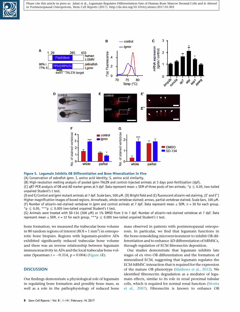
Figure 5. Legumain Inhibits OB Differentiation and Bone Mineralization In Vivo(A) Conservation of zebrafish lgmn. I, amino acid identity; S, amino acid similarity.(B) High-resolution melting analysis of pooled lgmn-TALEN and control-injected animals at 3 days post-fertilization (dpf).(C) qRT-PCR analysis of OB and AD marker genes at 5 dpf. Data represent mean ± SEM of three pools of ten animals. *p% 0.05, two-tailedunpaired Student’s t test.(D and E) Control and lgmnmutant animals at 7 dpf. Scale bars, 500 mM. (D) Bright field and (E) fluorescent alizarin red staining. (E0 and E00)Higher-magnification images of boxed regions. Arrowheads, whole vertebrae stained; arrows, partial vertebrae stained. Scale bars, 100 mM.(F) Number of alizarin-red-stained vertebrae in lgmn and control animals at 7 dpf. Data represent mean ± SEM, n > 30 for each group.*p % 0.05, ***p % 0.005 two-tailed unpaired Student’s t test.(G) Animals were treated with SD-134 (500 mM) or 1% DMSO from 3 to 7 dpf. Number of alizarin-red-stained vertebrae at 7 dpf. Datarepresent mean ± SEM, n = 12 for each group. ***p % 0.005 two-tailed unpaired Student’s t test.
Please cite this article in press as: Jafari et al., Legumain Regulates Differentiation Fate of Human Bone Marrow Stromal Cells and Is Alteredin Postmenopausal Osteoporosis, Stem Cell Reports (2017), http://dx.doi.org/10.1016/j.stemcr.2017.01.003
bone formation, we measured the trabecular bone volume
in 80 random regions of interest (ROI = 1mm2) in osteopo-
rotic bone biopsies. Regions with legumain-positive ADs
exhibited significantly reduced trabecular bone volume
and there was an inverse relationship between legumain
immunoreactivity in ADs and the local trabecular bone vol-
ume (Spearman r = �0.314, p = 0.004) (Figure 6E).
DISCUSSION
Our findings demonstrate a physiological role of legumain
in regulating bone formation and possibly bone mass, as
well as a role in the pathophysiology of reduced bone
8 Stem Cell Reports j Vol. 8 j 1–14 j February 14, 2017
mass observed in patients with postmenopausal osteopo-
rosis. In particular, we find that legumain functions in
the bone-remodeling microenvironment to inhibit OB dif-
ferentiation and to enhance AD differentiation of hBMSCs,
through regulation of ECM fibronectin deposition.
Our studies demonstrate that legumain inhibits late
stages of ex vivo OB differentiation and the formation of
mineralized ECM, suggesting that legumain regulates the
ECM-hBMSC interaction that is required for the expression
of the mature OB phenotype (Mathews et al., 2012). We
identified fibronectin degradation as a mediator of legu-
main effects, similar to its role in renal proximal tubular
cells, which is required for normal renal function (Morita
et al., 2007). Fibronectin is known to enhance OB
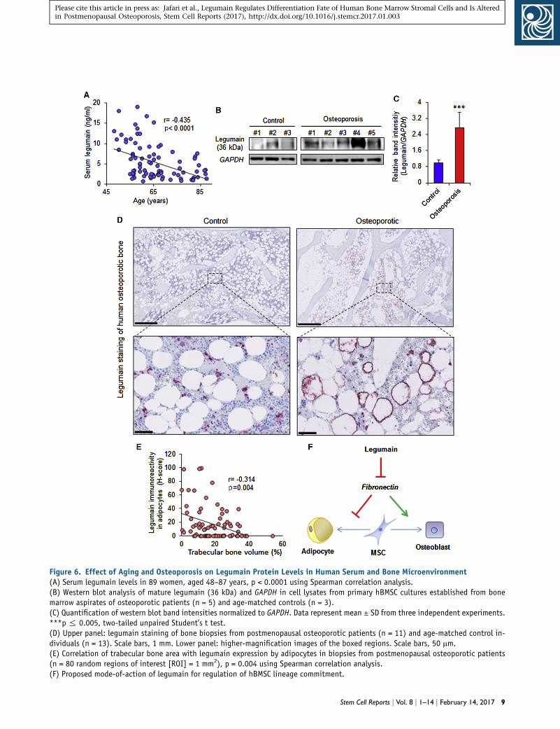
Figure 6. Effect of Aging and Osteoporosis on Legumain Protein Levels in Human Serum and Bone Microenvironment(A) Serum legumain levels in 89 women, aged 48–87 years, p < 0.0001 using Spearman correlation analysis.(B) Western blot analysis of mature legumain (36 kDa) and GAPDH in cell lysates from primary hBMSC cultures established from bonemarrow aspirates of osteoporotic patients (n = 5) and age-matched controls (n = 3).(C) Quantification of western blot band intensities normalized to GAPDH. Data represent mean ± SD from three independent experiments.***p % 0.005, two-tailed unpaired Student’s t test.(D) Upper panel: legumain staining of bone biopsies from postmenopausal osteoporotic patients (n = 11) and age-matched control in-dividuals (n = 13). Scale bars, 1 mm. Lower panel: higher-magnification images of the boxed regions. Scale bars, 50 mm.(E) Correlation of trabecular bone area with legumain expression by adipocytes in biopsies from postmenopausal osteoporotic patients(n = 80 random regions of interest [ROI] = 1 mm2), p = 0.004 using Spearman correlation analysis.(F) Proposed mode-of-action of legumain for regulation of hBMSC lineage commitment.
Stem Cell Reports j Vol. 8 j 1–14 j February 14, 2017 9
Please cite this article in press as: Jafari et al., Legumain Regulates Differentiation Fate of Human Bone Marrow Stromal Cells and Is Alteredin Postmenopausal Osteoporosis, Stem Cell Reports (2017), http://dx.doi.org/10.1016/j.stemcr.2017.01.003

Please cite this article in press as: Jafari et al., Legumain Regulates Differentiation Fate of Human Bone Marrow Stromal Cells and Is Alteredin Postmenopausal Osteoporosis, Stem Cell Reports (2017), http://dx.doi.org/10.1016/j.stemcr.2017.01.003
differentiation through interaction with the a5b1 integrin
receptor and is required for OB maturation, survival, and
matrix mineralization (Brunner et al., 2011; Linsley et al.,
2013; Mathews et al., 2012; Moursi et al., 1997). In
contrast, fibronectin exerts inhibitory effects on lipid accu-
mulation and AD differentiation (Antras et al., 1989; Rodri-
guez Fernandez and Ben-Ze’ev, 1989; Spiegelman and
Ginty, 1983).
Our data suggest that the proteolytic activity of legumain
is important for its effects on hBMSC differentiation and
bone formation. Legumain has been reported to regulate
bone resorption through inhibition of osteoclast formation
and function by its C-terminal fragment (17 kDa), which is
enzymatically inactive, suggesting that legumain exerts
protease-independent functions (Choi et al., 1999). In
addition, legumain exhibits carboxypeptidase (Dall and
Brandstetter, 2013) and peptide ligase activity (Dall et al.,
2015). The possible involvement of these actions of legu-
main in regulating skeletal homeostasis requires further
studies.
We observed that genetic loss- and gain-of-function of le-
gumain were associated with changes in BMSC prolifera-
tion. However, regulation of BMSC proliferation by legu-
main is independent of its enzymatic activity, since
small-molecule inhibition of legumain activity did not
alter BMSC proliferation. This observation corroborates
previous reports showing regulation of cell proliferation
by legumain, independent of its enzymatic activity (An-
drade et al., 2011). Further support of the potential role
of legumain in osteoprogenitor cell proliferation is based
on its colocalization with the proliferating osteoblastic
cells in human bone biopsies. The proliferation status of
osteoprogenitor cells near the bone-forming surfaces, as
determined by immunohistochemical analysis of Ki-67,
has been previously reported in human bone specimens
(Kristensen et al., 2014). Interestingly, the pattern of Ki-
67 immunoreactivity coincided with legumain expression,
as the legumain-positive osteoprogenitor cells (such as can-
opy cells) are also Ki-67 positive, whereas the mature oste-
oblastic cells (such as bone-lining cells) are both legumain
negative and Ki-67 negative.
We employed the zebrafish model to investigate the
developmental and pharmacological effects of legumain
inhibition in vivo. Zebrafish is an attractive model for
in vivo screening studies, due to the molecular and cellular
conservation of skeletal development and its predictive
value when studying human diseases (Fisher et al., 2003;
Grimes et al., 2016; Hayes et al., 2014; Li et al., 2009). For
example, mutations in zebrafish collagen type IA1 repro-
duce many aspects of osteogenesis imperfecta (Fisher
et al., 2003), and ptk7 mutant zebrafish have been identi-
fied as suitable models for scoliosis (Grimes et al., 2016).
We extend the usefulness of this model by showing that
10 Stem Cell Reports j Vol. 8 j 1–14 j February 14, 2017
late developmental events such as the mineralization of
vertebrae are amenable to analysis in TALEN-injected ani-
mals. Our finding that legumain-deficient zebrafish exhibit
enhanced OB differentiation and bone mineralization is
consistent with the in vitro effects of legumain knock-
down. In addition, we show that post-embryonic pharma-
cological inhibition of legumain recapitulates the osteo-
genic effects of genetic legumain ablation.
The serum levels of legumain decreased with aging,
which was counterintuitive in relation to the proposed
function of legumain in bone. However, the major tissue
source(s) of serum legumain are not known and the
decline in serum levels of legumain with aging may be
connected to age-related decrease in renal function that
may influence both clearance and production of legumain
(the kidneys could be a major source of circulating legu-
main, but this is not clear at the moment). Thus, direct ex-
amination of legumain within the bone microenviron-
ment is required for understanding its biological role in
skeletal homeostasis.
Osteoporosis is a systemic bone disease, characterized
by decreased bone formation, reduced bone mass, and
disruption of normal bone architecture, resulting in
bone fragility and increased risk of fractures (Compston,
2010). Legumain expression was elevated in hBMSCs
from osteoporotic patients and, at single-cell resolution,
legumain overexpression in ADs inversely correlated
with local trabecular bone volume. Bioinformatic analysis
revealed the presence of NF-kB binding sites in the legu-
main promoter, suggesting that the proinflammatory cy-
tokines (e.g., TNF-a and IL6), known to be upregulated
in osteoporotic bone marrow microenvironment (Char-
atcharoenwitthaya et al., 2007), could regulate legumain
expression.
In summary, our findings identified legumain as a ‘‘mo-
lecular switch’’ with opposing effects on bone and fat
formation by hBMSCs through degradation of the ECM
protein fibronectin (Figure 6F) and that altered expression
of legumain in the bone microenvironment contributes
to the pathophysiology of trabecular bone loss in osteopo-
rosis. Finally, our data suggest that inhibition of legumain
activity would be a promising approach to enhance bone
regeneration.
EXPERIMENTAL PROCEDURES
Cell Culturing, Differentiation, and siRNA
TransfectionWe have employed our well-characterized hBMSC-TERT cell line
(as amodel of hBMSC) established by ectopic expression of the cat-
alytic subunit of human telomerase, as described previously (Ab-
dallah et al., 2005; Simonsen et al., 2002). Primary hBMSC cultures
were established from bone marrow aspirates of osteoporotic

Please cite this article in press as: Jafari et al., Legumain Regulates Differentiation Fate of Human Bone Marrow Stromal Cells and Is Alteredin Postmenopausal Osteoporosis, Stem Cell Reports (2017), http://dx.doi.org/10.1016/j.stemcr.2017.01.003
patients and age-matched control subjects, as described before (Lee
et al., 2004). Informed consent was obtained from all donors and
the study was approved by the Scientific Ethics Committee of
the Region of Southern Denmark (issue no. 2003-41-3206, 2008-
00-92). Cells were cultured in standard culture medium (SCM)
containingminimal essential medium (MEM) (Invitrogen) supple-
mented with 10% fetal bovine serum (FBS) and 1% penicillin/
streptomycin (P/S) (Gibco) at 37�C in a humidified atmosphere
containing 5% CO2. All employed cell types were regularly tested
for mycoplasma contamination.
Cell culture plates were coated with collagen type 1 (6 mg/cm2;
Sigma), gelatin (1 mg/cm2; Sigma), or fibronectin (1 mg/cm2; Sigma)
and incubated at room temperature for 1 hr. The solutionwas then
removed and the plates were air dried for 45 min, rinsed with PBS
(without Ca2+ or Mg2+) followed by seeding cells.
For OB differentiation, 20 3 103 cells/cm2 were seeded and
induced with OB induction medium (OIM) containing 10 mM
b-glycerophosphate (Calbiochem), 10 nM dexamethasone
(Sigma), 50 mg/ml L-ascorbic acid (Wako Pure Chemicals Indus-
tries), 10 nM 1,25-dihydroxyvitamin D3 (LEO Pharma) in MEM
supplemented with 10% FBS and 1% P/S. The medium was
changed every third day. Control cells were cultured in SCM.
Quantification of ALP activity and alizarin red staining were per-
formed as described previously (Jafari et al., 2015).
For AD differentiation, cells were plated at a density of 35 3
103 cells/cm2 and induced with adipogenic induction medium
containing 10% horse serum (Gibco), 100 nM dexamethasone
(Sigma), 500 nM insulin (Sigma), 1 mM BRL49653 (Sigma),
and 0.25 mM 3-isobutyl-1-methylxanthine (Sigma) in MEM
medium supplemented with 10% FBS and 1% P/S. The medium
was changed every third day. Control cells were cultured in
SCM. Oil red O staining of the accumulated lipid droplets
in mature ADs was performed as described previously (Jafari
et al., 2015).
Small interfering RNA (siRNA) transfection was carried out as
described previously (Jafari et al., 2015). In brief, Lipofectamine
2000wasusedas transfection reagentanda reverse-transfectionpro-
tocol was employed according to the manufacturer’s instructions
(Invitrogen) and OB differentiation was induced 48 hr after
siRNA transfection.Non-targetingno. 1 siRNA (Ambion) and siRNA
against fibronectin (sense: GGCUCAGCAAAUGGUUCAGtt) (Am-
bion) were used at 25 nM (Hammond and Kruger, 1988) (Schuck
et al., 2004).
Legumain Activity AssayCells were washed with PBS and lysed using lysis buffer containing
1 mM disodium EDTA, 100 mM sodium citrate, 1% n-octyl-b-D-
glucopyranoside (pH 5.8). The total protein concentration in the
lysates was measured using Bradford assay (Hammond and Kruger,
1988). To measure legumain activity, 10 mg total protein in a total
volumeof 20 mLwas added to 100 mL of assay buffer (39.5mMcitric
acid, 121 mM Na2HPO4, 1 mM Na2EDTA, 0.1% 3-[(3-cholamido-
propyl)dimethylammonio]-1-propanesulfonate, and 1 mM DTT)
in a dark 96-well plate, followed by adding 50 mL of peptide sub-
strate (Z-Ala-Ala-Asn-AMC; 10 mM final concentration) (Bachem)
in assay buffer as described previously (Johansen et al., 1999).
The plate was then incubated at 30�C for 1 hr and the fluorescent
intensity (380EX/460EM) was measured using a FLUOstar Omega
multimode microplate reader.
Heterotopic Bone Formation Assay In VivoTo evaluate the in vivo bone formation, 5 3 105 cells of either
shLGMN or shCtrl cell lines were loaded on scaffolds containing
40 mg hydroxyapatite/tricalcium phosphate ceramic powder
(Zimmer Scandinavia), incubated at 37�C overnight, and im-
planted subcutaneously on the dorsal side of 8-week-old female
non-obese diabetic.CB17-Prkdcscid/J mice as described previously
(Abdallah et al., 2008). A simple randomization method was used
to assign mice to different groups. After 8 weeks, implants (n = 4
implants/cell line) were retrieved and fixed in 4% formaldehyde
for 24 hr, decalcified in formic acid for 3 days, and embedded in
paraffin. sections (4 mm, n = 9/implant) were cut and stained
with H&E(Bie & Berntsen). Bone volume per total volume was
blindly quantified using pixel scoring method as described previ-
ously (Abdallah et al., 2008). Human-specific vimentin staining
(Thermo Scientific, RM-9120) was used to show that the bone
formed in the implants was of human origin. Mice experiments
were carried out in accordance with permissions issued by the
Danish Animal Experiments Inspectorate (2012-DY-2934-00006).
Zebrafish StudiesZebrafish (Danio rerio) were housed at 28�C, in a 14 hr light and
10 hr dark cycle. Embryos were collected by natural spawning
and raised at 28�C in E3 solution according to standard protocols
(Westerfield, 2007). A pair of TALENs recognizing exon 4 of the ze-
brafish lgmn gene were constructed by the platinum gate method
(Sakuma et al., 2013). lgmn_TAL1: NN-NG-NN-NG-NG-NG-NI-
NN-NG-NI-NI-HD-HD-HD-NG-NI-HD. lgmn_TAL2: HD-HD-NI-
NG-NG-NG-NN-NN-NG-HD-NG-NN-NG-NG. TALEN mRNA was
synthesized by in vitro transcription using the T7 mMESSAGE
mMACHINE Kit (Ambion). mRNA (125 pg) encoding each TALEN
heterodimer was injected into the cytoplasm of the cell of one-cell-
stage wild-type zebrafish embryos. Control injections used
lgmn_TAL1 and an irrelevant TAL2. TALEN efficiency was moni-
tored by HRMA using KAPA-HRM master mix (Kapa Biosystems)
on an Eco Real-Time PCR machine (Illumina). HRMA_F: 50-TGA
TTT GTC AGT TCT TGC TCC TT-30. HRMA_R: 50-ACT TAC GTC
CCC AAT GTA GTC C-30. ON/OUT PCR was performed as
described using HRMA_F + HRMA_R as outer primers and
HRMA_F + lgmn_ON: 50-CCA TTT GGT CTG TTT ATG ACC ACT-
30 for ‘‘ON’’ PCR (Shah et al., 2015). Total RNA was extracted
from cultured cells using TRIzol reagent (Invitrogen) and the
aqueous phase was isolated by column purification (Zymo
Research). qRT-PCRwas performed using a KAPAOne-Step Univer-
sal Master Mix (KAPA Biosystems) on an Eco Real-Time PCR ma-
chine (Illumina) using the DDCT method. Table S3 shows the
primers used for qRT-PCR. Alizarin red staining and blind quanti-
fications of the vertebrae were performed as described previously
(Westerfield, 2007) and imaged with a Leica M165 FC microscope.
SD-134 was added to E3medium at 500 mM in 1%DMSO from 3 to
7 dpf. Simple randomization was used to assign the fish to SD-134
or DMSO groups. The use and treatment of zebrafish in this project
were in accordance with and approved by the Animal Ethics Re-
view Committee, Garvan Institute of Medical Research.
Stem Cell Reports j Vol. 8 j 1–14 j February 14, 2017 11

Please cite this article in press as: Jafari et al., Legumain Regulates Differentiation Fate of Human Bone Marrow Stromal Cells and Is Alteredin Postmenopausal Osteoporosis, Stem Cell Reports (2017), http://dx.doi.org/10.1016/j.stemcr.2017.01.003
Immunohistochemical Analyses of Human Bone
SpecimensBone biopsy specimens from 13 postmenopausal osteoporotic pa-
tients (mean age, 77 years; range, 68–86 years) and 11 age-matched
controls (mean age, 75 years; range, 67–83 years) were included in
the study (Kristensen et al., 2014). In accordance with approval
from theDanishNationalCommitteeonBiomedical Research Ethics
(journal no S-2007.01.21), formal consent was not required for the
controls, whose biopsies were included in the study retrospectively;
oral and written informed content was obtained from the osteopo-
rotic donors included in the study. Sections (3.5-mm thick) from the
decalcified paraffin-embedded bone specimens from postmeno-
pausal osteoporotic patients and controls were immunostained
with mouse anti-legumain (1:400, Santa Cruz, SC-133234, clone
B-8) antibodies, which were labeled with peroxidase-conjugated
anti-mouse IgG polymers (1:10; DPVM110HRP, BrightVision).
The signal was amplified with digoxigen-conjugated peroxidase-
reactive tyramide (1:900; NEL748B001KT, Perkin-Elmer), labeled
with alkaline-phosphatase-conjugated sheep anti-digoxigen (1:500;
11093274910, Roche), and visualized with Liquid Permanent Red
(DAKO). The stained sections were counterstained with Mayer’s he-
matoxylin and mounted with Aqua-Mount. The semi-quantitative
analyses estimated the prevalence of cells with either weak (+1),
moderate (+2), or strong (+3) staining within the different cell popu-
lations. We employed systematic random analysis for selection of
visual fields following the central axis in the biopsies. The H score
(range, 0–300) was calculated using the previously published equa-
tion (Andersen et al., 2013; Detre et al., 1995). H score =P
Pi 3 i,
where i is the staining intensity (0–3) and Pi is the percentage of cells
stained with each intensity (0%–100%).
Statistical AnalysisStatistical analyses were performed using Prism 6.0 (GraphPad) or
Microsoft Excel 2010. Data are represented as mean ± SD of at least
three independent experiments, unless otherwise stated. Normal
distribution of the data was tested using the D’Agostino-Pearson
omnibus normality test. Testing for statistical differences between
variables was carried out using two-tailed unpaired Student’s t test
on the normally distributed data. Statistical differences between
variables that did not follow normal distribution were determined
using the Mann-Whitney test. Linear regression analysis and the
Spearman correlation coefficient (r) were employed for the correla-
tion analyses. p % 0.05 was considered statistically significant.
SUPPLEMENTAL INFORMATION
Supplemental Information includes Supplemental Experimental
Procedures, two figures, and three tables and can be found with
this article online at http://dx.doi.org/10.1016/j.stemcr.2017.
01.003.
AUTHOR CONTRIBUTIONS
A.J., D.Q., T.L.A., Y.Z., B.P., S.P., L.C., and N.D., performed the ex-
periments. T.L.A., S.K., J.M.D., and P.K.A. collected human sam-
ples. A.J., D.Q., B.M.A., H.T.J., D.H., R.S., andM.K. wrote the initial
draft of the manuscript. A.J., D.Q., T.L.A., S.K., H.T.J., J.M.D.,
B.M.A., D.H., R.S., and M.K., contributed to the conceptual
12 Stem Cell Reports j Vol. 8 j 1–14 j February 14, 2017
idea, the experimental design of the study, and editing of the
manuscript.
ACKNOWLEDGMENTS
We would like to thank Dr. Henrik Daa Schrøder, Department of
Pathology, University of Southern Denmark, for providing bone
sections for initial evaluation of legumain expression and help
with staining interpretation and Dr. Charles Edward Frary for col-
lecting human bonemarrow samples. The SD-134 legumain inhib-
itor was kindly provided by Dr. Matt Bogyo, Stanford University,
California, USA. The excellent technical assistance of Bianca Jør-
gensen, Vivianne Joosten, Lone Christiansen, Hilde Nilsen, Kaja
Rau Laursen, and Birgit MacDonald is highly appreciated. D.Q.
received a fellowship from the Ministry of Higher Education,
Kurdistan Regional Government (KRG). The project received sup-
port from the Nordisk Research Committee, Novo Nordisk Foun-
dation (projects 10309 and 16284), the Lundbeck Foundation
(R77-A7208), and the Region of Southern Denmark, University
ofOslo andAnders Jahres Foundation for the Promotion of Science
(to R.S.), and NHRMC, and NIH AG027065 (to S.K.).
Received: October 11, 2016
Revised: January 3, 2017
Accepted: January 4, 2017
Published: February 2, 2017
REFERENCES
Abdallah, B.M., and Kassem, M. (2008). Human mesenchymal
stem cells: from basic biology to clinical applications. Gene Ther.
15, 109–116.
Abdallah, B.M., Haack-Sorensen, M., Burns, J.S., Elsnab, B., Jakob,
F., Hokland, P., and Kassem, M. (2005). Maintenance of differenti-
ation potential of human bone marrow mesenchymal stem cells
immortalized by human telomerase reverse transcriptase gene
despite [corrected] extensive proliferation. Biochem. Biophys.
Res. Commun. 326, 527–538.
Abdallah, B.M., Ditzel, N., and Kassem, M. (2008). Assessment of
bone formation capacity using in vivo transplantation assays: pro-
cedure and tissue analysis. Methods Mol. Biol. 455, 89–100.
Andersen, T.L., Abdelgawad, M.E., Kristensen, H.B., Hauge, E.M.,
Rolighed, L., Bollerslev, J., Kjaersgaard-Andersen, P., and Delaisse,
J.M. (2013). Understanding coupling between bone resorption
and formation: are reversal cells the missing link? Am. J. Pathol.
183, 235–246.
Andrade, V., Guerra,M., Jardim, C.,Melo, F., Silva,W., Ortega, J.M.,
Robert, M., Nathanson, M.H., and Leite, F. (2011). Nucleoplasmic
calcium regulates cell proliferation through legumain. J. Hepatol.
55, 626–635.
Antras, J., Hilliou, F., Redziniak, G., and Pairault, J. (1989).
Decreased biosynthesis of actin and cellular fibronectin during
adipose conversion of 3T3-F442A cells. Reorganization of the cy-
toarchitecture and extracellular matrix fibronectin. Biol. Cell 66,
247–254.
Ashley, S.L., Xia, M., Murray, S., O’Dwyer, D.N., Grant, E., White,
E.S., Flaherty, K.R., Martinez, F.J., and Moore, B.B. (2016).

Please cite this article in press as: Jafari et al., Legumain Regulates Differentiation Fate of Human Bone Marrow Stromal Cells and Is Alteredin Postmenopausal Osteoporosis, Stem Cell Reports (2017), http://dx.doi.org/10.1016/j.stemcr.2017.01.003
Six-SOMAmer index relating to immune, protease and angiogenic
functions predicts progression in IPF. PLoS One 11, e0159878.
Bey, E., Prat, M., Duhamel, P., Benderitter, M., Brachet, M., Tromp-
ier, F., Battaglini, P., Ernou, I., Boutin, L., Gourven,M., et al. (2010).
Emerging therapy for improving wound repair of severe radiation
burns using local bone marrow-derived stem cell administrations.
Wound Repair Regen. 18, 50–58.
Brunner, M.,Millon-Fremillon, A., Chevalier, G., Nakchbandi, I.A.,
Mosher, D., Block, M.R., Albiges-Rizo, C., and Bouvard, D. (2011).
Osteoblast mineralization requires beta1 integrin/ICAP-1-depen-
dent fibronectin deposition. J. Cell Biol. 194, 307–322.
Charatcharoenwitthaya, N., Khosla, S., Atkinson, E.J., McCready,
L.K., and Riggs, B.L. (2007). Effect of blockade of TNF-alpha and
interleukin-1 action on bone resorption in early postmenopausal
women. J. Bone Miner Res. 22, 724–729.
Chen, J.M., Fortunato, M., Stevens, R.A., and Barrett, A.J. (2001).
Activation of progelatinase A by mammalian legumain, a recently
discovered cysteine proteinase. Biol. Chem. 382, 777–783.
Choi, S.J., Reddy, S.V., Devlin, R.D., Menaa, C., Chung, H., Boyce,
B.F., and Roodman, G.D. (1999). Identification of human aspara-
ginyl endopeptidase (legumain) as an inhibitor of osteoclast for-
mation and bone resorption. J. Biol. Chem. 274, 27747–27753.
Choi, S.J., Kurihara, N., Oba, Y., and Roodman, G.D. (2001). Oste-
oclast inhibitory peptide 2 inhibits osteoclast formation via its
C-terminal fragment. J. Bone Miner Res. 16, 1804–1811.
Clerin, V., Shih, H.H., Deng, N., Hebert, G., Resmini, C., Shields,
K.M., Feldman, J.L., Winkler, A., Albert, L., Maganti, V., et al.
(2008). Expression of the cysteine protease legumain in vascular le-
sions and functional implications in atherogenesis. Atheroscle-
rosis 201, 53–66.
Compston, J. (2010). Osteoporosis: social and economic impact.
Radiol. Clin. North Am. 48, 477–482.
Dall, E., and Brandstetter, H. (2013). Mechanistic and structural
studies on legumain explain its zymogenicity, distinct activation
pathways, and regulation. Proc. Natl. Acad. Sci. USA 110, 10940–
10945.
Dall, E., Fegg, J.C., Briza, P., and Brandstetter, H. (2015). Structure
andmechanism of an aspartimide-dependent peptide ligase in hu-
man legumain. Angew. Chem. Int. Ed. Engl. 54, 2917–2921.
Delaisse, J.M. (2014). The reversal phase of the bone-remodeling
cycle: cellular prerequisites for coupling resorption and formation.
Bonekey Rep. 3, 561.
Deryugina, E.I., andQuigley, J.P. (2006).Matrixmetalloproteinases
and tumor metastasis. Cancer Metastasis Rev. 25, 9–34.
Detre, S., Saclani Jotti, G., and Dowsett, M. (1995). A ‘‘quickscore’’
method for immunohistochemical semiquantitation: validation
for oestrogen receptor in breast carcinomas. J. Clin. Pathol. 48,
876–878.
Ewald, S.E., Lee, B.L., Lau, L., Wickliffe, K.E., Shi, G.P., Chapman,
H.A., and Barton, G.M. (2008). The ectodomain of Toll-like recep-
tor 9 is cleaved to generate a functional receptor. Nature 456,
658–662.
Ewald, S.E., Engel, A., Lee, J., Wang, M., Bogyo, M., and Barton,
G.M. (2011). Nucleic acid recognition by Toll-like receptors is
coupled to stepwise processing by cathepsins and asparagine endo-
peptidase. J. Exp. Med. 208, 643–651.
Fisher, S., Jagadeeswaran, P., and Halpern, M.E. (2003). Radio-
graphic analysis of zebrafish skeletal defects. Dev. Biol. 264, 64–76.
Grimes, D.T., Boswell, C.W., Morante, N.F., Henkelman, R.M., Bur-
dine, R.D., and Ciruna, B. (2016). Zebrafish models of idiopathic
scoliosis link cerebrospinal fluid flow defects to spine curvature.
Science 352, 1341–1344.
Guo, P., Zhu, Z., Sun, Z., Wang, Z., Zheng, X., and Xu, H. (2013).
Expression of legumain correlates with prognosis and metastasis
in gastric carcinoma. PLoS One 8, e73090.
Halper, J., and Kjaer, M. (2014). Basic components of connective
tissues and extracellular matrix: elastin, fibrillin, fibulins, fibrin-
ogen, fibronectin, laminin, tenascins and thrombospondins.
Adv. Exp. Med. Biol. 802, 31–47.
Hammond, J.B., and Kruger, N.J. (1988). The Bradford method for
protein quantitation. Methods Mol. Biol. 3, 25–32.
Hare, J.M., Traverse, J.H., Henry, T.D., Dib, N., Strumpf, R.K.,
Schulman, S.P., Gerstenblith, G., DeMaria, A.N., Denktas, A.E.,
Gammon, R.S., et al. (2009). A randomized, double-blind, pla-
cebo-controlled, dose-escalation study of intravenous adult hu-
man mesenchymal stem cells (prochymal) after acute myocardial
infarction. J. Am. Coll. Cardiol. 54, 2277–2286.
Hayes, M., Gao, X., Yu, L.X., Paria, N., Henkelman, R.M., Wise,
C.A., and Ciruna, B. (2014). ptk7 mutant zebrafish models of
congenital and idiopathic scoliosis implicate dysregulatedWnt sig-
nalling in disease. Nat. Commun. 5, 4777.
Hernigou, P., Poignard, A., Beaujean, F., and Rouard, H. (2005).
Percutaneous autologous bone-marrow grafting for nonunions. In-
fluence of the number and concentration of progenitor cells.
J. Bone Joint Surg. Am. 87, 1430–1437.
Hoshiba, T., Kawazoe,N., andChen,G. (2012). The balance of oste-
ogenic and adipogenic differentiation in human mesenchymal
stem cells by matrices that mimic stepwise tissue development.
Biomaterials 33, 2025–2031.
Jafari, A., Siersbaek, M.S., Chen, L., Qanie, D., Zaher, W., Abdallah,
B.M., and Kassem, M. (2015). Pharmacological inhibition of pro-
tein kinase G1 enhances bone formation by human skeletal stem
cells through activation of RhoA-Akt signaling. Stem Cells 33,
2219–2231.
Johansen, H.T., Knight, C.G., and Barrett, A.J. (1999). Colorimetric
and fluorimetric microplate assays for legumain and a staining
reaction for detection of the enzyme after electrophoresis. Anal.
Biochem. 273, 278–283.
Kimmel, C.B., Ballard,W.W., Kimmel, S.R., Ullmann, B., and Schil-
ling, T.F. (1995). Stages of embryonic development of the zebrafish.
Dev. Dyn. 203, 253–310.
Kitamoto, S., Sukhova, G.K., Sun, J., Yang, M., Libby, P., Love, V.,
Duramad, P., Sun, C., Zhang, Y., Yang, X., et al. (2007). Cathepsin
L deficiency reduces diet-induced atherosclerosis in low-density li-
poprotein receptor-knockout mice. Circulation 115, 2065–2075.
Kristensen, L.P., Chen, L., Nielsen, M.O., Qanie, D.W., Kratchmar-
ova, I., Kassem, M., and Andersen, J.S. (2012). Temporal profiling
and pulsed SILAC labeling identify novel secreted proteins during
Stem Cell Reports j Vol. 8 j 1–14 j February 14, 2017 13
Please cite this article in press as: Jafari et al., Legumain Regulates Differentiation Fate of Human Bone Marrow Stromal Cells and Is Alteredin Postmenopausal Osteoporosis, Stem Cell Reports (2017), http://dx.doi.org/10.1016/j.stemcr.2017.01.003
ex vivo osteoblast differentiation of human stromal stem cells.
Mol. Cell. Proteomics 11, 989–1007.
Kristensen, H.B., Andersen, T.L., Marcussen, N., Rolighed, L., and
Delaisse, J.M. (2014). Osteoblast recruitment routes in human
cancellous bone remodeling. Am. J. Pathol. 184, 778–789.
Lee, J., and Bogyo, M. (2012). Synthesis and evaluation of aza-pep-
tidyl inhibitors of the lysosomal asparaginyl endopeptidase, legu-
main. Bioorg. Med. Chem. Lett. 22, 1340–1343.
Lee, R.H., Kim, B., Choi, I., Kim, H., Choi, H.S., Suh, K., Bae, Y.C.,
and Jung, J.S. (2004). Characterization and expression analysis of
mesenchymal stem cells from human bone marrow and adipose
tissue. Cell Physiol. Biochem. 14, 311–324.
Li, N., Felber, K., Elks, P., Croucher, P., and Roehl, H.H. (2009).
Tracking gene expression during zebrafish osteoblast differentia-
tion. Dev. Dyn. 238, 459–466.
Lin, Y., Qiu, Y., Xu, C., Liu, Q., Peng, B., Kaufmann, G.F., Chen, X.,
Lan, B., Wei, C., Lu, D., et al. (2014). Functional role of asparaginyl
endopeptidase ubiquitination by TRAF6 in tumor invasion and
metastasis. J. Natl. Cancer Inst. 106, dju012.
Linsley, C., Wu, B., and Tawil, B. (2013). The effect of fibrinogen,
collagen type I, and fibronectin onmesenchymal stem cell growth
and differentiation into osteoblasts. Tissue Eng. Part A. 19, 1416–
1423.
Liu, C., Sun, C., Huang, H., Janda, K., and Edgington, T. (2003).
Overexpression of legumain in tumors is significant for invasion/
metastasis and a candidate enzymatic target for prodrug therapy.
Cancer Res. 63, 2957–2964.
Lunde, N.N., Holm, S., Dahl, T.B., Elyouncha, I., Sporsheim, B.,
Gregersen, I., Abbas, A., Skjelland, M., Espevik, T., Solberg, R.,
et al. (2016). Increased levels of legumain in plasma and plaques
from patients with carotid atherosclerosis. Atherosclerosis http://
dx.doi.org/10.1016/j.atherosclerosis.2016.11.026.
Manoury, B., Hewitt, E.W., Morrice, N., Dando, P.M., Barrett, A.J.,
and Watts, C. (1998). An asparaginyl endopeptidase processes a
microbial antigen for class II MHC presentation. Nature 396,
695–699.
Mathews, S., Bhonde, R., Gupta, P.K., and Totey, S. (2012). Extracel-
lular matrix proteinmediated regulation of the osteoblast differen-
tiation of bone marrow derived human mesenchymal stem cells.
Differentiation 84, 185–192.
Mattock, K.L., Gough, P.J., Humphries, J., Burnand, K., Patel, L.,
Suckling, K.E., Cuello, F., Watts, C., Gautel, M., Avkiran, M., et al.
(2010). Legumain and cathepsin-L expression in human unstable
carotid plaque. Atherosclerosis 208, 83–89.
Miller, G., Matthews, S.P., Reinheckel, T., Fleming, S., andWatts, C.
(2011). Asparagine endopeptidase is required for normal kidney
physiology and homeostasis. FASEB J. 25, 1606–1617.
Morita, Y., Araki, H., Sugimoto, T., Takeuchi, K., Yamane, T.,Maeda,
T., Yamamoto, Y., Nishi, K., Asano, M., Shirahama-Noda, K., et al.
(2007). Legumain/asparaginyl endopeptidase controls extracel-
lular matrix remodeling through the degradation of fibronectin
in mouse renal proximal tubular cells. FEBS Lett. 581, 1417–1424.
Moursi, A.M., Globus, R.K., and Damsky, C.H. (1997). Interactions
between integrin receptors and fibronectin are required for calva-
rial osteoblast differentiation in vitro. J. Cell Sci. 110, 2187–2196.
14 Stem Cell Reports j Vol. 8 j 1–14 j February 14, 2017
Papaspyridonos, M., Smith, A., Burnand, K.G., Taylor, P., Padaya-
chee, S., Suckling, K.E., James, C.H., Greaves, D.R., and Patel, L.
(2006). Novel candidate genes in unstable areas of human athero-
sclerotic plaques. Arterioscler. Thromb. Vasc. Biol. 26, 1837–1844.
Rodriguez Fernandez, J.L., and Ben-Ze’ev, A. (1989). Regulation of
fibronectin, integrin and cytoskeleton expression in differenti-
ating adipocytes: inhibition by extracellular matrix and polyly-
sine. Differentiation 42, 65–74.
Sakuma, T., Ochiai, H., Kaneko, T., Mashimo, T., Tokumasu, D.,
Sakane, Y., Suzuki, K., Miyamoto, T., Sakamoto, N., Matsuura, S.,
et al. (2013). Repeating pattern of non-RVD variations in DNA-
binding modules enhances TALEN activity. Sci. Rep. 3, 3379.
Schuck, S., Manninen, A., Honsho, M., Fullekrug, J., and Simons,
K. (2004). Generation of single and double knockdowns in polar-
ized epithelial cells by retrovirus-mediated RNA interference.
Proc. Natl. Acad. Sci. USA 101, 4912–4917.
Sepulveda, F.E., Maschalidi, S., Colisson, R., Heslop, L., Ghirelli, C.,
Sakka, E., Lennon-Dumenil, A.M., Amigorena, S., Cabanie, L., and
Manoury, B. (2009). Critical role for asparagine endopeptidase in
endocytic Toll-like receptor signaling in dendritic cells. Immunity
31, 737–748.
Shah, A.N., Davey, C.F., Whitebirch, A.C., Miller, A.C., andMoens,
C.B. (2015). Rapid reverse genetic screening usingCRISPR in zebra-
fish. Nat. Methods 12, 535–540.
Simonsen, J.L., Rosada, C., Serakinci, N., Justesen, J., Stenderup, K.,
Rattan, S.I., Jensen, T.G., and Kassem, M. (2002). Telomerase
expression extends the proliferative life-span and maintains the
osteogenic potential of human bone marrow stromal cells. Nat.
Biotechnol. 20, 592–596.
Smith, R., Johansen, H.T., Nilsen, H., Haugen,M.H., Pettersen, S.J.,
Maelandsmo,G.M., Abrahamson,M., and Solberg, R. (2012). Intra-
and extracellular regulation of activity and processing of legumain
by cystatin E/M. Biochimie 94, 2590–2599.
Solberg, R., Smith, R., Almlof, M., Tewolde, E., Nilsen, H., and
Johansen, H.T. (2015). Legumain expression, activity and secretion
are increased duringmonocyte-to-macrophage differentiation and
inhibited by atorvastatin. Biol. Chem. 396, 71–80.
Spiegelman, B.M., and Ginty, C.A. (1983). Fibronectinmodulation
of cell shape and lipogenic gene expression in 3T3-adipocytes. Cell
35, 657–666.
Wang, L., Chen, S., Zhang, M., Li, N., Chen, Y., Su, W., Liu, Y., Lu,
D., Li, S., Yang, Y., et al. (2012). Legumain: a biomarker for diag-
nosis and prognosis of human ovarian cancer. J. Cell Biochem.
113, 2679–2686.
Westerfield, M. (2007). The Zebrafish Book: A Guide for the Labo-
ratory Use of Zebrafish (Danio Rerio) (University of Oregon Press).
Wu, M., Shao, G.R., Zhang, F.X., Wu, W.X., Xu, P., and Ruan, Z.M.
(2014). Legumain protein as a potential predictive biomarker for
Asian patients with breast carcinoma. Asian Pac. J. Cancer Prev.
15, 10773–10777.
Yamout,B.,Hourani,R., Salti,H., Barada,W., El-Hajj, T., Al-Kutoubi,
A., Herlopian, A., Baz, E.K., Mahfouz, R., Khalil-Hamdan, R., et al.
(2010). Bone marrow mesenchymal stem cell transplantation in
patients with multiple sclerosis: a pilot study. J. Neuroimmunol.
227, 185–189.

Stem Cell Reports, Volume 8
Supplemental Information
Legumain Regulates Differentiation Fate of Human Bone Marrow Stro-
mal Cells and Is Altered in Postmenopausal Osteoporosis
Abbas Jafari, Diyako Qanie, Thomas L. Andersen, Yuxi Zhang, Li Chen, BennoPostert, Stuart Parsons, Nicholas Ditzel, Sundeep Khosla, Harald ThidemannJohansen, Per Kjærsgaard-Andersen, Jean-Marie Delaisse, Basem M. Abdallah, DanielHesselson, Rigmor Solberg, and Moustapha Kassem

Supplemental information
Suplemental figures
Figure S1. Genetic or pharmacological manipulation of legumain alters proliferation and differentiation capacity
of human bone marrow stromal cells, Related to figure 2 and figure 3. (A) Quantification of cell number in hBMSC
cultures transduced with legumain shRNA (shLGMN) and non-targeting control shRNA (shCtrl). **P ≤ 0.01, two-tailed
unpaired Student t-test. Data represent mean ± s.d. from three independent experiments. (B) Quantification of legumain
activity in cell lysates of hBMSC treated with the legumain inhibitor SD-134 (50, 100, and 500 nM). Data represent mean
± s.d. from three independent experiments. **P ≤ 0.01, two-tailed unpaired Student t-test. hBMSC were treated with SD-
134 (50 nM) during osteogenic induction. (C) Quantification of cell number in hBMSC cultures in the presence of
legumain inhibitor SD-134 (50, 100, and 500 nM) for 6 days. Data represent mean ± s.d. from three independent
experiments. (D) ALP activity was quantified on day 6 and (E) matrix mineralization was quantified on day 12 of OB
differentiation. Data represent mean ± s.d. from three independent experiments. **P ≤ 0.01, two-tailed unpaired Student
t-test. SD-134 (50 nM) was added to adipogenic hBMSC cultures and (F, G) accumulation of lipid droplets was
measured by elution and quantification of the oil red O staining, Scale bar: 150 µm, Data represent mean ± s.d. from three
independent experiments. *P ≤ 0.05, two-tailed unpaired Student t-test. (H) Quantification of cell number in cultures of
hBMSC stably over-expressing legumain (LGMN) and control cell line (E.V.). *P ≤ 0.05, two-tailed unpaired Student t-
test. Data represent mean ± s.d. from three independent experiments.

Figure S2. Fibronectin expression during OB differentiation and its degradation by legumain, Related to figure 4.
(A) qRT-PCR analysis of fibronectin (FN1) mRNA expression in osteogenic cultures of hBMSC with or without over-
expression of legumain. Data represent mean ± s.d. from three independent experiments. P > 0.05, two-tailed unpaired
Student t-test. (B) Degradation of purified human fibronectin after incubations with lysate from legumain over-expressing
HEK293 cell line (M38L), analyzed by immunoblotting using human fibronectin antibody. As a control, fibronectin was
incubated for 24 hours with active bovine legumain (Ctr; purified from bovine kidneys). Various dilutions of the M38L
cell lysate was incubated with fibronectin for 24 hours (left lanes) or undiluted lysate was incubated with fibronectin for
various time periods (0-24 h; right lanes).

Supplemental tables
Supplemental table 1. List of human primers for qRT-PCR analysis (related to figure 1, figure 2, and figure 3).
Gene symbol Forward primer Reverse primer Temp
ADIPOQ TGT TGC TGG GAG CTG TTC TAC TG ATG TCT CCC TTA GGA CCA ATA AG 59
ALP ACG TGG CTA AGA ATG TCA TC CTG GTA GGC GAT GTC CTT A 58
FABP4 GCC AGG AAT TTG ACG AAG TC TGG TTG ATT TTC CAT CCC AT 58
IBSP GAT TTC CAG TTC AGG GCA GT TCC TCT CCA TAG CCC AGT GT 60
Col1a1 AGG GCT CCA ACG AGA TCG AGA TCC G TAC AGG AAG CAG ACA GGG CCA ACG TCG 60
FN1 AAA CTT GCA TCT GGA GGC AAA CCC AGC TCT GAT CAG CAT GGA CCA CTT 62
HPRT1 TGACACTGGCAAAACAATGCA GGTCCTTTTCACCAGCAAGCT 60
LGMN CAGACGCGTGCCATGCCTACCAGA GACTTTGCCGGATCCTATGCCCTTCAC 62
LPL CTT GGA GAT GTG GAC CAG C GTG CCA TAC AGA GAA ATC TC 58
BGLAP CAT GAG AGC CCT CAC A AGA GCG ACA CCC TAG AC 60
SPP1 CCA AGT AAG TCC AAC GAA AG GGT GAT GTC CTC GTC TGT A 58
PPARG2 TTC TCC TAT TGA CCC AGA AAG C CTC CAC TTT GAT TGC ACT TTG G 58
UBC ATTTGGGTCGCGGTTCTTG TGCCTTGACATTCTCGATGGT 60

Supplemental table 2. Sequences of control/scrambled and LGMN shRNA (related to figure 2 and figure 4).
Name Sequence
Control/scrambled
sense
TGAAGGCCAGACGCGAATTATTCAAGAGATAATTCGCGTCTGGCCTTCTTTTTTC
Control/scrambled
antisense
TCGAGAAAAAAGAAGGCCAGACGCGAATTATCTCTTGAATAATTCGCGTCTGGCCTTCA
LGMN sense TGTCCTACGCCTGTTACTATTTCAAGAGAATAGTAACAGGCGTAGGACTTTTTTC
LGMN antisense TCGAGAAAAAAGTCCTACGCCTGTTACTATTCTCTTGAAATAGTAACAGGCGTAGGACA
Supplemental table 3. List of zebrafish primers for qRT-PCR analysis (related to figure 5).
Gene symbol Forward primer Reverse primer Temp
bmp2b GTGAGGGTCAGTCGTTCCCT AGCATGTCGCCTACAGTTCG 60
runx2a TTTGAGCGTCAATTCCCAAG GGTACGGTGGAGGCAGGTAT 60
sp7 GCGTCGATTCTGGAGGAG AATCTCGGACTGGACTGGTG 60
alpl CGCAATTAAGCAGGGAATCA CCTGCGTTTACGGATTTTCA 60
spp1 ATGATCTGGAGGACGGGAAC GCTGGGAGAGTCCCTAGCAC 60
pparg GGTTTCATTACGGCGTTCAC TGCGGCTCTTCTTGTGTATG 60
actb2 GTGGTCTCGTGGATACCGCAA CTATGAGCTGCCTGACGGTCA 60

Supplemental Experimental Procedures
RNA extraction and quantitative reverse transcription polymerase chain reaction (qRT-PCR)
Total RNA was extracted from cultured cells using Trizol reagent (Invitrogen) according to the manufacturer’s
instructions. A NanoDrop ND-1000 spectrophotometer was used to measure the concentration of the extracted RNA.
Quality and purity of the total RNA was assessed by measuring A260/230 and A260/280 ratio, respectively. First strand
complementary DNA (cDNA) was synthesized from 2 μg of the total RNA, using the revertAid H minus first strand
cDNA synthesis kit (Fermentas) according to the manufacturer’s protocol. qRT-PCR was performed using a StepOne
Plus cycler (Applied Biosystem). The comparative threshold cycle (CT) method was used to measure the expression level
of each target gene and the reference genes ubiquitin C (UBC) and hypoxanthine phosphoribosyltransferase 1 (HPRT1)
using 1/(2CT
) in which CT is the difference between the CT value of the target gene and the CT value of the reference
genes. Supplemental table 1 shows the primers used for qRT-PCR.
Western blotting
Cells were lysed using RIPA buffer (Sigma) containing 50× protease inhibitor cocktail (Sigma) and 100X phosphatase
inhibitor cocktail (Sigma). Cell lysates were centrifuged at 12000 g for 10 minutes at 4 C. Total protein concentrations
were measured using Bradford (Hammond and Kruger, 1988) and equal amount of protein was loaded on a 10%
polyacrylamide gel (Invitrogen). Blotted PVDF membranes were incubated overnight at 4 °C with antibodies against
legumain (R&D Systems, MAB21992), fibronectin (Santa Cruz, sc-73611), and GAPDH (Cell signaling, 2118).
Membranes were incubated with HRP-conjugated secondary antibody (Santa Cruz Biotechnology) for 45 min at room
temperature, and protein bands were visualized with Amersham ECL chemiluminescence detection system (GE
Healthcare Bio-Sciences Corp). All antibodies were used at a 1:1000 dilution in PBS/T buffer with 5% milk. Human
fibronectin antibody (MAB1918, R&D) was used to assess degradation of purified human fibronectin (33 μg/ml; R&D
Systems) by legumain.
Generation of hBMSC with stable knock down or over-expression of LGMN
To establish the legumain over-expressing hBMSC-TERT cell line, the full length human LGMN was cloned from
hBMSC cDNA using the forward primer GCGCGCGAATTCATGGTTTGGAAAGTAGCTGTAT and the reverse
primer GCGCGCGTCGACTCAGTAGTGACCAAGGCA, and sub-cloned into the pBABE-puro retroviral vector
(Addgene). The Phoenix gag-pol packaging cell line (Addgene) was used for virus production as described previously
(Schuck et al., 2004). Transduced cells were cultured in the presence of 0.8 µg/ml puromycin to select for positive cells.
A control cell line was generated by transducing hBMSC-TERT with virus containing the empty vector.
To establish the LGMN stably knockdown hBMSC-TERT cell line (shLGMN), LGMN targeting oligos (supplemental
table 2) was cloned into pSicoR-Puro lentiviral construct (Addgene). HEK293T cell line was used for virus production.
After 2 days, virus containing media was collected from transfected HEK cells, passed through 0.45-μm syringe filter and
used for infection, in the presence of 6 µg/ml polybrene. Cells were cultured in the presence of 0.8 µg/ml puromycin to
select for transduced cell. A control cell line (shCtrl) was generated by transducing hBMSC-TERT with virus containing
the vector with a non-targeting/scrambled oligo.
ELISA measurements of legumain concentrations
ELISA measurements of legumain concentrations in conditioned media and human serum samples were performed using
an established ELISA procedure (R&D Systems) according to the manufacturer’s instructions previously described
(Smith et al., 2012). Collection and handling of the fasting serum samples is previously published (Abdallah et al., 2011).

In situ hybridization
The specificity of the immunostaining was validated by comparing the immunolocalization with the staining obtained by
in situ hybridization using a modified version of the RNAScope 2.0 high definition procedure (310035, ACD
Bioscience). Sections (3.5-µm thick) were pretreated as recently reported (Abdelgawad et al., 2016). The hybridization
(Probe-Hs-LGMN, cat# 434291, ACD bioscience) and amplification was conducted according to the instructions provided
by the manufacturer. The subsequent DAB staining was exchanged with an additional amplification step using
digoxigenin (DIG)-labelled tyramide (diluted 1:500) from the TSA plus DIG Reagent kit (NEL748001KT, PerkinElmer)
for 5 min at room temperature, followed by labelling with alkaline phosphatase conjugated sheep anti-DIG FAB
fragments (1:1000; 11093274910, Roche) and visualized with Liquid Permanent Red (LPR, DAKO, Denmark). Finally,
the sections were counterstained with Mayer’s Haematoxylin and mounted with Aqua-Mount.