leiomyosarcoma in an adult llama jarrod ludwig rd. tyler jr., d. righter, k. potter, kh. baum, gk....
TRANSCRIPT
Leiomyosarcoma in an Adult Llama
Jarrod LudwigRD. Tyler Jr., D. Righter, K. Potter, KH. Baum, GK. Saunders Virginia-Maryland Regional College of Veterinary Medicine
Presented at SEVPAC 2008 – Permission granted for use on
SEVPAC website only
• 12-year-old neutered male llama
Signalment
Presented at SEVPAC 2008 – Permission granted for use on
SEVPAC website only

History and Clinical Signs
• Subsequently the llama was found dead
• Presented to the referring veterinarian with acute:– Lethargy– Anorexia– Ptyalism– Tachypnea
Presented at SEVPAC 2008 – Permission granted for use on
SEVPAC website only
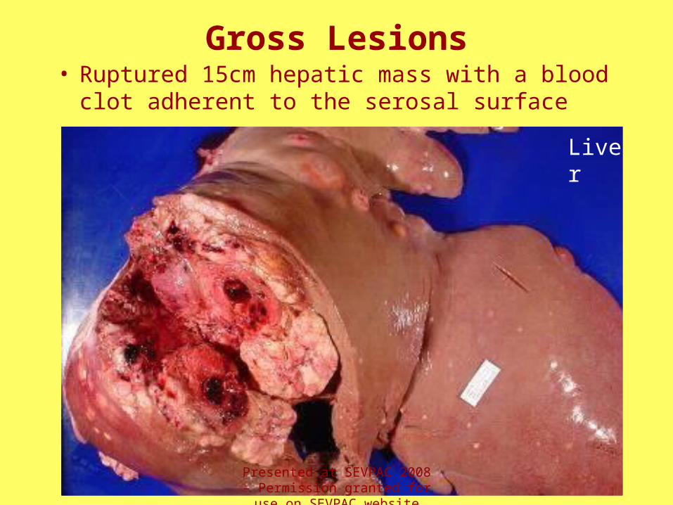
Gross Lesions• Ruptured 15cm hepatic mass with a blood clot
adherent to the serosal surface
Liver
Presented at SEVPAC 2008 – Permission granted for use on
SEVPAC website only
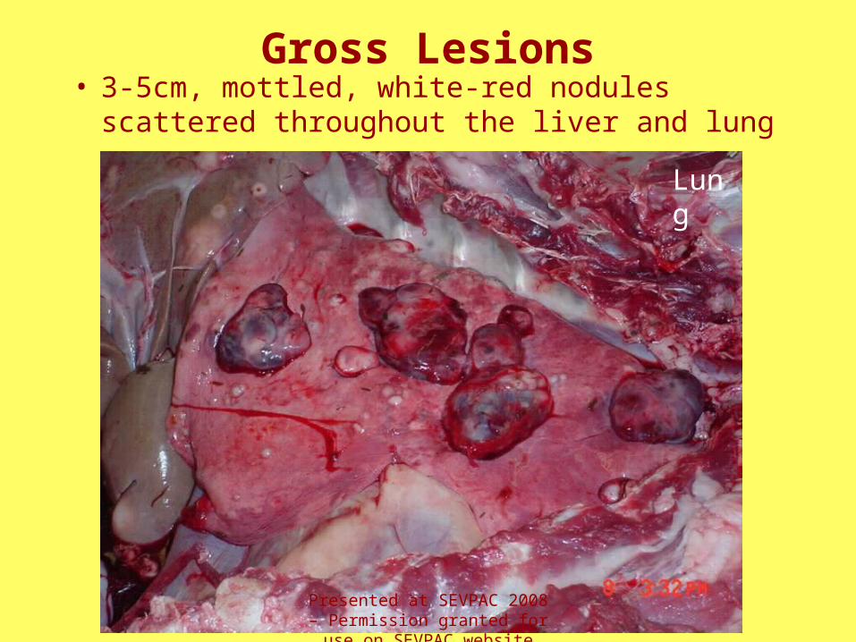
Gross Lesions• 3-5cm, mottled, white-red nodules scattered
throughout the liver and lung
Lung
Presented at SEVPAC 2008 – Permission granted for use on
SEVPAC website only

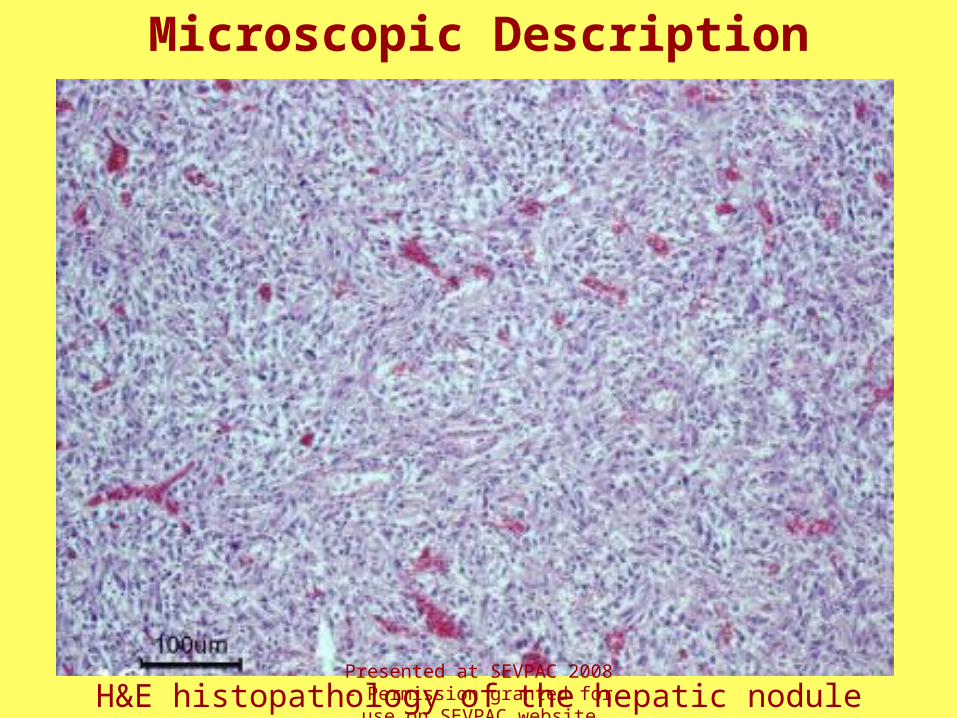
Microscopic Description• Hepatic mass and pulmonary nodules
– Extensive hemorrhage, fibrin, edema, and necrosis
– Normal hepatic architecture is obliterated and replaced by a densely cellular mass of pleomorphic spindle cells arranged in streams and bundles
– Marked anisocytosis, anisokaryosis, occasional multinucleated cells, and 2-4 mitotic figures per 400x field
– These findings are consistent with a neoplasm of mesenchymal origin (sarcoma)Presented at SEVPAC 2008 –
Permission granted for use on SEVPAC website only
Microscopic Description
H&E histopathology of the hepatic nodulePresented at SEVPAC 2008 – Permission granted for use on
SEVPAC website only
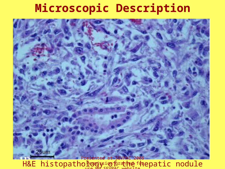
H&E histopathology of the hepatic nodule
Microscopic Description
Presented at SEVPAC 2008 – Permission granted for use on
SEVPAC website only

Immunohistochemistry
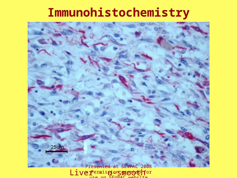
• Neoplastic cells are positive for:– 60-85% Vimentin immunoreactivity– 10-15% Muscle-specific actin immunoreactivity– 15-20% α-smooth muscle actin immunoreactivity
• Neoplastic cells are negative for:– Mac 387– Desmin– Cytokeratin
Presented at SEVPAC 2008 – Permission granted for use on
SEVPAC website only
Immunohistochemistry - Markers
• Vimentin - demonstrates that the neoplasm was of mesenchymal origin (sarcoma)
• Muscle-specific actin - recognizes all of the alpha actins (skeletal, cardiac, and smooth muscle) as well as gamma smooth muscle actin
• α-smooth muscle actin - recognizes only alpha smooth muscle actin
Presented at SEVPAC 2008 – Permission granted for use on
SEVPAC website only

Immunohistochemistry
Liver - Muscle actinPresented at SEVPAC 2008 – Permission granted for use on
SEVPAC website only
Immunohistochemistry
Liver - α-smooth muscle actinPresented at SEVPAC 2008 – Permission granted for use on
SEVPAC website only

Case Summary
• The large hepatic mass is suspected to be the primary neoplasm
• The mass likely originated from the smooth muscle of either a hepatic blood vessel or biliary duct
• Immunohistochemistry and histopathology support a diagnosis of leiomyosarcoma
• Sudden death was the result of hypovolemic shock secondary to acute hemoabdomen caused by the rupture of the large hepatic mass
Presented at SEVPAC 2008 – Permission granted for use on
SEVPAC website only
References
1. Cooper BJ, Valentine, BA: Tumors of the muscle. In: Tumours in Domestic Animals, ed. Mueten DJ, 4th ed., pp 319-340, Iowa State Press, IA 2002
2. Shapiro JL, Watson P, McEwen B, Carman S: Highlights of camelid diagnoses from necropsy submissions to the Animal Health Laboratory, University of Guelph, from 1998 to 2004. Can Vet J 46:317–318., 2005
3. Valentine BA, Martin, JM: Prevalence of neoplasia in llamas and alpacas (Oregon State University, 2001–2006). J Vet Diagn Invest 19:202-204, 2007
4. Valero R, et al.: Leiomiosarcoma hepatico: Reporte de un caso y revision de la literature. Casos Clinicos de Gastroenterologia, Casos Clinicos de Oncologia 2006: http://www.portalesmedicos.com/publicaciones/articles/256/2/Leiomiosarcoma-hepatico.-Caso-Clinico 2006
Presented at SEVPAC 2008 – Permission granted for use on
SEVPAC website only

Questions?
Presented at SEVPAC 2008 – Permission granted for use on
SEVPAC website only