lentiviral-mediated overexpression of akt1 reduces anoxia-reoxygenation injury in cardiomyocytes
TRANSCRIPT

Cell Biology International ISSN 1065-6995doi: 10.1002/cbin.10234
RESEARCH ARTICLE
Lentiviral-mediated overexpression of Akt1 reducesanoxia–reoxygenation injury in cardiomyocytesYanyan Du1y, Hong Zhu2y, Dongye Li1*, Lele Wang1, Lin Zhang1, Yuanyuan Luo2,Defeng Pan2 and Manman Huang1
1 Institute of Cardiovascular Disease Research, Xuzhou Medical College, No. 84 West Huaihai Road, Xuzhou, Jiangsu 221002, P.R. China2 Xuzhou Medical College Affiliated Hospital, Xuzhou 221006, P.R. China
Abstract
Activated PI3K/Akt signalling exerts a protective effect after myocardial ischemia by phosphorylating various substrates;however, the precise mechanism by which this occurs remains to be elucidated. We have constructed the recombinant lentiviralvector pLVX-Akt1-EGFP- 3FLAG (LV-Akt1) to determine the efficiency of LV-Akt1 infection, explore the protective role ofAkt1, and investigate the possible mechanism by which Akt1 signalling acts during anoxia/reoxygenation (A/R) ofcardiomyocytes in primary culture. Akt1 gene transfection increased cardiomyocyte pulsation, reduced cell mortality, anddecreased the concentration of lactate dehydrogenase (LDH) in myocardial cells supernatants. Akt1 transfection increased thelevels of intracellular p-Akt, enhanced the expression of the anti-apoptosis protein Bcl-2, and reduced that of the apoptosisprotein Bax (thereby increasing the Bcl-2/Bax ratio), and caused some increase in hypoxia-inducible factor1a (HIF-1a) andvascular endothelial growth factor (VEGF) expression after A/R. The protective role of Akt1 was partly suppressed by adding aphosphoinositide 3-kinase/Akt inhibitor (LY294002). In conclusion, LV-Akt1 was successfully constructed and neonatal ratcardiomyocytes were transfected efficiently. Akt1 overexpression significantly reduced A/R injury in cardiomyocytes, and thiscould be related to its effects on various targets of the PI3K/Akt signalling pathway, such as Bcl-2, Bax, HIF-1a and VEGF.
Keywords: Akt1; anoxia–reoxygenation; apoptosis; gene transfection; lentiviral vector; myocardium
Introduction
Akt, also called protein kinase B (PKB), is a serine/threonineprotein kinase family comprising three closely relatedmembers: Akt1 (PKBa), Akt2 (PKBb) and Akt3 (PKBg)(Hanada et al., 2004; Oudit et al., 2004). Akt/PKB contains anN-terminal PH domain, a central catalytic domain, and aC-terminal regulatory domain (Chan et al., 1999). As a targetof phosphoinositide 3-kinase (PI3K), it regulates awide rangeof biological responses including cell motility, growth,proliferation, and survival, as well as transcription, proteinsynthesis and nutrient metabolism (Brazil et al., 2002;Cantley, 2002). By phosphorylating a variety of downstreamsubstrates, activated Akt induces resistance to apoptosis,regulation of cell growth and proliferation, etc. (Dattaet al., 1999). Akt1 is expressed in almost all tissues but itsexpression is particularly high in brain, heart, and lungs,
while Akt2 is highly expressed in muscle and brown adiposetissue and Akt3 in brain tissue (Testa and Tsichlis, 2005).PI3K/Akt signalling is activated after myocardial ischemiaand Akt1 is a core node of the PI3K/Akt signalling pathway(Breivik et al., 2011). An adenovirus was used to transfect theAkt1 gene into a rat cardiac model, significantly improvingthe hemodynamics of ischemia/reperfusion (I/R), reducingmyocardial cell apoptosis and attenuating infarct size;however, its precise mechanism of action is not yetparticularly clear (Matsui et al., 2001).
Hypoxia-inducible factor-1 (HIF-1) is a heterodimerictranscription factor composed of HIF-1a and HIF-1bsubunits (Wang et al., 1995; Jiang et al., 1996). Inducedby hypoxia, growth factors and oncogenes, HIF-1a regulatesthe expression of many genes, including VEGF, hemeox-ygenase-1, inducible nitric oxide synthase (iNOS) and severalglycolytic enzymes (Semenza, 2002). In cancer cells, HIF-1a
�Corresponding author: e-mail: [email protected] authors contributed equally to this work.Abbreviations: A/R, anoxia/reoxygenation; LDH, lactate dehydrogenase; HIF-1a, hypoxia-inducible factor1a; VEGF, vascular endothelial growth factor
488 Cell Biol Int 38 (2014) 488–496 � 2013 International Federation for Cell Biology

expression is regulated by the PI3K/AKT signalling pathway(Zhong et al., 2000). Vascular endothelial growth factor(VEGF) is an important angiogenic factor and a number ofregulatory mechanisms modulate its expression, hypoxiabeing one of the most important. Low oxygen can increasethe levels of VEGF at both the mRNA and protein levels.VEGF acts on endothelial cells to stimulate angiogenesis,which in turn helps maintain an adequate supply of oxygen(Ferrara, 1999). Multiple lines of evidence indicate that thePI3K/AKT signalling pathway is required for VEGF expres-sion in the context of tumour angiogenesis and growth (Donget al., 2001).
Akt activation is necessary and sufficient to regulate HIF-1and VEGF expression in human cancer cells. HIF-1 andVEGF are potent inducers of angiogenesis (Jiang andLiu, 2008). By regulating the expression of HIF-1a tomodulate the expression of VEGF protein, the PI3K/AKTsignalling pathway promotes the germination of bloodvessels and facilitates tumour growth in tumour tissue. Innon-tumour tissue, constitutive overexpression of HIF-1a inmurine heart attenuates infarct size and improves cardiacfunction four weeks aftermyocardial infarction. Significantly,the authors also found increases in both capillary density andVEGF levels (Masakuni et al., 2005).
We have explored the effects of Akt1 overexpression on cellfunction, viability and gene expression in primary cardio-myocytes subjected to A/R, and investigated whether thePI3K/AKT signalling pathway regulates the expression/stabilisation of HIF-1a and VEGF by adding a PI3K/AKTinhibitor (LY294002), thereby demonstrating a protectiveeffect of Akt1 gene transfection on A/R injury incardiomyocytes.
Materials and methods
Animals, plasmids, vectors and cells
Neonatal Sprague–Dawley rats (1–3 days old) were obtainedfrom the Experimental Animal Centre of Xuzhou MedicalCollege (Xuzhou, Jiangsu, China). This study was approvedby the Animal Ethics Committee of Xuzhou Medical College(permit number: xz11-12540) and conforms to the Guide forthe Care and Use of Laboratory Animals published by the USNational Institutes of Health (NIH publication No. 85–23,revised 1996). PXZ208-AKT1 plasmids (Institute of Cardio-vascular Disease Research, Xuzhou, China), pLVX-EGFP-3FLAG (Sbo-bio, Shanghai, China) and DH5a competentcells (Takara, Japan) were acquired for use in this study.
Main reagents
Taq polymerase and dNTPs (Takara), In-FusionTM PCRCloning Kit (Clontech, CA), EcoR I (NEB, USA), DL 2,000
DNA Marker, Sepharose DNA recycling kit, BCA ProteinAssay Kit (HyClone-Pierce, USA), 1 kb DNA Ladder Marker,Prestained Protein Ladder (Fermentas, Canada) and the ECL-PLUS/Kit (Amersham, USA) were purchased from theindicated manufacturers. Mouse anti-GFP and goat anti-mouse IgG antibodies against caspase-3, Bcl-2 and Baxwere purchased from Santa Cruz Biotechnology (CA,USA). Rabbit monoclonal anti-Akt, rabbit monoclonalanti-p-Akt and LY294002 (CST, USA), mouse monoclonalanti-HIF-1a and mouse monoclonal anti-VEGF (Abcam,England), polybrene, 5-bromodeoxyuridine, collagenase I(St. Louis, MO, USA), Dulbecco’s modified Eagle’s medium(DMEM), fetal bovine serum (FBS) and 0.25% trypsin(GIBCO, USA), lactate dehydrogenase (LDH) assay kits(Jiancheng Bioengineering Institute, Nanjing, China), anti-rabbit IgG or anti-mouse IgG secondary antibody (Zhong-shan, Beijing, China) were purchased from the indicatedmanufacturers. Primers were synthesised by Generay(Shanghai, China). DNA from Akt1recombinant plasmid-positive clones was sequenced by BGI (Beijing, China).
Construction of lentiviral expression vector containingAkt1 gene
Within the template of the Akt1gene, a specific fragment wasamplified by PCR. The primers were as follows: AKT1-EcoRI-F, 50-CTCAAGCT TCGAATTCGCCACCATGAACGAC GTA GCC ATT GTG-30; and AKT1-EcoRI-R, 50-CCATGGTGGCGAATTCGGCTGTGCCACTGGCTGAG- 30.These primers contain homologous recombination andEcoRI restriction sites (underlined) as well as a Kozaksequence (double underlined). After isolation and purifica-tion of the PCR product, the Akt1 gene was inserted into theEcoRI site of the linearised expression vector pLVX-EGFP-3FLAG by incubating at 378C and 508C (both for 15min).The recombinant plasmid was obtained and transformed intoDH5a competent cells. Appropriate amounts of the trans-formed cells were spread on LB agar plates containingampicillin to select for the expression vector, and theplates were cultivated in a 378C incubator for 16 h (Zhaoet al., 2002).
Identification of positive recombinant clones, packagingof lentivirus, determining titer and analysing fusionprotein
Transformants were picked off the plate and suspended in10mL LB culturemedium; 1mLwas used as template for PCRto identify the positive colonies directly. The primersemployed were as follows: AKT1-F1, 50-CAA GGA TGAGGTTGCCCACAC-30 (binding upstreamof the Akt1 gene);and pEGFP-N-3, 50-CGTCGC CGTCCAGCTCGACCAG-30 (binding the downstream sequence of the multiple clone
Y. Du et al. LV-Akt1 reduces cardiomyocytes A/R injury
489Cell Biol Int 38 (2014) 488–496 � 2013 International Federation for Cell Biology

site). The positive recombinant clones were detected andvalidated by DNA sequencing. Subsequently, inoculation andplasmid extraction were conducted, and recombinantlentiviral particles were produced after co-transfection withpLVX-Akt1-EGFP-3FLAG and lentiviral packaging plasmids.The recombinant particles were then packaged, concentratedand amplified by 293FT cells to obtain LV-Akt1with highefficiency. Subsequently, the viral titer was determined byreal-time PCR and the expression level of the Akt1-EGFP-FLAG fusion protein was measured by Western blotting(Zhao et al., 2002).
The Akt1 gene was observed as a band at1.5–1 kb, whichwas consistent with the 1,478 bp band expected (seeSupporting Information, Figure S1A (a)). A 956 bp bandwas obtained from gel electrophoresis of the PCR amplifica-tion of the positive clone (see Figure S1A (b)). DNAsequencing confirmed that the sequence of the positiveclone was consistent with that in GenBank/NCBI. Greenfluorescent protein expression in 293FT cells was observedunder fluorescence microscopy after transfection with therecombinant lentiviral vector (see Figure S1B). A limitingdilution assay established that the titer of the virus was2.15� 108 IU/mL. Western blotting demonstrated that thefusion protein was effectively expressed in 293FT cells (seeFigure S1C).
Primary culture of cardiomyocytes
Neonatal rat cardiomyocytes were cultured using the methodpreviously described (Franke et al., 2007). Neonatal SD rats(1–3 days old) of both sexes were sacrificed and disinfectedwith 75% ethanol. On a very clean bench, the thoracic cavitywas cut open immediately and the heart was exposed,removed and sheared in cold PBS solution. The ventricleswere removed and rinsed four times with cold PBS to removeresidual blood. The tissue was minced, added to an equalvolume of 0.08% collagenase I and 0.08% trypsin, andincubated in a water bath with a magnetic stirrer (30 rpm,378C). Following water bath digestion (4–5min), naturalsedimentation and collection of the supernatant, the cells wereplaced in sterile centrifuge tube and incubated with culturemedium containing 10% fetal bovine serum to terminatedigestion. After passing through a 200mm mesh steel filter6–8 times, the filtrate was collected and centrifuged(1,000 rpm, 5min). The supernatant was discarded and thepellet was resuspended in 5mL of culture medium. Theresuspension and centrifugation steps were repeated threetimes to wash the cells completely free of trypsin andcollagenase. Finally, the cells were suspended in DMEMsupplemented with 100U/mL penicillin, 100mg/mL strepto-mycin, 20% fetal bovine serum and 0.1mM BrdU. Forsubsequent experiments, the cell suspensionwas inoculated at5� 105 cells/mL into 24-well or 96-well cell culture plates and
incubated with humidified air containing 5%CO2 at 378C. Toobtain a pure culture of cardiomyocytes and inhibit thegrowth of non-myocytes, a selective adhesion procedure wasperformed after incubation for 1.5 h. After the cardiomyocytecultures had been enriched for 24 h in complete medium,experiments were performed after 3 days in culture.
The efficiency of LV-Akt1 infection of myocardial cellculture
On the first day, neonatal rat cardiomyocytes were inoculatedat 5� 105 cells/mL into 24-well culture plates. On thethird day, LV-Akt1 virus-containing solution was thawed onice and added to the culture to MOIs of 5, 10, 15, 20,25, 30, 35, 40 and 45 along with polybrene (finalconcentration 8mg/mL). This culture was mixed andincubated in humidified air containing 5% CO2 at 378C.Green fluorescent protein expression was observed andphotographed at 24, 48 and 72 h using a fluorescencemicroscope (DP72, OLYMPUS). The rate of green fluorescentprotein expression was determined by dividing the number ofcells with green fluorescence by the total number of cells.
Cardiomyocyte anoxia–reoxygenation
When the cardiomyocytes grew closer to fusion, the culturemedium was deprived of oxygen and glucose to simulateischemia, oxygen and glucose were reintroduced to simulatereperfusion (Jun et al., 2011). The procedure was conductedas follows: the medium was discarded, nitrogen-saturatedD-Hank’s buffer solution was added, and the culture plateswere incubated for 180min in humidified air containing 5%CO2 and 95% N2 at 378C to simulate anoxia. The mediumwas then discarded and replaced with Hank’s buffer solutionsupplemented with glucose and Ca2þ, and the culture platewas incubated with humidified air containing 5% CO2 at378C for 120min to simulate reoxygenation. Control cellswere cultured in 20% fetal bovine serum in humidified aircontaining 5% CO2 at 378C; the remaining groups weretreated as described.
Experimental groups
Cells were randomly divided into control and experimentalgroups as follows. Normal control group (C): cardiomyocyteswere cultured under normal conditions without anoxia–reoxygenation (A/R) treatment. A/R group (A/R): culturedcardiomyocytes underwent A/R as described above. Emptycarrier group (EC): culturedmyocardial cells were transfectedwith LV-EGFP for 48 h, underwent A/R. Akt1 gene transfec-tion group (A): cultured cardiomyocytes were transfectedwith LV-Akt1 for 48 h, then underwent A/R.Akt1þ LY294002
LV-Akt1 reduces cardiomyocytes A/R injury Y. Du et al.
490 Cell Biol Int 38 (2014) 488–496 � 2013 International Federation for Cell Biology
(50mM) group (ALY): cultured cardiomyocytes were trans-fected with LV-Akt1 for 48 h, then LY294002 (50mM) wasadded to the supernatant and incubated for 2 h before A/R.The experiments were repeated three times.
Myocardial cell survival rate
Cell viability was assessed by trypan blue exclusion and bymeasuring LDH release into the medium (Das et al., 2005).Adherent myocardial cells were digested (0.25% trypsin) andisolated and 0.4% trypan blue was added. After 5minincubation, a small amount of the suspensionwas placed on amicroscope slide. The fraction of blue cells was quantifiedby microscopy. Several separate fields were observed underthe microscope, and cell viability was calculated by countingthe numbers of dead cells (blue-staining cells) and livingcells and using the following equation: cell survival rate¼non-blue-staining cells/(blue-staining cellsþ non-blue-staining cells)� 100%. For LDH measurements, the culturemedium of every groupwas collected and the enzyme activitywas monitored spectrophotometrically using an assay kit(Nanjing, China).
Western blotting to demonstrate Akt, p-Akt, Bcl-2, Bax,HIF-1a and VEGF protein expression
To investigate the possible mechanism(s) responsible for thecardioprotective effects of Akt1 overexpression, the proteinlevels of Akt, p-Akt, Bcl-2, Bax, HIF-1a and VEGF weredetermined by Western blotting, which was performed aspreviously described (Qi et al., 2011).
Statistical analysis
In each experimental series, data are presented as means�SD. GraphPad Prism 5.01 software (GraphPad; San Diego,CA, USA) was used for statistical analysis. One-way ANOVAwas used for multiple group comparisons. Differences withP< 0.05 were considered statistically significant.
Results
Determination of the rate of LV-Akt1 infection ofprimary cardiomyocytes
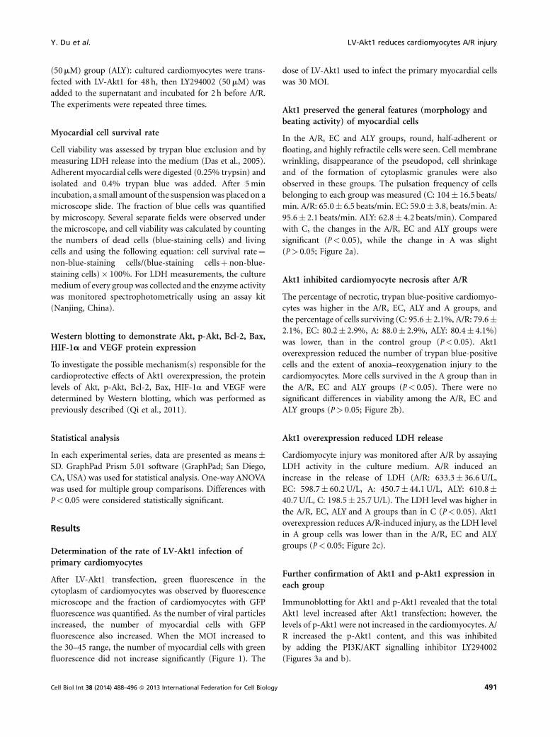
After LV-Akt1 transfection, green fluorescence in thecytoplasm of cardiomyocytes was observed by fluorescencemicroscope and the fraction of cardiomyocytes with GFPfluorescence was quantified. As the number of viral particlesincreased, the number of myocardial cells with GFPfluorescence also increased. When the MOI increased tothe 30–45 range, the number of myocardial cells with greenfluorescence did not increase significantly (Figure 1). The
dose of LV-Akt1 used to infect the primary myocardial cellswas 30 MOI.
Akt1 preserved the general features (morphology andbeating activity) of myocardial cells
In the A/R, EC and ALY groups, round, half-adherent orfloating, and highly refractile cells were seen. Cell membranewrinkling, disappearance of the pseudopod, cell shrinkageand of the formation of cytoplasmic granules were alsoobserved in these groups. The pulsation frequency of cellsbelonging to each group was measured (C: 104� 16.5 beats/min. A/R: 65.0� 6.5 beats/min. EC: 59.0� 3.8, beats/min. A:95.6� 2.1 beats/min. ALY: 62.8� 4.2 beats/min). Comparedwith C, the changes in the A/R, EC and ALY groups weresignificant (P< 0.05), while the change in A was slight(P> 0.05; Figure 2a).
Akt1 inhibited cardiomyocyte necrosis after A/R
The percentage of necrotic, trypan blue-positive cardiomyo-cytes was higher in the A/R, EC, ALY and A groups, andthe percentage of cells surviving (C: 95.6� 2.1%, A/R: 79.6�2.1%, EC: 80.2� 2.9%, A: 88.0� 2.9%, ALY: 80.4� 4.1%)was lower, than in the control group (P< 0.05). Akt1overexpression reduced the number of trypan blue-positivecells and the extent of anoxia–reoxygenation injury to thecardiomyocytes. More cells survived in the A group than inthe A/R, EC and ALY groups (P< 0.05). There were nosignificant differences in viability among the A/R, EC andALY groups (P> 0.05; Figure 2b).
Akt1 overexpression reduced LDH release
Cardiomyocyte injury was monitored after A/R by assayingLDH activity in the culture medium. A/R induced anincrease in the release of LDH (A/R: 633.3� 36.6U/L,EC: 598.7� 60.2U/L, A: 450.7� 44.1U/L, ALY: 610.8�40.7 U/L, C: 198.5� 25.7U/L). The LDH level was higher inthe A/R, EC, ALY and A groups than in C (P< 0.05). Akt1overexpression reduces A/R-induced injury, as the LDH levelin A group cells was lower than in the A/R, EC and ALYgroups (P< 0.05; Figure 2c).
Further confirmation of Akt1 and p-Akt1 expression ineach group
Immunoblotting for Akt1 and p-Akt1 revealed that the totalAkt1 level increased after Akt1 transfection; however, thelevels of p-Akt1 were not increased in the cardiomyocytes. A/R increased the p-Akt1 content, and this was inhibitedby adding the PI3K/AKT signalling inhibitor LY294002(Figures 3a and b).
Y. Du et al. LV-Akt1 reduces cardiomyocytes A/R injury
491Cell Biol Int 38 (2014) 488–496 � 2013 International Federation for Cell Biology
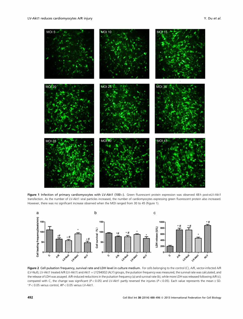
Figure 1 Infection of primary cardiomyocytes with LV-Akt1 (100�). Green fluorescent protein expression was observed 48 h post-eLV-Akt1
transfection. As the number of LV-Akt1 viral particles increased, the number of cardiomyocytes expressing green fluorescent protein also increased.
However, there was no significant increase observed when the MOI ranged from 30 to 45 (Figure 1).
Figure 2 Cell pulsation frequency, survival rate and LDH level in culture medium. For cells belonging to the control (C), A/R, vector-infected A/R
(LV-Null), LV-Akt1 treated A/R (LV-Akt1) and Akt1þ LY294002 (ALY) groups, the pulsation frequency wasmeasured, the survival rate was calculated, and
the release of LDHwas assayed. A/R-induced reductions in the pulsation frequency (a) and survival rate (b), while more LDHwas released following A/R (c);
compared with C, the change was significant (P< 0.05) and LV-Akt1 partly reversed the injuries (P< 0.05). Each value represents the mean� SD.�P< 0.05 versus control, #P< 0.05 versus LV-Akt1.
LV-Akt1 reduces cardiomyocytes A/R injury Y. Du et al.
492 Cell Biol Int 38 (2014) 488–496 � 2013 International Federation for Cell Biology

Figure 3 Expression of Akt1, p-Akt1, Bax, Bcl-2. Akt1 and p-Akt1expression was assayed in all groups. Akt1 transfection increased total Akt1
expression (P< 0.05, a) and A/R-induced p-Akt1, and the increase in Bax levels (b, c) and decrease in Bcl-2 expression (c). Compared with C, the change
was significant (P< 0.05). A/R induced an increase in Bax expression while Akt1 transfection decreased it (P< 0.05, c). Bcl-2 expression decreased
following A/R, but increased due to LV-Akt1 transfection (d). Compared with C, the change was significant (P< 0.05). As Bax decreased, Bcl-2 increased,
and the eBcl-2 to Bax ratio was higher in cells transfected with LV-Akt1 (e), than in the A/R group (P< 0.05). Each value represents the mean� SD.�P< 0.05 versus C, #P< 0.05 versus LV-Akt1.
Y. Du et al. LV-Akt1 reduces cardiomyocytes A/R injury
493Cell Biol Int 38 (2014) 488–496 � 2013 International Federation for Cell Biology

Effect of Akt1 overexpression on expression of apoptosisassociated proteins Bcl-2 and Bax
Immunoblotting for Bcl-2 and Bax showed that Baxexpression was higher in A/R than in the control cells(P< 0.05). However, Bcl-2 protein expression was signifi-cantly lower in by A/R than the control group (P< 0.05).Akt1 transfection caused upregulated Bcl-2 and down-regulated Bax protein expression, leading to a higher Bcl-2/Bax ratio than in the A/R group (Figures 3c–e).
Effect of Akt1 overexpression on expression of HIF-1aand VEGF
Immunoblotting for HIF-1a and VEGF revealed that boththese factors were more highly expressed in A/R than inthe control cells (P< 0.05). Akt1 transfection upregulatedHIF-1a and VEGF, though this effect was partly inhibitedby adding the PI3K/AKT signalling inhibitor LY294002(Figure 4).
Discussion
Gene therapy has become an increasingly popular molecularbiological technique during the past few years, and trans-genesis of target cells is one of the most important steps. Thebiological component includes retroviral, adenoviral andlentiviral vectors. In this study we used a lentiviral vectorcarrying an Akt1 gene fragment. The advantages of thismethod are a longer time for expression, diminishedcytotoxicity and immune response mediated by host cells,and the strong induction of infection in unseparated cellpopulations such as cardiomyocytes.
The PI3K/AKTsignalling pathway and several downstreampathways are activated in cardiomyocytes during the processof A/R, and this signalling is important in regulating cardiacstructure and function (Manning and Cantley 2007; Banet al., 2008; Oudit and Penninger, 2009).
Akt1 transfection increases the expression of Bcl-2, reducesthe expression of Bax, and increases the Bcl-2/Bax ratio whilereducing the myocardial apoptosis caused by A/R. Theseeffects of Akt1 overexpression were suppressed by the PI3K/AKT signaling pathway inhibitor LY29004, confirming thatAkt1 transfection has an anti-apoptotic effect by regulatingthe expression of the PI3K/AKT downstream proteins Bcl-2and Bax.
Anoxia constitutes a major component of many diseasestates and can have both proliferative (cancer) and injurious(stroke, heart attack) effects (Semenza, 2000). The HIF-1agene is upregulated in regions of myocardial infarction andischemia (Lee et al., 2000). Anoxia is also known to triggertranscription of the endogenous HIF-1a gene in severalorgans including human myocardium.
Much evidence indicates that PI3K/AKT signalling isrequired for VEGF and HIF-1a expression in the context oftumour angiogenesis and growth (Mazure et al., 1997). Wefound that HIF-1a and VEGF protein levels increasedfollowing myocardial A/R, and Akt1 transfection increasedtheir expression in cardiomyocytes. After the PI3K/Aktinhibitor (LY294002) was added, the protective roledescribed above was partly suppressed, indicating thatHIF-1a and VEGF expression could be regulated throughthe PI3K/AKT signalling pathway.
Continued hypoxia leads to ischemic necrosis of myocar-dial cells. Currently, reperfusion (the restoration of blood
Figure 4 Expression of HIF-1a and VEGF. The levels of HIF-1a and VEGF were measured in all groups. A/R induced an increase in HIF-1a and VEGF
expression (a, b). Compared with C, the change was significant (P< 0.05). Akt1 transfection enhanced the expression of HIF-1a and VEGF, as compared
with C; the changes were significant (P< 0.05). Each value represents the mean� SD. �P< 0.05 versus C, #P< 0.05 versus LV-Akt1.
LV-Akt1 reduces cardiomyocytes A/R injury Y. Du et al.
494 Cell Biol Int 38 (2014) 488–496 � 2013 International Federation for Cell Biology

flow) is the most effective method of preventing acutemyocardial necrosis (Hans and David, 2012). However, whilereperfusion rescues the myocardial cells and limits myocar-dial cell injury, it also aggravates such injury and causesirreversible necrosis at the same time (Martenian et al., 2006).The PI3K/AKT signalling pathway is protective duringmyocardial I/R, as ischemic preconditioning (IPC) (Murryet al., 1986) and ischemic post-conditioning (IPost) (Zhaoet al., 2003) activate this pathway to exert a protective effect inmyocardial ischemia-reperfusion injury. There is evidencethat the cardio-protective and anti-apoptotic effects ofLuteolin, a polyphenolic flavonoid, are associated withPI3K/Akt-pathway activation, at least partly (Fang et al.,2011; Sun et al., 2012). We found that Akt1 transfectionimproves the ischemic tolerance of myocardial cells andenhances the protective effects of the PI3K/AKT signallingpathway on A/R-injured cardiomyocytes. This could beuseful for further investigations of the mechanism responsi-ble for increased Akt1 expression in A/R cardiomyocytes afterIPC and IPost. Furthermore, gene therapy could be a futuremethod for the clinical treatment of myocardial ischemia.
Acknowledgments and funding
This work was supported by president in Xuzhou MedicalCollege (09KJZ30).
Author contributions
Dongye Li conceived and designed the study. Yanyan Du,Hong Zhu, Lele Wang et al. conducted the study and wrotethe paper. All authors contributed to the study and approvedthe final version of the paper.
References
Ban K, Cooper AJ, Samuel S, Bhatti A, Patel M, Izumo S (2008)
Phospha-tidylinositol 3-kinase gamma is a critical mediator of
myocardial ischemic and adenosine-mediated preconditioning.
Circ Res 103: 643–53.
Brazil DP, Park J, Hemmings BA (2002) PKB binding proteins.
Getting in on the Akt. Cell 111: 293–303.
Breivik L, Helgeland E, Aarnes EK, Mrdalj J, Jonassen AK (2011)
Remote postconditioning by humoral factors in eflluent from
ischemic preconditioned rat hearts is mediated via PI3K/Akt-
dependent cell-survival signaling at reperfusion. Basic Res
Cardiol 106: 135–45.
Cantley LC (2002) The phosphoinositide 3-kinase pathway.
Science 296: 1655–7.
Chan TO, Rittenhouse SE, Tsichlis PN (1999) AKT/PKB and other
D3 phosphoinositide-regulated kinases: kinase activation by
phosphoinositide-dependent phosphorylation. Annu Rev Bio-
chem 68: 965–1014.
Das A, Xi L, Kukreja RC (2005) Phosphodiesterase-5 inhibitor
sildenafil preconditions adult cardiac myocytes against necrosis
and apoptosis. Essential role of nitric oxide signaling. J Biol
Chem 280(13): 12944–55.
Datta SR, Brunet A, Greenberg ME (1999) Cellular survival: a play
in three Akts. Genes Dev 13(22): 2905–27.
Dong G, Chen Z, Li Z, Yeh Y, Bancroft NT, Van Waes CC (2001)
Hepatocyte growth factor/scatter fector-induced activation of
MEK and PI3K signal pathways contributes to expression of
proanogiogenic cytokines inter-leukin-8 and vascular endothe-
lial growth factor in hcad and neck squamous cell carcinoma.
Cancer Res 61: 5911–8.
Fang F, Li DY, Pan HJ, Chen D, Qi LL, Zhang R, Sun H (2011)
Luteolin inhibits apoptosis and improves cardiomyocyte
contractile function through the PI3K/Akt pathway in simulated
ischemia/reperfusion. Pharmacology 88: 149–58.
Ferrara N (1999) Molecular and biological properties of vascular
endothelial growth factor. J Mol Med 77: 527–43.
Franke WW, Schumacher H, Borrmann CM, (2007) The area
composita of adhering junctions connecting heart muscle cells
of vertebrates-III: assembly and sintegration of intercalated
disks in rat cardiomyocytes growing in culture. Eur J Cell Biol 86
(3): 127–42.
HanadaM, Feng J, Hemmings BA (2004) Structure, regulation and
function of PKB/AKT—a major therapeutic target. Biochim
Biophys Acta 1697: 3–16.
Hans MP, David GD (2012) Reducing the impact of myocardial
ischaemia/reperfusion injury. Cardiovasc Res 94: 165–7.
Jiang BH, Liu LZ (2008) AKTsignaling in regulating angiogenesis.
Curr Cancer Drug Targets 8(1): 19–26.
Jiang BH, Rue E, Wang GL, Roe R, Semenza GL (1996)
Dimerization DNA binding, and transactivation properties of
Hypoxia-inducible factor 1. J Biol Chem 271: 17771–8.
Peng J, Xu WJ, He BX, Liu Y, Zhang H, Ding QL (2011) Effects of
baicalein on rat myocardial ischemia and neonatal cardiomyo-
cyte injury. Chin J Nat Med 9(2): 0132–40.
Lee SH, Wolf PL, Escudero R, Deutsch R, Jamieson SW, Thistleth-
waite PA (2000) Early expression of angiogenesis factors in acute
myocardial ischemia and infarction. N Engl J Med 342:
626–33.
Manning BD, Cantley LC (2007) AKT/PKB signaling: navigating
downstream. Cell 129: 1261–74.
Martelli AM, Faenza I, Billi AM, Manzoli L, Evangelisti C, Falà F,
Cocco L (2006) Intranuclear 3-phosphoinositide metabolism
and Akt signaling: new mechanisms for tumorigenesis and
protection against apoptosis? Cell Signal 18(8): 1101–7.
Kido M, Du L, Sullivan CC, Li X, Deutsch R, Jamieson SW,
Thistlethwaite PA (2005) Hypoxia-inducible factor 1-alpha
reduces infarction and attenuates progression of cardiac
dysfunction after myocardial infarction in the mouse. JACC
46: 2116–24.
Matsui T, Tao J, del Monte F, Lee KH, Li L, Picard M, Force TL,
Franke TF, Hajjar RJ, Rosenzweig A (2001) Akt activation
preserves cardiac function and prevents injury after transient
cardiac ischemia in vivo. Criculation 104(3): 330–5.
Mazure NM, Chen EY, Ladcroute KR, Giaccia AJ (1997) Induction
of vascular endothetial growh factor by hypoxia is modulated by
Y. Du et al. LV-Akt1 reduces cardiomyocytes A/R injury
495Cell Biol Int 38 (2014) 488–496 � 2013 International Federation for Cell Biology

a phosphate-dylinositol 3-kinase/Akt signaling pathway in
Ha-ras-transformed cells through a hypoxia inducible factor-
1 transcriptional element. Blood 90: 3322–31.
Murry CE, Jennings RB, Reimer KA (1986) Preconditioning with
ischemia: a delay of lethal cell injury in ischemic myocardium.
Circulation 74(5): 1124–36.
Oudit GY, Penninger JM (2009) Cardiac regulation by phosphoi-
nositide 3-kinases and PTEN. Cardiovasc Res 82: 250–60.
Oudit GY, Sun H, Kerfant BG, Crackower MA, Penninger JM,
Backx PH (2004) The role of phosphoinositide-3 kinase and
PTEN in cardiovascular physiology and disease. J Mol Cell
Cardiol 37: 449–71.
Qi LL, Pan HJ, Li DY, Fang F, Chen D, Sun H (2011) Luteolin
Improves contractile function and attenuates apoptosis follow-
ing ischemia-reperfusion in adult rat cardiomyocytes. Eur J
Pharmacol 668: 201–7.
Semenza GL (2000) HIF-1 and human disease: one highly involved
factor. Genes Dev 14: 1983–91.
Semenza GL (2002) HIF-1 and tumor progression: pathophysiol-
ogy and therapeutics. Trends Mol Med 8: S62–S67.
Sun D, Huang J, Zhang Z, Gao H, Li J, Shen M, Cao F, Wang H
(2012) Luteolin limits infarct size and improves cardiac function
after myocardium ischemia/reperfusion injury in diabetic rats.
PLoS ONE V7N3: e33491.
Testa JR, Tsichlis PN (2005) AKT signaling in normal and
malignant cells. Oncogene 24(50): 7391–3.
Wang GL, Jiang BH, Rue EA, Semenza GL (1995) Hypoxia-
inducible factor 1 is a biasc-helix-loop-helix-PAS heterodimer
regulated by cellular O2 tension. Proc Natl Acad Sci 92: 5510–4.
Zhao J, PettigrewGJ, Thomas J, Vandenberg JI, Delriviere L, Bolton
EM, Carmichael A, Martin JL, Marber MS, Lever AML (2002)
Lentiviral vectors for delivery of genes into neonatal and adult
ventricular cardiac myocytes in vitro and in vivo. Basic Res
Cardiol 97(5): 348–58.
Zhao ZQ, Corvera JS, Halkos ME, Kerendi F, Wang NP, Guyton
RA, Vinten-Johansen J (2003) Inhibition of myocardial injury
by ischemic postconditioning during reperfusion: comparison
with ischemic preconditioning. Am J Physiol Heart Circ Physiol
285(2): H579–H588.
Zhong H, Chiles K, Feldser D, Laugher E, Hanrahan C, Georgescu
MM, Simons JW, Semenza G (2000) Modulation of Hypoxia-
inducible factor 1alpha expression by the epidermal growth
factor/phosphatidylinositol3-kinase/PTEN/AKT/FRAP path-
way in human prostate cancer cells: implication for tumor
angiogenesis and therapeutics. Cancer Res 60: 1541–5.
Received 1 September 2013; accepted 13 November 2013.Final version published online 16 January 2014 .
Supporting Information
Additional supporting information may be found in theonline version of this article at the publisher’s website.
Figure S1. The construction and identification of LV-Akt1
A. Electrophoregram of Akt1 gene amplified and positiveclones identified by PCR.B. The package of pLVX-Akt1-EGFP-3FLAG in 293 FT cells(�100).C. Akt1-EGFP-FLAG fusion protein analyzed by Westernblot.
LV-Akt1 reduces cardiomyocytes A/R injury Y. Du et al.
496 Cell Biol Int 38 (2014) 488–496 � 2013 International Federation for Cell Biology