leptin modulation of locomotor and emotional behaviors: the role of
TRANSCRIPT

Université de Montréal
Leptin modulation of locomotor and emotional behaviors: The role of
STAT3 signaling in dopamine neurons
par Maria Fernanda de Andrade Fernandes
Département de Physiologie
Faculté de Médecine
Thèse présentée à la Faculté de Médecine en vue de l’obtention du grade de Philosophia
Doctor (Ph.D.) en Physiologie
Juin 2014
© Maria Fernanda de Andrade Fernandes, 2014

ii
Université de Montréal
Faculté des études supérieures et postdoctorales
Cette thèse intitulée:
Leptin modulation of locomotor and emotional behaviors: The role of
STAT3 signaling in dopamine neurons
Présentée par :
Maria Fernanda de Andrade Fernandes
a été évaluée par un jury composé des personnes suivantes:
Dr. Alex Parker, président-rapporteur
Dr. Stephanie Fulton, directeur de recherche
Dr. Anne-Noël Samaha, membre du jury
Dr. Alfonso Abizaid, examinateur externe
Dr. Lise Coderre, représentant de la FESP

iii
Résumé
La leptine circule en proportion de la masse graisseuse du corps et la transduction de son
signal à travers la forme longue de son récepteur via un certain nombre de voies neurales , y
compris MAPK, PI3-K ,AMPK et JAK2 - STAT3 . Il faut noter que STAT3 constitue une
voie clée au récepteur de la leptine par laquelle la leptine module l'expression des gènes
impliqués dans la régulation du bilan énergétique.
La plupart des recherches ont porté sur la fonction du récepteur de la leptine au sein de l'
hypothalamus, en particulier la fonction du récepteur de la leptine dans le noyau arqué.
Toutefois, les récepteurs de la leptine sont également exprimés sur les neurones
dopaminergiques de l'aire tégmentale ventrale et la leptine agit sur cette région du cerveau
pour influencer la prise alimentaire, la motivation, la locomotion, l'anxiété et la transmission
de la dopamine. De plus, la leptine active la STAT3 dans les dopaminergiques et
GABAergiques populations neuronales. Bien que ces résultats contribuent à notre
compréhension des multiples actions de la leptine dans le système nerveux central, il reste à
résoudre les cellules et la signalisation du récepteur de la leptine qui sont responsables des
effets neurocomportementaux de la leptine dans le mésencéphale.
Visant à déterminer la contribution de la voie de signalisation STAT3 dans les neurones
dopaminergiques du mésencéphale, nous avons généré une lignée de souris knockout
conditionnel dans lequel l'activation du gène de STAT3 sur son résidu tyrosine 705 ( Tyr 705 )
est absent spécifiquement dans les neurones dopaminergiques. Avec l'utilisation de ce modèle
de souris génétique, nous avons évalué l'impact de l'ablation de la signalisation STAT3 dans
les neurones dopaminergiques sur un certain nombre de fonctions liées à la dopamine, y
compris l'alimentation, la locomotion, les comportements liés à la récompense, l'émotion et la
iv
libération de dopamine dans le noyau accumbens. Fait intéressant, nous avons observé un
dimorphisme sexuel dans le phénotype des souris STAT3DAT-KO. L'activation de la voie de
signalisation STAT3 dans les neurones dopaminergiques est responsable de l'action de la
leptine dans la réduction de la locomotion, récompense liée à l'activité physique, et de
l'augmentation de la libération et de la disponibilité de la dopamine chez les souris mâles.
Cependant, il ne module pas le comportement émotionnel. D'autre part, les souris femelles
STAT3DAT-KO augmentent les niveaux d'anxiété et les niveaux plasmatiques de corticostérone,
sans provoquer de changements de la dépression. Cependant, la perte d'activation de STAT3
dans les neurones dopaminergiques ne module pas le comportement locomoteur chez les
souris femelles. Notamment, les actions de la leptine dans le mésencéphale pour influencer le
comportement alimentaire ne sont pas médiées par l'activation de STAT3 dans les neurones
dopaminergiques, considérant que les souris mâles et femelles ont un comportement
alimentaire normal.
Nos résultats démontrent que la voie de signalisation STAT3 dans les neurones
dopaminergiques est responsable des effets anxiolytiques de la leptine, et soutient l'hypothèse
que la leptine communique l'état d'énergie du corps (i.e. la relation entre la dépense et les
apports énergétiques) pour les régions mésolimbiques pour atténuer les effets de motivation et
de récompense de plusieurs comportements qui servent à réhabiliter ou à épuiser les réserves
d'énergie. En outre, ce travail souligne l'importance d'étudier la modulation de la signalisation
de la leptine dans différente types de cellules, afin d'identifier les voies de signalisation et les
mécanismes cellulaires impliqués dans les différentes fonctions neuro-comportementales de la
leptine.

v
Mots-clés: leptine, STAT3, dopamine, mésencéphale, récompense liée à l'activité physique,
anxiété, comportement.

vi
Abstract
The adipocyte-derived hormone leptin circulates in proportion to the body fat content and
transduces its signal through the long form of its receptor via a number of neural pathways,
including MAPK, PI3-K, AMPK and JAK2-STAT3. Of note, STAT3 constitutes a key
pathway downstream to the leptin receptor by which leptin modulates the expression of genes
involved in energy balance.
Most research has focused on leptin receptor function within the hypothalamus, in particular
leptin receptor function within the arcuate nucleus. However, leptin receptors are also
expressed on dopaminergic neurons of the ventral tegmental area, and leptin has been shown
to target this brain region to influence feeding, motivation, locomotion, anxiety and dopamine
tone. Moreover, leptin activates STAT3 in both dopaminergic and GABAergic neuronal
populations. Although these findings contribute to our understanding of the multiple actions of
leptin in the central nervous system, it remains to be resolved which cells and leptin receptor
signaling pathway mediates the neurobehavioral effects of leptin in the midbrain.
Aiming at determining the contribution of STAT3 signaling in midbrain DA neurons, we
generated a line of conditional knockout mice in which the main activation site of STAT3
gene (tyr 705) is absent specifically in dopaminergic neurons (STAT3DAT-KO mice). Using this
genetic mouse model, we assessed the impact of ablation of STAT3 signaling in dopaminergic
neurons on a number of dopamine-related functions, including feeding, locomotion, reward-
related behaviors, emotion and nucleus accumbens dopamine release. Interestingly, we
observed a sexual dimorphism in the phenotype of STAT3DAT-KO mice. STAT3 signaling in
DA neurons mediates the actions of leptin in the midbrain to decrease locomotion and running
reward, and to increase dopamine release and availability in male mice. However, it does not

vii
modulate emotional behavior. On the other hand, STAT3DAT-KO female mice exhibited
increased anxiety-like behavior accompanied by increased plasma corticosterone levels,
without changes in behavioral despair relative to littermate controls. However, loss of STAT3
activation in dopaminergic neurons does not modulate locomotor behavior in female mice.
Notably, the actions of leptin in the midbrain to influence feeding behavior are not mediated
by STAT3 signaling in dopaminergic neurons, as both male and female STAT3DAT-KO mice
have normal feeding behavior as compared to littermate controls.
Our results demonstrate that STAT3 signaling in dopaminergic neurons mediates the
anxiolytic actions of leptin, and support the hypothesis that leptin communicates body energy
status (defined as a relationship between energy intake and energy expenditure) to mesolimbic
regions to adjust the motivational and rewarding effects of multiple behaviors that serve to
either restore or deplete energy stores. In addition, this work highlight the importance of
studying cell-type specific modulation of leptin signaling molecules to tease apart pathways
and the mechanisms involved in the different neurobehavioral functions of this adipocyte-
derived hormone.
Keywords: leptin, STAT3; dopamine, midbrain, running reward, anxiety, behavior.

viii
Index Resume ......................................................................................................................................iii Abstract ......................................................................................................................................vi Index ........................................................................................................................................viii List of Figures .............................................................................................................................x List of Abbreviations ................................................................................................................xv Dedication ..............................................................................................................................xviii Acknowledgements ..................................................................................................................xix Chapter I ....................................................................................................................................1 1. General introduction ............................................................................................................2 1.1. Central regulation of energy balance................................................................................2 1.2. Ingestive behavior: appetitive versus consummatory behavior......................................3 1.3. Feeding behavior.................................................................................................................5 1.4. Locomotion: ambulatory activity and voluntary exercise..............................................6 2. Leptin overview......................................................................................................................9 2.1. Leptin discovery and physiological actions......................................................................9 2.2. Leptin receptor signaling.................................................................................................12 2.3. Leptin activates STAT3 signaling in the hypothalamus................................................17 3. The mesolimbic and nigrostriatal dopamine systems.......................................................19 3.1. The ventral tegmental area (VTA) and substantia nigra (SN): receptors and neurotransmitters....................................................................................................................19 3.2. The dopaminergic reward circuit, feeding and running behavior...............................24 4. Leptin signaling in the mesolimbic dopamine system......................................................30 4.1. Leptin activates STAT3 signaling in extra-hypothalamic brain regions.....................30 4.2. Leptin and locomotion......................................................................................................33 4.3. The effect of leptin on reward..........................................................................................34 5. Leptin's role in emotion and mood.....................................................................................37 Chapter II ................................................................................................................................40 6. Objectives .............................................................................................................................41 6.1. First objective ....................................................................................................................41 6.2. Second objective ................................................................................................................41 7. General Methods....................................................................................................................42 Chapter III ...............................................................................................................................46 8. Articles ..................................................................................................................................47 8.1. Leptin suppresses the rewarding effects of running via STAT3 signaling in dopamine neurons.....................................................................................................................................48 8.2. Deletion of STAT3 in midbrain dopamine neurons increases anxiety-like behavior in female mice...............................................................................................................................91 Chapter IV .............................................................................................................................120 9. General discussion ..............................................................................................................121

ix
9.1. Role for STAT3 signaling in DA neurons in locomotion and running reward.....................................................................................................................................121 9.2. Loss of STAT3 signaling in DA neurons induces an anxiogenic phenotype in female mice..........................................................................................................................................130 10. General limitations...........................................................................................................136 11. Perspectives......................................................................................................................137 12. Conclusion........................................................................................................................140 13. Appendices .......................................................................................................................141 13.1. Appendix I: Unpublished results on the role for STAT3 signaling in DA neurons in the modulation of feeding behavior in male mice...............................................................142
13.2. Appendix II: Unpublished results on the role for STAT3 signaling in DA neurons in the modulation of feeding behavior and learning in female mice.....................................152 14. References.........................................................................................................................164

x
List of Figures Chapter I Figure 1. Leptin receptor signaling pathways schematics....................................................16 Figure 2. Schematic representation of mesocorticolimbic and nigrostriatal systems........23 Figure 3. Schematic representation of leptin receptor signaling in dopaminergic neurons of the midbrain.........................................................................................................................32 Chapter II Figure 4. Breeding strategy to obtain STAT3DAT-KO mice....................................................44 Chapter III Article #1 Figure 1. STAT3 in DA neurons regulates body weight, locomotor activity and voluntary wheel running. (A) Body weights of control (n=7) and STAT3DAT KO mice (n= 8). Inset: Weaning weight (n=20/group) (B) Total lean and fat mass of control (n=7) and STAT3DAT KO
(n=8) mice at 23 weeks of age. (C) Caloric intake (chow) normalized by body weight in control (n=7) and STAT3DAT KO (n=8) mice (D) Metabolic efficiency (total intake divided by weight gain) of control (n=7) and STAT3DAT KO (n=8) mice. (E) 24h ambulatory activity in control and STAT3DAT KO mice (n=9/group). (F) Dark phase locomotor activity is elevated in STAT3DAT KO mice relative to controls (n=9/group) (G) Ad libitum voluntary wheel running is increased in STAT3DAT KO (n=12) as compared to controls (n=7) (H) STAT3DAT KO (n=12) mice run much longer distances each day relative to controls (n=7). Mean±SEM; *p≤.05; ** p≤.01; ***p<0.001.....................................................................................................................85 Figure 2. STAT3 in dopamine neurons does not mediate the anorectic effects of central leptin and hedonic feeding but impairs food-motivated operant learning (A) Food intake of control and STAT3DAT KO mice receiving an intra-VTA injection of leptin (200ng/500μl/side) or vehicle (n=7-11/group) (B) Food intake of control and STAT3DAT KO
mice receiving an ICV injection of leptin (1μg/1μl) or vehicle (n=4/group) (C) Lever press responses on a fixed-ratio (FR)-1 operant conditioning task in control and STAT3DAT KO mice (n= 9/group; main effect of genotype). (D) Percentage of correct and incorrect responses by Day 8 of testing (n= 9/group) (E) Layout of the hedonic feeding test where mice learn to consume their daily chow ration in a 4-h time window (F) Test day intake of chow and sweetened high-fat diet (“dessert”) in control and STAT3DAT KO mice (n= 7/group). Mean±SEM; *p≤.05; ** p≤.01; ***p<0.001..........................................................................86 Figure 3. The rewarding effects of running are increased in STAT3DAT KO mice and are inhibited by leptin in the VTA. (A) Schema demonstrating the conditioned-place preference (CPP) task used to measure running reward. Alternating conditioning trials (paired one day, unpaired next) lasted 14 days (B) Control (n=16) and STAT3DAT KO (n=12) mice show significant preference for the “paired” side of the chamber associated with wheel running. (C). Preference for the paired side of the chamber is enhanced in STAT3DAT KO mice (n=12) relative to controls (n=16). (D) In a separate cohort, running CPP was assessed 1 hour after

xi
intra-VTA injection of vehicle in control (n=9) and STAT3DAT KO (n=8) mice. As in B, STAT3DAT KO mice show increases running CPP relative to controls (E) Intra-VTA leptin blocked the rewarding effects of running in control mice (n=14) but not in STAT3DAT KO
(n=16) mice. Running CPP was tested one hour after intra-VTA leptin (200ng, 500μl/side) injection. Mean±SEM; *p≤.05; ** p≤.01; ***p<0.001............................................................87 Figure 4. Lack of STAT3 in DA neurons decreases DA overflow in the NAc core and blocks amphetamine locomotor sensitization. (A) Illustration of brain slice preparation with placement of carbon fibre recording and stimulating electrodes for measurement of DA overflow using fast-scan cyclic voltammetry. (B) Representative current-time plot showing subsecond stimulation-evoked DA release and reuptake in a control and STAT3DAT KO mouse. Background current waveforms obtained immediately before the stimulation were subtracted from current waveforms obtained after stimulation to generate DA cyclic voltammograms. Oxidation current peaks for DA were obtained at potentials of -300 to -500 mV (versus Ag/AgCl) corresponding to 3.5–4.5 ms in the voltage waveform. The peak oxidation currents derived from voltammograms were converted to concentration by electrode calibration and used to generate current-time plots (C) DA overflow (mean of peak values from current-time plots) is reduced in STAT3DAT KO mice (n=6;3) relative to controls (n=6;3). (D) TH and D1 receptor protein expression is lower in in the NAc of STAT3DAT KO mice as compared to controls (n=5-10/group). Protein quantifications were normalized to GAPDH values. (E) AMPH locomotor sensitization: Locomotor activity over time following the first dose of AMPH. (F). Locomotor activity over time following the second dose of AMPH. (G). Area under the curve (AUC) locomotor activity data for control mice treated with leptin (n=9) or vehicle (n=9). Locomotor activity was increased following the second injection of AMPH in both groups of control mice, indicative of sensitization. (H) Area under the curve (AUC) locomotor activity data for STAT3DAT KO mice treated with leptin (n=6) or vehicle (n=7). STAT3DAT KO mice treated with vehicle failed to sensitize to repeat AMPH whereas leptin treatment to these mice restored AMPH sensitization. Mean±SEM; *p≤.05; ** p≤.01...........88 Supplemental Figure 1. Cre mediated STAT3 recombination. (A) PCR detection of STAT3 gene recombination. The recombined STAT3 gene (∆STAT3) was observed only in tissues/nuclei of STAT3DAT KO mice in which DAT is expressed. (B) Double immunohistochemistry for TH (VIP, purple) and pSTAT3(tyr705) (ni-DAB, black) from leptin treated STAT3DAT KO mice (n=4/group) reveals the absence of pSTAT3 from TH+ (dopamine) neurons (open arrows) and the presence of pSTAT3 in TH- cells (closed arrows) of the VTA, SN and MBH of STAT3DAT KO mice. CX: cortex; HPC: hippocampus; Liv: liver; MBH: mediobasal hypothalamus; Pan: pancreas; OB: olfactory bulb; VTA: ventral tegmental area; SN: substantia nigra..........................................................................................................89 Supplemental Figure 2, related to Figure 1. Similar wheel running activity between DAT::Cre and wildtype littermates. (n=4-5/group)....................................................................................90 Supplementary Figure S3, related to Figure 1. Comparable anxiety-like behavior in STAT3DAT
KO mice and controls. (A) Heat maps showing locomotor activity in the EPM in a STAT3DAT
KO mouse and control. (B,C) Anxiety-like behavior as measured in the EPM was similar between STAT3DAT KO mice and controls (n=11-12/group). Percentage of open arm and open

xii
arm time were not different between genotypes (C) No differences were observed in anxiety-like behavior in the open field test. Exploratory activity (distance travelled) was comparable between STAT3DAT KO mice and controls (n= 5-6/group).........................................................89 Article #2 Figure 1: Absence of STAT3 signaling in dopamine neurons enhances CORT levels and anxiety-like behavior. (A) Basal plasma corticosterone levels (ng/ml, ± SEM) is increased in STAT3DAT-KO mice (n=4) as compared to controls (n=4). ***p< 0.001. (B) 30’ restraint stress-induced plasma corticosterone levels (ng/ml, ± SEM) is significantly increased in STAT3DAT-
KO mice (n=6) as compared to littermate controls (n=6). ***p< 0.001 (C) Significant decrease in percentage of time spent in the centre of an open field arena in STAT3DAT-KO mice (n=10) as compared with the control group (n=8). ***p< 0.001. (D) Percentage time spent in the periphery of an open field arena is increased in STAT3DAT-KO mice (n=10) as compared to littermate controls (n=8). ***p< 0.001. (E) Percentage of open arm time and open arm entries is decreased in STAT3DAT-KO mice (n=7) as compared to littermate controls (n=8). ** p≤0.01, percentage open arm time; ** p<0.01.....................................................................................115 Figure 2. STAT3DAT-KO mice display normal locomotor activity, body weight and food intake. (A) 24h-locomotor activity, expressed as total distance travelled during the test in STAT3DAT-KO mice and respective controls (n=7-9). (B) Dark (active) phase locomotor activity in STAT3DAT-KO mice and respective controls (n=7-9). (C) Average caloric intake normalized by body weight of STAT3DAT-KO female mice and littermate controls on a regular chow diet (n=8-9).....................................................................................................................116 Figure 3: Lack of STAT3 in dopamine neurons does not affect depressive-like behavior. (A) Total immobility time in the forced swim test is similar between STAT3DAT-KO (n=6) and control mice (n=4). (B) Immobility time of STAT3DAT-KO (n=4) mice was comparable to controls (n=4) in the tail suspension test. ...............................................................................117 Figure 4: Lack of STAT3 in DA neurons alters the expression of DA-related proteins in the CeA. TH and DAT protein levels in CeA of STAT3DAT-KO mice (n=5-7) were reduced as compared to controls (n=4). ***p< 0.001. Phosphorylation of DARPP32 at Thr 34 was increased in STAT3DAT-KO mice (n=4) relative to controls (n=4). *p< 0.05...........................118 Figure 5: D1R antagonist (SCH23390) administration in the CeA reverses the anxiogenic phenotype of STAT3DAT KO mice. (A) Schema illustrating the placement of cannulae in the CeA (B) Percentage of time spent in the open arm of an EPM is comparable between STAT3DAT KO (n=5) and control mice (n=5) treated with SCH23390 (0.3 µg per mouse). Percentage of time spent in the open arm of an EPM is decreased in saline-treated STAT3DAT
KO mice (n=5) as compared to saline-treated controls (n=5). * p< 0.05. Similar proportion of time spent in the open arm time in saline- (n=5) versus SCH23390-treated (n=5) control mice. Increased percentage of time spent in open arm in SCH23390-treated STAT3DAT KO mice (n=5) as compared to saline-treated STAT3DAT KO mice (n=5). **p<0.01. (C) Percentage of entries made to the open arm of an EPM is similar between STAT3DAT KO (n=5) and control mice (n=5) treated with SCH23390. Percentage of entries to the open arm of an EPM is

xiii
decreased in saline-treated STAT3DAT KO mice (n=5) as compared to saline-treated controls (n=5). *p< 0.05. Comparable percentage of open arm entries in saline- (n=5) and SCH23390-treated (n=5) control mice. Increased percentage of open arm entries in SCH23390-treated STAT3DAT KO mice (n=5) as compared to saline-treated STAT3DAT KO mice (n=5). *p< 0.05..........................................................................................................................................119 Chapter IV General discussion Figure 5. The anxiolytic actions of leptin in female mice are mediated by increased LepRb-STAT3 signaling in midbrain DA neurons that target the CeA to decrease D1R signaling, perhaps via increased DA reuptake....................................................................135 Appendices Appendix I Figure 6. Long-term high-fat(HF) diet intake and body weight is not different between STAT3DAT-KO and control mice. (A) Cumulative HF caloric intake of STAT3DAT-KO and control mice. n= 8-11/group. (B) Long-term body weight in STAT3DAT-KO and control mice. n= 8-11/group..........................................................................................................................148
Figure 7. Food anticipatory activity (FAA) is not different between STAT3DAT-KO male mice and littermate controls. (A) 4h-locomotor activity prior to food access is not different in control and STAT3DAT-KO mice. n= 5/group. (B) Locomotor activity is not different between control and STAT3DAT-KO mice 60 minutes prior to access of food (n=5/group)..............................................................................................................................149 Figure 8. Food restriction-induced feeding is not different between STAT3DAT-KO male mice and littermate controls. (A) Protocol schematics. (B) Free-access to food after three days of 50% food restriction did not alter significantly regular chow consumption between genotypes. n=7/group................................................................................................................150 Figure 9: Food preference in STAT3DAT-KO mice. HF- versus LF-diet preference is similar between STAT3DAT-KO and littermate controls. n=10-14/group. ............................................151 Appendix II Figure 10. Caloric intake, body weight, and food preference is not different in STAT3DAT-
KO female mice as compared to controls. (A) Average long-term caloric intake and body weight of STAT3DAT-KO female mice and littermate controls on regular chow diet. n=8-9/group. (B) Food-preference test in STAT3DAT-KO and control mice. n=11-12/group. (C) Average long-term high-fat caloric intake of STAT3DAT-KO female mice and littermate controls. n=12-13/group. .......................................................................................................160 Figure 11. Locomotor activity and voluntary exercise is not different in STAT3DAT-KO female mice as compared to controls. (A) Dark (active) phase locomotor activity expressed as total distance travelled during the test in STAT3DAT-KO female mice as compared to

xiv
controls. n=7-9/group. (B) Voluntary wheel running exercise in female mice, expressed as average number of wheel revolutions per week. n=10 mice/group.........................................161 Figure 12. Operant conditioning task and D1R protein expression in STAT3DAT-KO and control mice. (A) Total lever press. n= 8-10/group ***p<0.001 (B) Percentage of correct over incorrect responses by day 10. n= 8-10/group. **p<0.01 (C) Significant decrease in D1R protein levels in the dorsolateral striatum (DLS) of STAT3DAT-KO mice as compared to controls. n=4-7/group. **p<0.01.............................................................................................162 Figure 13. STAT3DAT-KO female mice acquired CPP for a sweetened high-fat food. (A) CPP protocol schematics (B) Preference for the food-paired side of the CPP apparatus after minus before the CPP test. n=11/group..................................................................................163
xv
List of Abbreviations 5-HT: serotonin 6-OHDA: 6-hydroxydopamine AAV: adeno-associated virus ABA: activity-based anorexia AgRP: agouti related protein AMPH: amphetamine AMPK: 5′adenosine monophosphate-activated protein kinase ARC: hypothalamic arcuate nucleus BBB: blood–brain barrier BDNF: brain derived neurotrophic factor BMR: basal metabolic rate BNST: basal nucleus of the stria terminalis BSR: brain-stimulation-reward cAMP: cyclic adenosine monophosphate CART: cocaine- and amphetamine-regulated transcript CCK: cholecystokinin CeA: central nucleus of the amygdala CLi: caudal linear nucleus CNS: central nervous system CNTF: ciliary neurotrophic factor CPP: conditioned place-preference D1R: dopamine 1-like receptor D2R: dopamine 2-like receptor DA: dopamine DARPP: dopamine- and cAMP-regulated phosphoprotein DAT: dopamine transporter db: diabetes gene DIO: diet-induced obesity DLS: dorsolateral striatum DNA: deoxyribonucleic acid DRN: dorsal raphe nucleus ECBs: endocannabinoids EGF: epidermal growth factor EPM: elevated-plus maze ER: estrogen receptor ERK 1/2: extracellular signal-regulated kinase 1/2 FAA: food anticipatory activity fMRI: functional magnetic resonance imaging FR1: fixed ratio 1 FST: forced-swim test GABA: γ-aminobutyric acid G-CSF: granulocyte-colony stimulating factor HF: high-fat HPA: hypothalamic-pituitary axis

xvi
ICV: intracerebroventricular IFN: interferon IGF-1: insulin-like growth factor-1 IL: interleukin IRS: insulin receptor substrate JAK2: janus tyrosine kinase 2 KO: knockout LepR: leptin receptor LepRa: short-isoform of the leptin receptor LepRb: long-isoform of the leptin receptor LF: low fat LH: lateral hypothalamus LHA: lateral hypothalamic area LPOA: lateral preoptic area MAPK: mitogen-activated protein kinase MHb: medial habenular nucleus mTOR: mammalian target of rapamycin NAc: nucleus accumbens NE: norepinephine NMDA: N-methyl-D-aspartate NPY: neuropeptide Y NT: neurotensin NTS: nucleus of the solitary tract ob: obese gene OFT: open field test OT: tuberculum olfactorium PAG: periaqueductal gray PFC: prefrontal cortex PI3-K: phosphatidylinositol-4,5-bisphosphate 3-kinase PKA: cAMP-dependent protein kinase PMT: pontomesencephalic tegmentum POMC: proopiomelanocortin PTP1B: protein-tyrosine phosphatase 1B PVN: paraventricular hypothalamic nuclei RLi: rostral linear nucleus RRF: retrorubral field RRF: retrorubral field SH2: Src homology 2 domain containing proteins SHP2: protein tyrosine phosphatase 2 SN: substantia nigra SNc: substantia nigra pars compacta SNr: substantia nigra pars reticulada SOCS 3: suppressor of cytokine signaling 3 STAT: signal transducer and activator of transcription STAT3DAT-KO: dopamine-specific STAT3 knockout mice STAT3 N-/- : neural STAT3 mutant mice

xvii
TH: tyrosine hydroxylase TST: tail suspension test Tyr: tyrosine VMH: ventromedial hypothalamus VTA: ventral tegmental area

xviii
This thesis is dedicated to my parents Maria Amelia Fernandes and Manoel Fernandes. All I
have and will accomplish are only possible due to their endless love and sacrifices.

xix
Acknowledgements
I would like to acknowledge my research director, Dr. Stephanie Fulton, for welcoming me as
her first grad student and for giving me the precious opportunity to work and learn in her
laboratory. I am deeply thankful for all her time, her mentoring and willingness to keep me
focused and motivated. I will never forget and cannot thank her enough for her enormous
support, especially when I was going through a really tough time in my personal life...Many
thanks for teaching me the importance of doing good science and, above of all, for inspiring
me to become a better scientist.
I also have to express my sincere gratitude to Dr. Thierry Alquier and Dr. Pierrette Gaudreau
for their time and insightful ideas into this project over the years, as well as their help,
encouragement and support.
I warmly thank everyone in the Fulton lab, specially Sandeep Sharma, Cecile Hryhorczuk and
Stephanie Auguste, who were part of the "first generation" of the lab, for the great working
environment.
Sandeep Sharma and Cecile Hryhorczuk, your friendship and support has made all the
difference during this long journey! A huge thanks for the encouragement, for always being
willing to lend a helping hand with my experiments, for telling me to stand up for myself and
for keeping me sane in the lab... All our scientific and non-scientific conversations will be
missed dearly. I will always hold you both close to my heart.

xx
I am also very grateful to Shangang Zhao and Bouchra Taib, for always putting a smile on my
face and for being so kind to me. Big thanks to Demetra Rodaros (Demi) for her assistance,
friendship and for the laughing time.
I especially need to thank my dear family back in Brazil. I thank my mother, Maria Amelia,
and my father, Manoel, for all the love, unconditional support and for their understanding
when I was moving away... Without you none of this would be possible! To my sister Andrea,
thank you for being the best sister ever and for taking such good care of mom and dad. I miss
you all every single day, and love you deeply!
I want to thank my beloved husband Vincent for his infinite love, patience, constant support,
understanding and for being there for me through the rough and busy times during grad
school. There are no words to describe how much I love you.
Finally, I thank the Faculty of Graduate Studies (FESP/ Université de Montréal) and the
Canadian Diabetes Association (CDA) for providing doctoral fellowships.

1
Chapter I
Introduction and Literature Review

2
1. General Introduction
1.1. Central regulation of energy balance
In order to maintain an ideal body weight, an organism must balance energy intake with energy
expenditure. Under normal "steady-state" conditions, daily caloric requirements are estimated
based on a balance between energy expenditure and energy needs, to maintain normal growth,
thermogenesis, locomotion and reproduction (Bouchard and Perusse 1993, Wynne et al. 2005,
Lenard and Berthoud 2008). Energy balance regulation depends on a complex interaction
between central and peripheral signals to influence food intake and energy expenditure (Kalra et
al. 1999, Wynne et al. 2005, Matsuda et al. 2011). Excessive caloric intake in relation to
metabolic needs can lead to positive energy balance and, as a consequence, weight gain (Wynne
et al. 2005).
In modern societies, it is very common to eat beyond need, given that food consumption is also a
gratification for the palate (Cota et al. 2006). Along with overfeeding, sedentary life style greatly
contribute to an imbalance in total daily energy expenditure, resulting in body fat gain (Levine et
al. 1999), which will eventually cause obesity. On the other hand, body energy stores can be
depleted when daily energy expenditure exceeds caloric consumption. The major components of
total daily energy expenditure are: 1) caloric needs; 2) thermic effect of food; 3) locomotion-
induced thermogenesis (D'Alessio et al. 1988, Levine 2004). Of note, in most cases, these
homeostatic mechanisms matching energy expenditure to energy intake, regulates body weight
tightly, aiming to maintain body weight over a long period of time. However, this is not always
the case as in modern societies, it is very common to eat beyond physiological or nutritional
needs.

3
To preserve energy homeostasis, peripheral signal informs the brain about the body energy stores
through either the autonomic nervous system, or via central actions of nutrients, hormones and
metabolites (Lenard and Berthoud 2008). Within the central nervous system (CNS), there are
several structures involved in energy homeostasis, including the brainstem, parts of the cortex
and limbic system, and the hypothalamus (Lenard and Berthoud 2008). In fact, the hypothalamus
has been implicated in the control of food intake and body weight since a long ago, when
experiments demonstrated that lesions of this brain structure lead to a dysregulation of food
consumption (Brobeck 1946, Anand and Brobeck 1951). Within the hypothalamus, there are two
primary populations of neurons located in the arcuate nucleus (ARC) which are responsible for
the integration of peripheral signals of nutritional status (Cone et al. 2001): 1) the anorexigenic
proopiomelanocortin (POMC)- and cocaine- and amphetamine-regulated transcript (CART)-
containing neurons (Elias et al. 1998, Kristensen et al. 1998); 2) the orexigenic neurons
coexpressing neuropeptide Y (NPY) and agouti related protein (AgRP) (Broberger et al. 1998,
Hahn et al. 1998). These ARC neuronal populations form complex neural circuits with
hypothalamic second-order neurons to modulate energy homeostasis (Lopaschuk et al. 2010).
Thus, the above mentioned hypothalamic pathways allow transduction of peripheral signals into
behavioral and metabolic responses aiming at maintaining body energy stores at a constant level.
1.2. Ingestive behavior: appetitive versus consummatory behavior
Given the ever-increasing rise in obesity in our society, mostly due to energy intake chronically
exceeding energy output (Ogden et al. 2006), it remains very important to identify the
mechanisms underlying ingestive and locomotor behavior. In fact, ingestive behavior
encompasses all drinking and eating behaviors, and is thought to be comprised of two main

4
phases that contribute to the overall energy intake of animals: the appetitive and consummatory
phases (Kearney 2010, Keen-Rhinehart et al. 2013). The appetitive phase is characterized by
foraging behavior, when animal acquire and transport food, along with hoarding behavior, when
the animal stores the food; the consummatory phase is the phase that follows, when the food is
consumed, and involves basic motor movements that control chewing, swallowing, etc, as well
as central mechanisms responsible for food consumption (Foltin 2001, Bartness et al. 2011).
Both appetitive and consummatory behaviors can increase in conditions where energy reserves
are further challenged, such as during pregnancy/lactation, exposure to cold temperatures, hunger
and food cravings (the desire to immediately consume food and to replenish the energy stores)
(Day and Bartness 2003). Although both consummatory and ingestive behaviors involve
motivation, reward-related pleasure and locomotion, the important distinction between these two
phases of ingestive behavior lies in the fact that consummatory behaviors also reflects the ability
to perform motor actions; while appetitive behaviors reflects willingness to aquire fuels either for
eating or for storage (Keen-Rhinehart et al. 2013). Despite the importance of appetitive behavior
to the etiology of obesity and related diseases, more focus has been given to the neuroendocrine
mechanisms underlying consummatory behavior (Kalra et al. 1999, Abizaid and Horvath 2012,
Kageyama et al. 2012).
Appetitive ingestive behavior is a motivated behavior which aims to ensure that sufficient energy
will be available to fulfill the energy needs of the body (Everitt et al. 1984). Therefore, this phase
of ingestive behavior is greatly influenced by the dopaminergic reward circuit, which will be
discussed in more detail later in this thesis. For now, it is sufficient to mention that food hoarding
increases the activation of tyrosine hydroxylase (TH) (a rate limiting enzyme for the synthesis of
dopamine) within dopaminergic neurons in the mesolimbic reward circuitry (Yang et al. 2011,

5
Zhang et al. 2011), suggesting that the act of hoard food can be naturally rewarding and
reinforcing for animals which engage in such behavior.
1.3. Feeding behavior
Feeding is a physiological process required for survival (Blundell and Tremblay 1995). There are
multiple and complex factors involved in feeding behavior, which are characterized by
interactions between peripheral and central mechanisms that sense and respond to environmental
changes, such as nutrient supply, temperature and plasma glucose, among others, to adjust
energy intake (Stubbs 1999). In fact, the composition and amount of ingested food is variable
between individuals, and as mentioned previously in this thesis, is not well correlated with daily
energy expenditure (Edholm 1977, de Castro 1998). Thus, chronic consumption of excessive
calories coupled with physical inactivity often observed nowadays, will eventually cause obesity.
The "thrifty genotype" theory (Neel 1962) can serve as a possible explanation on how alterations
in feeding behavior throughout time are contributing to the increased rates of overweight and
obesity. According to this theory, for thousands of years our ancestors lived by hunting, farming
and fishing to obtain food. However, these people experienced alternating periods of food
abundance and starvation. Thus, to adapt to these extreme changes in caloric needs, our ancestors
developed a thrifty genotype that allowed them to eat in abundance and store fat during times of
plenty, so that they would not starve during times of famine (Neel 1962). With today's
obesogenic environment, including less physical activity coupled to high availability of caloric
foods, the "thrifty genotype" is no longer adaptive. In fact, the biological mechanisms stemming
from this began to work against us by continuing to store calories in preparation for famine, by

6
promoting increased food seeking and ingestion, and thus contributing to increased body fat
stores.
The decision whether or not to eat a palatable food depends on how the CNS processes and
integrates information about different aspects of eating behavior with hormonal signals related to
hunger and satiety to promote energy balance (Smith and Campfield 1993, Morton et al. 2006).
Thus, nutrient sensing, absorption and repletion of the body energy stores regulates the
expression and release of several metabolic hormones that play a fundamental role in the control
of energy homeostasis by the CNS (Fernandes et al. 2013). So, when energy stores are depleted,
the brain intercommunicates with peripheral signals to provide the energetic needs to the body,
making sure that adequate amounts of calories are available to tissues via the circulation
(Figlewicz et al. 1995, Riedy et al. 1995, Barrachina et al. 1997, Woods et al. 2000). Actually,
under usual circumstances, blood energy supply does not necessarily decrease below the
threshold to trigger a feeding episode - it is very common that animals initiate meals even though
the body energy stores are full, suggesting that timing and frequency of meals are not always
driven by energy needs (Woods et al. 2000). In the last decade, great progress has been made in
the identification of these peripheral signals which affects food intake and energy balance by
communicating metabolic information to the brain. Moreover, the realization that specific genes
have a profound influence on food intake and body weight, played a pivotal role in generating
new targets for drug development in the treatment of obesity and related disorders.
1.4. Locomotion: ambulatory activity and voluntary exercise
As previously mentioned, there are three main components of energy expenditure: 1) basal
metabolic rate (BMR); 2) thermic effect of food; 3) activity-induced thermogenesis, including

7
both voluntary exercise and spontaneous locomotor activity, or "non-exercise activity
thermogenesis" (Levine 2004). By definition, voluntary exercise can be seen as a purposeful
activity and/or movement that expends a significant amount of energy (Knab and Lightfoot
2010); while ambulatory activity represents the energy expenditure associated with all activities
we undertake, including those associated with mating, predator avoidance, food- and shelter-
seeking (Garland et al. 2011). These processes can be very different among species and
individuals, resulting in a large variation in daily energy expenditure and efficiency of energy
stored. Efficient energy storage, calculated by dividing the excess calories stored by the excess
calories consumed, is beneficial because it allows longer survival during periods of scarcity of
food (Levine et al. 1999). However, as discussed in Section 1.3, in the obesogenic environment
of modern societies where locomotion necessary for daily activities has diminished and high-
density food supply has become abundant, efficient energy storage leads to energy imbalance
and predisposes to obesity (Levine et al. 1999, Garland et al. 2011). This energetic efficiency
with which non-exercise activities are performed is one of the major determinants of how much
energy an individual will spend performing daily trivial activities. The link between non-exercise
ambulatory activity and eating is highlighted in studies showing that severe underfeeding or food
deprivation triggers substantial increases in locomotor activity, possibly due to an increase in
motivation for foraging behavior in rodents (Overton and Williams 2004, Adan et al. 2011).
Similarly, voluntary exercise is important to human health for many reasons, including the
prevention of obesity (Schrauwen and Westerterp 2000, Connelly et al. 2007). It is well known
that insufficient physical activity disturbs energy balance, and thus contributes to increased
overweight, obesity and associated comorbidities (Dishman et al. 2006). Conversely, exercise

8
reduces body weight and body fat mass in obese compared to lean rodents (Mayer et al. 1954),
and is associated with increased BMR (Hill et al. 1983).
Peripheral and central metabolic mechanisms greatly influence physical activity behavior and in
turn, physical activity affect the CNS. Exercise was shown to induce changes in central
neuropeptide systems involved in the regulation of energy homeostasis (Levin and Dunn-
Meynell 2004). Furthermore, regular physical activity protects the brain against the deleterious
effects of poor lifestyle (including those elicited by consumption of a high-saturated fat diet)
possibly by modulating molecular signaling pathways, known to be disrupted by the intake of
junk food (Molteni et al. 2004, Wu et al. 2004).

9
2. Leptin overview
2.1. Leptin discovery and physiological actions
According to Kennedy's "lipostatic theory", the amount of food ingested is determined by a
circulating "limiting factor" produced by the adipose tissue and sensed by the CNS, so that the
brain monitors this blood metabolite to adjust the body fat stores accordingly (Kennedy 1953). In
the late 50's, Hervey's parabiosis experiment (Hervey 1959), aiming at a better understanding of
the mechanisms involved in the central regulation of food intake, strongly supported the
lipostatic theory of body weight control. This parabiosis study of Hervey involved surgically
connecting the circulatory system of two animals: lean and obese ventromedial hypothalamus
(VMH)-lesioned rat, to produce a common blood supply. Hervey observed that the parabiosis
significantly reduced both food intake and body fat mass of the parabiotic partners of obese
VMH-lesioned rats, as compared to parabiotic partners of lean rats. This result suggested that a
circulating humoral factor was produced in excess by the lesioned parabiont as body fat
accumulated, and this factor was responsible for the reduced food intake and body weight
observed in the non-lesioned parabiont (Hervey 1959). A couple of decades later, parabiotic
experiments performed among wild-type and two different strains of mice with unknown genetic
mutations, ob/ob and db/db, demonstrated that the obese gene (ob) mutation lacked the
production of a circulating anorexic factor, whereas the diabetes gene (db) mutation impaired the
response to this factor (Coleman 1973). This circulating factor was later identified by Jeffrey
Friedman's laboratory and named leptin (from the Greek leptos, meaning thin) (Zhang et al.
1994). Therefore, leptin was the circulating satiety factor working as a negative feedback signal
to control energy balance. However, it must be taken into consideration that both human and
animals will eat beyond need should the opportunity arise. Thus, an important limitation to the

10
above-described studies is that a "circulating limiting factor" cannot be the only determinant of
the amount of food consumed.
Leptin, a 16-kDa protein, is a hormone synthesized mainly by the adipose tissue that circulates in
proportion to body fat mass and informs the CNS about the status of the body's energy stores, as
well as acute changes in caloric intake (Zhang et al. 1994, Boden et al. 1996, Considine et al.
1996, Schwartz and Seeley 1997, Chan and Mantzoros 2005). It is secreted in a pulsatile fashion
and has a significant diurnal variation with higher levels in late evening and early morning hours,
possibly aiming to suppress appetite during the night while sleeping. (Sinha et al. 1996, Licinio
et al. 1997). Leptin can be transported across the blood–brain barrier (BBB) to enter the brain,
where it binds to specific leptin receptors (LepR), expressed throughout the body, to modulate a
number of biological functions (Seeley and Woods 2003).
Early in the 90's it was observed that leptin injections reversed the obese phenotype of the leptin-
deficient ob/ob mouse and also restored fertility to female ob/ob mice (Campfield et al. 1995,
Halaas et al. 1995, Pelleymounter et al. 1995, Chehab et al. 1996). Importantly, additional
studies determined that the db mutation resides in the gene encoding the leptin receptor (Chua et
al. 1996, Lee et al. 1996) and that leptin administration to db/db mouse does not reduce food
ingestion or body weight (Halaas et al. 1995). In summary, overfeeding will lead to increased
body fat stores and consequently increased leptin levels. Increased plasma leptin will then
activate LepR signaling within the brain, which will in turn, decrease food intake and body
weight by stimulating energy expenditure.
Leptin has a number of other biological actions, including regulation of neuroendocrine function.
It was previously reported that when starvation induces a fall in leptin, this acute energy
deprivation will trigger a series of neuroendocrine responses, including decreases in reproductive

11
hormone levels in order to prevent energy-requiring processes, such as pregnancy; reductions in
the metabolic rate by decreasing thyroid hormone levels; increases in growth hormone levels,
that may mobilize energy stores; and reductions in growth-related processes by modulating
insulin-like growth factor-1 (IGF-1) levels (Ahima et al. 1996, Cunningham et al. 1999,
Amstalden et al. 2000, Chan et al. 2003, Chan et al. 2008). Other situations of extreme energy
deprivation, such as eating disorders (i.e.: bulimia, anorexia nervosa), cachexia and exercise-
induced amenorrhea are also associated with hypoleptinemia (Mantzoros et al. 1997, Audi et al.
1998, Licinio et al. 1998, Miller et al. 1998, Jimerson et al. 2000) and will indeed trigger a
neuroendocrine dysfunction with subsequent anovulation and osteoporosis, suggesting that leptin
is required for normal reproductive and neuroendocrine function (Welt et al. 2004). Moreover,
leptin acts as a permissive signal that allows initiation of puberty, as its administration advances
sexual maturation in mice (Barash et al. 1996, Ahima et al. 1997). Of note, leptin deficient ob/ob
mice is sterile and its sterility can be reversed by leptin treatment (Chehab et al. 1996, Mounzih
et al. 1997). Thus, leptin acts as a signal to the neuroendocrine reproductive system, so that
under hypoleptinemia conditions, this adipocyte hormone acts as a metabolic gate to inhibit the
neuroendocrine reproductive axis in males and females (Cunningham et al. 1999).
Leptin also plays a pivotal role in coupling the immune system and energy balance (Finck et al.
1998, Fantuzzi 2009, Procaccini et al. 2009), as inflammatory cytokines can influence
leptinemia, and leptin can also induce the synthesis of inflammatory cytokines (Granowitz 1997,
Sarraf et al. 1997, Loffreda et al. 1998, Yamaguchi et al. 1998). Other pleiotropic actions of
leptin include the promotion of linear growth through its influence on energy balance, and the
stimulation of secretion of pituitary growth hormone (Gat-Yablonski and Phillip 2008).
Moreover, leptin signaling is critical for neural development, as leptin-deficient ob/ob and leptin

12
receptor-deficient db/db mice have reduced brain weight and DNA content - of note, this
phenotype can be reversed in ob/ob mice by leptin administration (Steppan and Swick 1999).
This adipocyte-derived hormone was also shown to promote mitosis in many different brain
regions (Udagawa et al. 2006, Udagawa et al. 2007) and to stimulate the formation of neuronal
projections among hypothalamic nuclei, thereby influencing the maturation of the hypothalamic
feeding control circuitry (Bouret and Simerly 2007). Finally, leptin also plays a role in bone
development, growth and homeostasis (Steppan et al. 2000, Driessler and Baldock 2010), thyroid
function (Ghamari-Langroudi et al. 2010) and stress response (Ahima and Osei 2004).
2.2. Leptin receptor signaling
The LepR is the product of the db gene and belongs to cytokine class I receptor family (Myers
2004). The full-length receptor has similar signaling capacities as Interleukin 6- (IL6)-type
receptor and its helical structure resembles the structure of this cytokine (Baumann et al. 1996,
Tartaglia 1997). It has been shown to be expressed in the anterior pituitary gland, granulosa and
theca cells of the ovarian follicle, several brain regions and other peripheral tissues (Zachow and
Magoffin 1997, Finn et al. 1998).
Alternative splicing generates several isoforms of LepRs - to date, six splice variants of this
receptor have been identified in several species, all of them capable of binding leptin (Chua et al.
1997, Tartaglia 1997). The short-isoform of the leptin receptor, LepRa, has a truncated
intracellular domain and is detected in many organs and apparently lacks signaling capability
(Campfield et al. 1996, Wang et al. 1997). However, it plays a pivotal role in transporting leptin
across the BBB (Bjorbaek et al. 1998). Of note, the long isoform, LepRb, has the complete
intracellular domain, mediates signal transduction and is highly expressed in the hypothalamus
13
(Lee et al. 1996, Elmquist et al. 1998, Chan et al. 2002, Zabeau et al. 2003). Other than the
hypothalamus, leptin acts via the LepRb in the mesolimbic system, to modulate dopamine (DA)-
related behaviors including food motivation, reward and emotion (Figlewicz 2003, Fulton et al.
2006, Hommel et al. 2006, Leinninger and Myers 2008, Scott et al. 2009, Leshan et al. 2010)
and in the nucleus of the solitary tract (NST) of the brainstem to contribute to satiety (Robertson
et al. 2008). Among other events, LepRb signal transduction is triggered by activation of signal
transducer and activator of transcription (STAT), a protein key for leptin-induced gene
transcription (Wang et al. 1997).
Except for LepRa, all other isoforms contain a conserved intracellular proline-rich box1 domain,
required for recruiting janus tyrosine kinase 2 (JAK2), a tyrosine kinase associated to the
receptor, which provides the receptor with the required kinase activity for signal transduction
(Bjorbaek et al. 1997, Ghilardi and Skoda 1997). Thus, following leptin binding to LepRb, JAK2
is autophosphorylated and activated, promoting the subsequent phosphorylation of three LepRb
tyrosine residues (Tyr985, Tyr1077, and Tyr1138). Each LepRb tyrosine phosphorylation site
serve as docking sites for Src homology 2 (SH2) domain containing proteins, so that: Tyr985
leads to the recruitment of SH2-containing protein tyrosine phosphatase 2 (SHP2), which will
trigger the activation of extracellular signal-regulated kinase 1/2 (ERK1/2) (Li and Friedman
1999, Banks et al. 2000, Bjorbaek et al. 2001, Zhang et al. 2004). Alternatively, LepR Tyr985 was
shown to recruit suppressor of cytokine signaling 3 (SOCS3), thereby attenuating LepRb
signaling (Banks et al. 2000, Bjorbak et al. 2000, Bjornholm et al. 2007); Tyr1077 recruits
STAT5 (Gong et al. 2007); and Tyr1138 specifically binds and activates the latent transcription
factor STAT3 (Vaisse et al. 1996, Eyckerman et al. 1999, Banks et al. 2000, Villanueva and
Myers 2008). After binding to the LepRb, STAT3 is phosphorylated on tyrosine residue 705 by
14
JAK2. Once phosphorylated, STAT3 dimerizes and translocates to the nucleus to induce
transcription of a number of genes, including SOCS3, which will in turn negatively feedback on
LepRb signaling (Zabeau et al. 2003). Importantly, STAT3 is also activated by a variety of
cytokines, members of the interferon (IFN) and interleukin (IL) families, including IL-6, IL-10,
IL-2, by tyrosine kinase receptors, granulocyte-colony stimulating factor (G-CSF), epidermal
growth factor (EGF), ciliary neurotrophic factor (CNTF), among others (Takeda et al. 1998,
Akira 1999, Levy and Lee 2002).
LepRb signals by a number of neural pathways, including JAK2/STAT3 (Tartaglia 1997, Banks
et al. 2000), phosphatidylinositol-4,5-bisphosphate 3-kinase (PI3-K) (Kellerer et al. 1997,
Niswender et al. 2001) and other signaling pathways, including mitogen-activated protein kinase
(MAPK) (Rahmouni et al. 2009, Trinko et al. 2011), 5′adenosine monophosphate-activated
protein kinase (AMPK), and the mammalian target of rapamycin (mTOR) (Robertson et al.
2008) (For leptin receptor signaling pathways, see Figure 1).
With respect to LepRb signaling in the modulation of energy balance, it was reported by Zhang
and coworkers that conditional deletion of SHP2 in the forebrain, with subsequent impairment in
ERK1/2 activation by LepR, induces obesity in mice, although the specific role of ERK1/2 in
mediating the feeding effect of leptin needs further clarification (Zhang et al. 2004). Another
important signaling pathway mediating leptin's effect on energy balance is PI3-K pathway
(Niswender et al. 2001, Niswender et al. 2003). It was previously demonstrated that ICV
administration of LY294002, a potent inhibitor of PI3-K signal transduction, prevented leptin-
induced anorexia in rats (Niswender et al. 2001). In addition, it was suggested that JAK2
activation induces PI3-K activity through the insulin receptor substrate 2 (IRS-2) in the
hypothalamus (Niswender et al. 2001). As shown in Figure 1, a key pathway downstream of the

15
LepRb by which leptin regulates gene expression and energy homeostasis, involves the
activation of LepRb-STAT3 signaling pathway (Bjorbaek et al. 1997, Bates et al. 2003,
Munzberg et al. 2003, Zabeau et al. 2003, Gao et al. 2004).
In humans, homozygous mutations of the ob gene leading to complete leptin deficiency and
severe obesity have been described in extremely rare cases. Most obese humans are characterized
by high circulating leptin levels (Considine et al. 1996) accompanied by leptin-resistance (El-
Haschimi et al. 2000). Clinically, these patients with congenital leptin deficiency due to
mutations in the ob gene or leptin resistance due to mutations of the db gene are obese due to
severe hyperphagia (Strobel et al. 1998, Farooqi et al. 2007). Leptin replacement therapy to these
patients will normalize body weight and food consumption by altering the rewarding value of the
food and enhancing the response to satiety signals (Farooqi et al. 1999, Farooqi et al. 2007). Of
note, for the vast majority of leptin-resistant obese humans leptin replacement is largely
ineffective, inducing little if any weight loss (Heymsfield et al. 1999, Roth et al. 2008). Thus,
leptin's anorectic effect is attenuated during conditions of high-fat (HF) feeding and diet-induced
obesity (DIO) (Myers et al. 2012). Among the hypothalamic mechanisms involved in resistance
to leptin's effect are: a) defects at/or downstream to the LepRb signaling, including impaired
STAT3 activation; b) induction of inhibitors of leptin signaling by molecules such as SOCS 3
(Bjorbaek et al. 1999) and protein-tyrosine phosphatase 1B (PTP1B) (Zabolotny et al. 2002); c)
alterations in leptin's transport across the BBB (Munzberg 2008, Myers et al. 2008). On the other
hand, conditions such as dieting, which reduce body fat stores, lower plasma leptin levels and
therefore disinhibit neural circuits driving feeding. Thus, overall leptin serves as a homeostatic
regulator for long-term food intake and body weight maintenance (Ahima et al. 1996).
16
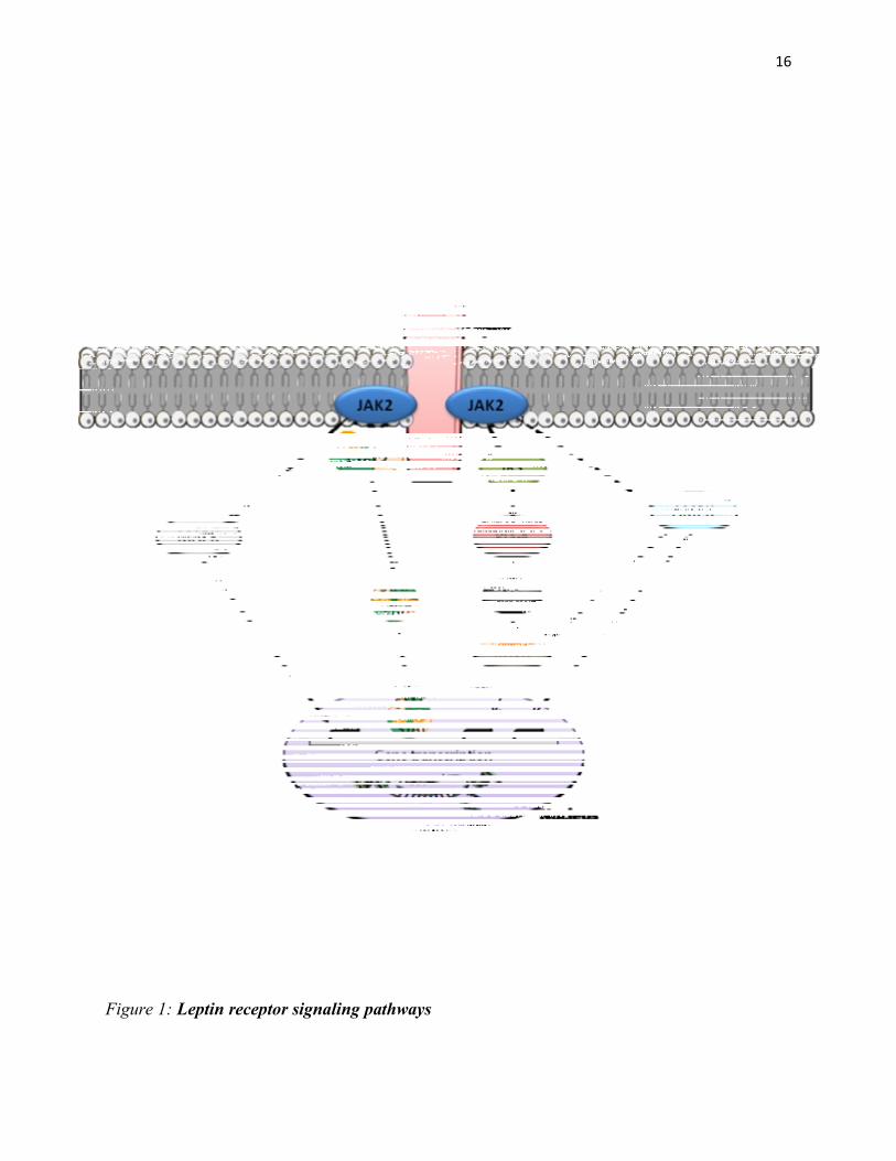
Figure 1: Leptin receptor signaling pathways
17
2.3. Leptin activates STAT3 signaling in the hypothalamus
STATs are a family of cytokine-activated signaling proteins that can directly bind to DNA and
modulate gene transcription (Gao et al. 2004). These proteins are characterized by a specific
structure consisting of several highly conserved domains: an N-terminal domain, a helical coil
domain, the DNA-binding domain, a linker domain, the dimerization domain and a
transactivation domain (Horvath et al. 1995, Mao et al. 2005). The dimerization domain contains
a SH2 domain, required for STATs to be recruited to the phosphorylated receptors and for the
subsequent formation of dimers (Shuai et al. 1994). Thus, after its recruitment to the receptor,
STATs becomes phosphorylated on a single tyrosine residue by the JAKs. The following events
are: dimerization by reciprocal interaction between phophotyrosine-SH2 domain and nuclear
translocation to induce target genes (Levy and Darnell 2002). Of note, all STATs are dimeric
proteins in the absence of activating tyrosine phosphorylation (Braunstein et al. 2003).
Different STAT proteins were identified in mammals: STAT1, STAT2, STAT3, STAT4,
STAT5A, STAT5B, STAT6. However, STAT3 has the most pleiotropic functions, playing an
important role in embryogenesis, macrophage function, immune regulation, glial and neuron
differentiation, among others (Bonni et al. 1997, Niwa et al. 1998, Takeda et al. 1999, Takizawa
et al. 2001, Levy and Lee 2002, Moon et al. 2002, Yoo et al. 2002, Welte et al. 2003).
Moreover, STAT3 is widely distributed throughout the CNS, and has been well implicated in
glial and neuronal differentiation (Bonni et al. 1997, Takizawa et al. 2001, Moon et al. 2002)
and, most importantly, in leptin-mediated energy homeostasis (Bates et al. 2003, Bates et al.
2004, Gao et al. 2004). STAT3 has a critical tyrosine residue (Tyr 705) in the SH2 region, which
is essential for its activation (Calo et al. 2003). When phosphorylated/activated, STAT3 induces

18
the transcription of several genes, including SOCS3, which inhibits JAK2/STAT3 signaling and
triggers central leptin resistance (Bjorbaek et al. 1999, Flier 2004, Howard et al. 2004).
Of note, STAT3 can be activated by a number of cytokines and stressors, via receptor-associated
kinases (Akira et al. 1994, Wegenka et al. 1994, Zhong et al. 1994, Vaisse et al. 1996,
Wishingrad et al. 1997, Peterson et al. 2000). Similarly, leptin can activate a number of STAT
proteins (i.e.: STAT3, STAT5 and STAT6) (Ghilardi et al. 1996). However, only STAT3 is
activated in the hypothalamus following leptin administration (Darnell 1996, Vaisse et al. 1996),
giving strength to the notion that STAT3 plays a pivotal role in leptin's action in the
hypothalamus to mediate energy balance.
Consistent with leptin's crucial role in the regulation of energy homeostasis and reproduction,
neural STAT3 mutant mice (STAT3 N-/-) causes all of the major phenotypes of leptin signaling,
recapitulating the obese and infertile phenotype of both LepRb deficient db/db mice and leptin
deficient ob/ob mice (Gao et al. 2004). Of note, STAT3 N-/- are leptin-resistant, have reduced
energy expenditure, decreased linear growth, increased corticosterone levels, infertility and
became hypothermic after exposure to fasting or cold stress (Gao et al. 2004). Furthermore, mice
with LepRb point mutations, that do not bind STAT3, are hyperphagic and obese with reduced
energy expenditure. However, these mice are less hyperglycaemic as compared to db/db mice,
and have normal reproductive and growth function (Bates et al. 2003, Bates et al. 2004).
Importantly, the major role of STAT3 in energy homeostasis is further supported by evidence
that other cytokines that activate STAT3, such as CNTF and IL-6, also decrease body weight
(Lambert et al. 2001, Wallenius et al. 2002).

19
3. The mesolimbic and nigrostriatal dopamine systems
3.1. The ventral tegmental area (VTA) and substantia nigra (SN): receptors and
neurotransmitters
In the ventral midbrain, areas containing dopaminergic neurons were classified into three nuclei:
A8 cells in the retrorubral field (RRF), A9 cells in the SN, and A10 cells in the VTA and related
nuclei (Oades and Halliday 1987, German and Manaye 1993). Neurons containing TH-positive
cell bodies are mainly found in all VTA (Swanson 1982) and SN regions (mostly SN pars
compacta) (Aumann et al. 2011). Of note, TH is the rate limiting enzyme in the synthesis of the
neurotransmitters DA and norepinephine (NE), whose distribution in the midbrain parallels the
distribution of DA (Bannon and Roth 1983). Eight major dopaminergic pathways were identified
by dissecting DA circuits in the brain and the specific locations of the DA receptors in these
circuits (Björklund et al. 1984). In the present section, I will focus on two of these pathways that
are especially important: the nigrostriatal and the mesolimbic pathway, described below.
The nigrostriatal and mesolimbic systems are functionally interconnected (Haber et al. 2000,
Everitt and Robbins 2005) and play complementary roles in the hedonic regulation of food
intake, reward, motivation, emotion-related behavior, learning and locomotion (Barbeau 1974,
McDonald and White 1993, D'Ardenne et al. 2008, Phillips et al. 2008, Smith and Villalba
2008). The SN lies in the midbrain immediately dorsal to the cerebral peduncles (Frankle et al.
2006) and consists of two parts with very different connections and functions: the dorsal part of
the SN, known as SN pars compacta (SNc), contains most of the DA neurons of the A9 area
(Anden et al. 1964, Dahlstroem and Fuxe 1964) and projects to the striatum; the ventral part, or
SN pars reticulata (SNr), also contains some DA cells and comprises GABA neurons which
projects to the thalamus (Deniau et al. 1978, Lynd-Balta and Haber 1994, Yung et al. 1998, Joel

20
and Weiner 2000). The main function of the SNc is motor control (Hodge and Butcher 1980),
evidenced by the fact that degeneration of SNc DA neurons leads to the muscle tremors of
Parkinson's disease (Kim et al. 2003, Pioli et al. 2008). However, besides locomotion, SN is also
implicated in other behaviors such as addiction (See et al. 2007), reward-oriented behaviors
(Hikosaka et al. 1993, Sato and Hikosaka 2002) and learning (Ljungberg et al. 1992, Da Cunha
et al. 2003, Da Cunha et al. 2006). The VTA represents a significant portion of the
extrapyramidal system of the basal ganglia, and is home to one of the major populations of DA
cells in the brain (Kalivas 1993, Fields et al. 2007). It is located close to the midline on the floor
of the midbrain, and surrounded by the SN (laterally), mammillary bodies and posterior
hypothalamus (rostrally), and by the pons and hindbrain (caudally) (Oades and Halliday 1987).
The VTA is composed by approximately of 55-60% dopaminergic, 5-33% GABAergic and 1-
15% of glutamatergic neurons (Kalivas 1993, Margolis et al. 2006, Yamaguchi et al. 2007, Dobi
et al. 2010). Of note, there's evidence indicating that some DA neurons may also use glutamate
as a neurotransmitter (Descarries et al. 2008). Selective stimulation and intracellular recording of
synaptic responses of VTA DA neurons suggested that both DA and glutamate were coreleased
by the same cell (Sulzer et al. 1998, Joyce and Rayport 2000). Furthermore, the ability of DA
neurons to release glutamate was explained by demonstrating that they selectively express
VGLUT2, one of the three vesicular glutamate transporters (Dal Bo et al. 2004).
The VTA receives afferents from a numbers of brain regions, such as the amygdala (Wallace et
al. 1992), the nucleus accumbens (NAc) (Heimer et al. 1991), the prefrontal cortex (PFC)
(Sesack and Pickel 1992), the dorsal raphe nucleus (DRN) (Gervais and Rouillard 2000), the
medial habenular nucleus (MHb) (Cuello et al. 1978), the lateral preoptic area (LPOA) and the
medial hypothalamus (Swanson 1982). The major neuronal efferents of the VTA are divided into

21
mesolimbic and mesocortical projections. Mesolimbic projections include outputs to the NAc,
tuberculum olfactorium (OT), amygdala, basal nucleus of the stria terminalis (BNST) and lateral
septum, hippocampus, the medial part of the thalamus and supraoptic nucleus of the
hypothalamus. Mesocortical projections include outputs to the prefrontal, entorhinal, cingulate
and occipital cortices, in addition to the periaqueductal gray, parabrachial nucleus, locus
coeruleus and dorsal and medial raphe nuclei (Swanson 1982, Oades and Halliday 1987, Pierce
and Kumaresan 2006) (For mesocorticolimbic and nigrostriatal systems, see Figure 2).
Dopamine's action on neuronal circuits depends on two other neurotransmitters: glutamate and
GABA (Beaulieu and Gainetdinov 2011). The main inhibitory inputs to the VTA are GABAergic
and includes local interneurons and projections from the NAc, ventral pallidum and
pontomesencephalic tegmentum (PMT) (Mao and McGehee 2010). As for the excitatory inputs
to the VTA, the great majority are glutamatergic projections from the PFC, BNST, amygdala and
also from the PMT (Mao and McGehee 2010). In fact, projections from the PMT are both
GABAergic and glutamatergic and play an important role in reward-relevant behavior (Lanca et
al. 2000, Corrigall et al. 2002).
Dopamine's physiological actions are mediated by G protein-coupled receptors, which according
to their structural, pharmacological, and biochemical properties were divided into D1 and D2
classes of DA receptors (Andersen et al. 1990, Tiberi et al. 1991, Niznik and Van Tol 1992,
Sokoloff et al. 1992, Civelli et al. 1993, Vallone et al. 2000). Dopamine signals through D1-like
receptors (D1R) or D2-like receptors (D2R) to stimulate or inhibit, respectively, regional brain
activity (Girault and Greengard 2004, Volkow et al. 2013). Among D1R are: DA receptors D1
and D5; among D2R are: DA receptors D2, D3 and D4 (Girault and Greengard 2004). According
to autoradiography studies, D2R are expressed both post-synaptically on DA target cells and pre-

22
synaptically on DA neurons (Bouthenet et al. 1987, Sales et al. 1989, Chen and Pan 2000, Pickel
et al. 2002, Sokoloff et al. 2006, Rondou et al. 2010). D2R are known to inhibit cyclic adenosine
monophosphate (cAMP) production from adenylate cyclase. Conversely, the D1R family,
activates G proteins to stimulate cAMP production from adenylate cyclase. These receptors are
found post-synaptically on DA-receptive neurons (Beaulieu and Gainetdinov 2011). Moreover,
D1R has been shown to be involved in the direct membrane depolarization of cholinergic cells
(Aosaki et al. 1998, Pisani et al. 2000) and fast-spiking GABAergic interneurons (Bracci et al.
2002).
Dopamine receptors are highly expressed in the periphery and the CNS. In the brain, D1R is
mostly expressed in striatal regions, NAc, SN, olfactory bulb, PFC and amygdala (Missale et al.
1998, Gerfen 2000, Rankin and Sibley 2010). As for the D2R expression, it is mostly expressed
in the striatum, NAc, olfactory tubercle, SN, VTA, hypothalamus, cortex, hippocampus, septum
and amygdala (Missale et al. 1998, Gerfen 2000, Vallone et al. 2000).
Previous neuroanatomical and functional studies have detected the presence of GABAA and
GABAB receptors in the VTA, densely distributed in the paranigral nucleus (Bayer and Pickel
1991, Churchill et al. 1992, Ciccarelli et al. 2012). Importantly, a large interconnected network
of GABAergic neurons in the VTA projects to the PFC and NAc (Van Bockstaele and Pickel
1995, Carr and Sesack 2000).
Among other neurotransmitters affecting DA neurotransmission in the midbrain, a subpopulation
of DA neurons containing neurotensin (NT) and cholecystokinin (CCK) were already described
in this region (Seroogy et al. 1987, Schalling et al. 1990, Kiyama et al. 1991, Binder et al. 2001).
Moreover, the presence of nicotinic, endocannabinoid (ECB), serotonin (5-HT), N-methyl-D-
aspartate (NMDA), glutamate and Mu opioid receptors were also detected in the VTA and NAc

23
(Doherty and Pickel 2000, Swanson and Kalivas 2000, Klink et al. 2001, Zheng and Johnson
2003, Pickel et al. 2004). Of note, 5-HT signaling was previously demonstrated to tightly
regulate a number of DA-related functions in both inhibitory and excitatory ways (Fletcher et al.
1999, Fletcher et al. 2012).

24
Figure 2: Schematic representation of mesocorticolimbic and nigrostriatal systems.

25
3.2. The dopaminergic reward circuit, feeding and running behavior
Rewards are defined as those objects or goals which we will work to acquire despite of time,
energy or effort; simply because they are highly pleasurable (Schultz 2010). Rewards typically
serve as reinforcers. By definition, a reinforcer is something that, when presented after a
behavior, causes the probability of that behavior's occurrence to increase. Importantly however,
not all rewards are reinforcers and not all reinforcers are rewards. A reward can be defined as a
reinforcer only if it increases the probability of behaviors that lead to the procurement and/or
consumption of the reward (Fulton 2010).
Given the importance of rewards to basic vital functions such as eating, drinking and
reproduction, it has been proposed that there exists a neural system responsible for the reward
processing (Shizgal 1997). This neural system was identified as the mesolimbic DA pathway,
which responds to rewards through the neurotransmitter DA (Arias-Carrion and Poppel 2007,
Phillips et al. 2008).
As described in a previous section, the mesolimbic pathway is one of the dopaminergic pathways
in the brain whose neurons originate in the VTA and connect to the limbic system, including the
NAc, the amygdala, the hippocampus and the PFC (Goodman 2008). Dopaminergic activity
within the VTA depends on both inhibitory and excitatory inputs that are critically involved in
brain mechanisms of reward, reinforcement and emotional arousal (Wise and Rompre 1989). The
VTA also send dopaminergic projections to several brain areas that are part of the so-called
"brain reward regions", which are interconnected in a highly complex circuitry allowing
discerning and reacting to both reward and aversive stimuli in the environment (Russo and
Nestler 2013). Of note, dopaminergic projections from VTA to the NAc represent the best
characterized reward circuit in the brain (Koob 1992, Russo and Nestler 2013). The

26
identification of this circuit dates back to 1954, when the Canadian researchers James Olds and
Peter Milner discovered that rodents will learn to work for direct electrical stimulation of certain
brain regions, and such stimulation seemed to be highly rewarding (Olds and Milner 1954) - this
experimental setup is referred to as brain-stimulation-reward (BSR). Similarly, there is data
showing that brain self-stimulation also exert a powerful feeling of pleasure in humans (Bishop
et al. 1963). Electrical brain stimulation is considered as one of the most powerful rewards since
it directly activates the reward circuitry (Wise and Rompre 1989). BSR studies demonstrated that
electrical stimulation of reward-related areas in the brain was rewarding and reinforcing, and
thus, it has provided a useful tool for understanding how natural rewards are processed by the
brain and the anatomical structures and neural mechanisms associated with reward circuitry
(Rolls 1975, Wise 2002). In addition, BSR experiments provided first evidence that the reward
circuitry is subdivided along functional and anatomical lines. Of note, besides playing a role in
recognizing rewards in the environment and initiating their consumption, brain reward circuitry
also modulates pleasure, motivation, value, and decision making (Koob and Le Moal 2008,
Pujara and Koenigs 2014).
The "Dopamine hypothesis" postulates that DA, released in the mesolimbic pathway, promotes
reward-related activities (Fibiger et al. 1987, Di Chiara and Imperato 1988). An extension of this
hypothesis is the "Reward Deficiency Syndrome", stating that animals will engage in behaviors
known to increase DA release solely to maintain high levels of this neurotransmitter (Palmiter
2007). In fact, there are two hypothesis on how DA and reward sensitivity influences the
expression of motivated behaviors: 1) The above mentioned "Reward Deficiency Syndrome",
which hypothesises that individuals with low dopaminergic function seek rewarding substances
or behaviors to increase endogenous DA levels and improve mood (Blum et al. 2000); 2)

27
Conversely, when reward hypersensitivity is associated with increased dopaminergic
transmission, individuals may feel more motivated to seek for additional reward stimuli solely
due to the fact that a specific reward is such a powerful reinforcer (Davis et al. 2008).
As for the role of DA signaling in the pursuit of natural rewards, such as food, it was previously
described that DA signaling in the NAc mediates the reinforcing effects of food (Wise 2008). On
the other hand Baldo and coworkers demonstrated that bilateral injections of either D1R or D2R
antagonist into the NAc show little effect on ad libitum food intake, although according to the
authors, an important caveat to this interpretation is that the doses of D1R or D2R antagonist
used may not have been high enough to completely block the effects of DA in food consumption
(Baldo et al. 2002), suggesting that higher antagonist doses would have decreased feeding,
Furthermore, when DA is depleted by intracranial injection of the DA-specific neurotoxin 6-
hydroxydopamine (6-OHDA) rodents develop severe motivational and regulatory deficits in
feeding and drinking and will die of starvation if DA is not replaced (Ungerstedt 1971, Zigmond
and Stricker 1972, Marshall et al. 1974). Complementary to these studies are those on DA-
specific deficient mice, suggesting that DA is pivotal for vital functions such as feeding, drinking
and locomotion, but is not required for the development of neural circuits involved in these
behaviors (Zhou and Palmiter 1995). In regards to feeding behavior, rodents lacking DA in
dopaminergic neurons (but retaining noradrenergic neurons) can execute behaviors necessary to
seek and ingest food, but they do not eat enough to survive (Szczypka et al. 1999). In light of the
"DA hypothesis", this could be explained by a lack of motivation to respond to the presence of
food - i.e.: the animal no longer shows goal-directed behavior because it does not anticipate a
reward.

28
In fact, food has long been known as a powerful behavioral modulator. For example, behaviorist
Edward Thorndike used food reward to motivate hungry animals learning to escape from early
operant chamber (Thorndike 1898). Consistent with this, a study using an appetitive operant
conditioning paradigm demonstrated that dopaminergic transmission within the NAc is required
for food-seeking behavior through mechanisms related to hedonic value of food, suggesting DA
as an important component of food motivation (Zhang et al. 2003). Similarly, dopamine
transporter (DAT) KO mice, that show chronically elevated extracellular DA, display increased
goal-directed behavior for food reward (Cagniard et al. 2006). Moreover, mesoaccumbens DA
signaling has been implicated in behavioral responses to cues that predict reward and
mechanisms involved in addiction (Robinson and Berridge 2003, Vezina 2004, Nicola et al.
2005, Hyman et al. 2006). Taken together, these studies suggest that DA is important to acquire
information about cues and stimuli related to food rewards and to the behavioral responses to
obtain these rewards (goal-directed behavior). However, apparently this neurotransmitter is not
involved in the events related to food consumption per se.
Associations between the dopamine reward pathway and physical activity behavior have been
found both in animal models and in humans. Consistent with that, voluntary wheel running
exercise has two relevant components: a motor (movement) component and a motivational
(rewarding) component (Knab and Lightfoot 2010), and has been suggested as a strong natural
reinforcer and as such, a self-rewarding behavior. Moreover, voluntary exercise is related to
changes in gene transcription in reward circuitry (Greenwood et al. 2011). Interestingly, a role
for the DA system on the regulation of motivation for physical activity was previously described
in studies of addiction and feeding showing that some animals will engage in physical exercise in
order to obtain food (Pirke et al. 1993, Casper 2006, Palmiter 2007). Likewise, animals will

29
increase drinking and/or eating when these activities were conditioned with the opportunity to
run on wheels (Premack et al. 1964), demonstrating that voluntary wheel running is a highly
motivated behavior.
Notably, goal-oriented behaviors that impose high energy costs, such as running exercise are
often motivated by psychological changes associated with prolonged physical activity, better
known as "runner's high" (Panksepp et al. 2002, Dietrich and McDaniel 2004). Humans usually
report this feeling during or right after long distance running, and it likely plays a major role in
humans’ motivation to keep running. In addition, rodents will readily begin to run when a wheel
is introduced (Wagemaker and Goldstein 1980, Partin 1983, Eikelboom and Mills 1988, Looy
and Eikelboom 1989), will indeed work by lever pressing to gain access to a running wheel
(Belke 2006, Belke 2006, Belke and Christie-Fougere 2006) and spend more time in a place
previously associated with the aftereffects of running (Lett et al. 2000, Lett et al. 2001),
indicating that voluntary wheel running activity is naturally rewarding and reinforcing.
Interestingly, rats trained to run long distances in a daily basis showed withdrawal signs, such as
displaced aggression (i.e.: digging and biting in the test cage), when access to the running wheels
is denied (Hoffmann et al. 1987). In addition, response to short-term wheel running deprivation
is similar to response to short-term deprivation on other reinforcers (such as food): rodents will
compensate with an elevation in the behavior when access is restored (Mueller et al. 1999). In
agreement with this data, other reports demonstrated that a couple of days of running deprivation
for rats with extensive wheel experience will induce a substantial increase in their subsequent
daily running (Hill 1956, Hill 1958, Hill 1961, Mueller et al. 1997). Together, these data point to
the conclusion that voluntary running exercise is a powerful natural reinforcer and shares neural
reward pathways with other reinforced behaviors such as food (Chaouloff 1989). Consistent with

30
this idea, mice selectively bred for high wheel running exhibited increased running motivation
mediated by reduced D1R signaling in the midbrain (Rhodes and Garland 2003) and as a result
of this alteration, high-running mice are more motivated than the respective controls to seek for
"running reward" (Rhodes et al. 2005). Other studies performed on this same mice suggest that
selective breeding based on wheel running rates raise reward threshold in high-running mice by
reducing the reinforcing value of shorter running durations (Belke and Garland 2007). The
altered DA transmission in mice bred for high wheel running exercise can probably be explained
by a decrease in DA concentration, decreased dopamine receptor densities, or reduced second-
messenger signaling (Rhodes et al. 2005).
Besides being rewarding, when taken to extremes, voluntary wheel running activity can develop
into an addictive-like behavior, as suggested by data obtained from surveys among highly
committed human runners, reporting: 1) runner's high (a feeling or euphoria after a strenuous
bout of exercise), 2) tolerance (the need to increase the distance run to achieve feelings of well-
being), 3) addiction (difficulties in job performance and social interactions) 4) symptoms of
withdrawal, including depression, irritability, and anxiety, when prohibited from running (Rudy
and Estok 1989, Chapman and De Castro 1990, Furst and Germone 1993) and by the number of
parallels between wheel running activity and drug-induced reward. For example, rats with a
tendency toward addiction develop compulsive running, probably due to a downregulation of
dopamine receptors (Werme et al. 1999). Moreover, similar to drugs of abuse, chronic running is
related to increased delta FosB expression in the NAc of addiction-prone Lewis rats (Werme et
al. 2002). Of note, the transcription factor delta FosB has been implicated in the development of
drug addiction and control of the reward system in the brain (McClung and Nestler 2003, Nestler
2008). Importantly, there is growing data demonstrating that running and drugs of abuse activate

31
overlapping neural systems (Kanarek et al. 1998, Werme et al. 2000, Ferreira et al. 2006). For
example, endurance exercise stimulates the release of endogenous opioid peptides in rats and
humans (Christie and Chesher 1982, Janal et al. 1984, Werme et al. 2000, Angelopoulos 2001)
and wheel running attenuates the rewarding effects of morphine in a conditioned place
preference test (Lett et al. 2002).Providing further evidence of the parallel between addiction and
voluntary exercise, experiments conducted in humans demonstrated that bouts of moderate
exercise can help attenuate symptoms of daily cannabis use, nicotine and alcohol withdrawal
(Ussher et al. 2001, Daniel et al. 2004, Ussher et al. 2004, Buchowski et al. 2011). Moreover,
the beneficial effects of exercise as an adjunct in treatment of drug addiction was previously
suggested to be related to its ability to facilitate DA transmission, normalize glutamatergic and
dopaminergic signaling, and reverse drug-induced changes in chromatin via epigenetic
interactions with BDNF in the reward pathway (Lynch et al. 2013). Thereby, as a natural reward,
voluntary running exercise modulates the brain reward circuitry and changes the rewarding effect
of addictive substances, thus contributing to reduce the incidence and severity of drug-addictive
behavior.

32
4. Leptin signaling in the mesolimbic dopamine system
4.1. Leptin activates STAT3 signaling in extra-hypothalamic brain regions
Besides being influenced by satiety factors such as leptin, eating behavior is also modulated by
dopaminergic neurons of the mesolimbic system (Cota et al. 2006, Fulton 2010, Fernandes et al.
2013). It is very well appreciated that LepRb activation in hypothalamic regions triggers STAT3
recruitment, phosphorylation and nuclear translocation to induce specific gene transcription and
influence energy balance (Li and Friedman 1999). However, leptin also induces STAT3
phosphorylation at residue Tyr 705 in extra-hypothalamic regions.
More recently, a number of reports have focused on the role of LepRb signaling in brain sites
other than the hypothalamus, including the hippocampus and VTA (Fulton et al. 2006, Hommel
et al. 2006, Harvey 2007, Robertson et al. 2008), where leptin activates LepRb-STAT3 in both
DA and GABAergic neuronal populations (Fulton et al. 2006). Consistent with this, LepRb
mRNA was already detected in the VTA, SN, DRN, thalamus, cerebellum, NTS, among other
areas (Elmquist et al. 1998, Figlewicz et al. 2003, Fulton et al. 2006, Hommel et al. 2006, Myers
et al. 2009, Scott et al. 2009). Most importantly, colocalization of the LepRb and the
dopaminergic marker TH was observed in different brain regions, including the hypothalamic
preoptic nucleus, ARC, paraventricular hypothalamic nuclei (PVN) and caudal linear nucleus
(Cli) of the midbrain. In addition, a small population of neurons coexpressing LepRb and TH
was found in the DRN, periaqueductal gray (PAG), rostral linear nucleus (RLi) and retrorubral
field (RRF) nucleus (Hakansson et al. 1998, Hay-Schmidt et al. 2001, Scott et al. 2009).
However, not all these LepRb-expressing DA cells can sense peripheral leptin, as observed by
lack of STAT3 phosphorylation following systemic leptin administration (Scott et al. 2009). In
fact, besides the VTA/SN area (Figlewicz et al. 2003, Fulton et al. 2006, Hommel et al. 2006),
LepRb- expressing DA neurons only induces pSTAT3 in the PAG and DRN, suggesting that
33
while many TH neurons express LepRb, very few cells are leptin-responsive (Scott et al. 2009)
(For LepRb signaling in the midbrain, see Figure 3).
Interestingly, evidence for the significant role of DA in leptin action, comes from studies
performed in mice lacking both leptin and DA. Contrary to leptin deficiency alone (ob/ob mice),
mice lacking both leptin and DA are hypophagic and lean, developing obesity only when treated
with the DA precursor L-DOPA. These findings are suggestive that dopaminergic
neurotransmission is required for feeding in ob/ob mice (Szczypka et al. 2000). Moreover, ob/ob
mice exhibit a substantial reduction in NAc DA release, do not sensitize to the locomotor-
stimulating effects of AMPH (Fulton et al. 2006) and have their obese hyperphagic phenotype
reversed by peripheral administration of D1R and D2R agonists (Scislowski et al. 1999).

34
Figure 3: Schematic representation of leptin receptor signaling in dopaminergic neurons of
the midbrain.

35
4.2. Leptin and locomotion
Other than feeding, leptin influences a wide range of DA-related behaviors, including
ambulatory activity (Fulton et al. 2006, Hommel et al. 2006) and voluntary wheel-running
exercise (Bates et al. 2003, Coppari et al. 2005, Shapiro et al. 2011, Verhagen et al. 2011).
However, there is inconsistency in the literature concerning leptin's effect on locomotion. Leptin-
deficient ob/ob mice have reduced ambulatory activity (Clark and Gay 1972, Dauncey 1986) and
reduced voluntary wheel running exercise (Jurgens et al. 2006), which can be reversed by leptin
administration (Pelleymounter et al. 1995, Coppari et al. 2005, Fulton et al. 2006, Huo et al.
2009, Morton et al. 2011). Similarly, either an ICV leptin treatment (Ainslie et al. 2001, Choi et
al. 2008) or leptin overexpression in the brain increases voluntary running exercise in rodents,
while a partial blockade of LepRb diminishes this behavior (Matheny et al. 2009). On the other
hand, other studies reported that an intraperitoneal leptin treatment in fed C57Bl/6 wild-type
mice, did not affect either ambulatory activity or voluntary exercise (Pelleymounter et al. 1995,
Morton et al. 2011). In contrast, leptin administration reduces hyperactivity in rodents (Exner et
al. 2000, Morton et al. 2011, Verhagen et al. 2011) and lack of LepRb signaling in the VTA
increases locomotion in male rats (Hommel et al. 2006).
Several factors may contribute to these inconsistent reports, including animal's physiological
variation in leptin levels (Morton et al. 2011). Interestingly, fasting, a condition characterized by
reduced leptinemia, is known to increase locomotor activity and voluntary exercise in rodents
(Sakkou et al. 2007, Morton et al. 2011). Consistent with these findings, leptin was shown to
abolish hyperactivity triggered by severe caloric restriction in both rodents and humans (Exner et
al. 2000, Hebebrand et al. 2003).

36
The mechanisms involved in fasting-induced hyperactivity remain poorly elucidated and may
represent a manifestation of increased food seeking behavior, driven by a reduced leptin
signaling (Overton and Williams 2004, Williams et al. 2004). Alternatively, it may also involve
increased hyperactivity-induced DA transmission in the reward circuitry (Mathes et al. 2010).
However the specific role of leptin in the modulation of ambulatory activity and voluntary
running exercise as well as the downstream signaling mechanisms needs to be further clarified.
4.3. The effect of leptin on reward
Observations that LepRb is expressed in the brain reward circuitry (Elmquist et al. 1998,
Figlewicz et al. 2003) strongly suggest that there is a crosstalk between energy regulatory signals
and the mesolimbic system.
Consistent with the notion that leptin influences the activity on neural circuits controlling food
intake to impair the perception of food rewards, animal studies using the palatable food self-
administration paradigm demonstrated that leptin inhibits motivated behavior for palatable food
(Figlewicz 2003) and decreases operant responding for sucrose (Figlewicz et al. 2006, Davis et
al. 2008). Furthermore, leptin reverses CPP for both sucrose and HF diet (Figlewicz et al. 2001,
Figlewicz et al. 2004) and rodents fed a HF diet showed reduced CPP for AMPH (Davis et al.
2008). Notably, a human study using functional magnetic resonance imaging (fMRI)
demonstrated that leptin administration to patients with congenital leptin deficiency increases the
ability to discriminate between the rewarding properties of food (Farooqi et al. 2007), further
highlighting the notion that leptin markedly affects food reward.
With respect to leptin's role in rewards other than food, it was previously reported that leptin
decreases rewarding electrical brain stimulation (Fulton et al. 2000, Fulton et al. 2004), evoked

37
DA release in the NAc (Krugel et al. 2003) and inhibits DA neuronal firing (Hommel et al.
2006). In addition, leptin influences sexual motivation (Wade et al. 1997, Schneider 2006,
Schneider et al. 2007) and drug self-administration. For example, this adipocyte hormone was
shown to attenuate the effect of food restriction to cause heroin relapse (Shalev et al. 2001) and
increased response to psychostimulant drugs, such as AMPH (Hao et al. 2004, Fulton et al.
2006), suggesting an excitatory role of leptin on reward pathways. Moreover, obese individuals
self-administer drugs of abuse less often than lean individuals (Davis et al. 2010), suggesting a
potential role for leptin in drug reward.
As previously mentioned in this thesis, voluntary wheel running exercise is a naturally rewarding
and reinforcing behavior and shares similar neurobehavioral characteristics with either drugs or
foods as reinforcers (Pierce et al. 1986, de Visser et al. 2007). Although a role for leptin in the
modulation of running reward has yet to be described, Girard and coworkers reported that mice
selectively bred for high wheel running activity have reduced plasma leptin levels which
correlates with increased running speed and distance (Girard et al. 2007), suggesting enhanced
motivation for foraging behavior (Exner et al. 2000). Alternatively, the low body fat and
hypoleptinemia observed in high-runner rodents could be explained by "reward substitution",
meaning that for these rodents, the rewarding value of running became much greater than the
rewarding value of food (Meek et al. 2012). This "reward substitution" was also observed in
studies conducted by Scarpace and co-workers, demonstrating that wheel running activity
reduces the preference of rodents for a HF diet and modulates LepRb-STAT3 signaling in the
brain reward circuitry (Scarpace et al. 2010). Thus, it is possible that, in the case of high-runner
rodents, voluntary running is a highly valued reward, which can be even greater than food
reward and is also modulated by leptin. Important to remember that mice bred for high rates of
38
running wheel activity have dysregulation of DA system in the brain, which is linked to a
number of aberrant behaviors, such as addiction, hyperphagia and compulsive exercise (Mathes
et al. 2010)
Leptin's ability to reduce reward-related behaviors occurs via an interaction with midbrain DA
signaling pathways (Carr 2002). However, the specific cells and signaling pathways mediating
the actions of leptin in the mesolimbic DA reward circuit remain to be better characterized.

39
5. Leptin's role in emotion and mood
The localization of the LepRb in limbic structures implicated in the control of mood and
emotion, such as the hippocampus, cortex and amygdala, suggests a potential role for leptin in
emotional behavior (Mercer et al. 1996, Elmquist et al. 1998, Leshan et al. 2006, Scott et al.
2009). Consistent with that, there is data showing that this adipocyte-derived hormone affects
emotional behavior via its action in limbic regions (Harvey 2007). Moreover, analyses of leptin-
induced c-fos mRNA expression revealed a link between behavioral actions of leptin and
neuronal activity of specific limbic areas, particularly the hippocampus (Lu et al. 2006).
Providing a connection between mood disorders and adiposity signals, many reports suggested
that leptin has antidepressant-like properties (Charnay et al. 2000, Lu et al. 2006, Lu 2007, Garza
et al. 2012, Guo et al. 2012, Milaneschi et al. 2012, Guo et al. 2013). Leptin administration
reduces behavioral despair in rats via its action in the hippocampus (Sahay and Hen 2007) and
stimulates hippocampal neurogenesis - a feature shared by antidepressants (Santarelli et al.
2003). In addition, ob/ob mice exhibited a depressive-like behavior in the forced-swim test
(FST), as compared to their lean littermates (Collin et al. 2000), and peripheral leptin
administration improved behavioral despair in rats (Lu et al. 2006). Briefly, FST or Porsolt test
is a well characterized model used to screen depression-like behavior in rodents (Porsolt et al.
1978).
Chronic stress is a well known trigger for depression in humans (Lu 2007). In agreement, rodents
exposed to chronic unpredictable stress or chronic social defeat stress showed decreased plasma
leptin levels and increased despair, which was reversed by systemic leptin administration (Lu et
al. 2006). In a more recent study, Yamada and coworkers demonstrated that peripheral leptin
administration reduces depressive-like behavior in lean mice via an upregulation of brain derived

40
neurotrophic factor (BDNF) in the hippocampus (Yamada et al. 2011). Importantly, BDNF
signaling in the hippocampus has been strongly implicated in the control of depression
(Shirayama et al. 2002, Karege et al. 2005). Similarly, lack of LepRb signaling in the dorsal
cerebral cortex and in the dentate gyrus in the hippocampus, also increases behavioral despair in
mice (Guo et al. 2012). Furthermore, plasma leptin levels are reduced either in humans with
major depression or in rodents with depression-like behavior, and both peripheral and
hippocampal administration of this adipocyte hormone improved symptoms (Kraus et al. 2001,
Atmaca et al. 2002, Westling et al. 2004, Jow et al. 2006, Lu et al. 2006). Consistent with these
findings, post-mortem studies of depressed patients who committed suicide revealed a
downregulation of leptin receptors in the frontal cortex (Lalovic et al. 2010).
Depressive disorder can often coexist with other mood disorders, such as anxiety (Sartorius et al.
1996). Providing another means of influencing the mesolimbic DA system, Liu and coworkers
demonstrated that leptin has anxiolytic properties such that lack of LepRb signaling specifically
in DA neurons induces an anxiogenic phenotype and increases burst firing of DA and D1R
signaling in the central amygdala (CeA) (Liu et al. 2010, Liu et al. 2011). In agreement with that,
both leptin receptor- (db/db) and leptin-deficient (ob/ob) mice displayed an anxiogenic
phenotype (Finger et al. 2010, Dinel et al. 2011), which, in the case of the ob/ob mice, can be
ameliorated by systemic leptin treatment (Asakawa et al. 2003). Providing further evidence of
leptin as a neurotrophic hormone with anxiolytic and antidepressant properties, there is literature
showing that depression and anxiety are common illnesses in patients with disorders
characterized by hypoleptinemia, such as anorexia nervosa (Braun et al. 1994, Grinspoon et al.
1996, Pollice et al. 1997, Holtkamp et al. 2004, Hebebrand et al. 2007) and functional
hypothalamic amenorrhoea (Fava et al. 1984, Warren et al. 1999). Furthermore, Lawson and

41
coworkers observed that low leptin levels are associated with increased symptoms of anxiety in
women, independently of total body fat mass (Lawson et al. 2012).
In spite of all these findings, the neural pathways as well as the specific functional role of leptin
receptor signaling in midbrain DA neurons in the regulation of emotional behavior needs further
clarification.

42
Chapter II
Objectives & General Methods

43
6. Objectives
6.1. First Objective
Our first objective was to investigate whether STAT3 signaling in DA neurons could modulate
the impact of leptin on feeding, food-motivated behavior, locomotion and NAc DA release.
Moreover, we sought to determine if leptin influences running reward and if it depends on
STAT3 signaling in midbrain DA neurons. This was accomplished by generation and phenotypic
characterization of conditional KO mice lacking the main phosphorylation site (tyr705) of the
STAT3 gene specifically in DA neurons, hereafter referred to as STAT3DAT-KO mice. This work
constitutes the first article given in Chapter III: "Leptin suppresses the rewarding effects of
running via STAT3 signaling in dopamine neurons" by Fernandes, M.F.; Sharma, S.; Mogra, S.
and Fulton, S.
6.2. Second Objective
This study was undertaken to assess the contribution of STAT3 signaling in DA neurons on
emotion-related behaviors. To address this question we generated DA-specific STAT3 KO mice
(STAT3DAT-KO) by crossing dopamine transporter DATCre mice with STAT3lox/lox mice. Then, we
studied the behavioral phenotype of STAT3DAT-KO female mice in anxiety and depression-like
behaviors. Pharmacological and molecular approaches were used to shed light on the potential
signaling mechanisms underlying the anxiogenic behavior observed in STAT3DAT-KO female
mice. This study constitutes the second article given in Chapter III: "Deletion of STAT3 in
midbrain dopamine neurons increases anxiety-like behavior in female mice" by Fernandes, M.F.;
Sharma, S. and Fulton, S.

44
7. General methods
The Cre-loxP system overview and STAT3DAT-KO mice generation
The Cre-loxP system is a special type of site-specific recombination which allows for
conditional mutagenesis of a gene of interest, thus representing a powerful tool in making
alterations to the mouse genome (Nagy 2000, Novak et al. 2000). This technology allows the
investigation of the role of specific genes in different regions or cell types, without confounding
effects caused by systemic influences. It was first described in bacteriophage P1 and consists of a
sequence-specific DNA recombinase (Cre) and a DNA sequence flanked by loxP sites, which is
recognized by the Cre-recombinase (Zou et al. 1994). Because both Cre gene and loxP sites are
not native to the mouse genome, they must be introduced by transgenic technology (Nagy 2000).
Thus, when the enzyme Cre-recombinase is introduced to a system expressing loxP sites in the
genome, it will catalyse a recombination event between the two loxP sites. Of note, the results of
the recombination depend on the orientation of the loxP sites: 1) if both loxP-sites have the same
orientation on a chromosome segment, the loxP-flanked DNA will be excised and eliminated; 2)
if the loxP sites are oriented in opposite directions, Cre recombinase mediates the inversion of
the floxed segment; 3) if the loxP sites are located on different chromosomes, Cre recombinase
mediates a chromosomal translocation (Nagy 2000).
On the present work, Cre and loxP mice colonies were developed separately and crossed to
produce a Cre-lox strain. More specifically, we generated STAT3DAT-KO mice, whereby
dopamine transporter-Cre (DATIresCre) mice were crossed with STAT3lox/lox mice (obtained from
Dr. Shizuo Akira, Osaka, Japan) in which loxP sites flank exon 22 of the STAT3 gene, encoding
the tyrosine residue 705 (For breeding schematics, see Figure 4). Of note, DATIresCre (or DAT

45
knock-in) have Cre recombinase expression directed to DA neurons, without disrupting
endogenous DAT expression (Backman et al. 2006, The Jackson Laboratory 2014).
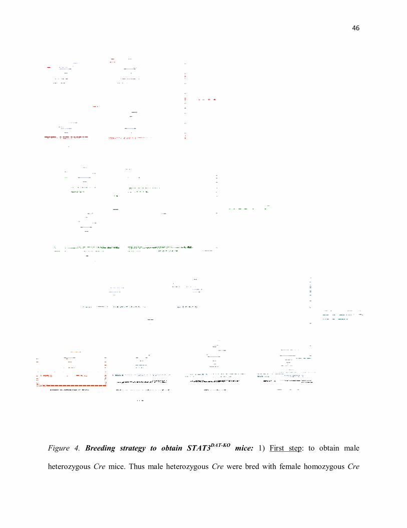
46
Figure 4. Breeding strategy to obtain STAT3DAT-KO mice: 1) First step: to obtain male
heterozygous Cre mice. Thus male heterozygous Cre were bred with female homozygous Cre

47
mice, to generate male heterozygous Cre mice; 2) Second step: to obtain male heterozygous Cre
/ heterozygous Lox mice. Thus, male heterozygous Cre / homozygous Lox were bred with female
homozygous Cre / homozygous Lox (STAT3lox/lox), to generate male heterozygous Cre /
heterozygous Lox mice; 3) Third step: to obtain the heterozygous Cre / homozygous Lox mice
(STAT3DAT-KO mice). Thus, male heterozygous Cre / heterozygous Lox were bred with female
homozygous Cre / homozygous Lox, to generate STAT3DAT-KO and respective control mice.

48
Chapter III
Articles

49
8. Articles
8.1 Article #1
Contributions:
Maria Fernanda A. Fernandes, contributed by performing most of the experiments (95%), except
for the qPCR to detect the disrupted STAT3 gene (performed by Shabana Mogra) and the fast-
scan cyclic voltammetry (performed by Dr. Stephanie Fulton). Sandeep Sharma contributed for
the setting up of the behavioral experiments. Maria Fernanda A. Fernandes also created the
graphics and wrote the manuscript. Dr. Stephanie Fulton also contributed in the results
discussion, redaction and correction of the manuscript.

50
Short article
Leptin suppresses the rewarding effects of running via Stat3 signaling in dopamine neurons
Maria F. Fernandes1,3, Sandeep Sharma1,2, Shabana Mogra1 & Stephanie Fulton1,2
1CRCHUM and Montreal Diabetes Research Center; Departments of: 2Nutrition & 3Physiology, Faculty of Medicine,
University of Montreal, Québec, Canada *Corresponding author: Stephanie Fulton, PhD CRCHUM-Tour Viger 900 rue St-Denis - Room 8-428 Montreal QC H2X0A9 Canada

51
Summary
Dopaminergic (DA) neurons of the midbrain are part of a neural circuit involved in reward and
motivation. To determine the role of the Signal Transducer and Activator of Transcription 3
(STAT3) in this circuitry, we inactivated STAT3 signaling in DA neurons of mice (STAT3DAT-
KO mice). Lack of STAT3 in DA neurons resulted in no changes in feeding, while it increased
locomotion, running wheel exercise and running reward. Moreover, STAT3 DAT-KO mice
exhibited impairments in learning an appetitive operant conditioning task and reduced NAc DA
release. Taken together, these results suggest that STAT3 mediates the action of leptin in the
midbrain to decrease locomotor behavior and to increase DA tone, while it does not affect food
intake, implying that different LepRb signaling molecules regulate distinct behavioral and
biochemical actions of midbrain leptin. Additionally, STAT3 signaling in DA neurons enhances
the rewarding value of running and facilitates instrumental learning. The findings described here
shed light on the cells and neural pathways mediating the actions of leptin in the midbrain.
Highlights
► STAT3 signaling in midbrain dopamine neurons is a key modulator of spontaneous and
voluntary physical activity ► Leptin in the ventral tegmental area abolishes the rewarding
effects of running in a manner that depends on STAT3 in dopamine neurons ► Loss of STAT3
in dopamine neurons blunts DA overflow in the nucleus accumbens

52
Introduction
Behavior is a central component of energy balance and all behavior entails energy expenditure in
the form of physical activity. Activity that increases the likelihood that a physiological need will
be met is referred to as appetitive, and appetitive behaviors are crucial to the fulfillment of
energy demands. Food-directed appetitive behaviors such as foraging, scavenging and hunting
may benefit from increased physical activity and endurance (Bramble and Lieberman, 2004).
The capacity for endurance running in cursorial mammals, particularly humans, is considered to
have evolved as a means to increase the payoff of food-directed behaviors targeting distant or
shifting food sources (Bramble and Lieberman, 2004; Carrier, 1984).
Physical activity is strongly influenced by the metabolic state of the organism. The adipocyte-
derived hormone leptin is a critical signal controlling feeding and energy expenditure and a fall
in leptin levels is a major component of the physiological response to fasting (Ahima et al.,
1996). The influence of leptin on energy expenditure includes its ability to alter spontaneous
locomotor activity and voluntary running in a manner that largely depends on food availability.
Leptin increases locomotor activity and/or voluntary running during fed states in leptin-deficient
humans (Farooqi et al., 1999; Licinio et al., 2004), ob/ob mice (Ahima et al., 1999; Morton et al.,
2011; Pelleymounter et al., 1995), lean mice (Morton et al., 2011) and rats (Choi et al., 2008;
Meek et al., 2012; Morton et al., 2011), or has no effect on running in fed wildtype mice (Morton
et al., 2011; Pelleymounter et al., 1995). In contrast, leptin decreases locomotor activity and
voluntary wheel running when food is restricted in ob/ob and lean mice (Morton et al., 2011) and
rats (Exner et al., 2000; Verhagen et al., 2011). Physical activity escalates with food restriction, a
phenomenon viewed as an expression of increased food acquisition behaviors (Bartness et al.,
2011; Overton and Williams, 2004; Sakkou et al., 2007). That leptin could be involved in this

53
process is supported by evidence that circulating concentrations are inversely related to physical
activity or voluntary running in humans (Franks et al., 2003; Hebebrand et al., 2003), rats (Exner
et al., 2000; Hebebrand et al., 2003), wildtype mice (Morton et al., 2011; Thorburn et al., 2000)
and in mice selectively bred for high running capacity (Girard et al., 2007). Furthermore, plasma
leptin levels correlate negatively with marathon run-time independent of BMI (Bobbert et al.,
2012) and with run speed and duration in high-running mice (Girard et al., 2007), suggesting that
leptin could impact the motivational and rewarding effects of running. Indeed, endurance
running is rewarding in humans (“runners high”) (Morgan, 1985; Partin, 1983; Wagemaker and
Goldstein, 1980) and rodents (Belke and Wagner, 2005; Collier and Hirsch, 1971; Kagan and
Berkun, 1954; Novak et al., 2012), yet little is known about the neural pathways and signaling
mechanisms involved.
Leptin modulates multiple components of brain reward circuitry (Fulton et al., 2000) and inhibits
the rewarding effects of food (Figlewicz et al., 2004; Figlewicz et al., 2006). DA neurons of the
midbrain ventral tegmental area (VTA) that project to limbic and cortical sites are essential for
the control of appetitive and reward-relevant behaviors (Salamone and Correa, 2012; Schultz,
2013). Leptin receptors (LepR) are expressed on midbrain DA neurons (Figlewicz et al., 2003;
Scott et al., 2009), and functional LepR signaling in DA neurons of the VTA is well-documented
(Domingos et al., 2011; Fulton et al., 2006; Hommel et al., 2006; Liu et al., 2011). Leptin has
anorectic actions in the VTA (Bruijnzeel et al., 2011; Hommel et al., 2006; Trinko et al., 2011)
whereas LepR knockdown in the VTA increases food intake, spontaneous locomotor activity
(Hommel et al., 2006) and food-motivated behavior (Davis et al., 2011). LepR signaling involves
the activation of signal transducer and activator of transcription 3 (STAT3) (Banks et al., 2000;
Tartaglia, 1997). Leptin phosphorylates STAT3 in DA and GABA neurons of the midbrain

54
(Fulton et al., 2006; Hommel et al., 2006), however leptin-induced pSTAT3 is observed only in a
subset of LepR-positive DA neurons (Scott et al., 2009) suggesting that DA neurons responsive
to leptin are heterogeneous and that LepR-STAT3 signaling may have unique neurobehavioral
actions.
Few studies have investigated the mechanisms underlying the effects of leptin on locomotor
activity. The hypoactivity of obese, leptin receptor deficient mice is corrected by restoration of
LepR to the mediobasal hypothalamus (Coppari et al., 2005) or proopiomelanocortin (POMC)
neurons (Huo et al., 2009) underscoring the role of the hypothalamus in leptin control of
locomotor behavior. However, the divergent effects of leptin on physical activity in conditions
where food is limited versus freely available suggest that separate neural processes are involved.
We sought to investigate the absence of STAT3 in DA neurons in the control of physical
activity, feeding and DA tone and to determine if leptin impacts the rewarding effects of running
via STAT3 signaling in VTA DA neurons.

55
Experimental procedures
Animals
All experiments were carried out in accordance with the guidelines and approval of the
Institutional Animal Care Committee of the CHUM Research Center. Floxed STAT3 mice
(C57Bl6 background) in which loxP sites flank exon 22 of the STAT3 gene that encodes a
tyrosine residue (tyr705) essential for STAT3 activation were graciously provided by Dr. Shizuo
Akira (Osaka, Japan) (Takeda et al., 1998). Female mice homozygous for the floxed STAT3
allele were crossed with male mice heterozygous for the floxed STAT3 allele and heterozygous
for the DAT::Cre transgene (B6.SJL-Slc6a3tm1.1(cre)Bkmn/J) (Backman et al., 2006) to generate
DATCre;STAT3fl/fl mice and littermate controls (STAT3fl/fl or STAT3fl). In addition to littermate
controls, DAT::Cre mice were used as secondary controls for wheel running experiments. Male
mice were weaned at P28 and housed in a temperature and humidity controlled room that was
maintained on a 12:12 hour reverse light/dark cycle. All experiments were conducted in the dark
phase of the cycle.
Metabolic assessments
Mice were individually housed with free access to standard chow and water. Food intake and
body weight was measured 3 times per week. Lean and fat mass were determined at 23 weeks of
age using an Echo MRI quantitative nuclear magnetic resonance system (Echo Medical Systems,
Houston, TX, USA). Metabolic efficiency (inverse of feed efficiency) was calculated for each
subject by dividing energy intake (Kcal) over the 12-week period by body weight gain in grams.
Following two days of habituation to cages, spontaneous locomotor activity was measured for 24
hours in mice (11-12 weeks of age) using automated metabolic cages (Accuscan Instruments
56
Inc., Columbus, OH, USA) consisting of 16 light beam arrays in x, y and z axes. Distance
travelled (horizontal activity) was measured by computer-controlled software.
Voluntary wheel running
Voluntary wheel running was assessed in a separate cohort of individually-housed mice with
continuous access to a low profile wireless running wheel in the cage (Med Associates, Inc., VT,
USA). Wheel revolutions were constantly monitored via transmitters and recorded by data
acquisition software (Med Associates Inc., St. Albans, VT, USA) on a nearby computer. Each
full wheel revolution is equivalent 36.9 cm distance travelled. Mice had free access to food and
water throughout and were not used for any other experiment.
Intra-cerebral cannulations and food intake
Anesthetised mice were placed in an ultraprecise mouse stereotaxic frame (Kopf; Tujunga, CA,
USA) and maintained on Isoflurane (1.5%+oxygen). VTA cannula implantation: With bregma
and lambda in the same horizontal plane, a bilateral guide cannula (26 gauge; 0.8mm distance
between cannulae; Plastics One, Roanoke, VA, USA) was lowered in the VTA (AP, –3.4 mm;
ML, ± 0.4 mm; DV, −4.7 mm). Intracerebroventricular (ICV) cannula implantation: A single
guide cannula (26 gauge; Plastics One, Roanoke, VA, USA) was lowered into the right lateral
ventricle (AP, –0.5 mm; ML, ± 1 mm; DV, −2 mm). Cannulae were anchored to the skull surface
with dental cement and occluded with metal obturators of the same length. Following recovery
and weight regain, ICV cannula placements were assessed by verifying the drinking response to
injection of angiotensin II (10ng/1µl). Mice were habituated to a powder chow diet for 3 days for
food intake measures. Powder food was placed in food cups that were fixed to the bottom of a
larger container to catch spillage. Following an overnight fast, separate groups of mice received a
bilateral VTA injection of leptin (200ng/500nl/side) or vehicle (PBS) or an ICV injection of

57
leptin (1ug/1ul; recombinant mouse leptin from Dr. A.F. Parlow, NIDDK) or vehicle right
before dark cycle onset using internal cannulas (Plastics One) attached to 1µl Hamilton
microsyringe.
Operant responding for food rewards
Mice were trained to press a lever for sucrose pellets according to published procedures (Sharma
et al., 2012). Briefly, mice were trained in operant chambers, house in sound-attenuating boxes,
equipped with two retractable levers (Med Associates Inc., St. Albans, VT, USA) and a pellet
dispenser delivering 20mg sucrose pellets (Bio-Serv, Frenchtown, NJ, USA) to a receptacle
positioned between the levers. One lever was designated active (triggers reward delivery) and
other as inactive (no reward) in a counterbalanced manner. Operant training on a fixed ratio-1
(FR1) schedule of reinforcement (1 lever press = 1 sucrose pellet) was carried out for 8 days and
the number of lever presses on each lever and rewards earned calculated by computer-controlled
software (Med Associates Inc.)
Hedonic feeding – “Dessert test”
We utilized a paradigm in which sated rodents voluntarily over consume a palatable test diet
(Choi et al., 2010). First, mice were entrained over 19 consecutive days to consume standard
chow in a 4-hour window in the first half of dark cycle (Fig. 2E). Prior to test day (Day 20) mice
were exposed to a small amount of high-fat/sugar food (D12231; Research Diets, Inc., New
Brunswick, NJ) to prevent neophobia. On the day of testing, chow intake was measured each
hour during 4 hours. Following the fourth hour of chow access, a separate set of food cups
containing high-fat/sugar food were placed in each cage and weighed one hour later.
Running reward

58
A new cohort of singly-housed mice (11-14 weeks of age) had free access to homecage running
wheels for 3 weeks. Mice were then subjected to a conditioned place preference (CPP) task using
a three-compartment, automated mouse CPP apparatus (Fig. 3A; Med Associates Inc., St.
Albans, VT, USA). Pre-test: Mice were confined to both the black and white compartments of
the CPP apparatus for 5-min to permit habituation to each compartment. Mice were then allowed
to move freely in the apparatus for 15-min during which the amount of time spent in each
compartment was recorded. Conditioning trials: Mice had 2-hour access either to a running
wheel (paired trial) or a locked running wheel (unpaired trail) and were then confined to either
the black or white compartment for 30 min. Paired and unpaired conditioning trials took place in
opposite compartments (counterbalanced across mice) on alternating days, thus each mouse ran
every-other day (Fig. 3A). Post-test: Following the last day of conditioning (Day 14), mice were
placed back in the open CPP apparatus for 15 min. The amount of time spent in each
compartment was once again recorded and the proportion of time spent in the paired side was
compared to that obtained during the pre-test.
Fast scan cyclic voltammetry
Carbon-fibre electrodes were constructed and prepared according to Kawagoe et al. (Kawagoe et
al., 1993). Electrodes were calibrated in vitro using known concentrations of DA (1µM
dopamine HCL; Sigma). Mice (6-7 weeks of age) were anesthetised with Isoflurane prior to
decapitation. Of note, at high clinical concentrations (<0.7 mM), Isoflurane was shown to inhibit
the release of glutamate without significantly affect dopamine release (Westphalen et al. 2013).
Coronal slices (300μm) were cut using a vibratome at the level of the nucleus accumbens (NAc)
(+0.90 to +1.30 mm from Bregma) in ice-cold, carbogenated ACSF containing 125 mM NaCl,
26 mM NaHCO3, 2.5 mM KCl, 2.4 mM CaCl2, 1.3 mM MgSO4, 0.3 mM KH2PO4 and 10 mM

59
D-Glucose. Fast-scan cyclic voltammetry was used to measure electrically-evoked DA release
according to published procedures (Fulton et al., 2011). Briefly, DA release in the NAc core
(Fig. 4A) was evoked at 5-min intervals by a single 400μA, 1.2 ms, rectangular electrical pulse
delivered by an S-900 stimulator (Dagan Corporation, Minneapolis, MN) and a bipolar
stimulating electrode (Plastics One, Roanoke, VI) placed ~200µm away from the carbon fibre
electrode. The carbon fibre electrode potential was linearly scanned from -0.4 to +1.2 V and back
to -0.4 V versus Ag/AgCl using a 400V/s scan rate controlled by an Axopatch 200B amplifier
connected to a computer and pClamp 10 software through a DigiData 1200B analog to digital
interface (Molecular Devices). One recording and stimulation site was tested per slice. DA
overflow was evoked 5 to 8 times per slice, until recordings were stable. The mean values of the
last three recordings from each slice were used for statistical comparisons. The sample size is
indicated as n=(x;y), x referring to the number of slices and y the number of mice.
Western immunoblotting
Mice were decapitated under Isoflurane anaesthesia, brains rapidly dissected and immersed in
-10oC isopentane and then stored at -80oC. Frozen brains were sliced into 0.5mm coronal
sections using an aluminum brain matrix. Coronal sections were mounted onto slides and
maintained on dry ice. Nuclei were micro-dissected using brain tissue punches (Stoelting Inc.,
Wood Dale, IL). Bilateral punches of 0.75mm diameter were obtained from the VTA and 1.0mm
diameter punches from the NAc. Micro-dissected tissues were homogenized on ice in 100 ml of
cell lysis buffer (100mM Tris, pH 7.5; 750mM NaCl; 5mM Na2EDTA; 5mM EGTA, pH 7.5; 5%
Triton x-100; 12.5mM sodium pyrophosphate; 5mM beta-glycerophosphate; 1mM, 5mM
Na3VO4; 5µg/ml leupeptin) with added protease (PMSF 1mM) and phosphatase inhibitors
(Sigma cocktails I and II) in 1.5 ml tubes using a motorized pestle. Tubes were centrifuged for

60
15 min at 14000 g. Protein concentrations were measured using BCA protein assay (Pierce
Biotechnology, IL, USA). Protein samples were separated by electrophoresis on a 10-12%
polyacrylamide gel and electro transferred to a nitrocellulose membrane (EMD Millipore
Corporation, Billerica, MA, USA). Non-specific binding sites were blocked in TBS 5% low-fat
milk and 0.1% Tween-20 or 5% BSA. Membranes were rinsed in buffer (0.1% Tween-20 in
TBS) and then incubated with anti-DAT (1:1000; Millipore, EMD Millipore Corporation,
Billerica, MA, USA), anti-DA D1A receptor (1:1000; Millipore, EMD Millipore Corporation,
Billerica, MA, USA), anti-D2 receptor (1:1000; Millipore, EMD Millipore Corporation,
Billerica, MA, USA), anti-TH (1:500; Millipore, EMD Millipore Corporation, Billerica, MA,
USA) or anti-GAPDH (1:1000) followed by anti-rabbit, anti-rat or anti-mouse IgG horseradish
peroxidase-conjugate (1:5000). After rinsing with buffer, immunocomplexes were visualized by
chemiluminescence using the western lighting plus ECL kit (PerkinElmer, Waltham, MA, USA).
Protein size was assessed using precision plus protein ladder (Bio-Rad, Bedford, MA, USA). The
film signals were digitally scanned and then density quantified using ImageJ software. GAPDH
(1:10000, Cell Signaling, Cell Signaling Technology, Inc., Danvers, MA, USA) was used as an
internal control, such that data were standardized according to GAPDH values.
Amphetamine locomotor sensitization
This experiment was performed as described previously (Fulton et al., 2006). A Comprehensive
Lab Animal Monitoring System (CLAMS; Columbus Instruments) was used for automated
measurement of locomotor activity following amphetamine treatment. Following a 2-day
habituation period to CLAMS cages, mice were implanted with a 14-day osmotic mini-pump
(Alzet model 1002, DURECT Corporation, Cupertino, CA, USA) into the intrascapular region
under Isoflurane anaesthesia. Pumps delivered leptin (500 ng/hr; 12 g/day) or vehicle (PBS) for

61
the duration of the experiment. Beginning the following day, each mouse received three IP
injections, each separated by 2 days, in the following order: (1) 1 mg/kg amphetamine (low
dose), (2) 4 mg/kg amphetamine (high dose), (3) 1 mg/kg amphetamine (repeat low dose).
Locomotor activity was recorded for 2 hours following injections. D-amphetamine (Sigma-
Aldrich, Dorset, UK) was dissolved in 0.9 % saline.
Statistical analyses
Data were analyzed using GraphPad 5 and are presented as mean±standard error of the mean
(SEM). A two-way ANOVA with Bonferonni post-hoc tests was used to compare control and
KO mice with regards to body weight, food intake, hedonic feeding, food-motivated operant
responding, spontaneous locomotor activity and voluntary running. Unpaired t-tests were used
for comparisons of body composition, metabolic efficiency, 24h-food intake following acute
leptin injection, locomotor activity during the dark cycle, total running distance (12 weeks),
running reward, electrochemistry and immunoblotting experiments. Area under the curve (AUC)
was calculated for dark cycle spontaneous locomotor activity and amphetamine-induced
locomotor sensitization and compared via unpaired t-tests. AMPH-induced locomotor
sensitization to the second low-dose of amphetamine was assessed using a one-way ANOVA
that compared activity counts between the first and second AMPH treatment for each treatment
group. Criterion for significance was set to p≤0.05 for all comparisons.

62
Results and Discussion
Ablation of STAT3 in dopamine neurons
To remove STAT3 function in midbrain DA neurons we bred STAT3 mice in which loxP sites
flank exon 22 of the STAT3 gene which encodes tyrosine residue 705 that is essential for LepR-
STAT3 activation (Takeda et al., 1998) with heterozygous knock-in mice that express Cre
recombinase under the control of the regulatory sequence of the dopamine transporter (DAT)
(Backman et al., 2006). STAT3fl/fl;DATCre knockout mice (hereafter referred to as STAT3DAT KO)
are born at expected frequencies, are of normal size and appearance and have similar weaning
weights (Fig. 1A, inset). A PCR product corresponding to the recombined STAT3 gene
(∆STAT3) was observed in brain nuclei of STAT3DAT KO mice in which DAT is expressed, but
was absent from control nuclei and tissues (Fig. S1A). Double immunohistochemistry for
phospho-STAT3 (pSTAT3) and tyrosine hydroxylase (TH) in slices from leptin-treated mice
confirmed that Cre recombination was specific to DA neurons (Fig. S1B). pSTAT3 nuclear
staining was absent from DA (TH+) neurons of the VTA and substantia nigra (SN) of STAT3DAT
KO mice, yet was apparent in non-dopaminergic (TH-) cells of the midbrain and throughout the
mediobasal hypothalamus (MBH) (Fig. S1B). The number of pSTAT3-postive cells in the VTA
and substantia nigra (SN) was reduced by 66.46±4.37% (n=4/group) relative to controls as
determined by counts in a separate series of immunolabelled slices.
STAT3 in dopamine neurons controls body weight and composition
To determine the contribution of STAT3 signaling in DA neurons in the regulation of body
composition and feeding, we measured body weight, fat and lean mass and food intake in chow
fed mice (Fig. 1A-D). As of 19 weeks of age, STAT3DAT KO mice weighed significantly less than

63
controls (Fig. 1A), a difference associated with a reduction in total fat mass (Fig. 1B). Caloric
intake normalized to body weight values was comparable between STAT3DAT KO mice and
controls (Figure 1C). As shown in Figure 1D, metabolic efficiency (total caloric intake divided
by body weight gain) was increased in STAT3DAT KO mice relative to controls indicating that
STAT3DAT KO mice utilize more energy per input unit. These findings suggest that reduced body
weight and fat composition of STAT3DAT KO mice is due to elevations in energy expenditure.
STAT3 in dopamine neurons controls spontaneous and voluntary physical activity
As midbrain DA neurons are strongly implicated in the control of locomotion, we next
set out to determine if spontaneous locomotor activity is increased in STAT3DAT KO mice.
Significant increases in horizontal movement (Fig. 1E) and total distance travelled (Fig. 1F) in
the dark cycle demonstrate that nocturnal activity is elevated in STAT3DAT KO mice relative to
controls. Previous work of Hommel et al. established that viral-mediated knockdown of LepR in
the VTA increases both food intake and locomotor activity without affecting body weight
(Hommel et al., 2006). Thus, findings here suggest that inhibition of locomotor activity by VTA
leptin involves STAT3 signaling in DA neurons. Reduced leptin signaling is also linked with
heightened voluntary running or exercise: Hypoleptinemia is associated with increased exercise
(Franks et al., 2003) and faster marathon run-times (Bobbert et al., 2012) in men independent of
BMI, with hyperactivity in individuals with anorexia (Hebebrand et al., 2003; Holtkamp et al.,
2003) and increased wheel running in wildtype mice (Thorburn et al., 2000) and mice bred for
high-running capacity (Girard et al., 2007; Vaanholt et al., 2008). In view of these findings, we
next measured voluntary wheel running in a separate cohort of mice with free access to a home
cage running wheel. STAT3DAT KO mice ran substantially more than controls as evidenced by an
64
increased number of wheel revolutions (Figure 1G). Remarkably, STAT3DAT KO mice ran ~11
km/day as compared to the ~6 km/day run by controls (Figure 1H). Similar wheel running
activity between DAT::Cre mice and wildtype littermates confirmed that increased running
activity of STAT3DAT KO mice is not attributable to the Cre transgene alone (Fig. S2).
We next sought to determine if hyperactivity of STAT3DAT KO mice is an expression of
increased anxiety-like behavior in view of findings implicating LepR signaling in DA neurons in
the anxiolytic actions of leptin (Liu et al., 2011). Using the elevated-plus maze (EPM), the most
commonly used rodent test of anxiety, we found that proportion of time spent and entries made
in the open arm was similar between STAT3DAT KO and controls (Fig. S3A-C). To confirm this
result, we also used an open field (OF) test whereby reduced exploration in a novel, open arena
reflects increased anxiety. Exploration in the OF was comparable between STAT3DAT KO and
controls (Fig. S3D). Together, these findings demonstrate that loss of STAT3 in DA neurons
increasing spontaneous locomotor activity and endurance running capacity in a manner unrelated
to anxiety. These results are in accordance with observations that VTA leptin administration
inhibits wheel running activity in a rat model of anorexia-induced hyperactivity (Verhagen et al.,
2011). Hyperactivity is a detrimental clinical feature of 30-80% of patients with anorexia nervosa
that can propagate negative energy balance (Davis et al., 1994). As anorexia-induced
hyperactivity is tied to hypoleptinemia, findings here may also have implications for increased
physical activity and motor restlessness in individuals with anorexia.
STAT3 in dopamine neurons does not mediate the anorectic effects of central leptin
Leptin has central actions to inhibit food intake when administered into the cerebral
ventricles and into select brain nuclei such as the VTA. To ascertain if STAT3 signaling in DA

65
neurons mediates the anorectic effects of central leptin, we measured food intake following intra-
VTA and ICV injection of leptin. Intra-VTA leptin attenuated chow intake relative to vehicle-
injected controls to a comparable extent in STAT3DAT KO mice and control mice (Fig. 2A). Thus,
similar to what has been reported in rats (Bruijnzeel et al., 2011; Hommel et al., 2006; Trinko et
al., 2011) leptin has anorectic effects when injected into the VTA of mice. Similarly, reductions
in food intake following ICV injection of leptin were not different between STAT3DAT KO mice
and controls (Figure 2B). These data suggest that LepR-Stat3 signaling in DA neurons does not
mediate the anorectic actions of leptin. LepR signal transduction also involves
phosphatidylinositol 3-OH-kinase (PI3K) (Kellerer et al., 1997; Niswender et al., 2001) and
extracellular signal-regulated kinase (ERK) signaling pathways (Rahmouni et al., 2009). Leptin
not only activates Erk1/2 signaling in the VTA but pharmacological inhibition of Erk1/2
phosphorylation was shown to block the anorectic action of VTA leptin (Trinko et al., 2011). It
remains to be determined if Erk1/2 signaling in DA and/or GABA neurons is involved in the
effects of VTA leptin on feeding.
Absence of STAT3 dampens instrumental learning but does not affect hedonic feeding
Mesolimbic and nigrostriatal DA neurons are key modulators of ingestive behavior, and
specifically the appetitive, conditioned and rewarding aspects of feeding (Fulton, 2010; Palmiter,
2007). Leptin decreases the rewarding effects of food (Figlewicz et al., 2004; Figlewicz et al.,
2006) whereas viral-mediated knockdown of LepR in the midbrain increases food-motivated
instrumental responding (Davis et al., 2011). To determine the role of STAT3 signaling in DA
neurons in food reward, we used an effort-based, operant conditioning task whereby mice learn
to press a lever in order to receive a sucrose pellet (Sharma et al., 2012). Mice were trained to
respond on a fixed-ratio 1 (FR1) schedule of reinforcement such that each lever press was

66
reinforced with a 20mg sugar pellet. Training could not progress towards more complex
schedules of reinforcement (e.g., progressive ratio) since STAT3DAT KO mice exhibited
significant response impairments on the FR1 schedule as evidenced by a reduced number of
lever presses (Fig. 2C) and decreased percentage of correct (active lever) responses (Fig. 2D)
relative to controls. Numerous reports have tied diminished performance specifically in operant
conditioning tasks to DA signaling deficits in the nucleus accumbens (NAc) (Nowend et al.,
2001; Smith-Roe and Kelley, 2000; Steinberg et al., 2014), and thus STAT3DAT KO mice may
have impaired NAc DA signaling. As an alternate method to investigate changes in food reward,
we employed a hedonic feeding test whereby sated mice voluntarily overeat palatable food, also
known as the dessert test (Fig. 2E) (Choi et al., 2010). As shown in Figure 2F, all mice
consumed around 50% of their daily caloric intake within the 1st hour of exposure to the chow
diet on the test day, eating progressively less over the remaining 3hours. As illustrated in Figure
2F, when exposed to sweetened high-fat food (dessert) during the last hour, both groups
increased their caloric intake to a similar degree suggesting that STAT3 in DA neurons does not
modulate hedonic feeding.
Leptin supresses the rewarding effects of running via Stat3 signaling in DA neurons
Endurance running or exercise can promote a lasting sense of well-being that includes feelings of
euphoria and pleasantness (Janal et al., 1984) and reduced anxiety and stress (Morgan, 1985;
Rosch, 1985), commonly referred to as the runner’s high (Boecker et al., 2008; Morgan, 1985;
Partin, 1983; Wagemaker and Goldstein, 1980). Running also has natural rewarding properties in
rodents: rats will work to have access to a running wheel (Belke, 1997; Pierce et al., 1986) and
spend more time in a place previously associated with the aftereffects of wheel running (Belke

67
and Wagner, 2005; Lett et al., 2000). Addictive and compulsive-like behavior directed at
reinstating or maintaining high levels of physical activity are reported in humans (Berczik et al.,
2012; Davis et al., 1995) and rodents (Lattanzio and Eikelboom, 2003; Werme et al., 1999), and
the rewarding effects of running can substitute for other rewards such as food (Routtenberg and
Kuznesof, 1967; Satvat and Eikelboom, 2006) and drugs of abuse (Cosgrove et al., 2002;
Kanarek et al., 1995). Running engages opioid (Lett et al., 2002; Vargas-Perez et al., 2004), DA
(de Castro and Hill, 1988; Greenwood et al., 2011; Mathes et al., 2010; Vargas-Perez et al.,
2004) and endocannabinoid systems (Chaouloff et al., 2011; Raichlen et al., 2012). The euphoric
effects of running have been associated with increased opioid signaling in corticolimbic brain
areas (Boecker et al., 2008), however, very little is known about the neural pathways and
signaling molecules that give rise to the rewarding impact of running. We next set out to
determine if running reward is increased in STAT3DAT KO mice and if leptin modulates the
rewarding effects of running via the VTA in a manner that relies on STAT3 signaling in DA
neurons. Using the conditioned place preference (CPP) test (Fig. 3A), a behavioral tool for
examining changes in conditioned reward, our data demonstrate that the rewarding effect of
running is increased in STAT3DAT KO mice as compared to controls (Fig. 3B,C). STAT3DAT KO
mice spend more time in the paired side of the chamber previously associated with wheel
running (Fig. 3C). Next, a separate cohort of mice with intra-VTA bilateral cannulae went
through the conditioning procedure and then received an intra-VTA injection of leptin, at a dose
shown to inhibit feeding (Fig. 2A) (Hommel et al., 2006), or vehicle prior to the CPP post-test.
Once again, STAT3DAT KO mice treated with vehicle showed increased conditioned rewarding
effects associated with wheel running as compared to controls (Fig. 3D). Confirming our
hypothesis that the increased running reward observed in STAT3DAT KO mice is mediated by

68
LepR-STAT3 signaling in VTA DA neurons, VTA leptin completely suppressed the rewarding
effects of running in control mice yet was unable to block running reward in STAT3DAT KO mice
(Fig. 3E). In agreement with previous studies suggesting a role for mesolimbic DA signaling in
wheel running performance (Dubreucq et al., 2013; Mathes et al., 2010), here we identify the
influence of leptin to inhibit running reward via the VTA in a manner that is contingent on
STAT3 in DA neurons.
Lack of Stat3 in dopamine neuron reduces dopamine overflow and expression of TH and
D1R in the nucleus accumbens
The NAc is a critical efferent target of VTA DA neurons strongly implicated in motivation,
appetitive responses and reward learning (Salamone and Correa, 2012; Schultz, 2013). As DA
signaling in the NAc is important for sustaining reward-related operant responding (Salamone
and Correa, 2012), our finding that operant performance for food is impaired in STAT3DAT KO
mice (Fig. 2C,D) are suggestive of perturbations in NAc DA signaling. Correspondingly,
complete leptin-deficiency was previously reported to supress DA overflow in the NAc and to
diminish the expression of TH, the rate-limiting enzyme for DA biosynthesis (Fulton et al.,
2006). To determine if lack of STAT3 modulates evoked DA overflow in the NAc we used fast
scan cyclic voltammetry, an electrochemical method that permits real-time, quantal measurement
of DA exocytosis. Axonal DA overflow in the NAc core of acute slice preparations was evoked
with a stimulating electrode and recorded with a carbon fibre electrode (Fig. 4A). The NAc core
was selected based on evidence linking this NAc sub-compartment to the regulation of wheel
running (Greenwood et al., 2011; Vargas-Perez et al., 2003). As shown in Figure 4B and C, the
amplitude of evoked DA overflow was significantly reduced in STAT3DAT KO mice relative to
69
controls. To determine if lower evoked DA overflow is coupled to changes in DA-relevant
protein expression, we next carried out immunoblotting experiments. Protein levels of TH and
D1 receptor were decreased in the NAc of STAT3DAT KO mice relative to controls (Fig. 4D).
Similar reductions in D1R and TH expression in the NAc are reported in mice selectively bred
for their high-running capacity (Knab et al., 2009). Collectively, these results identify a role for
NAc DA signaling in running, physical endurance and the rewarding properties of running.
Lack of Stat3 in DA neurons impairs amphetamine-induced locomotor sensitization
To corroborate the electrochemical data showing attenuated DA overflow in the NAc we studied
behavioral sensitization in response to the psychostimulant drug amphetamine (AMPH). The
acute locomotor-activating effects of AMPH rely on its actions to increase extracellular DA in
the mesoaccumbens pathway by blocking DA reuptake and eliciting reverse transport via DAT.
Mice received systemic leptin or vehicle via subcutaneous osmotic mini-pumps for the 10-day
sensitization protocol as reported previously (Fulton et al., 2006). As illustrated in Figure 4E,
there was no difference in locomotor activity in response to low-dose AMPH between vehicle
and leptin-treated mice of either genotype. As expected, locomotion was increased following
repeat AMPH administration in control mice (sensitization effect - Figure 4F & G). However,
vehicle-treated STAT3DAT KO mice failed to sensitize to AMPH as shown by similar locomotor
responses to AMPH following the two injections (Fig. 4H). Despite the absence of AMPH
sensitization in STAT3DAT KO mice, chronic leptin treatment restored AMPH locomotor
sensitization in these mice, although not quite to levels observed in controls (Fig. 4H). These
results suggest that STAT3 signaling in DA neurons is important but not necessary for leptin
modulation of DA-dependent, AMPH-induced locomotor sensitization. In agreement with these

70
results, Leinninger et al. found that deletion of LepR in neurotensin neurons of the lateral
hypothalamus (LH) that project to VTA DA neurons supresses the locomotor effects of AMPH
and inhibit NAc DA overflow (Leinninger et al., 2011). Thus, leptin acts directly on DA neurons
to modulate mesolimbic DA signaling and function, in part via STAT3 signaling, while also
having important indirect actions via LH inputs to these neurons.
71
General Discussion
The present findings uncover a new function for leptin in the control of the rewarding
effects of running and identify the key contribution of LepR-STAT3 signaling in DA neurons to
this process. STAT3 loss-of-function elicited substantial increases in spontaneous locomotor
activity and voluntary endurance running capacity without affecting the anorectic actions of
leptin or hedonic feeding, results that highlight the involvement of DA-specific STAT3 in the
central control of physical activity. By tying behavioral changes to decreased NAc DA overflow
and a dampening of mesolimbic DA function our findings also reveal STAT3 as an important
regulator of mesoaccumbens DA tone and highlight the contribution of midbrain DA signaling in
the rewarding impact of running.
The present data suggest that reduced LepR-Stat3 signaling in midbrain DA neurons
could be an important element mediating restriction-induced hyperactivity and the coinciding fall
in leptin levels. Furthermore, as chronic (but not acute) exercise by itself has been shown to
suppress leptin levels in men (Perusse et al., 1997), endurance training may be inherently
reinforcing by inhibiting LepR-STAT3 signaling in DA neurons. Although energetically costly,
the capacity for endurance running in cursorial mammals is considered to enable food acquisition
when food is at a distance or requires pursuit (see examples of distance hunters in Carrier;
(Bramble and Lieberman, 2004; Carrier, 1984). Correspondingly, the rewarding effects of
running or the runner’s high may have evolved to encourage stamina and therefore increase the
probability of return on this energetic investment. The motivational effects of physical activity
would accordingly rise during conditions when food is scarce (or costly) to promote activity-
based behaviors such as hunting, scavenging or food hoarding (accumulation) then decrease as a
function of food availability and increasing leptin. Interestingly, food hoarding increases with
72
food restriction and weight loss (Cabanac and Swiergiel, 1989), is inhibited by leptin (Buckley
and Schneider, 2003; Keen-Rhinehart and Bartness, 2008) and was previously speculated as the
underlying behavioral function of a brain reward circuit inhibited by leptin (Fulton et al., 2000).
Otherwise, the rewarding effects of running could presumably encourage physical activity and
thus help to counter overweight, obesity and associated health risks. Conversely, in some
individuals the rewarding properties of physical activity can foster excessive and compulsive
patterns of exercise that are characteristic of an addiction (Berczik et al., 2012). Consistent with
findings here, an intriguing new report shows that exercise addiction in men is associated with
low, fat-adjusted leptin levels (Lichtenstein et al., 2014). This finding may also have relevance to
the issue of hyperactivity associated with hypoleptinemia in anorexia since individuals with
anorexia exhibit heightened sensitivity to rewards (Jappe et al., 2011). It is not known if there are
genetic or physiological factors that contribute to a larger drop in leptin levels in response to
food restriction or exercise in normal or pathological states that could increase the propensity for
physical activity and sensitivity to its rewarding effects
In view of its well-established role in reward-relevant behaviour, motivation and
addiction, the contribution of DA in leptin control of running reward is not surprising. Previous
studies implicated opioids in frontolimbic circuitry in the euphoric effects of running in humans
(Boecker et al., 2008) and endocannabinoid signaling via CB1 receptors in the VTA that
modulate DA neurotransmission in the running performance of mice (Dubreucq et al., 2013).
Endocannabinoid, DA and opioid systems converge in basal ganglia circuitry and several studies
highlight their interaction in the control and drug and food reward. CB1 signaling in the VTA
fine-tunes phasic DA release and thereby contributes to appetitive behaviors and reward-relevant
learning. Striatal DA can modulate the release of opioid peptides that have been connected to the

73
hedonic effects of rewarding stimuli. Parceling out the precise contribution of each these
substrates, the manner in which they cooperate and the corticolimbic neuronal populations
involved in the rewarding effects of running requires further investigation.

74
Funding and Disclosure
The project described here was supported by a grant from the Canadian Institutes of Health
Research to Stephanie Fulton and doctoral awards to Maria F. Fernandes from the Canadian
Diabetes Association (CDA) and FESP-Université de Montréal. The authors do not have any
conflict of interest to report.

75
Supplemental Information
Leptin modulates locomotor activity and running reward via STAT3 signaling in midbrain
dopamine neurons
Fernandes, MF; Sharma, S; Mogra, S.; Fulton, S.
Supplemental Experimental Procedures
Cre-mediated recombination
Genomic DNA (200ng) was isolated using from brain tissue punches and peripheral tissue
dissections using Trizol and subjected to PCR to detect recombination (“∆STAT3”) (Takeda et
al., 1998) using the following primers: a- 5’-CAC ACA AGC CAT CAA ACT CTG GTC TCC-
3’ (specific for exon 23 of the STAT3 gene) b- 5’-GAT TTG AGT CAG GGA TCC ATA ACT
TCG -3’ (specific for loxP site upstream of the targeting construct).
Immunohistochemistry
STAT3DAT KO mice and controls were injected IP with either leptin (5 mg/kg body weight) or
PBS following an overnight fast. Recombinant leptin was obtained from Dr. A.F. Parlow
(National Hormone and Peptide Program, National Institute of Diabetes and Digestive Kidney
Diseases, Torrance, California). Two hours after treatment, mice were anesthetised with sodium
pentobarbital (50mg/kg, IP) and perfused transcardially with 0.9% ice-cold saline followed by
10% neutral buffered formalin. Tissue preparation for pSTAT3 staining was as described (Fulton
et al., 2006). Following pSTAT3 primary antibody labeling, sections were rinsed and incubated
with a secondary biotinylated anti-rabbit antibody (1:1000; Vector Laboratories), labeled with
avidin-biotin complex, and then stained with nickel-enhanced diaminobenzidine (DAB). Floating
sections were blocked in avidin/biotin solution followed by 3% goat serum, and then incubated
in anti-TH antibody (1:1000, mouse, Millipore, EMD Millipore Corporation, Billerica, MA,

76
USA) overnight at 4°C. Sections were rinsed and incubated with a secondary biotinylated anti-
mouse antibody (1:300; Vector Laboratories), labeled with avidin-biotin complex, and then
stained with Vector-VIP substrate. In a separate series of slices (n=4/group) stained for pSTAT3
alone as above, the number of pSTAT3+ cells were counted using ImageJ software in slices at
coronal levels corresponding to plates 59 to 61 of the mouse brain atlas (Paxinos and Franklin,
2004).
Elevated plus maze test (EPM)
The EPM apparatus consists of two closed arms that oppose two open arms in a plus design
(Med Associates, Inc., St Albans, VT, USA). Decreased time spent in the open, exposed arm is
an indicator of increased anxiety-like behavior. The apparatus is placed 60 cm above the floor
and has a video camera fixed overhead. Each mouse was placed in the middle of the maze facing
the open arm opposing the experimenter. Movement in the maze was recorded and tracked for 5
min by an overhead video camera connected to a PC with Ethovision XT software (Med
Associates, Inc., St Albans, VT, USA).
Open field test
As an additional test of anxiety-like behavior, STAT3DAT KO
mice and controls were tested in the
open field test. The open field consists of a Plexiglas box (50 x 50 x 30 cm) in a lit room. Each
mouse was placed in the middle of the arena and allowed to explore the field for 5 min.
Movement in the field was recorded and tracked by an overhead video camera connected to a PC
with Ethovision XT software (Med Associates, Inc., St Albans, VT, USA).
.

77
References
Ahima, R.S., Bjorbaek, C., Osei, S., and Flier, J.S. (1999). Regulation of neuronal and glial proteins by leptin: implications for brain development. Endocrinology 140, 2755-2762.
Ahima, R.S., Prabakaran, D., Mantzoros, C., Qu, D., Lowell, B., Maratos-Flier, E., and Flier, J.S. (1996). Role of leptin in the neuroendocrine response to fasting. Nature 382, 250-252.
Backman, C.M., Malik, N., Zhang, Y., Shan, L., Grinberg, A., Hoffer, B.J., Westphal, H., and Tomac, A.C. (2006). Characterization of a mouse strain expressing Cre recombinase from the 3' untranslated region of the dopamine transporter locus. Genesis 44, 383-390.
Banks, A.S., Davis, S.M., Bates, S.H., and Myers, M.G., Jr. (2000). Activation of downstream signals by the long form of the leptin receptor. The Journal of biological chemistry 275, 14563-14572.
Bartness, T.J., Keen-Rhinehart, E., Dailey, M.J., and Teubner, B.J. (2011). Neural and hormonal control of food hoarding. American journal of physiology. Regulatory, integrative and comparative physiology 301, R641-655.
Belke, T.W. (1997). Running and responding reinforced by the opportunity to run: effect of reinforcer duration. Journal of the experimental analysis of behavior 67, 337-351.
Belke, T.W., and Wagner, J.P. (2005). The reinforcing property and the rewarding aftereffect of wheel running in rats: a combination of two paradigms. Behav Processes 68, 165-172.
Berczik, K., Szabo, A., Griffiths, M.D., Kurimay, T., Kun, B., Urban, R., and Demetrovics, Z. (2012). Exercise addiction: symptoms, diagnosis, epidemiology, and etiology. Substance use & misuse 47, 403-417.
Bobbert, T., Mai, K., Brechtel, L., Schulte, H.M., Weger, B., Pfeiffer, A.F., Spranger, J., and Diederich, S. (2012). Leptin and endocrine parameters in marathon runners. Int J Sports Med 33, 244-248.
Boecker, H., Sprenger, T., Spilker, M.E., Henriksen, G., Koppenhoefer, M., Wagner, K.J., Valet, M., Berthele, A., and Tolle, T.R. (2008). The runner's high: opioidergic mechanisms in the human brain. Cerebral cortex 18, 2523-2531.
Bramble, D.M., and Lieberman, D.E. (2004). Endurance running and the evolution of Homo. Nature 432, 345-352.
Bruijnzeel, A.W., Corrie, L.W., Rogers, J.A., and Yamada, H. (2011). Effects of insulin and leptin in the ventral tegmental area and arcuate hypothalamic nucleus on food intake and brain reward function in female rats. Behav Brain Res 219, 254-264.
Buckley, C.A., and Schneider, J.E. (2003). Food hoarding is increased by food deprivation and decreased by leptin treatment in Syrian hamsters. American journal of physiology. Regulatory, integrative and comparative physiology 285, R1021-1029.
Cabanac, M., and Swiergiel, A.H. (1989). Rats eating and hoarding as a function of body weight and cost of foraging. The American journal of physiology 257, R952-957.
Carrier, D.R. (1984). The energetic paradox of human running and hominid evolution. Current Anthropology 25, 483-495.

78
Chaouloff, F., Dubreucq, S., Bellocchio, L., and Marsicano, G. (2011). Endocannabinoids and motor behavior: CB1 receptors also control running activity. Physiology 26, 76-77; author reply 78.
Choi, D.L., Davis, J.F., Fitzgerald, M.E., and Benoit, S.C. (2010). The role of orexin-A in food motivation, reward-based feeding behavior and food-induced neuronal activation in rats. Neuroscience 167, 11-20.
Choi, Y.H., Li, C., Hartzell, D.L., Little, D.E., Della-Fera, M.A., and Baile, C.A. (2008). ICV leptin effects on spontaneous physical activity and feeding behavior in rats. Behav Brain Res 188, 100-108.
Collier, G., and Hirsch, E. (1971). Reinforcing properties of spontaneous activity in the rat. Journal of comparative and physiological psychology 77, 155-160.
Cosgrove, K.P., Hunter, R.G., and Carroll, M.E. (2002). Wheel-running attenuates intravenous cocaine self-administration in rats: sex differences. Pharmacology, biochemistry, and behavior 73, 663-671.
Davis, C., Kennedy, S.H., Ralevski, E., Dionne, M., Brewer, H., Neitzert, C., and Ratusny, D. (1995). Obsessive compulsiveness and physical activity in anorexia nervosa and high-level exercising. Journal of psychosomatic research 39, 967-976.
Davis, C., Kennedy, S.H., Ravelski, E., and Dionne, M. (1994). The role of physical activity in the development and maintenance of eating disorders. Psychological medicine 24, 957-967.
Davis, J.F., Choi, D.L., Schurdak, J.D., Fitzgerald, M.F., Clegg, D.J., Lipton, J.W., Figlewicz, D.P., and Benoit, S.C. (2011). Leptin regulates energy balance and motivation through action at distinct neural circuits. Biological psychiatry 69, 668-674.
de Castro, J.M., and Hill, J.O. (1988). Exercise and brain catecholamine relationships with brown adipose tissue and whole-body oxygen consumption in rats. Physiology & behavior 43, 9-12.
Domingos, A.I., Vaynshteyn, J., Voss, H.U., Ren, X., Gradinaru, V., Zang, F., Deisseroth, K., de Araujo, I.E., and Friedman, J. (2011). Leptin regulates the reward value of nutrient. Nat Neurosci 14, 1562-1568.
Dubreucq, S., Durand, A., Matias, I., Benard, G., Richard, E., Soria-Gomez, E., Glangetas, C., Groc, L., Wadleigh, A., Massa, F., et al. (2013). Ventral tegmental area cannabinoid type-1 receptors control voluntary exercise performance. Biological psychiatry 73, 895-903.
Exner, C., Hebebrand, J., Remschmidt, H., Wewetzer, C., Ziegler, A., Herpertz, S., Schweiger, U., Blum, W.F., Preibisch, G., Heldmaier, G., et al. (2000). Leptin suppresses semi-starvation induced hyperactivity in rats: implications for anorexia nervosa. Molecular psychiatry 5, 476-481.
Farooqi, I.S., Jebb, S.A., Langmack, G., Lawrence, E., Cheetham, C.H., Prentice, A.M., Hughes, I.A., McCamish, M.A., and O'Rahilly, S. (1999). Effects of recombinant leptin therapy in a child with congenital leptin deficiency. The New England journal of medicine 341, 879-884.
Figlewicz, D.P., Bennett, J., Evans, S.B., Kaiyala, K., Sipols, A.J., and Benoit, S.C. (2004). Intraventricular insulin and leptin reverse place preference conditioned with high-fat diet in rats. Behavioral neuroscience 118, 479-487.
Figlewicz, D.P., Bennett, J.L., Naleid, A.M., Davis, C., and Grimm, J.W. (2006). Intraventricular insulin and leptin decrease sucrose self-administration in rats. Physiology & behavior 89, 611-616.

79
Figlewicz, D.P., Evans, S.B., Murphy, J., Hoen, M., and Baskin, D.G. (2003). Expression of receptors for insulin and leptin in the ventral tegmental area/substantia nigra (VTA/SN) of the rat. Brain Res 964, 107-115.
Franks, P.W., Farooqi, I.S., Luan, J., Wong, M.Y., Halsall, I., O'Rahilly, S., and Wareham, N.J. (2003). Does physical activity energy expenditure explain the between-individual variation in plasma leptin concentrations after adjusting for differences in body composition? The Journal of clinical endocrinology and metabolism 88, 3258-3263.
Fulton, S. (2010). Appetite and reward. Front Neuroendocrinol 31, 85-103.
Fulton, S., Pissios, P., Manchon, R.P., Stiles, L., Frank, L., Pothos, E.N., Maratos-Flier, E., and Flier, J.S. (2006). Leptin regulation of the mesoaccumbens dopamine pathway. Neuron 51, 811-822.
Fulton, S., Thibault, D., Mendez, J.A., Lahaie, N., Tirotta, E., Borrelli, E., Bouvier, M., Tempel, B.L., and Trudeau, L.E. (2011). Contribution of Kv1.2 voltage-gated potassium channel to D2 autoreceptor regulation of axonal dopamine overflow. The Journal of biological chemistry 286, 9360-9372.
Fulton, S., Woodside, B., and Shizgal, P. (2000). Modulation of brain reward circuitry by leptin. Science 287, 125-128.
Girard, I., Rezende, E.L., and Garland, T., Jr. (2007). Leptin levels and body composition of mice selectively bred for high voluntary locomotor activity. Physiol Biochem Zool 80, 568-579.
Greenwood, B.N., Foley, T.E., Le, T.V., Strong, P.V., Loughridge, A.B., Day, H.E., and Fleshner, M. (2011). Long-term voluntary wheel running is rewarding and produces plasticity in the mesolimbic reward pathway. Behav Brain Res 217, 354-362.
Hebebrand, J., Exner, C., Hebebrand, K., Holtkamp, C., Casper, R.C., Remschmidt, H., Herpertz-Dahlmann, B., and Klingenspor, M. (2003). Hyperactivity in patients with anorexia nervosa and in semistarved rats: evidence for a pivotal role of hypoleptinemia. Physiology & behavior 79, 25-37.
Holtkamp, K., Herpertz-Dahlmann, B., Mika, C., Heer, M., Heussen, N., Fichter, M., Herpertz, S., Senf, W., Blum, W.F., Schweiger, U., et al. (2003). Elevated physical activity and low leptin levels co-occur in patients with anorexia nervosa. The Journal of clinical endocrinology and metabolism 88, 5169-5174.
Hommel, J.D., Trinko, R., Sears, R.M., Georgescu, D., Liu, Z.W., Gao, X.B., Thurmon, J.J., Marinelli, M., and DiLeone, R.J. (2006). Leptin receptor signaling in midbrain dopamine neurons regulates feeding. Neuron 51, 801-810.
Huo, L., Gamber, K., Greeley, S., Silva, J., Huntoon, N., Leng, X.H., and Bjorbaek, C. (2009). Leptin-dependent control of glucose balance and locomotor activity by POMC neurons. Cell metabolism 9, 537-547.
Jappe, L.M., Frank, G.K., Shott, M.E., Rollin, M.D., Pryor, T., Hagman, J.O., Yang, T.T., and Davis, E. (2011). Heightened sensitivity to reward and punishment in anorexia nervosa. The International journal of eating disorders 44, 317-324.
Kagan, J., and Berkun, M. (1954). The reward value of running activity. Journal of comparative and physiological psychology 47, 108.
80
Kanarek, R.B., Marks-Kaufman, R., D'Anci, K.E., and Przypek, J. (1995). Exercise attenuates oral intake of amphetamine in rats. Pharmacology, biochemistry, and behavior 51, 725-729.
Kawagoe, K.T., Zimmerman, J.B., and Wightman, R.M. (1993). Principles of voltammetry and microelectrode surface states. Journal of neuroscience methods 48, 225-240.
Keen-Rhinehart, E., and Bartness, T.J. (2008). Leptin inhibits food-deprivation-induced increases in food intake and food hoarding. American journal of physiology. Regulatory, integrative and comparative physiology 295, R1737-1746.
Kellerer, M., Koch, M., Metzinger, E., Mushack, J., Capp, E., and Haring, H.U. (1997). Leptin activates PI-3 kinase in C2C12 myotubes via janus kinase-2 (JAK-2) and insulin receptor substrate-2 (IRS-2) dependent pathways. Diabetologia 40, 1358-1362.
Knab, A.M., Bowen, R.S., Hamilton, A.T., Gulledge, A.A., and Lightfoot, J.T. (2009). Altered dopaminergic profiles: implications for the regulation of voluntary physical activity. Behavioural brain research 204, 147-152.
Lattanzio, S.B., and Eikelboom, R. (2003). Wheel access duration in rats: I. Effects on feeding and running. Behavioral neuroscience 117, 496-504.
Leinninger, G.M., Opland, D.M., Jo, Y.H., Faouzi, M., Christensen, L., Cappellucci, L.A., Rhodes, C.J., Gnegy, M.E., Becker, J.B., Pothos, E.N., et al. (2011). Leptin action via neurotensin neurons controls orexin, the mesolimbic dopamine system and energy balance. Cell metabolism 14, 313-323.
Lett, B.T., Grant, V.L., Byrne, M.J., and Koh, M.T. (2000). Pairings of a distinctive chamber with the aftereffect of wheel running produce conditioned place preference. Appetite 34, 87-94.
Lett, B.T., Grant, V.L., Koh, M.T., and Flynn, G. (2002). Prior experience with wheel running produces cross-tolerance to the rewarding effect of morphine. Pharmacology, biochemistry, and behavior 72, 101-105.
Lichtenstein, M.B., Andries, A., Hansen, S., Frystyk, J., and Stoving, R.K. (2014). Exercise Addiction in Men Is Associated With Lower Fat-Adjusted Leptin Levels. Clinical journal of sport medicine : official journal of the Canadian Academy of Sport Medicine.
Licinio, J., Caglayan, S., Ozata, M., Yildiz, B.O., de Miranda, P.B., O'Kirwan, F., Whitby, R., Liang, L., Cohen, P., Bhasin, S., et al. (2004). Phenotypic effects of leptin replacement on morbid obesity, diabetes mellitus, hypogonadism, and behavior in leptin-deficient adults. Proceedings of the National Academy of Sciences of the United States of America 101, 4531-4536.
Liu, J., Perez, S.M., Zhang, W., Lodge, D.J., and Lu, X.Y. (2011). Selective deletion of the leptin receptor in dopamine neurons produces anxiogenic-like behavior and increases dopaminergic activity in amygdala. Molecular psychiatry 16, 1024-1038.
Mathes, W.F., Nehrenberg, D.L., Gordon, R., Hua, K., Garland, T., Jr., and Pomp, D. (2010). Dopaminergic dysregulation in mice selectively bred for excessive exercise or obesity. Behav Brain Res 210, 155-163.
Meek, T.H., Dlugosz, E.M., Vu, K.T., and Garland, T., Jr. (2012). Effects of leptin treatment and Western diet on wheel running in selectively bred high runner mice. Physiology & behavior 106, 252-258.

81
Morgan, W.P. (1985). Affective beneficence of vigorous physical activity. Medicine and science in sports and exercise 17, 94-100.
Morton, G.J., Kaiyala, K.J., Fisher, J.D., Ogimoto, K., Schwartz, M.W., and Wisse, B.E. (2011). Identification of a physiological role for leptin in the regulation of ambulatory activity and wheel running in mice. American journal of physiology. Endocrinology and metabolism 300, E392-401.
Niswender, K.D., Morton, G.J., Stearns, W.H., Rhodes, C.J., Myers, M.G., Jr., and Schwartz, M.W. (2001). Intracellular signalling. Key enzyme in leptin-induced anorexia. Nature 413, 794-795.
Novak, C.M., Burghardt, P.R., and Levine, J.A. (2012). The use of a running wheel to measure activity in rodents: relationship to energy balance, general activity, and reward. Neuroscience and biobehavioral reviews 36, 1001-1014.
Nowend, K.L., Arizzi, M., Carlson, B.B., and Salamone, J.D. (2001). D1 or D2 antagonism in nucleus accumbens core or dorsomedial shell suppresses lever pressing for food but leads to compensatory increases in chow consumption. Pharmacology, biochemistry, and behavior 69, 373-382.
Overton, J.M., and Williams, T.D. (2004). Behavioral and physiologic responses to caloric restriction in mice. Physiology & behavior 81, 749-754.
Palmiter, R.D. (2007). Is dopamine a physiologically relevant mediator of feeding behavior? Trends Neurosci 30, 375-381.
Partin, C. (1983). Runner's "high". JAMA : the journal of the American Medical Association 249, 21.
Paxinos, G., and Franklin, K. (2004). The Mouse Brain in Stereotaxic Coordinates. (Gulf Professional Publishing).
Pelleymounter, M.A., Cullen, M.J., Baker, M.B., Hecht, R., Winters, D., Boone, T., and Collins, F. (1995). Effects of the obese gene product on body weight regulation in ob/ob mice. Science 269, 540-543.
Perusse, L., Collier, G., Gagnon, J., Leon, A.S., Rao, D.C., Skinner, J.S., Wilmore, J.H., Nadeau, A., Zimmet, P.Z., and Bouchard, C. (1997). Acute and chronic effects of exercise on leptin levels in humans. Journal of applied physiology 83, 5-10.
Pierce, W.D., Epling, W.F., and Boer, D.P. (1986). Deprivation and satiation: The interrelations between food and wheel running. Journal of the experimental analysis of behavior 46, 199-210.
Rahmouni, K., Sigmund, C.D., Haynes, W.G., and Mark, A.L. (2009). Hypothalamic ERK mediates the anorectic and thermogenic sympathetic effects of leptin. Diabetes 58, 536-542.
Raichlen, D.A., Foster, A.D., Gerdeman, G.L., Seillier, A., and Giuffrida, A. (2012). Wired to run: exercise-induced endocannabinoid signaling in humans and cursorial mammals with implications for the 'runner's high'. The Journal of experimental biology 215, 1331-1336.
Routtenberg, A., and Kuznesof, A.W. (1967). Self-starvation of rats living in activity wheels on a restricted feeding schedule. Journal of comparative and physiological psychology 64, 414-421.

82
Sakkou, M., Wiedmer, P., Anlag, K., Hamm, A., Seuntjens, E., Ettwiller, L., Tschop, M.H., and Treier, M. (2007). A role for brain-specific homeobox factor Bsx in the control of hyperphagia and locomotory behavior. Cell metabolism 5, 450-463.
Salamone, J.D., and Correa, M. (2012). The mysterious motivational functions of mesolimbic dopamine. Neuron 76, 470-485.
Satvat, E., and Eikelboom, R. (2006). Dissociation of conditioned and unconditioned factors in the running-induced feeding suppression. Physiology & behavior 89, 428-437.
Schultz, W. (2013). Updating dopamine reward signals. Current opinion in neurobiology 23, 229-238.
Scott, M.M., Lachey, J.L., Sternson, S.M., Lee, C.E., Elias, C.F., Friedman, J.M., and Elmquist, J.K. (2009). Leptin targets in the mouse brain. J Comp Neurol 514, 518-532.
Sharma, S., Hryhorczuk, C., and Fulton, S. (2012). Progressive-ratio responding for palatable high-fat and high-sugar food in mice. J Vis Exp, e3754.
Smith-Roe, S.L., and Kelley, A.E. (2000). Coincident activation of NMDA and dopamine D1 receptors within the nucleus accumbens core is required for appetitive instrumental learning. The Journal of neuroscience : the official journal of the Society for Neuroscience 20, 7737-7742.
Steinberg, E.E., Boivin, J.R., Saunders, B.T., Witten, I.B., Deisseroth, K., and Janak, P.H. (2014). Positive reinforcement mediated by midbrain dopamine neurons requires d1 and d2 receptor activation in the nucleus accumbens. PLoS One 9, e94771.
Takeda, K., Kaisho, T., Yoshida, N., Takeda, J., Kishimoto, T., and Akira, S. (1998). STAT3 activation is responsible for IL-6-dependent T cell proliferation through preventing apoptosis: generation and characterization of T cell-specific STAT3-deficient mice. J Immunol 161, 4652-4660.
Tartaglia, L.A. (1997). The leptin receptor. The Journal of biological chemistry 272, 6093-6096.
Thorburn, A.W., Holdsworth, A., Proietto, J., and Morahan, G. (2000). Differential and genetically separable associations of leptin with obesity-related traits. International journal of obesity and related metabolic disorders : journal of the International Association for the Study of Obesity 24, 742-750.
Trinko, R., Gan, G., Gao, X.B., Sears, R.M., Guarnieri, D.J., and DiLeone, R.J. (2011). Erk1/2 mediates leptin receptor signaling in the ventral tegmental area. PLoS One 6, e27180.
Vaanholt, L.M., Jonas, I., Doornbos, M., Schubert, K.A., Nyakas, C., Garland, T., Jr., Visser, G.H., and van Dijk, G. (2008). Metabolic and behavioral responses to high-fat feeding in mice selectively bred for high wheel-running activity. International journal of obesity 32, 1566-1575.
Vargas-Perez, H., Borrelli, E., and Diaz, J.L. (2004). Wheel running use in dopamine D2L receptor knockout mice. Neuroscience letters 366, 172-175.
Vargas-Perez, H., Mena-Segovia, J., Giordano, M., and Diaz, J.L. (2003). Induction of c-fos in nucleus accumbens in naive male Balb/c mice after wheel running. Neuroscience letters 352, 81-84.
Verhagen, L.A., Luijendijk, M.C., and Adan, R.A. (2011). Leptin reduces hyperactivity in an animal model for anorexia nervosa via the ventral tegmental area. Eur Neuropsychopharmacol 21, 274-281.

83
Wagemaker, H., and Goldstein, L. (1980). The runner's high. J Sports Med Phys Fitness 20, 227-229.
Werme, M., Thoren, P., Olson, L., and Brene, S. (1999). Addiction-prone Lewis but not Fischer rats develop compulsive running that coincides with downregulation of nerve growth factor inducible-B and neuron-derived orphan receptor 1. The Journal of neuroscience : the official journal of the Society for Neuroscience 19, 6169-6174.
Westphalen, R. I., K. M. Desai and H. C. Hemmings, Jr. (2013). "Presynaptic inhibition of the release of multiple major central nervous system neurotransmitter types by the inhaled anaesthetic isoflurane." Br J Anaesth 110(4): 592-599.

84
Figure Legends
Figure 1. STAT3 in DA neurons regulates body weight, locomotor activity and voluntary
wheel running. (A) Body weights of control (n=7) and STAT3DAT KO mice (n= 8). Inset:
Weaning weight (n=20/group) (B) Total lean and fat mass of control (n=7) and STAT3DAT KO
(n=8) mice at 23 weeks of age. (C) Caloric intake (chow) normalized by body weight in control
(n=7) and STAT3DAT KO (n=8) mice (D) Metabolic efficiency (total intake divided by weight
gain) of control (n=7) and STAT3DAT KO (n=8) mice. (E) 24h ambulatory activity in control and
STAT3DAT KO mice (n=9/group). (F) Dark phase locomotor activity is elevated in STAT3DAT KO
mice relative to controls (n=9/group) (G) Ad libitum voluntary wheel running is increased in
STAT3DAT KO (n=12) as compared to controls (n=7) (H) STAT3DAT KO (n=12) mice run much
longer distances each day relative to controls (n=7). Mean±SEM; *p<0.05; ** p<.001;
***p<0.001
Figure 2. STAT3 in dopamine neurons does not mediate the anorectic effects of central
leptin and hedonic feeding but impairs food-motivated operant learning (A) Food intake of
control and STAT3DAT KO mice receiving an intra-VTA injection of leptin (200ng/500μl/side) or
vehicle (n=7-11/group) (B) Food intake of control and STAT3DAT KO mice receiving an ICV
injection of leptin (1μg/1μl) or vehicle (n=4/group) (C) Lever press responses on a fixed-ratio
(FR)-1 operant conditioning task in control and STAT3DAT KO mice (n= 9/group; main effect of
genotype). (D) Percentage of correct and incorrect responses by Day 8 of testing (n= 9/group)
(E) Layout of the hedonic feeding test where mice learn to consume their daily chow ration in a
4-h time window (F) Test day intake of chow and sweetened high-fat diet (“dessert”) in control
and STAT3DAT KO mice (n= 7/group). Mean±SEM; *p<.05; ** p<.01; ***p<0.001

85
Figure 3. The rewarding effects of running are increased in STAT3DAT KO mice and are
inhibited by leptin in the VTA. (A) Schema demonstrating the conditioned-place preference
(CPP) task used to measure running reward. Alternating conditioning trials (paired one day,
unpaired next) lasted 14 days (B) Control (n=16) and STAT3DAT KO (n=12) mice show significant
preference for the “paired” side of the chamber associated with wheel running. (C). Preference
for the paired side of the chamber is enhanced in STAT3DAT KO mice (n=12) relative to controls
(n=16). (D) In a separate cohort, running CPP was assessed 1 hour after intra-VTA injection of
vehicle in control (n=9) and STAT3DAT KO (n=8) mice. As in B, STAT3DAT KO mice show
increases running CPP relative to controls (E) Intra-VTA leptin blocked the rewarding effects of
running in control mice (n=14) but not in STAT3DAT KO (n=16) mice. Running CPP was tested
one hour after intra-VTA leptin (200ng, 500μl/side) injection. Mean±SEM; *p<0.05; ** p<0.01;
***p<0.001
Figure 4. Lack of STAT3 in DA neurons decreases DA overflow in the NAc core and blocks
amphetamine locomotor sensitization. (A) Illustration of brain slice preparation with
placement of carbon fibre recording and stimulating electrodes for measurement of DA overflow
using fast-scan cyclic voltammetry. (B) Representative current-time plot showing subsecond
stimulation-evoked DA release and reuptake in a control and STAT3DAT KO mouse. Background
current waveforms obtained immediately before the stimulation were subtracted from current
waveforms obtained after stimulation to generate DA cyclic voltammograms. Oxidation current
peaks for DA were obtained at potentials of -300 to -500 mV (versus Ag/AgCl) corresponding to
3.5–4.5 ms in the voltage waveform. The peak oxidation currents derived from voltammograms

86
were converted to concentration by electrode calibration and used to generate current-time plots
(C) DA overflow (mean of peak values from current-time plots) is reduced in STAT3DAT KO mice
(n=6;3) relative to controls (n=6;3). (D) TH and D1 receptor protein expression is lower in in the
NAc of STAT3DAT KO mice as compared to controls (n=5-10/group). Protein quantifications were
normalized to GAPDH values. (E) AMPH locomotor sensitization: Locomotor activity over time
following the first dose of AMPH. (F). Locomotor activity over time following the second dose
of AMPH. (G). Area under the curve (AUC) locomotor activity data for control mice treated
with leptin (n=9) or vehicle (n=9). Locomotor activity was increased following the second
injection of AMPH in both groups of control mice, indicative of sensitization. (H) Area under the
curve (AUC) locomotor activity data for STAT3DAT KO mice treated with leptin (n=6) or vehicle
(n=7). STAT3DAT KO mice treated with vehicle failed to sensitize to repeat AMPH whereas leptin
treatment to these mice restored AMPH sensitization. Mean±SEM; *p<0.05; ** p<0.01.
Supplementary Figures
Supplementary Figure S1. Cre mediated STAT3 recombination. (A) PCR detection of STAT3
gene recombination. The recombined STAT3 gene (∆STAT3) was observed only in
tissues/nuclei of STAT3DAT KO mice in which DAT is expressed. (B) Double
immunohistochemistry for TH (VIP, purple) and pSTAT3(tyr705) (ni-DAB, black) from leptin
treated STAT3DAT KO mice (n=4/group) reveals the absence of pSTAT3 from TH+
(dopamine) neurons (open arrows) and the presence of pSTAT3 in TH- cells (closed arrows) of
the VTA, SN and MBH of STAT3DAT KO mice. CX: cortex; HPC: hippocampus; Liv: liver;
MBH: mediobasal hypothalamus; Pan: pancreas; OB: olfactory bulb; VTA: ventral tegmental
area; SN: substantia nigra

87
Supplementary Figure S2, related to Figure 1. Similar wheel running activity between
DAT::Cre and wildtype littermates. (n=4-5/group).
Supplementary Figure S3, related to Figure 1. Comparable anxiety-like behavior in
STAT3DAT KO mice and controls. (A) Heat maps showing locomotor activity in the EPM in a
STAT3DAT KO
mouse and control. (B,C) Anxiety-like behavior as measured in the EPM was
similar between STAT3DAT KO
mice and controls (n=11-12/group). Percentage of open arm and
open arm time were not different between genotypes (C) No differences were observed in
anxiety-like behavior in the open field test. Exploratory activity (distance travelled) was
comparable between STAT3DAT KO
mice and controls (n= 5-6/group).
.

88
Fernandes et al.
Figure 1

89
Fernandes et al.
Figure 2

90
Fernandes et al.
Figure 3

91
Fernandes et al.
Figure 4
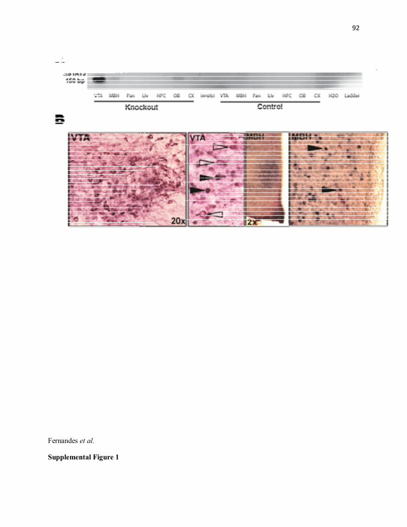
92
Fernandes et al.
Supplemental Figure 1

93
Fernandes et al.
Supplemental Figure 2
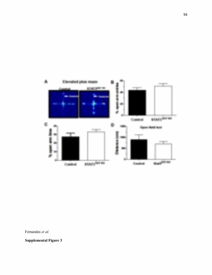
94
Fernandes et al.
Supplemental Figure 3

95
8.2 Article #2
Contributions:
Maria Fernanda A. Fernandes, contributed by performing most of the experiments (97%),
creating the graphics and writing the manuscript. Sandeep Sharma helped setting up the elevated
plus maze (EPM) and open field (OFT) tests. Dr. Stephanie Fulton contributed in the results
discussion, redaction and correction of the manuscript.

96
Deletion of STAT3 in midbrain dopamine neurons increases anxiety-like behavior in female mice
Maria F. Fernandes1,3, Sandeep Sharma1,2 & Stephanie Fulton1,2
1CRCHUM and Montreal Diabetes Research Center; Departments of: 2Nutrition & 3Physiology, Faculty of Medicine,
University of Montreal, Québec, Canada *Corresponding author: Stephanie Fulton, PhD CRCHUM-Tour Viger 900 rue St-Denis - Room 8-428 Montreal QC H2X0A9 Canada

97
Abstract
The adipocyte-derived hormone leptin targets the long form of its receptor in the central nervous
system to produce several behavioral actions compatible with the maintenance of energy
balance. Leptin activates a number of signaling pathways including JAK2-STAT3, MAPK, PI3-
K and AMPK. JAK2-STAT3 constitutes a key pathway by which leptin modulates gene
expression and energy balance. Leptin has anxiolytic effects which have been tied to LepRb
signaling on mesolimbic dopamine neurons that project to the central nucleus of the amygdala,
however, it remains to be resolved which leptin signaling pathways are involved. To assess the
contribution of STAT3 signaling in dopaminergic neurons on anxiety-related behavior we
generated a dopamine-specific STAT3 knockout (STAT3DAT-KO) by crossing dopamine
transporter DATCre mice with STAT3lox/lox mice. STAT3DAT-KO female mice exhibited normal
feeding, locomotion, body weight and behavioral despair relative to littermate controls.
However, STAT3DAT-KO mice showed increased anxiety-like behavior in both the elevated plus
maze and open field tests, as well as increased plasma corticosterone levels. Blocking dopamine
D1-receptors in the central nucleus of the amygdala by local microinjection of the D1-receptor
antagonist SCH23390 reversed the anxiogenic phenotype of STAT3DAT-KO mice. These results
suggest that activation of STAT3 in midbrain dopamine neurons that project to the central
amygdala mediates the anxiolytic effects of leptin.
Keywords: leptin, dopamine, central amygdala, anxiety

98
Introduction
The ob gene product leptin is a well described satiety factor which acts via the long form of its
receptor (LepRb) to inform the hypothalamic centres about the status of body fat stores (Inui,
1999; Zhang et al, 1994). Leptin's actions in the hypothalamus as an "anti-obesity" hormone
have been exhaustively studied. However, LepRb is distributed throughout the brain, including
mesolimbic regions involved in emotional behavior (Elmquist et al, 1998; Leshan et al, 2006;
Mercer et al, 1996; Scott et al, 2009). Via the LepRb, leptin targets the ventral tegmental area
(VTA) to inhibit feeding-related behaviors (Davis et al, 2011; Hommel et al, 2006), locomotion
(Hommel et al, 2006; Verhagen et al, 2011), and anxiety (Liu et al, 2011).
Of note, the therapeutic targets for obesity and mood disorders, such as anxiety, are the same
(Zheng et al, 2006) and a strong relationship between feeding and anxiolytic behavior has
already been described (Pallister and Waller, 2008; Swinbourne and Touyz, 2007). Consistent
with this idea, previous studies pointed out to the anxiolytic effects of leptin in rodents and
humans. There is data showing that plasma leptin levels in women is strictly related to symptoms
of anxiety, independent of body fat composition (Lawson et al, 2012). Peripheral administration
of leptin was previously demonstrated to decrease anxiety-like behaviors in mice (Finger et al,
2010; Liu et al, 2010; Lu et al, 2006). Moreover, leptin-deficient ob/ob mice display an
anxiogenic phenotype, which can be reversed by systemic leptin administration (Asakawa et al,
2003; Finger et al, 2010). Although the specific signaling mechanisms by which leptin
modulates anxiety-like behavior needs further clarification, Liu and coworkers demonstrated that
inhibition of dopaminergic transmission via dopamine D1-receptor (D1R) may be involved (Liu
et al, 2011).
99
Estrogen acts downstream of the LepRb to directly activate signal transducer and activator of
transcription-3 (STAT3) pathway in a leptin-independent manner and thus, to influence gene
transcription (Gao and Horvath, 2008). Similarly to the leptin receptor, estrogen receptors (ER)
are also expressed in both dopaminergic and non-dopaminergic neurons in the midbrain. More
specifically, ERs were detected in and around the A8, A9, and A10 dopaminergic cell groups in
rodents (Creutz and Kritzer, 2002). Moreover, both leptin and estrogens significantly contribute
to the incidence and intensity of mood disorders, including anxiety and depression, with females
being at higher risk than males (Lund et al, 2005; Walf and Frye, 2006; Williams, 2010). Based
on these findings, it can be speculated that estrogens and leptin may act synergistically to
modulate STAT3 signaling in DA neurons. However, further experiments are required to test this
hypothesis.
Given that STAT3 constitutes a predominant signal whereby leptin regulates gene expression
(Bates et al, 2003); leptin induces STAT3 phosphorylation in dopamine (DA) and GABA
neurons of the VTA (Fulton et al, 2006); and that both leptin and estrogen activates STAT3
signaling pathway, we hypothesized that this transcription factor might be a strong potential
candidate molecule to mediate the anxiolytic effects of leptin in female mice. Using Cre-LoxP
cell-specific gene targeting to abolish STAT3 activation in DA neurons, our goal was to
determine whether LepRb-STAT3 could mediate the role of leptin in the midbrain to affect
emotional processes.
100
Materials and methods
Animals
DA-specific STAT3 KO mice ("STAT3DAT-KO") were generated as described (Fernandes et al,
2014, submitted to Cell Metabolism). Briefly, STAT3DAT-KO mice have been generated by
crossing the DATCre mice with STAT3lox/lox mice, which have the main activation site of STAT3
gene (tyr705) flanked by loxP sites. STAT3DAT-KO mice and littermate controls were housed in
groups of five at 22-24°C on a 12:12 h light–dark cycle (lights on at 10:00 am) with ad libitum
access to food and water. Mice were genotyped using PCR-based procedures. Estrous cycle was
not monitored during these studies. Therefore, the effects of inactivation of STAT3 in DAT
expressing cells on anxiety- and depression-like behaviors were determined in randomly cycling
females. All animal procedures were approved by the CRCHUM Animal Care Committee.
Long term food intake and body weight
Mice were individually housed with free access to standard chow and water. Food intake and
body weight was measured 3 times per week.
Ambulatory activity
Following two days of habituation to cages, spontaneous locomotor activity was measured for 24
hours in automated Accuscan metabolic cages (Accuscan Instruments Inc., Columbus, OH,
USA) consisting of 16 light beam arrays in x, y and z axes. Distance travelled (horizontal
activity) was measured by computer-controlled software.
Open field test (OFT)

101
To assess anxiety-like behavior, STAT3DAT-KO mice and controls were tested in both the OFT
and elevated plus maze (below). The open field consisted of a Plexiglas box (50 x 50 x 30 cm) in
a brightly lit room. Each mouse was placed in the middle of the arena and allowed to explore the
field for 5 min. Movement in the field was recorded and tracked by an overhead video camera
connected to a PC with Ethovision XT software (Med Associates, Inc., St Albans, VT, USA).
Elevated plus maze test (EPM)
The EPM test served as an additional measure of anxiety-like behavior. The EPM apparatus
consists of two closed arms that oppose two open arms in a plus design (Med Associates, Inc., St
Albans, VT, USA). Decreased time spent in the open, exposed arm is an indicator of increased
anxiety-like behavior. The apparatus is placed 60 cm above the floor and has a video camera
fixed overhead. Each mouse was placed in the middle of the maze facing the open arm opposing
the experimenter. Movement in the maze was recorded and tracked for 5 min by an overhead
video camera connected to a PC with Ethovision XT software (Med Associates, Inc., St Albans,
VT, USA).
Forced swim test (FST)
The FST was used to screen depressive-like behavior in STAT3DAT-KO and littermate controls. In
this test, animals display "behavioral despair" as indicated by increased immobility and less
escape-oriented behaviors. When forced to swim in a glass cylinder filled with water in which
they are confined mice eventually cease escape attempts and become immobile. The increasing
immobility time reflects a state of despair. For this experiment, all mice were forced to swim in a
glass cylinder (height, 15 cm; diameter, 12 cm) containing water (23oC) at a 10-cm depth. A

102
video camera located above the apparatus recorded each test. The duration of immobility during
the last 4 min of the 6-min testing period (2 min habituation) was calculated.
Tail suspension test (TST)
The TST test served as an additional measure of depressive-like behavior. In this test, mice first
struggle in an attempt to escape and over time animals become immobile, which is believed to
reflect a state of "behavioral despair". Mice were suspended by the tail to a movement-sensitive
hook for 10 min. Movement of the mouse during the procedure is automatically recorded via the
transmission of electrical signals to a central unit. When movement was lower than a pre-
established threshold, the mouse was considered immobile.
Surgical procedures
For intra-CeA cannula implantation, animals were anesthetized with Isoflurane (1.5%+oxygen)
and placed in a stereotaxic frame (Kopf; Tujunga, CA, USA). Unilateral guide cannulae (26
gauge; Plastics One, Roanoke, VA, USA) were lowered into both sides of the CeA, using the
following coordinates: AP, –1.25 mm; ML, ± 2.70 mm; DV, −4.2 mm (Crock et al, 2012).
Cannulae were anchored to the skull surface with dental cement and occluded with metal
obturators of the same length. Following cannula implantation mice were individually housed
and body weights monitored daily to ensure appropriate post-surgical weight recovery. Mice
recovered for at least 7 days before testing. At the end of the experiment, the cannulae placement
was confirmed by the administration of methylene blue dye (0.3ul/side). Mice without staining
restricted to the CeA (n=3) were excluded from subsequent analysis.
103
D1 dopamine receptor antagonist (SCH23390) injections in the CeA
For this experiment all mice were subjected to repeated handling to allow habituation and
remained in their respective home cages until the infusion of the D1R antagonist SCH23390
(Sigma-Aldrich, St Louis, MO, USA) or vehicle (isotonic sterile saline) bilaterally into the
central nucleus of the amygdala (CeA) (0.1ug/ul; 0.3ul/side delivered over two minutes).
Following injection, all mice returned to their home cages and were tested in the EPM 30
minutes following the injections, as previously described (Liu et al, 2011).
Basal and stress-induced corticosterone measures
Both basal and stress-potentiated plasma corticosterone levels were measured. To measure basal
corticosterone, blood samples were obtained via cardiac puncture at the time of sacrifice. For
restraint stress experiments, mice were restrained in decapicones (Braintree Scientific
Inc.,Braintree, MA, USA) for 30 min, and blood samples were obtained immediately after via
cardiac puncture. All animals were sacrificed during the light cycle, at 6 am, when corticosterone
levels are lower, according to the circadian pattern of corticosterone secretion. Plasma was
separated by centrifugation and measured by an ELISA corticosterone kit according to
manufacture instructions (Enzo Life Sciences, Farmingdale, NY, USA). Each sample was
measured in triplicates. Inter assay variability was less than 10%.
Western immunoblotting
Mice were decapitated under Isoflurane anaesthesia and brains rapidly dissected and stored at -
80oC. Frozen brains were sliced into 0.5mm coronal sections using a brain matrix. Coronal
sections were mounted onto slides maintained on dry ice. Bilateral punches were obtained from

104
NAc (figure 18, Mouse Brain Atlas), DLS (figure 21, Mouse Brain Atlas) and CeA (figure 39,
Mouse Brain Atlas) (Franklin and Paxinos 2008) using brain tissue punches (Stoelting Inc.,
Wood Dale, IL). Microdissected tissues were homogenized on ice in 100 ml of cell lysis buffer
(100mM Tris, pH 7.5; 750mM NaCl; 5mM Na2EDTA; 5mM EGTA, pH 7.5; 5% Triton x-100;
12.5mM sodium pyrophosphate; 5mM beta-glycerophosphate; 1mM, 5mM Na3VO4; 5µg/ml
leupeptin) with added protease (PMSF 1mM) and phosphotase inhibitors (Sigma phosphatase
inhibitor cocktails I and II) in 1.5 ml aliquots using a motorized pestle. Aliquots were centrifuged
for 15 min at 14000 g. Protein concentrations were measured using BCA protein assay (Pierce
Biotechnology, IL, USA). Protein samples were separated by electrophoresis on a 12-10%
polyacrylamide gel and electro transferred to a nitrocellulose membrane (EMD Millipore
Corporation, Billerica, MA, USA). Non-specific binding sites were blocked in TBS 5% low-fat
milk and 0.1% Tween-20 or 5% BSA. Membranes were rinsed in buffer (0.1% Tween-20 in
TBS) and then incubated with anti-DAT (1:1000; Millipore, EMD Millipore Corporation,
Billerica, MA, USA), anti-D2 receptor (1:1000; Millipore, EMD Millipore Corporation,
Billerica, MA, USA), anti-TH (1:500; Millipore, EMD Millipore Corporation, Billerica, MA,
USA), anti- pDARPP-32 (Thr 34) (1:1000; Santa Cruz Biotechnology, Santa Cruz, CA, USA)
followed by anti-rabbit, anti-rat or anti-mouse IgG horseradish peroxidase-conjugate (1:5000).
After rinsing with buffer, immunocomplexes were visualized by chemiluminescence using the
western lighting plus ECL kit (PerkinElmer, Waltham, MA, USA). Protein size was assessed
with a precision plus protein ladder (Bio-Rad, Bedford, MA, USA). The film signals were
digitally scanned; density quantified using ImageJ software. GAPDH (1:10000, Cell Signaling
Technology, Inc., Danvers, MA, USA) was used as an internal control, such that data were
standardized according to GAPDH values.
105
Statistical analyses
Data were analyzed using GraphPad 5 and presented as means and respective standard errors of
the mean (SEM). Unpaired t-tests were used to compare control and STAT3DAT-KO mice in all
studies, except for 24h-ambulatory activity and food intake studies, which were analysed using
2-way ANOVA with Bonferoni post-hoc tests. Criterion for significance was set to p≤0.05 in all
comparisons.

106
Results
Absence of STAT3 signaling in dopamine neurons enhances CORT levels and anxiety-like
behavior. Basal plasma corticosterone (CORT) levels were elevated in female STAT3DAT-KO
mice as compared to respective controls at the middle of the light cycle (6am) (***p< 0.001,
Figure 1A). Consistent with these findings, 30-minutes of restraint increased corticosterone
levels in both groups, however, stress-induced elevations of CORT were more pronounced in
STAT3DAT-KO mice (***p< 0.001, Figure 1B). To determine the role of STAT3 signaling in DA
neurons in anxiety-like behavior, STAT3DAT-KO and control mice were tested in an OFT and
EPM - two well-validated behavioral tests of anxiety (Archer, 1973; Carobrez and Bertoglio,
2005; Rodgers and Dalvi, 1997; Stanford, 2007). Our results demonstrated that STAT3DAT-KO
mice avoid the center of the open field arena and spend more time in the periphery as compared
to controls (***p < 0.001, Figure 1C and 1D). In addition, as shown in, Figure 1E both
percentage of time spent into the open arm and percentage of entries made into the open arm of
an EPM is lower in STAT3DAT-KO mice as compared to controls (**p<0.01. Figure 2E). Taken
together, our results reveal an anxiogenic phenotype in STAT3DAT-KO female mice.
STAT3DAT-KO mice display normal locomotor activity, body weight and food intake
To determine if the increased anxiety-like behavior was a result of changes in locomotor activity
we examined 24 hours locomotor activity in STAT3DAT-KO mice and respective controls (Figure
2A and 2B). STAT3DAT-KO mice and littermate controls exhibited similar locomotor activity
during 24 hours (Figure 2A) and similar locomotor activity during the dark (active) phase
(Figure 2B). Moreover, we assessed the effects of loss of STAT3 in DA neurons on long-term
chow intake and body weight regulation. There is no difference in body weight and food
consumption between genotypes (Figure 2C).

107
Lack of STAT3 in dopamine neurons does not affect depressive-like behavior
To determine if the absence of STAT3 signaling in DA neurons impacts depressive-like
behavior, mice were tested in the FST and TST, two well-validated tests of behavioral despair in
mice (Castagne et al, 2010, 2011). As shown in Figure 3A and 3B, there were no differences in
immobility time, an index of "behavioral despair", in either the FST and TST between
genotypes, demonstrating that loss of pSTAT3 in DA neurons does not influence depressive-like
behaviors .
Lack of STAT3 in DA neurons alters the expression of DA-related proteins in the CeA
To determine if changes in the expression of DA-related proteins could be involved in the
heightened anxiety of STAT3DAT-KO mice we studied the expression of TH, DAT and phospho-
DARPP32(thr34) protein in the CeA. As shown in Figure 4, TH and DAT expression is
significantly decreased in the CeA of STAT3DAT-KO female mice as compared to controls
(***p<0.001, Figure 4) whereas phospho-DARPP32 (thr34), a marker of D1R activation, was
increased in the CeA of STAT3DAT-KO mice relative to controls (*p<0.05). The increases in
pDARPP32(thr34) observed are suggestive of increased CeA D1R signaling in STAT3 DAT-KO
female mice.
D1R antagonist (SCH23390) administration in the CeA reverses the anxiogenic phenotype
of STAT3DAT KO mice
As we obtained biochemical evidence to suggest that D1R signaling is increased in the CeA of
STAT3DAT-KO mice, we next set out to determine if blocking D1R in the CeA could reverse the
increased anxiety-like behavior of STAT3DAT-KO mice. Mice received a bilateral injection of the
D1 receptor antagonist SCH23390 (0.3 µg per mouse) or vehicle into the CeA and were then
screened for anxiety-like behavior 30 minutes later. Supporting our earlier results, saline-treated
108
STAT3DAT KO mice spent less time (*p<0.05, Figure 5A) and made less entries (*p<0.05, Figure
5B) into the open arm as compared to controls. There were no differences in either the
percentage of time spent in the open arm (Figure 5A) or percentage of entries made into the open
arm (Figure 5B) of the EPM between SCH23390-treated STAT3DAT-KO and control mice. In
addition, the percentage of time spent in the open arm (Figure 5A) or percentage of entries made
to this arm (Figure 5B) was similar between vehicle- and SCH23390-treated control mice.
However, we observed that SCH23390-treated STAT3DAT-KO mice increased the percentage of
time spent in the open arms (**p<0.01, Figure 5A) as well as the percentage of entries made into
this arm (*p<0.05, Figure 5B), as compared to vehicle-treated STAT3DAT-KO mice. Thus, D1
blockade in the CeA reversed the increased anxiety-like behavior of STAT3DAT KO mice.

109
Discussion
Leptin has been shown to have anxiolytic actions mediated by LepRb signaling on mesolimbic
dopamine (DA) neurons that project to the central nucleus of the amygdala (CeA) (Liu et al,
2011). Here we find that absence of STAT3 signaling, an important signaling molecule
downstream of LepRb, specifically in DA neurons induces anxiety in female mice by increasing
DA signaling in the CeA. However, depressive-like behavior is similar between STAT3DAT-KO
mice and respective controls, suggesting that STAT3 signaling in dopaminergic neurons
specifically modulates anxiety in female mice.
Besides its well-described role in controlling food intake and body weight, the adipocyte-derived
hormone leptin also plays an important role in the modulation of mood (Leshan et al, 2006; Liu
et al, 2010; Scott et al, 2009). Leptin's anxiolytic properties has already been described (Liu et
al, 2010). Furthermore, systemic leptin ameliorates anxiety-like behavior in ob/ob mice
(Asakawa et al, 2003) and reduced depressive-like behavior in rodents (Lu et al, 2006).
However, the specific functional role of LepRb signaling in midbrain DA neurons in the
regulation of emotional behavior as well as the signaling molecules involved remained elusive
until now. Our findings that lack of STAT3 signaling in DA neurons of female mice results in
increased anxiety-like behavior (OFT and EPM) and elevated basal and stress-induced
corticosterone levels without changes in behavioral despair are consistent with those of Liu and
coworkers, showing that LepRb KO in DA neurons increase anxiety but not despair in male mice
(Liu et al, 2011). Alternatively, the anti-depressant effects of leptin appear to be more associated
with hippocampal leptin signaling (Guo et al, 2013; Harvey, 2007). In contrast with Liu and
coworkers, we failed to observe an anxiogenic phenotype in STAT3DAT-KO male mice. The
difference between our results and those from Liu and collaborators as well as the sexual

110
dimorphism observed in anxiety-like behavior in STAT3DAT-KO mice can be explained from a
few speculative perspectives. First, the authors do not mention the reason why they chose to
include only male mice in their study, given that there is literature showing that females are
much more prone to be affected by mood disorders than males (Bekker and van Mens-Verhulst,
2007; Kessler et al, 2005). Furthermore, a dense population of estrogen-expressing neurons was
observed in the CeA of female rats (Pfaff and Keiner, 1973) and estrogen increases extracellular
DA levels by inhibiting the dopamine transporter (DAT) (Watson et al, 2006). Of note, estrogen
modulation of DAT seems to occur either via an interaction between estrogen receptors (ERs)
and DAT, or between ERs and a variety of signaling mediators responsible for second
messengers and response cascades affecting DAT (Alyea et al, 2008). Thus, with regards to the
gender differences observed between male and female STAT3DAT-KO mice, a possible
explanation could be related to the previously suggested gender differences in estrogen's effect
on dopaminergic function (Alyea et al, 2008). One hypothesis is that estrogen may be
contributing to increase extracellular DA concentrations in the CeA of STAT3DAT-KO female
mice by inhibiting DA reuptake. Moreover, given the growing literature suggesting marked
sexual dimorphisms in brain wiring, morphology and chemistry (Cosgrove et al, 2007; De Vries
and Boyle, 1998), one can speculate that the gender differences observed in our studies may be
related to a difference in the number of ERs in the CeA of males versus females; an altered
sensitivity to the anxiolytic actions of estrogen or even due to a different effect of estrogen in
male and female brain. However, more research is necessary to test these hypotheses.
In agreement with the anxiogenic phenotype observed in STAT3DAT-KO female mice, our results
showed an increased basal and restraint stress-induced plasma corticosterone levels in mutant
mice as compared to littermate controls. These findings are consistent with previous in vivo and
111
in vitro studies suggesting that leptin inhibits corticosterone secretion by the adrenal gland
(Ahima et al, 1996; Nowak et al, 2002; Pralong et al, 1998; Szucs et al, 2001). On the other
hand, other studies reported that central leptin increases the activity in the hypothalamic-
pituitary-adrenal axis (HPA) axis which in turn activates corticotropin-releasing hormone (CRH)
neurons to drive corticosterone secretion in male rats (Morimoto et al, 2000; van Dijk et al,
1997). The apparent discrepancy between these findings can be due to the metabolic status of the
animal, gender and/or species differences.
To try to identify potential signaling molecules underlying the anxiogenic behavior of
STAT3DAT-KO mice, our immunoblotting studies found that absence of STAT3 signaling in DA
neurons reduces DAT and increases phospho-DARPP32 (Thr34) protein levels in the CeA of
STAT3DAT-KO female mice. Of note, DARPP-32 is a critical downstream target of D1R: D1R
signaling increases cAMP levels and PKA activity via the stimulation of adenylyl cyclase by
stimulatory G-proteins. PKA-catalyzed phosphorylation will then activate DARPP-32 (Beaulieu
and Gainetdinov 2011) Thus, increased pDARPP32(thr34) is suggestive of enhanced D1R
activation, which our findings suggest could be due to increased extracellular DA concentrations
elicited by reduced DAT function. A very speculative hypothesis on the link between STAT3
and DA reuptake would be that STAT3 binds to the DAT promoter to regulate its function. This
hypothesis needs to be tested in future studies.
Taken into consideration previous data demonstrating that absence of LepRb in DA neurons
potentiates anxiogenic behavior and D1R signaling in the CeA (Liu et al. 2011), we next
investigated whether increased D1R in the CeA was contributing to anxiety-like behavior in
STAT3DAT-KO mice. By blocking DA transmission specifically in the CeA via an intra-CeA
administration of a D1R antagonist (SCH23390) we observed that the anxiogenic phenotype of

112
STAT3DAT-KO female mice normalized. This finding is consistent with observations in DA-
specific LepRb KO mice (Liu et al, 2011). Of note, the effect of D1R antagonist could include a
mixture of postsynaptic and presynaptic mechanisms, blocking the activity of D1R localized at
postsynaptic elements of CeA neurons as well as at presynaptic nerve terminals.
Importantly, the results of this study unravel the specific signaling pathway by which leptin acts
on dopaminergic neurons to modulate anxiogenic behavior. Taken together, our findings suggest
that the anxiolytic actions of leptin in female mice are mediated by increased LepRb-STAT3
signaling in midbrain DA neurons that target the CeA to decrease D1R signaling, perhaps via
increased DA reuptake.
Funding and Disclosure
The project described here was supported by a grant from the Canadian Institutes of Health
Research to Stephanie Fulton and doctoral awards to Maria F. Fernandes from the Canadian
Diabetes Association (CDA) and FESP-Université de Montréal. The authors do not have any
conflict of interest to report.

113
References
Ahima RS, Prabakaran D, Mantzoros C, Qu D, Lowell B, Maratos-Flier E, et al (1996). Role of leptin in the neuroendocrine response to fasting. Nature 382(6588): 250-252.
Alyea RA, Laurence SE, Kim SH, Katzenellenbogen BS, Katzenellenbogen JA, Watson CS (2008). The roles of membrane estrogen receptor subtypes in modulating dopamine transporters in PC-12 cells. Journal of neurochemistry 106(4): 1525-1533.
Archer J (1973). Tests for emotionality in rats and mice: a review. Animal behavior 21(2): 205-235.
Asakawa A, Inui A, Inui T, Katsuura G, Fujino MA, Kasuga M (2003). Leptin treatment ameliorates anxiety in ob/ob obese mice. Journal of diabetes and its complications 17(2): 105-107.
Bates SH, Stearns WH, Dundon TA, Schubert M, Tso AW, Wang Y, et al (2003). STAT3 signaling is required for leptin regulation of energy balance but not reproduction. Nature 421(6925): 856-859.
Bekker MH, van Mens-Verhulst J (2007). Anxiety disorders: sex differences in prevalence, degree, and background, but gender-neutral treatment. Gender medicine 4 Suppl B: S178-193.
Carobrez AP, Bertoglio LJ (2005). Ethological and temporal analyses of anxiety-like behavior: the elevated plus-maze model 20 years on. Neuroscience and biobehavioral reviews 29(8): 1193-1205.
Castagne V, Moser P, Roux S, Porsolt RD (2010). Rodent models of depression: forced swim and tail suspension behavioral despair tests in rats and mice. Current protocols in pharmacology / editorial board, SJ Enna Chapter 5: Unit 5 8.
Castagne V, Moser P, Roux S, Porsolt RD (2011). Rodent models of depression: forced swim and tail suspension behavioral despair tests in rats and mice. Current protocols in neuroscience / editorial board, Jacqueline N Crawley [et al] Chapter 8: Unit 8 10A.
Cosgrove KP, Mazure CM, Staley JK (2007). Evolving knowledge of sex differences in brain structure, function, and chemistry. Biological psychiatry 62(8): 847-855.
Creutz LM, Kritzer MF (2002). Estrogen receptor-beta immunoreactivity in the midbrain of adult rats: regional, subregional, and cellular localization in the A10, A9, and A8 dopamine cell groups. The Journal of comparative neurology 446(3): 288-300.
Crock LW, Kolber BJ, Morgan CD, Sadler KE, Vogt SK, Bruchas MR, et al (2012). Central amygdala metabotropic glutamate receptor 5 in the modulation of visceral pain. The Journal of neuroscience : the official journal of the Society for Neuroscience 32(41): 14217-14226.

114
Davis JF, Choi DL, Schurdak JD, Fitzgerald MF, Clegg DJ, Lipton JW, et al (2011). Leptin regulates energy balance and motivation through action at distinct neural circuits. Biological psychiatry 69(7): 668-674.
De Vries GJ, Boyle PA (1998). Double duty for sex differences in the brain. Behavioral brain research 92(2): 205-213.
Elmquist JK, Bjorbaek C, Ahima RS, Flier JS, Saper CB (1998). Distributions of leptin receptor mRNA isoforms in the rat brain. The Journal of comparative neurology 395(4): 535-547.
Fernandes MF, Sharma S, Mogra S, Fulton S (2014). Leptin suppresses locomotion and running reward via STAT3 signaling in dopamine neurons. submitted to Cell Metabolism.
Finger BC, Dinan TG, Cryan JF (2010). Leptin-deficient mice retain normal appetitive spatial learning yet exhibit marked increases in anxiety-related behaviors. Psychopharmacology 210(4): 559-568.
Fulton S, Pissios P, Manchon RP, Stiles L, Frank L, Pothos EN, et al (2006). Leptin regulation of the mesoaccumbens dopamine pathway. Neuron 51(6): 811-822.
Gao Q, Horvath TL (2008). Cross-talk between estrogen and leptin signaling in the hypothalamus. American journal of physiology Endocrinology and metabolism 294(5): E817-826.
Guo M, Huang TY, Garza JC, Chua SC, Lu XY (2013). Selective deletion of leptin receptors in adult hippocampus induces depression-related behaviors. The international journal of neuropsychopharmacology / official scientific journal of the Collegium Internationale Neuropsychopharmacologicum 16(4): 857-867.
Harvey J (2007). Leptin regulation of neuronal excitability and cognitive function. Current opinion in pharmacology 7(6): 643-647.
Hommel JD, Trinko R, Sears RM, Georgescu D, Liu ZW, Gao XB, et al (2006). Leptin receptor signaling in midbrain dopamine neurons regulates feeding. Neuron 51(6): 801-810.
Inui A (1999). Feeding and body-weight regulation by hypothalamic neuropeptides--mediation of the actions of leptin. Trends in neurosciences 22(2): 62-67.
Kessler RC, Berglund P, Demler O, Jin R, Merikangas KR, Walters EE (2005). Lifetime prevalence and age-of-onset distributions of DSM-IV disorders in the National Comorbidity Survey Replication. Archives of general psychiatry 62(6): 593-602.

115
Lawson EA, Miller KK, Blum JI, Meenaghan E, Misra M, Eddy KT, et al (2012). Leptin levels are associated with decreased depressive symptoms in women across the weight spectrum, independent of body fat. Clinical endocrinology 76(4): 520-525.
Leshan RL, Bjornholm M, Munzberg H, Myers MG, Jr. (2006). Leptin receptor signaling and action in the central nervous system. Obesity 14 Suppl 5: 208S-212S.
Liu J, Garza JC, Bronner J, Kim CS, Zhang W, Lu XY (2010). Acute administration of leptin produces anxiolytic-like effects: a comparison with fluoxetine. Psychopharmacology 207(4): 535-545.
Liu J, Perez SM, Zhang W, Lodge DJ, Lu XY (2011). Selective deletion of the leptin receptor in dopamine neurons produces anxiogenic-like behavior and increases dopaminergic activity in amygdala. Molecular psychiatry 16(10): 1024-1038.
Lu XY, Kim CS, Frazer A, Zhang W (2006). Leptin: a potential novel antidepressant. Proceedings of the National Academy of Sciences of the United States of America 103(5): 1593-1598.
Lund TD, Rovis T, Chung WC, Handa RJ (2005). Novel actions of estrogen receptor-beta on anxiety-related behaviors. Endocrinology 146(2): 797-807.
Mercer JG, Hoggard N, Williams LM, Lawrence CB, Hannah LT, Trayhurn P (1996). Localization of leptin receptor mRNA and the long form splice variant (Ob-Rb) in mouse hypothalamus and adjacent brain regions by in situ hybridization. FEBS letters 387(2-3): 113-116.
Morimoto I, Yamamoto S, Kai K, Fujihira T, Morita E, Eto S (2000). Centrally administered murine-leptin stimulates the hypothalamus-pituitary- adrenal axis through arginine-vasopressin. Neuroendocrinology 71(6): 366-374.
Nowak KW, Pierzchala-Koziec K, Tortorella C, Nussdorfer GG, Malendowicz LK (2002). Effects of prolonged leptin infusion on rat pituitary-adrenocortical function. International journal of molecular medicine 9(1): 61-64.
Pallister E, Waller G (2008). Anxiety in the eating disorders: understanding the overlap. Clinical psychology review 28(3): 366-386.
Pfaff D, Keiner M (1973). Atlas of estradiol-concentrating cells in the central nervous system of the female rat. The Journal of comparative neurology 151(2): 121-158.
Pralong FP, Roduit R, Waeber G, Castillo E, Mosimann F, Thorens B, et al (1998). Leptin inhibits directly glucocorticoid secretion by normal human and rat adrenal gland. Endocrinology 139(10): 4264-4268.

116
Rodgers RJ, Dalvi A (1997). Anxiety, defence and the elevated plus-maze. Neuroscience and biobehavioral reviews 21(6): 801-810.
Scott MM, Lachey JL, Sternson SM, Lee CE, Elias CF, Friedman JM, et al (2009). Leptin targets in the mouse brain. The Journal of comparative neurology 514(5): 518-532.
Stanford SC (2007). The Open Field Test: reinventing the wheel. Journal of psychopharmacology 21(2): 134-135.
Swinbourne JM, Touyz SW (2007). The co-morbidity of eating disorders and anxiety disorders: a review. European eating disorders review : the journal of the Eating Disorders Association 15(4): 253-274.
Szucs N, Varga I, Jakab C, Patocs A, Glaz E, Toth M, et al (2001). Leptin inhibits cortisol and corticosterone secretion in pathologic human adrenocortical cells. Pituitary 4(1-2): 71-77.
van Dijk G, Donahey JC, Thiele TE, Scheurink AJ, Steffens AB, Wilkinson CW, et al (1997). Central leptin stimulates corticosterone secretion at the onset of the dark phase. Diabetes 46(11): 1911-1914.
Verhagen LA, Luijendijk MC, Adan RA (2011). Leptin reduces hyperactivity in an animal model for anorexia nervosa via the ventral tegmental area. European neuropsychopharmacology : the journal of the European College of Neuropsychopharmacology 21(3): 274-281.
Walf AA, Frye CA (2006). A review and update of mechanisms of estrogen in the hippocampus and amygdala for anxiety and depression behavior. Neuropsychopharmacology : official publication of the American College of Neuropsychopharmacology 31(6): 1097-1111.
Watson CS, Alyea RA, Hawkins BE, Thomas ML, Cunningham KA, Jakubas AA (2006). Estradiol effects on the dopamine transporter - protein levels, subcellular location, and function. Journal of molecular signaling 1: 5.
Williams GP (2010). The role of oestrogen in the pathogenesis of obesity, type 2 diabetes, breast cancer and prostate disease. European journal of cancer prevention : the official journal of the European Cancer Prevention Organisation 19(4): 256-271.
Zhang Y, Proenca R, Maffei M, Barone M, Leopold L, Friedman JM (1994). Positional cloning of the mouse obese gene and its human homologue. Nature 372(6505): 425-432.
Zheng CJ, Han LY, Yap CW, Ji ZL, Cao ZW, Chen YZ (2006). Therapeutic targets: progress of their exploration and investigation of their characteristics. Pharmacological reviews 58(2): 259-279.

117
Figure Legends
Figure 1: Absence of STAT3 signaling in dopamine neurons enhances CORT levels and
anxiety-like behavior. (A) Basal plasma corticosterone levels (ng/ml, ± SEM) is increased in
STAT3DAT-KO mice (n=4) as compared to controls (n=4). ***p< 0.001. (B) 30’ restraint stress-
induced plasma corticosterone levels (ng/ml, ± SEM) is significantly increased in STAT3DAT-KO
mice (n=6) as compared to littermate controls (n=6). ***p< 0.001 (C) Significant decrease in
percentage of time spent in the centre of an open field arena in STAT3DAT-KO mice (n=10) as
compared with the control group (n=8). ***p < 0.001. (D) Percentage time spent in the
periphery of an open field arena is increased in STAT3DAT-KO mice (n=10) as compared to
littermate controls (n=8). ***p < 0.001. (E) Percentage of open arm time and open arm entries is
decreased in STAT3DAT-KO mice (n=7) as compared to littermate controls (n=8). **p<0.01,
percentage open arm time; **p<0.01.
Figure 2. STAT3DAT-KO mice display normal locomotor activity, body weight and food
intake. (A) 24h-locomotor activity, expressed as total distance travelled during the test in
STAT3DAT-KO mice and respective controls (n=7-9). (B) Dark (active) phase locomotor activity
in STAT3DAT-KO mice and respective controls (n=7-9). (C) Average caloric intake normalized by
body weight of STAT3DAT-KO female mice and littermate controls on a regular chow diet (n=8-
9).
Figure 3: Lack of STAT3 in dopamine neurons does not affect depressive-like behavior. (A)
Total immobility time in the forced swim test is similar between STAT3DAT KO (n=6) and control

118
mice (n=4). (B) Immobility time of STAT3DAT KO (n=4) mice was comparable to controls (n=4)
in the tail suspension test.
Figure 4: Lack of STAT3 in DA neurons alters the expression of DA-related proteins in the
CeA. TH and DAT protein levels in CeA of STAT3DAT KO mice (n=5-7) were reduced as
compared to controls (n=4). ***p< 0.001. Phosphorylation of DARPP32 at Thr 34 was increased
in STAT3DAT KO mice (n=4) relative to controls (n=4). *p<0.05.
Figure 5: D1R antagonist (SCH23390) administration in the CeA reverses the anxiogenic
phenotype of STAT3DAT KO mice. (A) Schema illustrating the placement of cannulae in the CeA
(B) Percentage of time spent in the open arm of an EPM is comparable between STAT3DAT KO
(n=5) and control mice (n=5) treated with SCH23390 (0.3 µg per mouse). Percentage of time
spent in the open arm of an EPM is decreased in saline-treated STAT3DAT KO mice (n=5) as
compared to saline-treated controls (n=5). *p<0.05. Similar proportion of time spent in the open
arm time in saline- (n=5) versus SCH23390-treated (n=5) control mice. Increased percentage of
time spent in open arm in SCH23390-treated STAT3DAT KO mice (n=5) as compared to saline-
treated STAT3DAT KO mice (n=5). **p<0.01. (C) Percentage of entries made to the open arm of
an EPM is similar between STAT3DAT KO (n=5) and control mice (n=5) treated with SCH23390.
Percentage of entries to the open arm of an EPM is decreased in saline-treated STAT3DAT KO
mice (n=5) as compared to saline-treated controls (n=5). *p<0.05. Comparable percentage of
open arm entries in saline- (n=5) and SCH23390-treated (n=5) control mice. Increased

119
percentage of open arm entries in SCH23390-treated STAT3DAT KO mice (n=5) as compared to
saline-treated STAT3DAT KO mice (n=5). *p<0.05.
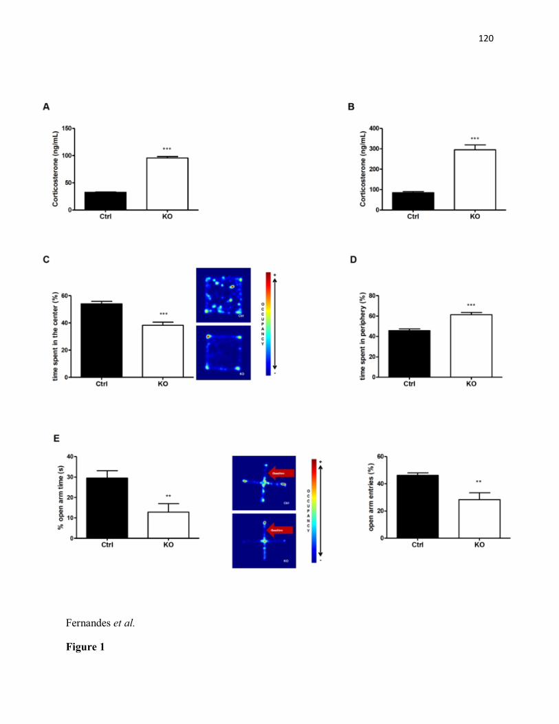
120
Fernandes et al.
Figure 1
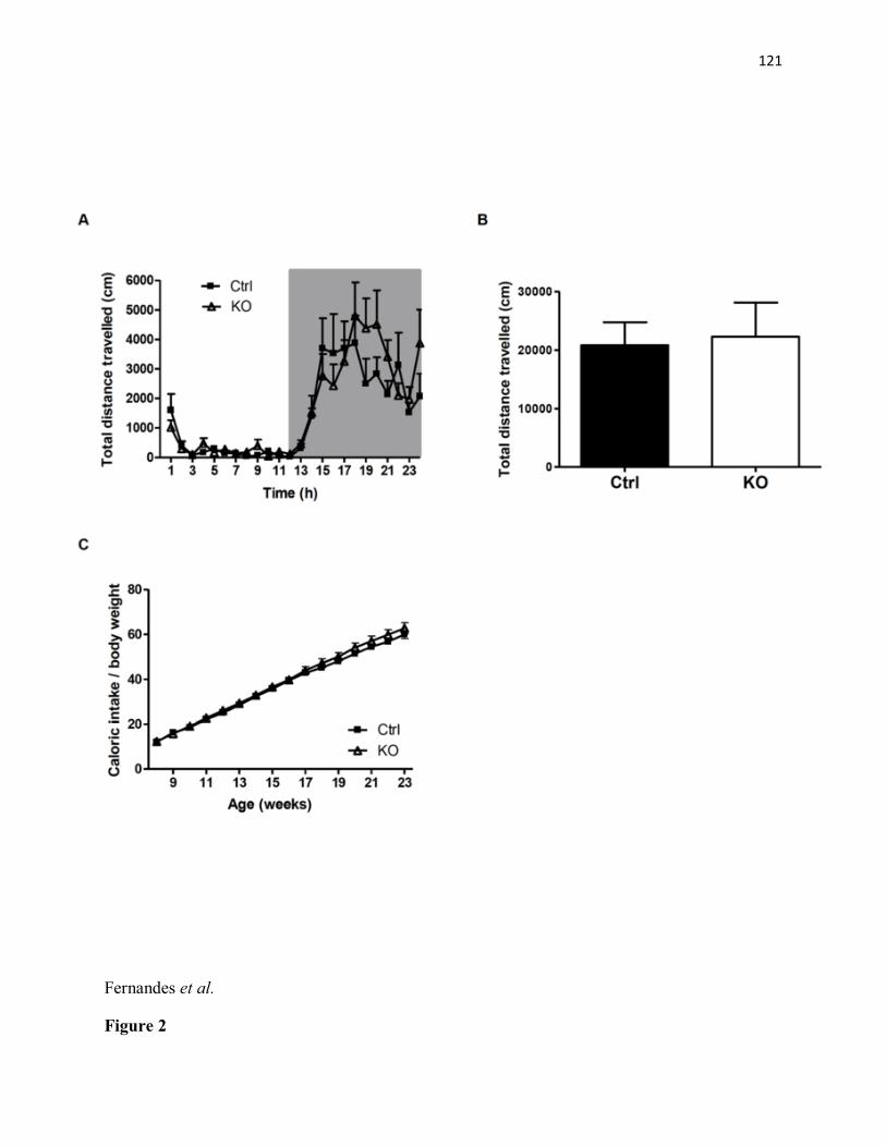
121
Fernandes et al.
Figure 2
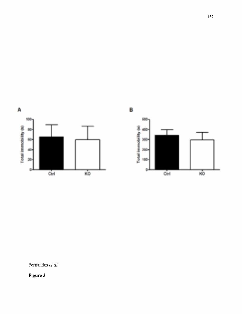
122
Fernandes et al.
Figure 3
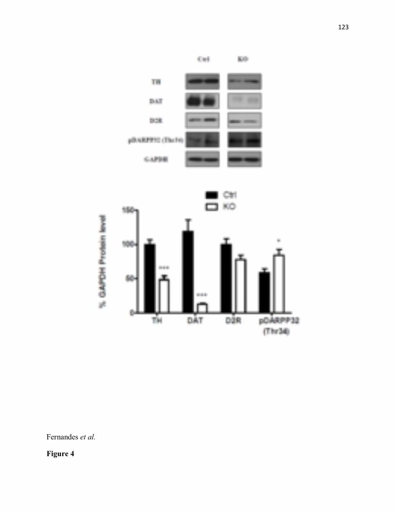
123
Fernandes et al.
Figure 4

124
Fernandes et al.
Figure 5

125
Chapter IV
General Discussion & Conclusion

126
9. General Discussion
The studies described in this thesis hone in on the LepRb signaling mechanisms by which leptin
affects mesolimbic DA signaling previously described by Fulton et al, Hommel et al and
Domingos et al. (Fulton et al. 2000, Fulton et al. 2006, Hommel et al. 2006, Domingos et al.
2011). The objective of the present work was to further define the specific cells and signaling
mechanisms by which leptin impacts the mesolimbic DA system. More specifically, this thesis
tested the hypothesis that leptin acts in the midbrain to modulate multiple behaviors via STAT3
signaling in DA neurons. Thus, to isolate the STAT3 gene and study its role in the brain DA
system, we generated a line of conditional KO mice lacking the main activation site of STAT3
gene (tyr705) selectively on DA neurons, as described in General Methods. It is important to
highlight that STAT3 ablation was observed in DA neurons within the VTA/SN. No significant
Cre-mediated STAT3 recombination was observed in other regions (Article#1, supplemental fig.
1). Of note, we are the first to identify a role for DA-specific STAT3 signaling in the modulation
of locomotion, running reward, anxiety-like behavior and DA tone.
9.1. Role for STAT3 signaling in DA neurons in locomotion and running reward
The first study described in this thesis shows for the first time that STAT3 signaling in DA
neurons mediates the action of leptin in the VTA to decrease spontaneous and voluntary
locomotor activity and running reward, and to decrease DA overflow in the NAc of male mice.
Moreover, our findings provide firm evidence that the influence of leptin in the midbrain to
decrease food intake does not require STAT3 signaling in DA neurons.

127
Previous reports demonstrated that a direct intra-VTA leptin injection induces short-term
decreases in food consumption in rats (Hommel et al. 2006, Bruijnzeel et al. 2011), while viral-
mediated LepRb knockdown in the VTA has an opposite effect by increasing food intake without
affecting body weight (Hommel et al. 2006). Thus, we sought to determine the contribution of
STAT3 signaling in DA neurons on the previously described anorectic effects of leptin in the
midbrain. We observed that either a single ICV or intra-VTA leptin injection reduced food intake
to a similar extent in both control and STAT3DAT-KO mice. Our results are the first to show that
leptin acts in the VTA to decrease food intake in mice, and imply that pSTAT3 in DA neurons
does not mediate the acute anorectic effect of leptin. Alternatively, Hommel and coworkers
found that adeno-associated virus (AAV)-mediated LepRb knockdown in the VTA, targeting
both dopaminergic and non-dopaminergic neurons in this region, increases food intake (Hommel
et al. 2006). This raises the possibility that either non-dopaminergic neurons (i.e.: GABA
neurons) or other LepRb signaling pathway modulates the effect of leptin in the midbrain to
decrease food consumption. Consistent with this hypothesis, Trinko and coworkers demonstrated
that the anorexigenic effects of an intra-VTA leptin injection are probably mediated by ERK1/2
pathway, and STAT3 (tyr705) activation is not sufficient for the effects of leptin in the VTA to
reduce food intake (Trinko et al. 2011). Alternatively, JAK2 signaling pathway has also been
suggested as a possible mediator of the anorectic effects of leptin in the VTA (Morton et al.
2009).
We next checked long-term food intake and body weight of control and STAT3DAT-KO mice fed a
chow diet and we observed that STAT3DAT-KO male mice exhibited reduced body weight and
reduced adiposity in spite of no changes in caloric intake, when normalized by the body weight,
suggesting that STAT3 signaling in DA neurons are not involved in long-term food intake. Our

128
findings are in agreement with previous studies by Davis and coworkers showing that
knockdown of LepRb in the midbrain does not affect feeding (Davis et al. 2011), and suggest
that signaling pathways other than STAT3 mediates the effects of leptin in the VTA to reduce
feeding. To determine whether the observed reduced body weight/body fat mass in STAT3DAT-KO
mice is due to increased energy expenditure, we assessed metabolic efficiency in these mice.
Metabolic efficiency, defined as the ratio of calories consumed divided by body weight gain over
a specific time interval, constitutes a initial method from which we can infer change in energy
expenditure (Butler and Kozak 2010, Ellacott et al. 2010). Our data show that metabolic
efficiency, was enhanced in STAT3DAT-KO mice fed a standard chow diet, as compared to
littermate controls. Given that normalized chow intake is not different between genotypes, we
hypothesized that the decreased body fat mass and increased metabolic efficiency observed in
STAT3DAT-KO mice might be due to increased locomotion. To address this question, we
examined ambulatory activity in STAT3DAT-KO and littermate controls. We found a significant
increase in dark-phase (active cycle) locomotor activity in STAT3DAT-KO male mice relative to
controls. This is in agreement with a previous report that viral-mediated VTA LepRb knockdown
stimulates locomotor activity in male rats (Hommel et al. 2006), and thereby suggests that DA-
specific STAT3 signaling mediates this effect. Furthermore, increased ambulatory activity in
STAT3DAT-KO male mice may contribute to the observed enhanced metabolic efficiency. Of note,
there's growing evidence of a link between metabolic signals and DA function, indicating a
potential role for this neurotransmitter in energy balance. It has been suggested that the DA
system acts to optimize the utilization of body energy resources, as DA favors energy
expenditure (Beeler et al. 2012). In fact, dopamine can shift obese animals from a state of
behavioral energy conservation to a state of behavioral energy expenditure (Beeler et al. 2012).

129
Consistent with this idea, dopamine was shown to increase resting energy expenditure in
humans(Ruttimann et al. 1991, Nakagawa et al. 1994) As previously mentioned in this thesis,
LepRb are also expressed on midbrain DA neurons and activation of these receptors on
dopaminergic cells decreases dopamine function (Hommel et al. 2006, Figlewicz and Benoit
2009) , enhances locomotor activity and energy expenditure (Ribeiro et al. 2011).
VTA DA neurons also projects to the caudal brainstem, and VTA-SNc DA neurons modulate
locomotor behaviors via projections to the basal ganglia which projects down to the brainstem
mesencephalic locomotor region (Kravitz et al. 2010, Stephenson-Jones et al. 2011). Therefore,
it could be possible that inactivation of STAT3 in midbrain DA neurons that project to the
brainstem mediates the increases in locomotion observed in STAT3DAT-KO mice and thus,
contributes to increased energy expenditure.
Food intake is regulated by an interaction between homeostatic and hedonic pathways.
Moreover, leptin is involved in the signaling of food reward (Fulton et al. 2000, Fulton et al.
2004), and reduces the hedonic response to palatable foods (Rangel 2013). Thus, we assessed
hedonic-based feeding behavior in control and STAT3DAT-KO mice using the "Dessert-Effect"
test. In this test paradigm, the intake of a palatable food (“dessert”) is assessed in calorically-
sated mice (Davis et al. 2012). Our results demonstrated that there are no differences in the
hedonic responses to palatable food between genotypes, suggesting that STAT3 signaling in DA
neurons does not modulate either the incentive motivational properties of food or food hedonics.
Likewise, we did not observe differences in other aspects of feeding behavior, including food
anticipatory activity (FAA), restriction-induced feeding and food preference between STAT3DAT-
KO and control mice (Figures 7, 8, 9, respectively Appendix I). Thus, taken together, these

130
results strongly suggest that STAT3 signaling in DA neurons does not modulate any aspect of
feeding behavior in mice.
As discussed in previous sections, leptin also affects energy balance by influencing the
rewarding effects of food as well as goal-oriented behaviors that contribute to obtaining food
(Rangel 2013). Accordingly, LepRb knockdown in the midbrain increases the motivation to
obtain food (Davis et al. 2011). To test our hypothesis that STAT3 signaling in DA neurons
mediates leptin's effect in food reward, we assessed food-motivated learning in STAT3DAT-KO
and control mice. Interestingly, STAT3DAT-KO male mice show impaired food-motivated learning
in a Fixed Ratio 1 (FR1) schedule of reinforcement. During appetitive instrumental learning
tasks, animals are expected to learn to emit a behavior (lever press) to obtain a reinforcer (i.e.:
sucrose pellet) (Robinson et al. 2007). A number of reports suggested that DA signaling (mainly
in the NAc) is greatly involved in instrumental (i.e.: operant conditioning) and Pavlovian or
classical conditioning (i.e.: CPP) learning, invoked during acquisition of behaviors associated
with food reward (Wise et al. 1978, Smith and Schneider 1988, Beninger and Miller 1998, Di
Chiara 2002, Wise 2004). Instrumental conditioning is a form of associative learning in which a
voluntary response is strengthened or diminished by its consequences (i.e.: reward, punishment,
etc.) (Thorndike 1911, Skinner 1966). In this kind of conditioning, the animal has to perform an
operant response and learn the relationship between the response and the response-associated
reward (Domjan 2005). Pavlovian or classical conditioning is another form of associative
learning in which a stimulus initially incapable of evoking a response acquire the ability to do so,
through repeated pairing with another stimulus (Pavlov 1927). Notably, the neurocircuitry
underlying the classical conditioning processes are distinct from those underlying the
instrumental learning (Bardo and Bevins 2000). Moreover, DA has been implicated in the

131
association between actions and outcomes in instrumental learning tasks (Schultz 1998, Dayan
and Balleine 2002, Robinson et al. 2006), while in classical conditioning tests DA has been
implicated in associations between neutral stimuli and rewards (Wilkinson et al. 1998, Dickinson
et al. 2000, Di Ciano et al. 2001, Dalley et al. 2005). However, the role of DA in the distinct
learning processes is not completely clear. For example, slightly hyperdopaminergic mice (DA
levels elevated 70% above controls) display normal instrumental learning (Yin et al. 2006), and
DA-depleted mice can acquire CPP for morphine reward (Hnasko et al. 2005), although they do
not perform tasks that require intentional movement (Szczypka et al. 1999, Hnasko et al. 2006),
such as operant responding. A recent report demonstrated that DA projections to the NAc
(mediated by D1R) are critical for operant conditioning (Steinberg et al. 2014). Moreover,
impaired D1R signaling in the NAc decreases lever pressing for food reinforcers (Nakajima
1989, Smith-Roe and Kelley 2000, Gore and Zweifel 2013). Likewise, reducing DA in the NAc
using intra-NAc injections of either 6-OHDA or D1R antagonist, substantially decreased lever
pressing for palatable food, without causing a general reduction in food motivation, as rats
remained directed toward the acquisition and consumption of food when it is freely available
(Salamone et al. 1991, Nowend et al. 2001). These findings suggest that diminishing DA in the
NAc may impair the ability of animals to overcome work-related response costs in order to
obtain food in a instrumental responding task (Nowend et al. 2001). Consistent with this idea, we
observed decreased D1R protein expression and decreased DA release in the NAc of STAT3DAT-
KO male mice, as compared to controls. Based on this result, we speculate that the significant
impairment in food-motivated learning in a FR1 schedule of reinforcement observed in our
model might be due to an impaired D1R transmission within the NAc of STAT3DAT-KO mice.
Notably, in spite of their poor performance in a food-motivated learning task, STAT3DAT-KO male

132
mice acquired CPP for running reward, suggesting that their learning impairment is specific to
the appetitive-operant conditioning task. The ability of STAT3DAT-KO mice to demonstrate
associative learning in a CPP task but not in a simple operant conditioning task (FR1)
underscores the point that there are multiple subtypes of reward-related learning, which are
differently affected by LepRb-STAT3 signaling in DA neurons.
In view of the well-described role of mesolimbic DA system in physical activity and the
reinforcing effects of running exercise, we measured voluntary wheel running in a separate
cohort of mice. Voluntary running wheel exercise was shown to reduce the preference of rodents
for palatable food, in addition to influence STAT3 activation selectively in the VTA (Scarpace et
al. 2010, Shapiro et al. 2011). Furthermore, a single leptin injection into the VTA suppresses
voluntary running exercise (Verhagen et al. 2011). Our results demonstrated that STAT3DAT-KO
male mice exercise significantly more than their respective controls, suggesting that the ability of
leptin in the VTA to suppress voluntary wheel running rely on STAT3 activation in DA neurons.
Similarly to food, voluntary exercise can also be rewarding. As previously mentioned in this
thesis, regular physical exercise can produce a rewarding effect in humans often referred to as
"runner’s high" (Panksepp et al. 2002, Dietrich and McDaniel 2004), and rats will work to have
access to a running wheel (Belke 2006, Belke 2006, Belke and Christie-Fougere 2006) and spend
more time in a place previously associated with the aftereffects of wheel running (Lett et al.
2000, Lett et al. 2001). To test the hypothesis whether the enhanced voluntary exercise in
STAT3DAT-KO mice reflects an increase in the rewarding aftereffects of wheel running, we
assessed the reward value of running in STAT3DAT-KO and control mice using a CPP paradigm.
In the CPP paradigm, the reinforcing effects of objects and/or behaviors are evaluated by
measuring the amount of time the animal spends in an environment previously associated with
133
that object/behavior (Tzschentke 2007). As the most striking finding of this study, we observed
that STAT3DAT-KO mice spend more time in a place previously associated with the aftereffects of
wheel running, relative to controls. Moreover, a single intra-VTA leptin injection one hour prior
to the CPP test impaired running CPP acquisition to a greater extent in the control group, firstly
suggesting that STAT3DAT-KO mice exercise more because the rewarding value of wheel running
is much higher in these mice as compared to controls. Most importantly, the increased running
reward observed in our model is modulated by LepRb-STAT3 signaling in DA neurons, and not
by other activators for STAT3 such as CNTF or IL-6. In fact, given that LepRb-deficient db/db
mice does not show STAT3 activation in the VTA after leptin administration (Fulton et al.
2006), and intra-VTA leptin decreases running reward in control mice but not in STAT3DAT-KO, it
is very unlikely that other STAT3 activator is involved in mediating the phenotypic
characteristics of STAT3DAT-KO mice. Very little is known about the neural controls of voluntary
running and its rewarding effect, and we are the first to show that leptin modulates the rewarding
aftereffects of running via LepRb-STAT3 signaling in DA neurons.
Besides the above-mentioned reduced D1R protein expression in NAc of STAT3DAT-KO mice, the
observed behavioral changes are accompanied by decreases in DA biosynthesis (decreased TH
protein expression) in the NAc, which is consistent with the effects of central leptin to increase
TH expression (Fulton et al. 2006, Leinninger et al. 2009), and suggests that STAT3 signaling in
DA neurons is involved. Interestingly, our findings matches with those by Knab and coworkers,
demonstrating that both D1R and TH expression were significantly downregulated in the NAc of
mice bred for high wheel running exercise (Knab et al. 2009), and thus suggest that D1R as well
as TH may be associated with the regulation of physical activity in STAT3DAT-KO male mice.
Moreover, we measured evoked DA release in the NAc core using fast scan cyclic voltammetry
134
in acute brain slice preparations and found that electrically stimulated DA release was reduced in
STAT3DAT-KO male mice. This is in agreement with a previous report from Fulton and coworkers
demonstrating that electrically evoked release of DA in the NAc is drastically reduced in ob/ob
mice (Fulton et al. 2006), and suggest that diminished evoked DA release in STAT3DAT-KO male
mice is induced by loss of DA-specific STAT3 signaling and could be partially related to a
reduction in DA available for release, potentially due to diminished synthesis reflected by the
observed reduced TH protein expression. Of note, there are no differences in DAT and D2R
protein expression in the VTA, NAc, DLS and CeA.
As an alternative means to assess mesoaccumbens DA function, we investigated AMPH-induced
locomotor sensitization in STAT3DAT-KO and control mice. Of note, one of the primary actions of
AMPH is to increase dopaminergic concentration in the synaptic cleft by inhibiting DA reuptake
and promoting reverse transport, resulting in efflux of DA via the DAT (Besson et al. 1971, Von
Voigtlander and Moore 1973, Hoebel et al. 1983, Sulzer et al. 2005) - the resultant dramatic
increase in extracellular DA has been suggested to be of major importance for AMPH-induced
psychomotor activation (Kelly et al. 1975, Pijnenburg et al. 1975, Sulzer et al. 2005). Consistent
with previous reports showing that leptin increases AMPH-induced locomotion in rodents (Hao
et al. 2004, Fulton et al. 2006), a sensitized locomotor response was observed in control mice
receiving continuous treatment with leptin or vehicle delivered via a 14-days subcutaneous mini-
osmotic pump implants. Interestingly however, subcutaneous infusion of leptin also enhances the
locomotor responses to AMPH in STAT3DAT-KO mice suggesting that LepRb-STAT3 signaling is
important but not necessary for the actions of leptin to increase AMPH sensitization.
Accordingly, Leinninger and coworkers reported that there is a population of LepRb neurons in
the LHA expressing the neuropeptide NT, which is activated by leptin and modulates the

135
mesolimbic DA system (Leinninger et al. 2011). Most importantly, these authors observed that
leptin signaling in NT neurons participates in the action of AMPH to stimulate locomotor
activity (Leinninger et al. 2011). Thus, the fact that STAT3DAT-KO male mice show increased
AMPH-induced locomotor response when treated with leptin, could well be related to leptin's
action via an alternative signaling mechanism, such as the NT neurons in the LHA, to decrease
DAT activity and consequently increase AMPH sensitization. Experiments needs to be
conducted to test this hypothesis.
Taken together, our findings suggest an important role for leptin in attenuating ambulatory
activity, voluntary exercise and running reward in male mice, that are mediated via LepRb-
STAT3 signaling in midbrain DA neurons and changes in DA tone. We posit that leptin acts in
mesolimbic brain reward circuitry as a signal of plenty to dampen the motivational and
rewarding effects of several behaviors that serve to replenish energy stores. Consistent with this
idea, our findings further suggest that LepRb-STAT3 signaling in DA neurons constitute a
potential mechanism involved in hypoleptinemia-induced hyperactivity. Based on the fact that
STAT3DAT-KO male mice show reduced food intake and reduced body fat mass due to increased
locomotion and wheel running hyperactivity, these transgenic mice could potentially serve as a
useful model for the study of activity-based anorexia (ABA).

136
9.2. Loss of STAT3 signaling in DA neurons induces an anxiogenic phenotype in female mice
It is well known that DA signaling in the VTA is implicated in anxiety-like behavior. Previous
studies demonstrated that electrical stimulation of VTA neurons induces anxiogenic responses,
while lesions of the VTA or inhibition of DA signaling within this region by stimulation of the
inhibitory D2R have anxiolytic effects (Borowski and Kokkinidis 1996, Munro and Kokkinidis
1997, Gifkins et al. 2002). As mentioned previously, the VTA is known to project to the
amygdala, among other areas. The amygdala is a subcortical structure in the limbic system which
has long been highlighted as a major constituent of the neural circuits processing emotional
behavior (Weiskrantz 1956, Aggleton 1993, LeDoux 2000). In regards to its anatomical
organization, the amygdala consists of a group of anatomically and functionally distinct nuclei
(Amaral and Insausti 1992, Saygin et al. 2011). Among these nuclei, the CeA is strongly
modulated by DA and, most importantly, it is located in a strategic position to mediate many
aspects of anxiety-like behavior, given that CeA neurons project to sites involved in mediating
different aspects of mood and emotion (Amaral and Insausti 1992, Davis 1992). Consistent with
these observations, DA signaling within the CeA is enhanced in response to a variety of
emotional stimuli (Inglis and Moghaddam 1999, Torres et al. 2002) and D1R signaling in the
CeA has been highly associated with anxiety behavior (Guarraci et al. 1999, de la Mora et al.
2005). In addition, leptin modulates DA signaling within the CeA via D1R (Liu et al. 2011) and
play an important role in emotional behavior (Leshan et al. 2006, Scott et al. 2009, Liu et al.
2010). The anxiolytic and antidepressant properties of leptin were already described (Asakawa et
al. 2003, Lu et al. 2006, Liu et al. 2010), however, the specific LepRb signaling pathway and
brain sites has, until now, remained poorly understood.

137
As the most striking finding of this study, we observed that STAT3DAT-KO female mice exhibited
an anxiogenic phenotype, which was consistently observed in two widely used tests of anxiety:
the open field test (OFT) and the elevated-plus maze (EPM). Of note, both OFT and EPM tests
have proven to be efficient to detect anxiety-like behavior in studies other than those testing
anxiolytic drugs (Teegarden and Bale 2007, Liu et al. 2011, Sharma et al. 2013).
Importantly, the anxiogenic behavior is not the result of general hypolocomotion or
abnormalities in feeding activity as STAT3DAT-KO female mice showed normal feeding behavior
and locomotion when compared to littermate controls (Figures 10 and 11 respectively,
Appendix II). Given that symptoms of anxiety and depression often co-occur (Sartorius et al.
1996), we assessed depressive-like behavior in STAT3DAT-KO mice by determining immobility
time in the forced-swim and tail suspension tests. In agreement with findings that LepRb
signaling in DA neurons does not modulate behavioral despair in mice (Liu et al. 2011),
depressive-like behavior is similar between genotypes. These findings suggest that leptin acts via
distinct neural pathways to modulate anxiety and depression in mice. In fact, there is literature
strongly supporting the involvement of LepRb signaling in the hippocampus in the modulation of
depression-like behavior (Lu et al. 2006, Yamada et al. 2011, Garza et al. 2012, Guo et al.
2013). Taken together, our results points to the conclusion that STAT3 signaling in DA neurons
does not affect DA signaling in brain structures involved in depression-like behavior (i.e.:
hippocampus) or, alternatively, leptin's antidepressant-like effects is not mediated by STAT3
signaling in DA neurons.
Evidence from our immunoblotting studies on brain nuclei of STAT3DAT-KO female mice
demonstrates that DAT protein expression is reduced, while threonine-34 phosphorylation of
DARPP32 is increased in the CeA of STAT3DAT-KO as compared to controls. Briefly, D1R
138
signaling activates cAMP-dependent protein kinase (PKA), which will, in turn, directly stimulate
DARPP32-Thr 34 phosphorylation (Nishi et al. 2000). These results suggest an increased DA
signaling via D1R due to reduced DA reuptake, elicited by reduced DAT function, in midbrain
DA neurons that projects to the CeA. Moreover, it represents a potential mechanism by which
increased D1R signaling within the CeA mediates the anxiogenic-like behavior in STAT3DAT-KO
female mice. Consistent with this hypothesis and observations in DA-specific LepRb KO mice
(Liu et al. 2011), we found that pharmacological blockage of D1R signaling in the CeA
normalized anxiogenic behavior in STAT3DAT-KO mice, suggesting that lack of STAT3 activation
in DA neurons results in increased anxiety by somehow increasing D1R signaling.
As discussed in Article 2, our studies complement those of Liu and collaborators (Liu et al.
2011) and indicate that selective inactivation of STAT3 (tyr705) in DA neurons results in an
anxiogenic phenotype, mediated by increased D1R signaling in the CeA of female but not male
mice. Furthermore, STAT3 signaling in DA neurons does not affect despair-like behavior,
suggesting that leptin's antidepressant-like action is mediated by other brain circuits or even
other neurotransmitters (Harvey 2007, Guo et al. 2013). Intriguingly, in contrast to a previous
study suggesting that lack of LepRb signaling in DA neurons increases anxiety in male mice (Liu
et al. 2011), we found that loss of STAT3 activation in DA neurons did not affect anxiety-like
behavior in STAT3DAT-KO male mice. There are a few hypothesis to explain the sexual
dimorphism observed in our mice model. First of all, there is literature showing that females are
much more prone to be afflicted by mood disorders than males (Kessler et al. 2005, Bekker and
van Mens-Verhulst 2007), suggesting that gonadal hormones, such as estrogen and testosterone,
may play an essential role in the etiology of anxiety and depression. Likewise, an important role
for estrogen in augmenting both pre- and postsynaptic dopaminergic function was already
139
described (Morissette et al. 1990, Levesque and Di Paolo 1991, Hu et al. 2006, Becker and Hu
2008). Moreover, estrogens are known to regulate circulating leptin levels and leptin sensitivity
(Rosenbaum et al. 1996, Shimizu et al. 1997, Clegg et al. 2006) and to improve mood (Joffe and
Cohen 1998); while testosterone or androgens suppress leptin concentration (Hislop et al. 1999)
and have anxiolytic and antidepressant benefits (Wang et al. 1996, Hermans et al. 2006,
DiBlasio et al. 2008). It was already reported that there are differences in estrogen levels
between males and females, with females showing higher levels than males (Clegg et al. 2006).
In addition, leptinemia also varies between males and females (Maffei et al. 1995, Castracane et
al. 1998), and female rats exhibits increased sensitivity to the central effects of leptin as
compared to males (Clegg et al. 2003). Most importantly, estrogen receptors (ERs) colocalize
with TH positive cells in mesolimbic regions (Kritzer 1997, Creutz and Kritzer 2002), including
the amygdala (Asmus and Newman 1993, Cavanaugh and Lonstein 2010, Cavanaugh and
Lonstein 2010), suggesting a potential role for estrogens on TH cell activity. In agreement with
these findings, circulating levels of both estrogen receptors (ERα and ERβ) are correlated with
DA hyperactivity within the mesolimbic system (Thompson and Moss 1997). Of note, estrogen
modulates STAT3 in the hypothalamus (Gao and Horvath 2008) and targets ERβ in the VTA
(Creutz and Kritzer 2002). Furthermore, a dense population of estrogen-expressing neurons has
already been described in limbic regions, such as the CeA, in female rats (Pfaff and Keiner
1973). Given the growing literature suggesting marked sexual dimorphisms in brain wiring,
morphology and chemistry (De Vries and Boyle 1998, Cosgrove et al. 2007), we speculate that
sex differences observed in our studies may involve a difference in the number of ERs in the
CeA of males versus females; an altered sensitivity to the anxiolytic actions of estrogen; a
140
different effect of estrogen in male and female brain or even due to estrogen action to increase
DA reuptake within the CeA. However, these hypotheses awaits further experimentation.
Besides suggesting that leptin regulates feeding or emotion-related behavior by distinct neuronal
targets and in a sex-specific fashion, the results presented on this study unravel the signaling
pathway by which leptin acts on DA neurons to modulate anxiety-like behavior. Importantly our
studies contribute to the mechanistic understanding of the role of leptin in anxiogenic behavior
and provide insights into novel therapeutic approaches for the treatment of anxiety disorders
(For a graphic representation of our main conclusion, see Figure 5).

141
Figure 5: The anxiolytic actions of leptin in female mice are mediated by increased LepRb-
STAT3 signaling in midbrain DA neurons that target the CeA to decrease D1R signaling,
perhaps via increased DA reuptake.

142
10. General limitations
Limitations of this study include:
we cannot totally exclude the possibility of developmental compensation that might
have occurred in STAT3DAT-KO mice;
we cannot affirm that all the phenotype of STAT3DAT-KO mice was caused by lack of
LepRb-STAT3 signaling in DA neurons, as not all experiments were performed in leptin-
treated mice;
we did not assess DA release in the CeA of STAT3DAT-KO mice, in order to better
understand the mechanisms underlying the observed anxiogenic phenotype in our model;
we did not investigate whether estrogen and leptin signaling interact in the VTA.
the estrous cycle was not monitored or controlled in our study. Thus, the contribution of
the hormonal milieu to the gender differences reported cannot be determined.
143
11. Perspectives
The identification of a major role for STAT3 signaling in DA neurons in the modulation of
locomotion, running reward, operant learning, DA tone and anxiety-like behavior in addition to
the sexual dimorphic phenotype observed in STAT3DAT-KO mice will undoubtedly lead to a great
amount of future research. The following are a select few studies that may shed further insight on
STAT3 signaling in midbrain DA neurons.
Which DA-relevant genes are directly modulated by STAT3?
To address this question, future studies can evaluate the direct binding of STAT3 to promoters
using pSTAT3 chromatin immunoprecipitation.
Which are the LepRb signaling pathways mediating the effects of leptin in feeding behavior?
As mentioned in previous sections, leptin's effect in eating behavior is well described (Fulton
2010, Narayanan et al. 2010, Fernandes et al. 2013). However, we found that DA specific
STAT3 activation does not mediate the effects of leptin on any aspect of feeding behavior,
suggesting that either GABA neurons or other LepRb signaling pathway is involved. To further
our understanding of the LepRb signaling pathways mediating the effects of leptin in feeding
behavior, it would be interesting to perform experiments involving the use of viral-mediated
knockdown of STAT3 in the VTA by administering AAV delivery of Cre-recombinase directly
into the VTA of STAT3lox/lox mice, targeting both DA and GABA neurons. Alternatively, if this
KO model also does not show any effect on feeding behavior, it would be relevant to use
144
pharmacological manipulations to modulate the activity of LepRb relevant signaling pathways
(i.e.: PI3-K, MAPK and AMPK) in the VTA. These experiments will allow a better
understanding of the contribution of specific LepRb signaling pathways in the actions of leptin in
the midbrain to impact feeding behavior.
Which signaling molecules are involved in leptin-induced increased AMPH sensitization in
STAT3 DAT-KO mice?
Leptin has been shown to play important roles in the control of the mesolimbic DA system,
including in the behavioral responses to AMPH (Sulzer et al. 1995, Figlewicz et al. 1998, Kahlig
et al. 2005, Fulton et al. 2006). Our data demonstrated that leptin peripheral administration
induced locomotor sensitization to AMPH in STAT3DAT-KO mice. This is suggestive that leptin's
effect to enhance AMPH sensitization (Fulton et al. 2006) does not only involve STAT3
signaling in DA neurons. It was already reported that:
1) VTA NT signaling contributes to AMPH psychomotor activation (Panayi et al. 2005);
2) leptin action via NT neurons modulates the locomotor stimulating effects of AMPH
(Leinninger et al. 2011);
3) LepRb neurons expressing NT projects to the VTA (Leinninger et al. 2011)
Thus, we hypothesize that leptin may be acting via NT neurons to induce AMPH sensitization in
STAT3DAT-KO mice. As a means to test this hypothesis, I propose to determine whether an intra-
VTA injection of the NT receptor antagonist (SR142948A) would prevent leptin-induced AMPH

145
sensitization in STAT3DAT-KO mice. This experiment will determine whether LepRb-NT
signaling mediates leptin action to promote AMPH sensitization.
What is the contribution of estrogen signaling for the sexual dimorphism observed in this
study?
The answer to this question could be found by determining whether central estradiol
administration activates STAT3 in DA neurons. If it is the case, it would be very interesting and
informative to investigate whether ovariectomized STAT3DAT-KO mice results in a behavioral
phenotype similar to that observed in STAT3DAT-KO male mice. These studies could open up a
compelling line of research on the crosstalk between leptin and estrogen signaling in DA
neurons.
146
12. Conclusion
The salient findings of this thesis are:
I. Our studies with STAT3DAT-KO mice suggest that the influence of leptin in the midbrain
to decrease free-feeding intake does not require STAT3 activation in DA neurons.
Alternatively, previous data suggest that JAK2 (Morton et al. 2009)or ERK1/2-MAPK
signaling (Trinko et al. 2011) is likely involved in the anorectic responses of leptin in the
VTA. Beyond that, we found that LepRb-STAT3 signaling in DA neurons does not
modulate feeding behavior.
II. Based on our findings that LepRb-STAT3 signaling in DA neurons mediates the effect
of leptin in the midbrain to decrease locomotor activity, voluntary exercise and running
reward in male mice, we hypothesize that leptin acts in mesolimbic brain reward circuitry
as a satiety signal to reduce the motivational and rewarding value of behaviors that
contribute to replenish energy stores. Consistent with this idea, lack of STAT3 activation
in midbrain DA neurons reduced both DA biosynthesis and NAc evoked DA release.
III. Loss of STAT3 signaling in DA neurons increases anxiety in female mice without
affecting depressive-like behavior. Furthermore, the anxiolytic action of leptin in female
mice is mediated by increased LepRb-STAT3 signaling in midbrain DA neurons that
target the CeA to decrease D1R signaling, perhaps via increased DA reuptake.

147
Appendices
The aim of this section is to describe relevant unpublished data obtained from STAT3DAT-KO mice
which will hopefully contribute for a broader understanding of the function of STAT3 signaling
in DA neurons.

148
13. Appendices
13.1. Appendix I
Unpublished results on the role for STAT3 signaling in DA neurons in the modulation of
feeding behavior in male mice
In addition to the chow intake studies described in Article 1, we also conducted experiments to
check long-term HF intake and body weight of control and in STAT3DAT-KO mice. In agreement
with what was observed in the chow-fed groups, there are no differences in HF consumption
and/or body weight between genotypes (Figure 6A-B, Appendix I). Notably, besides the
hypothalamus, the VTA has been suggested as another susceptible site for leptin resistance
(Matheny et al. 2011, Bruijnzeel et al. 2013), given the observations that leptin-induced STAT3
phosphorylation is impaired in this region in long-term HF-fed animals (Matheny et al. 2011).
Thus, a possible hypothesis to explain why food intake and body weights are not different
between HF-fed STAT3DAT-KO and control mice could be that the long term exposure to this
palatable food triggers leptin resistance in the VTA of control mice, by increasing SOCS 3
expression. This way, VTA leptin-resistant control mice would resemble the phenotype of
STAT3DAT-KO mice in which concerns to HF intake and body weight. In another words, we
hypothesize that in a HF condition LepRb-STAT3 signaling is also disrupted in the VTA of
control mice, so that both STAT3DAT-KO and control would exhibit a similar phenotype. This is a
very speculative explanation, which would require further experimentation.
In regards to other aspects of feeding behavior, we also failed to observe differences between
genotypes in FAA (Figure 7A-B, Appendix I) restriction-induced feeding (Figure 8, Appendix
I) and food preference (Figure 9, Appendix I) tests. FAA is defined as a behavior which
includes increased locomotion prior to receiving a daily scheduled meal (Merkestein et al. 2012).

149
As shown in Figure 7, STAT3 signaling in midbrain dopaminergic neurons does not affect FAA
in mice, when using a food-restriction protocol. Similarly, lack of leptin in ob/ob mice failed to
affect critically the expression of FAA (Gunapala et al. 2011). On the other hand Ribeiro and
coworkers observed that leptin administration abolishes FAA in ob/ob mice (Ribeiro et al. 2011).
Moreover, leptin receptor-deficient Zucker rats exhibited enhanced FAA after 22 days of food
restriction (Mistlberger and Marchant 1999). The apparent inconsistency regarding leptin's effect
on the expression of FAA is likely related to different experimental conditions, including the
duration of the food restriction protocol (long versus short duration) and also to the type of
restriction used (caloric restriction versus food intake restriction). To access restriction-induced
feeding in STAT3DAT-KO mice, we used a relatively short-term food restriction protocol (12 days)
during which the animals were habituated to have a limited access to standard chow daily (4h
only), while most of the studies suggesting that leptin reduces FAA used a long-term restriction
protocol (22-28 days). Furthermore, our findings suggest that locomotor behavior and FAA are
regulated independently by LepRb-STAT3 signaling in DA neurons.
We also failed to observe differences in chow consumption between STAT3DAT-KO and control
mice, following a 3-days food restriction-induced feeding, which is consistent with our previous
observation that there are no differences in regular chow consumption between genotypes. Given
food-restriction is known to induce stress (Ilott et al. 2014), and leptin has been suggested to
affect stress-induced eating (Appelhans 2010, Tomiyama et al. 2012), a possible explanation for
the lack of difference in restriction-induced feeding between groups could be that leptin does not
modulate this behavior via LepRb-STAT3 signaling in midbrain DA neurons. Alternatively,
Tomiyama and coworkers reported that the influence of leptin in stress-induced eating in humans
is specific for sweetened HF diet, as they failed to find a relationship between leptin and stress-

150
induced feeding in other food category (Tomiyama et al. 2012). Consistent with these findings,
other studies in rodents and humans suggested that stress affects sweet food intake to a much
greater extent than it affects the intake of regular chow or salty foods (Kawai et al. 2000, Epel et
al. 2001, Dallman et al. 2003, Habhab et al. 2009). Therefore, the inconsistency between our
results and these findings could be due to the use of different protocols and also to the fact that
we used standard chow in our experiments, instead of opting for a more palatable diet.
A role for DA in the modulation of food choice was previously described (Cooper and Al-Naser
2006). In addition, leptin has been suggested to modulate food preference in humans and rodents
(Wetzler et al. 2004, Licinio et al. 2007, Tomiyama et al. 2012), although the molecular
mechanisms and cell types involved remain unknown. Moreover, to our knowledge, the effects
of STAT3 signaling in DA neurons have not been previously reported in any form of food-
preference situation. Thus, we assessed food choice (high-fat versus low-fat diet) in control and
STAT3DAT-KO mice and we failed to observe differences between genotypes, further suggesting
that leptin's modulation of food preference does not involve STAT3 signaling in DA neurons.
Along with the findings presented on Article 1, these results strengthen our conclusion that
STAT3 signaling in DA neurons does not influence any aspect of feeding behavior.

151
Methods
Long-term HF intake and body weight
STAT3DAT-KO and control mice were housed individually and had free access to a HF diet
(D12231; Research Diets, Inc., New Brunswick, NJ, USA) containing 58% kcal from fat; 16.4%
kcal from protein and 25.5% kcal from carbohydrates and tap water throughout the experiment
(8 to 23 weeks of age). The animal room was kept at 22–24°C in a 12:12 h light–dark cycle.
Body weight and HF intake were assessed three times a week at 10am (onset of the dark period).
In addition to the amount of food consumption, weekly HF caloric intake for each mouse was
normalized by its respective body weight.
Food anticipatory activity
Mice were habituated to have a 4 hour-limited access to standard chow (Teklad Global 18%
Protein Rodent Diet, Harlan Laboratories, Indianapolis, Indiana, USA) daily (12-4pm) during 19
days in a novel entrainment cage (CLAMS metabolic chambers). On day 20, mice were placed in
their entrainment cage 4h prior to food access (8am) and locomotor activity was measured using
an automated comprehensive laboratory animal monitoring system (CLAMS, Columbus
Instruments, Columbus, OH, USA). Locomotor activity was defined as successive linear infrared
beam breaks recorded in 20min bins (breaks/20min).
Food restriction-induced feeding

152
All mice were housed individually with free access to standard chow and water. Food intake was
measured for 3 consecutive days. After that (on the following 3 days) STAT3DAT-KO and control
mice were 50% food restricted. On day 4 all mice were given free access to standard chow and
food consumption was measured after 2, 6, 12 and 24h.
Food preference test
Food choice was assessed in a food preference test for the HF diet (D12231; Research Diets,
Inc., New Brunswick, NJ) containing 58% Kcal from fat; 16.4% Kcal from protein and 25.5%
Kcal from carbohydrates, or ingredient-matched LF diet (D12328; Research Diets, Inc., New
Brunswick, NJ), containing 10.5% Kcal from fat; 16.4% Kcal from protein and 73.1% Kcal from
carbohydrates. All mice were single housed with free access to tap water. In each cage two cups
were provided in opposite corners one filled with HF diet and the other filled with LF diet. The
location of the cups was switched every 24 hour and food choice was assessed for 3 consecutive
days by measuring the amount of diets consumed. The results were presented as a ratio of HF
preference.
Statistical analyses
Data were analyzed using GraphPad 5 and presented as means and respective standard errors. A
two-way ANOVA with Bonferoni post-hoc tests was used to compare STAT3DAT-KO and control
mice body weight, HF caloric intake, chow intake in a food restriction-induced feeding test, FAA
and food preference. Criterion for significance was set to p≤0.05 in all comparisons.

153
Figure legends
Figure 6. Long-term high-fat(HF) diet intake and body weight is not different between
STAT3DAT-KO and control mice. (A) Cumulative HF caloric intake of STAT3DAT-KO and control
mice. n= 8-11/group. (B) Long-term body weight in STAT3DAT-KO and control mice. n= 8-
11/group.
Figure 7. Food anticipatory activity (FAA) is not different between STAT3DAT-KO male mice
and littermate controls. (A) 4h-locomotor activity prior to food access is not different in control
and STAT3DAT-KO mice. n= 5/group. (B) Locomotor activity is not different between control and
STAT3DAT-KO mice 60 minutes prior to access of food (n=5/group).
Figure 8. Food restriction-induced feeding is not different between STAT3DAT-KO male mice
and littermate controls. (A) Protocol schematics. (B) Free-access to food after three days of 50%
food restriction did not alter significantly regular chow consumption between
genotypes.n=7/group.
Figure 9: Food preference in STAT3DAT-KO mice. HF- versus LF-diet preference is similar
between STAT3DAT-KO and littermate controls. n=10-14/group.

154
A)
9 11 13 15 17 19 21 230
500
1000
1500
2000
CtrlKO
Age (weeks)
Cum
ulat
ive
high
-fat c
alor
ic in
take
(Kca
l)
B)
9 11 13 15 17 19 21 230
10
20
30
40
50
CtrlKO
Age (weeks)
Body
wei
ght (
g)
Figure 6

155
A)
B)
Ctrl KO0
50000
100000
150000
AUC
Figure 7
0 60 120 180 2400
1000
2000
3000
CtrlKO
Light cycle Dark cycle
Circadian time (minutes)
4h-lo
com
otor
act
ivity
(AU
C)
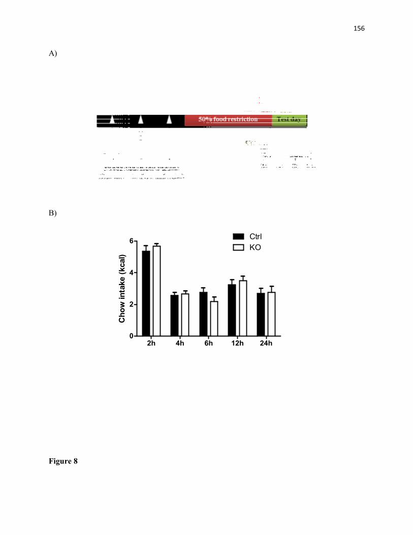
156
A)
B)
2h 4h 6h 12h 24h0
2
4
6 CtrlKO
Cho
w in
take
(kca
l)
Figure 8

157
day1 day2 day30.0
0.2
0.4
0.6
0.8 CtrlKO
Kca
l fro
m h
igh-
fat f
ood
Figure 9
158
13.2. Appendix II
Unpublished results on the role for STAT3 signaling in DA neurons in the modulation of
feeding behavior and learning in female mice
In agreement with our findings in STAT3DAT-KO male mice, our studies in female mice also
demonstrated that STAT3 signaling in DA neurons does not influence food intake and body
weight in chow-fed animals (Figure 10A, Appendix II). Likewise, food preference (HF versus
LF diet) as well as HF diet intake was also unaltered in STAT3DAT-KO female mice (Figure 10B-
C, Appendix II). However, we found that gender significantly affects general locomotion in
STAT3DAT-KO mice as, opposed to what was observed in males, ambulatory activity and
voluntary wheel running exercise is similar between STAT3DAT-KO and control female mice
(Figure 11A-B, Appendix II). The reason for the gender difference in locomotor behavior is not
known although, we hypothesize that estrogen signaling in DA neurons could be involved, as it
is well described that there are sex differences in dopaminergic function in the brain (Kuhn et al.
2010). Furthermore, circulating leptin levels are modulated by different cytokines and hormones
(including estrogen) that are also under the influence of gender (Considine 2001, Janeckova
2001). Of note, leptin levels are higher in females than in males (Frederich et al. 1995, Considine
2001, Janeckova 2001), also suggestive of a possible sexual dimorphism of LepRb signaling.
Similar to what was observed in STAT3DAT-KO male mice, lack of STAT3 signaling in DA
neurons also suppresses lever pressing for food in females - STAT3DAT-KO female mice showed
severe impairment in learning a FR1 schedule of reinforcement in an appetitive operant
conditioning task (Figure 12A-B, Appendix II). There is data implicating DA signaling in the
dorsolateral striatum (DLS) in the modulation of learning, including operant learning (Frank et
al. 2004, Yin et al. 2006, Balleine et al. 2007, Balleine and O'Doherty 2010). Consistent with
159
this idea, we found that D1R protein expression is downregulated in the DLS of STAT3DAT-KO
female mice as compared to controls, which we hypothesize may be related to the severe
impairment in learning a FR1 schedule of reinforcement for food pellet reward (Figure 12C,
Appendix II). Furthermore, STAT3DAT-KO female mice exhibited normal performance in a CPP
for palatable food (sweetened HF pellets), strengthening our hypothesis that loss of STAT3 in
DA neurons mediates leptin's effect in appetitive operant learning, but not in a classical
conditioning task (Figure 13, Appendix II). Of note, the learning impairment observed in
STAT3DAT-KO mice is not related to locomotor deficits or loss of appetite, as these mice exhibited
normal ambulatory activity and feeding behavior as compared to controls. Although our findings
suggest that STAT3 signaling in DA neurons is critical for operant learning, more experiments
needs to be conducted to shed light on the detailed mechanisms underlying the effects of leptin
signaling in different forms learning.

160
Methods
Long-term food intake and body weight
STAT3DAT-KO and control mice were housed individually with free access to standard chow
(Teklad Global 18% Protein Rodent Diet, Harlan Laboratories, Indianapolis, Indiana, USA) and
tap water throughout the experiment (8 to 23 weeks of age). The animal room was kept at 22–
24°C in a 12:12 h light–dark cycle. Body weight and food intake were assessed three times a
week at the onset of the dark period.
Food preference test
Food choice was assessed in a food preference test for the HF diet (D12231; Research Diets,
Inc., New Brunswick, NJ) containing 58% Kcal from fat; 16.4% Kcal from protein and 25.5%
Kcal from carbohydrates, or ingredient-matched LF diet (D12328; Research Diets, Inc., New
Brunswick, NJ), containing 10.5% Kcal from fat; 16.4% Kcal from protein and 73.1% Kcal from
carbohydrates. All mice were single housed with free access to tap water. In each cage two cups
were provided in opposite corners one filled with HF diet and the other filled with LF diet. The
location of the cups was switched every 24 hour and food choice was assessed for 3 consecutive
days by measuring the amount of diets consumed. The results were presented as a ratio of HF
preference.
Long-term HF intake
STAT3DAT-KO and control mice were housed individually and had free access to a HF diet
(D12231; Research Diets, Inc., New Brunswick, NJ, USA) containing 58% kcal from fat; 16.4%
kcal from protein and 25.5% kcal from carbohydrates and tap water throughout the experiment

161
(8 to 23 weeks of age). The animal room was kept at 22–24°C in a 12:12 h light–dark cycle.
Food intake was assessed three times a week at the onset of the dark period.
Ambulatory activity
To access 24-hour spontaneous locomotor activity STAT3DAT-KO and littermate controls were
individually placed into metabolic cages (Accuscan Instruments Inc., Columbus, OH, USA)
consisting of 16 light beams arrays in x, y and z axes. Distance travelled (horizontal activity) was
measured by nearby computer-controlled software.
Voluntary wheel running activity
To record daily running wheel behavior, animals were single housed into standard rat cages
(26,03cm width x 48,26cm depth x 20,32cm height) containing plastic wireless running wheels
(one wheel revolution=36.9 cm, Med Associates Inc., St. Albans, VT, USA). Wheel running
activity was continuously monitored through wireless transmitters and recorded using the
Wireless Running Wheel Manager Data Acquisition Software (Med Associates Inc., St. Albans,
VT, USA). Data were exported from the wheel manager database for further analysis. The
parameter evaluated was the total wheel running activity (revolutions per 24 h).
Appetitive food-motivated operant learning
Fixed ratio 1 (1:1) schedule of reinforcement
We used mouse operant chambers (Med Associates Inc., St. Albans, VT, USA) equipped with
two levers and a pellet dispenser delivering 20mg of sucrose pellets (Bio-Serv, Frenchtown, NJ,
USA). During the operant conditioning sessions, the mouse is expected to press the designated

162
active lever (triggering food reward) to get a reinforcer. The allocation of right and left levers
were counterbalanced between mice. All mice were trained in 60 min sessions to press the active
lever on a continuous reinforcement schedule (FR1), in which every lever press is reinforced
with a 20 mg sucrose pellet. All training was conducted in the dark cycle and the animals were
50% food restricted during the operant training. Operant training was carried out over 10 days.
Conditioned Place Preference for sweetened HF food
The place preference apparatus (Med Associates Inc., St. Albans, VT, USA) consisted of three
chambers with overall inside dimensions of: 46.5cm length x 12.7cm width x 12.5cm height. The
two choice compartments consist of a black compartment with a stainless steel grid rod floor and
a white compartment with a spaced stainless steel mesh floor. These two chambers are separated
by a small center compartment that is 11.7 cm long with a neutral gray finish and a PVC floor.
Preference test: On day 0 each experimental mouse was confined to both the black and white
compartment of the CPP apparatus for 5 minutes to allow habituation to each chamber. On this
same day, animals were allowed to move freely within the apparatus for 15 minutes, during
which the amount of time spent in each compartment was recorded. Conditioning training: In the
following 14 days, mice were randomly assigned to a pairing compartment (black or white)
associated with palatable food (sweetened HF pellets, containing 48.9% Kcal as fat, Bio-Serv,
Frenchtown, NJ, USA) on even days or no food on even days. Mice were confined to the
appropriate compartment for 30 min during the 14-days conditioning period. CPP test: On day
15, each mouse was placed back in the CPP apparatus and allowed to move freely across
compartments for 15 min. The amount of time spent in each compartment was recorded and the
proportion of time spent on the paired side was compared to that obtained during the pre-test.

163
Western immunoblotting
Mice were decapitated under Isoflurane anaesthesia. Brains were rapidly dissected and stored at -
80oC. Frozen brains were sliced into 0.5mm coronal sections using a brain matrix. Coronal
sections were mounted onto slides and maintained on dry ice. Nuclei were microdissected using
brain tissue punches (Stoelting Inc., Wood Dale, IL). Bilateral punches of 1.0mm diameter were
obtained from the DLS. Microdissected tissues were homogenized on ice in 100 ml of cell lysis
buffer (100mM Tris, pH 7.5; 750mM NaCl; 5mM Na2EDTA; 5mM EGTA, pH 7.5; 5% Triton x-
100; 12.5mM sodium pyrophosphate; 5mM beta-glycerophosphate; 1mM, 5mM Na3VO4;
5µg/ml leupeptin) with added protease (PMSF 1mM) and phosphotase inhibitors (Sigma
phosphatase inhibitor cocktails I and II) in 1.5 ml tubes using a motorized pestle, and after that
centrifuged for 15 min at 14000 g. Protein concentrations were measured using BCA protein
assay (Pierce Biotechnology, IL, USA). Protein samples were separated by electrophoresis on a
10% polyacrylamide gel and electrotransferred to a nitrocellulose membrane (EMD Millipore
Corporation, Billerica, MA, USA). Non-specific binding sites were blocked in TBS 5% low-fat
milk and 0.1% Tween-20 or 5% BSA. Membranes were rinsed in buffer (0.1% Tween-20 in
TBS) and then incubated with anti-D1R (1:1000; Millipore, EMD Millipore Corporation,
Billerica, MA, USA) followed by anti-rabbit IgG horseradish peroxidase-conjugate (1:5000).
After rinsing with buffer, immunocomplexes were visualized by chemiluminescence using the
western lighting plus ECL kit (PerkinElmer, Waltham, MA, USA). Protein size was compared
by using precision plus protein ladder (Bio-Rad, Bedford, MA, USA). The film signals were
digitally scanned and then density quantified using ImageJ software. GAPDH (1:10000, Cell

164
Signaling, Cell Signaling Technology, Inc., Danvers, MA, USA) was used as an internal control,
such that data were standardized according to GAPDH values.
Statistical analyses
Data were analyzed using GraphPad 5 and presented as means and respective standard errors. A
two-way ANOVA with Bonferoni post-hoc tests was used to compare control and STAT3DAT-KO
mice body weight, food intake (chow and HF diet), food preference, food motivation and
voluntary exercise. Unpaired t-tests were used to compare control and STAT3DAT-KO mice
locomotor activity, food CPP and D1R protein expression. Criterion for significance was set to
p≤0.05 in all comparisons.

165
Figure legends
Figure 10. Caloric intake, body weight, and food preference is not different in STAT3DAT-KO
female mice as compared to controls. (A) Average long-term caloric intake and body weight of
STAT3DAT-KO female mice and littermate controls on regular chow diet. n=8-9/group (B) Food-
preference test in STAT3DAT-KO and control mice. n=11-12/group. (C) Average long-term high-
fat caloric intake of STAT3DAT-KO female mice and littermate controls. n=12-13/group.
Figure 11. Locomotor activity and voluntary exercise is not different in STAT3DAT-KO female
mice as compared to controls. (A) Dark (active) phase locomotor activity expressed as total
distance travelled during the test in STAT3DAT-KO female mice as compared to controls. n=7-
9/group. (B) Voluntary wheel running exercise in female mice, expressed as average number of
wheel revolutions per week. n=10 mice/group.
Figure 12. Operant conditioning task and D1R protein expression in STAT3DAT-KO and control
mice. (A) Total lever press. n= 8-10/group ***p<0.001 (B) Percentage of correct over incorrect
responses by day 10. n= 8-10/group. **p<0.01 (C) Significant decrease in D1R protein levels in
the dorsolateral striatum (DLS) of STAT3DAT-KO mice as compared to controls. n=4-7/group.
**p<0.01.
Figure 13. STAT3DAT-KO female mice acquired CPP for a sweetened high-fat food. (A) CPP
protocol schematics (B) Preference for the food-paired side of the CPP apparatus after minus
before the CPP test. n=11/group.
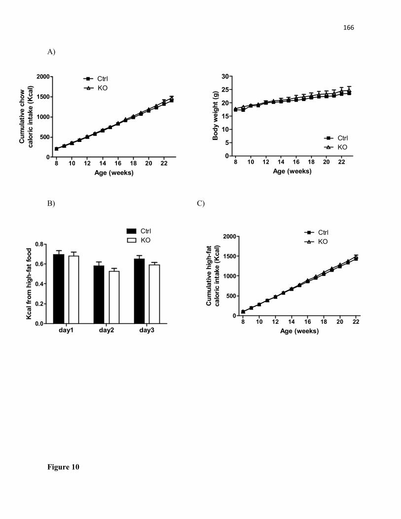
166
A)
B) C)
Figure 10
8 10 12 14 16 18 20 220
5
10
15
20
25
30
CtrlKO
Age (weeks)
Body
wei
ght (
g)
8 10 12 14 16 18 20 220
500
1000
1500
2000 CtrlKO
Age (weeks)
Cum
ulat
ive
chow
calo
ric in
take
(Kca
l)
day1 day2 day30.0
0.2
0.4
0.6
0.8
CtrlKO
Kcal
from
hig
h-fa
t foo
d
8 10 12 14 16 18 20 220
500
1000
1500
2000 CtrlKO
Age (weeks)
Cum
ulat
ive
high
-fat
calo
ric in
take
(Kca
l)

167
A)
B)
Figure 11
Ctrl KO0
10000
20000
30000
Tota
l dis
tanc
e tr
avel
led
(cm
)
9 11 13 15 17 19 210
50000
100000
150000
200000
250000
300000
CtrlKO
Age (weeks)
Num
ber o
f whe
el re
volu
tions
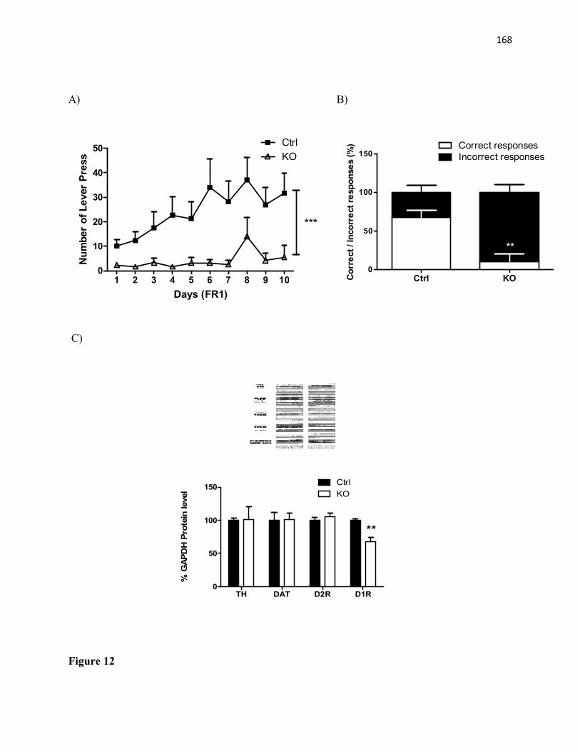
168
A) B)
1 2 3 4 5 6 7 8 9 100
10
20
30
40
50 CtrlKO
***
Days (FR1)
Num
ber o
f Lev
er P
ress
C)
TH DAT D2R D1R0
50
100
150 CtrlKO
**
% G
APD
H Pr
otei
n le
vel
Figure 12
Ctrl KO0
50
100
150Correct responsesIncorrect responses
**
Corr
ect /
Inco
rrec
t res
pons
es (%
)
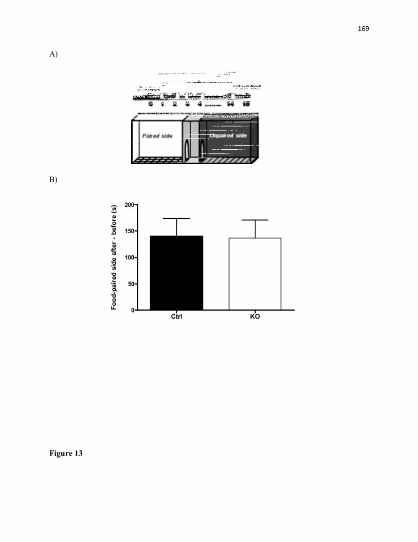
169
A)
B)
Ctrl KO0
50
100
150
200
Food
-pai
red
side
afte
r - b
efor
e (s
)
Figure 13

170
14. References
Abizaid, A. and T. L. Horvath (2012). "Ghrelin and the central regulation of feeding and energy balance." Indian J Endocrinol Metab 16(Suppl 3): S617-626.
Adan, R. A., J. J. Hillebrand, U. N. Danner, S. Cardona Cano, M. J. Kas and L. A. Verhagen (2011). "Neurobiology driving hyperactivity in activity-based anorexia." Curr Top Behav Neurosci 6: 229-250.
Aggleton, J. P. (1993). "The contribution of the amygdala to normal and abnormal emotional states." Trends Neurosci 16(8): 328-333.
Ahima, R. S., J. Dushay, S. N. Flier, D. Prabakaran and J. S. Flier (1997). "Leptin accelerates the onset of puberty in normal female mice." J Clin Invest 99(3): 391-395.
Ahima, R. S. and S. Y. Osei (2004). "Leptin signaling." Physiol Behav 81(2): 223-241.
Ahima, R. S., D. Prabakaran, C. Mantzoros, D. Qu, B. Lowell, E. Maratos-Flier and J. S. Flier (1996). "Role of leptin in the neuroendocrine response to fasting." Nature 382(6588): 250-252.
Ainslie, D. A., M. J. Morris, G. Wittert, H. Turnbull, J. Proietto and A. W. Thorburn (2001). "Estrogen deficiency causes central leptin insensitivity and increased hypothalamic neuropeptide Y." Int J Obes Relat Metab Disord 25(11): 1680-1688.
Akira, S. (1999). "Functional roles of STAT family proteins: lessons from knockout mice." Stem Cells 17(3): 138-146.
Akira, S., Y. Nishio, M. Inoue, X. J. Wang, S. Wei, T. Matsusaka, K. Yoshida, T. Sudo, M. Naruto and T. Kishimoto (1994). "Molecular cloning of APRF, a novel IFN-stimulated gene factor 3 p91-related transcription factor involved in the gp130-mediated signaling pathway." Cell 77(1): 63-71.
Amaral, D. G. and R. Insausti (1992). "Retrograde transport of D-[3H]-aspartate injected into the monkey amygdaloid complex." Exp Brain Res 88(2): 375-388.
Amstalden, M., M. R. Garcia, S. W. Williams, R. L. Stanko, S. E. Nizielski, C. D. Morrison, D. H. Keisler and G. L. Williams (2000). "Leptin gene expression, circulating leptin, and luteinizing hormone pulsatility are acutely responsive to short-term fasting in prepubertal heifers: relationships to circulating insulin and insulin-like growth factor I(1)." Biol Reprod 63(1): 127-133.
Anand, B. K. and J. R. Brobeck (1951). "Localization of a "feeding center" in the hypothalamus of the rat." Proc Soc Exp Biol Med 77(2): 323-324.
Anden, N. E., A. Carlsson, A. Dahlstroem, K. Fuxe, N. A. Hillarp and K. Larsson (1964). "Demonstration and Mapping out of Nigro-Neostriatal Dopamine Neurons." Life Sci 3: 523-530.
Andersen, P. H., J. A. Gingrich, M. D. Bates, A. Dearry, P. Falardeau, S. E. Senogles and M. G. Caron (1990). "Dopamine receptor subtypes: beyond the D1/D2 classification." Trends Pharmacol Sci 11(6): 231-236.

171
Angelopoulos, T. J. (2001). "Beta-endorphin immunoreactivity during high-intensity exercise with and without opiate blockade." Eur J Appl Physiol 86(1): 92-96.
Aosaki, T., K. Kiuchi and Y. Kawaguchi (1998). "Dopamine D1-like receptor activation excites rat striatal large aspiny neurons in vitro." J Neurosci 18(14): 5180-5190.
Appelhans, B. M. (2010). "Circulating leptin moderates the effect of stress on snack intake independent of body mass." Eat Behav 11(3): 152-155.
Arias-Carrion, O. and E. Poppel (2007). "Dopamine, learning, and reward-seeking behavior." Acta Neurobiol Exp (Wars) 67(4): 481-488.
Asakawa, A., A. Inui, T. Inui, G. Katsuura, M. A. Fujino and M. Kasuga (2003). "Leptin treatment ameliorates anxiety in ob/ob obese mice." J Diabetes Complications 17(2): 105-107.
Asmus, S. E. and S. W. Newman (1993). "Tyrosine hydroxylase mRNA-containing neurons in the medial amygdaloid nucleus and the reticular nucleus of the thalamus in the Syrian hamster." Brain Res Mol Brain Res 20(3): 267-273.
Atmaca, M., M. Kuloglu, E. Tezcan, B. Ustundag, O. Gecici and B. Firidin (2002). "Serum leptin and cholesterol values in suicide attempters." Neuropsychobiology 45(3): 124-127.
Audi, L., C. S. Mantzoros, A. Vidal-Puig, D. Vargas, M. Gussinye and A. Carrascosa (1998). "Leptin in relation to resumption of menses in women with anorexia nervosa." Mol Psychiatry 3(6): 544-547.
Aumann, T. D., K. Egan, J. Lim, W. C. Boon, C. R. Bye, H. K. Chua, N. Baban, C. L. Parish, L. Bobrovskaya, P. Dickson and M. K. Horne (2011). "Neuronal activity regulates expression of tyrosine hydroxylase in adult mouse substantia nigra pars compacta neurons." J Neurochem 116(4): 646-658.
Backman, C. M., N. Malik, Y. Zhang, L. Shan, A. Grinberg, B. J. Hoffer, H. Westphal and A. C. Tomac (2006). "Characterization of a mouse strain expressing Cre recombinase from the 3' untranslated region of the dopamine transporter locus." Genesis 44(8): 383-390.
Baldo, B. A., K. Sadeghian, A. M. Basso and A. E. Kelley (2002). "Effects of selective dopamine D1 or D2 receptor blockade within nucleus accumbens subregions on ingestive behavior and associated motor activity." Behav Brain Res 137(1-2): 165-177.
Balleine, B. W., M. R. Delgado and O. Hikosaka (2007). "The role of the dorsal striatum in reward and decision-making." J Neurosci 27(31): 8161-8165.
Balleine, B. W. and J. P. O'Doherty (2010). "Human and rodent homologies in action control: corticostriatal determinants of goal-directed and habitual action." Neuropsychopharmacology 35(1): 48-69.
Banks, A. S., S. M. Davis, S. H. Bates and M. G. Myers, Jr. (2000). "Activation of downstream signals by the long form of the leptin receptor." J Biol Chem 275(19): 14563-14572.
Bannon, M. J. and R. H. Roth (1983). "Pharmacology of mesocortical dopamine neurons." Pharmacol Rev 35(1): 53-68.
172
Barash, I. A., C. C. Cheung, D. S. Weigle, H. Ren, E. B. Kabigting, J. L. Kuijper, D. K. Clifton and R. A. Steiner (1996). "Leptin is a metabolic signal to the reproductive system." Endocrinology 137(7): 3144-3147.
Barbeau, A. (1974). "High-level levodopa therapy in Parkinson's disease: five years later." Trans Am Neurol Assoc 99: 160-163.
Bardo, M. T. and R. A. Bevins (2000). "Conditioned place preference: what does it add to our preclinical understanding of drug reward?" Psychopharmacology (Berl) 153(1): 31-43.
Barrachina, M. D., V. Martinez, L. Wang, J. Y. Wei and Y. Tache (1997). "Synergistic interaction between leptin and cholecystokinin to reduce short-term food intake in lean mice." Proc Natl Acad Sci U S A 94(19): 10455-10460.
Bartness, T. J., E. Keen-Rhinehart, M. J. Dailey and B. J. Teubner (2011). "Neural and hormonal control of food hoarding." Am J Physiol Regul Integr Comp Physiol 301(3): R641-655.
Bates, S. H., T. A. Dundon, M. Seifert, M. Carlson, E. Maratos-Flier and M. G. Myers, Jr. (2004). "LRb-STAT3 signaling is required for the neuroendocrine regulation of energy expenditure by leptin." Diabetes 53(12): 3067-3073.
Bates, S. H., W. H. Stearns, T. A. Dundon, M. Schubert, A. W. Tso, Y. Wang, A. S. Banks, H. J. Lavery, A. K. Haq, E. Maratos-Flier, B. G. Neel, M. W. Schwartz and M. G. Myers, Jr. (2003). "STAT3 signalling is required for leptin regulation of energy balance but not reproduction." Nature 421(6925): 856-859.
Baumann, H., K. K. Morella, D. W. White, M. Dembski, P. S. Bailon, H. Kim, C. F. Lai and L. A. Tartaglia (1996). "The full-length leptin receptor has signaling capabilities of interleukin 6-type cytokine receptors." Proc Natl Acad Sci U S A 93(16): 8374-8378.
Bayer, V. E. and V. M. Pickel (1991). "GABA-labeled terminals form proportionally more synapses with dopaminergic neurons containing low densities of tyrosine hydroxylase-immunoreactivity in rat ventral tegmental area." Brain Res 559(1): 44-55.
Beaulieu, J. M. and R. R. Gainetdinov (2011). "The physiology, signaling, and pharmacology of dopamine receptors." Pharmacol Rev 63(1): 182-217.
Becker, J. B. and M. Hu (2008). "Sex differences in drug abuse." Front Neuroendocrinol 29(1): 36-47.
Beeler, J. A., C. R. Frazier and X. Zhuang (2012). "Putting desire on a budget: dopamine and energy expenditure, reconciling reward and resources." Front Integr Neurosci 6: 49.
Bekker, M. H. and J. van Mens-Verhulst (2007). "Anxiety disorders: sex differences in prevalence, degree, and background, but gender-neutral treatment." Gend Med 4 Suppl B: S178-193.
Belke, T. W. (2006). "Concurrent schedules of wheel-running reinforcement: choice between different durations of opportunity to run in rats." Learn Behav 34(1): 61-70.
Belke, T. W. (2006). "Responding for sucrose and wheel-running reinforcement: effect of pre-running." Behav Processes 71(1): 1-7.

173
Belke, T. W. and M. M. Christie-Fougere (2006). "Investigations of timing during the schedule and reinforcement intervals with wheel-running reinforcement." Behav Processes 73(3): 240-247.
Belke, T. W. and T. Garland, Jr. (2007). "A brief opportunity to run does not function as a reinforcer for mice selected for high daily wheel-running rates." J Exp Anal Behav 88(2): 199-213.
Beninger, R. J. and R. Miller (1998). "Dopamine D1-like receptors and reward-related incentive learning." Neurosci Biobehav Rev 22(2): 335-345.
Besson, M., A. Cheramy, P. Feltz and J. Glowinski (1971). "Dopamine: spontaneous and drug-induced release from the caudate nucleus in the cat." Brain Res 32(2): 407-424.
Binder, E. B., B. Kinkead, M. J. Owens and C. B. Nemeroff (2001). "Neurotensin and dopamine interactions." Pharmacol Rev 53(4): 453-486.
Bishop, M. P., S. T. Elder and R. G. Heath (1963). "Intracranial self-stimulation in man." Science 140(3565): 394-396.
Bjorbaek, C., R. M. Buchholz, S. M. Davis, S. H. Bates, D. D. Pierroz, H. Gu, B. G. Neel, M. G. Myers, Jr. and J. S. Flier (2001). "Divergent roles of SHP-2 in ERK activation by leptin receptors." J Biol Chem 276(7): 4747-4755.
Bjorbaek, C., K. El-Haschimi, J. D. Frantz and J. S. Flier (1999). "The role of SOCS-3 in leptin signaling and leptin resistance." J Biol Chem 274(42): 30059-30065.
Bjorbaek, C., J. K. Elmquist, P. Michl, R. S. Ahima, A. van Bueren, A. L. McCall and J. S. Flier (1998). "Expression of leptin receptor isoforms in rat brain microvessels." Endocrinology 139(8): 3485-3491.
Bjorbaek, C., S. Uotani, B. da Silva and J. S. Flier (1997). "Divergent signaling capacities of the long and short isoforms of the leptin receptor." J Biol Chem 272(51): 32686-32695.
Bjorbak, C., H. J. Lavery, S. H. Bates, R. K. Olson, S. M. Davis, J. S. Flier and M. G. Myers, Jr. (2000). "SOCS3 mediates feedback inhibition of the leptin receptor via Tyr985." J Biol Chem 275(51): 40649-40657.
Björklund, A., T. Hökfelt and M. J. Kuhar (1984). Classical transmitters in the CNS. Amsterdam ; New York, Elsevier.
Bjornholm, M., H. Munzberg, R. L. Leshan, E. C. Villanueva, S. H. Bates, G. W. Louis, J. C. Jones, R. Ishida-Takahashi, C. Bjorbaek and M. G. Myers, Jr. (2007). "Mice lacking inhibitory leptin receptor signals are lean with normal endocrine function." J Clin Invest 117(5): 1354-1360.
Blum, K., E. R. Braverman, J. M. Holder, J. F. Lubar, V. J. Monastra, D. Miller, J. O. Lubar, T. J. Chen and D. E. Comings (2000). "Reward deficiency syndrome: a biogenetic model for the diagnosis and treatment of impulsive, addictive, and compulsive behaviors." J Psychoactive Drugs 32 Suppl: i-iv, 1-112.
Blundell, J. E. and A. Tremblay (1995). "Appetite control and energy (fuel) balance." Nutr Res Rev 8(1): 225-242.

174
Boden, G., X. Chen, M. Mozzoli and I. Ryan (1996). "Effect of fasting on serum leptin in normal human subjects." J Clin Endocrinol Metab 81(9): 3419-3423.
Bonni, A., Y. Sun, M. Nadal-Vicens, A. Bhatt, D. A. Frank, I. Rozovsky, N. Stahl, G. D. Yancopoulos and M. E. Greenberg (1997). "Regulation of gliogenesis in the central nervous system by the JAK-STAT signaling pathway." Science 278(5337): 477-483.
Borowski, T. B. and L. Kokkinidis (1996). "Contribution of ventral tegmental area dopamine neurons to expression of conditional fear: effects of electrical stimulation, excitotoxin lesions, and quinpirole infusion on potentiated startle in rats." Behav Neurosci 110(6): 1349-1364.
Bouchard, C. and L. Perusse (1993). "Genetic aspects of obesity." Ann N Y Acad Sci 699: 26-35.
Bouret, S. G. and R. B. Simerly (2007). "Development of leptin-sensitive circuits." J Neuroendocrinol 19(8): 575-582.
Bouthenet, M. L., M. P. Martres, N. Sales and J. C. Schwartz (1987). "A detailed mapping of dopamine D-2 receptors in rat central nervous system by autoradiography with [125I]iodosulpride." Neuroscience 20(1): 117-155.
Bracci, E., D. Centonze, G. Bernardi and P. Calabresi (2002). "Dopamine excites fast-spiking interneurons in the striatum." J Neurophysiol 87(4): 2190-2194.
Braun, D. L., S. R. Sunday and K. A. Halmi (1994). "Psychiatric comorbidity in patients with eating disorders." Psychol Med 24(4): 859-867.
Braunstein, J., S. Brutsaert, R. Olson and C. Schindler (2003). "STATs dimerize in the absence of phosphorylation." J Biol Chem 278(36): 34133-34140.
Brobeck, J. R. (1946). "Mechanism of the development of obesity in animals with hypothalamic lesions." Physiol Rev 26(4): 541-559.
Broberger, C., J. Johansen, C. Johansson, M. Schalling and T. Hokfelt (1998). "The neuropeptide Y/agouti gene-related protein (AGRP) brain circuitry in normal, anorectic, and monosodium glutamate-treated mice." Proc Natl Acad Sci U S A 95(25): 15043-15048.
Bruijnzeel, A. W., L. W. Corrie, J. A. Rogers and H. Yamada (2011). "Effects of insulin and leptin in the ventral tegmental area and arcuate hypothalamic nucleus on food intake and brain reward function in female rats." Behav Brain Res 219(2): 254-264.
Bruijnzeel, A. W., X. Qi and L. W. Corrie (2013). "Anorexic effects of intra-VTA leptin are similar in low-fat and high-fat-fed rats but attenuated in a subgroup of high-fat-fed obese rats." Pharmacol Biochem Behav 103(3): 573-581.
Buchowski, M. S., N. N. Meade, E. Charboneau, S. Park, M. S. Dietrich, R. L. Cowan and P. R. Martin (2011). "Aerobic exercise training reduces cannabis craving and use in non-treatment seeking cannabis-dependent adults." PLoS One 6(3): e17465.
175
Butler, A. A. and L. P. Kozak (2010). "A recurring problem with the analysis of energy expenditure in genetic models expressing lean and obese phenotypes." Diabetes 59(2): 323-329.
Cagniard, B., P. D. Balsam, D. Brunner and X. Zhuang (2006). "Mice with chronically elevated dopamine exhibit enhanced motivation, but not learning, for a food reward." Neuropsychopharmacology 31(7): 1362-1370.
Calo, V., M. Migliavacca, V. Bazan, M. Macaluso, M. Buscemi, N. Gebbia and A. Russo (2003). "STAT proteins: from normal control of cellular events to tumorigenesis." J Cell Physiol 197(2): 157-168.
Campfield, L. A., F. J. Smith and P. Burn (1996). "The OB protein (leptin) pathway--a link between adipose tissue mass and central neural networks." Horm Metab Res 28(12): 619-632.
Campfield, L. A., F. J. Smith, Y. Guisez, R. Devos and P. Burn (1995). "Recombinant mouse OB protein: evidence for a peripheral signal linking adiposity and central neural networks." Science 269(5223): 546-549.
Carr, D. B. and S. R. Sesack (2000). "GABA-containing neurons in the rat ventral tegmental area project to the prefrontal cortex." Synapse 38(2): 114-123.
Carr, K. D. (2002). "Augmentation of drug reward by chronic food restriction: behavioral evidence and underlying mechanisms." Physiol Behav 76(3): 353-364.
Casper, R. C. (2006). "The 'drive for activity' and "restlessness" in anorexia nervosa: potential pathways." J Affect Disord 92(1): 99-107.
Castracane, V. D., R. R. Kraemer, M. A. Franken, G. R. Kraemer and T. Gimpel (1998). "Serum leptin concentration in women: effect of age, obesity, and estrogen administration." Fertil Steril 70(3): 472-477.
Cavanaugh, B. L. and J. S. Lonstein (2010). "Androgenic and oestrogenic influences on tyrosine hydroxylase-immunoreactive cells of the prairie vole medial amygdala and bed nucleus of the stria terminalis." J Neuroendocrinol 22(4): 217-225.
Cavanaugh, B. L. and J. S. Lonstein (2010). "Social novelty increases tyrosine hydroxylase immunoreactivity in the extended olfactory amygdala of female prairie voles." Physiol Behav 100(4): 381-386.
Chan, J. L., S. Bluher, N. Yiannakouris, M. A. Suchard, J. Kratzsch and C. S. Mantzoros (2002). "Regulation of circulating soluble leptin receptor levels by gender, adiposity, sex steroids, and leptin: observational and interventional studies in humans." Diabetes 51(7): 2105-2112.
Chan, J. L., K. Heist, A. M. DePaoli, J. D. Veldhuis and C. S. Mantzoros (2003). "The role of falling leptin levels in the neuroendocrine and metabolic adaptation to short-term starvation in healthy men." J Clin Invest 111(9): 1409-1421.
Chan, J. L. and C. S. Mantzoros (2005). "Role of leptin in energy-deprivation states: normal human physiology and clinical implications for hypothalamic amenorrhoea and anorexia nervosa." Lancet 366(9479): 74-85.

176
Chan, J. L., C. J. Williams, P. Raciti, J. Blakeman, T. Kelesidis, I. Kelesidis, M. L. Johnson, M. O. Thorner and C. S. Mantzoros (2008). "Leptin does not mediate short-term fasting-induced changes in growth hormone pulsatility but increases IGF-I in leptin deficiency states." J Clin Endocrinol Metab 93(7): 2819-2827.
Chaouloff, F. (1989). "Physical exercise and brain monoamines: a review." Acta Physiol Scand 137(1): 1-13.
Chapman, C. L. and J. M. De Castro (1990). "Running addiction: measurement and associated psychological characteristics." J Sports Med Phys Fitness 30(3): 283-290.
Charnay, Y., I. Cusin, P. G. Vallet, P. Muzzin, F. Rohner-Jeanrenaud and C. Bouras (2000). "Intracerebroventricular infusion of leptin decreases serotonin transporter binding sites in the frontal cortex of the rat." Neurosci Lett 283(2): 89-92.
Chehab, F. F., M. E. Lim and R. Lu (1996). "Correction of the sterility defect in homozygous obese female mice by treatment with the human recombinant leptin." Nat Genet 12(3): 318-320.
Chen, N. N. and W. H. Pan (2000). "Regulatory effects of D2 receptors in the ventral tegmental area on the mesocorticolimbic dopaminergic pathway." J Neurochem 74(6): 2576-2582.
Choi, Y. H., C. Li, D. L. Hartzell, D. E. Little, M. A. Della-Fera and C. A. Baile (2008). "ICV leptin effects on spontaneous physical activity and feeding behavior in rats." Behav Brain Res 188(1): 100-108.
Christie, M. J. and G. B. Chesher (1982). "Physical dependence on physiologically released endogenous opiates." Life Sci 30(14): 1173-1177.
Chua, S. C., Jr., W. K. Chung, X. S. Wu-Peng, Y. Zhang, S. M. Liu, L. Tartaglia and R. L. Leibel (1996). "Phenotypes of mouse diabetes and rat fatty due to mutations in the OB (leptin) receptor." Science 271(5251): 994-996.
Chua, S. C., Jr., I. K. Koutras, L. Han, S. M. Liu, J. Kay, S. J. Young, W. K. Chung and R. L. Leibel (1997). "Fine structure of the murine leptin receptor gene: splice site suppression is required to form two alternatively spliced transcripts." Genomics 45(2): 264-270.
Churchill, L., R. P. Dilts and P. W. Kalivas (1992). "Autoradiographic localization of gamma-aminobutyric acidA receptors within the ventral tegmental area." Neurochem Res 17(1): 101-106.
Ciccarelli, A., A. Calza, P. Panzanelli, A. Concas, M. Giustetto and M. Sassoe-Pognetto (2012). "Organization of GABAergic synaptic circuits in the rat ventral tegmental area." PLoS One 7(10): e46250.
Civelli, O., J. R. Bunzow and D. K. Grandy (1993). "Molecular diversity of the dopamine receptors." Annu Rev Pharmacol Toxicol 33: 281-307.
Clark, L. D. and P. E. Gay (1972). "Activity and body-weight relationships in genetically obese animals." Biol Psychiatry 4(3): 247-250.
Clegg, D. J., L. M. Brown, S. C. Woods and S. C. Benoit (2006). "Gonadal hormones determine sensitivity to central leptin and insulin." Diabetes 55(4): 978-987.
177
Clegg, D. J., C. A. Riedy, K. A. Smith, S. C. Benoit and S. C. Woods (2003). "Differential sensitivity to central leptin and insulin in male and female rats." Diabetes 52(3): 682-687.
Coleman, D. L. (1973). "Effects of parabiosis of obese with diabetes and normal mice." Diabetologia 9(4): 294-298.
Collin, M., M. L. Hakansson-Ovesjo, I. Misane, S. O. Ogren and B. Meister (2000). "Decreased 5-HT transporter mRNA in neurons of the dorsal raphe nucleus and behavioral depression in the obese leptin-deficient ob/ob mouse." Brain Res Mol Brain Res 81(1-2): 51-61.
Cone, R. D., M. A. Cowley, A. A. Butler, W. Fan, D. L. Marks and M. J. Low (2001). "The arcuate nucleus as a conduit for diverse signals relevant to energy homeostasis." Int J Obes Relat Metab Disord 25 Suppl 5: S63-67.
Connelly, J. B., M. J. Duaso and G. Butler (2007). "A systematic review of controlled trials of interventions to prevent childhood obesity and overweight: a realistic synthesis of the evidence." Public Health 121(7): 510-517.
Considine, R. V. (2001). "Regulation of leptin production." Rev Endocr Metab Disord 2(4): 357-363.
Considine, R. V., M. K. Sinha, M. L. Heiman, A. Kriauciunas, T. W. Stephens, M. R. Nyce, J. P. Ohannesian, C. C. Marco, L. J. McKee, T. L. Bauer and et al. (1996). "Serum immunoreactive-leptin concentrations in normal-weight and obese humans." N Engl J Med 334(5): 292-295.
Cooper, S. J. and H. A. Al-Naser (2006). "Dopaminergic control of food choice: contrasting effects of SKF 38393 and quinpirole on high-palatability food preference in the rat." Neuropharmacology 50(8): 953-963.
Coppari, R., M. Ichinose, C. E. Lee, A. E. Pullen, C. D. Kenny, R. A. McGovern, V. Tang, S. M. Liu, T. Ludwig, S. C. Chua, Jr., B. B. Lowell and J. K. Elmquist (2005). "The hypothalamic arcuate nucleus: a key site for mediating leptin's effects on glucose homeostasis and locomotor activity." Cell Metab 1(1): 63-72.
Corrigall, W. A., K. M. Coen, J. Zhang and L. Adamson (2002). "Pharmacological manipulations of the pedunculopontine tegmental nucleus in the rat reduce self-administration of both nicotine and cocaine." Psychopharmacology (Berl) 160(2): 198-205.
Cosgrove, K. P., C. M. Mazure and J. K. Staley (2007). "Evolving knowledge of sex differences in brain structure, function, and chemistry." Biol Psychiatry 62(8): 847-855.
Cota, D., J. G. Barrera and R. J. Seeley (2006). "Leptin in energy balance and reward: two faces of the same coin?" Neuron 51(6): 678-680.
Creutz, L. M. and M. F. Kritzer (2002). "Estrogen receptor-beta immunoreactivity in the midbrain of adult rats: regional, subregional, and cellular localization in the A10, A9, and A8 dopamine cell groups." J Comp Neurol 446(3): 288-300.
Cuello, A. C., P. C. Emson, G. Paxinos and T. Jessell (1978). "Substance P containing and cholinergic projections from the habenula." Brain Res 149(2): 413-429.

178
Cunningham, M. J., D. K. Clifton and R. A. Steiner (1999). "Leptin's actions on the reproductive axis: perspectives and mechanisms." Biol Reprod 60(2): 216-222.
D'Alessio, D. A., E. C. Kavle, M. A. Mozzoli, K. J. Smalley, M. Polansky, Z. V. Kendrick, L. R. Owen, M. C. Bushman, G. Boden and O. E. Owen (1988). "Thermic effect of food in lean and obese men." J Clin Invest 81(6): 1781-1789.
D'Ardenne, K., S. M. McClure, L. E. Nystrom and J. D. Cohen (2008). "BOLD responses reflecting dopaminergic signals in the human ventral tegmental area." Science 319(5867): 1264-1267.
Da Cunha, C., M. H. Silva, S. Wietzikoski, E. C. Wietzikoski, M. M. Ferro, I. Kouzmine and N. S. Canteras (2006). "Place learning strategy of substantia nigra pars compacta-lesioned rats." Behav Neurosci 120(6): 1279-1284.
Da Cunha, C., S. Wietzikoski, E. C. Wietzikoski, E. Miyoshi, M. M. Ferro, J. A. Anselmo-Franci and N. S. Canteras (2003). "Evidence for the substantia nigra pars compacta as an essential component of a memory system independent of the hippocampal memory system." Neurobiol Learn Mem 79(3): 236-242.
Dahlstroem, A. and K. Fuxe (1964). "Evidence for the Existence of Monoamine-Containing Neurons in the Central Nervous System. I. Demonstration of Monoamines in the Cell Bodies of Brain Stem Neurons." Acta Physiol Scand Suppl: SUPPL 232:231-255.
Dal Bo, G., F. St-Gelais, M. Danik, S. Williams, M. Cotton and L. E. Trudeau (2004). "Dopamine neurons in culture express VGLUT2 explaining their capacity to release glutamate at synapses in addition to dopamine." J Neurochem 88(6): 1398-1405.
Dalley, J. W., K. Laane, D. E. Theobald, H. C. Armstrong, P. R. Corlett, Y. Chudasama and T. W. Robbins (2005). "Time-limited modulation of appetitive Pavlovian memory by D1 and NMDA receptors in the nucleus accumbens." Proc Natl Acad Sci U S A 102(17): 6189-6194.
Dallman, M. F., N. Pecoraro, S. F. Akana, S. E. La Fleur, F. Gomez, H. Houshyar, M. E. Bell, S. Bhatnagar, K. D. Laugero and S. Manalo (2003). "Chronic stress and obesity: a new view of "comfort food"." Proc Natl Acad Sci U S A 100(20): 11696-11701.
Daniel, J., M. Cropley, M. Ussher and R. West (2004). "Acute effects of a short bout of moderate versus light intensity exercise versus inactivity on tobacco withdrawal symptoms in sedentary smokers." Psychopharmacology (Berl) 174(3): 320-326.
Darnell, J. E., Jr. (1996). "Reflections on STAT3, STAT5, and STAT6 as fat STATs." Proc Natl Acad Sci U S A 93(13): 6221-6224.
Dauncey, M. J. (1986). "Activity-induced thermogenesis in lean and genetically obese (ob/ob) mice." Experientia 42(5): 547-549.
Davis, C., R. D. Levitan, A. S. Kaplan, J. Carter, C. Reid, C. Curtis, K. Patte, R. Hwang and J. L. Kennedy (2008). "Reward sensitivity and the D2 dopamine receptor gene: A case-control study of binge eating disorder." Prog Neuropsychopharmacol Biol Psychiatry 32(3): 620-628.
179
Davis, J. F., D. L. Choi and S. C. Benoit (2010). "Insulin, leptin and reward." Trends Endocrinol Metab 21(2): 68-74.
Davis, J. F., D. L. Choi, J. D. Schurdak, M. F. Fitzgerald, D. J. Clegg, J. W. Lipton, D. P. Figlewicz and S. C. Benoit (2011). "Leptin regulates energy balance and motivation through action at distinct neural circuits." Biol Psychiatry 69(7): 668-674.
Davis, J. F., M. Perello, D. L. Choi, I. J. Magrisso, H. Kirchner, P. T. Pfluger, M. Tschoep, J. M. Zigman and S. C. Benoit (2012). "GOAT induced ghrelin acylation regulates hedonic feeding." Horm Behav 62(5): 598-604.
Davis, J. F., A. L. Tracy, J. D. Schurdak, M. H. Tschop, J. W. Lipton, D. J. Clegg and S. C. Benoit (2008). "Exposure to elevated levels of dietary fat attenuates psychostimulant reward and mesolimbic dopamine turnover in the rat." Behav Neurosci 122(6): 1257-1263.
Davis, M. (1992). "The role of the amygdala in fear and anxiety." Annu Rev Neurosci 15: 353-375.
Day, D. E. and T. J. Bartness (2003). "Fasting-induced increases in food hoarding are dependent on the foraging-effort level." Physiol Behav 78(4-5): 655-668.
Dayan, P. and B. W. Balleine (2002). "Reward, motivation, and reinforcement learning." Neuron 36(2): 285-298.
de Castro, J. M. (1998). "Genes and environment have gender-independent influences on the eating and drinking of free-living humans." Physiol Behav 63(3): 385-395.
de la Mora, M. P., L. Cardenas-Cachon, M. Vazquez-Garcia, M. Crespo-Ramirez, K. Jacobsen, M. Hoistad, L. Agnati and K. Fuxe (2005). "Anxiolytic effects of intra-amygdaloid injection of the D1 antagonist SCH23390 in the rat." Neurosci Lett 377(2): 101-105.
de Visser, L., R. van den Bos, A. K. Stoker, M. J. Kas and B. M. Spruijt (2007). "Effects of genetic background and environmental novelty on wheel running as a rewarding behaviour in mice." Behav Brain Res 177(2): 290-297.
De Vries, G. J. and P. A. Boyle (1998). "Double duty for sex differences in the brain." Behav Brain Res 92(2): 205-213.
Deniau, J. M., C. Hammond, A. Riszk and J. Feger (1978). "Electrophysiological properties of identified output neurons of the rat substantia nigra (pars compacta and pars reticulata): evidences for the existence of branched neurons." Exp Brain Res 32(3): 409-422.
Descarries, L., N. Berube-Carriere, M. Riad, G. D. Bo, J. A. Mendez and L. E. Trudeau (2008). "Glutamate in dopamine neurons: synaptic versus diffuse transmission." Brain Res Rev 58(2): 290-302.
Di Chiara, G. (2002). "Nucleus accumbens shell and core dopamine: differential role in behavior and addiction." Behav Brain Res 137(1-2): 75-114.

180
Di Chiara, G. and A. Imperato (1988). "Drugs abused by humans preferentially increase synaptic dopamine concentrations in the mesolimbic system of freely moving rats." Proc Natl Acad Sci U S A 85(14): 5274-5278.
Di Ciano, P., R. N. Cardinal, R. A. Cowell, S. J. Little and B. J. Everitt (2001). "Differential involvement of NMDA, AMPA/kainate, and dopamine receptors in the nucleus accumbens core in the acquisition and performance of pavlovian approach behavior." J Neurosci 21(23): 9471-9477.
DiBlasio, C. J., J. Hammett, J. B. Malcolm, B. A. Judge, J. H. Womack, M. C. Kincade, M. L. Ogles, J. G. Mancini, A. L. Patterson, R. W. Wake and I. H. Derweesh (2008). "Prevalence and predictive factors for the development of de novo psychiatric illness in patients receiving androgen deprivation therapy for prostate cancer." Can J Urol 15(5): 4249-4256; discussion 4256.
Dickinson, A., J. Smith and J. Mirenowicz (2000). "Dissociation of Pavlovian and instrumental incentive learning under dopamine antagonists." Behav Neurosci 114(3): 468-483.
Dietrich, A. and W. F. McDaniel (2004). "Endocannabinoids and exercise." Br J Sports Med 38(5): 536-541.
Dinel, A. L., C. Andre, A. Aubert, G. Ferreira, S. Laye and N. Castanon (2011). "Cognitive and emotional alterations are related to hippocampal inflammation in a mouse model of metabolic syndrome." PLoS One 6(9): e24325.
Dishman, R. K., H. R. Berthoud, F. W. Booth, C. W. Cotman, V. R. Edgerton, M. R. Fleshner, S. C. Gandevia, F. Gomez-Pinilla, B. N. Greenwood, C. H. Hillman, A. F. Kramer, B. E. Levin, T. H. Moran, A. A. Russo-Neustadt, J. D. Salamone, J. D. Van Hoomissen, C. E. Wade, D. A. York and M. J. Zigmond (2006). "Neurobiology of exercise." Obesity (Silver Spring) 14(3): 345-356.
Dobi, A., E. B. Margolis, H. L. Wang, B. K. Harvey and M. Morales (2010). "Glutamatergic and nonglutamatergic neurons of the ventral tegmental area establish local synaptic contacts with dopaminergic and nondopaminergic neurons." J Neurosci 30(1): 218-229.
Doherty, M. D. and V. M. Pickel (2000). "Ultrastructural localization of the serotonin 2A receptor in dopaminergic neurons in the ventral tegmental area." Brain Res 864(2): 176-185.
Domingos, A. I., J. Vaynshteyn, H. U. Voss, X. Ren, V. Gradinaru, F. Zang, K. Deisseroth, I. E. de Araujo and J. Friedman (2011). "Leptin regulates the reward value of nutrient." Nat Neurosci 14(12): 1562-1568.
Domjan, M. (2005). The essentials of conditioning and learning. Southbank, Vic., Australia ; Belmont, CA, Thomson/Wadsworth.
Driessler, F. and P. A. Baldock (2010). "Hypothalamic regulation of bone." J Mol Endocrinol 45(4): 175-181.
Edholm, O. G. (1977). "Energy balance in man studies carried out by the Division of Human Physiology, National Institute for Medical Research." J Hum Nutr 31(6): 413-431.
Eikelboom, R. and R. Mills (1988). "A microanalysis of wheel running in male and female rats." Physiol Behav 43(5): 625-630.

181
El-Haschimi, K., D. D. Pierroz, S. M. Hileman, C. Bjorbaek and J. S. Flier (2000). "Two defects contribute to hypothalamic leptin resistance in mice with diet-induced obesity." J Clin Invest 105(12): 1827-1832.
Elias, C. F., C. Lee, J. Kelly, C. Aschkenasi, R. S. Ahima, P. R. Couceyro, M. J. Kuhar, C. B. Saper and J. K. Elmquist (1998). "Leptin activates hypothalamic CART neurons projecting to the spinal cord." Neuron 21(6): 1375-1385.
Ellacott, K. L., G. J. Morton, S. C. Woods, P. Tso and M. W. Schwartz (2010). "Assessment of feeding behavior in laboratory mice." Cell Metab 12(1): 10-17.
Elmquist, J. K., C. Bjorbaek, R. S. Ahima, J. S. Flier and C. B. Saper (1998). "Distributions of leptin receptor mRNA isoforms in the rat brain." J Comp Neurol 395(4): 535-547.
Epel, E., R. Lapidus, B. McEwen and K. Brownell (2001). "Stress may add bite to appetite in women: a laboratory study of stress-induced cortisol and eating behavior." Psychoneuroendocrinology 26(1): 37-49.
Everitt, B. J., T. Hokfelt, L. Terenius, K. Tatemoto, V. Mutt and M. Goldstein (1984). "Differential co-existence of neuropeptide Y (NPY)-like immunoreactivity with catecholamines in the central nervous system of the rat." Neuroscience 11(2): 443-462.
Everitt, B. J. and T. W. Robbins (2005). "Neural systems of reinforcement for drug addiction: from actions to habits to compulsion." Nat Neurosci 8(11): 1481-1489.
Exner, C., J. Hebebrand, H. Remschmidt, C. Wewetzer, A. Ziegler, S. Herpertz, U. Schweiger, W. F. Blum, G. Preibisch, G. Heldmaier and M. Klingenspor (2000). "Leptin suppresses semi-starvation induced hyperactivity in rats: implications for anorexia nervosa." Mol Psychiatry 5(5): 476-481.
Eyckerman, S., W. Waelput, A. Verhee, D. Broekaert, J. Vandekerckhove and J. Tavernier (1999). "Analysis of Tyr to Phe and fa/fa leptin receptor mutations in the PC12 cell line." Eur Cytokine Netw 10(4): 549-556.
Fantuzzi, G. (2009). "Three questions about leptin and immunity." Brain Behav Immun 23(4): 405-410.
Farooqi, I. S., E. Bullmore, J. Keogh, J. Gillard, S. O'Rahilly and P. C. Fletcher (2007). "Leptin regulates striatal regions and human eating behavior." Science 317(5843): 1355.
Farooqi, I. S., S. A. Jebb, G. Langmack, E. Lawrence, C. H. Cheetham, A. M. Prentice, I. A. Hughes, M. A. McCamish and S. O'Rahilly (1999). "Effects of recombinant leptin therapy in a child with congenital leptin deficiency." N Engl J Med 341(12): 879-884.
Farooqi, I. S., T. Wangensteen, S. Collins, W. Kimber, G. Matarese, J. M. Keogh, E. Lank, B. Bottomley, J. Lopez-Fernandez, I. Ferraz-Amaro, M. T. Dattani, O. Ercan, A. G. Myhre, L. Retterstol, R. Stanhope, J. A. Edge, S. McKenzie, N. Lessan, M. Ghodsi, V. De Rosa, F. Perna, S. Fontana, I. Barroso, D. E. Undlien and S. O'Rahilly (2007). "Clinical and molecular genetic spectrum of congenital deficiency of the leptin receptor." N Engl J Med 356(3): 237-247.
Fava, G. A., G. Trombini, S. Grandi, M. Bernardi, L. P. Evangelisti, G. Santarsiero and C. Orlandi (1984). "Depression and anxiety associated with secondary amenorrhea." Psychosomatics 25(12): 905-908.

182
Fernandes, M. F., S. Sharma, C. Hryhorczuk, S. Auguste and S. Fulton (2013). "Nutritional controls of food reward." Can J Diabetes 37(4): 260-268.
Ferreira, A., S. Lamarque, P. Boyer, F. Perez-Diaz, R. Jouvent and C. Cohen-Salmon (2006). "Spontaneous appetence for wheel-running: a model of dependency on physical activity in rat." Eur Psychiatry 21(8): 580-588.
Fibiger, H. C., F. G. LePiane, A. Jakubovic and A. G. Phillips (1987). "The role of dopamine in intracranial self-stimulation of the ventral tegmental area." J Neurosci 7(12): 3888-3896.
Fields, H. L., G. O. Hjelmstad, E. B. Margolis and S. M. Nicola (2007). "Ventral tegmental area neurons in learned appetitive behavior and positive reinforcement." Annu Rev Neurosci 30: 289-316.
Figlewicz, D. P. (2003). "Adiposity signals and food reward: expanding the CNS roles of insulin and leptin." Am J Physiol Regul Integr Comp Physiol 284(4): R882-892.
Figlewicz, D. P., J. Bennett, S. B. Evans, K. Kaiyala, A. J. Sipols and S. C. Benoit (2004). "Intraventricular insulin and leptin reverse place preference conditioned with high-fat diet in rats." Behav Neurosci 118(3): 479-487.
Figlewicz, D. P., J. L. Bennett, A. M. Naleid, C. Davis and J. W. Grimm (2006). "Intraventricular insulin and leptin decrease sucrose self-administration in rats." Physiol Behav 89(4): 611-616.
Figlewicz, D. P. and S. C. Benoit (2009). "Insulin, leptin, and food reward: update 2008." Am J Physiol Regul Integr Comp Physiol 296(1): R9-R19.
Figlewicz, D. P., S. B. Evans, J. Murphy, M. Hoen and D. G. Baskin (2003). "Expression of receptors for insulin and leptin in the ventral tegmental area/substantia nigra (VTA/SN) of the rat." Brain Res 964(1): 107-115.
Figlewicz, D. P., M. S. Higgins, S. B. Ng-Evans and P. J. Havel (2001). "Leptin reverses sucrose-conditioned place preference in food-restricted rats." Physiol Behav 73(1-2): 229-234.
Figlewicz, D. P., T. A. Patterson, L. B. Johnson, A. Zavosh, P. A. Israel and P. Szot (1998). "Dopamine transporter mRNA is increased in the CNS of Zucker fatty (fa/fa) rats." Brain Res Bull 46(3): 199-202.
Figlewicz, D. P., A. J. Sipols, R. J. Seeley, M. Chavez, S. C. Woods and D. Porte, Jr. (1995). "Intraventricular insulin enhances the meal-suppressive efficacy of intraventricular cholecystokinin octapeptide in the baboon." Behav Neurosci 109(3): 567-569.
Finck, B. N., K. W. Kelley, R. Dantzer and R. W. Johnson (1998). "In vivo and in vitro evidence for the involvement of tumor necrosis factor-alpha in the induction of leptin by lipopolysaccharide." Endocrinology 139(5): 2278-2283.
Finger, B. C., T. G. Dinan and J. F. Cryan (2010). "Leptin-deficient mice retain normal appetitive spatial learning yet exhibit marked increases in anxiety-related behaviours." Psychopharmacology (Berl) 210(4): 559-568.

183
Finn, P. D., M. J. Cunningham, K. Y. Pau, H. G. Spies, D. K. Clifton and R. A. Steiner (1998). "The stimulatory effect of leptin on the neuroendocrine reproductive axis of the monkey." Endocrinology 139(11): 4652-4662.
Fletcher, P. J., K. M. Korth and J. W. Chambers (1999). "Selective destruction of brain serotonin neurons by 5,7-dihydroxytryptamine increases responding for a conditioned reward." Psychopharmacology (Berl) 147(3): 291-299.
Fletcher, P. J., Z. Rizos, K. Noble, A. D. Soko, L. B. Silenieks, A. D. Le and G. A. Higgins (2012). "Effects of the 5-HT2C receptor agonist Ro60-0175 and the 5-HT2A receptor antagonist M100907 on nicotine self-administration and reinstatement." Neuropharmacology 62(7): 2288-2298.
Flier, J. S. (2004). "Obesity wars: molecular progress confronts an expanding epidemic." Cell 116(2): 337-350.
Foltin, R. W. (2001). "Effects of amphetamine, dexfenfluramine, diazepam, and other pharmacological and dietary manipulations on food "seeking" and "taking" behavior in non-human primates." Psychopharmacology (Berl) 158(1): 28-38.
Frank, M. J., L. C. Seeberger and C. O'Reilly R (2004). "By carrot or by stick: cognitive reinforcement learning in parkinsonism." Science 306(5703): 1940-1943.
Frankle, W. G., M. Laruelle and S. N. Haber (2006). "Prefrontal cortical projections to the midbrain in primates: evidence for a sparse connection." Neuropsychopharmacology 31(8): 1627-1636.
Franklin, K. B. J. and G. Paxinos (2008). The mouse brain in stereotaxic coordinates. Amsterdam ; New York, Elsevier/Academic Press.
Frederich, R. C., A. Hamann, S. Anderson, B. Lollmann, B. B. Lowell and J. S. Flier (1995). "Leptin levels reflect body lipid content in mice: evidence for diet-induced resistance to leptin action." Nat Med 1(12): 1311-1314.
Fulton, S. (2010). "Appetite and reward." Front Neuroendocrinol 31(1): 85-103.
Fulton, S., P. Pissios, R. P. Manchon, L. Stiles, L. Frank, E. N. Pothos, E. Maratos-Flier and J. S. Flier (2006). "Leptin regulation of the mesoaccumbens dopamine pathway." Neuron 51(6): 811-822.
Fulton, S., D. Richard, B. Woodside and P. Shizgal (2004). "Food restriction and leptin impact brain reward circuitry in lean and obese Zucker rats." Behav Brain Res 155(2): 319-329.
Fulton, S., B. Woodside and P. Shizgal (2000). "Modulation of brain reward circuitry by leptin." Science 287(5450): 125-128.
Furst, D. M. and K. Germone (1993). "Negative addiction in male and female runners and exercisers." Percept Mot Skills 77(1): 192-194.
Gao, Q. and T. L. Horvath (2008). "Cross-talk between estrogen and leptin signaling in the hypothalamus." Am J Physiol Endocrinol Metab 294(5): E817-826.

184
Gao, Q., M. J. Wolfgang, S. Neschen, K. Morino, T. L. Horvath, G. I. Shulman and X. Y. Fu (2004). "Disruption of neural signal transducer and activator of transcription 3 causes obesity, diabetes, infertility, and thermal dysregulation." Proc Natl Acad Sci U S A 101(13): 4661-4666.
Garland, T., Jr., H. Schutz, M. A. Chappell, B. K. Keeney, T. H. Meek, L. E. Copes, W. Acosta, C. Drenowatz, R. C. Maciel, G. van Dijk, C. M. Kotz and J. C. Eisenmann (2011). "The biological control of voluntary exercise, spontaneous physical activity and daily energy expenditure in relation to obesity: human and rodent perspectives." J Exp Biol 214(Pt 2): 206-229.
Garza, J. C., M. Guo, W. Zhang and X. Y. Lu (2012). "Leptin restores adult hippocampal neurogenesis in a chronic unpredictable stress model of depression and reverses glucocorticoid-induced inhibition of GSK-3beta/beta-catenin signaling." Mol Psychiatry 17(8): 790-808.
Gat-Yablonski, G. and M. Phillip (2008). "Leptin and regulation of linear growth." Curr Opin Clin Nutr Metab Care 11(3): 303-308.
Gerfen, C. R. (2000). "Molecular effects of dopamine on striatal-projection pathways." Trends Neurosci 23(10 Suppl): S64-70.
German, D. C. and K. F. Manaye (1993). "Midbrain dopaminergic neurons (nuclei A8, A9, and A10): three-dimensional reconstruction in the rat." J Comp Neurol 331(3): 297-309.
Gervais, J. and C. Rouillard (2000). "Dorsal raphe stimulation differentially modulates dopaminergic neurons in the ventral tegmental area and substantia nigra." Synapse 35(4): 281-291.
Ghamari-Langroudi, M., K. R. Vella, D. Srisai, M. L. Sugrue, A. N. Hollenberg and R. D. Cone (2010). "Regulation of thyrotropin-releasing hormone-expressing neurons in paraventricular nucleus of the hypothalamus by signals of adiposity." Mol Endocrinol 24(12): 2366-2381.
Ghilardi, N. and R. C. Skoda (1997). "The leptin receptor activates janus kinase 2 and signals for proliferation in a factor-dependent cell line." Mol Endocrinol 11(4): 393-399.
Ghilardi, N., S. Ziegler, A. Wiestner, R. Stoffel, M. H. Heim and R. C. Skoda (1996). "Defective STAT signaling by the leptin receptor in diabetic mice." Proc Natl Acad Sci U S A 93(13): 6231-6235.
Gifkins, A., Q. Greba and L. Kokkinidis (2002). "Ventral tegmental area dopamine neurons mediate the shock sensitization of acoustic startle: a potential site of action for benzodiazepine anxiolytics." Behav Neurosci 116(5): 785-794.
Girard, I., E. L. Rezende and T. Garland, Jr. (2007). "Leptin levels and body composition of mice selectively bred for high voluntary locomotor activity." Physiol Biochem Zool 80(6): 568-579.
Girault, J. A. and P. Greengard (2004). "The neurobiology of dopamine signaling." Arch Neurol 61(5): 641-644.
Gong, Y., R. Ishida-Takahashi, E. C. Villanueva, D. C. Fingar, H. Munzberg and M. G. Myers, Jr. (2007). "The long form of the leptin receptor regulates STAT5 and ribosomal protein S6 via alternate mechanisms." J Biol Chem 282(42): 31019-31027.

185
Goodman, A. (2008). "Neurobiology of addiction. An integrative review." Biochem Pharmacol 75(1): 266-322.
Gore, B. B. and L. S. Zweifel (2013). "Genetic reconstruction of dopamine D1 receptor signaling in the nucleus accumbens facilitates natural and drug reward responses." J Neurosci 33(20): 8640-8649.
Granowitz, E. V. (1997). "Transforming growth factor-beta enhances and pro-inflammatory cytokines inhibit ob gene expression in 3T3-L1 adipocytes." Biochem Biophys Res Commun 240(2): 382-385.
Greenwood, B. N., T. E. Foley, T. V. Le, P. V. Strong, A. B. Loughridge, H. E. Day and M. Fleshner (2011). "Long-term voluntary wheel running is rewarding and produces plasticity in the mesolimbic reward pathway." Behav Brain Res 217(2): 354-362.
Grinspoon, S., T. Gulick, H. Askari, M. Landt, K. Lee, E. Anderson, Z. Ma, L. Vignati, R. Bowsher, D. Herzog and A. Klibanski (1996). "Serum leptin levels in women with anorexia nervosa." J Clin Endocrinol Metab 81(11): 3861-3863.
Guarraci, F. A., R. J. Frohardt and B. S. Kapp (1999). "Amygdaloid D1 dopamine receptor involvement in Pavlovian fear conditioning." Brain Res 827(1-2): 28-40.
Gunapala, K. M., C. M. Gallardo, C. T. Hsu and A. D. Steele (2011). "Single gene deletions of orexin, leptin, neuropeptide Y, and ghrelin do not appreciably alter food anticipatory activity in mice." PLoS One 6(3): e18377.
Guo, M., T. Y. Huang, J. C. Garza, S. C. Chua and X. Y. Lu (2013). "Selective deletion of leptin receptors in adult hippocampus induces depression-related behaviours." Int J Neuropsychopharmacol 16(4): 857-867.
Guo, M., Y. Lu, J. C. Garza, Y. Li, S. C. Chua, W. Zhang, B. Lu and X. Y. Lu (2012). "Forebrain glutamatergic neurons mediate leptin action on depression-like behaviors and synaptic depression." Transl Psychiatry 2: e83.
Haber, S. N., J. L. Fudge and N. R. McFarland (2000). "Striatonigrostriatal pathways in primates form an ascending spiral from the shell to the dorsolateral striatum." J Neurosci 20(6): 2369-2382.
Habhab, S., J. P. Sheldon and R. C. Loeb (2009). "The relationship between stress, dietary restraint, and food preferences in women." Appetite 52(2): 437-444.
Hahn, T. M., J. F. Breininger, D. G. Baskin and M. W. Schwartz (1998). "Coexpression of Agrp and NPY in fasting-activated hypothalamic neurons." Nat Neurosci 1(4): 271-272.
Hakansson, M. L., H. Brown, N. Ghilardi, R. C. Skoda and B. Meister (1998). "Leptin receptor immunoreactivity in chemically defined target neurons of the hypothalamus." J Neurosci 18(1): 559-572.
Halaas, J. L., K. S. Gajiwala, M. Maffei, S. L. Cohen, B. T. Chait, D. Rabinowitz, R. L. Lallone, S. K. Burley and J. M. Friedman (1995). "Weight-reducing effects of the plasma protein encoded by the obese gene." Science 269(5223): 543-546.

186
Hao, J., S. Cabeza de Vaca and K. D. Carr (2004). "Effects of chronic ICV leptin infusion on motor-activating effects of D-amphetamine in food-restricted and ad libitum fed rats." Physiol Behav 83(3): 377-381.
Harvey, J. (2007). "Leptin regulation of neuronal excitability and cognitive function." Curr Opin Pharmacol 7(6): 643-647.
Harvey, J. (2007). "Leptin: a diverse regulator of neuronal function." J Neurochem 100(2): 307-313.
Hay-Schmidt, A., L. Helboe and P. J. Larsen (2001). "Leptin receptor immunoreactivity is present in ascending serotonergic and catecholaminergic neurons of the rat." Neuroendocrinology 73(4): 215-226.
Hebebrand, J., C. Exner, K. Hebebrand, C. Holtkamp, R. C. Casper, H. Remschmidt, B. Herpertz-Dahlmann and M. Klingenspor (2003). "Hyperactivity in patients with anorexia nervosa and in semistarved rats: evidence for a pivotal role of hypoleptinemia." Physiol Behav 79(1): 25-37.
Hebebrand, J., T. D. Muller, K. Holtkamp and B. Herpertz-Dahlmann (2007). "The role of leptin in anorexia nervosa: clinical implications." Mol Psychiatry 12(1): 23-35.
Heimer, L., D. S. Zahm, L. Churchill, P. W. Kalivas and C. Wohltmann (1991). "Specificity in the projection patterns of accumbal core and shell in the rat." Neuroscience 41(1): 89-125.
Hermans, E. J., P. Putman, J. M. Baas, H. P. Koppeschaar and J. van Honk (2006). "A single administration of testosterone reduces fear-potentiated startle in humans." Biol Psychiatry 59(9): 872-874.
Hervey, G. R. (1959). "The effects of lesions in the hypothalamus in parabiotic rats." J Physiol 145(2): 336-352.
Heymsfield, S. B., A. S. Greenberg, K. Fujioka, R. M. Dixon, R. Kushner, T. Hunt, J. A. Lubina, J. Patane, B. Self, P. Hunt and M. McCamish (1999). "Recombinant leptin for weight loss in obese and lean adults: a randomized, controlled, dose-escalation trial." JAMA 282(16): 1568-1575.
Hikosaka, O., M. Sakamoto and N. Miyashita (1993). "Effects of caudate nucleus stimulation on substantia nigra cell activity in monkey." Exp Brain Res 95(3): 457-472.
Hill, J. O., J. R. Davis and A. R. Tagliaferro (1983). "Effects of diet and exercise training on thermogenesis in adult female rats." Physiol Behav 31(1): 133-135.
Hill, W. F. (1956). "Activity as an autonomous drive." J Comp Physiol Psychol 49(1): 15-19.
Hill, W. F. (1958). "The effect of long confinement on voluntary wheel-running by rats." J Comp Physiol Psychol 51(6): 770-773.
Hill, W. F. (1961). "Effects of activity deprivation on choice of an activity incentive." J Comp Physiol Psychol 54: 78-82.
Hislop, M. S., B. D. Ratanjee, S. G. Soule and A. D. Marais (1999). "Effects of anabolic-androgenic steroid use or gonadal testosterone suppression on serum leptin concentration in men." Eur J Endocrinol 141(1): 40-46.
187
Hnasko, T. S., F. A. Perez, A. D. Scouras, E. A. Stoll, S. D. Gale, S. Luquet, P. E. Phillips, E. J. Kremer and R. D. Palmiter (2006). "Cre recombinase-mediated restoration of nigrostriatal dopamine in dopamine-deficient mice reverses hypophagia and bradykinesia." Proc Natl Acad Sci U S A 103(23): 8858-8863.
Hnasko, T. S., B. N. Sotak and R. D. Palmiter (2005). "Morphine reward in dopamine-deficient mice." Nature 438(7069): 854-857.
Hodge, G. K. and L. L. Butcher (1980). "Pars compacta of the substantia nigra modulates motor activity but is not involved importantly in regulating food and water intake." Naunyn Schmiedebergs Arch Pharmacol 313(1): 51-67.
Hoebel, B. G., A. P. Monaco, L. Hernandez, E. F. Aulisi, B. G. Stanley and L. Lenard (1983). "Self-injection of amphetamine directly into the brain." Psychopharmacology (Berl) 81(2): 158-163.
Hoffmann, P., P. Thoren and D. Ely (1987). "Effect of voluntary exercise on open-field behavior and on aggression in the spontaneously hypertensive rat (SHR)." Behav Neural Biol 47(3): 346-355.
Holtkamp, K., J. Hebebrand and B. Herpertz-Dahlmann (2004). "The contribution of anxiety and food restriction on physical activity levels in acute anorexia nervosa." Int J Eat Disord 36(2): 163-171.
Hommel, J. D., R. Trinko, R. M. Sears, D. Georgescu, Z. W. Liu, X. B. Gao, J. J. Thurmon, M. Marinelli and R. J. DiLeone (2006). "Leptin receptor signaling in midbrain dopamine neurons regulates feeding." Neuron 51(6): 801-810.
Horvath, C. M., Z. Wen and J. E. Darnell, Jr. (1995). "A STAT protein domain that determines DNA sequence recognition suggests a novel DNA-binding domain." Genes Dev 9(8): 984-994.
Howard, J. K., B. J. Cave, L. J. Oksanen, I. Tzameli, C. Bjorbaek and J. S. Flier (2004). "Enhanced leptin sensitivity and attenuation of diet-induced obesity in mice with haploinsufficiency of Socs3." Nat Med 10(7): 734-738.
Hu, M., C. J. Watson, R. T. Kennedy and J. B. Becker (2006). "Estradiol attenuates the K+-induced increase in extracellular GABA in rat striatum." Synapse 59(2): 122-124.
Huo, L., K. Gamber, S. Greeley, J. Silva, N. Huntoon, X. H. Leng and C. Bjorbaek (2009). "Leptin-dependent control of glucose balance and locomotor activity by POMC neurons." Cell Metab 9(6): 537-547.
Hyman, S. E., R. C. Malenka and E. J. Nestler (2006). "Neural mechanisms of addiction: the role of reward-related learning and memory." Annu Rev Neurosci 29: 565-598.
Ilott, N. E., T. Schneider, J. Mill, L. Schalkwyk, G. Brolese, L. Bizarro, I. P. Stolerman, E. Dempster and P. Asherson (2014). "Long-term effects of gestational nicotine exposure and food-restriction on gene expression in the striatum of adolescent rats." PLoS One 9(2): e88896.
Inglis, F. M. and B. Moghaddam (1999). "Dopaminergic innervation of the amygdala is highly responsive to stress." J Neurochem 72(3): 1088-1094.
188
Janal, M. N., E. W. Colt, W. C. Clark and M. Glusman (1984). "Pain sensitivity, mood and plasma endocrine levels in man following long-distance running: effects of naloxone." Pain 19(1): 13-25.
Janeckova, R. (2001). "The role of leptin in human physiology and pathophysiology." Physiol Res 50(5): 443-459.
Jimerson, D. C., C. Mantzoros, B. E. Wolfe and E. D. Metzger (2000). "Decreased serum leptin in bulimia nervosa." J Clin Endocrinol Metab 85(12): 4511-4514.
Joel, D. and I. Weiner (2000). "The connections of the dopaminergic system with the striatum in rats and primates: an analysis with respect to the functional and compartmental organization of the striatum." Neuroscience 96(3): 451-474.
Joffe, H. and L. S. Cohen (1998). "Estrogen, serotonin, and mood disturbance: where is the therapeutic bridge?" Biol Psychiatry 44(9): 798-811.
Jow, G. M., T. T. Yang and C. L. Chen (2006). "Leptin and cholesterol levels are low in major depressive disorder, but high in schizophrenia." J Affect Disord 90(1): 21-27.
Joyce, M. P. and S. Rayport (2000). "Mesoaccumbens dopamine neuron synapses reconstructed in vitro are glutamatergic." Neuroscience 99(3): 445-456.
Jurgens, H. S., A. Schurmann, R. Kluge, S. Ortmann, S. Klaus, H. G. Joost and M. H. Tschop (2006). "Hyperphagia, lower body temperature, and reduced running wheel activity precede development of morbid obesity in New Zealand obese mice." Physiol Genomics 25(2): 234-241.
Kageyama, H., F. Takenoya, S. Hirako, N. Wada, Y. Kintaka, S. Inoue, E. Ota, T. Ogawa and S. Shioda (2012). "Neuronal circuits involving neuropeptide Y in hypothalamic arcuate nucleus-mediated feeding regulation." Neuropeptides 46(6): 285-289.
Kahlig, K. M., F. Binda, H. Khoshbouei, R. D. Blakely, D. G. McMahon, J. A. Javitch and A. Galli (2005). "Amphetamine induces dopamine efflux through a dopamine transporter channel." Proc Natl Acad Sci U S A 102(9): 3495-3500.
Kalivas, P. W. (1993). "Neurotransmitter regulation of dopamine neurons in the ventral tegmental area." Brain Res Brain Res Rev 18(1): 75-113.
Kalra, S. P., M. G. Dube, S. Pu, B. Xu, T. L. Horvath and P. S. Kalra (1999). "Interacting appetite-regulating pathways in the hypothalamic regulation of body weight." Endocr Rev 20(1): 68-100.
Kanarek, R. B., A. V. Gerstein, R. P. Wildman, W. F. Mathes and K. E. D'Anci (1998). "Chronic running-wheel activity decreases sensitivity to morphine-induced analgesia in male and female rats." Pharmacol Biochem Behav 61(1): 19-27.
Karege, F., G. Vaudan, M. Schwald, N. Perroud and R. La Harpe (2005). "Neurotrophin levels in postmortem brains of suicide victims and the effects of antemortem diagnosis and psychotropic drugs." Brain Res Mol Brain Res 136(1-2): 29-37.
189
Kawai, K., K. Sugimoto, K. Nakashima, H. Miura and Y. Ninomiya (2000). "Leptin as a modulator of sweet taste sensitivities in mice." Proc Natl Acad Sci U S A 97(20): 11044-11049.
Kearney, J. (2010). "Food consumption trends and drivers." Philos Trans R Soc Lond B Biol Sci 365(1554): 2793-2807.
Keen-Rhinehart, E., K. Ondek and J. E. Schneider (2013). "Neuroendocrine regulation of appetitive ingestive behavior." Front Neurosci 7: 213.
Kellerer, M., M. Koch, E. Metzinger, J. Mushack, E. Capp and H. U. Haring (1997). "Leptin activates PI-3 kinase in C2C12 myotubes via janus kinase-2 (JAK-2) and insulin receptor substrate-2 (IRS-2) dependent pathways." Diabetologia 40(11): 1358-1362.
Kelly, P. H., P. W. Seviour and S. D. Iversen (1975). "Amphetamine and apomorphine responses in the rat following 6-OHDA lesions of the nucleus accumbens septi and corpus striatum." Brain Res 94(3): 507-522.
Kennedy, G. C. (1953). "The role of depot fat in the hypothalamic control of food intake in the rat." Proc R Soc Lond B Biol Sci 140(901): 578-596.
Kessler, R. C., P. Berglund, O. Demler, R. Jin, K. R. Merikangas and E. E. Walters (2005). "Lifetime prevalence and age-of-onset distributions of DSM-IV disorders in the National Comorbidity Survey Replication." Arch Gen Psychiatry 62(6): 593-602.
Kim, S. J., J. Y. Sung, J. W. Um, N. Hattori, Y. Mizuno, K. Tanaka, S. R. Paik, J. Kim and K. C. Chung (2003). "Parkin cleaves intracellular alpha-synuclein inclusions via the activation of calpain." J Biol Chem 278(43): 41890-41899.
Kiyama, H., E. M. McGowan and P. C. Emson (1991). "Co-expression of cholecystokinin mRNA and tyrosine hydroxylase mRNA in populations of rat substantia nigra cells; a study using a combined radioactive and non-radioactive in situ hybridization procedure." Brain Res Mol Brain Res 9(1-2): 87-93.
Klink, R., A. de Kerchove d'Exaerde, M. Zoli and J. P. Changeux (2001). "Molecular and physiological diversity of nicotinic acetylcholine receptors in the midbrain dopaminergic nuclei." J Neurosci 21(5): 1452-1463.
Knab, A. M., R. S. Bowen, A. T. Hamilton, A. A. Gulledge and J. T. Lightfoot (2009). "Altered dopaminergic profiles: implications for the regulation of voluntary physical activity." Behav Brain Res 204(1): 147-152.
Knab, A. M. and J. T. Lightfoot (2010). "Does the difference between physically active and couch potato lie in the dopamine system?" Int J Biol Sci 6(2): 133-150.
Koob, G. F. (1992). "Drugs of abuse: anatomy, pharmacology and function of reward pathways." Trends Pharmacol Sci 13(5): 177-184.
Koob, G. F. and M. Le Moal (2008). "Addiction and the brain antireward system." Annu Rev Psychol 59: 29-53.

190
Kraus, T., M. Haack, A. Schuld, D. Hinze-Selch and T. Pollmacher (2001). "Low leptin levels but normal body mass indices in patients with depression or schizophrenia." Neuroendocrinology 73(4): 243-247.
Kravitz, A. V., B. S. Freeze, P. R. Parker, K. Kay, M. T. Thwin, K. Deisseroth and A. C. Kreitzer (2010). "Regulation of parkinsonian motor behaviours by optogenetic control of basal ganglia circuitry." Nature 466(7306): 622-626.
Kristensen, P., M. E. Judge, L. Thim, U. Ribel, K. N. Christjansen, B. S. Wulff, J. T. Clausen, P. B. Jensen, O. D. Madsen, N. Vrang, P. J. Larsen and S. Hastrup (1998). "Hypothalamic CART is a new anorectic peptide regulated by leptin." Nature 393(6680): 72-76.
Kritzer, M. F. (1997). "Selective colocalization of immunoreactivity for intracellular gonadal hormone receptors and tyrosine hydroxylase in the ventral tegmental area, substantia nigra, and retrorubral fields in the rat." J Comp Neurol 379(2): 247-260.
Krugel, U., T. Schraft, H. Kittner, W. Kiess and P. Illes (2003). "Basal and feeding-evoked dopamine release in the rat nucleus accumbens is depressed by leptin." Eur J Pharmacol 482(1-3): 185-187.
Kuhn, C., M. Johnson, A. Thomae, B. Luo, S. A. Simon, G. Zhou and Q. D. Walker (2010). "The emergence of gonadal hormone influences on dopaminergic function during puberty." Horm Behav 58(1): 122-137.
Lalovic, A., T. Klempan, A. Sequeira, G. Luheshi and G. Turecki (2010). "Altered expression of lipid metabolism and immune response genes in the frontal cortex of suicide completers." J Affect Disord 120(1-3): 24-31.
Lambert, P. D., K. D. Anderson, M. W. Sleeman, V. Wong, J. Tan, A. Hijarunguru, T. L. Corcoran, J. D. Murray, K. E. Thabet, G. D. Yancopoulos and S. J. Wiegand (2001). "Ciliary neurotrophic factor activates leptin-like pathways and reduces body fat, without cachexia or rebound weight gain, even in leptin-resistant obesity." Proc Natl Acad Sci U S A 98(8): 4652-4657.
Lanca, A. J., K. L. Adamson, K. M. Coen, B. L. Chow and W. A. Corrigall (2000). "The pedunculopontine tegmental nucleus and the role of cholinergic neurons in nicotine self-administration in the rat: a correlative neuroanatomical and behavioral study." Neuroscience 96(4): 735-742.
Lawson, E. A., K. K. Miller, J. I. Blum, E. Meenaghan, M. Misra, K. T. Eddy, D. B. Herzog and A. Klibanski (2012). "Leptin levels are associated with decreased depressive symptoms in women across the weight spectrum, independent of body fat." Clin Endocrinol (Oxf) 76(4): 520-525.
LeDoux, J. E. (2000). "Emotion circuits in the brain." Annu Rev Neurosci 23: 155-184.
Lee, G. H., R. Proenca, J. M. Montez, K. M. Carroll, J. G. Darvishzadeh, J. I. Lee and J. M. Friedman (1996). "Abnormal splicing of the leptin receptor in diabetic mice." Nature 379(6566): 632-635.
Leinninger, G. M., Y. H. Jo, R. L. Leshan, G. W. Louis, H. Yang, J. G. Barrera, H. Wilson, D. M. Opland, M. A. Faouzi, Y. Gong, J. C. Jones, C. J. Rhodes, S. Chua, Jr., S. Diano, T. L. Horvath, R. J. Seeley, J. B. Becker, H. Munzberg and M. G. Myers, Jr. (2009). "Leptin acts via leptin receptor-expressing lateral hypothalamic neurons to modulate the mesolimbic dopamine system and suppress feeding." Cell Metab 10(2): 89-98.

191
Leinninger, G. M. and M. G. Myers, Jr. (2008). "LRb signals act within a distributed network of leptin-responsive neurones to mediate leptin action." Acta Physiol (Oxf) 192(1): 49-59.
Leinninger, G. M., D. M. Opland, Y. H. Jo, M. Faouzi, L. Christensen, L. A. Cappellucci, C. J. Rhodes, M. E. Gnegy, J. B. Becker, E. N. Pothos, A. F. Seasholtz, R. C. Thompson and M. G. Myers, Jr. (2011). "Leptin action via neurotensin neurons controls orexin, the mesolimbic dopamine system and energy balance." Cell Metab 14(3): 313-323.
Lenard, N. R. and H. R. Berthoud (2008). "Central and peripheral regulation of food intake and physical activity: pathways and genes." Obesity (Silver Spring) 16 Suppl 3: S11-22.
Leshan, R. L., M. Bjornholm, H. Munzberg and M. G. Myers, Jr. (2006). "Leptin receptor signaling and action in the central nervous system." Obesity (Silver Spring) 14 Suppl 5: 208S-212S.
Leshan, R. L., D. M. Opland, G. W. Louis, G. M. Leinninger, C. M. Patterson, C. J. Rhodes, H. Munzberg and M. G. Myers, Jr. (2010). "Ventral tegmental area leptin receptor neurons specifically project to and regulate cocaine- and amphetamine-regulated transcript neurons of the extended central amygdala." J Neurosci 30(16): 5713-5723.
Lett, B. T., V. L. Grant, M. J. Byrne and M. T. Koh (2000). "Pairings of a distinctive chamber with the aftereffect of wheel running produce conditioned place preference." Appetite 34(1): 87-94.
Lett, B. T., V. L. Grant and M. T. Koh (2001). "Naloxone attenuates the conditioned place preference induced by wheel running in rats." Physiol Behav 72(3): 355-358.
Lett, B. T., V. L. Grant, M. T. Koh and G. Flynn (2002). "Prior experience with wheel running produces cross-tolerance to the rewarding effect of morphine." Pharmacol Biochem Behav 72(1-2): 101-105.
Levesque, D. and T. Di Paolo (1991). "Dopamine receptor reappearance after irreversible receptor blockade: effect of chronic estradiol treatment of ovariectomized rats." Mol Pharmacol 39(5): 659-665.
Levin, B. E. and A. A. Dunn-Meynell (2004). "Chronic exercise lowers the defended body weight gain and adiposity in diet-induced obese rats." Am J Physiol Regul Integr Comp Physiol 286(4): R771-778.
Levine, J. A. (2004). "Non-exercise activity thermogenesis (NEAT)." Nutr Rev 62(7 Pt 2): S82-97.
Levine, J. A., N. L. Eberhardt and M. D. Jensen (1999). "Role of nonexercise activity thermogenesis in resistance to fat gain in humans." Science 283(5399): 212-214.
Levy, D. E. and J. E. Darnell, Jr. (2002). "Stats: transcriptional control and biological impact." Nat Rev Mol Cell Biol 3(9): 651-662.
Levy, D. E. and C. K. Lee (2002). "What does Stat3 do?" J Clin Invest 109(9): 1143-1148.
Li, C. and J. M. Friedman (1999). "Leptin receptor activation of SH2 domain containing protein tyrosine phosphatase 2 modulates Ob receptor signal transduction." Proc Natl Acad Sci U S A 96(17): 9677-9682.
192
Licinio, J., C. Mantzoros, A. B. Negrao, G. Cizza, M. L. Wong, P. B. Bongiorno, G. P. Chrousos, B. Karp, C. Allen, J. S. Flier and P. W. Gold (1997). "Human leptin levels are pulsatile and inversely related to pituitary-adrenal function." Nat Med 3(5): 575-579.
Licinio, J., A. B. Negrao, C. Mantzoros, V. Kaklamani, M. L. Wong, P. B. Bongiorno, A. Mulla, L. Cearnal, J. D. Veldhuis, J. S. Flier, S. M. McCann and P. W. Gold (1998). "Synchronicity of frequently sampled, 24-h concentrations of circulating leptin, luteinizing hormone, and estradiol in healthy women." Proc Natl Acad Sci U S A 95(5): 2541-2546.
Licinio, J., L. Ribeiro, J. V. Busnello, T. Delibasi, S. Thakur, R. M. Elashoff, A. Sharma, P. M. Jardack, A. M. Depaoli and M. L. Wong (2007). "Effects of leptin replacement on macro- and micronutrient preferences." Int J Obes (Lond) 31(12): 1859-1863.
Liu, J., J. C. Garza, J. Bronner, C. S. Kim, W. Zhang and X. Y. Lu (2010). "Acute administration of leptin produces anxiolytic-like effects: a comparison with fluoxetine." Psychopharmacology (Berl) 207(4): 535-545.
Liu, J., S. M. Perez, W. Zhang, D. J. Lodge and X. Y. Lu (2011). "Selective deletion of the leptin receptor in dopamine neurons produces anxiogenic-like behavior and increases dopaminergic activity in amygdala." Mol Psychiatry 16(10): 1024-1038.
Ljungberg, T., P. Apicella and W. Schultz (1992). "Responses of monkey dopamine neurons during learning of behavioral reactions." J Neurophysiol 67(1): 145-163.
Loffreda, S., S. Q. Yang, H. Z. Lin, C. L. Karp, M. L. Brengman, D. J. Wang, A. S. Klein, G. B. Bulkley, C. Bao, P. W. Noble, M. D. Lane and A. M. Diehl (1998). "Leptin regulates proinflammatory immune responses." FASEB J 12(1): 57-65.
Looy, H. and R. Eikelboom (1989). "Wheel running, food intake, and body weight in male rats." Physiol Behav 45(2): 403-405.
Lopaschuk, G. D., J. R. Ussher and J. S. Jaswal (2010). "Targeting intermediary metabolism in the hypothalamus as a mechanism to regulate appetite." Pharmacol Rev 62(2): 237-264.
Lu, X. Y. (2007). "The leptin hypothesis of depression: a potential link between mood disorders and obesity?" Curr Opin Pharmacol 7(6): 648-652.
Lu, X. Y., C. S. Kim, A. Frazer and W. Zhang (2006). "Leptin: a potential novel antidepressant." Proc Natl Acad Sci U S A 103(5): 1593-1598.
Lynch, W. J., A. B. Peterson, V. Sanchez, J. Abel and M. A. Smith (2013). "Exercise as a novel treatment for drug addiction: a neurobiological and stage-dependent hypothesis." Neurosci Biobehav Rev 37(8): 1622-1644.
Lynd-Balta, E. and S. N. Haber (1994). "The organization of midbrain projections to the striatum in the primate: sensorimotor-related striatum versus ventral striatum." Neuroscience 59(3): 625-640.

193
Maffei, M., J. Halaas, E. Ravussin, R. E. Pratley, G. H. Lee, Y. Zhang, H. Fei, S. Kim, R. Lallone, S. Ranganathan and et al. (1995). "Leptin levels in human and rodent: measurement of plasma leptin and ob RNA in obese and weight-reduced subjects." Nat Med 1(11): 1155-1161.
Mantzoros, C., J. S. Flier, M. D. Lesem, T. D. Brewerton and D. C. Jimerson (1997). "Cerebrospinal fluid leptin in anorexia nervosa: correlation with nutritional status and potential role in resistance to weight gain." J Clin Endocrinol Metab 82(6): 1845-1851.
Mao, D. and D. S. McGehee (2010). "Nicotine and behavioral sensitization." J Mol Neurosci 40(1-2): 154-163.
Mao, X., Z. Ren, G. N. Parker, H. Sondermann, M. A. Pastorello, W. Wang, J. S. McMurray, B. Demeler, J. E. Darnell, Jr. and X. Chen (2005). "Structural bases of unphosphorylated STAT1 association and receptor binding." Mol Cell 17(6): 761-771.
Margolis, E. B., H. Lock, G. O. Hjelmstad and H. L. Fields (2006). "The ventral tegmental area revisited: is there an electrophysiological marker for dopaminergic neurons?" J Physiol 577(Pt 3): 907-924.
Marshall, J. F., J. S. Richardson and P. Teitelbaum (1974). "Nigrostriatal bundle damage and the lateral hypothalamic syndrome." J Comp Physiol Psychol 87(5): 808-830.
Matheny, M., A. Shapiro, N. Tumer and P. J. Scarpace (2011). "Region-specific diet-induced and leptin-induced cellular leptin resistance includes the ventral tegmental area in rats." Neuropharmacology 60(2-3): 480-487.
Matheny, M., Y. Zhang, A. Shapiro, N. Tumer and P. J. Scarpace (2009). "Central overexpression of leptin antagonist reduces wheel running and underscores importance of endogenous leptin receptor activity in energy homeostasis." Am J Physiol Regul Integr Comp Physiol 297(5): R1254-1261.
Mathes, W. F., D. L. Nehrenberg, R. Gordon, K. Hua, T. Garland, Jr. and D. Pomp (2010). "Dopaminergic dysregulation in mice selectively bred for excessive exercise or obesity." Behav Brain Res 210(2): 155-163.
Matsuda, K., K. S. Kang, A. Sakashita, S. Yahashi and H. Vaudry (2011). "Behavioral effect of neuropeptides related to feeding regulation in fish." Ann N Y Acad Sci 1220: 117-126.
Mayer, J., N. B. Marshall, J. J. Vitale, J. H. Christensen, M. B. Mashayekhi and F. J. Stare (1954). "Exercise, food intake and body weight in normal rats and genetically obese adult mice." Am J Physiol 177(3): 544-548.
McClung, C. A. and E. J. Nestler (2003). "Regulation of gene expression and cocaine reward by CREB and DeltaFosB." Nat Neurosci 6(11): 1208-1215.
McDonald, R. J. and N. M. White (1993). "A triple dissociation of memory systems: hippocampus, amygdala, and dorsal striatum." Behav Neurosci 107(1): 3-22.
Meek, T. H., E. M. Dlugosz, K. T. Vu and T. Garland, Jr. (2012). "Effects of leptin treatment and Western diet on wheel running in selectively bred high runner mice." Physiol Behav 106(2): 252-258.
194
Mercer, J. G., N. Hoggard, L. M. Williams, C. B. Lawrence, L. T. Hannah and P. Trayhurn (1996). "Localization of leptin receptor mRNA and the long form splice variant (Ob-Rb) in mouse hypothalamus and adjacent brain regions by in situ hybridization." FEBS Lett 387(2-3): 113-116.
Merkestein, M., L. A. Verhagen and R. A. Adan (2012). Food-Anticipatory Activity: Rat Models and Underlying Mechanisms #. T Animal Models of Eating Disorders. 74: 291-317.
Milaneschi, Y., E. M. Simonsick, N. Vogelzangs, E. S. Strotmeyer, K. Yaffe, T. B. Harris, M. I. Tolea, L. Ferrucci, B. W. Penninx, A. Health and S. Body Composition (2012). "Leptin, abdominal obesity, and onset of depression in older men and women." J Clin Psychiatry 73(9): 1205-1211.
Miller, K. K., M. S. Parulekar, E. Schoenfeld, E. Anderson, J. Hubbard, A. Klibanski and S. K. Grinspoon (1998). "Decreased leptin levels in normal weight women with hypothalamic amenorrhea: the effects of body composition and nutritional intake." J Clin Endocrinol Metab 83(7): 2309-2312.
Missale, C., S. R. Nash, S. W. Robinson, M. Jaber and M. G. Caron (1998). "Dopamine receptors: from structure to function." Physiol Rev 78(1): 189-225.
Mistlberger, R. E. and E. G. Marchant (1999). "Enhanced food-anticipatory circadian rhythms in the genetically obese Zucker rat." Physiol Behav 66(2): 329-335.
Molteni, R., A. Wu, S. Vaynman, Z. Ying, R. J. Barnard and F. Gomez-Pinilla (2004). "Exercise reverses the harmful effects of consumption of a high-fat diet on synaptic and behavioral plasticity associated to the action of brain-derived neurotrophic factor." Neuroscience 123(2): 429-440.
Moon, C., J. Y. Yoo, V. Matarazzo, Y. K. Sung, E. J. Kim and G. V. Ronnett (2002). "Leukemia inhibitory factor inhibits neuronal terminal differentiation through STAT3 activation." Proc Natl Acad Sci U S A 99(13): 9015-9020.
Morissette, M., D. Biron and T. Di Paolo (1990). "Effect of estradiol and progesterone on rat striatal dopamine uptake sites." Brain Res Bull 25(3): 419-422.
Morton, G. J., J. E. Blevins, F. Kim, M. Matsen and D. P. Figlewicz (2009). "The action of leptin in the ventral tegmental area to decrease food intake is dependent on Jak-2 signaling." Am J Physiol Endocrinol Metab 297(1): E202-210.
Morton, G. J., D. E. Cummings, D. G. Baskin, G. S. Barsh and M. W. Schwartz (2006). "Central nervous system control of food intake and body weight." Nature 443(7109): 289-295.
Morton, G. J., K. J. Kaiyala, J. D. Fisher, K. Ogimoto, M. W. Schwartz and B. E. Wisse (2011). "Identification of a physiological role for leptin in the regulation of ambulatory activity and wheel running in mice." Am J Physiol Endocrinol Metab 300(2): E392-401.
Mounzih, K., R. Lu and F. F. Chehab (1997). "Leptin treatment rescues the sterility of genetically obese ob/ob males." Endocrinology 138(3): 1190-1193.
Mueller, D. T., G. Herman and R. Eikelboom (1999). "Effects of short- and long-term wheel deprivation on running." Physiol Behav 66(1): 101-107.

195
Mueller, D. T., A. Loft and R. Eikelboom (1997). "Alternate-day wheel access: effects on feeding, body weight, and running." Physiol Behav 62(4): 905-908.
Munro, L. J. and L. Kokkinidis (1997). "Infusion of quinpirole and muscimol into the ventral tegmental area inhibits fear-potentiated startle: implications for the role of dopamine in fear expression." Brain Res 746(1-2): 231-238.
Munzberg, H. (2008). "Differential leptin access into the brain--a hierarchical organization of hypothalamic leptin target sites?" Physiol Behav 94(5): 664-669.
Munzberg, H., L. Huo, E. A. Nillni, A. N. Hollenberg and C. Bjorbaek (2003). "Role of signal transducer and activator of transcription 3 in regulation of hypothalamic proopiomelanocortin gene expression by leptin." Endocrinology 144(5): 2121-2131.
Myers, M. G., M. A. Cowley and H. Munzberg (2008). "Mechanisms of leptin action and leptin resistance." Annu Rev Physiol 70: 537-556.
Myers, M. G., Jr. (2004). "Leptin receptor signaling and the regulation of mammalian physiology." Recent Prog Horm Res 59: 287-304.
Myers, M. G., Jr., S. B. Heymsfield, C. Haft, B. B. Kahn, M. Laughlin, R. L. Leibel, M. H. Tschop and J. A. Yanovski (2012). "Challenges and opportunities of defining clinical leptin resistance." Cell Metab 15(2): 150-156.
Myers, M. G., Jr., H. Munzberg, G. M. Leinninger and R. L. Leshan (2009). "The geometry of leptin action in the brain: more complicated than a simple ARC." Cell Metab 9(2): 117-123.
Nagy, A. (2000). "Cre recombinase: the universal reagent for genome tailoring." Genesis 26(2): 99-109.
Nakagawa, M., Y. Shinozawa, N. Ando, N. Aikawa and M. Kitajima (1994). "The effects of dopamine infusion on the postoperative energy expenditure, metabolism, and catecholamine levels of patients after esophagectomy." Surg Today 24(8): 688-693.
Nakajima, S. (1989). "Subtypes of dopamine receptors involved in the mechanism of reinforcement." Neurosci Biobehav Rev 13(2-3): 123-128.
Narayanan, N. S., D. J. Guarnieri and R. J. DiLeone (2010). "Metabolic hormones, dopamine circuits, and feeding." Front Neuroendocrinol 31(1): 104-112.
Neel, J. V. (1962). "Diabetes mellitus: a "thrifty" genotype rendered detrimental by "progress"?" Am J Hum Genet 14: 353-362.
Nestler, E. J. (2008). "Review. Transcriptional mechanisms of addiction: role of DeltaFosB." Philos Trans R Soc Lond B Biol Sci 363(1507): 3245-3255.
Nicola, S. M., S. A. Taha, S. W. Kim and H. L. Fields (2005). "Nucleus accumbens dopamine release is necessary and sufficient to promote the behavioral response to reward-predictive cues." Neuroscience 135(4): 1025-1033.
196
Nishi, A., J. A. Bibb, G. L. Snyder, H. Higashi, A. C. Nairn and P. Greengard (2000). "Amplification of dopaminergic signaling by a positive feedback loop." Proc Natl Acad Sci U S A 97(23): 12840-12845.
Niswender, K. D., B. Gallis, J. E. Blevins, M. A. Corson, M. W. Schwartz and D. G. Baskin (2003). "Immunocytochemical detection of phosphatidylinositol 3-kinase activation by insulin and leptin." J Histochem Cytochem 51(3): 275-283.
Niswender, K. D., G. J. Morton, W. H. Stearns, C. J. Rhodes, M. G. Myers, Jr. and M. W. Schwartz (2001). "Intracellular signalling. Key enzyme in leptin-induced anorexia." Nature 413(6858): 794-795.
Niwa, H., T. Burdon, I. Chambers and A. Smith (1998). "Self-renewal of pluripotent embryonic stem cells is mediated via activation of STAT3." Genes Dev 12(13): 2048-2060.
Niznik, H. B. and H. H. Van Tol (1992). "Dopamine receptor genes: new tools for molecular psychiatry." J Psychiatry Neurosci 17(4): 158-180.
Novak, A., C. Guo, W. Yang, A. Nagy and C. G. Lobe (2000). "Z/EG, a double reporter mouse line that expresses enhanced green fluorescent protein upon Cre-mediated excision." Genesis 28(3-4): 147-155.
Nowend, K. L., M. Arizzi, B. B. Carlson and J. D. Salamone (2001). "D1 or D2 antagonism in nucleus accumbens core or dorsomedial shell suppresses lever pressing for food but leads to compensatory increases in chow consumption." Pharmacol Biochem Behav 69(3-4): 373-382.
Oades, R. D. and G. M. Halliday (1987). "Ventral tegmental (A10) system: neurobiology. 1. Anatomy and connectivity." Brain Res 434(2): 117-165.
Ogden, C. L., M. D. Carroll, L. R. Curtin, M. A. McDowell, C. J. Tabak and K. M. Flegal (2006). "Prevalence of overweight and obesity in the United States, 1999-2004." JAMA 295(13): 1549-1555.
Olds, J. and P. Milner (1954). "Positive reinforcement produced by electrical stimulation of septal area and other regions of rat brain." J Comp Physiol Psychol 47(6): 419-427.
Overton, J. M. and T. D. Williams (2004). "Behavioral and physiologic responses to caloric restriction in mice." Physiol Behav 81(5): 749-754.
Palmiter, R. D. (2007). "Is dopamine a physiologically relevant mediator of feeding behavior?" Trends Neurosci 30(8): 375-381.
Panayi, F., J. Colussi-Mas, L. Lambas-Senas, B. Renaud, H. Scarna and A. Berod (2005). "Endogenous neurotensin in the ventral tegmental area contributes to amphetamine behavioral sensitization." Neuropsychopharmacology 30(5): 871-879.
Panksepp, J., B. Knutson and J. Burgdorf (2002). "The role of brain emotional systems in addictions: a neuro-evolutionary perspective and new 'self-report' animal model." Addiction 97(4): 459-469.
Partin, C. (1983). "Runner's "high"." JAMA 249(1): 21.
Pavlov, I. P. (1927). Conditioned reflexes; an investigation of the physiological activity of the cerebral cortex. London, Oxford university press: Humphrey Milford.

197
Pelleymounter, M. A., M. J. Cullen, M. B. Baker, R. Hecht, D. Winters, T. Boone and F. Collins (1995). "Effects of the obese gene product on body weight regulation in ob/ob mice." Science 269(5223): 540-543.
Peterson, W. M., Q. Wang, R. Tzekova and S. J. Wiegand (2000). "Ciliary neurotrophic factor and stress stimuli activate the Jak-STAT pathway in retinal neurons and glia." J Neurosci 20(11): 4081-4090.
Pfaff, D. and M. Keiner (1973). "Atlas of estradiol-concentrating cells in the central nervous system of the female rat." J Comp Neurol 151(2): 121-158.
Phillips, A. G., G. Vacca and S. Ahn (2008). "A top-down perspective on dopamine, motivation and memory." Pharmacol Biochem Behav 90(2): 236-249.
Pickel, V. M., J. Chan, T. L. Kash, J. J. Rodriguez and K. MacKie (2004). "Compartment-specific localization of cannabinoid 1 (CB1) and mu-opioid receptors in rat nucleus accumbens." Neuroscience 127(1): 101-112.
Pickel, V. M., J. Chan and M. J. Nirenberg (2002). "Region-specific targeting of dopamine D2-receptors and somatodendritic vesicular monoamine transporter 2 (VMAT2) within ventral tegmental area subdivisions." Synapse 45(2): 113-124.
Pierce, R. C. and V. Kumaresan (2006). "The mesolimbic dopamine system: the final common pathway for the reinforcing effect of drugs of abuse?" Neurosci Biobehav Rev 30(2): 215-238.
Pierce, W. D., W. F. Epling and D. P. Boer (1986). "Deprivation and satiation: The interrelations between food and wheel running." J Exp Anal Behav 46(2): 199-210.
Pijnenburg, A. J., W. M. Honig and J. M. Van Rossum (1975). "Inhibition of d-amphetamine-induced locomotor activity by injection of haloperidol into the nucleus accumbens of the rat." Psychopharmacologia 41(2): 87-95.
Pioli, E. Y., W. Meissner, R. Sohr, C. E. Gross, E. Bezard and B. H. Bioulac (2008). "Differential behavioral effects of partial bilateral lesions of ventral tegmental area or substantia nigra pars compacta in rats." Neuroscience 153(4): 1213-1224.
Pirke, K. M., A. Broocks, T. Wilckens, R. Marquard and U. Schweiger (1993). "Starvation-induced hyperactivity in the rat: the role of endocrine and neurotransmitter changes." Neurosci Biobehav Rev 17(3): 287-294.
Pisani, A., P. Bonsi, D. Centonze, P. Calabresi and G. Bernardi (2000). "Activation of D2-like dopamine receptors reduces synaptic inputs to striatal cholinergic interneurons." J Neurosci 20(7): RC69.
Pollice, C., W. H. Kaye, C. G. Greeno and T. E. Weltzin (1997). "Relationship of depression, anxiety, and obsessionality to state of illness in anorexia nervosa." Int J Eat Disord 21(4): 367-376.
Porsolt, R. D., G. Anton, N. Blavet and M. Jalfre (1978). "Behavioural despair in rats: a new model sensitive to antidepressant treatments." Eur J Pharmacol 47(4): 379-391.

198
Premack, D., R. W. Schaeffer and A. Hundt (1964). "Reinforcement of Drinking by Running: Effect of Fixed Ratio and Reinforcement Time." J Exp Anal Behav 7: 91-96.
Procaccini, C., E. V. Lourenco, G. Matarese and A. La Cava (2009). "Leptin signaling: A key pathway in immune responses." Curr Signal Transduct Ther 4(1): 22-30.
Pujara, M. and M. Koenigs (2014). "Mechanisms of reward circuit dysfunction in psychiatric illness: prefrontal-striatal interactions." Neuroscientist 20(1): 82-95.
Rahmouni, K., C. D. Sigmund, W. G. Haynes and A. L. Mark (2009). "Hypothalamic ERK mediates the anorectic and thermogenic sympathetic effects of leptin." Diabetes 58(3): 536-542.
Rangel, A. (2013). "Regulation of dietary choice by the decision-making circuitry." Nat Neurosci 16(12): 1717-1724.
Rankin, M. L. and D. R. Sibley (2010). "Constitutive phosphorylation by protein kinase C regulates D1 dopamine receptor signaling." J Neurochem 115(6): 1655-1667.
Rhodes, J. S., S. C. Gammie and T. Garland, Jr. (2005). "Neurobiology of Mice Selected for High Voluntary Wheel-running Activity." Integr Comp Biol 45(3): 438-455.
Rhodes, J. S. and T. Garland (2003). "Differential sensitivity to acute administration of Ritalin, apomorphine, SCH 23390, but not raclopride in mice selectively bred for hyperactive wheel-running behavior." Psychopharmacology (Berl) 167(3): 242-250.
Ribeiro, A. C., G. Ceccarini, C. Dupre, J. M. Friedman, D. W. Pfaff and A. L. Mark (2011). "Contrasting effects of leptin on food anticipatory and total locomotor activity." PLoS One 6(8): e23364.
Riedy, C. A., M. Chavez, D. P. Figlewicz and S. C. Woods (1995). "Central insulin enhances sensitivity to cholecystokinin." Physiol Behav 58(4): 755-760.
Robertson, S. A., G. M. Leinninger and M. G. Myers, Jr. (2008). "Molecular and neural mediators of leptin action." Physiol Behav 94(5): 637-642.
Robinson, S., A. J. Rainwater, T. S. Hnasko and R. D. Palmiter (2007). "Viral restoration of dopamine signaling to the dorsal striatum restores instrumental conditioning to dopamine-deficient mice." Psychopharmacology (Berl) 191(3): 567-578.
Robinson, S., B. N. Sotak, M. J. During and R. D. Palmiter (2006). "Local dopamine production in the dorsal striatum restores goal-directed behavior in dopamine-deficient mice." Behav Neurosci 120(1): 196-200.
Robinson, T. E. and K. C. Berridge (2003). "Addiction." Annu Rev Psychol 54: 25-53.
Rolls, E. T. (1975). "The neural basis of brain-stimulation reward." Prog Neurobiol 3: 73-160.
Rondou, P., G. Haegeman and K. Van Craenenbroeck (2010). "The dopamine D4 receptor: biochemical and signalling properties." Cell Mol Life Sci 67(12): 1971-1986.

199
Rosenbaum, M., M. Nicolson, J. Hirsch, S. B. Heymsfield, D. Gallagher, F. Chu and R. L. Leibel (1996). "Effects of gender, body composition, and menopause on plasma concentrations of leptin." J Clin Endocrinol Metab 81(9): 3424-3427.
Roth, J. D., B. L. Roland, R. L. Cole, J. L. Trevaskis, C. Weyer, J. E. Koda, C. M. Anderson, D. G. Parkes and A. D. Baron (2008). "Leptin responsiveness restored by amylin agonism in diet-induced obesity: evidence from nonclinical and clinical studies." Proc Natl Acad Sci U S A 105(20): 7257-7262.
Rudy, E. B. and P. J. Estok (1989). "Measurement and significance of negative addiction in runners." West J Nurs Res 11(5): 548-558.
Russo, S. J. and E. J. Nestler (2013). "The brain reward circuitry in mood disorders." Nat Rev Neurosci 14(9): 609-625.
Ruttimann, Y., Y. Schutz, E. Jequier, T. Lemarchand and R. Chiolero (1991). "Thermogenic and metabolic effects of dopamine in healthy men." Crit Care Med 19(8): 1030-1036.
Sahay, A. and R. Hen (2007). "Adult hippocampal neurogenesis in depression." Nat Neurosci 10(9): 1110-1115.
Sakkou, M., P. Wiedmer, K. Anlag, A. Hamm, E. Seuntjens, L. Ettwiller, M. H. Tschop and M. Treier (2007). "A role for brain-specific homeobox factor Bsx in the control of hyperphagia and locomotory behavior." Cell Metab 5(6): 450-463.
Salamone, J. D., R. E. Steinpreis, L. D. McCullough, P. Smith, D. Grebel and K. Mahan (1991). "Haloperidol and nucleus accumbens dopamine depletion suppress lever pressing for food but increase free food consumption in a novel food choice procedure." Psychopharmacology (Berl) 104(4): 515-521.
Sales, N., M. P. Martres, M. L. Bouthenet and J. C. Schwartz (1989). "Ontogeny of dopaminergic D-2 receptors in the rat nervous system: characterization and detailed autoradiographic mapping with [125I]iodosulpride." Neuroscience 28(3): 673-700.
Santarelli, L., M. Saxe, C. Gross, A. Surget, F. Battaglia, S. Dulawa, N. Weisstaub, J. Lee, R. Duman, O. Arancio, C. Belzung and R. Hen (2003). "Requirement of hippocampal neurogenesis for the behavioral effects of antidepressants." Science 301(5634): 805-809.
Sarraf, P., R. C. Frederich, E. M. Turner, G. Ma, N. T. Jaskowiak, D. J. Rivet, 3rd, J. S. Flier, B. B. Lowell, D. L. Fraker and H. R. Alexander (1997). "Multiple cytokines and acute inflammation raise mouse leptin levels: potential role in inflammatory anorexia." J Exp Med 185(1): 171-175.
Sartorius, N., T. B. Ustun, Y. Lecrubier and H. U. Wittchen (1996). "Depression comorbid with anxiety: results from the WHO study on psychological disorders in primary health care." Br J Psychiatry Suppl(30): 38-43.
Sato, M. and O. Hikosaka (2002). "Role of primate substantia nigra pars reticulata in reward-oriented saccadic eye movement." J Neurosci 22(6): 2363-2373.

200
Saygin, Z. M., D. E. Osher, J. Augustinack, B. Fischl and J. D. Gabrieli (2011). "Connectivity-based segmentation of human amygdala nuclei using probabilistic tractography." Neuroimage 56(3): 1353-1361.
Scarpace, P. J., M. Matheny and Y. Zhang (2010). "Wheel running eliminates high-fat preference and enhances leptin signaling in the ventral tegmental area." Physiol Behav 100(2): 173-179.
Schalling, M., K. Friberg, K. Seroogy, P. Riederer, E. Bird, S. N. Schiffmann, P. Mailleux, J. J. Vanderhaeghen, S. Kuga, M. Goldstein and et al. (1990). "Analysis of expression of cholecystokinin in dopamine cells in the ventral mesencephalon of several species and in humans with schizophrenia." Proc Natl Acad Sci U S A 87(21): 8427-8431.
Schneider, J. E. (2006). "Metabolic and hormonal control of the desire for food and sex: implications for obesity and eating disorders." Horm Behav 50(4): 562-571.
Schneider, J. E., J. F. Casper, A. Barisich, C. Schoengold, S. Cherry, J. Surico, A. DeBarba, F. Fabris and E. Rabold (2007). "Food deprivation and leptin prioritize ingestive and sex behavior without affecting estrous cycles in Syrian hamsters." Horm Behav 51(3): 413-427.
Schrauwen, P. and K. R. Westerterp (2000). "The role of high-fat diets and physical activity in the regulation of body weight." Br J Nutr 84(4): 417-427.
Schultz, W. (1998). "Predictive reward signal of dopamine neurons." J Neurophysiol 80(1): 1-27.
Schultz, W. (2010). "Subjective neuronal coding of reward: temporal value discounting and risk." Eur J Neurosci 31(12): 2124-2135.
Schwartz, M. W. and R. J. Seeley (1997). "Seminars in medicine of the Beth Israel Deaconess Medical Center. Neuroendocrine responses to starvation and weight loss." N Engl J Med 336(25): 1802-1811.
Scislowski, P. W., E. Tozzo, Y. Zhang, S. Phaneuf, R. Prevelige and A. H. Cincotta (1999). "Biochemical mechanisms responsible for the attenuation of diabetic and obese conditions in ob/ob mice treated with dopaminergic agonists." Int J Obes Relat Metab Disord 23(4): 425-431.
Scott, M. M., J. L. Lachey, S. M. Sternson, C. E. Lee, C. F. Elias, J. M. Friedman and J. K. Elmquist (2009). "Leptin targets in the mouse brain." J Comp Neurol 514(5): 518-532.
See, R. E., J. C. Elliott and M. W. Feltenstein (2007). "The role of dorsal vs ventral striatal pathways in cocaine-seeking behavior after prolonged abstinence in rats." Psychopharmacology (Berl) 194(3): 321-331.
Seeley, R. J. and S. C. Woods (2003). "Monitoring of stored and available fuel by the CNS: implications for obesity." Nat Rev Neurosci 4(11): 901-909.
Seroogy, K. B., A. Mehta and J. H. Fallon (1987). "Neurotensin and cholecystokinin coexistence within neurons of the ventral mesencephalon: projections to forebrain." Exp Brain Res 68(2): 277-289.

201
Sesack, S. R. and V. M. Pickel (1992). "Prefrontal cortical efferents in the rat synapse on unlabeled neuronal targets of catecholamine terminals in the nucleus accumbens septi and on dopamine neurons in the ventral tegmental area." J Comp Neurol 320(2): 145-160.
Shalev, U., J. Yap and Y. Shaham (2001). "Leptin attenuates acute food deprivation-induced relapse to heroin seeking." J Neurosci 21(4): RC129.
Shapiro, A., K. Y. Cheng, Y. Gao, D. O. Seo, S. Anton, C. S. Carter, Y. Zhang, N. Tumer and P. J. Scarpace (2011). "The act of voluntary wheel running reverses dietary hyperphagia and increases leptin signaling in ventral tegmental area of aged obese rats." Gerontology 57(4): 335-342.
Sharma, S., M. F. Fernandes and S. Fulton (2013). "Adaptations in brain reward circuitry underlie palatable food cravings and anxiety induced by high-fat diet withdrawal." Int J Obes (Lond) 37(9): 1183-1191.
Shimizu, H., Y. Shimomura, Y. Nakanishi, T. Futawatari, K. Ohtani, N. Sato and M. Mori (1997). "Estrogen increases in vivo leptin production in rats and human subjects." J Endocrinol 154(2): 285-292.
Shirayama, Y., A. C. Chen, S. Nakagawa, D. S. Russell and R. S. Duman (2002). "Brain-derived neurotrophic factor produces antidepressant effects in behavioral models of depression." J Neurosci 22(8): 3251-3261.
Shizgal, P. (1997). "Neural basis of utility estimation." Curr Opin Neurobiol 7(2): 198-208.
Shuai, K., C. M. Horvath, L. H. Huang, S. A. Qureshi, D. Cowburn and J. E. Darnell, Jr. (1994). "Interferon activation of the transcription factor Stat91 involves dimerization through SH2-phosphotyrosyl peptide interactions." Cell 76(5): 821-828.
Sinha, M. K., J. P. Ohannesian, M. L. Heiman, A. Kriauciunas, T. W. Stephens, S. Magosin, C. Marco and J. F. Caro (1996). "Nocturnal rise of leptin in lean, obese, and non-insulin-dependent diabetes mellitus subjects." J Clin Invest 97(5): 1344-1347.
Skinner, B. F. (1966). The behavior of organisms. New York,, Appleton-Century-Crofts.
Smith-Roe, S. L. and A. E. Kelley (2000). "Coincident activation of NMDA and dopamine D1 receptors within the nucleus accumbens core is required for appetitive instrumental learning." J Neurosci 20(20): 7737-7742.
Smith, F. J. and L. A. Campfield (1993). "Meal initiation occurs after experimental induction of transient declines in blood glucose." Am J Physiol 265(6 Pt 2): R1423-1429.
Smith, G. P. and L. H. Schneider (1988). "Relationships between mesolimbic dopamine function and eating behavior." Ann N Y Acad Sci 537: 254-261.
Smith, Y. and R. Villalba (2008). "Striatal and extrastriatal dopamine in the basal ganglia: an overview of its anatomical organization in normal and Parkinsonian brains." Mov Disord 23 Suppl 3: S534-547.

202
Sokoloff, P., J. Diaz, B. Le Foll, O. Guillin, L. Leriche, E. Bezard and C. Gross (2006). "The dopamine D3 receptor: a therapeutic target for the treatment of neuropsychiatric disorders." CNS Neurol Disord Drug Targets 5(1): 25-43.
Sokoloff, P., M. P. Martres, B. Giros, M. L. Bouthenet and J. C. Schwartz (1992). "Heterogeneity and regulation of central dopamine receptor subtypes studied by cDNA cloning methodology." Neurochem Int 20 Suppl: 27S-32S.
Steinberg, E. E., J. R. Boivin, B. T. Saunders, I. B. Witten, K. Deisseroth and P. H. Janak (2014). "Positive reinforcement mediated by midbrain dopamine neurons requires d1 and d2 receptor activation in the nucleus accumbens." PLoS One 9(4): e94771.
Stephenson-Jones, M., E. Samuelsson, J. Ericsson, B. Robertson and S. Grillner (2011). "Evolutionary conservation of the basal ganglia as a common vertebrate mechanism for action selection." Curr Biol 21(13): 1081-1091.
Steppan, C. M., D. T. Crawford, K. L. Chidsey-Frink, H. Ke and A. G. Swick (2000). "Leptin is a potent stimulator of bone growth in ob/ob mice." Regul Pept 92(1-3): 73-78.
Steppan, C. M. and A. G. Swick (1999). "A role for leptin in brain development." Biochem Biophys Res Commun 256(3): 600-602.
Strobel, A., T. Issad, L. Camoin, M. Ozata and A. D. Strosberg (1998). "A leptin missense mutation associated with hypogonadism and morbid obesity." Nat Genet 18(3): 213-215.
Stubbs, R. J. (1999). "Peripheral signals affecting food intake." Nutrition 15(7-8): 614-625.
Sulzer, D., T. K. Chen, Y. Y. Lau, H. Kristensen, S. Rayport and A. Ewing (1995). "Amphetamine redistributes dopamine from synaptic vesicles to the cytosol and promotes reverse transport." J Neurosci 15(5 Pt 2): 4102-4108.
Sulzer, D., M. P. Joyce, L. Lin, D. Geldwert, S. N. Haber, T. Hattori and S. Rayport (1998). "Dopamine neurons make glutamatergic synapses in vitro." J Neurosci 18(12): 4588-4602.
Sulzer, D., M. S. Sonders, N. W. Poulsen and A. Galli (2005). "Mechanisms of neurotransmitter release by amphetamines: a review." Prog Neurobiol 75(6): 406-433.
Swanson, C. J. and P. W. Kalivas (2000). "Regulation of locomotor activity by metabotropic glutamate receptors in the nucleus accumbens and ventral tegmental area." J Pharmacol Exp Ther 292(1): 406-414.
Swanson, L. W. (1982). "The projections of the ventral tegmental area and adjacent regions: a combined fluorescent retrograde tracer and immunofluorescence study in the rat." Brain Res Bull 9(1-6): 321-353.
Szczypka, M. S., M. A. Rainey, D. S. Kim, W. A. Alaynick, B. T. Marck, A. M. Matsumoto and R. D. Palmiter (1999). "Feeding behavior in dopamine-deficient mice." Proc Natl Acad Sci U S A 96(21): 12138-12143.
Szczypka, M. S., M. A. Rainey and R. D. Palmiter (2000). "Dopamine is required for hyperphagia in Lep(ob/ob) mice." Nat Genet 25(1): 102-104.

203
Takeda, K., B. E. Clausen, T. Kaisho, T. Tsujimura, N. Terada, I. Forster and S. Akira (1999). "Enhanced Th1 activity and development of chronic enterocolitis in mice devoid of Stat3 in macrophages and neutrophils." Immunity 10(1): 39-49.
Takeda, K., T. Kaisho, N. Yoshida, J. Takeda, T. Kishimoto and S. Akira (1998). "Stat3 activation is responsible for IL-6-dependent T cell proliferation through preventing apoptosis: generation and characterization of T cell-specific Stat3-deficient mice." J Immunol 161(9): 4652-4660.
Takizawa, T., K. Nakashima, M. Namihira, W. Ochiai, A. Uemura, M. Yanagisawa, N. Fujita, M. Nakao and T. Taga (2001). "DNA methylation is a critical cell-intrinsic determinant of astrocyte differentiation in the fetal brain." Dev Cell 1(6): 749-758.
Takizawa, T., M. Yanagisawa, W. Ochiai, K. Yasukawa, T. Ishiguro, K. Nakashima and T. Taga (2001). "Directly linked soluble IL-6 receptor-IL-6 fusion protein induces astrocyte differentiation from neuroepithelial cells via activation of STAT3." Cytokine 13(5): 272-279.
Tartaglia, L. A. (1997). "The leptin receptor." J Biol Chem 272(10): 6093-6096.
Teegarden, S. L. and T. L. Bale (2007). "Decreases in dietary preference produce increased emotionality and risk for dietary relapse." Biol Psychiatry 61(9): 1021-1029.
The Jackson Laboratory. (2014). "Mouse Mutant Resource Web Site." Retrieved 16 May, 2014, from http://jaxmice.jax.org/strain/006660.html.
Thompson, T. L. and R. L. Moss (1997). "Modulation of mesolimbic dopaminergic activity over the rat estrous cycle." Neurosci Lett 229(3): 145-148.
Thorndike, E. (1898). "Some Experiments on Animal Intelligence." Science 7(181): 818-824.
Thorndike, E. L. (1911). Animal intelligence : experimental studies. New York, Macmillan.
Tiberi, M., K. R. Jarvie, C. Silvia, P. Falardeau, J. A. Gingrich, N. Godinot, L. Bertrand, T. L. Yang-Feng, R. T. Fremeau, Jr. and M. G. Caron (1991). "Cloning, molecular characterization, and chromosomal assignment of a gene encoding a second D1 dopamine receptor subtype: differential expression pattern in rat brain compared with the D1A receptor." Proc Natl Acad Sci U S A 88(17): 7491-7495.
Tomiyama, A. J., I. Schamarek, R. H. Lustig, C. Kirschbaum, E. Puterman, P. J. Havel and E. S. Epel (2012). "Leptin concentrations in response to acute stress predict subsequent intake of comfort foods." Physiol Behav 107(1): 34-39.
Torres, I. L., G. D. Gamaro, A. P. Vasconcellos, R. Silveira and C. Dalmaz (2002). "Effects of chronic restraint stress on feeding behavior and on monoamine levels in different brain structures in rats." Neurochem Res 27(6): 519-525.
Trinko, R., G. Gan, X. B. Gao, R. M. Sears, D. J. Guarnieri and R. J. DiLeone (2011). "Erk1/2 mediates leptin receptor signaling in the ventral tegmental area." PLoS One 6(11): e27180.
Tzschentke, T. M. (2007). "Measuring reward with the conditioned place preference (CPP) paradigm: update of the last decade." Addict Biol 12(3-4): 227-462.
204
Udagawa, J., R. Hashimoto, H. Suzuki, T. Hatta, Y. Sotomaru, K. Hioki, Y. Kagohashi, T. Nomura, Y. Minami and H. Otani (2006). "The role of leptin in the development of the cerebral cortex in mouse embryos." Endocrinology 147(2): 647-658.
Udagawa, J., T. Hatta, R. Hashimoto and H. Otani (2007). "Roles of leptin in prenatal and perinatal brain development." Congenit Anom (Kyoto) 47(3): 77-83.
Ungerstedt, U. (1971). "Adipsia and aphagia after 6-hydroxydopamine induced degeneration of the nigro-striatal dopamine system." Acta Physiol Scand Suppl 367: 95-122.
Ussher, M., P. Nunziata, M. Cropley and R. West (2001). "Effect of a short bout of exercise on tobacco withdrawal symptoms and desire to smoke." Psychopharmacology (Berl) 158(1): 66-72.
Ussher, M., A. K. Sampuran, R. Doshi, R. West and D. C. Drummond (2004). "Acute effect of a brief bout of exercise on alcohol urges." Addiction 99(12): 1542-1547.
Vaisse, C., J. L. Halaas, C. M. Horvath, J. E. Darnell, Jr., M. Stoffel and J. M. Friedman (1996). "Leptin activation of Stat3 in the hypothalamus of wild-type and ob/ob mice but not db/db mice." Nat Genet 14(1): 95-97.
Vallone, D., R. Picetti and E. Borrelli (2000). "Structure and function of dopamine receptors." Neurosci Biobehav Rev 24(1): 125-132.
Van Bockstaele, E. J. and V. M. Pickel (1995). "GABA-containing neurons in the ventral tegmental area project to the nucleus accumbens in rat brain." Brain Res 682(1-2): 215-221.
Verhagen, L. A., M. C. Luijendijk and R. A. Adan (2011). "Leptin reduces hyperactivity in an animal model for anorexia nervosa via the ventral tegmental area." Eur Neuropsychopharmacol 21(3): 274-281.
Vezina, P. (2004). "Sensitization of midbrain dopamine neuron reactivity and the self-administration of psychomotor stimulant drugs." Neurosci Biobehav Rev 27(8): 827-839.
Villanueva, E. C. and M. G. Myers, Jr. (2008). "Leptin receptor signaling and the regulation of mammalian physiology." Int J Obes (Lond) 32 Suppl 7: S8-12.
Volkow, N. D., D. Tomasi, G. J. Wang, F. Telang, J. S. Fowler, J. Logan, L. J. Maynard and C. T. Wong (2013). "Predominance of D2 receptors in mediating dopamine's effects in brain metabolism: effects of alcoholism." J Neurosci 33(10): 4527-4535.
Von Voigtlander, P. F. and K. E. Moore (1973). "Involvement of nigro-striatal neurons in the in vivo release of dopamine by amphetamine, amantadine and tyramine." J Pharmacol Exp Ther 184(3): 542-552.
Wade, G. N., R. L. Lempicki, A. K. Panicker, R. M. Frisbee and J. D. Blaustein (1997). "Leptin facilitates and inhibits sexual behavior in female hamsters." Am J Physiol 272(4 Pt 2): R1354-1358.
Wagemaker, H. and L. Goldstein (1980). "The runner's high." J Sports Med Phys Fitness 20(2): 227-229.

205
Wallace, D. M., D. J. Magnuson and T. S. Gray (1992). "Organization of amygdaloid projections to brainstem dopaminergic, noradrenergic, and adrenergic cell groups in the rat." Brain Res Bull 28(3): 447-454.
Wallenius, V., K. Wallenius, B. Ahren, M. Rudling, H. Carlsten, S. L. Dickson, C. Ohlsson and J. O. Jansson (2002). "Interleukin-6-deficient mice develop mature-onset obesity." Nat Med 8(1): 75-79.
Wang, C., G. Alexander, N. Berman, B. Salehian, T. Davidson, V. McDonald, B. Steiner, L. Hull, C. Callegari and R. S. Swerdloff (1996). "Testosterone replacement therapy improves mood in hypogonadal men--a clinical research center study." J Clin Endocrinol Metab 81(10): 3578-3583.
Wang, Y., K. K. Kuropatwinski, D. W. White, T. S. Hawley, R. G. Hawley, L. A. Tartaglia and H. Baumann (1997). "Leptin receptor action in hepatic cells." J Biol Chem 272(26): 16216-16223.
Warren, M. P., F. Voussoughian, E. B. Geer, E. P. Hyle, C. L. Adberg and R. H. Ramos (1999). "Functional hypothalamic amenorrhea: hypoleptinemia and disordered eating." J Clin Endocrinol Metab 84(3): 873-877.
Wegenka, U. M., C. Lutticken, J. Buschmann, J. Yuan, F. Lottspeich, W. Muller-Esterl, C. Schindler, E. Roeb, P. C. Heinrich and F. Horn (1994). "The interleukin-6-activated acute-phase response factor is antigenically and functionally related to members of the signal transducer and activator of transcription (STAT) family." Mol Cell Biol 14(5): 3186-3196.
Weiskrantz, L. (1956). "Behavioral changes associated with ablation of the amygdaloid complex in monkeys." J Comp Physiol Psychol 49(4): 381-391.
Welt, C. K., J. L. Chan, J. Bullen, R. Murphy, P. Smith, A. M. DePaoli, A. Karalis and C. S. Mantzoros (2004). "Recombinant human leptin in women with hypothalamic amenorrhea." N Engl J Med 351(10): 987-997.
Welte, T., S. S. Zhang, T. Wang, Z. Zhang, D. G. Hesslein, Z. Yin, A. Kano, Y. Iwamoto, E. Li, J. E. Craft, A. L. Bothwell, E. Fikrig, P. A. Koni, R. A. Flavell and X. Y. Fu (2003). "STAT3 deletion during hematopoiesis causes Crohn's disease-like pathogenesis and lethality: a critical role of STAT3 in innate immunity." Proc Natl Acad Sci U S A 100(4): 1879-1884.
Werme, M., C. Messer, L. Olson, L. Gilden, P. Thoren, E. J. Nestler and S. Brene (2002). "Delta FosB regulates wheel running." J Neurosci 22(18): 8133-8138.
Werme, M., P. Thoren, L. Olson and S. Brene (1999). "Addiction-prone Lewis but not Fischer rats develop compulsive running that coincides with downregulation of nerve growth factor inducible-B and neuron-derived orphan receptor 1." J Neurosci 19(14): 6169-6174.
Werme, M., P. Thoren, L. Olson and S. Brene (2000). "Running and cocaine both upregulate dynorphin mRNA in medial caudate putamen." Eur J Neurosci 12(8): 2967-2974.
Westling, S., B. Ahren, L. Traskman-Bendz and A. Westrin (2004). "Low CSF leptin in female suicide attempters with major depression." J Affect Disord 81(1): 41-48.
206
Westphalen, R. I., K. M. Desai and H. C. Hemmings, Jr. (2013). "Presynaptic inhibition of the release of multiple major central nervous system neurotransmitter types by the inhaled anaesthetic isoflurane." Br J Anaesth 110(4): 592-599.
Wetzler, S., V. Dumaz, M. Goubern, D. Tome and C. Larue-Achagiotis (2004). "Intraperitoneal leptin modifies macronutrient choice in self-selecting rats." Physiol Behav 83(1): 65-72.
Wilkinson, L. S., T. Humby, A. S. Killcross, E. M. Torres, B. J. Everitt and T. W. Robbins (1998). "Dissociations in dopamine release in medial prefrontal cortex and ventral striatum during the acquisition and extinction of classical aversive conditioning in the rat." Eur J Neurosci 10(3): 1019-1026.
Williams, G., X. J. Cai, J. C. Elliott and J. A. Harrold (2004). "Anabolic neuropeptides." Physiol Behav 81(2): 211-222.
Wise, R. A. (2002). "Brain reward circuitry: insights from unsensed incentives." Neuron 36(2): 229-240.
Wise, R. A. (2004). "Dopamine, learning and motivation." Nat Rev Neurosci 5(6): 483-494.
Wise, R. A. (2008). "Dopamine and reward: the anhedonia hypothesis 30 years on." Neurotox Res 14(2-3): 169-183.
Wise, R. A. and P. P. Rompre (1989). "Brain dopamine and reward." Annu Rev Psychol 40: 191-225.
Wise, R. A., J. Spindler, H. deWit and G. J. Gerberg (1978). "Neuroleptic-induced "anhedonia" in rats: pimozide blocks reward quality of food." Science 201(4352): 262-264.
Wishingrad, M. A., S. Koshlukova and S. W. Halvorsen (1997). "Ciliary neurotrophic factor stimulates the phosphorylation of two forms of STAT3 in chick ciliary ganglion neurons." J Biol Chem 272(32): 19752-19757.
Woods, S. C., M. W. Schwartz, D. G. Baskin and R. J. Seeley (2000). "Food intake and the regulation of body weight." Annu Rev Psychol 51: 255-277.
Wu, A., Z. Ying and F. Gomez-Pinilla (2004). "The interplay between oxidative stress and brain-derived neurotrophic factor modulates the outcome of a saturated fat diet on synaptic plasticity and cognition." Eur J Neurosci 19(7): 1699-1707.
Wynne, K., S. Stanley, B. McGowan and S. Bloom (2005). "Appetite control." J Endocrinol 184(2): 291-318.
Yamada, N., G. Katsuura, Y. Ochi, K. Ebihara, T. Kusakabe, K. Hosoda and K. Nakao (2011). "Impaired CNS leptin action is implicated in depression associated with obesity." Endocrinology 152(7): 2634-2643.
Yamaguchi, M., T. Murakami, T. Tomimatsu, Y. Nishio, N. Mitsuda, T. Kanzaki, H. Kurachi, K. Shima, T. Aono and Y. Murata (1998). "Autocrine inhibition of leptin production by tumor necrosis factor-alpha (TNF-alpha) through TNF-alpha type-I receptor in vitro." Biochem Biophys Res Commun 244(1): 30-34.
Yamaguchi, T., W. Sheen and M. Morales (2007). "Glutamatergic neurons are present in the rat ventral tegmental area." Eur J Neurosci 25(1): 106-118.

207
Yang, H. D., Q. Wang, Z. Wang and D. H. Wang (2011). "Food hoarding and associated neuronal activation in brain reward circuitry in Mongolian gerbils." Physiol Behav 104(3): 429-436.
Yin, H. H., B. J. Knowlton and B. W. Balleine (2006). "Inactivation of dorsolateral striatum enhances sensitivity to changes in the action-outcome contingency in instrumental conditioning." Behav Brain Res 166(2): 189-196.
Yin, H. H., X. Zhuang and B. W. Balleine (2006). "Instrumental learning in hyperdopaminergic mice." Neurobiol Learn Mem 85(3): 283-288.
Yoo, J. Y., D. L. Huso, D. Nathans and S. Desiderio (2002). "Specific ablation of Stat3beta distorts the pattern of Stat3-responsive gene expression and impairs recovery from endotoxic shock." Cell 108(3): 331-344.
Yung, K. K., Y. C. Tse, Z. G. Gao, S. Y. Choi and F. S. Kwok (1998). "Localization of GABA transaminase immunoreactivity in the rat substantia nigra pars reticulata." Neurosci Lett 253(1): 57-60.
Zabeau, L., D. Lavens, F. Peelman, S. Eyckerman, J. Vandekerckhove and J. Tavernier (2003). "The ins and outs of leptin receptor activation." FEBS Lett 546(1): 45-50.
Zabolotny, J. M., K. K. Bence-Hanulec, A. Stricker-Krongrad, F. Haj, Y. Wang, Y. Minokoshi, Y. B. Kim, J. K. Elmquist, L. A. Tartaglia, B. B. Kahn and B. G. Neel (2002). "PTP1B regulates leptin signal transduction in vivo." Dev Cell 2(4): 489-495.
Zachow, R. J. and D. A. Magoffin (1997). "Direct intraovarian effects of leptin: impairment of the synergistic action of insulin-like growth factor-I on follicle-stimulating hormone-dependent estradiol-17 beta production by rat ovarian granulosa cells." Endocrinology 138(2): 847-850.
Zhang, E. E., E. Chapeau, K. Hagihara and G. S. Feng (2004). "Neuronal Shp2 tyrosine phosphatase controls energy balance and metabolism." Proc Natl Acad Sci U S A 101(45): 16064-16069.
Zhang, M., C. Balmadrid and A. E. Kelley (2003). "Nucleus accumbens opioid, GABaergic, and dopaminergic modulation of palatable food motivation: contrasting effects revealed by a progressive ratio study in the rat." Behav Neurosci 117(2): 202-211.
Zhang, X. Y., H. D. Yang, Q. Zhang, Z. Wang and D. H. Wang (2011). "Increased feeding and food hoarding following food deprivation are associated with activation of dopamine and orexin neurons in male Brandt's voles." PLoS One 6(10): e26408.
Zhang, Y., R. Proenca, M. Maffei, M. Barone, L. Leopold and J. M. Friedman (1994). "Positional cloning of the mouse obese gene and its human homologue." Nature 372(6505): 425-432.
Zheng, F. and S. W. Johnson (2003). "Dual modulation of gabaergic transmission by metabotropic glutamate receptors in rat ventral tegmental area." Neuroscience 119(2): 453-460.
Zhong, Z., Z. Wen and J. E. Darnell, Jr. (1994). "Stat3: a STAT family member activated by tyrosine phosphorylation in response to epidermal growth factor and interleukin-6." Science 264(5155): 95-98.

208
Zhou, Q. Y. and R. D. Palmiter (1995). "Dopamine-deficient mice are severely hypoactive, adipsic, and aphagic." Cell 83(7): 1197-1209.
Zigmond, M. J. and E. M. Stricker (1972). "Deficits in feeding behavior after intraventricular injection of 6-hydroxydopamine in rats." Science 177(4055): 1211-1214.
Zou, Y. R., W. Muller, H. Gu and K. Rajewsky (1994). "Cre-loxP-mediated gene replacement: a mouse strain producing humanized antibodies." Curr Biol 4(12): 1099-1103.