lepto spiros is
TRANSCRIPT
10.1128/CMR.14.2.296-326.2001.
2001, 14(2):296. DOI:Clin. Microbiol. Rev. Paul N. Levett Leptospirosis
http://cmr.asm.org/content/14/2/296Updated information and services can be found at:
These include:
REFERENCEShttp://cmr.asm.org/content/14/2/296#ref-list-1free at:
This article cites 556 articles, 200 of which can be accessed
CONTENT ALERTS more»articles cite this article),
Receive: RSS Feeds, eTOCs, free email alerts (when new
http://cmr.asm.org/site/misc/reprints.xhtmlInformation about commercial reprint orders: http://journals.asm.org/site/subscriptions/To subscribe to to another ASM Journal go to:
on May 6, 2012 by guest
http://cmr.asm
.org/D
ownloaded from
CLINICAL MICROBIOLOGY REVIEWS,0893-8512/01/$04.0010 DOI: 10.1128/CMR.14.2.296–326.2001
Apr. 2001, p. 296–326 Vol. 14, No. 2
Copyright © 2001, American Society for Microbiology. All Rights Reserved.
LeptospirosisPAUL N. LEVETT*
University of the West Indies, School of Clinical Medicine & Research, and Leptospira Laboratory,Ministry of Health, Barbados
INTRODUCTION .......................................................................................................................................................296HISTORICAL ASPECTS...........................................................................................................................................297BACTERIOLOGY.......................................................................................................................................................297
Taxonomy and Classification ................................................................................................................................297Serological classification....................................................................................................................................297Genotypic classification......................................................................................................................................297
Biology of Leptospires ............................................................................................................................................299Culture Methods .....................................................................................................................................................299Molecular Biology ...................................................................................................................................................299
EPIDEMIOLOGY.......................................................................................................................................................300CLINICAL FEATURES OF LEPTOSPIROSIS .....................................................................................................302
Anicteric Leptospirosis...........................................................................................................................................303Icteric Leptospirosis ...............................................................................................................................................304Ocular Involvement ................................................................................................................................................305Other Complications ..............................................................................................................................................305Chronic or Latent Infection ..................................................................................................................................305Pathology..................................................................................................................................................................306Treatment.................................................................................................................................................................306Immunization ..........................................................................................................................................................306
PATHOGENESIS........................................................................................................................................................307Toxin Production ....................................................................................................................................................307Attachment...............................................................................................................................................................307Immune Mechanisms .............................................................................................................................................307Surface Proteins......................................................................................................................................................308Immunity..................................................................................................................................................................308
LABORATORY DIAGNOSIS....................................................................................................................................308General Clinical Laboratory Findings.................................................................................................................308Microscopic Demonstration...................................................................................................................................308Antigen Detection....................................................................................................................................................309Isolation of Leptospires .........................................................................................................................................309
Identification of leptospiral isolates.................................................................................................................309Susceptibility testing ..........................................................................................................................................309
Serological Diagnosis .............................................................................................................................................309Microscopic agglutination test ..........................................................................................................................309Other serological tests .......................................................................................................................................311
Molecular Diagnosis...............................................................................................................................................312Molecular Typing....................................................................................................................................................313
CONCLUSION............................................................................................................................................................314ACKNOWLEDGMENTS ...........................................................................................................................................314REFERENCES ............................................................................................................................................................314
INTRODUCTION
Leptospirosis is now identified as one of the emerging in-fectious diseases, exemplified by recent large outbreaks in Nic-aragua (78, 100, 349, 507, 581), Brazil, India (645), southeastAsia, the United States (98, 102), and most recently in severalcountries as a result of the EcoChallenge Sabah 2000 compe-tition in Malaysia (99, 126, 204). In the landmark Institute ofMedicine report “Emerging Infections: Microbial Threats toHealth in the United States,” leptospirosis was used as an
example of an infection which had in the past caused signifi-cant morbidity in military personnel deployed in tropical areas(340).
Much of the resurgent international interest in leptospirosisstems from several large clusters of cases which have occurredin Central and South America following flooding as a result ofEl Nino-related excess rainfall (201, 332, 436, 581, 664). How-ever, the occurrence of large outbreaks of leptospirosis follow-ing severe floods is not a new phenomenon and is not restrictedto tropical regions (226, 232, 425, 442, 526, 590).
In this review, the epidemiology and clinical features ofleptospirosis are described, recent taxonomic changes affectingthe genus Leptospira are discussed, and advances in the diag-
* Mailing address: University of the West Indies, School of ClinicalMedicine & Research, Queen Elizabeth Hospital, Barbados. Phone:(246) 427-5586. Fax: (246) 429-6738. E-mail: [email protected].
296
on May 6, 2012 by guest
http://cmr.asm
.org/D
ownloaded from
nosis of leptospirosis by serological and molecular methods areanalyzed.
HISTORICAL ASPECTS
Leptospirosis is a zoonosis of ubiquitous distribution, causedby infection with pathogenic Leptospira species. The spectrumof human disease caused by leptospires is extremely wide,ranging from subclinical infection to a severe syndrome ofmultiorgan infection with high mortality. This syndrome, ic-teric leptospirosis with renal failure, was first reported over 100years ago by Adolf Weil in Heidelberg (624). However, anapparently identical syndrome occurring in sewer workers wasdescribed several years earlier (337, 338). Earlier descriptionsof diseases that were probably leptospirosis were reviewedrecently (207, 211). Leptospirosis was certainly recognized asan occupational hazard of rice harvesting in ancient China(211), and the Japanese name akiyami, or autumn fever, per-sists in modern medicine. With hindsight, clear descriptions ofleptospiral jaundice can be recognized as having appearedearlier in the 19th century, some years before the descriptionby Weil (211). It has been suggested that Leptospira interrogansserovar icterohaemorrhagiae was introduced to western Eu-rope in the 18th century by westward extension of the range ofof Rattus norvegicus from Eurasia (24).
The etiology of leptospirosis was demonstrated indepen-dently in 1915 in Japan and Germany (207). In Japan, Inadaand Ido detected both spirochetes and specific antibodies inthe blood of Japanese miners with infectious jaundice, and twogroups of German physicians studied German soldiers afflictedby “French disease” in the trenches of northeast France.Uhlenhuth and Fromme (588) and Hubener and Reiter (289)detected spirochetes in the blood of guinea pigs inoculatedwith the blood of infected soldiers. Unfortunately, these twogroups became so embroiled in arguments over priority thatthey overlooked the first publications in English (296) andGerman of papers by Inada’s group, whose initial publicationspredated their own by 8 months (207). Confirmation of theoccurrence of leptospirosis on both sides of the Western Frontwas obtained rapidly after the publication in Europe of Inada’swork (131, 145, 543, 630).
Given the initial controversy over nomenclature, it is ironicthat the organism had first been described almost 10 yearsbefore (542). Stimson demonstrated by silver staining the pres-ence of clumps of spirochetes in the kidney tubules of a patientwho reportedly died of yellow fever. The spirochetes hadhooked ends, and Stimson named them Spirochaeta interrogansbecause of their resemblance to a question mark. Unfortu-nately, this sentinel observation was overlooked for many years(211).
The importance of occupation as a risk factor was recog-nized early. The role of the rat as a source of human infectionwas discovered in 1917 (293), while the potential for leptospiraldisease in dogs was recognized, but clear distinction betweencanine infection with L. interrogans serovars icterohaemorrha-giae and canicola took several years (329). Leptospirosis inlivestock was recognized some years later (24). Several mono-graphs provide extensive information on the early develop-ment of knowledge on leptospirosis (24, 211, 213, 596, 634).
BACTERIOLOGY
Taxonomy and Classification
Serological classification. Prior to 1989, the genus Lepto-spira was divided into two species, L. interrogans, comprising allpathogenic strains, and L. biflexa, containing the saprophyticstrains isolated from the environment (217, 309). L. biflexa wasdifferentiated from L. interrogans by the growth of the formerat 13°C and growth in the presence of 8-azaguanine (225 mg/ml) and by the failure of L. biflexa to form spherical cells in 1M NaCl.
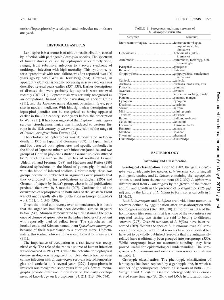
Both L. interrogans and L. biflexa are divided into numerousserovars defined by agglutination after cross-absorption withhomologous antigen (162, 309, 330). If more than 10% of thehomologous titer remains in at least one of the two antisera onrepeated testing, two strains are said to belong to differentserovars (297). Over 60 serovars of L. biflexa have been re-corded (309). Within the species L. interrogans over 200 sero-vars are recognized; additional serovars have been isolated buthave yet to be validly published. Serovars that are antigenicallyrelated have traditionally been grouped into serogroups (330).While serogroups have no taxonomic standing, they haveproved useful for epidemiological understanding. The sero-groups of L. interrogans and some common serovars are shownin Table 1.
Genotypic classification. The phenotypic classification ofleptospires has been replaced by a genotypic one, in which anumber of genomospecies include all serovars of both L. in-terrogans and L. biflexa. Genetic heterogeneity was demon-strated some time ago (80, 260), and DNA hybridization stud-
TABLE 1. Serogroups and some serovars ofL. interrogans sensu lato
Serogroup Serovar(s)
icterohaemorrhagiae, .................................Icterohaemorrhagiae,copenhageni, lai,zimbabwe
Hebdomadis .................................................hebdomadis, jules,kremastos
Autumnalis ...................................................autumnalis, fortbragg, bim,weerasinghe
Pyrogenes......................................................pyrogenesBataviae ........................................................bataviaeGrippotyphosa..............................................grippotyphosa, canalzonae,
ratnapuraCanicola ........................................................canicolaAustralis ........................................................australis, bratislava, loraPomona .........................................................pomonaJavanica.........................................................javanicaSejroe ............................................................sejroe, saxkoebing, hardjoPanama .........................................................panama, mangusCynopteri ......................................................cynopteriDjasiman.......................................................djasimanSarmin ...........................................................sarminMini ...............................................................mini, georgiaTarassovi .......................................................tarassoviBallum...........................................................ballum, aroboreaCelledoni.......................................................celledoniLouisiana ......................................................louisiana, lankaRanarum .......................................................ranarumManhao .........................................................manhaoShermani.......................................................shermaniHurstbridge...................................................hurstbridge
VOL. 14, 2001 LEPTOSPIROSIS 297
on May 6, 2012 by guest
http://cmr.asm
.org/D
ownloaded from
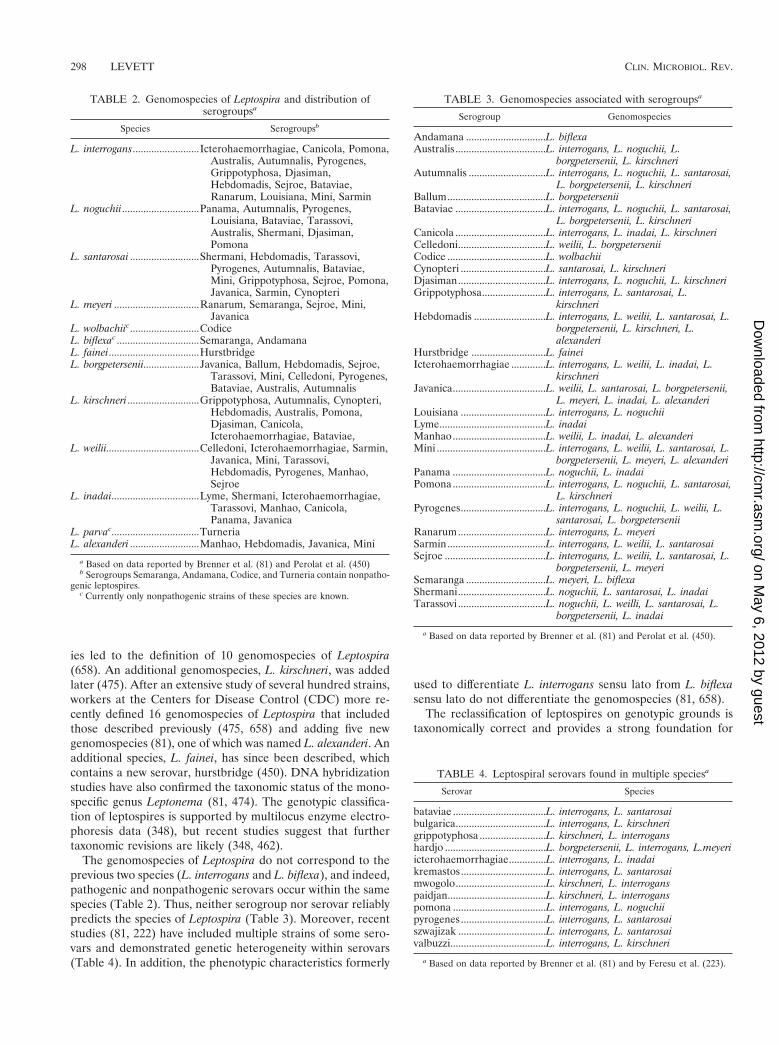
ies led to the definition of 10 genomospecies of Leptospira(658). An additional genomospecies, L. kirschneri, was addedlater (475). After an extensive study of several hundred strains,workers at the Centers for Disease Control (CDC) more re-cently defined 16 genomospecies of Leptospira that includedthose described previously (475, 658) and adding five newgenomospecies (81), one of which was named L. alexanderi. Anadditional species, L. fainei, has since been described, whichcontains a new serovar, hurstbridge (450). DNA hybridizationstudies have also confirmed the taxonomic status of the mono-specific genus Leptonema (81, 474). The genotypic classifica-tion of leptospires is supported by multilocus enzyme electro-phoresis data (348), but recent studies suggest that furthertaxonomic revisions are likely (348, 462).
The genomospecies of Leptospira do not correspond to theprevious two species (L. interrogans and L. biflexa), and indeed,pathogenic and nonpathogenic serovars occur within the samespecies (Table 2). Thus, neither serogroup nor serovar reliablypredicts the species of Leptospira (Table 3). Moreover, recentstudies (81, 222) have included multiple strains of some sero-vars and demonstrated genetic heterogeneity within serovars(Table 4). In addition, the phenotypic characteristics formerly
used to differentiate L. interrogans sensu lato from L. biflexasensu lato do not differentiate the genomospecies (81, 658).
The reclassification of leptospires on genotypic grounds istaxonomically correct and provides a strong foundation for
TABLE 2. Genomospecies of Leptospira and distribution ofserogroupsa
Species Serogroupsb
L. interrogans .........................Icterohaemorrhagiae, Canicola, Pomona,Australis, Autumnalis, Pyrogenes,Grippotyphosa, Djasiman,Hebdomadis, Sejroe, Bataviae,Ranarum, Louisiana, Mini, Sarmin
L. noguchii .............................Panama, Autumnalis, Pyrogenes,Louisiana, Bataviae, Tarassovi,Australis, Shermani, Djasiman,Pomona
L. santarosai ..........................Shermani, Hebdomadis, Tarassovi,Pyrogenes, Autumnalis, Bataviae,Mini, Grippotyphosa, Sejroe, Pomona,Javanica, Sarmin, Cynopteri
L. meyeri ................................Ranarum, Semaranga, Sejroe, Mini,Javanica
L. wolbachiic ..........................CodiceL. biflexac ...............................Semaranga, AndamanaL. fainei ..................................HurstbridgeL. borgpetersenii.....................Javanica, Ballum, Hebdomadis, Sejroe,
Tarassovi, Mini, Celledoni, Pyrogenes,Bataviae, Australis, Autumnalis
L. kirschneri ...........................Grippotyphosa, Autumnalis, Cynopteri,Hebdomadis, Australis, Pomona,Djasiman, Canicola,Icterohaemorrhagiae, Bataviae,
L. weilii...................................Celledoni, Icterohaemorrhagiae, Sarmin,Javanica, Mini, Tarassovi,Hebdomadis, Pyrogenes, Manhao,Sejroe
L. inadai.................................Lyme, Shermani, Icterohaemorrhagiae,Tarassovi, Manhao, Canicola,Panama, Javanica
L. parvac.................................TurneriaL. alexanderi ..........................Manhao, Hebdomadis, Javanica, Mini
a Based on data reported by Brenner et al. (81) and Perolat et al. (450)b Serogroups Semaranga, Andamana, Codice, and Turneria contain nonpatho-
genic leptospires.c Currently only nonpathogenic strains of these species are known.
TABLE 3. Genomospecies associated with serogroupsa
Serogroup Genomospecies
Andamana ..............................L. biflexaAustralis..................................L. interrogans, L. noguchii, L.
borgpetersenii, L. kirschneriAutumnalis .............................L. interrogans, L. noguchii, L. santarosai,
L. borgpetersenii, L. kirschneriBallum.....................................L. borgpeterseniiBataviae ..................................L. interrogans, L. noguchii, L. santarosai,
L. borgpetersenii, L. kirschneriCanicola ..................................L. interrogans, L. inadai, L. kirschneriCelledoni.................................L. weilii, L. borgpeterseniiCodice .....................................L. wolbachiiCynopteri ................................L. santarosai, L. kirschneriDjasiman.................................L. interrogans, L. noguchii, L. kirschneriGrippotyphosa........................L. interrogans, L. santarosai, L.
kirschneriHebdomadis ...........................L. interrogans, L. weilii, L. santarosai, L.
borgpetersenii, L. kirschneri, L.alexanderi
Hurstbridge ............................L. faineiIcterohaemorrhagiae .............L. interrogans, L. weilii, L. inadai, L.
kirschneriJavanica...................................L. weilii, L. santarosai, L. borgpetersenii,
L. meyeri, L. inadai, L. alexanderiLouisiana ................................L. interrogans, L. noguchiiLyme........................................L. inadaiManhao...................................L. weilii, L. inadai, L. alexanderiMini .........................................L. interrogans, L. weilii, L. santarosai, L.
borgpetersenii, L. meyeri, L. alexanderiPanama ...................................L. noguchii, L. inadaiPomona...................................L. interrogans, L. noguchii, L. santarosai,
L. kirschneriPyrogenes................................L. interrogans, L. noguchii, L. weilii, L.
santarosai, L. borgpeterseniiRanarum.................................L. interrogans, L. meyeriSarmin.....................................L. interrogans, L. weilii, L. santarosaiSejroe ......................................L. interrogans, L. weilii, L. santarosai, L.
borgpetersenii, L. meyeriSemaranga ..............................L. meyeri, L. biflexaShermani.................................L. noguchii, L. santarosai, L. inadaiTarassovi .................................L. noguchii, L. weilli, L. santarosai, L.
borgpetersenii, L. inadai
a Based on data reported by Brenner et al. (81) and Perolat et al. (450).
TABLE 4. Leptospiral serovars found in multiple speciesa
Serovar Species
bataviae ...................................L. interrogans, L. santarosaibulgarica..................................L. interrogans, L. kirschnerigrippotyphosa .........................L. kirschneri, L. interroganshardjo ......................................L. borgpetersenii, L. interrogans, L.meyeriicterohaemorrhagiae..............L. interrogans, L. inadaikremastos................................L. interrogans, L. santarosaimwogolo..................................L. kirschneri, L. interroganspaidjan.....................................L. kirschneri, L. interroganspomona ...................................L. interrogans, L. noguchiipyrogenes ................................L. interrogans, L. santarosaiszwajizak .................................L. interrogans, L. santarosaivalbuzzi....................................L. interrogans, L. kirschneri
a Based on data reported by Brenner et al. (81) and by Feresu et al. (223).
298 LEVETT CLIN. MICROBIOL. REV.
on May 6, 2012 by guest
http://cmr.asm
.org/D
ownloaded from
future classifications. However, the molecular classification isproblematic for the clinical microbiologist, because it is clearlyincompatible with the system of serogroups which has servedclinicians and epidemiologists well for many years. Until sim-pler DNA-based identification methods are developed andvalidated, it will be necessary for clinical laboratories to retainthe serological classification of pathogenic leptospires for theforeseeable future. In addition, the retention of L. interrogansand L. biflexa as specific names in the genomic classificationalso allows nomenclatural confusion. In the following pages,specific names refer to the genomospecies, including L. inter-rogans sensu stricto and L. biflexa sensu stricto.
Biology of Leptospires
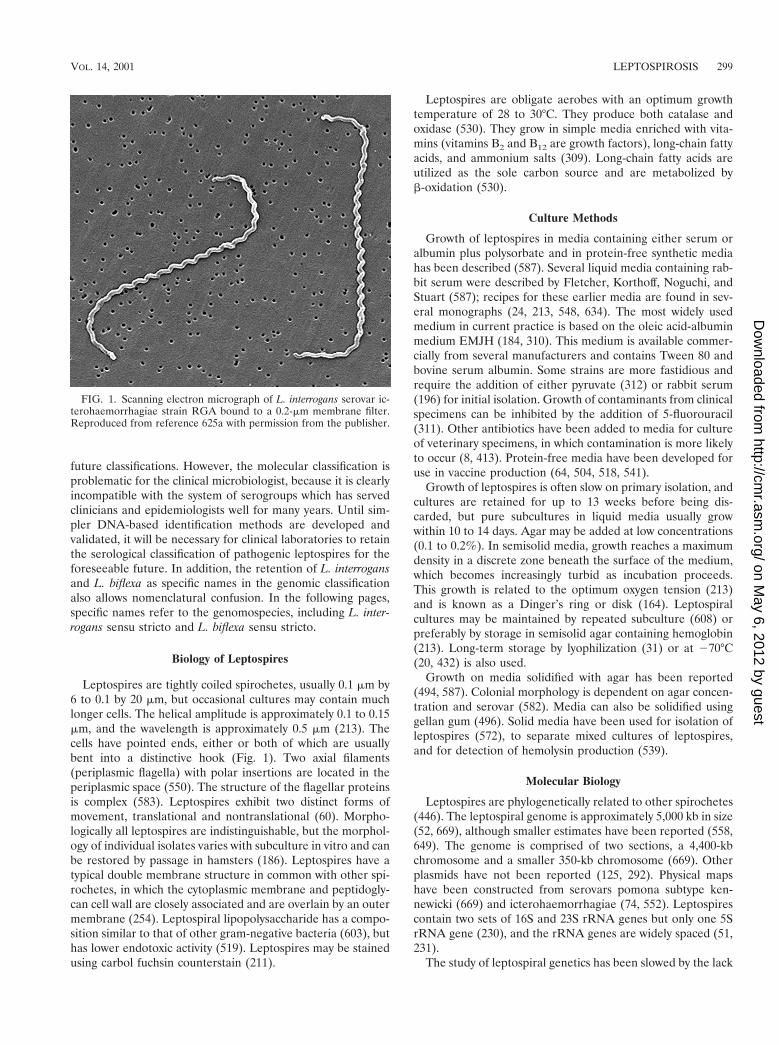
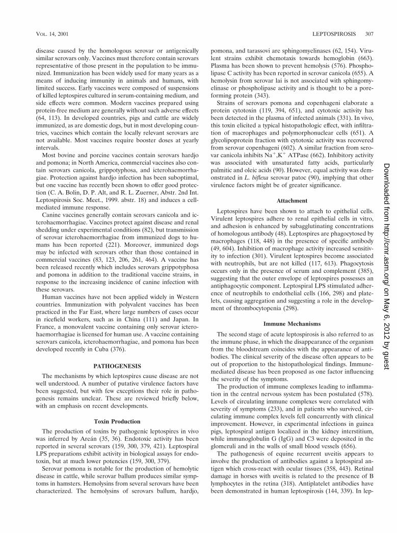
Leptospires are tightly coiled spirochetes, usually 0.1 mm by6 to 0.1 by 20 mm, but occasional cultures may contain muchlonger cells. The helical amplitude is approximately 0.1 to 0.15mm, and the wavelength is approximately 0.5 mm (213). Thecells have pointed ends, either or both of which are usuallybent into a distinctive hook (Fig. 1). Two axial filaments(periplasmic flagella) with polar insertions are located in theperiplasmic space (550). The structure of the flagellar proteinsis complex (583). Leptospires exhibit two distinct forms ofmovement, translational and nontranslational (60). Morpho-logically all leptospires are indistinguishable, but the morphol-ogy of individual isolates varies with subculture in vitro and canbe restored by passage in hamsters (186). Leptospires have atypical double membrane structure in common with other spi-rochetes, in which the cytoplasmic membrane and peptidogly-can cell wall are closely associated and are overlain by an outermembrane (254). Leptospiral lipopolysaccharide has a compo-sition similar to that of other gram-negative bacteria (603), buthas lower endotoxic activity (519). Leptospires may be stainedusing carbol fuchsin counterstain (211).
Leptospires are obligate aerobes with an optimum growthtemperature of 28 to 30°C. They produce both catalase andoxidase (530). They grow in simple media enriched with vita-mins (vitamins B2 and B12 are growth factors), long-chain fattyacids, and ammonium salts (309). Long-chain fatty acids areutilized as the sole carbon source and are metabolized byb-oxidation (530).
Culture Methods
Growth of leptospires in media containing either serum oralbumin plus polysorbate and in protein-free synthetic mediahas been described (587). Several liquid media containing rab-bit serum were described by Fletcher, Korthoff, Noguchi, andStuart (587); recipes for these earlier media are found in sev-eral monographs (24, 213, 548, 634). The most widely usedmedium in current practice is based on the oleic acid-albuminmedium EMJH (184, 310). This medium is available commer-cially from several manufacturers and contains Tween 80 andbovine serum albumin. Some strains are more fastidious andrequire the addition of either pyruvate (312) or rabbit serum(196) for initial isolation. Growth of contaminants from clinicalspecimens can be inhibited by the addition of 5-fluorouracil(311). Other antibiotics have been added to media for cultureof veterinary specimens, in which contamination is more likelyto occur (8, 413). Protein-free media have been developed foruse in vaccine production (64, 504, 518, 541).
Growth of leptospires is often slow on primary isolation, andcultures are retained for up to 13 weeks before being dis-carded, but pure subcultures in liquid media usually growwithin 10 to 14 days. Agar may be added at low concentrations(0.1 to 0.2%). In semisolid media, growth reaches a maximumdensity in a discrete zone beneath the surface of the medium,which becomes increasingly turbid as incubation proceeds.This growth is related to the optimum oxygen tension (213)and is known as a Dinger’s ring or disk (164). Leptospiralcultures may be maintained by repeated subculture (608) orpreferably by storage in semisolid agar containing hemoglobin(213). Long-term storage by lyophilization (31) or at 270°C(20, 432) is also used.
Growth on media solidified with agar has been reported(494, 587). Colonial morphology is dependent on agar concen-tration and serovar (582). Media can also be solidified usinggellan gum (496). Solid media have been used for isolation ofleptospires (572), to separate mixed cultures of leptospires,and for detection of hemolysin production (539).
Molecular Biology
Leptospires are phylogenetically related to other spirochetes(446). The leptospiral genome is approximately 5,000 kb in size(52, 669), although smaller estimates have been reported (558,649). The genome is comprised of two sections, a 4,400-kbchromosome and a smaller 350-kb chromosome (669). Otherplasmids have not been reported (125, 292). Physical mapshave been constructed from serovars pomona subtype ken-newicki (669) and icterohaemorrhagiae (74, 552). Leptospirescontain two sets of 16S and 23S rRNA genes but only one 5SrRNA gene (230), and the rRNA genes are widely spaced (51,231).
The study of leptospiral genetics has been slowed by the lack
FIG. 1. Scanning electron micrograph of L. interrogans serovar ic-terohaemorrhagiae strain RGA bound to a 0.2-mm membrane filter.Reproduced from reference 625a with permission from the publisher.
VOL. 14, 2001 LEPTOSPIROSIS 299
on May 6, 2012 by guest
http://cmr.asm
.org/D
ownloaded from
of a transformation system (317, 677). Recently, a shuttle vec-tor was developed using the temperate bacteriophage LE1from L. biflexa (498). This advance offers the prospect of morerapid progress in the understanding of Leptospira at the mo-lecular level.
Several repetitive elements have been identified (73, 317,553, 641, 673), of which several are insertion sequences (IS)coding for transposases. IS1533 has a single open readingframe (668), while IS1500 has four (73). Both IS1500 andIS1533 are found in many serovars (73, 672), but the copynumber varies widely between different serovars and amongisolates of the same serovar (74). A role for these insertionsequences in transposition and genomic rearrangements hasbeen identified (73, 74, 668, 677). Other evidence for horizon-tal transfer within the genus Leptospira has been reported(468).
A number of leptospiral genes have been cloned and ana-lyzed, including several for amino acid synthesis (163, 486,674), rRNA (228, 229), ribosomal proteins (676), RNA poly-merase (483), DNA repair (540), heat shock proteins (47, 441),sphingomyelinase (508, 509), hemolysins (154, 343), outermembrane proteins (168, 255, 256, 515), flagellar proteins(354, 355, 398, 584, 640), and lipopolysaccharide (LPS) synthe-sis (88, 152, 317, 397).
Within serovar icterohaemorrhagiae, the genome appears tobe conserved (281, 552). This conservation allowed the iden-tification of at least one new serovar by recognition of distinctpulsed-field gel electrophoresis (PFGE) profiles (280). How-ever, the recent demonstration of heterogeneity within sero-vars (81, 222) indicates the need for further study of multipleisolates of individual serovars.
EPIDEMIOLOGY
Leptospirosis is presumed to be the most widespread zoo-nosis in the world (646). The source of infection in humans isusually either direct or indirect contact with the urine of aninfected animal. The incidence is significantly higher in warm-climate countries than in temperate regions (208, 479); this isdue mainly to longer survival of leptospires in the environmentin warm, humid conditions. However, most tropical countriesare also developing countries, and there are greater opportu-nities for exposure of the human population to infected ani-mals, whether livestock, domestic pets, or wild or feral animals.The disease is seasonal, with peak incidence occurring in sum-mer or fall in temperate regions, where temperature is thelimiting factor in survival of leptospires, and during rainy sea-sons in warm-climate regions, where rapid dessication wouldotherwise prevent survival.
The reported incidence of leptospirosis reflects the availabil-ity of laboratory diagnosis and the clinical index of suspicion asmuch as the incidence of the disease. Within the United States,the highest incidence is found in Hawaii (101). Leptospirosisceased to be a notifiable infection within the United Statesafter December 1994 (97).
The usual portal of entry is through abrasions or cuts in theskin or via the conjunctiva; infection may take place via intactskin after prolonged immersion in water, but this usually oc-curs when abrasions are likely to occur and is thus difficult tosubstantiate. Water-borne transmission has been documented;
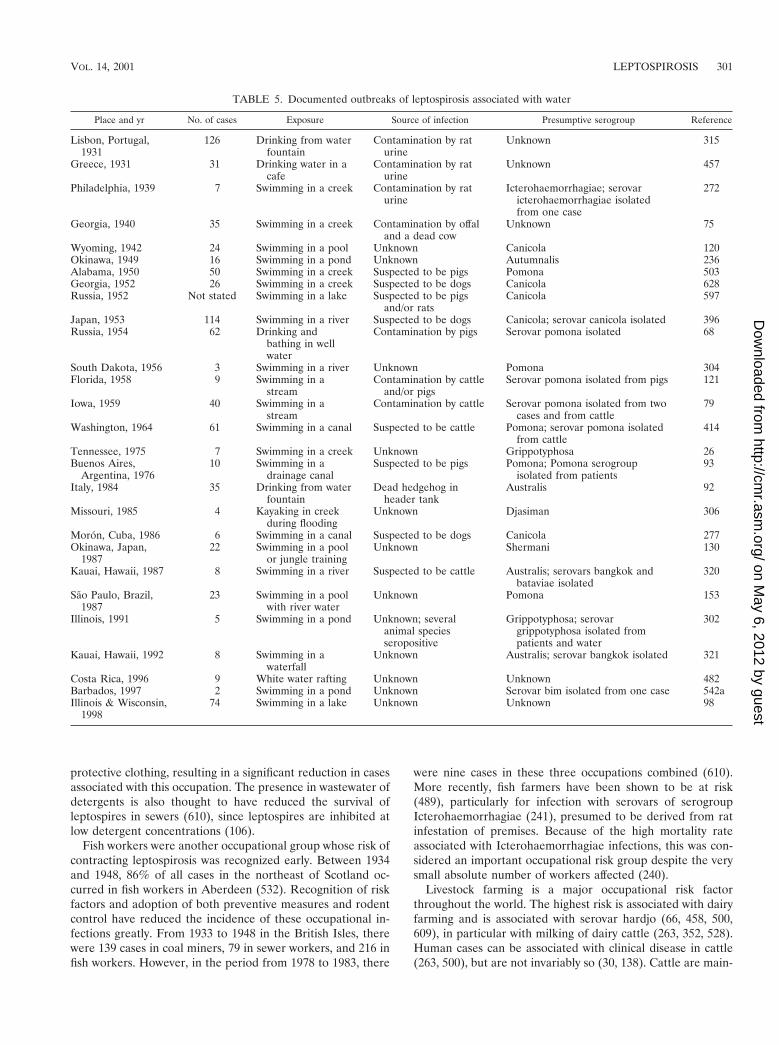
point contamination of water supplies has resulted in severaloutbreaks of leptospirosis (Table 5). Inhalation of water oraerosols also may result in infection via the mucous mem-branes of the respiratory tract. Rarely, infection may followanimal bites (55, 158, 244, 360, 525). Direct transmission be-tween humans has been demonstrated rarely (see Other Com-plications, below). However, excretion of leptospires in humanurine months after recovery has been recorded (46, 307). It isthought that the low pH of human urine limits survival ofleptospires after excretion. Transmission by sexual intercourseduring convalescence has been reported (167, 262).
Animals, including humans, can be divided into mainte-nance hosts and accidental (incidental) hosts. The disease ismaintained in nature by chronic infection of the renal tubulesof maintenance hosts (43). A maintenance host is defined as aspecies in which infection is endemic and is usually transferredfrom animal to animal by direct contact. Infection is usuallyacquired at an early age, and the prevalence of chronic excre-tion in the urine increases with the age of the animal. Otheranimals (such as humans) may become infected by indirectcontact with the maintenance host. Animals may be mainte-nance hosts of some serovars but incidental hosts of others,infection with which may cause severe or fatal disease. Themost important maintenance hosts are small mammals, whichmay transfer infection to domestic farm animals, dogs, andhumans. The extent to which infection is transmitted dependson many factors, including climate, population density, and thedegree of contact between maintenance and accidental hosts.Different rodent species may be reservoirs of distinct serovars,but rats are generally maintenance hosts for serovars of theserogroups lcterohaemorrhagiae and Ballum, and mice are themaintenance hosts for serogroup Ballum. Domestic animalsare also maintenance hosts; dairy cattle may harbor serovarshardjo, pomona, and grippotyphosa; pigs may harbor pomona,tarassovi, or bratislava; sheep may harbor hardjo and pomona;and dogs may harbor canicola (69). Distinct variations in main-tenance hosts and the serovars they carry occur throughout theworld (266). A knowledge of the prevalent serovars and theirmaintenance hosts is essential to understanding the epidemi-ology of the disease in any region.
Human infections may be acquired through occupational,recreational, or avocational exposures. Occupation is a signif-icant risk factor for humans (609). Direct contact with infectedanimals accounts for most infections in farmers, veterinarians,abattoir workers (95, 104, 570), meat inspectors (65), rodentcontrol workers (155), and other occupations which requirecontact with animals (27, 357). Indirect contact is important forsewer workers, miners, soldiers (87, 314, 361), septic tankcleaners, fish farmers (241, 489), gamekeepers, canal workers(29), rice field workers (219, 430, 615), taro farmers (25),banana farmers (535), and sugar cane cutters (132).
Miners were the first occupational risk group to be recog-nized (86, 296). The occurrence of Weil’s disease in sewerworkers was first reported in the 1930s (23, 218, 308, 545).Serovar icterohaemorrhagiae was isolated by guinea pig inoc-ulation from patients, from rats trapped in sewers (23, 308),and from the slime lining the sewers (23). In Glasgow, Scot-land, a seroprevalence among sewer workers of 17% was re-ported (545). The recognition of this important risk activity ledto the adoption of rodent control programs and the use of
300 LEVETT CLIN. MICROBIOL. REV.
on May 6, 2012 by guest
http://cmr.asm
.org/D
ownloaded from
protective clothing, resulting in a significant reduction in casesassociated with this occupation. The presence in wastewater ofdetergents is also thought to have reduced the survival ofleptospires in sewers (610), since leptospires are inhibited atlow detergent concentrations (106).
Fish workers were another occupational group whose risk ofcontracting leptospirosis was recognized early. Between 1934and 1948, 86% of all cases in the northeast of Scotland oc-curred in fish workers in Aberdeen (532). Recognition of riskfactors and adoption of both preventive measures and rodentcontrol have reduced the incidence of these occupational in-fections greatly. From 1933 to 1948 in the British Isles, therewere 139 cases in coal miners, 79 in sewer workers, and 216 infish workers. However, in the period from 1978 to 1983, there
were nine cases in these three occupations combined (610).More recently, fish farmers have been shown to be at risk(489), particularly for infection with serovars of serogroupIcterohaemorrhagiae (241), presumed to be derived from ratinfestation of premises. Because of the high mortality rateassociated with Icterohaemorrhagiae infections, this was con-sidered an important occupational risk group despite the verysmall absolute number of workers affected (240).
Livestock farming is a major occupational risk factorthroughout the world. The highest risk is associated with dairyfarming and is associated with serovar hardjo (66, 458, 500,609), in particular with milking of dairy cattle (263, 352, 528).Human cases can be associated with clinical disease in cattle(263, 500), but are not invariably so (30, 138). Cattle are main-
TABLE 5. Documented outbreaks of leptospirosis associated with water
Place and yr No. of cases Exposure Source of infection Presumptive serogroup Reference
Lisbon, Portugal,1931
126 Drinking from waterfountain
Contamination by raturine
Unknown 315
Greece, 1931 31 Drinking water in acafe
Contamination by raturine
Unknown 457
Philadelphia, 1939 7 Swimming in a creek Contamination by raturine
Icterohaemorrhagiae; serovaricterohaemorrhagiae isolatedfrom one case
272
Georgia, 1940 35 Swimming in a creek Contamination by offaland a dead cow
Unknown 75
Wyoming, 1942 24 Swimming in a pool Unknown Canicola 120Okinawa, 1949 16 Swimming in a pond Unknown Autumnalis 236Alabama, 1950 50 Swimming in a creek Suspected to be pigs Pomona 503Georgia, 1952 26 Swimming in a creek Suspected to be dogs Canicola 628Russia, 1952 Not stated Swimming in a lake Suspected to be pigs
and/or ratsCanicola 597
Japan, 1953 114 Swimming in a river Suspected to be dogs Canicola; serovar canicola isolated 396Russia, 1954 62 Drinking and
bathing in wellwater
Contamination by pigs Serovar pomona isolated 68
South Dakota, 1956 3 Swimming in a river Unknown Pomona 304Florida, 1958 9 Swimming in a
streamContamination by cattle
and/or pigsSerovar pomona isolated from pigs 121
Iowa, 1959 40 Swimming in astream
Contamination by cattle Serovar pomona isolated from twocases and from cattle
79
Washington, 1964 61 Swimming in a canal Suspected to be cattle Pomona; serovar pomona isolatedfrom cattle
414
Tennessee, 1975 7 Swimming in a creek Unknown Grippotyphosa 26Buenos Aires,
Argentina, 197610 Swimming in a
drainage canalSuspected to be pigs Pomona; Pomona serogroup
isolated from patients93
Italy, 1984 35 Drinking from waterfountain
Dead hedgehog inheader tank
Australis 92
Missouri, 1985 4 Kayaking in creekduring flooding
Unknown Djasiman 306
Moron, Cuba, 1986 6 Swimming in a canal Suspected to be dogs Canicola 277Okinawa, Japan,
198722 Swimming in a pool
or jungle trainingUnknown Shermani 130
Kauai, Hawaii, 1987 8 Swimming in a river Suspected to be cattle Australis; serovars bangkok andbataviae isolated
320
Sao Paulo, Brazil,1987
23 Swimming in a poolwith river water
Unknown Pomona 153
Illinois, 1991 5 Swimming in a pond Unknown; severalanimal speciesseropositive
Grippotyphosa; serovargrippotyphosa isolated frompatients and water
302
Kauai, Hawaii, 1992 8 Swimming in awaterfall
Unknown Australis; serovar bangkok isolated 321
Costa Rica, 1996 9 White water rafting Unknown Unknown 482Barbados, 1997 2 Swimming in a pond Unknown Serovar bim isolated from one case 542aIllinois & Wisconsin,
199874 Swimming in a lake Unknown Unknown 98
VOL. 14, 2001 LEPTOSPIROSIS 301
on May 6, 2012 by guest
http://cmr.asm
.org/D
ownloaded from
tenance hosts of serovar hardjo (192), and infection with thisserovar occurs throughout the world (45, 412, 466). Manyanimals are seronegative carriers (192, 267, 571). After infec-tion, leptospires localize in the kidneys (249, 427, 465, 571,626) and are excreted intermittently in the urine (189). Serovarhardjo causes outbreaks of mastitis (196) and abortion (190).Serovar hardjo is found in aborted fetuses and in prematurecalves (188, 194, 238, 268). In addition, hardjo has been iso-lated from normal fetuses (191), the genital tracts of pregnantcattle (191), vaginal discharge after calving (193), and the gen-ital tract and urinary tract of .50% of cows (197, 198) andbulls (185). In Australia, both serovars hardjo and pomonawere demonstrated in bovine abortions, but serological evi-dence suggested that the incidence of hardjo infection wasmuch higher (182, 305, 529). In Scotland, 42% of cattle wereseropositive for hardjo, representing 85% of all seropositiveanimals (187). In the United States, serovar hardjo is the mostcommonly isolated serovar in cattle (198), but pomona alsooccurs.
There is a significant risk associated with recreational expo-sures occurring in water sports (405), including swimming,canoeing (306, 517), white water rafting (482, 591, 627), freshwater fishing, and other sports where exposure is common,such as potholing and caving (611). The potential for exposureof large numbers of individuals occurs during competitiveevents (98, 99, 102, 126, 204). Several outbreaks of leptospiro-sis associated with water have been reported (Table 5). Manyof these outbreaks have followed extended periods of hot, dryweather, when pathogenic leptospires presumably have multi-plied in freshwater ponds or rivers. Cases of leptospirosis alsofollow extensive flooding (111, 153, 201, 226, 232, 425, 436, 442,526, 590, 645).
Pathogenic serovars have been isolated from water in trop-ical regions (19) and in the United States, where serovarsicterohaemorrhagiae, dakota, ballum, pomona, and grippoty-phosa have been recovered (137, 161, 242). Survival of patho-genic leptospires in the environment is dependent on severalfactors, including pH, temperature, and the presence of inhib-itory compounds. Most studies have used single serovars andquite different methodologies, but some broad conclusionsmay be drawn. Under laboratory conditions, leptospires inwater at room temperature remain viable for several months atpH 7.2 to 8.0 (106, 246), but in river water survival is shorterand is prolonged at lower temperatures (106, 137). The pres-ence of domestic sewage decreases the survival time to a mat-ter of hours (106), but in an oxidation ditch filled with cattleslurry, viable leptospires were detected for several weeks (160).In acidic soil (pH 6.2) taken from canefields in Australia,
serovar australis survived for up to 7 weeks, and in rainwater-flooded soil it survived for at least 3 weeks (531). When soilwas contaminated with urine from infected rats or voles, lep-tospires survived for approximately 2 weeks (319, 531). Inslightly different soil, serovar pomona survived for up to 7weeks under conditions approximating the New Zealand win-ter (274).
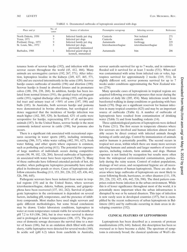
Many sporadic cases of leptospirosis in tropical regions areacquired following avocational exposures that occur during theactivities of daily life (205, 454). Many infections result frombarefooted walking in damp conditions or gardening with barehands (170). Dogs are a significant reservoir for human infec-tion in many tropical countries (623) and may be an importantsource of outbreaks (Table 6). A number of outbreaks ofleptospirosis have resulted from contamination of drinkingwater (Table 5) and from handling rodents (14).
Three epidemiological patterns of leptospirosis were definedby Faine (211). The first occurs in temperate climates wherefew serovars are involved and human infection almost invari-ably occurs by direct contact with infected animals thoughfarming of cattle and pigs. Control by immunization of animalsand/or humans is potentially possible. The second occurs intropical wet areas, within which there are many more serovarsinfecting humans and animals and larger numbers of reservoirspecies, including rodents, farm animals, and dogs. Humanexposure is not limited by occupation but results more oftenfrom the widespread environmental contamination, particu-larly during the rainy season. Control of rodent populations,drainage of wet areas, and occupational hygiene are all neces-sary for prevention of human leptospirosis. These are also theareas where large outbreaks of leptospirosis are most likely tooccur following floods, hurricanes, or other disasters (111, 158,201, 226, 232, 425, 436, 442, 526, 590). The third pattern com-prises rodent-borne infection in the urban environment. Whilethis is of lesser significance throughout most of the world, it ispotentially more important when the urban infrastructure isdisrupted by war or by natural disasters. This type of infectionis now rarely seen in developed countries (157), but is exem-plified by the recent rediscovery of urban leptospirosis in Bal-timore (601) and by outbreaks occurring in slum areas in de-veloping countries (332).
CLINICAL FEATURES OF LEPTOSPIROSIS
Leptospirosis has been described as a zoonosis of proteanmanifestations (456, 644). Indeed, this description has been sooverused as to have become a cliche. The spectrum of symp-toms is extremely broad; the classical syndrome of Weil’s dis-
TABLE 6. Documented outbreaks of leptospirosis associated with dogs
Place and yr No. ofcases Source of infection Presumptive serogroup Infecting serovar Reference
North Dakota, 1950 9 Infected family pet dog Canicola Not isolated 271Texas, 1971 7 Infected pet dogs Canicola canicola 54Portland, Oreg., 1972 9 Infected family pet dog Autumnalis fortbragg 225St. Louis, Mo., 1972 5 Infected pet dogs,
previously immunizedIcterohaemorrhagiae icterohaemorrhagiae 221
Barbados, 1988 1 Infected guard dogs inkennels, immunized
Autumnalis bim 206
302 LEVETT CLIN. MICROBIOL. REV.
on May 6, 2012 by guest
http://cmr.asm
.org/D
ownloaded from
ease represents only the most severe presentation. Formerly itwas considered that distinct clinical syndromes were associatedwith specific serogroups (596). However, this view was ques-tioned by some authorities (18, 180, 220), and more intensestudy over the past 30 years has refuted this hypothesis. Anexplanation for many of the observed associations may befound in the ecology of the maintenance animal hosts in ageographic region. A region with a richly varied fauna willsupport a greater variety of serogroups than will a region withfew animal hosts. In humans, severe leptospirosis is frequentlybut not invariably caused by serovars of the icterohaemorrha-giae serogroup. The specific serovars involved depend largelyon the geographic location and the ecology of local mainte-nance hosts. Thus in Europe, serovars copenhageni and ictero-haemorrhagiae, carried by rats, are usually responsible forinfectious, while in Southeast Asia, serovar lai is common.
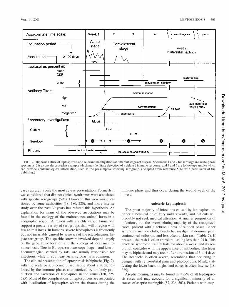
The clinical presentation of leptospirosis is biphasic (Fig. 2),with the acute or septicemic phase lasting about a week, fol-lowed by the immune phase, characterized by antibody pro-duction and excretion of leptospires in the urine (180, 325,585). Most of the complications of leptospirosis are associatedwith localization of leptospires within the tissues during the
immune phase and thus occur during the second week of theillness.
Anicteric Leptospirosis
The great majority of infections caused by leptospires areeither subclinical or of very mild severity, and patients willprobably not seek medical attention. A smaller proportion ofinfections, but the overwhelming majority of the recognizedcases, present with a febrile illness of sudden onset. Othersymptoms include chills, headache, myalgia, abdominal pain,conjunctival suffusion, and less often a skin rash (Table 7). Ifpresent, the rash is often transient, lasting less than 24 h. Thisanicteric syndrome usually lasts for about a week, and its res-olution coincides with the appearance of antibodies. The fevermay be biphasic and may recur after a remission of 3 to 4 days.The headache is often severe, resembling that occurring indengue, with retro-orbital pain and photophobia. Myalgia af-fecting the lower back, thighs, and calves is often intense (18,325).
Aseptic meningitis may be found in #25% of all leptospiro-sis cases and may account for a significant minority of allcauses of aseptic meningitis (57, 236, 503). Patients with asep-
FIG. 2. Biphasic nature of leptospirosis and relevant investigations at different stages of disease. Specimens 1 and 2 for serology are acute-phasespecimens, 3 is a convalescent-phase sample which may facilitate detection of a delayed immune response, and 4 and 5 are follow-up samples whichcan provide epidemiological information, such as the presumptive infecting serogroup. (Adapted from reference 586a with permission of thepublisher.)
VOL. 14, 2001 LEPTOSPIROSIS 303
on May 6, 2012 by guest
http://cmr.asm
.org/D
ownloaded from
tic meningitis have tended to be younger than those with ictericleptospirosis (57, 328, 522). In their series of 616 cases, Alstonand Broom (24) noted that 62% of children #14 years oldpresented with aseptic meningitis, whereas only 31% of pa-tients aged 15 to 29 years did so and only 10% of those over 30years of age. Mortality is almost nil in anicteric leptospirosis(180), but death resulting from massive pulmonary hemor-rhage occurred in 2.4% of the anicteric patients in a Chineseoutbreak (615).
The differential diagnosis must include common viral infec-tions, such as influenza (18), human immunodeficiency virusseroconversion (290), and, in the tropics, dengue (332, 350,501), in addition to the bacterial causes of fever of unknownorigin, such as typhoid. Turner (585) provided a comprehen-sive list of other conditions that may be mimicked by leptospi-rosis, including encephalitis, poliomyelitis, rickettsiosis, glan-dular fever (infectious mononucleosis), brucellosis, malaria,viral hepatitis, and pneumonitis. Hantavirus infections mustalso be considered in the differential diagnosis for patients withpulmonary involvement (32). Petechial or purpuric lesions mayoccur (18, 115), and recently, cases of leptospirosis resemblingviral hemorrhagic fevers have been reported in travelers re-turning from Africa (278, 402).
Icteric Leptospirosis
Icteric leptospirosis is a much more severe disease in whichthe clinical course is often very rapidly progressive. Severecases often present late in the course of the disease, and thiscontributes to the high mortality rate, which ranges between 5and 15%. Between 5 and 10% of all patients with leptospirosishave the icteric form of the disease (273). The jaundice occur-ring in leptospirosis is not associated with hepatocellular ne-crosis, and liver function returns to normal after recovery(476). Serum bilirubin levels may be high, and many weeks maybe required for normalization (177). There are moderate risesin transaminase levels, and minor elevation of the alkalinephosphatase level usually occurs.
The complications of severe leptospirosis emphasize themultisystemic nature of the disease. Leptospirosis is a commoncause of acute renal failure (ARF), which occurs in 16 to 40%of cases (2, 177, 473, 631). A distinction may be made betweenpatients with prerenal azotemia (non-ARF) and those withARF. Patients with prerenal azotaemia may respond to rehy-dration, and decisions regarding dialysis can be delayed for upto 72 h (417). In patients with ARF, oliguria (odds ratio [OR],9.98) was a significant predictor of death (142).
Serum amylase levels are often raised significantly in asso-ciation with ARF (18, 175, 422), but clinical symptoms ofpancreatitis are not a common finding (174, 401, 439). Necro-tizing pancreatitis has been detected at autopsy (175, 544).Thrombocytopenia (platelet count of ,100 3 109/liter) occursin $50% of cases and is a significant predictor for the devel-opment of ARF (176). However, thrombocytopenia in lepto-spirosis is transient and does not result from disseminatedintravascular coagulation (179, 419).
The occurrence of pulmonary symptoms in cases of lepto-spirosis was first noted by Silverstein (525). Subsequent reportshave shown that pulmonary involvement may be the majormanifestation of leptospirosis in some clusters of cases (294,510, 614, 664) and in some sporadic cases (63, 461). The se-verity of respiratory disease is unrelated to the presence ofjaundice (283, 294). Patients may present with a spectrum ofsymptoms, ranging from cough, dyspnea, and hemoptysis(which may be mild or severe) to adult respiratory distresssyndrome (15, 22, 59, 89, 110, 151, 165, 200, 399, 426, 472, 527,664, 666). Intra-alveolar hemorrhage was detected in the ma-jority of patients, even in the absence of overt pulmonarysymptoms (171). Pulmonary hemorrhage may be severeenough to cause death (294, 581, 659, 664).
The incidence of respiratory involvement varies. In a Chi-nese series of anicteric cases, more than half had respiratorysymptoms, while 67% had radiographic changes (614); in asimilar Korean series, 67% of patients had respiratory symp-toms and 64% had radiographic abnormalities (294), whereas
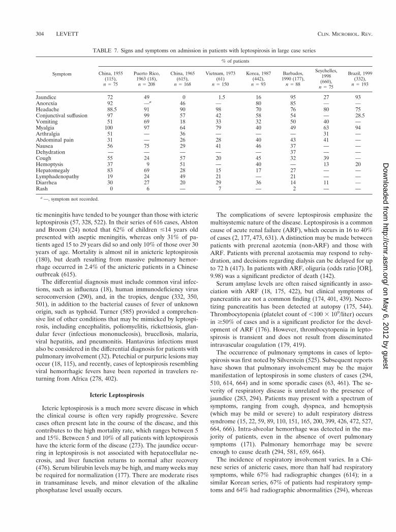
TABLE 7. Signs and symptoms on admission in patients with leptospirosis in large case series
Symptom
% of patients
China, 1955(115),
n 5 75
Puerto Rico,1963 (18),n 5 208
China, 1965(615),
n 5 168
Vietnam, 1973(61)
n 5 150
Korea, 1987(442),
n 5 93
Barbados,1990 (177),
n 5 88
Seychelles,1998
(660),n 5 75
Brazil, 1999(332),
n 5 193
Jaundice 72 49 0 1.5 16 95 27 93Anorexia 92 —a 46 — 80 85 — —Headache 88.5 91 90 98 70 76 80 75Conjunctival suffusion 97 99 57 42 58 54 — 28.5Vomiting 51 69 18 33 32 50 40 —Myalgia 100 97 64 79 40 49 63 94Arthralgia 51 — 36 — — — 31 —Abdominal pain 31 — 26 28 40 43 41 —Nausea 56 75 29 41 46 37 — —Dehydration — — — — — 37 — —Cough 55 24 57 20 45 32 39 —Hemoptysis 37 9 51 — 40 — 13 20Hepatomegaly 83 69 28 15 17 27 — —Lymphadenopathy 19 24 49 21 — 21 — —Diarrhea 30 27 20 29 36 14 11 —Rash 0 6 — 7 — 2 — —
a —, symptom not recorded.
304 LEVETT CLIN. MICROBIOL. REV.
on May 6, 2012 by guest
http://cmr.asm
.org/D
ownloaded from
in a series of jaundiced patients in Brazil, only 17% had clinicalevidence of pulmonary involvement, but 33% had radiographicabnormalities (415). In a large Chinese series, moist rales werenoted in 17% of cases (115). Rales are more common in ictericthan in nonicteric leptospirosis (18). Concurrent hemoptysisand pulmonary infiltrates on chest radiographs were noted in12% of 69 nonfatal cases in the Seychelles (659). Cigarettesmoking was reported as a risk factor for the development ofpulmonary symptoms (375).
Radiography generally reveals diffuse small opacities whichmay be widely disseminated or which may coalesce into largerareas of consolidation, with increasing severity of symptoms(342, 415, 525, 614, 659, 664). Pleural effusions may occur (342,560). The patchy infiltrates which are commonly seen reflectareas of intra-alveolar and interstitial hemorrhage (294, 419,472, 614, 664). Both alveolar infiltrates (OR 7.3) and dyspnea(OR 11.7) are poor prognostic indicators in severe leptospiro-sis (172). Similarly, in icteric leptospirosis in Brazil, respiratoryinsufficiency (OR 4.6) was associated with death (332).
Cardiac involvement in leptospirosis is common but may beunderestimated. Fatal myocarditis was first described in 1935(400). Clinical evidence of myocardial involvement, includingabnormal T waves, was detected in 10% of 80 severe ictericcases in Louisiana (536), while similar electrocardiographic(ECG) abnormalities were detected in over 40% of patients inChina, India, Sri Lanka, and the Philippines (353, 467, 471,618), including both icteric and nonicteric cases. However, in aprospective study in Malaysia, identical ECG changes werefound in patients with either leptospirosis or malaria (445), andit was concluded that such ECG changes were nonspecific.Other ECG abnormalities have been reported less frequently(470). The presence of myocarditis was strongly associatedwith the severity of pulmonary symptoms in anicteric Chinesepatients (353). A mortality rate of 54% was reported in severeleptospirosis cases with myocarditis (341). Repolarization ab-normalities on ECG were considered a poor prognostic indi-cator (OR 5.9) in severe leptospirosis cases (172), as werearrhythmias (OR 2.83) in a Brazilian series (332).
Ocular Involvement
Ocular manifestations of severe leptospirosis were noted inearly reports (622, 624). Conjunctival suffusion is seen in themajority of patients in some series (377). Conjunctival suffu-sion in the presence of scleral icterus is said to be pathogno-monic of Weil’s disease (596). Anterior uveitis, either unilat-eral or bilateral, occurs after recovery from the acute illness ina minority of cases (53). Uveitis may present weeks, months, oroccasionally years after the acute stage. Chronic visual distur-bance, persisting 20 years or more after the acute illness, hasbeen reported (521).
The incidence of ocular complications is variable, but thisprobably reflects the long time scale over which they mayoccur. In the United States the incidence was estimated at 3%(273), while in Romania an incidence of 2% was estimatedbetween 1979 and 1985 (28). However, in abattoir workers withevidence of recent leptospirosis, the latter authors reported anincidence of 40% (28).
In most cases uveitis is presumed to be an immune phenom-enon, but leptospires have been isolated from human and
equine eyes (16, 209), and more recently, leptospiral DNA hasbeen demonstrated in aqueous humor by PCR (114, 209, 389).Late-onset uveitis may result from an autoimmune reaction tosubsequent exposure (211).
Recently, a large cluster of cases of uveitis was reportedfrom Madurai in southern India following an outbreak of lep-tospirosis which occurred after heavy flooding (114, 477, 478).The majority of affected patients were males, with a mean ageof 35 years (477). Eyes were involved bilaterally in 38 patients(52%), and panuveitis was present in 96% of eyes. Other sig-nificant ocular findings included anterior chamber cells, vitre-ous opacities, and vasculitis in the absence of visual deficit(114).
Other Complications
Acute infection in pregnancy has been reported to causeabortion (116) and fetal death (122, 214), but not invariably so.In one of the cases reported by Chung et al. (116), leptospireswere isolated from amniotic fluid, placenta, and cord blood;the infant was mildly ill and was discharged at 2 weeks of age.In another case, a neonate developed jaundice and died 2 daysafter birth (356). Leptospires were demonstrated in the liverand kidneys by silver staining, but serological evidence of lep-tospiral infection in the mother was only obtained 2 weeksafter delivery. Leptospires have been isolated from humanbreast milk (116), and in one case serovar hardjo was probablytransmitted from an infected mother to her infant by breast-feeding (70).
Rare complications include cerebrovascular accidents (224,346), rhabdomyolysis (133, 374, 537), thrombotic thrombocy-topenic purpura (336), acute acalculous cholecystitis (44, 401,600), erythema nodosum (157), aortic stenosis (91), Kawasakisyndrome (291, 636), reactive arthritis (633), epididymitis(285), nerve palsy (516, 578), male hypogonadism (437), andGuillain-Barre syndrome (403). Cerebral arteritis, resemblingMoyamoya disease, has been reported in a series of patientsfrom China (650).
Chronic or Latent Infection
Anecdotal reports suggest that leptospirosis may inducechronic symptoms analogous to those produced by other spi-rochetal infections, such as Lyme disease. However, there isvery little objective evidence to support or disprove this hy-pothesis. The possibility of chronic human infection was sug-gested, without evidence of infection other than serology (420).A single case of late-onset meningitis following icteric lepto-spirosis has been described (406), in which leptospires wereisolated from both cerebrospinal fluid (CSF) and urine. Thispatient exhibited a negligible antibody response to the infect-ing strain, suggesting the presence of an immune defect.
Of the sequelae of acute leptospirosis described above, uve-itis is a potentially chronic condition and is a recognizedchronic sequel of leptospirosis in humans and horses. Equinerecurrent uveitis appears to be an autoimmune disease (358,443), and Faine (211) suggested that late-onset uveitis in hu-mans may result from an autoimmune reaction to subsequentexposure. Immune involvement in retinal pathology has beendemonstrated in horses with spontaneous uveitis (318). Lep-tospires have been isolated from the human eye (16), and more
VOL. 14, 2001 LEPTOSPIROSIS 305
on May 6, 2012 by guest
http://cmr.asm
.org/D
ownloaded from
recently, leptospiral DNA has been amplified from aqueoushumor (114, 367, 389) of patients with uveitis. In these cases,uveitis has occurred relatively soon after the acute illness.
One follow-up study of 11 patients with a mean time of 22years (range, 6 to 34 years) after recovery from acute leptospi-rosis has been reported (521). Four patients complained ofpersistent headaches since their acute illness. Two patientscomplained of visual disturbances; both had evidence of pastbilateral anterior uveitis. No biochemical or hematologic ab-normalities were detected to suggest continuing liver or renalimpairment. No studies to date have attempted to confirm thepersistence of leptospires in the tissues of patients who havesubsequently died of other causes.
Pathology
Leptospirosis is characterized by the development of vasculitis,endothelial damage, and inflammatory infiltrates composed ofmoncytic cells, plasma cells, histiocytes, and neutrophils. On grossexamination, petechial hemorrhages are common and may beextensive (35), and organs are often discolored due to thedegree of icterus (459). The histopathology is most marked inthe liver, kidneys, heart, and lungs (665), but other organs mayalso be affected according to the severity of the individualinfection. The overall structure of the liver is not significantlydisrupted, but there may be intrahepatic cholestasis (35, 169).Hypertrophy and hyperplasia of Kupffer cells is evident (148),and erythrophagocytosis has been reported (35, 169). In thekidneys, interstitial nephritis is the major finding, accompaniedby an intense cellular infiltration composed of neutrophils andmoncytes (447). Leptospires can be seen within the renal tu-bules (35, 447, 665). By electron microscopy, the tubular cellbrush borders are denuded, the tubular basement membrane isthickened, and tubular cells exhibit mitochondrial depletion(147). In addition, minor changes are seen in the glomeruli,suggesting an anatomical basis for proteinuria in leptospirosis(147).
Pathological findings in the heart include interstitial myo-carditis with infiltration of predominantly lymphocytes andplasma cells, petechial hemorrhages (particularly in the epicar-dium), mononuclear infiltration in the epicardium, pericardialeffusions, and coronary arteritis (34, 146, 149, 202, 341, 472). Inthe lungs, pulmonary congestion and hemorrhage are common(35, 664), and infiltration of alveolar spaces by monocytes andneutrophils occurs (472). Hyaline membrane formation mayoccur (472, 666). Leptospires may be seen within endothelialcells in interalveolar septa, and attached to capillary endothe-lial cells (419).
In skeletal muscles, particularly of the leg, focal necrosis ofisolated muscle fibers occurs, with infiltration of histiocytes,neutrophils, and plasma cells (169, 589). This evidence of my-ositis correlates with the intense myalgia reported by somepatients (325). In brain, perivascular cuffing is observed (35,665).
Treatment
Treatment of leptospirosis differs depending on the severityand duration of symptoms at the time of presentation. Patientswith mild, flu-like symptoms require only symptomatic treat-ment but should cautioned to seek further medical help if they
develop jaundice. Patients who present with more severe an-icteric leptospirosis will require hospital admission and closeobservation. If the headache is particularly severe, a lumbarpuncture usually produces a dramatic improvement.
The management of icteric leptospirosis requires admissionof the patient to the intensive care unit initially. Patients withprerenal azotemia can be rehydrated initially while their renalfunction is observed, but patients in acute renal failure requiredialysis as a matter of urgency. This is accomplished satisfac-torily by peritoneal dialysis (250, 408, 556). Cardiac monitoringis also desirable during the first few days after admission (172).
Specific antibiotic treatment was reported soon after peni-cillin became available, with mixed results (42). Oxytetracy-cline was also used (497). Early experience was summarized byAlston and Broom in their monograph (24). Few well-designedand well-controlled studies of antibiotic treatment have beenreported (252). A major difficulty in assessing the efficacy ofantibiotic treatment results from the late presentation of manypatients with severe disease, after the leptospires have local-ized in the tissues.
Doxycycline (100 mg twice a day for 7 days) was shown toreduce the duration and severity of illness in anicteric lepto-spirosis by an average of 2 days (382). Patients with severedisease were excluded from this study. Two randomized stud-ies of penicillin produced conflicting results. One study in-cluded 42 patients with severe leptospirosis, of whom 19 werejaundiced (619); no patient required dialysis and there were nodeaths. Intravenous penicillin was given at a dosage of 6 MU/day for 7 days and found to halve the duration of fever. Asecond study included 79 patients with icteric leptospirosis, ofwhom 4 died (178). Patients in the treatment group receivedintravenous penicillin at a dose of 8 MU/day for 5 days. Nodifference was observed between treatment and control groupsin outcome or duration of the illness. There have been nocontrolled trials of penicillin versus doxycycline for treatmentof leptospirosis.
A consistent finding of these studies has been the preventionof leptospiruria or a significant reduction in its duration (178,382, 619). This finding alone is sufficient justification for anti-biotic use, but any antibiotic treatment should be started asearly as possible and should not replace other therapeuticmeasures. Jarisch-Herxheimer reactions have been reportedafter penicillin administration (200, 227, 598, 615). However,the apparently low risk should not preclude the use of penicil-lin (620).
Doxycycline (200 mg orally, once weekly) has been shown tobe effective for short-term prophylaxis in high-risk environ-ments (245, 511, 551). Similar findings have been reported inrhesus monkeys challenged experimentally (199). In a recentcontrolled trial, doxycycline significantly reduced the incidenceof clinical disease but not serological evidence of infection(511). Anecdotal evidence suggests that doxycycline but notpenicillin may be used successfully after exposure in laboratoryaccidents (239). An evidence-based review of antibiotic pro-phylaxis has been published (251).
Immunization
Immunity to leptospirosis is largely humoral (7) and is rel-atively serovar specific. Thus, immunization protects against
306 LEVETT CLIN. MICROBIOL. REV.
on May 6, 2012 by guest
http://cmr.asm
.org/D
ownloaded from
disease caused by the homologous serovar or antigenicallysimilar serovars only. Vaccines must therefore contain serovarsrepresentative of those present in the population to be immu-nized. Immunization has been widely used for many years as ameans of inducing immunity in animals and humans, withlimited success. Early vaccines were composed of suspensionsof killed leptospires cultured in serum-containing medium, andside effects were common. Modern vaccines prepared usingprotein-free medium are generally without such adverse effects(64, 113). In developed countries, pigs and cattle are widelyimmunized, as are domestic dogs, but in most developing coun-tries, vaccines which contain the locally relevant serovars arenot available. Most vaccines require booster doses at yearlyintervals.
Most bovine and porcine vaccines contain serovars hardjoand pomona; in North America, commercial vaccines also con-tain serovars canicola, grippotyphosa, and icterohaemorrha-giae. Protection against hardjo infection has been suboptimal,but one vaccine has recently been shown to offer good protec-tion (C. A. Bolin, D. P. Alt, and R. L. Zuerner, Abstr. 2nd Int.Leptospirosis Soc. Meet., 1999. abstr. 18) and induces a cell-mediated immune response.
Canine vaccines generally contain serovars canicola and ic-terohaemorrhagiae. Vaccines protect against disease and renalshedding under experimental conditions (82), but transmissionof serovar icterohaemorrhagiae from immunized dogs to hu-mans has been reported (221). Moreover, immunized dogsmay be infected with serovars other than those contained incommercial vaccines (83, 123, 206, 261, 464). A vaccine hasbeen released recently which includes serovars grippotyphosaand pomona in addition to the traditional vaccine strains, inresponse to the increasing incidence of canine infection withthese serovars.
Human vaccines have not been applied widely in Westerncountries. Immunization with polyvalent vaccines has beenpracticed in the Far East, where large numbers of cases occurin ricefield workers, such as in China (111) and Japan. InFrance, a monovalent vaccine containing only serovar ictero-haemorrhagiae is licensed for human use. A vaccine containingserovars canicola, icterohaemorrhagiae, and pomona has beendeveloped recently in Cuba (376).
PATHOGENESIS
The mechanisms by which leptospires cause disease are notwell understood. A number of putative virulence factors havebeen suggested, but with few exceptions their role in patho-genesis remains unclear. These are reviewed briefly below,with an emphasis on recent developments.
Toxin Production
The production of toxins by pathogenic leptospires in vivowas inferred by Arean (35, 36). Endotoxic activity has beenreported in several serovars (159, 300, 379, 421). LeptospiralLPS preparations exhibit activity in biological assays for endo-toxin, but at much lower potencies (159, 300, 379).
Serovar pomona is notable for the production of hemolyticdisease in cattle, while serovar ballum produces similar symp-toms in hamsters. Hemolysins from several serovars have beencharacterized. The hemolysins of serovars ballum, hardjo,
pomona, and tarassovi are sphingomyelinases (62, 154). Viru-lent strains exhibit chemotaxis towards hemoglobin (663).Plasma has been shown to prevent hemolysis (576). Phospho-lipase C activity has been reported in serovar canicola (655). Ahemolysin from serovar lai is not associated with sphingomy-elinase or phospholipase activity and is thought to be a pore-forming protein (343).
Strains of serovars pomona and copenhageni elaborate aprotein cytotoxin (119, 394, 651), and cytotoxic activity hasbeen detected in the plasma of infected animals (331). In vivo,this toxin elicited a typical histopathologic effect, with infiltra-tion of macrophages and polymorphonuclear cells (651). Aglycolipoprotein fraction with cytotoxic activity was recoveredfrom serovar copenhageni (602). A similar fraction from sero-var canicola inhibits Na1,K1 ATPase (662). Inhibitory activitywas associated with unsaturated fatty acids, particularlypalmitic and oleic acids (90). However, equal activity was dem-onstrated in L. biflexa serovar patoc (90), implying that othervirulence factors might be of greater significance.
Attachment
Leptospires have been shown to attach to epithelial cells.Virulent leptospires adhere to renal epithelial cells in vitro,and adhesion is enhanced by subagglutinating concentrationsof homologous antibody (48). Leptospires are phagocytosed bymacrophages (118, 448) in the presence of specific antibody(49, 604). Inhibition of macrophage activity increased sensitiv-ity to infection (301). Virulent leptospires become associatedwith neutrophils, but are not killed (117, 613). Phagocytosisoccurs only in the presence of serum and complement (385),suggesting that the outer envelope of leptospires possesses anantiphagocytic component. Leptospiral LPS stimulated adher-ence of neutrophils to endothelial cells (166, 298) and plate-lets, causing aggregation and suggesting a role in the develop-ment of thrombocytopenia (298).
Immune Mechanisms
The second stage of acute leptospirosis is also referred to asthe immune phase, in which the disappearance of the organismfrom the bloodstream coincides with the appearance of anti-bodies. The clinical severity of the disease often appears to beout of proportion to the histopathological findings. Immune-mediated disease has been proposed as one factor influencingthe severity of the symptoms.
The production of immune complexes leading to inflamma-tion in the central nervous system has been postulated (578).Levels of circulating immune complexes were correlated withseverity of symptoms (233), and in patients who survived, cir-culating immune complex levels fell concurrently with clinicalimprovement. However, in experimental infections in guineapigs, leptospiral antigen localized in the kidney interstitium,while immunoglobulin G (IgG) and C3 were deposited in theglomeruli and in the walls of small blood vessels (656).
The pathogenesis of equine recurrent uveitis appears toinvolve the production of antibodies against a leptospiral an-tigen which cross-react with ocular tissues (358, 443). Retinaldamage in horses with uveitis is related to the presence of Blymphocytes in the retina (318). Antiplatelet antibodies havebeen demonstrated in human leptospirosis (144, 339). In lep-
VOL. 14, 2001 LEPTOSPIROSIS 307
on May 6, 2012 by guest
http://cmr.asm
.org/D
ownloaded from
tospirosis and septicemia, such antibodies are directed againstcryptantigens exposed on damaged platelets and do not play acausal role in the development of thrombocytopenia (592).Other autoantibodies have been detected in acute illness, in-cluding IgG anticardiolipin antibodies (495) and antineutro-phil cytoplasmic antibodies (127). However, the significance ofantineutrophil cytoplasmic antibodies in the pathogenesis ofvascular injury in leptospirosis has been questioned (1).
Virulent leptospires induce apoptosis in vivo and in vitro(388, 391). In mice, apoptosis of lymphocytes is elicited by LPSvia induction of tumor necrosis factor alpha (TNF-a) (299).Elevated levels of inflammatory cytokines such as TNF-a havebeen reported in patients with leptospirosis (203).
Surface Proteins
The outer membrane of leptospires contains LPS and sev-eral lipoproteins (outer membrane proteins [OMPs]) (254).The LPS is highly immunogenic and is responsible for serovarspecificity (107, 152). An inverse relationship between expres-sion of transmembrane OMPs and virulence was demonstratedin serovar grippotyphosa (259). Outer membrane lipoproteinLipL36 is downregulated in vivo (56) and is not recognized bythe humoral immune response to host-adapted leptospirosis inhamsters (257). Other OMPs are also downregulated in vivo(418). Outer membrane components may be important in thepathogenesis of interstitial nephritis (56, 256). A fibronectin-binding protein produced only by virulent strains was describedrecently (390).
Immunity
Immunity to leptospirosis is largely humoral in nature (7).Passive immunity can be conferred by antibodies alone (6, 316,505). A serovar-specific antigen (F4) extracted from LPS (215)lacked endotoxic activity and induced protective immunity inrabbits, guinea pigs, and mice (216). A similar antigen (TM),which inhibited agglutination by homologous antisera (3), wasshown to be distinct from F4 (10) but had a common epitope(12). Sodium dodecyl sulfate extracts of whole cells inducedproduction of protective antibody, which was also agglutinatingand complement fixing (326). Immunity is strongly restricted tothe homologous serovar or closely related serovars. Serovarspecificity is conferred by the LPS antigens (317, 392, 605).Broadly reactive genus-specific antigens have also been de-scribed (13, 411, 431, 538).
Several of the leptospiral OMPs are highly conserved (256,515), and the potential for subunit vaccines which can generatebroadly cross-protective immunity has been suggested by re-cent studies using OmpL1 and LipL41 (258), which inducedsynergistic protection.
Cell-mediated immune responses to leptospirosis have beenreported (480). However, suppression of the cell-mediated im-mune response has been reported (652), with reduction in thenumber of CD41 lymphocytes and in their responsiveness tosome mitogens. Anecdotal evidence for lack of a significantcell-mediated component in the immune response to leptospi-rosis was provided by the clinical course of cases occurring inpatients with AIDS (143, 416).
LABORATORY DIAGNOSIS
General Clinical Laboratory Findings
In anicteric disease, the erythrocyte sedimentation rate iselevated, and white cell counts range from below normal tomoderately elevated (180). Liver function tests show a slightelevation in aminotransferases, bilirubin, and alkaline phos-phatase in the absence of jaundice. Urinalysis shows protein-uria, pyuria, and often microscopic hematuria. Hyaline andgranular casts may also be present during the first week ofillness (180).
Lumbar puncture will usually reveal a normal or slightlyelevated CSF pressure (57) and may serve to reduce the in-tensity of headache. CSF examination may initially show apredominance of polymorphs or lymphocytes, but later exam-ination almost invariably shows that lymphocytes predominate(57, 96). CSF protein may be normal or elevated, while CSFglucose is usually normal. In patients with severe jaundice,xanthochromia may occur (96, 180, 634). CSF abnormalitiesare common in the second week of illness, and CSF pleocytosiscan persist for weeks (180).
In severe leptospirosis, a peripheral leukocytosis occurs witha shift to the left, whereas in dengue, atypical lymphocytes arecommonly observed. Thrombocytopenia is common and maybe marked (176). Renal function impairment is indicated byraised plasma creatinine levels. The degree of azotemia varieswith severity of illness (24). In icteric leptospirosis, liver func-tion tests generally show a significant rise in bilirubin, withlesser increases in transaminases and marginal increases inalkaline phosphatase levels (177). The increase in bilirubin isgenerally out of proportion to the other liver function testvalues (179). Similar findings were reported for serum creati-nine phophokinase levels (313). Serum amylase may also beelevated, particularly in patients with ARF.
The nonspecific nature of these changes can only suggest adiagnosis of leptospirosis. For confirmation of the diagnosis,specific microbiological tests are necessary.
Microscopic Demonstration
Leptospires may be visualized in clinical material by dark-field microscopy or by immunofluorescence or light micros-copy after appropriate staining. Dark-field microscopic exam-ination of body fluids such as blood, urine, CSF, and dialysatefluid has been used but is both insensitive and lacking speci-ficity. Approximately 104 leptospires/ml are necessary for onecell per field to be visible by dark-field microscopy (587). Aquantitative buffy coat method was recently shown to have asensitivity of approximately 103 leptospires/ml (335). A methodwhich involved repeated microscopic examination of double-centrifuged anticoagulated blood demonstrated leptospires in32% of patients whose leptospirosis was confirmed by animalinoculation (634). Microscopy of blood is of value only duringthe first few days of the acute illness, while leptospiremiaoccurs. In volunteers infected with serovar grippotyphosa, lep-tospires were detected as early as 4 days prior to the develop-ment of symptoms (24). None of the positive samples reportedby Wolff (634) were taken more than 6 days after onset ofsymptoms. Most authorities agree that there are too few lep-tospires in CSF for detection by dark-field microscopy (24,
308 LEVETT CLIN. MICROBIOL. REV.
on May 6, 2012 by guest
http://cmr.asm
.org/D
ownloaded from
634). Direct dark-field microscopy of blood is also subject tomisinterpretation of fibrin or protein threads, which may showBrownian motion (213, 587, 634).
Staining methods have been applied to increase the sensi-tivity of direct microscopic examination. These have includedimmunofluorescence staining of bovine urine (72, 284), water,and soil (275) and immunoperoxidase staining of blood andurine (562). A variety of histopathological stains have beenapplied to the detection of leptospires in tissues. Leptospireswere first visualized by silver staining (542), and the Warthin-Starry stain is widely used for histologic examination. Immu-nofluorescence microscopy is used extensively to demonstrateleptospires in veterinary specimens (195). More recently, im-munohistochemical methods have been applied (256, 589, 664,665).
Antigen Detection
Detection of leptospiral antigens in clinical material wouldoffer greater specificity than dark-field microscopy while hav-ing the potential for greater sensitivity. An evaluation of sev-eral methods concluded that radioimmunoassay (RIA) coulddetect 104 to 105 leptospires/ml and an enzyme-linked immu-nosorbent assay (ELISA) method could detect 105 leptospires/ml, but countercurrent immunoelectrophoresis and staphylo-coccal coagglutination were much less sensitive (4). RIA wasmore sensitive than dark-field microscopy but less sensitivethan culture when applied to porcine urine (109). A double-sandwich ELISA could detect 104 leptospires of serovar hardjobut was less sensitive for other serovars (103). A chemilumi-nescent immunoassay (612) was applied to human blood andurine (433) but was no more sensitive than earlier ELISA.More recently, immunomagnetic antigen capture was com-bined with fluoroimmunoassay to detect as few as 102 lepto-spires/ml in urine of cattle infected with serovar hardjo (654).Inhibitory substances have been reported in urine (4, 109, 654),indicating the need for treatment of urine prior to testing.
Isolation of Leptospires
Leptospiremia occurs during the first stage of the disease,beginning before the onset of symptoms, and has usually fin-ished by the end of the first week of the acute illness (384).Therefore, blood cultures should be taken as soon as possibleafter the patient’s presentation. One or two drops of blood areinoculated into 10 ml of semisolid medium containing 5-flu-orouracil at the patient’s bedside. For the greatest recoveryrate, multiple cultures should be performed, but this is rarelypossible. Inoculation of media with dilutions of blood samplesmay increase recovery (548). Rapid detection of leptospires byradiometric methods has been described (366). Leptospiressurvive in conventional blood culture media for a number ofdays (434). Rarely, leptospires have been isolated from bloodweeks after the onset of symptoms (303).
Other samples that may be cultured during the first week ofillness include CSF and dialysate. Urine can be cultured fromthe beginning of the second week of symptomatic illness. Theduration of urinary excretion varies but may last for severalweeks (46). Survival of leptospires in voided human urine islimited, so urine should be processed immediately (587) bycentrifugation, followed by resuspending the sediment in phos-
phate-buffered saline (to neutralize the pH) and inoculatinginto semisolid medium containing 5-fluorouracil.
Cultures are incubated at 28 to 30°C and examined weeklyby dark-field microscopy for up to 13 weeks before being dis-carded. Contaminated cultures may be passed through a0.2-mm or 0.45-mm filter before subculture into fresh medium(487).
Identification of leptospiral isolates. Isolated leptospires areidentified either by serological methods or, more recently, bymolecular techniques. Traditional methods relied on cross-agglutinin absorption (162). The number of laboratories whichcan perform these identification methods is very small. The useof panels of monoclonal antibodies (327, 333, 334, 520, 563,564) allows laboratories which can perform the microscopicagglutination test to identify isolates with relative rapidity.Molecular methods have become more widely used (279, 451)and are discussed below.
Susceptibility testing. Leptospires are susceptible to b-lac-tams, macrolides, tetracyclines, fluoroquinolones, and strepto-mycin (21, 213). MBCs are several orders of magnitude higherthan MICs (423, 554). Problems in the determination of sus-ceptibility include the long incubation time required (183), theuse of media containing serum (423, 648), and the difficulty inquantifying growth accurately. These constraints have limitedthe development of rapid, standardized methods for suscepti-bility testing.
Serological Diagnosis
Most cases of leptospirosis are diagnosed by serology. An-tibodies are detectable in the blood approximately 5 to 7 daysafter the onset of symptoms. Serological methods can be di-vided into two groups: those which are genus specific (Table 8)and those which are serogroup specific. The use of agglutina-tion tests was described soon after the first isolation of theorganism (373, 506). At that time few serovars were recog-nized, and there was little attempt to standardize the method-ology between laboratories. Many other methodologies havesince been applied to serological diagnosis, but the definitiveserological investigation in leptospirosis remains the micro-scopic agglutination test (MAT).
Microscopic agglutination test. The reference method forserological diagnosis of leptospirosis is the MAT, in whichpatient sera are reacted with live antigen suspensions of lep-
TABLE 8. Genus-specific serological tests for diagnosis ofleptospirosis
Method Reference(s)
Complement fixation test....................................463Sensitized erythrocyte lysis .................................105Macroscopic slide agglutination.........................234Immunfluorescence..............................................33, 580Patoc slide agglutination test..............................76, 362, 381, 557Indirect hemagglutination...................................295, 431, 499, 546, 547Counterimmunoelectrophoresis .........................410, 567ELISA....................................................................11, 363, 565, 566Microcapsule agglutination.................................38, 39, 514Dot-ELISA............................................................438, 617, 350aIgM dipstick..........................................................253, 533, 350aLatex agglutination ..............................................534
VOL. 14, 2001 LEPTOSPIROSIS 309
on May 6, 2012 by guest
http://cmr.asm
.org/D
ownloaded from
tospiral serovars. After incubation, the serum-antigen mixturesare examined microscopically for agglutination, and the titersare determined. Formerly, the method was known as the ag-glutination-lysis test because of the formation of lysis balls(506) or lysis globules (596) of cellular debris in the presenceof high-titered antiserum. However, these are tightly aggluti-nated clumps of leptospires containing live cells and not debris(586).
Several modifications of earlier methods (124, 235, 549, 634)led to an MAT method which can be performed and read inmicrotiter trays. Protocols for performing the MAT have beendescribed in detail (17, 210, 322, 548). The MAT is a complextest to control, perform, and interpret (586). Live cultures ofall serovars required for use as antigens must be maintained.This applies equally whether the test is performed with live orformalin-killed antigens. The repeated weekly subculture oflarge numbers of strains presents hazards for laboratory work-ers, and laboratory-acquired infections have been reported (16,460). Other drawbacks include the continuous risk of cross-contamination of the antigen cultures, necessitating periodicverification of each serovar. MAT titers are affected by theculture medium in which the antigens are grown (409).
The range of antigens used should include serovars repre-sentative of all serogroups (210, 586) and all locally commonserovars (579). Antibody titers to local isolates are often higherthan titers to laboratory stock strains of serovars within thesame serogroup. It is usual to include one of the serovars of thenonpathogenic species L. biflexa (276, 557). Such a wide rangeof antigens is used in order to detect infections with uncom-mon or previously undetected serovars (320). Contrary to awidely held belief, the MAT is a serogroup-specific assay. Inmany reports which purport to show serovar specificity, a lim-ited range of serogroups were tested, each represented by onlya single serovar. Moreover, few studies have attempted tocorrelate the presumptive serogroup determined by MAT withthe results of culture. However, the ability of convalescent-phase MAT titers to predict even the infecting serogroup maybe as low as 40% (P. N. Levett, Abstr. 2nd Int. LeptospirosisSoc. Meet. 1999, abstr. 29).
The MAT is read by dark-field microscopy. The end point isthe highest dilution of serum at which 50% agglutination oc-curs. Because of the difficulty in detecting when 50% of theleptospires are agglutinated, the end point is determined by thepresence of approximately 50% free, unagglutinated lepto-spires compared to the control suspension (210). Considerableeffort is required to reduce the subjective effect of observervariation, even within laboratories.
Interpretation of the MAT is complicated by the high degreeof cross-reaction that occurs between different serogroups, es-pecially in acute-phase samples. This is to some extent predict-able, and patients often have similar titers to all serovars of anindividual serogroup. Of note, “paradoxical” reactions (Fig. 3),in which the highest titers are detected to a serogroup unre-lated to the infecting one, are also common (24, 577). Thebroad cross-reactivity in the acute phase, followed by relativeserogroup specificity in convalescent-phase samples, resultsfrom the detection in the MAT of both IgM and IgG antibod-ies (6, 41, 112, 404, 431, 491, 578) and the presence of severalcommon antigens among leptospires (6, 108, 355).
Paired sera are required to confirm a diagnosis with cer-
tainty. A fourfold or greater rise in titer between paired seraconfirms the diagnosis regardless of the interval between sam-ples. The interval between the first and second samples greatlydepends on the delay between onset of symptoms and presen-tation of the patient. If symptoms of overt leptospirosis arepresent, an interval of 3 to 5 days may be adequate to detectrising titers. However, if the patient presents earlier in thecourse of the disease or if the date of onset is not knownprecisely, then an interval of 10 to 14 days between samples ismore appropriate. Less often, seroconversion does not occurwith such rapidity, and a longer interval between samples (orrepeated sampling) is necessary. MAT serology is insensitive,particularly in early acute-phase specimens (33, 77, 140).Moreover, patients with fulminant leptospirosis may die beforeseroconversion occurs (84, 140, 484).
Acute infection is suggested by a single elevated titer de-tected in association with an acute febrile illness. The magni-tude of such a titer is dependent on the background level ofexposure in the population and hence the seroprevalence.Thus, in the current CDC case definition, a titer of $200 isused to define a probable case with a clinically compatibleillness (97). Although this may be appropriate for use in apopulation in which exposure to leptospirosis is uncommon, ahigher cut-off titer is necessary for defining probable cases ofleptospirosis in most tropical countries. In areas where lepto-spirosis is endemic, a single titer of $800 in symptomaticpatients is generally indicative of leptospirosis (212), but titersas high as $1,600 have been recommended (17).
Titers following acute infection may be extremely high($25,600) and may take months or even years to fall to lowlevels (24, 67, 359, 493). Often, it is not possible to distinguisha predominant serogroup until months after infection, as cross-reacting titers decline at different rates (359). If possible, it isimportant to examine several sera taken at intervals after theacute disease in order to determine the presumptive infectingserogroup. Rarely, seroconversion may be delayed for manyweeks after recovery, and longer serological follow-up will benecessary to confirm the diagnosis.
Some patients have serological evidence of previous infec-tion with a different leptospiral serogroup. In these cases, se-rological diagnosis is complicated further by the “anamnesticresponse,” in which the first rise in antibody titer is usuallydirected against the infecting serovar from the previous expo-sure. Only later does it become possible to identify the serovaror serogroup responsible for the current infection, as the titerof specific antibody rises. Paradoxical reactions also occur inpatients who have such infections, and interpretation of serol-ogy is further complicated.
Formalized antigens have been used in the MAT to over-come some of the difficulties associated with the use of liveantigens. Titers obtained with these antigens are somewhatlower, and more cross-reactions are detected (210, 243, 368,435, 548, 634). Agglutination of formalin-treated antigens isqualitatively different from that seen with live antigens (17);however, for laboratories without the staff or expertise to main-tain live antigens, formalin-treated and lyophilized antigensmay represent a good alternative.
The MAT is also the most appropriate test to employ inepidemiological serosurveys, since it can be applied to serafrom any animal species and the range of antigens used can be
310 LEVETT CLIN. MICROBIOL. REV.
on May 6, 2012 by guest
http://cmr.asm
.org/D
ownloaded from
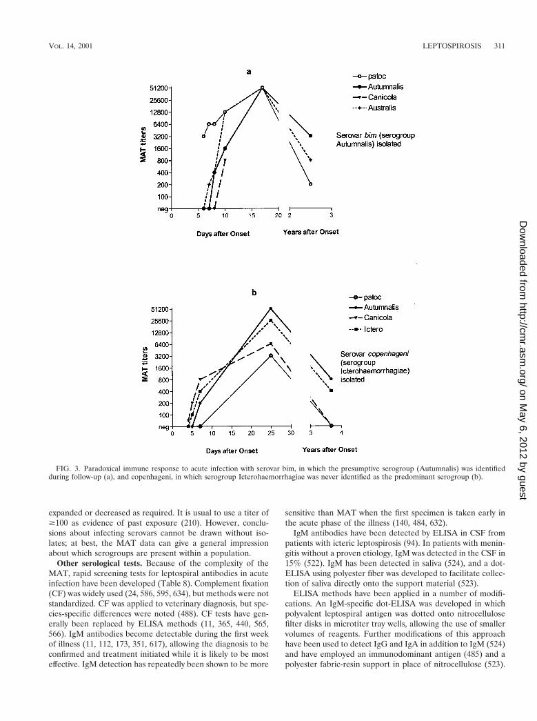
expanded or decreased as required. It is usual to use a titer of$100 as evidence of past exposure (210). However, conclu-sions about infecting serovars cannot be drawn without iso-lates; at best, the MAT data can give a general impressionabout which serogroups are present within a population.
Other serological tests. Because of the complexity of theMAT, rapid screening tests for leptospiral antibodies in acuteinfection have been developed (Table 8). Complement fixation(CF) was widely used (24, 586, 595, 634), but methods were notstandardized. CF was applied to veterinary diagnosis, but spe-cies-specific differences were noted (488). CF tests have gen-erally been replaced by ELISA methods (11, 365, 440, 565,566). IgM antibodies become detectable during the first weekof illness (11, 112, 173, 351, 617), allowing the diagnosis to beconfirmed and treatment initiated while it is likely to be mosteffective. IgM detection has repeatedly been shown to be more
sensitive than MAT when the first specimen is taken early inthe acute phase of the illness (140, 484, 632).
IgM antibodies have been detected by ELISA in CSF frompatients with icteric leptospirosis (94). In patients with menin-gitis without a proven etiology, IgM was detected in the CSF in15% (522). IgM has been detected in saliva (524), and a dot-ELISA using polyester fiber was developed to facilitate collec-tion of saliva directly onto the support material (523).
ELISA methods have been applied in a number of modifi-cations. An IgM-specific dot-ELISA was developed in whichpolyvalent leptospiral antigen was dotted onto nitrocellulosefilter disks in microtiter tray wells, allowing the use of smallervolumes of reagents. Further modifications of this approachhave been used to detect IgG and IgA in addition to IgM (524)and have employed an immunodominant antigen (485) and apolyester fabric-resin support in place of nitrocellulose (523).
FIG. 3. Paradoxical immune response to acute infection with serovar bim, in which the presumptive serogroup (Autumnalis) was identifiedduring follow-up (a), and copenhageni, in which serogroup Icterohaemorrhagiae was never identified as the predominant serogroup (b).
VOL. 14, 2001 LEPTOSPIROSIS 311
on May 6, 2012 by guest
http://cmr.asm
.org/D
ownloaded from
A commercial IgM dot-ELISA dipstick has been shown to beas sensitive as a microtiter plate IgM-ELISA (350a). Anotherdipstick assay (253) has been extensively evaluated in severalpopulations (512, 533, 661). A dot immunoblot assay usingcolloidal gold conjugate allowed completion of the assay within30 min (455).
In contrast to the applications of ELISA for diagnosis ofhuman infection, in which broadly reactive assays are generallydesirable and few serovar-specific assays have been developed(395), veterinary applications have been directed towards de-tection of serovar-specific antibodies, particularly for detectionof infection in food animals. ELISA methods have describedfor detection of serovar pomona (134, 573) and hardjo (5, 58,573, 653) infection in cattle and hardjo in sheep (9). Severalassays are available commercially for serodiagnosis of bovinehardjo infection and have been evaluated (642). IgM detectionby ELISA has also been applied to canine diagnosis (264, 265,623).
A macroscopic slide agglutination test was described inwhich 12 serovars were combined into four pools for the rapidscreening of sera from humans and animals (234). Despite theuse of an expanded antigen range, false-negative results werereported for sera from populations in areas of endemic lepto-spirosis (635). Several modifications of this test have used asingle serovar antigen, usually serovar patoc (76, 364, 369,621). Some studies have reported that the patoc slide test isinsensitive (369, 546, 616), but a commercial slide agglutina-tion assay was recently found to be as sensitive and specific asan IgM-ELISA while remaining reactive for a shorter timeafter recovery than either the IgM-ELISA or the MAT (77).
A number of methods using sensitized red blood cells havebeen described. The extraction of an erythrocyte-sensitizingsubstance led to the development of both a hemolytic assayrequiring complement (135, 136) and a hemagglutination assay(383, 547), and a number of modifications of the latter havebeen described (295, 499). These assays detect both IgM andIgG antibodies (351, 431). The indirect hemagglutination assay(IHA) developed at CDC (547) was shown to have a sensitivityof 92% and specificity of 95% compared with the MAT (546).This assay is available commercially and for many years as theonly U.S. Food and Drug Administration-approved productfor serological diagnosis of leptospirosis. Recent estimates ofthe sensitivity of the IHA in populations in which leptospirosisis endemic have varied. In one study, IHA detected all patientswith leptospirosis but was positive in only 44% of first acute-phase samples taken a mean of 5 days after onset of symptoms(351). Other studies have reported lower overall sensitivities,partly due to differences in case ascertainment and study de-sign (181, 661).
A microcapsule agglutination test using a synthetic polymerin place of red blood cells (39, 514) has been evaluated exten-sively in Japan and China (40, 139). In an international mul-ticenter evaluation, the microcapsule agglutination test wasmore sensitive than either the MAT or an IgM-ELISA inearly-acute-phase samples (38), but failed to detect infectionscaused by some serovars (38, 513). An advantage of this directagglutination method is that it can be applied without modifi-cation to sera from other animal species (37).
Other techniques applied to the detection of leptospiralantibodies include immunofluorescence (33, 580), RIA (323),
counterimmunoelectrophoresis (410, 567, 657), and thin-layerimmunoassay (50). These methods have not been widely used.
Molecular Diagnosis
Leptospiral DNA has been detected in clinical material bydot-blotting (393, 569) and in situ hybridization (568). A re-combinant probe specific for pathogenic serovars was preparedfrom serovar lai (143). Probes specific for serovar hardjoboviswere developed (344, 594, 673) and applied to the detection ofleptospires in bovine urine (72). However, the sensitivity of32P-labeled probes was approximately 103 leptospires (393,569, 673), much lower than the sensitivity of PCR, and probeshave not been used extensively for diagnosis since PCR be-came available.
Several primer pairs for PCR detection of leptospires havebeen described, some based on specific gene targets (483),most frequently 16S or 23S rRNA genes (287, 386, 407, 607,637, 639, 667) and repetitive elements (428, 502, 643, 670, 671),while others have been constructed from genomic libraries(247, 248, 324, 594). However, few have been shown to amplifyleptospiral DNA from either human (247, 386) or veterinary(378, 559, 594, 606, 643, 670) clinical material, and of these,only two methods have been subject to extensive clinical eval-uation (84, 387). Both methods were found to be more sensi-tive than culture, but differences in analysis of the data renderdirect comparisons between the two approaches impossible.
In one analysis, culture and PCR were positive in 48 and62% of confirmed cases of leptospirosis, respectively, but se-rology was positive in 97% (84). However, PCR was positivefor two patients who died before seroconversion and was alsopositive for 18% of seronegative first acute-phase samples.
Both these approaches have limitations. The primers de-scribed by Merien et al. (386) amplify a 331-bp fragment of therrs (16S rRNA) gene of both pathogenic and nonpathogenicleptospires, which in the unlikely event of contamination ofspecimens with nonpathogenic leptospires might produce afalse-positive result, whereas the G1 and G2 primers describedby Gravekamp et al. (247) do not amplify L. kirschneri sero-vars, necessitating the use of two primer pairs for detection ofall pathogenic serovars (248).
Despite these potential shortcomings, these two primer pairshave been the most widely used for clinical studies. LeptospiralDNA has been amplified from serum (84, 247, 387), urine (46,84, 387), aqueous humor (389), CSF (387, 492, 601), and anumber of tissues obtained at autopsy (unpublished data).
The detection of leptospiral DNA in bovine urine has alsobeen investigated. Primers which amplified several serovars ofserogroup Sejroe were described (593), and a method specificfor serovar hardjo genotype hardjobovis was developed (643).An assay based on the IS1533 insertion sequence (670) facili-tated both detection and identification of serovars directlyfrom urine. Another assay was developed and applied to bothbovine and porcine urine samples (607). To overcome theproblem of inhibitors present in bovine urine, a magnetic im-munocapature PCR assay for serovar hardjo was developed(559).
A recent study evaluated five PCR methods, culture, andimmunofluorescence for detection of serovar hardjo in bovineurine samples (606). Primers derived from rRNA gene se-
312 LEVETT CLIN. MICROBIOL. REV.
on May 6, 2012 by guest
http://cmr.asm
.org/D
ownloaded from
quences were the least specific, and none of the methods was100% sensitive. A combination of two detection methods cho-sen from PCR, immunofluorescence, and culture was the mostsensitive.
A limitation of PCR-based diagnosis of leptospirosis is theinability of most PCR assays to identify the infecting serovar.While this is not significant for individual patient management,the identity of the serovar has significant epidemiological andpublic health value. Strategies designed to overcome this ob-stacle have included restriction endonuclease digestion of PCRproducts (85, 502), direct sequencing of amplicons (424), andsingle-strand conformation analysis (SSCP) (380, 647). Lepto-spiral genomospecies but not individual serovars can be differ-entiated following PCR by electrophoresis in nondenaturingpolyacrylamide gels, followed by silver staining (424), withoutthe additional step of purification and denaturing.
PCR has been used to distinguish pathogenic from non-pathogenic serovars (407, 444, 639). Recently, a fluorescent-probe 59 exonuclease PCR assay was described for the rapiddetection of pathogenic leptospires (637).
Molecular Typing
Because of the difficulties associated with serological iden-tification of leptospiral isolates, there has been great interest inmolecular methods for identification and subtyping (279, 561).Methods employed have included digestion of chromosomalDNA by restriction endonucleases (REA), restriction frag-ment length polymorphism (RFLP), ribotyping, PFGE, and anumber of PCR-based approaches.
REA has been studied extensively (270, 288, 370, 372, 490,555, 563, 575). Distinct genotypes within serovar hardjo weredemonstrated (490). Bovine isolates from North America haveall been found to be of genotype hardjobovis, of which sub-types A, B, and C could be recognized (574). In NorthernIreland, both genotypes hardjobovis and hardjoprajitno werefound among bovine isolates (371). Antigenic differences werealso reported among hardjobovis isolates (345). Moreover, se-rovar balcanica isolates in North America were indistinguish-able from genotype hardjobovis isolates by REA (574). Fur-ther analysis of RFLP in genotype hardjobovis isolates byREA, Southern blotting, and PFGE has shown the existence ofmultiple genetic clones resulting from genomic rearrangement(675). These clones were usually localized within geographicallocations and thus are of epidemiological significance (675).
Similar subserovar differences were detected within serovarpomona, isolates from North America being identified as sub-type kennewicki (563, 575), while European isolates were ofserovar pomona (270, 563) or mozdok (269, 270). More re-cently, differences between subtype kennewicki isolates werecorrelated with host animal source (71). Differences betweenserovars copenhageni and icterohaemorrhagiae were demon-strated by some workers (372), but not all (288, 555). However,all isolates of these two serovars are indistinguishable by PFGE(281).
Ribotyping has demonstrated reasonably good correlationwith the phylogenetic classification of leptospira into 11 geno-mospecies. Using EcoRI for digestion and 16S and 23S rRNAfrom Escherichia coli as the probe, a large database was con-structed (451, 452). Many serovars gave unique profiles, while
other serovars could not be distinguished from each other byribotyping, particularly those that were known previously to beclosely related, such as icterohaemorrhagiae and copenhageni(288, 555). Ribotypes of serovars within genomospecies couldbe grouped together by the possession of common fragments.This database is available at the Institut Pasteur website (http://www.pasteur.fr/recherche/Leptospira/Ribotyping.html). Ribotyp-ing has been shown to discriminate accurately between the sero-var hardjo genotypes hardjobovis and hardoprajitno (453).
An alternative approach to ribotyping used three restrictionenzymes and a PCR-derived 16S rDNA probe. Use of only 16SrDNA gave fewer bands, but this was counterbalanced to someextent by the use of multiple restriction enzymes, giving threedifferent patterns for each serovar (286). Relatively few sero-vars were examined but all gave distinct ribotypes with theexception of serovars icterohaemorrhagiae and copenhageni.A range of other probes have been used to generate RFLPs(429, 593, 672, 673). A probe based on the repetitive sequenceelement from serovar hardjo genotype hardjobovis was alsoused to detect leptospires in bovine urine (673).
PFGE has proven useful to characterize leptospiral serovars(279). In contrast to its application in strain typing of otherorganisms, PFGE has shown that the genomes of leptospiralserovars are remarkably conserved, both over time and acrosswide geographical distributions (279, 281). Importantly, recentclinical isolates gave the same banding patterns as referencestrains of the same serovar which have been maintained formany years by repeated subculture (279). Using the enzymeNotI, most but not all serovars gave unique PFGE patterns. L.interrogans serovars bratislava, lora, jalna, and muenchen gaveidentical patterns when digested with NotI but were differen-tiated when digested with SgrAI (282). Other serovars whichwere difficult to differentiate included L. borgpetersenii serovarsarborea and castellonis. The L. interrogans serovars copenhag-eni and icterohaemorrhagiae were indistinguishable by PFGE,confirming their close relationship. PFGE analysis has becomethe de facto standard for molecular characterization of lepto-spiral isolates, and other molecular typing methods will infuture have to be validated against this method.
A limiting factor in all methods which analyze chromosomalDNA is the requirement for large quantities of purified DNA.As a result, several methods based on the analysis of PCR-amplified sections of leptospiral DNA have been employed.Sequence variation within the 285-bp fragment amplified bythe G1 and G2 primers (248) led to different electrophoreticmobilities which were detected by polyacrylamide gel electro-phoresis and silver staining (424). This approach allowed se-rovars of L. interrogans sensu stricto to be differentiated fromL. noguchii serovars.
Sequence variation is also exploited in SSCP. Using thismethod, serovars prevalent in China were shown to have dif-ferent mobilities corresponding to L. interrogans and L. borg-petersenii (647). The Chinese isolates were studied using asequence amplified from the 16S rRNA gene, the highly con-served nature of which may account for the inability to distin-guish serovars from one another. In contrast, SSCP analysis ofthe G1-G2 amplicon allows serovar identification within eachgenomospecies studied (380). A restriction on the use of thelatter sequence is the inability of the G1 and G2 primers toamplify L. kirschneri (248). An alternative application of these
VOL. 14, 2001 LEPTOSPIROSIS 313
on May 6, 2012 by guest
http://cmr.asm
.org/D
ownloaded from
primers is their use under low-stringency conditions, generat-ing a mixture of specific and nonspecific products (150). Underthese conditions, the G1 and G2 primers amplify all species,including L. biflexa. Polymorphisms were detected which al-lowed discrimination of serovars with the exception of closelyrelated serovars, including copenhageni and icterohaemorrha-giae (85, 150).
The presence of multiple copy insertion sequences has beenexploited for serovar identification (481, 502, 670, 671). Meth-ods based on IS1533 have limited application because of theabsence of this insertion sequence in L. interrogans (sensustricto) and L. noguchii (481, 670). By amplifying the sequencesbetween adjacent copies of IS1500, numerous genetic sub-groups within serovar pomona type kennewickii were distin-guished (671).
RFLP analysis of PCR-amplified 16S and 23S rRNA genesallowed the grouping of 48 serovars into 16 mapped restrictionsite polymorphism profiles (469). Using this approach, thegenomospecies of Leptospira could be identified, and the ge-notypes hardjobovis and hardjoprajitno of serovar hardjo wereclearly distinguished (453). The method was simplified to yieldonly five profiles by using a single restriction enzyme (638).One of the potential advantages of this RFLP approach is theability to amplify leptospiral DNA from clinical material and toidentify the infecting serovar or genomospecies rapidly in theabsence of an isolate. Other workers have used primers thatamplify only a restricted range of serovars (85, 502), limitingthe utility of the approach unless several primer sets are used(85).
DNA fingerprinting using arbitrary primers (625, 629) hasbeen studied extensively (85, 128, 129, 237, 453, 469), usingdifferent primers and conditions. Direct comparison betweenthe results of these studies is therefore impossible, but it isclear that reproducibility is difficult to achieve without absolutestandardization of experimental procedure. Profiles are af-fected markedly by the primer used, the quantity and quality ofthe DNA template (128, 380, 599), and the electrophoresisconditions (129). The greatest value of arbitrary primer tech-niques lies in their ability to differentiate between isolateswhen the range of potential serovars is limited, allowing rapididentification of freshly isolated strains (85, 128, 237). Arbi-trary-primed PCR was used to derive species-specific probesfor identification of L. interrogans (sensu stricto), L. borg-petersenii, and L. kirschneri by dot blotting (347). A cluster of43 L. interrogans sensu strico isolates from a number of Bra-zilian outbreaks were shown to have identical arbitrary-primedPCR fingerprints (449) despite the inclusion of isolates ofserovars copenhageni and canicola.
CONCLUSION
The etiology and epidemiology of leptospirosis have beenunderstood for many years, and this knowledge has led to thedevelopment of effective preventive strategies. In developedcountries, leptospirosis continues to be a disease of consider-able economic significance in animal husbandry, but the majorburden of human disease remains in tropical and subtropicaldeveloping countries. Several recent outbreaks of leptospirosishave drawn attention to the potential effects of climate changeand human activity on the incidence of the disease and the
broad spectrum of clinical manifestations. The development ofseveral promising approaches to rapid diagnosis has beenbased largely on the recognition that early initiation of antibi-otic therapy is important in acute disease, but also on the needfor simpler assays which can be used more widely. However,many of these diagnostic advances will be unavailable to thosepopulations for which they would be most useful. At a morefundamental level, understanding of the mechanisms of patho-genesis remains incomplete, but recent advances in the molec-ular biology of leptospires offer the prospect of more rapidprogress in the future.
ACKNOWLEDGMENTS
I thank the many colleagues who have assisted me with access to theliterature, particularly Esther Maria Fajardo, Peter Gaskin, Albert Ko,Diane Van Alstyne, and Rob Weyant and the staff of the InformationCenter, CDC, Atlanta, Ga.
REFERENCES
1. Abdulkader, R., E. Sabbaga, L. Meireles, and A. Radu. 1993. Vascularinjury in acute renal failure due to leptospirosis is not associated withantineutrophil cytoplasmic antibody. Nephron 65:156.
2. Abdulkader, R. C. R. M. 1997. Acute renal failure in leptospirosis. RenalFail. 19:191–198.
3. Adachi, Y., and R. Yanagawa. 1977. Inhibition of leptospiral agglutinationby the type-specific main antigens of leptospiras. Infect. Immun. 17:466–467.
4. Adler, B., R. J. Chappel, and S. Faine. 1982. The sensitivities of differentimmunoassays for detecting leptospiral antigen. Zentbl. Bakteriol. 252:405–413.
5. Adler, B., D. V. Cousins, S. Faine, and G. M. Robertson. 1982. Bovine IgMand IgG response to Leptospira interrogans serovar hardjo as measured byenzyme immunoassay. Vet. Microbiol. 7:577–585.
6. Adler, B., and S. Faine. 1978. The antibodies involved in the human im-mune response to leptospiral infection. J. Med. Microbiol. 11:387–400.
7. Adler, B., and S. Faine. 1977. Host immunological mechanisms in theresistance of mice to leptospiral infections. Infect. Immun. 17:67–72.
8. Adler, B., S. Faine, W. L. Christopher, and R. J. Chappel. 1986. Develop-ment of an improved selective medium for isolation of leptospires fromclinical material. Vet. Microbiol. 12:377–381.
9. Adler, B., S. Faine, and L. M. Gordon. 1981. The enzyme-linked immu-nosorbent assay (ELISA) as a serological test for detecting antibodiesagainst Leptospira interrogans serovar hardjo in sheep. Aust. Vet. J. 57:414–417.
10. Adler, B., S. Faine, and R. Yanagawa. 1980. Comparative studies on twoantigens (F4 and TM) extracted from leptospires. J. Clin. Microbiol. 12:7–9.
11. Adler, B., A. M. Murphy, S. A. Locarnini, and S. Faine. 1980. Detection ofspecific anti-leptospiral immunoglobulins M and G in human serum bysolid-phase enzyme-linked immunosorbent assay. J. Clin. Microbiol. 11:452–457.
12. Adler, B. A., and S. Faine. 1983. A Pomona serogroup-specific, agglutinat-ing antigen in Leptospira, identified by monoclonal antibodies. Pathology15:247–250.
13. Adler, B. A., and S. Faine. 1983. Species- and genus-specific antigens inLeptospira, revealed by monoclonal antibodies and enzyme immunoassay.Zentbl. Bakteriol. 255:317–322.
14. Agrawal, P. K., and D. K. Srivastava. 1986. Outbreak of Weil’s disease ina food fad commune in India. BMJ 293:1646–1647.
15. Alani, F. S. S., M. P. Mahoney, L. P. Ormerod, P. A. Wright, and M.Garrues. 1993. Leptospirosis presenting as atypical pneumonia, respiratoryfailure and pyogenic meningitis. J. Infect. 27:281–283.
16. Alexander, A., A. Baer, J. R. Fair, W. S. Gochenour, J. H. King, and R. N.Yager. 1952. Leptospiral uveitis: report of a bacteriologically confirmedcase. Arch. Ophthalmol. 48:292–297.
17. Alexander, A. D. 1986. Serological diagnosis of leptospirosis, p. 435–439. InN. R. Rose, H. Friedman, and J. L. Fahey (ed.), Manual of clinical labo-ratory immunology, 3rd ed. American Society for Microbiology, Washing-ton, D.C.
18. Alexander, A. D., A. S. Benenson, R. J. Byrne, R. S. Dıaz-Rivera, L. B.Evans, W. S. Gochenour, H. E. Hall, J. A. Hightower, H. Jeffries, J. deJesus, E. Martınez, M. Paniagua, J. A. Pons, F. Ramos-Morales, R. Rodrı-guez-Molina, K. Y. Swisher, T. E. Woodward, and R. H. Yager. 1963.Leptospirosis in Puerto Rico. Zoonoses Res. 2:152–227.
19. Alexander, A. D., L. B. Evans, M. F. Baker, H. J. Baker, D. Ellison, and M.Marriapan. 1975. Pathogenic leptospiras isolated from Malaysian surfacewaters. Appl. Microbiol. 29:30–33.
314 LEVETT CLIN. MICROBIOL. REV.
on May 6, 2012 by guest
http://cmr.asm
.org/D
ownloaded from
20. Alexander, A. D., E. F. Lessel, L. B. Evans, E. Franck, and S. S. Green.1972. Preservation of leptospiras by liquid-nitrogen refrigeration. Int. J.Syst. Bacteriol. 22:165–169.
21. Alexander, A. D., and P. L. Rule. 1986. Penicillins, cephalosporins, andtetracyclines in treatment of hamsters with fatal leptospirosis. Antimicrob.Agents Chemother. 30:835–839.
22. Allen, P., S. Raftery, and D. Phelan. 1989. Massive pulmonary haemorrhagedue to leptospirosis. Intensive Care Med. 15:322–324.
23. Alston, J. M. 1935. Leptospiral jaundice among sewer-workers. Lanceti:806–809.
24. Alston, J. M., and J. C. Broom. 1958. Leptospirosis in man and animals. E.& S. Livingstone, Edinburgh, U.K.
25. Anderson, B. S., and H. P. Minette. 1986. Leptospirosis in Hawaii: shiftingtrends in exposure, 1907–1984. Int. J. Zoonoses 13:76–88.
26. Anderson, D. C., D. S. Folland, M. D. Fox, C. M. Patton, and A. F.Kaufmann. 1978. Leptospirosis: a common-source outbreak due to lepto-spires of the grippotyphosa serogroup. Am. J. Epidemiol. 107:538–544.
27. Anderson, D. C., J. G. Geistfeld, H. M. Maetz, C. M. Patton, and A. F.Kaufmann. 1978. Leptospirosis in zoo workers associated with bears. Am. J.Trop. Med. Hyg. 27:210–211.
28. Andreescu, N., D. Tacorian, E. Duminica, M. Filip, and A. Sosin. 1988.Investigarea serologica in complicatiile oculare ale leptospirozelor la om.Bacteriol. Virusol. Parazitol. Epidemiol. 33:41–46.
29. Andre-Fontaine, G., X. Peslerbe, and J. P. Ganiere. 1992. Occupationalhazard of unnoticed leptospirosis in water ways maintenance staff. Eur. J.Epidemiol. 8:228–232.
30. Andrew, D. E., and G. R. Marrocco. 1977. Leptospirosis in New England.JAMA 238:2027–2028.
31. Annear, D. I. 1974. Recovery of leptospires after dry storage for ten years.Int. J. Syst. Bacteriol. 24:399–401.
32. Antoniadis, A., S. Alexiou-Daniel, L. Fidani, and E. F. K. Bautz. 1995.Comparison of the clinical and serologic diagnosis of haemorrhagic feverwith renal syndrome (HFRS) and leptospirosis. Eur. J. Epidemiol. 11:91–92.
33. Appassakij, H., K. Silpapojakul, R. Wansit, and J. Woodtayakorn. 1995.Evaluation of the immunofluorescent antibody test for the diagnosis ofhuman leptospirosis. Am. J. Trop. Med. Hyg. 52:340–343.
34. Arean, V. M. 1957. Leptospiral myocarditis. Lab. Investig. 6:462–471.35. Arean, V. M. 1962. The pathologic anatomy and pathogenesis of fatal
human leptospirosis (Weil’s disease). Am. J. Pathol. 40:393–423.36. Arean, V. M., G. Sarasin, and J. H. Green. 1964. The pathogenesis of
leptospirosis: toxin production by Leptospira icterohaemorrhagiae. Am. J.Vet. Res. 25:836–843.
37. Arimitsu, Y., K. Fukumura, and Y. Shintaki. 1989. Distribution of lepto-spirosis among stray dogs in the Okinawa Islands, Japan: comparison of themicrocapsule and microscopic agglutination tests. Br. Vet. J. 145:473–477.
38. Arimitsu, Y., E. Kmety, Y. Ananyina, G. Baranton, I. R. Ferguson, L.Smythe, and W. J. Terpstra. 1994. Evaluation of the one-point microcap-sule agglutination test for diagnosis of leptospirosis. Bull. WHO 72:395–399.
39. Arimitsu, Y., S. Kobayashi, K. Akama, and T. Matuhasi. 1982. Develop-ment of a simple serological method for diagnosing leptospirosis: a micro-capsule agglutination test. J. Clin. Microbiol. 15:835–841.
40. Arimitsu, Y., T. Matuhasi, S. Kobayashi, T. Sato, and J. J. Cui. 1987.Serodiagnosis of leptospirosis in China by the one-point MCA method.Epidemiol. Infect. 99:393–398.
41. Awad-Masalmeh, A., and H. Willinger. 1983. Evaluation of 2-mercapto-ethanol treatment in serodiagnosis of swine leptoispirosis. Microbiologica6:133–143.
42. Baber, M. D., and R. D. Stuart. 1946. Leptospirosis canicola: a case treatedwith penicillin. Lancet ii:594–596.
43. Babudieri, B. 1958. Animal reservoirs of leptospirosis. Ann. N.Y. Acad. Sci.70:393–413.
44. Baelen, E., and J. Roustan. 1997. Leptospirosis associated with acute acal-culous cholecystitis. J. Clin. Gastroenterol. 25:704–706.
45. Bahaman, A. R., A. L. Ibrahim, N. D. Stallman, and R. D. Tinniswood.1988. The bacteriological prevalence of leptospiral infection in cattle andbuffaloes in West Malaysia. Epidemiol. Infect. 100:239–246.
46. Bal, A. E., C. Gravekamp, R. A. Hartskeerl, J. de Meza-Brewster, H.Korver, and W. J. Terpstra. 1994. Detection of leptospires in urine by PCRfor early diagnosis of leptospirosis. J. Clin. Microbiol. 32:1894–1898.
47. Ballard, S. A., R. P. A. M. Segers, N. Bleumink-Pluym, J. Fyfe, S. Faine, andB. Adler. 1993. Molecular analysis of the hsp (groE) operon of Leptospirainterrogans serovar copenhageni. Mol. Microbiol. 8:739–751.
48. Ballard, S. A., M. Williamson, B. Adler, T. Vinh, and S. Faine. 1986.Interactions of virulent and avirulent leptospires with primary cultures ofrenal epithelial cells. J. Med. Microbiol. 21:59–67.
49. Banfi, E., M. Cinco, M. Bellini, and M. R. Soranzo. 1982. The role ofantibodies and serum complement in the interaction between macrophagesand leptospires. J. Gen. Microbiol. 128:813–816.
50. Banfi, E., M. Cinco, S. Delia, L. Castagnari, V. Vullo, C. M. Mastroianni,
and C. Contini. 1984. New trends in the rapid serodiagnosis of leptospirosis.Zentbl. Bakteriol. 257:503–507.
51. Baril, C., J. L. Herrmann, C. Richaud, D. Margarita, and I. S. Girons. 1992.Scattering of the rRNA genes on the physical map of the circular chromo-some of Leptospira interrogans serovar icterohaemorrhagiae. J. Bacteriol.174:7566–7571.
52. Baril, C., and I. Saint Girons. 1990. Sizing of the Leptospira genome bypulsed-field agarose gel electrophoresis. FEMS Microbiol. Lett. 71:95–100.
53. Barkay, S., and H. Garzozi. 1984. Leptospirosis and uveitis. Ann. Ophthal-mol. 16:164–168.
54. Barkin, R. M., and J. W. Glosser. 1973. Leptospirosis—an epidemic inchildren. Am. J. Epidemiol. 98:184–191.
55. Barkin, R. M., J. C. Guckian, and J. W. Glosser. 1974. Infection by Lep-tospira ballum: a laboratory-associated case. South. Med. J. 67:155–156.
56. Barnett, J. K., D. Barnett, C. A. Bolin, T. A. Summers, E. A. Wagar, N. F.Cheville, R. A. Hartskeerl, and D. A. Haake. 1999. Expression and distri-bution of leptospiral outer membrane components during renal infection ofhamsters. Infect. Immun. 67:853–861.
57. Beeson, P. B., and D. D. Hankey. 1952. Leptospiral meningitis. Arch. Intern.Med. 89:575–583.
58. Bercovich, Z., R. Taaijke, and B. A. Bokhout. 1990. Evaluation of an ELISAfor the diagnosis of experimentally induced and naturally occurring Lepto-spira hardjo infections in cattle. Vet. Microbiol. 21:255–262.
59. Berendsen, H. H., J. H. Rommes, B. S. Hylkema, A. F. Meinesz, and H. J.Sluiter. 1984. Adult respiratory failure with leptospirosis. Ann. Intern. Med.101:402.
60. Berg, H. C., D. B. Bromley, and N. W. Charon. 1978. Leptospiral motility,p. 285–294. In R. Y. Stanier, H. J. Rogers, and J. B. Ward (ed.), Relationsbetween structure and function in the prokaryotic cell. 28th Symposium ofthe Society for General Microbiology. Cambridge University Press, Cam-bridge, U.K.
61. Berman, S. J., C. C. Tsai, K. K. Holmes, J. W. Fresh, and R. H. Watten.1973. Sporadic anicteric leptospirosis in South Vietnam. Ann. Intern. Med.79:167–173.
62. Bernheimer, A. W., and R. F. Bey. 1986. Copurification of Leptospira inter-rogans serovar pomona hemolysin and sphingomyelinase C. Infect. Immun.54:262–264.
63. Bethlem, N., A. Lemle, and N. G. Pereira. 1991. Leptospirosis. Semin.Respir. Med. 12:58–67.
64. Bey, R. F., and R. C. Johnson. 1978. Protein-free and low-protein media forthe cultivation of Leptospira. Infect. Immun. 19:562–569.
65. Blackmore, D. K., L. Bell, and L. Schollum. 1979. Leptospirosis in meatinspectors: preliminary results of a serological survey. N. Z. Med. J. 90:415–418.
66. Blackmore, D. K., and L. M. Schollum. 1982. Risks of contracting lepto-spirosis on the dairy farm. N. Z. Med. J. 95:649–652.
67. Blackmore, D. K., L. M. Schollum, and K. M. Moriarty. 1984. The magni-tude and duration of titres of leptospiral agglutinins in human sera. N. Z.Med. J. 97:83–86.
68. Blagoveshchenskaia, N. M. 1957. On the epidemiology of anicteric lepto-spirosis. J. Microbiol. Epidemiol. Immunobiol. 28:240–244.
69. Bolin, C. 2000. Leptospirosis, p. 185–200. In C. Brown and C. Bolin (ed.),Emerging diseases of animals. ASM Press, Washington, D.C.
70. Bolin, C. A., and P. Koellner. 1988. Human-to-human transmission ofLeptospira interrogans by milk. J. Infect. Dis. 158:246–247.
71. Bolin, C. A., and R. L. Zuerner. 1996. Correlation between DNA restrictionfragment length polymorphisms in Leptospira interrogans serovar pomonatype kennewicki and host animal source. J. Clin. Microbiol. 34:424–425.
72. Bolin, C. A., R. L. Zuerner, and G. Trueba. 1989. Comparison of threetechniques to detect Leptospira interrogans serovar hardjo type hardjo-bovisin bovine urine. Am. J. Vet. Res. 50:1001–1003.
73. Boursaux-Eude, C., I. Saint Girons, and R. L. Zuerner. 1995. IS1500, anIS3-like element from Leptospira interrogans. Microbiology 141:2165–2173.
74. Boursaux-Eude, C., I. Saint Girons, and R. L. Zuerner. 1998. Leptospiragenomics. Electrophoresis 19:589–592.
75. Bowdoin, C. D. 1942. A new disease identity (?). J. Med. Assoc. Ga.31:437–442.
76. Bragger, J. M., and B. Adler. 1976. A card test for the serodiagnosis ofhuman leptospirosis. J. Clin. Pathol. 29:198–202.
77. Brandao, A. P., E. D. Camargo, E. D. da Silva, M. V. Silva, and R. V. Abrao.1998. Macroscopic agglutination test for rapid diagnosis of human lepto-spirosis. J. Clin. Microbiol. 36:3138–3142.
78. Brandling-Bennett, A. D., and F. Pinheiro. 1996. Infectious diseases inLatin America and the Caribbean: are they really emerging and increasing?Emerg. Infect. Dis. 2:59–61.
79. Braun, J. L. 1961. Epidemiology of leptospirosis in Iowa-a study of sporadicand epidemic cases. J. Am. Vet. Med. Assoc. 138:532–536.
80. Brendle, J. J., M. Rogul, and A. D. Alexander. 1974. Deoxyribonucleic acidhybridization among selected leptospiral serotypes. Int. J. Syst. Bacteriol.24:205–214.
81. Brenner, D. J., A. F. Kaufmann, K. R. Sulzer, A. G. Steigerwalt, F. C.Rogers, and R. S. Weyant. 1999. Further determination of DNA relatedness
VOL. 14, 2001 LEPTOSPIROSIS 315
on May 6, 2012 by guest
http://cmr.asm
.org/D
ownloaded from
between serogroups and serovars in the family Leptospiraceae with a pro-posal for Leptospira alexanderi sp. nov. and four new Leptospira genomo-species. Int. J. Syst. Bacteriol. 49:839–858.
82. Broughton, E. S., and J. Scarnell. 1985. Prevention of renal carriage ofleptospirosis in dogs by vaccination. Vet. Rec. 117:307–311.
83. Brown, C. A., A. W. Roberts, M. A. Miller, D. A. David, S. A. Brown, C. A.Bolin, J. Jarecki-Black, C. E. Greene, and D. Miller-Liebl. 1996. Leptospirainterrogans serovar grippotyphosa infection in dogs. J. Am. Vet. Med. Assoc.209:1265–1267.
84. Brown, P. D., C. Gravekamp, D. G. Carrington, H. Van de Kemp, R. A.Hartskeerl, C. N. Edwards, C. O. R. Everard, W. J. Terpstra, and P. N.Levett. 1995. Evaluation of the polymerase chain reaction for early diag-nosis of leptospirosis. J. Med. Microbiol. 43:110–114.
85. Brown, P. D., and P. N. Levett. 1997. Differentiation of Leptospira speciesand serovars by PCR-restriction endonuclease analysis, arbitrarily primedPCR and low-stringency PCR. J. Med. Microbiol. 46:173–181.
86. Buchanan, G. 1927. Spirochaetal jaundice. Special Report Series, no. 113.Medical Research Council, London, U.K.
87. Buckland, F. E., and R. D. Stuart. 1945. Mud fever (leptospirosis) in theBritish army in France. Lancet ii:331–333.
88. Bulach, D. M., T. Kalambaheti, A. de La Pena-Moctezuma, and B. Adler.2000. Functional analysis of genes in the rfb locus of Leptospira borgpeterse-nii serovar hardjo subtype hardjobovis. Infect. Immun. 68:3793–3798.
89. Burke, B. J., J. F. Searle, and D. Mattingly. 1976. Leptospirosis presentingwith profuse haemoptysis. BMJ 2:982.
90. Burth, P., M. Younes-Ibrahim, F. H. F. S. Goncalez, E. R. Costa, and M. V.Castro Faria. 1997. Purification and characterization of a Na1, K1 ATPaseinhibitor found in an endotoxin of Leptospira interrogans. Infect. Immun.65:157–1560.
91. Butler, C. S., and S. A. Endara. 2000. Leptospirosis complicated by severeaortic stenosis. Anaesth. Intensive Care 28:434–437.
92. Cacciapuoti, B., L. Ciceroni, C. Maffei, F. Di Stanislao, P. Strusi, L. Cale-gari, R. Lupido, G. Scalise, G. Cagnoni, and G. Renga. 1987. A waterborneoutbreak of leptospirosis. Am. J. Epidemiol. 126:535–545.
93. Caccione, R. A., E. S. Cascelli, M. A. Saravı, and E. S. Martınez. 1977.Brote de leptospirosis en ninos de Longhamps, Pcia de Buenos Aires,Argentina: daignostico de laboratorio. Rev. Argent. Microbiol. 9:126–128.
94. Camargo, E. D., M. V. Silva, A. J. Vaz, L. Batista, A. P. Brandao, A. W.Ferreira, E. C. Romero, and P. R. S. Barbosa. 1995. ELISA-IgM applied tocerebrospinal fluid in human leptospirosis. Serodiagn. Immunother. Infect.Dis. 7:19–22.
95. Campagnolo, E. R., M. C. Warwick, H. L. Marx, R. P. Cowart, H. D.Donnell, M. D. Bajani, S. L. Bragg, J. E. Esteban, D. P. Alt, J. W. Tappero,C. A. Bolin, and D. A. Ashford. 2000. Analysis of the 1998 outbreak ofleptospirosis in Missouri in humans exposed to infected swine. J. Am. Vet.Med. Assoc. 216:676–682.
96. Cargill, W. H., and P. B. Beeson. 1947. The value of spinal fluid examina-tion as a diagnostic procedure in Weil’s disease. Ann. Intern. Med. 27:396–400.
97. Centers for Disease Control and Prevention. 1997. Case definitions forinfectious conditions under public health surveillance. Morb. Mortal. Wkly.Rep. 46(RR-10):49.
98. Centers for Disease Control and Prevention. 1998. Outbreak of acutefebrile illness among athletes participating in triathlons—Wisconsin andIllinois, 1998. Morb. Mortal. Wkly. Rep. 47:585–588.
99. Centers for Disease Control and Prevention. 2000. Outbreak of acutefebrile illness among participants in EcoChallenge Sabah 2000—Malaysia,2000. Morb. Mortal. Wkly. Rep. 49:816–817.
100. Centers for Disease Control and Prevention. 1995. Outbreak of acutefebrile illness and pulmonary hemorrhage—Nicaragua, 1995. Morb. Mor-tal. Wkly. Rep. 44:841–843.
101. Centers for Disease Control and Prevention. 1994. Summary of notifiablediseases, United States 1994. Morb. Mortal. Wkly. Rep. 43(53):1–80.
102. Centers for Disease Control and Prevention. 1998. Update: leptospirosisand unexplained acute febrile illness among athletes participating in tria-thlons—Illinois and Wisconsin, 1998. Morb. Mortal. Wkly. Rep. 47:673–676.
103. Champagne, M. J., R. Higgins, J. M. Fairbrother, and D. Dubreuil. 1991.Detection and characterization of leptospiral antigens using a biotin/avidindouble-antibody sandwich enzyme-linked immunosorbent assay and immu-noblot. Can. J. Vet. Res. 55:239–245.
104. Chan, O. Y., D. R. Paul, and E. H. Sng. 1987. Leptospirosis among abattoirworkers—a serological study. Singapore Med. J. 28:293–296.
105. Chang, R. S., D. J. W. Smith, D. E. McComb, C. F. Sharp, and J. I. Tonge.1957. The use of erythrocyte sensitizing substance in the diagnosis of lep-tospiroses. II. The sensitized erythrocyte lysis test. Am. J. Trop. Med. Hyg.6:101–107.
106. Chang, S. L., M. Buckingham, and M. P. Taylor. 1948. Studies on Lepto-spira icterohaemorrhagiae. IV. Survival in water and sewage: destruction inwater by halogen compounds, synthetic detergents, and heat. J. Infect. Dis.82:256–266.
107. Chapman, A. J., B. Adler, and S. Faine. 1988. Antigens recognised by the
human immune repsonse to infection with Leptospira interrogans serovarhardjo. J. Med. Microbiol. 25:269–278.
108. Chapman, A. J., B. Adler, and S. Faine. 1987. Genus-specific antigens inLeptospira revealed by immunoblotting. Zentbl. Bakteriol. 264:279–283.
109. Chappel, R. J., B. Adler, S. A. Ballard, S. Faine, R. T. Jones, B. D. Millar,and J. A. Swainger. 1984/85. Enzymatic radioimmunoassay for detectingLeptospira interrogans serovar pomona in the urine of experimentally-in-fected pigs. Vet. Microbiol. 10:279–286.
110. Chee, H. D., G. J. Ossenkoppele, W. Bronsveld, and L. G. Thijs. 1985. Adultrespiratory distress syndrome in Leptospira icterohaemorragiae infection.Intensive Care Med. 11:254–256.
111. Chen, T. 1985. Development and present status of leptospiral vaccine andtechnology of vaccine production in China. Jpn. J. Bacteriol. 40:755–762.
112. Chernukha, Y. G., Z. S. Shishkina, P. M. Baryshev, and I. L. Kokovin. 1976.The dynamics of IgM- and IgG-antibodies in leptospiral infection in man.Zentbl. Bakteriol. 236:336–343.
113. Christopher, W. L., B. Adler, and S. Faine. 1982. Immunogenicity of lep-tospiral vaccines grown in protein-free medium. J. Med. Microbiol. 15:493–501.
114. Chu, K. M., R. Rathinam, P. Namperumalsamy, and D. Dean. 1998. Iden-tification of Leptospira species in the pathogenesis of uveitis and determi-nation of clinical ocular characteristics in South India. J. Infect. Dis. 177:1314–1321.
115. Chung, H.-L., F.-H. Ch’iu, H.-T. Wu, T.-C. Hou, and C.-H. K’uang. 1958.Leptospirosis. A clinical and statistical study of 182 cases. Chin. Med. J.77:207–235.
116. Chung, H.-L., W.-C. Ts’ao, P.-S. Mo, and C. Yen. 1963. Transplacental orcongenital infection of leptospirosis. Chin. Med. J. 82:777–782.
117. Cinco, M., and E. Banfi. 1983. Interactions between human polymorpho-nuclear leukocytes and one strain of pathogenic Leptospira (L. interroganssp.) and one of saprophytic Leptospira (L. biflexa sp.). FEMS Microbiol.Lett. 19:51–54.
118. Cinco, M., E. Banfi, and M. R. Soranzo. 1981. Studies on the interactionbetween macrophages and leptospires. J. Gen. Microbiol. 124:409–413.
119. Cinco, M., M. Banfi, A. Furlani, and V. Scarcia. 1980. Cytotoxic activity ofsupernatant extracts of virulent and saprophytic leptospires. Zentbl. Bak-teriol. 248:260–267.
120. Cockburn, T. A., J. D. Vavra, S. S. Spencer, J. R. Dann, L. J. Peterson, andK. R. Reinhard. 1954. Human leptospirosis associated with a swimmingpool, diagnosed after eleven years. Am. J. Hyg. 60:1–7.
121. Coggins, W. J. 1962. Leptospirosis due to Leptospira pomona. An outbreakof nine cases. JAMA 181:1077–1078.
122. Coghlan, J. D., and A. D. Bain. 1969. Leptospirosis in human pregnancyfollowed by death of the foetus. BMJ 1:228–230.
123. Cole, J. R., L. T. Sangster, C. R. Sulzer, A. R. Pursell, and H. C. Elling-hausen. 1982. Infections with Encephalitozoon cuniculi and Leptospira in-terrogans, serovars grippotyphosa and ballum, in a kennel of foxhounds.J. Am. Vet. Med. Assoc. 180:435–437.
124. Cole, J. R., C. R. Sulzer, and A. R. Pursell. 1973. Improved microtechniquefor the leptospiral microscopic agglutination test. Appl. Microbiol. 25:976–980.
125. Collares-Pereira, M., J. A. G. M. Cristino, and A. T. Pereira. 1991. Plasmidanalysis of Leptospira, p. 462–473. In Y. Kobayashi (ed.), Leptospirosis.Proceedings of the Leptospirosis Research Conference 1990. University ofTokyo Press, Tokyo, Japan.
126. Communicable Disease Surveillance Centre. 2000. Leptospirosis outbreakin Eco Challenge 2000 participants. Commun. Dis. Rep. 10:341.
127. Constantin, A., F. Marin, F. Oksman, and G. Bouteiller. 1996. Antineutro-phil cytoplasmic antibodies in leptospirosis. J. Rheumatol. 23:411.
128. Corney, B. G., J. Colley, S. P. Djordjevic, R. Whittington, and G. C. Gra-ham. 1993. Rapid identification of some Leptospira isolates from cattle byrandom amplified polymorphic DNA fingerprinting. J. Clin. Microbiol.31:2927–2932.
129. Corney, B. G., J. Colley, and G. C. Graham. 1997. Simplified analysis ofpathogenic leptospiral serovars by random amplified polymorphic DNAfingerprinting. J. Med. Microbiol. 46:927–932.
130. Corwin, A., A. Ryan, W. Bloys, R. Thomas, B. Deniega, and D. Watts. 1990.A waterborne outbreak of leptospirosis among United States military per-sonnel in Okinawa, Japan. Int. J. Epidemiol. 19:743–748.
131. Costa, S., and J. Troisier. 1916. Un cas de spirochetose ictero-hemor-ragique. Bull. Mem. Soc. Med. Hopitaux de Paris 40:1635–1639.
132. Cotter, T. J. 1936. Weil’s disease in North Queensland. BMJ 1:51–56.133. Coursin, D. B., S. J. Updike, and D. G. Maki. 2000. Massive rhabdomyolysis
and multiple organ dysfunction syndrome caused by leptospirosis. IntensiveCare Med. 26:808–812.
134. Cousins, D. V., G. M. Robertson, and L. Hustas. 1985. The use of theenzyme-linked immunosorbent assay (ELISA) to detect the IgM and IgGantibody response to Leptospira interrogans serovars hardjo, pomona andtarassovi in cattle. Vet. Microbiol. 10:439–450.
135. Cox, C. D. 1955. Hemolysis of sheep erythrocytes sensitized with leptospiralextracts. Proc. Soc. Exp. Biol. Med. 90:610–615.
136. Cox, C. D., A. D. Alexander, and L. C. Murphy. 1957. Evaluation of the
316 LEVETT CLIN. MICROBIOL. REV.
on May 6, 2012 by guest
http://cmr.asm
.org/D
ownloaded from
hemolytic test in the serodiagnosis of human leptospirosis. J. Infect. Dis.101:210–218.
137. Crawford, R. P., J. M. Heinemann, W. F. McCulloch, and S. L. Diesch.1971. Human infections associated with waterborne leptospires, and sur-vival studies on serotype pomona. J. Am. Vet. Med. Assoc. 159:1477–1484.
138. Crawford, S. M., and D. W. Miles. 1980. Leptospira hebdomadis associatedwith an outbreak of illness in workers on a farm in North Yorkshire. Br. J.Ind. Med. 37:397–398.
139. Cui, J. J., G. X. Xiao, T. Z. Chen, G. F. Zhu, T. Sato, M. Seki, S. Kobayahsi,and Y. Arimitsu. 1991. Further evaluation of one-point microcapsule ag-glutination test for diagnosis of leptospirosis. Epidemiol. Infect. 106:561–565.
140. Cumberland, P. C., C. O. R. Everard, and P. N. Levett. 1999. Assessment ofthe efficacy of the IgM enzyme-linked immunosorbent assay (ELISA) andmicroscopic agglutination test (MAT) in the diagnosis of acute leptospiro-sis. Am. J. Trop. Med. Hyg. 61:731–734.
141. Daher, E., D. M. Zanetta, M. B. Cavalcante, and R. C. Abdulkader. 1999.Risk factors for death and changing patterns in leptospirosis acute renalfailure. Am. J. Trop. Med. Hyg. 61:630–634.
142. Dai, B., J. Xiao, and C. Shen. 1994. Identification of pathogenic leptospiresby recombinant DNA probes. Chin. Med. Sci. J. 9:209–214.
143. da Silva, M. V., L. Batista, E. D. Camargo, P. A. Leitao, V. G. Szalkay, C.Rosenthal, A. J. Vaz, and A. M. de Souza. 1990. Leptospirosis in patientswith anti-HIV antibodies: report of 2 cases. Rev. Soc. Bras. Med. Trop.23:229–231.
144. Davenport, A., F. P. Rugman, M. J. Desmond, and R. Ganta. 1989. Isthrombocytopenia seen in patients with leptospirosis immunologically me-diated? J. Clin. Pathol. 42:439–440.
145. Dawson, B., and W. E. Hume. 1916. Jaundice of infective origin. Q. J. Med.10:90–131.
146. De Biase, L., G. De Curtis, S. Paparoni, D. Sciarra, and P. P. Campa. 1987.Fatal leptospiral myocarditis. G. Ital. Cardiol. 17:992–994.
147. de Brito, T., E. Freymuller, D. O. Penna, H. S. Santos, S. Soares deAlmeida, P. A. A. Galvao, and V. G. Pereira. 1965. Electron microscopy ofthe biopsied kidney in human leptospirosis. Am. J. Trop. Med. Hyg. 14:397–403.
148. de Brito, T., M. M. Machado, S. D. Montans, S. Hosino, and E. Freymuller.1967. Liver biopsy in human leptospirosis: a light and electron microscopystudy. Virchows Arch. Pathol. Anat. 342:61–69.
149. de Brito, T., C. F. Morais, P. H. Yasuda, C. P. Lancellotti, S. Hoshino-Shimizu, E. Yamashiro, and V. A. F. Alves. 1987. Cardiovascular involve-ment in human and experimental leptospirosis: pathologic findings andimmunohistochemical detection of leptospiral antigen. Ann. Trop. Med.Parasitol. 81:207–214.
150. de Caballero, O. L. S. D., E. Dias Neto, M. C. Koury, A. J. Romanha, andA. J. G. Simpson. 1994. Low-stringency PCR with diagnostically usefulprimers for identification of Leptospira serovars. J. Clin. Microbiol. 32:1369–1372.
151. de Koning, J., J. G. van der Hoeven, and A. E. Meinders. 1995. Respiratoryfailure in leptospirosis (Weil’s disease). Neth. J. Med. 47:224–229.
152. de la Pena-Moctezuma, A., D. M. Bulach, T. Kalambaheti, and B. Adler.1999. Comparative analysis of the LPS biosynthetic loci of the geneticsubtypes of serovar Hardjo: Leptospira interrogans subtype Hardjoprajitnoand Leptospira borgpetersenii subtype Hardjobovis. FEMS Microbiol. Lett.177:319–326.
153. de Lima, S. C., E. E. Sakata, C. E. Santo, P. H. Yasuda, S. V. Stiliano, andF. A. Ribeiro. 1990. Outbreak of human leptospirosis by recreational activ-ity in the municipality of Sao Jose dos Campos, Sao Paulo: seroepidemio-logical study. Rev. Inst. Med. Trop. Sao Paulo 32:474–479.
154. del Real, G., R. P. Segers, B. A. van der Zeijst, and W. Gaastra. 1989.Cloning of a hemolysin gene from Leptospira interrogans serovar hardjo.Infect. Immun. 57:2588–2590.
155. Demers, R. Y., R. Frank, P. Demers, and M. Clay. 1985. Leptospiral expo-sure in Detroit rodent control workers. Am. J. Public Health 75:1090–1091.
156. Demers, R. Y., A. Thiermann, P. Demers, and R. Frank. 1983. Exposure toLeptospira icterohaemorrhagiae in inner-city and suburban children: a sero-logic comparison. J. Fam. Pract. 17:1007–1011.
157. Derham, R. L. J. 1976. Leptospirosis as a cause of erythema nodosum. BMJ2:403–404.
158. de Souza, D. 1986. Consideracoes sobre enchentes e leptospirose humanano municipio de Sao Paulo. Rev. Esc. Enferm. USP 20:243–250.
159. de Souza, L., and M. C. Koury. 1992. Chemical and biological properties ofendotoxin from Leptospira interrogans serovars canicola and icterohaem-orrhagiae. Braz. J. Med. Biol. Res. 25:467–475.
160. Diesch, S. L. 1971. Survival of leptospires in cattle manure. J. Am. Vet.Med. Assoc. 159:1513–1517.
161. Diesch, S. L., and W. F. McCulloch. 1966. Isolation of pathogenic lepto-spires from waters used for recreation. Public Health Rep. 81:299–304.
162. Dikken, H., and E. Kmety. 1978. Serological typing methods of leptospires,p. 259–307. In T. Bergan and J. R. Norris (ed.), Methods in Microbiology,vol. 11. Academic Press, London, U.K.
163. Ding, M., and D. B. Yelton. 1993. Cloning and analysis of the leuB gene of
Leptospira interrogans serovar pomona. J. Gen. Microbiol. 139:1093–1103.164. Dinger, J. E. 1932. Duurzaamheid der smetkracht van leptospirenkweeken.
Ned. Tijdschr. Geneeskd. 72:1511–1519.165. Dive, A. M., G. Bigaignon, and M. Reynaert. 1987. Adult respiratory dis-
tress syndrome in Leptospira icterohaemorrhagiae infection. Intensive CareMed. 13:214.
166. Dobrina, A., E. Nardon, E. Vecile, M. Cinco, and P. Patriarca. 1995. Lep-tospira icterohemorrhagiae and leptospire peptidoglycans induce endothelialcell adhesiveness for polymorphonuclear leukocytes. Infect. Immun. 63:2995–2999.
167. Doeleman, F. P. J. 1932. Ziekte van Weil, rechstreeks overgebracht vanmensch op mensch. Ned. Tijdschr. Geneeskd. 76:5057.
168. Doherty, J. P., B. Adler, J. I. Rood, S. J. Billington, and S. Faine. 1989.Expression of two conserved leptospiral antigens in Escherichia coli. J. Med.Microbiol. 28:143–149.
169. Dooley, J. R., and K. G. Ishak. 1976. Leptospirosis, p. 101–106. In C. H.Binford and D. H. Connor (ed.), Pathology of tropical and extraordinarydiseases, vol. 1. Armed Forces Institute of Pathology, Washington, D.C.
170. Douglin, C. P., C. Jordan, R. Rock, A. Hurley, and P. N. Levett. 1997. Riskfactors for severe leptospirosis in the parish of St. Andrew, Barbados.Emerg. Infect. Dis. 3:78–80.
171. du Couedic, L., J. P. Courtin, P. Poubeau, B. Tanguy, M. di Francia, andC. Arvin-Berod. 1998. Hemorragies intra-alveolaires patentes et occultes aucours des leptospiroses. Rev. Mal. Respir. 15:61–67.
172. Dupont, H., D. Dupont-Perdrizet, J. L. Perie, S. Zehner-Hansen, B. Jarrige,and J. B. Daijardin. 1997. Leptospirosis: prognostic factors associated withmortality. Clin. Infect. Dis. 25:720–724.
173. Edelweiss, E. L., and M. Mailloux. 1982. The curve of immunoglobulins inhuman leptospirosis. Int. J. Zoonoses 9:51–55.
174. Edwards, C. N. 1999. Leptospirosis and pancreatitis. Pediatr. Infect. Dis. J.18:399–400.
175. Edwards, C. N., and C. O. R. Everard. 1991. Hyperamylasemia and pan-creatitis in leptospirosis. Am. J. Gastroenterol. 86:1665–1668.
176. Edwards, C. N., G. D. Nicholson, and C. O. R. Everard. 1982. Thrombo-cytopenia in leptospirosis. Am. J. Trop. Med. Hyg. 31:827–829.
177. Edwards, C. N., G. D. Nicholson, T. A. Hassell, C. O. R. Everard, and J.Callender. 1990. Leptospirosis in Barbados: a clinical study. West IndianMed. J. 39:27–34.
178. Edwards, C. N., G. D. Nicholson, T. A. Hassell, C. O. R. Everard, and J.Callender. 1988. Penicillin therapy in icteric leptospirosis. Am. J. Trop.Med. Hyg. 39:388–390.
179. Edwards, C. N., G. D. Nicholson, T. A. Hassell, C. O. R. Everard, and J.Callender. 1986. Thrombocytopenia in leptospirosis: the absence of evi-dence for disseminated intravascular coagulation. Am. J. Trop. Med. Hyg.35:352–354.
180. Edwards, G. A., and B. M. Domm. 1960. Human leptospirosis. Medicine39:117–156.
181. Effler, P. V., H. Y. Domen, S. L. Bragg, T. Aye, and D. M. Sasaki. 2000.Evaluation of the indirect hemagglutination assay for diagnosis of acuteleptospirosis in Hawaii. J. Clin. Microbiol. 38:1081–1084.
182. Elder, J. K., P. M. Pepper, M. W. M. Hill, and W. H. Ward. 1985. Thesignificance of leptospiral titres associated with bovine abortion. Aust. Vet.J. 62:258–262.
183. Ellinghausen, H. C. 1983. Growth, cultural characteristics, and antibacterialsensitivity of Leptospira interrogans serovar hardjo. Cornell Vet. 73:225–239.
184. Ellinghausen, H. C., and W. G. McCullough. 1965. Nutrition of Leptospirapomona and growth of 13 other serotypes: fractionation of oleic albumincomplex and a medium of bovine albumin and polysorbate 80. Am. J. Vet.Res. 26:45–51.
185. Ellis, W. A., J. A. Cassells, and J. Doyle. 1986. Genital leptospirosis in bulls.Vet. Rec. 118:333.
186. Ellis, W. A., K. Hovind-Hougen, S. Moller, and A. Birch-Andresen. 1983.Morphological changes upon subculturing of freshly isolated strains ofLeptospira interrogans serovar hardjo. Zentralbl. Bakteriol. Mikrobiol.Hyg. A 255:323–335.
187. Ellis, W. A., and S. W. Michna. 1976. Bovine leptospirosis: a serological andclinical study. Vet. Rec. 99:387–391.
188. Ellis, W. A., and S. W. Michna. 1976. Bovine leptospirosis: demonstrationof leptospires of the Hebdomadis serogroup in aborted fetuses and a pre-mature calf. Vet. Rec. 99:430–432.
189. Ellis, W. A., and S. W. Michna. 1977. Bovine leptospirosis: experimentalinfection of pregnant heifers with a strain belonging to the Hebdomadisserogroup. Res. Vet. Sci. 22:229–236.
190. Ellis, W. A., and S. W. Michna. 1976. Bovine leptospirosis: infection by theHebdomadis serogroup and abortion-a herd study. Vet. Rec. 99:409–412.
191. Ellis, W. A., S. D. Neill, J. J. O’Brien, J. A. Cassells, and J. Hanna. 1982.Bovine leptospirosis: microbiological and serological findings in normalfetuses removed from the uteri after slaughter. Vet. Rec. 110:192–194.
192. Ellis, W. A., J. J. O’Brien, and J. Cassells. 2000. Role of cattle in themaintenance of Leptospira interrogans serovar hardjo infection in NorthernIreland. Vet. Rec. 108:555–557.
193. Ellis, W. A., J. J. O’Brien, J. A. Cassells, S. D. Neill, and J. Hanna. 1985.
VOL. 14, 2001 LEPTOSPIROSIS 317
on May 6, 2012 by guest
http://cmr.asm
.org/D
ownloaded from
Excretion of Leptospira interrogans serovar hardjo following calving or abor-tion. Res. Vet. Sci. 39:296–298.
194. Ellis, W. A., J. J. O’Brien, S. Neill, J. Hanna, and D. G. Bryson. 1976. Theisolation of a leptospire from an aborted bovine fetus. Vet. Rec. 99:458–459.
195. Ellis, W. A., J. J. O’Brien, S. D. Neill, H. W. Ferguson, and J. Hanna. 1982.Bovine leptospirosis: microbiological and serological findings in abortedfetuses. Vet. Rec. 110:147–150.
196. Ellis, W. A., J. J. O’Brien, J. K. L. Pearson, and D. S. Collins. 1976. Bovineleptospirosis: infection by the Hebdomadis serogroup and mastitis. Vet.Rec. 99:368–370.
197. Ellis, W. A., J. G. Songer, J. Montgomery, and J. A. Cassells. 1986. Prev-alence of Leptospira interrogans serovar hardjo in the genital and urinarytracts of non-pregnant cattle. Vet. Rec. 118:11–13.
198. Ellis, W. A., and A. B. Thiermann. 1986. Isolation of leptospires from thegenital tracts of Iowa cows. Am. J. Vet. Res. 47:1694–1696.
199. Elwell, M. R., G. S. Ward, P. Hansukjariya, and M. Tingpalapong. 1985.Doxycycline prophylaxis for experimental leptospira infection in non-hu-man primates and hamsters. Southeast Asian J. Trop. Med. Public Health.16:268–273.
200. Emmanouilides, C. E., O. F. Kohn, and R. Garibaldi. 1994. Leptospirosiscomplicated by a Jarisch-Herxheimer reaction and adult respiratory distresssyndrome: case report. Clin. Infect. Dis. 18:1004–1006.
201. Epstein, P. R., O. C. Pena, and J. B. Racedo. 1995. Climate and disease inColombia. Lancet 346:1243–1244.
202. Estavoyer, J.-M., D. Marquelet, G.-H. Baufle, O. Becque, Y. Michel-Briand,and G. Pageaut. 1980. Leptospirose grave avec localisation cardiaque.Presse Med. 9:2579.
203. Estavoyer, J. M., E. Racadot, G. Couetdic, J. Leroy, and L. Grosperrin.1991. Tumor necrosis factor in patients with leptospirosis. Rev. Infect. Dis.13:1245–1246.
204. Evans, M., and G. Baranton. 2000. Leptospirosis outbreak in Eco Chal-lenge 2000 participants, Eurosurveillance Weekly 4:000921. [Online.] http://www.eurosurv.org/2000/000921.htm.
205. Everard, C. O. R., S. Bennett, C. N. Edwards, G. D. Nicholson, T. A.Hassell, D. G. Carrington, and J. D. Everard. 1992. An investigation ofsome risk factors for severe leptospirosis on Barbados. J. Trop. Med. Hyg.95:13–32.
206. Everard, C. O. R., C. J. Jones, V. A. Inniss, D. G. Carrington, and A. W.Vaughan. 1987. Leptospirosis in dogs on Barbados. Isr. J. Vet. Med. 43:288–295.
207. Everard, J. D. 1996. Leptospirosis, p. 111–119, 416–418. In F. E. G. Cox(ed.), The Wellcome Trust illustrated history of tropical diseases. TheWellcome Trust, London, U.K.
208. Everard, J. D., and C. O. R. Everard. 1993. Leptospirosis in the Caribbean.Rev. Med. Microbiol. 4:114–122.
209. Faber, N. A., M. Crawford, R. B. LeFebvre, N. C. Buyukmihci, J. E. Ma-digan, and N. H. Willits. 2000. Detection of Leptospira spp. in the aqueoushumor of horses with naturally acquired recurrent uveitis. J. Clin. Micro-biol. 38:2731–2733.
210. Faine, S. 1982. Guidelines for the control of leptospirosis. World HealthOrganization, Geneva, Switzerland.
211. Faine, S. 1994. Leptospira and leptospirosis. CRC Press, Boca Raton, Fla.212. Faine, S. 1988. Leptospirosis, p. 344–352. In A. Balows, W. J. Hausler, M.
Ohashi, and A. Turano (ed.), Laboratory diagnosis of infectious diseases:principles and practice, vol. 1. Springer-Verlag, New York, N.Y.
213. Faine, S., B. Adler, C. Bolin, and P. Perolat. 1999. Leptospira and lepto-spirosis, 2nd ed. MedSci, Melbourne, Australia.
214. Faine, S., B. Adler, W. Christopher, and R. Valentine. 1984. Fatal congen-ital human leptospirosis. Zentbl. Bakteriol. 257:548.
215. Faine, S., B. Adler, and A. Palit. 1974. Chemical, serological and biologicalproperties of a serotype-specific polysaccharide antigen in Leptospira. Aust.J. Exp. Biol. Med. Sci. 52:311–319.
216. Faine, S., B. Adler, and G. Ruta. 1974. A mechanism of immunity toleptospirosis. Aust. J. Exp. Biol. Med. Sci. 52:301–310.
217. Faine, S., and N. D. Stallman. 1982. Amended descriptions of the genusLeptospira Noguchi 1917 and the species L.interrogans (Stimson 1907) We-nyon 1926 and L. biflexa (Wolbach and Binger 1914) Noguchi 1918. Int. J.Syst. Bacteriol. 32:461–463.
218. Fairley, N. H. 1934. Weil’s disease among sewer workers in London. BMJ2:10–14.
219. Famatiga, E. G., T. M. Topacio, M. H. Suva, and F. M. Oliveros. 1972.Studies on leptospirosis in animals and man in the Philippines. V. Serolog-ical survey of leptospirosis among occupationally exposed Filipinos. South-east Asian J. Trop. Med. Public Health 3:482–488.
220. Feigin, R. D., and D. C. Anderson. 1975. Human leptospirosis. Crit. Rev.Clin. Lab. Sci. 5:413–467.
221. Feigin, R. D., L. A. Lobes, D. Anderson, and L. Pickering. 1973. Humanleptospirosis from immunized dogs. Ann. Intern. Med. 79:777–785.
222. Feresu, S. B., C. A. Bolin, H. van de Kemp, and H. Korver. 1999. Identifi-cation of a serogroup Bataviae Leptospira strain isolated from an ox inZimbabwe. Zentralbl. Bakteriol. 289:19–29.
223. Feresu, S. B., A. G. Steigerwalt, and D. J. Brenner. 1999. DNA relatednessof Leptospira strains isolated from beef cattle in Zimbabwe. Int. J. Syst.Bacteriol. 49:1111–1117.
224. Forwell, M. A., P. G. Redding, M. J. Brodie, and D. de R. Gentleman. 1984.Leptospirosis complicated by fatal intracerebral haemorrhage. BMJ 289:1583.
225. Fraser, D. W., J. W. Glosser, D. P. Francis, C. J. Phillips, J. C. Feeley, andC. R. Sulzer. 1973. Leptospirosis caused by serotype fort-bragg—a suburbanoutbreak. Ann. Intern. Med. 79:786–789.
226. French, J. G., and K. W. Holt. 1989. Floods, p. 69–78. In M. D. Gregg (ed.),The public health consequences of disasters. Centers for Disease Control,Atlanta, Ga.
227. Friedland, J. S., and D. A. Warrell. 1991. The Jarisch-Herxheimer reactionin leptospirosis: possible pathogenesis and review. Rev. Infect. Dis. 13:207–210.
228. Fukunaga, M., I. Horie, I. Mifuchi, and M. Takemoto. 1991. Cloning,characterization and taxonomic significance of genes for the 5S ribosomalRNA of Leptonema illini strain 3055. J. Gen. Microbiol. 137:1523–1528.
229. Fukunaga, M., I. Horie, N. Okuzako, and I. Mifuchi. 1990. Nucleotidesequence of a 16S rRNA gene for Leptospira interrogans serovar canicolastrain Moulton. Nucleic Acids Res. 18:366.
230. Fukunaga, M., and I. Mifuchi. 1989. The number of large ribosomal RNAgenes in Leptospira interrogans and Leptospira biflexa. Microbiol. Immunol.33:459–466.
231. Fukunaga, M., and I. Mifuchi. 1989. Unique organization of Leptospirainterrogans rRNA genes. J. Bacteriol. 171:5763–5767.
232. Fuortes, L., and M. Nettleman. 1994. Leptospirosis: a consequence of theIowa flood. Iowa Med. 84:449–450.
233. Galli, M., R. Esposito, P. Crocchiolo, M. Chemotti, M. Gasparro, and P. P.Dall’Aglio. 1985. Immune complexes in leptospirosis. Infection 13:156.
234. Galton, M. M., D. K. Powers, A. M. Hall, and R. G. Cornell. 1958. A rapidmicroscopic-slide screening test for the serodiagnosis of leptospirosis.Am. J. Vet. Res. 19:505–512.
235. Galton, M. M., C. R. Sulzer, C. A. Santa Rosa, and M. J. Fields. 1965.Application of a microtechnique to the agglutination test for leptospiralantibodies. Appl. Microbiol. 13:81–85.
236. Gauld, R. L., W. L. Crouch, A. L. Kaminsky, R. L. Hullinghorst, W. S.Gochenour, and R. H. Yager. 1952. Leptospiral meningitis: report of anoutbreak among American troops on Okinawa. JAMA 149:228–231.
237. Gerritsen, M. A., M. A. Smits, and T. Olyhoek. 1995. Random amplifiedpolymorphic DNA fingerprinting for rapid identification of leptospiras ofserogroup Sejroe. J. Med. Microbiol. 42:336–339.
238. Giles, N., S. C. Hathaway, and A. E. Stevens. 1983. Isolation of Leptospirainterrogans serovar hardjo from a viable premature calf. Vet. Rec. 113:174–176.
239. Gilks, C. F., H. P. Lambert, E. S. Broughton, and C. C. Baker. 1988. Failureof penicillin prophylaxis in laboratory acquired leptospirosis. Postgrad.Med. J. 64:236–238.
240. Gill, N., S. A. Waitkins, and I. M. Calder. 1985. Further update on lepto-spirosis: continuing risk in fish farmers. BMJ 290:1988.
241. Gill, O. N., J. D. Coghlan, and I. M. Calder. 1985. The risk of leptospirosisin United Kingdom fish farm workers. J. Hyg. 94:81–86.
242. Gillespie, R. W. H., and J. Ryno. 1963. Epidemiology of leptospirosis.Am. J. Public Health 53:950–955.
243. Gochenour, W. S., C. A. Gleiser, and N. K. Ward. 1958. Laboratory diag-nosis of leptospirosis. Ann. N.Y. Acad. Sci. 70:421–426.
244. Gollop, J. H., A. R. Katz, R. C. Rudoy, and D. M. Sasaki. 1993. Rat-biteleptospirosis. West. J. Med. 159:76–77.
245. Gonsalez, C. R., J. Casseb, F. G. Monteiro, J. B. Paula-Neto, R. B. Fer-nandez, M. B. Silva, E. D. Camargo, J. M. Mairinque, and L. C. Tavares.1998. Use of doxycycline for leptospirosis after high-risk exposure in SaoPaulo, Brazil. Rev. Inst. Med. Trop. Sao Paulo 41:59–61.
246. Gordon Smith, C. E., and L. H. Turner. 1961. The effect of pH on thesurvival of leptospires in water. Bull. WHO 24:35–43.
247. Gravekamp, C., H. van de Kemp, D. Carrington, G. J. J. M. van Eys,C. O. R. Everard, and W. J. Terpstra. 1991. Detection of leptospiral DNAby PCR in serum from patients with copenhageni infections, p. 151–164. InY. Kobayashi (ed.), Leptospirosis. Proceedings of the Leptospirosis Re-search Conference 1990. University of Tokyo Press, Tokyo, Japan.
248. Gravekamp, C., H. van de Kemp, M. Franzen, D. Carrington, G. J.Schoone, G. J. J. M. van Eys, C. O. R. Everard, R. A. Hartskeerl, and W. J.Terpstra. 1993. Detection of seven species of pathogenic leptospires byPCR using two sets of primers. J. Gen. Microbiol. 139:1691–1700.
249. Gregoire, N., R. Higgins, and Y. Robinson. 1987. Isolation of leptospiresfrom nephritic kidneys of beef cattle at slaughter. Am. J. Vet. Res. 48:370–371.
250. Grell, G., H. Ho-Ping-Kong, M. M. S. Ragbeer, O. Barrow, E. E. Ward, andG. A. O. Alleyne. 1971. Peritoneal dialysis in severe leptospiral renal failure.West Indian Med. J. 20:76–82.
251. Guidugli, F., A. A. Castro, and A. N. Atallah. 2000. Antibiotics for prevent-ing leptospirosis (Cochrane Review), Cochrane Library, issue 4. UpdateSoftware, Oxford, U.K.
318 LEVETT CLIN. MICROBIOL. REV.
on May 6, 2012 by guest
http://cmr.asm
.org/D
ownloaded from
252. Guidugli, F., A. A. Castro, and A. N. Atallah. 2000. Antibiotics for treatingleptospirosis (Cochrane Review), Cochrane Library, Issue 2. Update Soft-ware, Oxford, U.K.
253. Gussenhoven, G. C., M. A. W. G. van der Hoorn, M. G. A. Goris, W. J.Terpstra, R. A. Hartskeerl, B. W. Mol, C. W. van Ingen, and H. L. Smits.1997. LEPTO dipstick, a dipstick assay for detection of Leptospira-specificimmunoglobulin M antibodies in human sera. J. Clin. Microbiol. 35:92–97.
254. Haake, D. A. 2000. Spirochaetal lipoproteins and pathogenesis. Microbiol-ogy 146:1491–1504.
255. Haake, D. A., C. I. Champion, C. Martinich, E. S. Shang, D. R. Blanco, J. N.Miller, and M. A. Lovett. 1993. Molecular cloning and sequence analysis ofthe gene encoding OmpL1, a transmembrane outer membrane protein ofpathogenic Leptospira spp. J. Bacteriol. 175:4225–4234.
256. Haake, D. A., G. Chao, R. L. Zuerner, J. K. Barnett, D. Barnett, M. Mazel,J. Matsunaga, P. N. Levett, and C. A. Bolin. 2000. The leptospiral majorouter membrane protein LipL32 is a lipoprotein expressed during mam-malian infection. Infect. Immun. 68:2276–2285.
257. Haake, D. A., C. Martinich, T. A. Summers, E. S. Shang, J. D. Pruetz, A. M.McCoy, M. K. Mazel, and C. A. Bolin. 1998. Characterization of leptospiralouter membrane lipoprotein LipL36: downregulation associated with late-log-phase growth and mammalian infection. Infect. Immun. 66:1579–1587.
258. Haake, D. A., M. K. Mazel, A. M. McCoy, F. Milward, G. Chao, J. Matsu-naga, and E. A. Wagar. 1999. Leptospiral outer membrane proteins OmpL1and LipL41 exhibit synergistic immunoprotection. Infect. Immun. 67:6572–6582.
259. Haake, D. A., E. M. Walker, D. R. Blanco, C. A. Bolin, M. N. Miller, andM. A. Lovett. 1991. Changes in the surface of Leptospira interrogans serovargrippotyphosa during in vitro cultivation. Infect. Immun. 59:1131–1140.
260. Haapala, D. K., M. Rogul, L. B. Evans, and A. D. Alexander. 1969. De-oxyribonucleic acid base composition and homology studies of Leptospira. J.Bacteriol. 98:421–428.
261. Harkin, K. R., and C. L. Gartrell. 1996. Canine leptospirosis in New Jerseyand Michigan: 17 cases (1990–1995). J. Am. Anim. Hosp. Assoc. 32:495–501.
262. Harrison, N. A., and W. R. Fitzgerald. 1988. Leptospirosis—can it be asexually transmitted disease? Postgrad. Med. J. 64:163–164.
263. Hart, R. J. C., J. Gallagher, and S. Waitkins. 1984. An outbreak of lepto-spirosis in cattle and man. BMJ 288:1983–1984.
264. Hartman, E. G. 1984. An IgM- and IgG-specific enzyme-linked immunosor-bent assay (ELISA) to detect anti-leptospiral immunoglobulins in dogs.Zentbl. Bakteriol. 257:508–510.
265. Hartman, E. G., M. Van Houten, J. A. Van der Donk, and J. F. Frik. 1984.Serodiagnosis of canine leptospirosis by solid-phase enzyme-linked immu-nosorbent assay. Vet. Immunol. Immunopathol. 7:33–42.
266. Hartskeerl, R. A., and W. J. Terpstra. 1996. Leptospirosis in wild animals.Vet. Q. 18(Suppl. 3):S149–S150.
267. Hathaway, S. C., T. W. A. Little, and D. G. Pritchard. 1986. Problemsassociated with the serological diagnosis of Leptospira interrogans serovarhardjo infection in bovine populations. Vet. Rec. 119:84–86.
268. Hathaway, S. C., T. W. A. Little, and A. E. Stevens. 1982. Isolation ofLeptospira interrogans serovar hardjo from aborted bovine fetuses in En-gland. Vet. Rec. 111:58.
269. Hathaway, S. C., R. B. Marshall, T. W. A. Little, S. A. Headlam, and P. J.Winter. 1985. Differentiation of reference strains of leptospires of thePomona serogroup by cross-agglutination absorption and restriction endo-nuclease analysis. Res. Vet. Sci. 39:145–150.
270. Hathaway, S. C., R. B. Marshall, T. W. A. Little, S. A. Headlam, and P. J.Winter. 1985. Identification by cross-agglutination absorption and restric-tion endonuclease analysis of leptospires of the Pomona serogroup isolatedin the United Kingdom. Res. Vet. Sci. 39:151–156.
271. Haunz, E. A., and J. D. Cardy. 1952. Canicola fever: report of nine cases inone family, with abstract of the world literature. Arch. Intern. Med. 89:978–993.
272. Havens, W. P., C. J. Bucher, and H. A. Reimann. 1941. Leptospirosis: apublic health hazard. Report of a small outbreak of Weil’s disease inbathers. JAMA 116:289–291.
273. Heath, C. W., A. D. Alexander, and M. M. Galton. 1965. Leptospirosis in theUnited States: 1949–1961. N. Engl. J. Med. 273:857–864, 915–922.
274. Hellstrom, J. S., and R. B. Marshall. 1978. Survival of Leptospira interrogansserovar pomona in an acidic soil under simulated New Zealand field con-ditions. Res. Vet. Sci. 25:29–33.
275. Henry, R. A., R. C. Johnson, B. B. Bohlool, and E. L. Schmidt. 1971.Detection of Leptospira in soil and water by immunofluorescence staining.Appl. Microbiol. 21:953–956.
276. Hergt, R. 1976. Meaning of serotype Patoc (biflexa complex) for the diag-nosis of leptospirosis by microscopic agglutination test. Zentbl. Bakteriol.235:506–511.
277. Hernandez, M. S., J. B. Aguila, L. P. Gonzalez, and V. G. Gonzalez. 1991.Brote de leptospirosis en ninos con predomino meningoencefalico, en elmunicipio Moron. Rev. Cuba. Med. Trop. 43:136–139.
278. Heron, L. G., E. A. Reiss-Levy, T. C. Jacques, D. J. Dickeson, L. D. Smythe,and T. C. Sorrell. 1997. Leptospirosis presenting as a haemorrhagic fever in
a traveller from Africa. Med. J. Aust. 167:477–479.279. Herrmann, J. L. 1993. Genomic techniques for identification of Leptospira
strains. Pathol. Biol. 41:943–950.280. Herrmann, J. L., P. Bakoss, H. Korver, A. A. Bulu, E. Bellenger, W. J.
Terpstra, I. Saint Girons, and G. Baranton. 1994. A new serovar in theGrippotyphosa serogroup comprising leptospiral isolates from different re-gions. Int. J. Syst. Bacteriol. 44:362–364.
281. Herrmann, J. L., C. Baril, E. Bellenger, P. Perolat, G. Baranton, and I.Saint Girons. 1991. Genome conservation in isolates of Leptospira interro-gans. J. Bacteriol. 173:7582–7588.
282. Herrmann, J. L., E. Bellenger, P. Perolat, G. Baranton, and I. Saint Girons.1992. Pulsed-field gel electrophoresis of NotI digests of leptospiral DNA: anew rapid method of serovar identification. J. Clin. Microbiol. 30:1696–1702.
283. Hill, M. K., and C. V. Sanders. 1997. Leptospiral pneumonia. Semin.Respir. Infect. 12:44–49.
284. Hodges, R. T., and M. O. Ekdahl. 1973. Use of a fluorescent antibodytechnique for the serological differentiation of leptospiral serotypes in cul-tures and in bovine urine. N. Z. Vet. J. 21:109–115.
285. Hoghton, M. A. R., and P. A. Proce. 1986. Leptospirosis hardjo epididymitis.BMJ 292:174.
286. Hookey, J. V. 1993. Characterization of Leptospiraceae by 16S DNA restric-tion length polymorphisms. J. Gen. Microbiol. 139:1681–1689.
287. Hookey, J. V. 1992. Detection of Leptospiraceae by amplification of 16Sribosomal DNA. FEMS Microbiol. Lett. 90:267–274.
288. Hookey, J. V., and M. F. Palmer. 1991. A comparative investigation andidentification of Leptospira interrogans serogroup icterohaemorrhagiaestrains by monoclonal antibody and DNA fingerprint analysis. Zentbl. Bak-teriol. 275:185–199.
289. Hubener, E. A., and H. Reiter. 1915. Beitrage zur Aetiologie der WeilschenKrankheit. Dtsch. Med. Wochenschr. 41:1275–1277.
290. Hudson, C. P., P. N. Levett, C. N. Edwards, R. Moosai, and T. C. Roach.1997. Severe primary HIV-1 infection among black persons in Barbados.Int. J. STD AIDS 8:393–397.
291. Humphry, T., S. Sanders, and M. Stadius. 1977. Leptospirosis mimickingMLNS. J. Pediatr. 91:853–854.
292. Hyde, F. W., and R. C. Johnson. 1984. Genetic relationship of lyme diseasespirochetes to Borrelia, Treponema, and Leptospira spp. J. Clin. Microbiol.20:151–154.
293. Ido, Y., R. Hoki, H. Ito, and H. Wani. 1917. The rat as a carrier ofSpirochaeta icterohaemorrhagiae, the causative agent of Weil’s disease (spi-rochaetosis icterohaemorrhagica). J. Exp. Med. 26:341–353.
294. Im, J.-G., K. M. Yeon, M. C. Han, C.-W. Kim, W. R. Webb, J. S. Lee, Y. C.Han, W. H. Chang, and J. G. Chi. 1989. Leptospirosis of the lung: radio-graphic findings in 58 patients. Am. J. Roentgenol. 152:955–959.
295. Imamura, S., H. Matsui, and Y. Ashizawa. 1974. Indirect hemagglutinationtest for detection of leptospiral antibodies. Jpn. J. Exp. Med. 44:191–197.
296. Inada, R., Y. Ido, R. Hoki, R. Kaneko, and H. Ito. 1916. The etiology, modeof infection, and specific therapy of Weil’s disease (spirochaetosis icteroh-aemorrhagica). J. Exp. Med. 23:377–402.
297. International Committee on Systematic Bacteriology Subcommittee on theTaxonomy of Leptospira. 1987. Minutes of the meeting, 5 and 6 September1986, Manchester, England. Int. J. Syst. Bacteriol. 37:472–473.
298. Isogai, E., K. Hirose, K. Kimura, S. Hayashi, T. Kubota, N. Fujii, and H.Isogai. 1997. Role of platelet-activating-factor (PAF) on cellular responsesafter stimulation with leptospire lipopolysaccharide. Microbiol. Immunol.41:271–275.
299. Isogai, E., H. Isogai, T. Kubota, N. Fujii, S. Hayashi, T. Indoh, S. Takagi,H. Miura, and K. Kimura. 1998. Apoptosis of lymphocytes in mice admin-istered lipopolysaccharide from Leptospira interrogans. Zentbl. Vetmed.Reihe B 45:529–537.
300. Isogai, E., H. Isogai, Y. Kurebayashi, and N. Ito. 1986. Biological activitiesof leptospiral lipopolysaccharide. Zentbl. Bakteriol. 261:53–64.
301. Isogai, E., H. Kitagawa, H. Isogai, Y. Kurebayashi, and N. Ito. 1986.Phagocytosis as a defense mechanism against infection with leptospiras.Zentbl. Bakteriol. 261:65–74.
302. Jackson, L. A., A. F. Kaufmann, W. G. Adams, M. B. Phelps, C. Andreasen,C. W. Langkop, B. J. Francis, and J. D. Wenger. 1993. Outbreak of lepto-spirosis associated with swimming. Pediatr. Infect. Dis. J. 12:48–54.
303. Jeandel, P., D. Raoult, Y. Rougier, C. Auger, and M. Mailloux. 1984. Latepositive blood cultures in leptospiroses (report of four cases). Trans. R. Soc.Trop. Med. Hyg. 78:143–145.
304. Jellison, W. L., H. G. Stoenner, and G. M. Berg. 1958. Leptospirosis amongIndians in the Dakotas. Rocky Mt. Med. J. 55:56–58, 121.
305. Jerrett, I. V., S. McOrist, J. Waddington, J. W. Browning, J. C. Malecki,and I. P. McCausland. 1984. Diagnostic studies of the fetus, placenta andmaternal blood from 265 bovine abortions. Cornell Vet. 74:8–20.
306. Jevon, T. R., M. P. Knudson, P. A. Smith, P. S. Whitecar, and R. L. Blake.1986. A point-source epidemic of leptospirosis: description of cases, cause,and prevention. Postgrad. Med. 80:121–129.
307. Johnson, D. W. 1950. The Australian leptospiroses. Med. J. Aust. 2:724–731.
VOL. 14, 2001 LEPTOSPIROSIS 319
on May 6, 2012 by guest
http://cmr.asm
.org/D
ownloaded from
308. Johnson, D. W., H. E. Brown, and E. H. Derrick. 1937. Weil’s disease inBrisbane. Med. J. Aust. 1:811–818.
309. Johnson, R. C., and S. Faine. 1984. Leptospira, p. 62–67. In N. R. Krieg andJ. G. Holt (ed.), Bergey’s manual of systematic bacteriology, vol. 1. Williams& Wilkins, Baltimore, Md.
310. Johnson, R. C., and V. G. Harris. 1967. Differentiation of pathogenic andsaprophytic leptospires. 1. Growth at low temperatures. J. Bacteriol. 94:27–31.
311. Johnson, R. C., and P. Rogers. 1964. 5-Fluorouracil as a selective agent forgrowth of leptospirae. J. Bacteriol. 87:422–426.
312. Johnson, R. C., J. Walby, R. A. Henry, and N. E. Auran. 1973. Cultivationof parasitic leptospires: effect of pyruvate. Appl. Microbiol. 26:118–119.
313. Johnson, W. D., I. C. Silva, and H. Rocha. 1975. Serum creatinine phos-phokinase in leptospirosis. JAMA 233:981–982.
314. Johnston, J. H., J. Lloyd, J. McDonald, and S. Waitkins. 1983. Leptospi-rosis-an occupational disease of soldiers. J. R. Army Med. Corps 129:111–114.
315. Jorge, R. 1932. Une epidemie, a Lisbonne, d’ictere hemorragique d’originehydrique contracte per os: nosologie, bacteriologie et epidemiologie. Bull.Off. Int. Hyg. Publique 24:88–117.
316. Jost, B. H., B. Adler, T. Vinh, and S. Faine. 1986. A monoclonal antibodyreacting with a determinant on leptospiral lipopolysaccharide protectsguinea pigs against leptospirosis. J. Med. Microbiol. 22:269–275.
317. Kalambaheti, T., D. M. Bulach, K. Rajakumar, and B. Adler. 1999. Geneticorganization of the lipopolysaccharide O-antigen biosynthetic locus of Lep-tospira borgpetersenii serovar Hardjobovis. Microb. Pathog. 27:105–117.
318. Kalsow, C. M., and A. E. Dwyer. 1998. Retinal immunopathology in horseswith uveitis. Ocul. Immunol. Inflamm. 6:239–251.
319. Karaseva, E. V., Y. G. Chernukha, and L. A. Piskunova. 1973. Results ofstudying the time of survival of pathogenic leptospira under natural condi-tions. J. Hyg. Epidemiol. Microbiol. Immunol. 17:339–345.
320. Katz, A. R., S. J. Manea, and D. M. Sasaki. 1991. Leptospirosis on Kauai:investigation of a common source waterborne outbreak. Am. J. PublicHealth 81:1310–1312.
321. Katz, A. R., D. M. Sasaki, A. H. Mumm, J. Escamilla, C. R. Middleton, andS. E. Romero. 1997. Leptospirosis on Oahu: an outbreak among militarypersonnel associated with recreational exposure. Mil. Med. 162:101–104.
322. Kaufmann, A. F., and R. S. Weyant. 1995. Leptospiraceae, p. 621–625. InP. R. Murray et al. (ed.), Manual of clinical microbiology, 6th ed. ASMPress, Washington, D.C.
323. Kawaoka, Y., M. Naiki, and R. Yanagawa. 1979. Radioimmunoassay systemusing a serovar-specific lipopolysaccharide antigen of Leptospira. J. Clin.Microbiol. 10:313–316.
324. Kee, S. H., I. K. Kim, M. S. Choi, and W. H. Chang. 1994. Detection ofleptospiral DNA by PCR. J. Clin. Microbiol. 32:1035–1039.
325. Kelley, P. W. 1998. Leptospirosis, p. 1580–1587. In S. L. Gorbach, J. G.Bartlett, and N. R. Blacklow (ed.), Infectious diseases, 2nd ed. W. B.Saunders, Philadelphia, Pa.
326. Kida, H., H. Watanabe, S. Yamamoto, and R. Yanagawa. 1976. Immuno-logical and morphological analysis of sodium dodecyl sulfate extract ofLeptospira. Zentbl. Bakteriol. 236:328–335.
327. Kim, M. J. 1987. Serovar identification of Korean leptospiral strains withmonoclonal antibodies. Korean J. Intern. Med. 32:571–578.
328. King, S. D., and A. E. Urquhart. 1975. Laboratory investigations on fourcases of leptospiral meningitis in Jamaica. West Indian Med. J. 24:196–201.
329. Klarenbeek, A., and W. A. P. Schuffner. 1933. Het voorkomen van eenafwijkend leptospira-ras in Nederland. Ned. Tijdschr. Geneeskd. 77:4271–4276.
330. Kmety, E., and H. Dikken. 1993. Classification of the species Leptospirainterrogans and history of its serovars. University Press Groningen, Gro-ningen, The Netherlands.
331. Knight, L. L., N. G. Miller, and R. J. White. 1973. Cytotoxic factor in theblood and plasma of animals during leptospirosis. Infect. Immun. 8:401–405.
332. Ko, A. I., M. Galvao Reis, C. M. Ribeiro Dourado, W. D. Johnson, L. W.Riley, and the Salvador Leptospirosis Study Group. 1999. Urban epidemicof severe leptospirosis in Brazil. Lancet 354:820–825.
333. Kobayashi, Y., T. Tamai, and E. Sada. 1985. Serological analysis of sero-group lcterohaemorrhagiae using monoclonal antibodies. Microbiol. Immu-nol. 29:1229–1235.
334. Korver, H., A. H. J. Kolk, J. Vingerhoed, J. van Leeuwen, and W. J.Terpstra. 1988. Classification of serovars of the lcterohaemorrhagiae sero-group by monoclonal antibodies. Isr. J. Vet. Med. 44:15–18.
335. Kramer, K. J., L. W. Pang, H. P. Minette, and J. B. Perrone. 1994. Eval-uation of the quantitative buffy coat analysis (QBC) system for the detec-tion of leptosira in human blood. Southeast. Asian J. Trop. Med. PublicHealth 25:788–789.
336. Laing, R. W., C. Teh, and C. H. Toh. 1990. Thrombotic thrombocytopenicpurpura (TTP) complicating leptospirosis: a previously undescribed phe-nomenon. J. Clin. Pathol. 43:961–962.
337. Landouzy, L. T. J. 1883. Fievre bilieuse ou hepatique. Gaz. Hopital 56:809.338. Landouzy, L. T. J. 1883. Typhus hepatique. Gaz. Hopital 56:913.
339. Law-Koune, J. D., P. Picard, T. Van Der Linden, A. Michault, J. C. Corbin,and G. Duval. 1988. Thrombocytopenia in leptospirosis: role of anti-plateletantibodies. Presse Med. 17:1315–1316.
340. Lederberg, J., R. E. Shope, and S. C. Oaks (ed.). 1992. Emerging infections:microbial threats to health in the United States. National Academy Press,Washington, D.C.
341. Lee, M. G., G. Char, S. Dianzumba, and P. Prussia. 1986. Cardiac involve-ment in severe leptospirosis. West Indian Med. J. 35:295–300.
342. Lee, R. E. J., S. I. Terry, T. M. Walker, and A. E. Urquhart. 1981. The chestradiograph in leptospirosis in Jamaica. Br. J. Radiol. 54:939–943.
343. Lee, S. H., K. A. Kim, Y. G. Park, I. W. Seong, M. J. Kim, and Y. J. Lee.2000. Identification and partial characterization of a novel hemolysin fromLeptospira interrogans serovar lai. Gene 254:19–28.
344. LeFebvre, R. B. 1987. DNA probe for detection of the Leptospira interrogansserovar hardjo genotype hardjo-bovis. J. Clin. Microbiol. 25:2236–2238.
345. LeFebvre, R. B., A. B. Thiermann, and J. Foley. 1987. Genetic and antigenicdifferences of serologically indistinguishable leptospires of serovar hardjo.J. Clin. Microbiol. 25:2094–2097.
346. Lessa, I., and E. Cortes. 1981. Cerebrovascular accident as a complicationof leptospirosis. Lancet ii:1113.
347. Letocart, M., G. Baranton, and P. Perolat. 1997. Rapid identification ofpathogenic Leptospira species (Leptospira interrogans, L. borgpetersenii, andL. kirschneri) with species-specific DNA probes produced by arbitrarilyprimed PCR. J. Clin. Microbiol. 35:248–253.
348. Letocart, M., P. Boerlin, F. Boerlin-Petzold, J. Goudet, G. Baranton, and P.Perolat. 1999. Genetic structure of the genus Leptospira by mutlilocusenzyme electrophoresis. Int. J. Syst. Bacteriol. 49:231–238.
349. Levett, P. N. 1999. Leptospirosis: re-emerging or re-discovered disease?J. Med. Microbiol. 48:417–418.
350. Levett, P. N., S. L. Branch, and C. N. Edwards. 2000. Detection of dengueinfection in patients investigated for leptospirosis in Barbados. Am. J. Trop.Med. Hyg. 62:112–114.
350a.Levett, P. N., S. L. Branch, C. U. Whittington, C. N. Edwards, and H.Paxton. 2001. Two methods for rapid serological diagnosis of acute lepto-spirosis. Clin. Diagn. Lab. Immunol. 8:349–351.
351. Levett, P. N., and C. U. Whittington. 1998. Evaluation of the indirecthemagglutination assay for diagnosis of acute leptospirosis. J. Clin. Micro-biol. 36:11–14.
352. Levine, D. F. 1989. Leptospirosis in the milking parlour. Br. J. Hosp. Med.42:340.
353. Lin, C., T. Ma, Y. C. Ch’en, and W. Cheng. 1965. Studies on anictericleptospirosis. II. Observations on electrocardiograms. Chin. Med. J. 84:291–298.
354. Lin, M., N. Bughio, and O. Surujballi. 1999. Expression in Escherichia coliof flaB, the gene coding for a periplasmic flagellin of Leptospira interrogansserovar pomona. J. Med. Microbiol. 48:977–982.
355. Lin, M., O. Surujballi, K. Nielsen, S. Nadin-Davis, and G. Randall. 1997.Identification of a 35-kilodalton serovar-cross-reactive flagellar protein,FlaB, from Leptospira interrogans by N-terminal sequencing, gene cloning,and sequence analysis. Infect. Immun. 65:4355–4359.
356. Lindsay, S., and I. W. Luke. 1949. Fatal leptospirosis (Weil’s disease) in anewborn infant. Case of intrauterine fetal infection with report of an au-topsy. J. Pediatr. 34:90–94.
357. Looke, D. F. M. 1986. Weil’s syndrome in a zoologist. Med. J. Aust. 144:597–601.
358. Lucchesi, P. M., and A. E. Parma. 1999. A DNA fragment of Leptospirainterrogans encodes a protein which shares epitopes with equine cornea.Vet. Immunol. Immunopathol. 71:173–179.
359. Lupidi, R., M. Cinco, D. Balanzin, E. Delprete, and P. E. Varaldo. 1991.Serological follow-up of patients in a localized outbreak of leptospirosis.J. Clin. Microbiol. 29:805–809.
360. Luzzi, G. A., L. M. Milne, and S. A. Waitkins. 1987. Rat-bite acquiredleptospirosis. J. Infect. 15:57–60.
361. Mackenzie, R. B., C. G. Reiley, A. G. Alexander, E. A. Bruckner, F. H.Diercks, and H. K. Beye. 1966. An outbreak of leptospirosis among U.S.army troops in the Canal Zone. I. Clinical and laboratory observations.Am. J. Trop. Med. Hyg. 15:57–63.
362. Mailloux, M. 1967. Use of the Leptospira biflexa Patoc antigen in theserodiagnosis of leptospirosis. Ann. Inst. Pasteur 112:121–125.
363. Mailloux, M., Y. Dufresne, J. Mazzonelli, and G. T. Dorta de Mazzonelli.1984. Interet de la methode ELISA dans le diagnostic des leptospiroses.Med. Mal. Infect. 14:107–109.
364. Mailloux, M., J. Mazzonelli, and G. T. Dorta de Mazzonelli. 1974. Ther-moresistant antigen in leptospires. Possibility of a macroscopic diagnosis ofleptospirosis with a single antigen. Zentbl. Bakteriol. Mikrobiol. Hyg. A229:238–241.
365. Mailloux, M., J. G. Mazzonelli, and Y. Dufresne. 1984. Application of animmuno-enzyme technique to titration of antibodies in leptospirosis:ELISA (enzyme-linked immunosorbent assay). Zentbl. Bakteriol. 257:511–513.
366. Manca, N., R. Verardi, D. Colombrita, G. Ravizzola, E. Savoldi, and A.Turano. 1986. Radiometric method for the rapid detection of Leptospira
320 LEVETT CLIN. MICROBIOL. REV.
on May 6, 2012 by guest
http://cmr.asm
.org/D
ownloaded from
organisms. J. Clin. Microbiol. 23:401–403.367. Mancel, E., F. Merien, L. Pesenti, D. Salino, G. Angibaud, and P. Perolat.
1999. Clinical aspects of ocular leptospirosis in New Caledonia (SouthPacific). Aust. N. Z. J. Ophthalmol. 27:380–386.
368. Manev, C. 1976. Serological characteristics of the leptospira serogroupPomona. II Changes in the agglutination and absorption properties of thereference strain after formalin- and heat-inactivation. Zentbl. Bakteriol.236:323–327.
369. Marın-Leon, I., M. J. Perez-Lozano, E. de Villar-Conde, C. Dastis-Bendala,J. Vargas-Romero, and T. Pumarola-Sune. 1997. Prospective evaluation ofthe macroagglutination slide test for Leptospira. Serodiagn. Immunother.Infect. Dis. 8:191–193.
370. Marshall, R. B., B. E. Wilton, and A. J. Robinson. 1981. Identification ofLeptospira serovars by restriction-endonuclease analysis. J. Med. Microbiol.14:163–166.
371. Marshall, R. B., P. J. Winter, A. B. Thiermann, and W. A. Ellis. 1985.Genotypes of Leptospira interrogans serovar hardjo in cattle in the UK. Vet.Rec. 117:669–670.
372. Marshall, R. B., P. J. Winter, and R. Yanagawa. 1984. Restriction endo-nuclease DNA analysis of Leptospira interrogans serovars icterohaemorrha-giae and hebdomadis. J. Clin. Microbiol. 20:808–810.
373. Martin, L., and A. Pettit. 1918. Sero-diagnostic de la spirochaetose ictero-haemorrhagique. Bull. Mem. Soc. Med. Hop. Paris 42:672–675.
374. Martinelli, R., M. A. Luna, and H. Rocha. 1994. Is rhabdomyolysis anadditonal factor in the pathogenesis of acute renal failure in leptospirosis?Rev. Inst. Med. Trop. Sao Paulo 36:111–114.
375. Martınez Garcıa, M. A., A. de Diego Damia, R. Menendez Villanueva, andJ. L. Lopez Hontagas. 2000. Pulmonary involvement in leptospirosis. Eur.J. Clin. Microbiol. Infect. Dis. 19:471–474.
376. Martinez Sanchez, R., A. M. Obregon Fuentes, A. Perez Sierra, A. Baly Gil,M. Diaz Gonzalez, M. Baro Suarez, R. Menendez Capote, A. Ruiz Perez, G.Sierra Gonzalez, and A. U. Lopez Chavez. 1998. The reactogenicity andimmunogenicity of the first Cuban vaccine against human leptospirosis.Rev. Cuba. Med. Trop. 50:159–166.
377. Martins, M. G., K. T. Matos, M. V. da Silva, and M. T. de Abreu. 1998.Ocular manifestations in the acute phase of leptospirosis. Ocul. Immunol.Inflamm. 6:75–79.
378. Masri, S. A., P. T. Nguyen, S. P. Gale, C. J. Howard, and S. C. Jung. 1997.A polymerase chain reaction assay for the detection of Leptospira spp. inbovine semen. Can. J. Vet. Res. 61:15–20.
379. Masuzawa, T., R. Nakamura, T. Shimizu, and Y. Yanagihara. 1990. Bio-logical activities and endotoxic activities of protective antigens (PAgs) ofLeptospira interrogans. Zentbl. Bakteriol. 274:109–117.
380. Matthias, M. A. 2000. Molecular characterization of pathogenic Leptospirafrom animals on the island of Barbados. Ph.D. thesis. University of theWest Indies, Barbados.
381. Mazzonelli, J., G. Dorta de Mazzonelli, and M. Mailloux. 1974. Possibilitede diagnostique serologique macroscopique des leptospires a l’aide d’unantigene unique. Med. Mal. Infect. 4:253–254.
382. McClain, J. B. L., W. R. Ballou, S. M. Harrison, and D. L. Steinweg. 1984.Doxycycline therapy for leptospirosis. Ann. Intern. Med. 100:696–698.
383. McComb, D. E., D. J. W. Smith, D. L. Coffin, R. A. MacCready, and R. S.Chang. 1957. The use of erythrocyte sensitizing substance in the diagnosisof leptospirosis. I. The sensitized erythrocyte agglutination test. Am. J.Trop. Med. Hyg. 6:90–100.
384. McCrumb, F. R., J. L. Stockard, C. R. Robinson, L. H. Turner, D. G. Levis,C. W. Maisey, M. F. Kelleher, C. A. Gleiser, and J. E. Smadel. 1957.Leptospirosis in Malaya. I. Sporadic cases among military and civilianpersonnel. Am. J. Trop. Med. Hyg. 6:238–256.
385. McGrath, H., B. Adler, T. Vinh, and S. Faine. 1984. Phagocytosis of virulentand avirulent leptospires by guinea-pig and human polymorphonuclearleukocytes in vitro. Pathology 16:243–249.
386. Merien, F., P. Amouriauz, P. Perolat, G. Baranton, and I. Saint Girons.1992. Polymerase chain reaction for detection of Leptospira spp. in clinicalsamples. J. Clin. Microbiol. 30:2219–2224.
387. Merien, F., G. Baranton, and P. Perolat. 1995. Comparison of polymerasechain reaction with microagglutination test and culture for diagnosis ofleptospirosis. J. Infect. Dis. 172:281–285.
388. Merien, F., G. Baranton, and P. Perolat. 1997. Invasion of Vero cells andinduction of apoptosis in macrophages by pathogenic Leptospira interrogansare correlated with virulence. Infect. Immun. 65:729–738.
389. Merien, F., P. Perolat, E. Mancel, D. Persan, and G. Baranton. 1993.Detection of leptospiral DNA by polymerase chain reaction in aqueoushumor of a patient with unilateral uveitis. J. Infect. Dis. 168:1335–1336.
390. Merien, F., J. Truccolo, G. Baranton, and P. Perolat. 2000. Identification ofa 36-kDa fibronectin-binding protein expressed by a virulent variant ofLeptospira interrogans serovar icterohaemorrhagiae. FEMS Microbiol. Lett.185:17–22.
391. Merien, F., J. Truccolo, Y. Rougier, G. Baranton, and P. Perolat. 1998. Invivo apoptosis of hepatocytes in guinea pigs infected with Leptospira inter-rogans serovar icterohaemorrhagiae. FEMS Microbiol. Lett. 169:95–102.
392. Midwinter, A., T. Vinh, S. Faine, and B. Adler. 1994. Characterization of an
antigenic oligosaccharide from Leptospira interrogans serovar pomona andits role in immunity. Infect. Immun. 62:5477–5482.
393. Millar, B. D., R. J. Chappel, and B. Adler. 1987. Detection of leptospires inbiological fluids using DNA hybridization. Vet. Microbiol. 15:71–78.
394. Miller, N. G., R. C. Froehling, and R. J. White. 1970. Activity of leptospiresand their products on L cell monolayers. Am. J. Vet. Res. 31:371–377.
395. Milner, A. R., K. B. Jackson, K. Woodruff, and I. J. Smart. 1985. Enzyme-linked immunosorbent assay for determining specific immunoglobulin M ininfections caused by Leptospira interrogans serovar hardjo. J. Clin. Micro-biol. 22:539–542.
396. Misao, T., S. Hiroyoshi, K. Katsuta, Y. Nishihara, Y. Kobayashi, K. Ku-washima, and M. Aso. 1956. Canicola fever in Japan. Am. J. Hyg. 63:294–307.
397. Mitchison, M., D. M. Bulach, T. Vinh, K. Rajakumar, S. Faine, and B.Adler. 1997. Identification and characterization of the dTDP-rhamnosebiosynthesis and transfer genes of the lipopolysaccharide-related rfb locusin Leptospira interrogans serovar copenhageni. J. Bacteriol. 179:1262–1267.
398. Mitchison, M., J. I. Rood, S. Faine, and B. Adler. 1991. Molecular analysisof a Leptospira borgpetersenii gene encoding an endoflagellar subunit pro-tein. J. Gen. Microbiol. 137:1529–1536.
399. Mohammedi, I., G. Chaumentin, J. M. Sab, J. M. Dubois, F. Mignot, andD. Robert. 1993. Syndrome de detresse respiratoire aigue par hemorragieintra-alveolaire au cours d’une leptospirose. Presse Med. 22:1148–1149.
400. Mollaret, P., and J. Ferroir. 1935. Two cases of icterohemorrhagic spiro-chetosis, one with fatal myocarditis; contribution to the study of meningealreactions of typical icterogenic forms of spirochetosis. Bull. Mem. Soc.Med. Hop. Paris 51:1622–1632.
401. Monno, S., and Y. Mizushima. 1993. Leptospirosis with acute acalculouscholecystitis and pancreatitis. J. Clin. Gastroenterol. 16:52–54.
402. Monsuez, J.-J., R. Kidouche, B. Le Gueno, and D. Postic. 1997. Leptospi-rosis presenting as haemorrhagic fever in visitor to Africa. Lancet 349:254–255.
403. Morgan, A. G., and F. Cawich. 1980. Ascending polyneuropathy in lepto-spirosis-a case study. Ann. Trop. Med. Parasitol. 74:567–568.
404. Morris, J. A., and S. N. Hussaini. 1974. Characterization of the antibodiesdetected by the microscopic agglutination test for bovine leptospirosis. J.Hyg. 73:425–432.
405. Mumford, C. J. 1989. Leptospirosis and water sports. Br. J. Hosp. Med.41:519.
406. Murgatroyd, F. 1937. Chronic meningitis in Weil’s disease. BMJ 1:7–11.407. Murgia, R., N. Riquelme, G. Baranton, and M. Cinco. 1997. Oligonucleo-
tides specific for pathogenic and saprophytic leptospira occurring in water.FEMS Microbiol. Lett. 148:27–34.
408. Muthusethupati, M. A., and S. Shivakumar. 1987. Acute renal failure dueto leptospirosis. J. Assoc. Phys. India 35:631–633.
409. Myers, D. M. 1976. Effect of culture medium on the agglutinability ofleptospires by the microscopic agglutination test. Rev. Argent. Microbiol.8:14–20.
410. Myers, D. M. 1987. Serodiagnosis of human leptospirosis by counterimmu-noelectrophoresis. J. Clin. Microbiol. 25:897–899.
411. Myers, D. M., and E. A. Coltorti. 1978. Broadly reacting precipitating andagglutinating antigen of leptospirae. J. Clin. Microbiol. 8:580–590.
412. Myers, D. M., and F. Jelambi. 1975. Isolation and identification of Lepto-spira hardjo from cattle in Argentina. Trop. Geogr. Med. 27:63–70.
413. Myers, D. M., and V. M. Varela-Diaz. 1973. Selective isolation of leptospi-ras from contaminated material by incorporation of neomycin to culturemedia. Appl. Microbiol. 25:781–786.
414. Nelson, K. E., E. A. Ager, M. M. Galton, R. W. H. Gillespie, and C. R.Sulzer. 1973. An outbreak of leptospirosis in Washington State. Am. J.Epidemiol. 98:336–347.
415. Nery, L. E., A. B. de Paula, J. Nakatani, M. L. dos Santos, and O. R. Ratto.1977. Clinical, radiological and functional pulmonary manifestations inpatients with leptospirosis. Rev. Inst. Med. Trop. Sao Paulo 19:366–373.
416. Neves, E. d. S., M. M. Pereira, M. C. Galhardo, A. Caroli, J. Andrade, M. G.Morgado, and R. P. Mendes. 1994. Leptospirosis patient with AIDS: thefirst case reported. Rev. Soc. Bras. Med. Trop. 27:39–42.
417. Nicholson, G. D., C. N. Edwards, T. A. Hassell, C. O. R. Everard, and J.Callender. 1989. Urinary diagnostic indices in the management of lepto-spirosis. West Indian Med. J. 38:33–38.
418. Nicholson, V. M., and J. F. Prescott. 1993. Outer membrane proteins ofthree pathogenic Leptospira species. Vet. Microbiol. 36:123–138.
419. Nicodemo, A. C., M. I. S. Duarte, V. A. F. Alves, C. F. H. Takakura, R. T. M.Santos, and E. L. Nicodemo. 1997. Lung lesions in human leptospirosis:microscopic, immunohistochemical, and ultrastructural features related tothrombocytopenia. Am. J. Trop. Med. Hyg. 56:181–187.
420. Nicolescu, M., and N. Andreescu. 1984. May human leptospirosis developas a chronic infection? Zentbl. Bakteriol. 257:531–534.
421. Nie, D. K., S. H. Wu, M. H. Shi, M. L. Lu, S. X. Jiang, G. F. Zhu, H. Q.Wang, and Y. M. Liu. 1984. Studies on endotoxin of Leptospira. I. Extrac-tion of lipopolysaccharides from Leptospira interrogans serovar Lai andanalysis of their chemical and biological properties. Chung Kuo I Hsueh KoHsueh Yuan Hsueh Pao 6:321–325.
VOL. 14, 2001 LEPTOSPIROSIS 321
on May 6, 2012 by guest
http://cmr.asm
.org/D
ownloaded from
422. O’Brien, M. M., J. M. Vincent, D. A. Person, and B. A. Cook. 1998. Lep-tospirosis and acute pancreatitis: a report of ten cases. Pediatr. Infect. Dis.J. 17:436–438.
423. Oie, S., K. Hironaga, A. Koshiro, H. Konishi, and Z. Yoshii. 1983. In vitrosusceptibilities of five Leptospira strains to 16 antimicrobial agents. Anti-microb. Agents Chemother. 24:905–908.
424. Oliveira, M. A. A., O. L. Caballero, E. Dias Neto, M. C. Koury, A. J.Romanha, J. Carvalho, R. A. Hartskeerl, and A. J. G. Simpson. 1995. Useof nondenaturing silver-stained polyacrylamide gel analysis of polymerasechain reaction amplification products for the differential diagnosis of Lep-tospira interrogans infection. Diagn. Microbiol. Infect. Dis. 22:343–348.
425. Oliveira, V. J. C., J. M. B. Rocha, G. B. Silva, and C. L. N. Cabral. 1977.Observations on a new epidemic outbreak of leptospirosis in greater Recife,Brazil, in 1975. Rev. Inst. Adolfo Lutz 37:33–36.
426. O’Neill, K. M., L. S. Rickman, and A. A. Lazarus. 1991. Pulmonary man-ifestations of leptospirosis. Rev. Infect. Dis. 13:705–709.
427. Orr, H. S., and T. W. A. Little. 1979. Isolation of leptospira of the serotypehardjo from bovine kidneys. Res. Vet. Sci. 27:343–346.
428. Pacciarini, M. L., M. L. Savio, G. Donini, and S. Tagliabue. 1993. Thesearch for improved methods for diagnosing leptospirosis: the approach ofa laboratory in Brescia, Italy. Rev. Sci. Tech. 12:647–663.
429. Pacciarini, M. L., M. L. Savio, S. Tagliabue, and C. Rossi. 1992. Repetitivesequences cloned from Leptospira interrogans serovar hardjo genotype hard-joprajitno and their application to serovar identification. J. Clin. Microbiol.30:1243–1249.
430. Padre, L. P., G. Watt, M. L. Tuazon, M. R. Gray, and L. W. Laughlin. 1988.A serologic survey of rice-field leptospirosis in central Luzon, Philippines.Southeast Asian J. Trop. Med. Public Health 19:197–199.
431. Palit, A., and J. Gulasekharam. 1973. Genus-specific leptospiral antigenand its possible use in laboratory diagnosis. J. Clin. Pathol. 26:7–16.
432. Palit, A., L. M. Haylock, and J. C. Cox. 1986. Storage of pathogenic lep-tospires in liquid nitrogen. J. Appl. Bacteriol. 61:407–411.
433. Palmer, M., and J. Hookey. 1992. The chemiluminescent detection of lep-tospiral antigen. Zentbl. Bakteriol. 277:300–308.
434. Palmer, M., S. A. Waitkins, and W. Zochowski. 1984. Survival of leptospiresin commercial blood culture systems. Zentbl. Bakteriol. Mikrobiol. Hyg. A257:480–487.
435. Palmer, M. F., S. A. Waitkins, and S. W. Wanyangu. 1987. A comparison oflive and formalised leptospiral microscopic agglutination test. Zentbl. Bak-teriol. 265:151–159.
436. Pan American Health Organization. 1998. Impact of Hurricane Mitch onCentral America. Epidemiol. Bull. 19:1–13.
437. Panidis, D., D. Rousso, S. Skiadopoulos, D. Vavilis, and A. Kalogeropoulos.1994. Hypothalamic-pituitary deficiency after Weil’s syndrome: a case re-port. Fertil. Steril. 62:1077–1079.
438. Pappas, M. G., R. Ballou, M. R. Gray, E. T. Takafuji, R. N. Miller, andW. T. Hockmeyer. 1985. Rapid serodiagnosis of leptospirosis using theIgM-specific dot-ELISA: comparison with the microscopic agglutinationtest. Am. J. Trop. Med. Hyg. 34:346–354.
439. Parenti, D. M., W. Steinberg, and P. Kang. 1996. Infectious causes of acutepancreatitis. Pancreas 13:256–371.
440. Park, K.-H., W.-H. Chang, J.-S. Lee, K.-W. Choi, K.-H. Park, and H.-B. Oh.1986. Diagnosis of leptospirosis by enzyme-linked immunosorbent assay. J.Korea Soc. Microbiol. 21:181–189.
441. Park, S. H., B. Y. Ahn, and M. J. Kim. 1999. Expression and immunologiccharacterization of recombinant heat shock protein 58 of Leptospira spe-cies: a major target antigen of the humoral immune response. DNA Cell.Biol. 18:903–910.
442. Park, S.-K., S.-H. Lee, Y.-K. Rhee, S.-K. Kang, K.-J. Kim, M.-C. Kim,K.-W. Kim, and W.-H. Chang. 1989. Leptospirosis in Chonbuk province ofKorea in 1987: a study of 93 patients. Am. J. Trop. Med. Hyg. 41:345–351.
443. Parma, A. E., A. S. Fernandez, C. G. Santisteban, R. A. Bowden, and S. I.Cerone. 1987. Tears and aqueous humor from horses inoculated with Lep-tospira contain antibodies which bind to cornea. Vet. Immunol. Immuno-pathol. 14:181–185.
444. Parma, A. E., A. Seijo, P. M. Lucchesi, B. Deodato, and M. E. Sanz. 1997.Differentiation of pathogenic and non-pathogenic leptospires by means ofthe polymerase chain reaction. Rev. Inst. Med. Trop. Sao Paulo 39:203–207.
445. Parsons, M. 1965. Electrocardiographic changes in leptospirosis. BMJ2:201–203.
446. Paster, B. J., F. E. Dewhirst, W. G. Weisburg, L. A. Tordoff, G. J. Fraser,R. B. Hespell, T. B. Stanton, L. Zablen, L. Mandelco, and C. R. Woese.1991. Phylogenetic analysis of the spirochetes. J. Bacteriol. 173:6101–6109.
447. Penna, D., T. de Brito, A. A. Pupo, M. M. Machado, P. A. A. Galvao, andS. Soares de Almeida. 1963. Kidney biopsy in human leptospirosis. Am. J.Trop. Med. Hyg. 12:896–901.
448. Pereira, M. M., J. Andrade, R. S. Marchevsky, and R. Ribeiro dos Santos.1998. Morphological characterization of lung and kidney lesions in C3H/HeJ mice infected with Leptospira interrogans serovar icterohaemorrhagiae:defect of a CD41 and CD81 T-cells are prognosticators of the diseaseprogression. Exp. Toxicol. Pathol. 50:191–198.
449. Pereira, M. M., M. G. Matsuo, A. R. Bauab, S. A. Vasconcelos, Z. M.
Moraes, G. Baranton, and I. Saint Girons. 2000. A clonal subpopulation ofLeptospira interrogans sensu stricto is the major cause of leptospirosis out-breaks in Brazil. J. Clin. Microbiol. 38:450–452.
450. Perolat, P., R. J. Chappel, B. Adler, G. Baranton, D. M. Bulach, M. L.Billinghurst, M. Letocart, F. Merien, and M. S. Serrano. 1998. Leptospirafainei sp. nov., isolated from pigs in Australia. Int. J. Syst. Bacteriol. 48:851–858.
451. Perolat, P., F. Grimont, B. Regnault, P. A. D. Grimont, E. Fournie, H.Thevenet, and G. Baranton. 1990. rRNA gene restriction patterns of Lep-tospira: a molecular typing system. Res. Microbiol. 141:159–171.
452. Perolat, P., I. Lecuyer, D. Postic, and G. Baranton. 1993. Diversity ofribosomal DNA fingerprints of Leptospira serovars provides a database forsubtyping and species assignation. Res. Microbiol. 144:5–15.
453. Perolat, P., F. Merien, W. A. Ellis, and G. Baranton. 1994. Characterizationof Leptospira isolates from serovar hardjo by ribotyping, arbitrarily primedPCR, and mapped restriction site polymorphisms. J. Clin. Microbiol. 32:1949–1957.
454. Perrocheau, A., and P. Perolat. 1997. Epidemiology of leptospirosis in NewCaledonia (South Pacific): a one-year study. Eur. J. Epidemiol. 13:161–167.
455. Petchclai, B., S. Hiranras, and U. Potha. 1991. Gold immunoblot analysisof IgM-specific antibody in the diagnosis of human leptospirosis. Am. J.Trop. Med. Hyg. 45:672–675.
456. Peter, G. 1982. Leptospirosis: a zoonosis of protean manifestations. Pediatr.Infect. Dis. J. 1:282–288.
457. Petzetakis, M. 1932. A propos d’une epidemie de spirochetose icterohe-mmorragique a l’ile de Syra: origine hydrique del’epidemie, presence desspirochetes chez les rats d’egout, en Grece. Bull. Soc. Pathol. Exot. 25:411–416.
458. Phillip, N. A. 1976. Leptospirosis: New Zealand’s no. 1 dairy occupationaldisease. N. Z. Vet. J. 24:6–8.
459. Pierce, P. F., J. P. Utz, and E. E. Lack. 1997. Leptospirosis, p. 615–619. InD. H. Connor, F. W. Chandler, D. A. Schwartz, H. J. Manz, and E. E. Lack(ed.), Pathology of infectious diseases, vol. 1. Appleton & Lange, Stamford,Conn.
460. Pike, R. M. 1976. Laboratory-associated infections: summary and analysisof 3921 cases. Health Lab. Sci. 13:105–114.
461. Poh, S. C., and C. S. Soh. 1970. Lung manifestations in leptospirosis.Thorax 25:751–755.
462. Postic, D., N. Riquelme-Sertour, F. Merien, P. Perolat, and G. Baranton.2000. Interest of partial 16S rDNA gene sequences to resolve heterogene-ities between Leptospira collections: application to L. meyeri. Res. Micro-biol. 151:333–341.
463. Pot, A. W., and C. G. T. Dornickx. 1936. The complement fixation test in thediagnosis of Weil’s disease. J. Pathol. Bacteriol. 43:367–372.
464. Prescott, J. F., R. L. Ferrier, V. M. Nicholson, K. M. Johnston, and B. Hoff.1991. Is canine leptospirosis underdiagnosed in southern Ontario? A casereport and serological survey. Can. Vet. J. 32:481–486.
465. Prescott, J. F., R. B. Miller, and V. M. Nicholson. 1987. Isolation of Lep-tospira hardjo from kidneys of Ontario cattle at slaughter. Can. J. Vet. Res.51:229–231.
466. Prescott, J. F., R. B. Miller, V. M. Nicholson, S. W. Martin, and T. Lesnick.1988. Seroprevalence and association with abortion of leptospirosis in cattlein Ontario. Can. J. Vet. Res. 52:210–215.
467. Rajiv, C., R. J. Manjuran, N. Sudhayakumar, and M. Haneef. 1996. Car-diovascular involvement in leptospirosis. Indian Heart J. 48:691–694.
468. Ralph, D., and M. McClelland. 1994. Phylogenetic evidence for horizontaltransfer of an intervening sequence between species in a spirochete genus.J. Bacteriol. 176:5982–5987.
469. Ralph, D., M. McClelland, J. Welsh, G. Baranton, and P. Perolat. 1993.Leptospira species categorized by arbitrarily primed polymerase chain re-action (PCR) and by mapped restriction polymorphisms in PCR-amplifiedrRNA genes. J. Bacteriol. 175:973–981.
470. Ram, P., and M. S. Chandra. 1985. Unusual electrocardiographic abnor-mality in leptospirosis: case reports. J. Vasc. Dis. 36:477–482.
471. Ramachandran, S. 1975. Electrocardiographic abnormalities in leptospiro-sis. J. Trop. Med. Hyg. 78:210–213.
472. Ramachandran, S., and M. V. F. Perera. 1977. Cardiac and pulmonaryinvolvement in leptospirosis. Trans. R. Soc. Trop. Med. Hyg. 71:56–59.
473. Ramachandran, S., C. N. A. Rajapakse, M. V. F. Perera, and M. Yoga-nathan. 1976. Patterns of acute renal failure in leptospirosis. J. Trop. Med.Hyg. 79:158–160.
474. Ramadass, P., B. D. W. Jarvis, R. J. Corner, M. Cinco, and R. B. Marshall.1990. DNA relatedness among strains of Leptospira biflexa. Int. J. Syst.Bacteriol. 40:231–235.
475. Ramadass, P., B. D. W. Jarvis, R. J. Corner, D. Penny, and R. B. Marshall.1992. Genetic characterization of pathogenic Leptospira species by DNAhybridization. Int. J. Syst. Bacteriol. 42:215–219.
476. Ramos-Morales, F., R. S. Dıaz-Rivera, A. A. Cintron-Rivera, J. A. Rullan,A. S. Benenson, and J. Acosta-Matienzo. 1959. The pathogenesis of lepto-spiral jaundice. Ann. Intern. Med. 51:861–878.
477. Rathinam, S. R., S. Rathnam, S. Selvaraj, D. Dean, R. A. Nozik, and P.Namperumalsamy. 1997. Uveitis associated with an epidemic outbreak of
322 LEVETT CLIN. MICROBIOL. REV.
on May 6, 2012 by guest
http://cmr.asm
.org/D
ownloaded from
leptospirosis. Am. J. Ophthalmol. 124:71–79.478. Rathinam, S., S. Ratnam, L. Sureshbabu, and K. Natarajaseenivasan.
1996. Leptospiral antibodies in patients with recurrent ophthalmic involve-ment. Indian J. Med. Res. 103:66–68.
479. Ratnam, S. 1994. Leptospirosis: an Indian perspective. Indian J. Med.Microbiol. 12:228–239.
480. Ratnam, S., T. Sundararaj, S. Subramanian, N. Madanagopalan, and V.Jayanthi. 1984. Humoral and cell-mediated immune responses to lepto-spires in different human cases. Trans. R. Soc. Trop. Med. Hyg. 78:539–542.
481. Redstone, J. S., and M. J. Woodward. 1996. The development of a ligasemediated PCR with potential for differentiation of serovars within Lepto-spira interrogans. Vet. Microbiol. 51:351–362.
482. Reisberg, B. E., R. Wurtz, P. Diaz, B. Francis, P. Zakowski, S. Fannin, D.Sesline, S. Waterman, R. Sanderson, T. McChesney, R. Boddie, M. Levy, G.Miller, and G. Herrera. 1997. Outbreak of leptospirosis among white-waterrafters— Costa Rica, 1996. Morb. Mortal. Wkly. Rep. 46:577–579.
483. Renesto, P., K. Lorvellec-Guillon, M. Drancourt, and D. Raoult. 2000. rpoBgene analysis as a novel strategy for identification of spirochetes from thegenera Borrelia, Treponema, and Leptospira. J. Clin. Microbiol. 38:2200–2203.
484. Ribeiro, M. A., C. S. N. Assis, and E. C. Romero. 1994. Serodiagnosis ofhuman leptospirosis employing immunodominant antigen. Serodiagn. Im-munother. Infect. Dis. 6:140–144.
485. Ribeiro, M. A., C. C. Souza, and S. H. P. Almeida. 1995. Dot-ELISA forhuman leptospirosis employing immunodominant antigen. J. Trop. Med.Hyg. 98:452–456.
486. Richaud, C., D. Margarita, G. Baranton, and I. Saint Girons. 1990. Cloningof genes required for amino acid biosynthesis from Leptospira interrogansserovar icterohaemorrhagiae. J. Gen. Microbiol. 136:651–656.
487. Rittenberg, M. B., W. D. Linscott, and M. G. Ball. 1958. Simple method forseparating leptospirae from contaminating microorganisms. J. Bacteriol.76:669–670.
488. Robertson, A., and P. Boulanger. 1963. Comparison of the complement-fixation test and the microscopic agglutination test (agglutination-lysis) forthe detection of leptospiral serogroup antibodies. Can. J. Comp. Med.27:113–120.
489. Robertson, M. H., I. R. Clarke, J. D. Coghlan, and O. N. Gill. 1981.Leptospirosis in trout farmers. Lancet ii:626–627.
490. Robinson, A. J., P. Ramadass, A. Lee, and R. B. Marshall. 1982. Differen-tiation of subtypes within Leptospira interrogans serovars hardjo, balcanicaand tarassovi, by bacterial restriction-endonuclease DNA analysis(BRENDA). J. Med. Microbiol. 15:331–338.
491. Roch, P., L. Sramkova, and J. Salak. 1976. The agglutinating and immu-nofluorescent activities of antileptospiral antibodies of human sera and ofimmunoglobulins M and G. J. Hyg. Epidemiol. Microbiol. Immunol. 20:341–352.
492. Romero, E. C., A. E. C. Billerbeck, V. S. Lando, E. D. Camargo, C. C. Souza,and P. H. Yasuda. 1998. Detection of Leptospira DNA in patients withaseptic meningitis by PCR. J. Clin. Microbiol. 36:1453–1455.
493. Romero, E. C., C. R. Caly, and P. H. Yasuda. 1998. The persistence ofleptospiral agglutinins titers in human sera diagnosed by the microscopicagglutination test. Rev. Inst. Med. Trop. Sao Paulo 40:183–184.
494. Roth, E. E., D. Linder, and W. V. Adams. 1961. The use of agar plates as anaid for the isolation of leptospires. Am. J. Vet. Res. 22:308–312.
495. Rugman, F. P., G. Pinn, M. F. Palmer, M. Waite, and C. R. Hay. 1991.Anticardiolipin antibodies in leptospirosis. J. Clin. Pathol. 44:517–519.
496. Rule, P. L., and A. D. Alexander. 1986. Gellan gum as a substitute for agarin leptospiral media. J. Clin. Microbiol. 23:500–504.
497. Russell, R. W. R. 1958. Treatment of leptospirosis with oxytetracycline.Lancet ii:1143–1145.
498. Saint Girons, I., P. Bourhy, C. Ottone, M. Picardeau, D. Yelton, R. W.Hendrix, P. Glaser, and N. Charon. 2000. The LE1 bacteriophage repli-cates as a plasmid within Leptospira biflexa: construction of an L. biflexa-Escherichia coli shuttle vector. J. Bacteriol. 182:5700–5705.
499. Sakamoto, N., R. Yanagawa, E. Ono, H. Kida, M. Mori, Y. Arimitsu, K.Akama, J. Yasuda, and K. Too. 1985. Detection of antibodies to leptospiralgenus-specific antigen in human and animal sera by indirect hemagglutina-tion test with a partially purified genus-specific protein antigen. Zentbl.Bakteriol. 259:548–556.
500. Sakula, A., and W. Moore. 1969. Benign leptospirosis: first reported out-break in the British Isles due to strains belonging to the Hebdomadisserogroup of Leptospira interrogans. BMJ i:226–228.
501. Sanders, E. J., J. G. Rigau-Perez, H. L. Smits, C. C. Deseda, V. A. Vorndam,T. Aye, R. A. Spiegel, R. S. Weyant, and S. L. Bragg. 1999. Increase ofleptospirosis in dengue-negative patients after a hurricane in Puerto Rico in1996. Am. J. Trop. Med. Hyg. 61:399–404.
502. Savio, M. L., C. Rossi, P. Fusi, S. Tagliabue, and M. L. Pacciarini. 1994.Detection and identification of Leptospira interrogans serovars by PCRcoupled with restriction endonuclease analysis of amplified DNA. J. Clin.Microbiol. 32:935–941.
503. Schaeffer, M. 1951. Leptospiral meningitis. Investigation of a water-borneepidemic due to L. pomona. J. Clin. Investig. 30:670–671.
504. Schonberg, A. 1983. Growth of 10 Leptospira interrogans serovars usingpolyvinylpyrrolidone(PVP)-treated Tween in protein-free medium. Zentbl.Bakteriol. Mikrobiol. Hyg. A 254:540–544.
505. Schoone, G. J., C. O. R. Everard, H. Korver, D. G. Carrington, V. A. Inniss,and W. J. Terpstra. 1989. An immunoprotective monoclonal antibody di-rected against Leptospira interrogans serovar copenhageni. J. Gen. Micro-biol. 135:73–78.
506. Schuffner, W., and A. Mochtar. 1927. Versuche sur Aufteilung von Lepto-spirenstammen, mit einleitenden Bemerkungen uber den Verlauf von Ag-glutination und Lysis. Zentbl. Bakteriol. 101:405–413.
507. Schwartz, D. A. 1997. Emerging and reemerging infections: progress andchallenges in the subspecialty of infectious disease pathology. Arch. Pathol.Lab. Med. 121:776–784.
508. Segers, R. P. A. M., A. van der Drift, A. de Njis, P. Corcione, B. A. M. vander Zeijst, and W. Gaastra. 1990. Molecular analysis of a sphingomyelinaseC gene from Leptospira interrogans serovar hardjo. Infect. Immun. 58:2177–2185.
509. Segers, R. P. A. M., J. A. van Gestel, G. J. J. M. van Eys, B. A. M. van derZeijst, and W. Gaastra. 1992. Presence of putative sphingomyelinase genesamong members of the family Leptospiraceae. Infect. Immun. 60:1707–1710.
510. Sehgal, S. C., M. V. Murhekar, and A. P. Sugunan. 1995. Outbreak ofleptospirosis with pulmonary involvement in North Andaman. IndianJ. Med. Res. 102:9–12.
511. Sehgal, S. C., A. P. Sugunan, M. V. Murhekar, S. Sharma, and P. Vijaya-chari. 2000. Randomized controlled trial of doxycycline prophylaxis againstleptospirosis in an endemic area. Int. J. Antimicrob. Agents 13:249–255.
512. Sehgal, S. C., P. Vijayachari, S. Sharma, and A. P. Sugunana. 1999. LEPTOdipstick: a rapid and simple method for serodiagnosis of acute leptospirosis.Trans. R. Soc. Trop. Med. Hyg. 93:161–164.
513. Sehgal, S. C., P. Vijayachari, and V. Subramaniam. 1997. Evaluation ofleptospira micro capsule agglutination test (MCAT) for serodiagnosis ofleptospirosis. Indian J. Med. Res. 106:504–507.
514. Seki, M., T. Sato, Y. Aritmitsu, T. Matuhasi, and S. Kobayashi. 1987.One-point method for serological diagnosis of leptospirosis: a microcapsuleagglutination test. Epidemiol. Infect. 99:399–405.
515. Shang, E. S., T. A. Summers, and D. A. Haake. 1996. Molecular cloning andsequence analysis of the gene encoding LipL41, a surface-exposed lipopro-tein of pathogenic Leptospira species. Infect. Immun. 64:2322–2330.
516. Sharma, O. P., N. Eltahir, and M. Roy. 1999. Facial palsy in a patient withleptospirosis: causal or accidental. Sarcoidosis Vasc. Diffuse Lung Dis.16:104–106.
517. Shaw, R. D. 1992. Kayaking as a risk factor for leptospirosis. Mo. Med.89:354–357.
518. Shenberg, E. 1967. Growth of pathogenic Leptospira in chemically definedmedia. J. Bacteriol. 93:1598–1606.
519. Shimizu, T., E. Matsusaka, K. Takayanagi, T. masuzawa, Y. Iwamoto, T.Morita, I. Mifuchi, and Y. Yanagihara. 1987. Biological activities of lipopo-lysaccharide-like substance (LLS) extracted from Leptospira interrogans se-rovar canicola strain Moulton. Microbiol. Immunol. 31:727–735.
520. Shinozaki, F., E. Sada, T. Tamai, and Y. Kobayashi. 1992. Characterizationof Leptospira strains HY-1, HY-2, and HY-10 isolated in Korea by meansof monoclonal antibodies and restriction endonuclease analysis. Am. J.Trop. Med. Hyg. 46:342–349.
521. Shpilberg, O., Y. Shaked, M. K. Maier, D. Samra, and Y. Samra. 1990.Long-term follow-up after leptospirosis. South. Med. J. 83:405–407.
522. Silva, M. V., E. D. Camargo, L. Batista, A. J. Vaz, A. W. Ferreira, andP. R. S. Barbosa. 1996. Application of anti-leptospira ELISA-IgM for theetiologic elucidation of meningitis. Rev. Inst. Med. Trop. Sao Paulo 38:153–156.
523. Silva, M. V., P. M. Nakamura, E. D. Camargo, L. Batista, A. J. Vaz, A. P.Brandao, and A. W. Ferreira. 1994. Dot-ELISA-IgM in saliva for thediagnosis of human leptospirosis using polyester fabric-resin as support.Rev. Inst. Med. Trop. Sao Paulo 36:475–478.
524. Silva, M. V., P. M. Nakamura, E. D. Camargo, L. Batista, A. J. Vaz, E. C.Romero, and A. P. Brandao. 1997. Immunodiagnosis of human leptospiro-sis by dot-ELISA for the detection of IgM, IgG, and IgA antibodies. Am. J.Trop. Med. Hyg. 56:650–655.
525. Silverstein, C. M. 1953. Pulmonary manifestations of leptospirosis. Radi-ology 61:327–334.
526. Simoes, J., J. F. d. Azevedo, and J. M. Palmeiro. 1969. Some aspects of theWeil’s disease epidemiology based on a recent epidemic after a flood inLisbon (1967). An. Esc. Nac. Saude Publica Med. Trop. 3:19–32.
527. Simpson, F. G., K. A. Green, G. J. Haug, and D. L. Brookes. 1998. Lepto-spirosis associated with severe pulmonary haemorrhage in Far NorthQueensland. Med. J. Aust. 169:151–153.
528. Skilbeck, N. W., and G. T. Miller. 1986. A serological survey of leptospirosisin Gippsland dairy farmers. Med. J. Aust. 144:565–567.
529. Slee, K. J., S. McOrist, and N. W. Skilbeck. 1983. Bovine abortion associ-ated with Leptospira interrogans serovar hardjo infection. Aust. Vet. J.60:204–206.
530. Smibert, R. M. 1977. The Spirochaetales, p. 195–228. In A. I. Laskin and
VOL. 14, 2001 LEPTOSPIROSIS 323
on May 6, 2012 by guest
http://cmr.asm
.org/D
ownloaded from
H. A. Lechavelier (ed.), CRC handbook of microbiology, 2nd ed, vol. 1.CRC Press, Cleveland, Ohio.
531. Smith, D. J. W., and H. R. M. Self. 1955. Observations on the survival ofLeptospira australis A in soil and water. J. Hyg. 53:436–444.
532. Smith, J. 1949. Weil’s disease in the north-east of Scotland. Br. J. Ind. Med.6:213–220.
533. Smits, H. L., Y. V. Ananyina, A. Chereshsky, L. Dancel, A. F. R. F. Lai,H. D. Chee, P. N. Levett, T. Masuzawa, Y. Yanagihara, M. A. Muthusethu-pathi, E. J. Sanders, D. M. Sasaki, H. Domen, C. Yersin, T. Aye, S. L.Bragg, G. C. Gussenhoven, M. G. Goris, W. J. Terpstra, and R. A. Harts-keerl. 1999. International multicenter evaluation of the clinical utility of adipstick assay for detection of Leptospira-specific immunoglobulin M anti-bodies in human serum specimens. J. Clin. Microbiol. 37:2904–2909.
534. Smits, H. L., M. A. van Der Hoorn, M. G. Goris, G. C. Gussenhoven, C.Yersin, D. M. Sasaki, W. J. Terpstra, and R. A. Hartskeerl. 2000. Simplelatex agglutination assay for rapid serodiagnosis of human leptospirosis.J. Clin. Microbiol. 38:1272–1275.
535. Smythe, L., M. Dohnt, M. Symonds, L. Barnett, M. Moore, D. Brookes, andM. Vallanjon. 2000. Review of leptospirosis notifications in Queenslandand Australia: January 1998–June 1999. Commun. Dis. Intell. 24:153–157.
536. Sodeman, W. A., and J. H. Killough. 1951. The cardiac manifestations ofWeil’s disease. Am. J. Trop. Med. Hyg. 31:479–488.
537. Solbrig, M. V., J. H. Sher, and R. W. Kula. 1987. Rhabdomyolysis inleptospirosis (Weil’s disease). J. Infect. Dis. 156:692–693.
538. Sonrier, C., C. Branger, V. Michel, N. Ruvoen-Clouet, J. P. Ganiere, and G.Andre-Fontaine. 2000. Evidence of cross-protection within Leptospira inter-rogans in an experimental model. Vaccine 19:86–94.
539. Stamm, L. V., and N. W. Charon. 1979. Plate assay for detection of Lep-tospira interrogans serovar pomona hemolysin. J. Clin. Microbiol. 10:590–592.
540. Stamm, L. V., E. A. Parrish, and F. C. Gherardini. 1991. Cloning of therecA gene from a free-living leptospire and distribution of RecA-like pro-tein among spirochetes. Appl. Environ. Microbiol. 57:183–189.
541. Staneck, J. L., R. C. Henneberry, and C. D. Cox. 1973. Growth require-ments of pathogenic Leptospira. Infect. Immun. 7:886–897.
542. Stimson, A. M. 1907. Note on an organism found in yellow-fever tissue.Public Health Rep. 22:541.
542a.St. John, M. A., S. King, S. E. M. Bullen, J. Cherian, and P. N. Levett. 2000.Leptospirosis occurring in two children after fresh water immersion. WestIndian Med. J. 49:340–343.
543. Stokes, A., J. A. Ryle, and W. H. Tytler. 1917. Weil’s disease (Spirochaetosislctero-haemorrhagica) in the British army in Flanders. Lancet i:142–153.
544. Stolze, E., J. D. G. Schaad, and P. H. Bool. 1959. Weil’s disease associatedwith pancreatic necrosis. Trop. Geogr. Med. 11:93–95.
545. Stuart, R. D. 1939. Weil’s disease in Glasgow sewer workers. BMJ i:324–326.
546. Sulzer, C. R., J. W. Glosser, F. Rogers, W. L. Jones, and M. Frix. 1975.Evaluation of an indirect hemagglutination test for the diagnosis of humanleptospirosis. J. Clin. Microbiol. 2:218–221.
547. Sulzer, C. R., and W. L. Jones. 1973. Evaluation of a hemagglutination testfor human leptospirosis. Appl. Microbiol. 26:655–657.
548. Sulzer, C. R., and W. L. Jones. 1978. Leptospirosis: methods in laboratorydiagnosis. U.S. Department of Health, Education and Welfare, Atlanta,Ga.
549. Sulzer, C. R., and W. L. Jones. 1973. A modified semi-micro method for thetest for leptospirosis. Health Lab. Sci. 10:13–17.
550. Swain, R. H. A. 1957. The electron-microscopical anatomy of Leptospiracanicola. J. Pathol. Bacteriol. 73:155–158.
551. Takafuji, E. T., J. W. Kirkpatrick, R. N. Miller, J. J. Karwacki, P. W. Kelley,M. R. Gray, K. M. McNeill, H. L. Timboe, R. E. Kane, and J. L. Sanchez.1984. An efficacy trial of doxycycline chemoprophylaxis against leptospiro-sis. N. Engl. J. Med. 310:497–500.
552. Takahashi, Y., K. Akase, H. Hirano, and M. Fukunaga. 1998. Physical andgenetic maps of the Leptospira interrogans serovar icterohaemorrhagiaestrain Ictero no. 1 chromosome and sequencing of a 19-kb region of thegenome containing the 5S rRNA gene. Gene 215:37–45.
553. Takahashi, Y., M. Kishida, S. Yamamoto, and M. Fukunaga. 1999. Repet-itive sequence of Leptospira interrogans serovar icterohaemorrhagiae strainIctero No. 1: a sensitive probe for demonstration of Leptospira interrogansstrains. Microbiol. Immunol. 43:669–678.
554. Takashima, I., M. Ngoma, and N. Hashimoto. 1993. Antimicrobial effects ofa new carboxyquinolone drug, Q-35, on five serogroups of Leptospira inter-roagns. Antimicrob. Agents Chemother. 37:901–902.
555. Tamai, T., E. Sada, and Y. Kobayashi. 1988. Restriction endonucleaseDNA analysis of Leptospira interrogans serovars Icterohaemorrhagiae andCopenhageni. Microbiol. Immunol. 32:887–894.
556. Tan, D. S. K., A. B. Suleiman, and S. Jeyaindran. 1986. 16 cases of acuterenal failure due to leptospirosis. Med. J. Malaysia 41:152–155.
557. Tan, D. S. K., and Q. B. Welch. 1974. Evaluation of Leptospira biflexaantigens for screening human sera by the microscopic agglutination (MA)test in comparison with the sensitized-erythrocyte-lysis (SEL) test. South-east Asian J. Trop. Med. Public Health 5:12–16.
558. Taylor, K. A., A. G. Barbour, and D. D. Thomas. 1991. Pulsed-field gelelectrophoretic analysis of leptospiral DNA. Infect. Immun. 59:323–329.
559. Taylor, M. J., W. A. Ellis, J. M. Montgomery, K. T. Yan, S. W. McDowell,and D. P. Mackie. 1997. Magnetic immuno capture PCR assay (MIPA):detection of Leptopsira borgpetersenii serovar hardjo. Vet. Microbiol. 56:135–145.
560. Teglia, O. F., C. Battagliotti, R. L. Villavicencio, and B. A. Cunha. 1995.Leptospiral pneumonia. Chest 108:874–875.
561. Terpstra, W. J. 1992. Typing leptospira from the perspective of a referencelaboratory. Acta Leidensia 60:79–87.
562. Terpstra, W. J., J. Jabboury-Postema, and H. Korver. 1983. Immunoper-oxidase staining of leptospires in blood and urine. Zentbl. Bakteriol. 254:534–539.
563. Terpstra, W. J., H. Korver, G. J. Schoone, J. van Leeuwen, C. E. Schone-mann, M. De Jonge-Aglibut, and A. H. J. Kolk. 1987. Comparative classi-fication of Leptospira serovars of the Pomona serogroup by monolconalantibodies and restriction-endonuclease analysis. Zentbl. Bakteriol. 266:412–421.
564. Terpstra, W. J., H. Korver, J. Van Leeuwen, P. R. Klatser, and A. H. J.Kolk. 1985. The classification of Sejroe group serovars of Leptospira inter-rogans with monoclonal antibodies. Zentbl. Bakteriol. 259:498–506.
565. Terpstra, W. J., G. S. Ligthart, and G. J. Schoone. 1985. ELISA for thedetection of specific IgM and IgG in human leptospirosis. J. Gen. Micro-biol. 131:377–385.
566. Terpstra, W. J., G. S. Ligthart, and G. J. Schoone. 1980. Serodiagnosis ofhuman leptospirosis by enzyme-linked-immunosorbent-assay (ELISA).Zentbl. Bakteriol. 247:400–405.
567. Terpstra, W. J., G. J. Schoone, and G. S. Ligthart. 1979. Counterimmuno-electrophoresis in the diagnosis of human leptospirosis. Zentbl. Bakteriol.244:285–290.
568. Terpstra, W. J., G. J. Schoone, G. S. Ligthart, and J. ter Schegget. 1987.Detection of Leptospira interrogans in clinical specimens by in situ hybrid-ization using biotin-labelled DNA probes. J. Gen. Microbiol. 133:911–914.
569. Terpstra, W. J., G. J. Schoone, and J. ter Schegget. 1986. Detection ofleptospiral DNA by nucleic acid hybridization with 32P- and biotin-labelledprobes. J. Med. Microbiol. 22:23–28.
570. Terry, J., M. Trent, and M. Bartlett. 2000. A cluster of leptospirosis amongabattoir workers. Commun. Dis. Intell. 24:158–160.
571. Thiermann, A. B. 1983. Bovine leptospirosis: bacteriologic versus serologicdiagnosis of cows at slaughter. Am. J. Vet. Res. 44:2244–2245.
572. Thiermann, A. B. 1981. Use of solid medium for isolation of leptospires ofthe hebdomadis serogroup from bovine milk and urine. Am. J. Vet. Res.42:2143–2145.
573. Thiermann, A. B., and L. A. Garrett. 1983. Enzyme-linked immunosorbentassay for the detection of antibodies to Leptospira interrogans serovarshardjo and pomona in cattle. Am. J. Vet. Res. 44:884–887.
574. Thiermann, A. B., A. L. Handsaker, J. W. Foley, F. W. White, and B. F.Kingscote. 1986. Reclassification of North American leptospiral isolatesbelonging to serogroups Mini and Sejroe by restriction endonuclease an-laysis. Am. J. Vet. Res. 47:61–66.
575. Thiermann, A. B., A. L. Handsaker, S. L. Moseley, and B. Kingscote. 1985.New method for classification of leptospiral isolates belonging to serogroupPomona by restriction endonuclease analysis: serovar kennewicki. J. Clin.Microbiol. 21:585–587.
576. Thompson, J. C., and B. W. Manktelow. 1986. Pathogenesis and red bloodcell destruction in haemoglobinaemic leptospirosis. J. Comp. Pathol. 96:529–540.
577. Tjalma, R. A., and M. M. Galton. 1965. Human leptospirosis in Iowa.Am. J. Trop. Med. Hyg. 14:387–396.
578. Tong, M. J., E. B. Rosenberg, B. A. Votteri, and C.-C. Tsai. 1971. Immu-nological response in leptospirosis. Report of three cases. Am. J. Trop.Med. Hyg. 20:625–630.
579. Torten, M. 1979. Leptospirosis, p. 363–420. In H. E. Stoenner, M. Torten,and W. Kaplan (ed.), CRC handbook series in zoonoses, section A: bacte-rial, rickettsial and mycotic diseases, vol. I. CRC Press, Boca Raton, Fla.
580. Torten, M., E. Shenberg, and J. van der Hoeden. 1966. The use of immu-nofluorescence in the diagnosis of human leptospirosis by a genus-specificantigen. J. Infect. Dis. 116:537–543.
581. Trevejo, R. T., J. G. Rigau-Perez, D. A. Ashford, E. M. McClure, C. Jar-quin-Gonzalez, J. J. Amador, J. O. de los Reyes, A. Gonzalez, S. R. Zaki,W. J. Shieh, R. G. McLean, R. S. Nasci, R. S. Weyant, C. A. Bolin, S. L.Bragg, B. A. Perkins, and R. A. Spiegel. 1998. Epidemic leptospirosisassociated with pulmonary hemorrhage—Nicaragua, 1995. J. Infect. Dis.178:1457–1463.
582. Tripathy, D. N., L. E. Hanson, and F. C. Jones. 1980. Growth of Hebdoma-dis group of leptospires in solid medium. Am. J. Vet. Res. 41:1153–1154.
583. Trueba, G. A., C. A. Bolin, and R. L. Zuerner. 1992. Characterization of theperiplasmic flagellum proteins of Leptospira interrogans. J. Bacteriol. 174:4761–4768.
584. Trueba, G. A., C. A. Bolin, and R. L. Zuerner. 1995. Cloning of the pfaPgene of Leptospira borgpetersenii. Gene 160:133–134.
324 LEVETT CLIN. MICROBIOL. REV.
on May 6, 2012 by guest
http://cmr.asm
.org/D
ownloaded from
585. Turner, L. H. 1967. Leptospirosis I. Trans. R. Soc. Trop. Med. Hyg. 61:842–855.
586. Turner, L. H. 1968. Leptospirosis II. Serology. Trans. R. Soc. Trop. Med.Hyg. 62:880–889.
586a.Turner, L. H. 1969. Leptospirosis. i:231–235.587. Turner, L. H. 1970. Leptospirosis III. Maintenance, isolation and demon-
stration of leptospires. Trans. R. Soc. Trop. Med. Hyg. 64:623–646.588. Uhlenhuth, P., and W. Fromme. 1915. Experimentelle Untersuchungen
uber die sogenannte Weilsche Krankheit (ansteckende Gelbsucht). Med.Klin. 44:1202–1203.
589. Uip, D. E., V. Amato Neto, and M. S. Duarte. 1992. Diagnostico precoce daleptospirose por demonstracao de antıgenos atraves de exame imuno-his-toquımino em musculo da panturrilha. Rev. Inst. Med. Trop. Sao Paulo34:375–381.
590. Vanasco, N. B., G. Sequeira, M. L. D. Fontana, S. Fusco, M. D. Sequeira,and D. Enria. 2000. Descripcion de un brote de leptospirosis en la ciudadde Santa Fe, Argentina, marzo–abril de 1998. Rev. Panam. Salud Publica7:35–40.
591. van Crevel, R., P. Speelman, C. Gravekamp, and W. J. Terpstra. 1994.Leptospirosis in travelers. Clin. Infect. Dis. 19:132–134.
592. van der Lelie, J., C. M. van der Plas-Van Dalen, and A. E. von dem Borne.1984. Platelet autoantibodies in septicaemia. Br. J. Haematol. 58:755–760.
593. Van Eys, G. J. J. M., M. J. Gerritsen, H. Korver, G. J. Schoone, C. C. M.Kroon, and W. J. Terpstra. 1991. Characterization of serovars of the genusLeptospira by DNA hybridization with hardjobovis and icterohaemorrha-giae recombinant probes with special attention to serogroup Sejroe. J. Clin.Microbiol. 29:1042–1048.
594. Van Eys, G. J. J. M., C. Gravekamp, M. J. Gerritsen, W. Quint, M. T. E.Cornelissen, J. ter Schegget, and W. J. Terpstra. 1989. Detection of lepto-spires in urine by the polymerase chain reaction. J. Clin. Microbiol. 27:2258–2262.
595. Van Eys, G. J. J. M., J. Zaal, G. J. Schoone, and W. J. Terpstra. 1988. DNAhybridization with hardjobovis-specific recombinant probes as a method fortype discrimination of Leptospira interrogans serovar hardjo. J. Gen. Micro-biol. 134:567–574.
596. van Thiel, P. H. 1948. The leptospiroses. University of Leiden, Leiden, TheNetherlands.
597. Varfolomeeva, A. A. 1957. Epidemiology and aetiology of an outbreak ofleptospirosis. J. Microbiol. Epidemiol. Immunobiol. 28:38–43.
598. Vaughan, C., C. C. Cronin, E. K. Walsh, and M. Whelton. 1994. TheJarisch-Herxheimer reaction in leptospirosis. Postgrad. Med. J. 70:118–121.
599. Veloso, I., M. Lopes, C. Salas, and E. Moreira. 2000. A comparison of threeDNA extractive procedures with Leptospira for polymerase chain reactionanalysis. Mem. Inst. Oswaldo Cruz 95:339–343.
600. Vilaichone, R. K., V. Mahachai, and H. Wilde. 1999. Acute acalculouscholecystitis in leptospirosis. J. Clin. Gastroenterol. 29:280–283.
601. Vinetz, J. M., G. E. Glass, C. E. Flexner, P. Mueller, and D. C. Kaslow.1996. Sporadic urban leptospirosis. Ann. Intern. Med. 125:794–798.
602. Vinh, T., B. Adler, and S. Faine. 1986. Glycolipoprotein cytotoxin fromLeptospira interrogans serovar copenhageni. J. Gen. Microbiol. 132:111–123.
603. Vinh, T., B. Adler, and S. Faine. 1986. Ultrastructure and chemical com-position of lipopolysaccharide extracted from Leptospira interrogans serovarcopenhageni. J. Gen. Microbiol. 132:103–109.
604. Vinh, T., B. Adler, and S. Faine. 1982. The role of macrophages in theprotection of mice against leptospirosis: in vitro and in vivo studies. Pathol-ogy 14:463–468.
605. Vinh, T., S. Faine, C. J. Handley, and B. Adler. 1994. Immunochemicalstudies of opsonic epitopes of the lipopolysaccharide of Leptospira interro-gans serovar hardjo. FEMS Immunol. Med. Microbiol. 8:99–108.
606. Wagenaar, J., R. L. Zuerner, D. Alt, and C. A. Bolin. 2000. Comparison ofpolymerase chain reaction assays with bacteriologic culture, immunofluo-rescence, and nucleic acid hybridization for detection of Leptospira borg-petersenii serovar hardjo in urine of cattle. Am. J. Vet. Res. 61:316–20.
607. Wagenaar, J. A., R. P. Segers, and B. A. Van der Zeijst. 1994. Rapid andspecific detection of pathogenic Leptospira species by amplification of ri-bosomal sequences. Mol. Biotechnol. 2:1–14.
608. Waitkins, S. 1984. Maintenance of Leptospira, p. 57–62. In B. E. Kirsop andJ. J. S. Snell (ed.), Maintenance of microorganisms. Academic Press, Lon-don, U.K.
609. Waitkins, S. A. 1986. Leptospirosis as an occupational disease. Br. J. Ind.Med. 43:721–725.
610. Waitkins, S. A. 1984. An update on leptospirosis. Commun. Dis. Rep.44:3–4.
611. Waitkins, S. A., and J. Buchan. 1987. Leptospirosis in British cavers inMalaysia—the Sarawak experience. Isr. J. Vet. Med. 43:346.
612. Waitkins, S. A., and J. Hookey. 1986. The detection of leptospires by achemiluminescent immunoassay. J. Med. Microbiol. 21:353–356.
613. Wang, B., J. Sullivan, G. W. Sullivan, and G. L. Mandell. 1984. Interactionof leptospires with human polymorphonuclear neutrophils. Infect. Immun.44:459–464.
614. Wang, C., C. Ch’i, and F. Lu. 1965. Studies on anicteric leptospirosis. III.
Roentgenologic observations of pulmonary changes. Chin. Med. J. 84:298–306.
615. Wang, C., L. John, T. Chang, W. Cheng, M. Luo, and A. Hung. 1965.Studies on anicteric leptospirosis. I. Clinical manifestations and antibiotictherapy. Chin. Med. J. 84:283–291.
616. Wanyangu, S. W., M. F. Palmer, W. J. Zochowski, and S. A. Waitkins. 1987.Comparison of the DIFCO and Patoc 1 slide antigens in the screening ofleptospirosis. Comp. Immunol. Microbiol. Infect. Dis. 10:155–161.
617. Watt, G., L. M. Alquiza, L. P. Padre, M. L. Tuazon, and L. W. Laughlin.1988. The rapid diagnosis of leptospirosis: a prospective comparison of thedot enzyme-linked immunosorbent assay and the genus-specific micro-scopic agglutination test at different stages of illness. J. Infect. Dis. 157:840–842.
618. Watt, G., L. P. Padre, M. Tuazon, and C. Caluaquib. 1990. Skeletal andcardiac muscle involvement in severe, late leptospirosis. J. Infect. Dis.162:266–269.
619. Watt, G., L. P. Padre, M. L. Tuazon, C. Calubaquib, E. Santiago, C. P.Ranoa, and L. W. Laughlin. 1988. Placebo-controlled trial of intravenouspenicillin for severe and late leptospirosis. Lancet i:433–435.
620. Watt, G., and D. A. Warrell. 1995. Leptospirosis and the Jarisch-Herxhei-mer reaction. Clin. Infect. Dis. 20:1437–1438.
621. Weber, A., G. Weber, and H. Krauss. 1984. Evaluation of the slide agglu-tination test for detection of leptospiral antibodies in serum samples ofslaughter pigs. Zentbl. Bakteriol. 257:498–500.
622. Weekers, L., and J. Firket. 1916. Les manifestations oculaires de la spiro-chetose ictero-hemorragique. Arch. Ophthalmol. 35:647–665.
623. Weekes, C. C., C. O. R. Everard, and P. N. Levett. 1997. Seroepidemiologyof canine leptospirosis on the island of Barbados. Vet. Microbiol. 51:215–222.
624. Weil, A. 1886. Ueber eine eigentumliche, mit Milztumor, Icterus und Ne-phritis einhergehende akute Infektionskrankheit. Dtsche. Arch. Klin. Med.39:209–232.
625. Welsh, J., and M. McClelland. 1990. Fingerprinting genomes using PCRwith arbitrary primers. Nucleic Acids Res. 18:7213–7218.
625a.Weyant, R. S., S. L. Bragg, and A. F. Kaufman. 1999. Leptospira andLeptonema, p. 739–745. In P. R. Murray, E. J. Baron, M. A. Pfaller, F. C.Tenover, and R. H. Yolken (ed.), Manual of clinical microbiology, 7th ed.ASM Press, Washington, D.C.
626. White, F. H., K. R. Sulzer, and R. W. Engel. 1982. Isolations of Leptospirainterrogans serovars hardjo, balcanica, and pomona from cattle at slaughter.Am. J. Vet. Res. 43:1172–1173.
627. Wilkins, E., A. Cope, and S. Waitkins. 1988. Rapids, rafts, and rats. Lancetii:283–284.
628. Williams, H. R., W. J. Murphy, J. E. McCroan, L. E. Starr, and M. K.Ward. 1956. An epidemic of Canicola fever in man with the demonstrationof Leptospira canicola infection in dogs, swine and cattle: clinical andepidemiological studies. Am. J. Hyg. 64:46–58.
629. Williams, J. G. K., A. R. Kubelik, K. J. Livak, J. A. Rafalski, and S. V.Tingey. 1990. DNA polymorphisms amplified by arbitrary primers are use-ful as genetic markers. Nucleic Acids Res. 18:6531–6535.
630. Wilmaers, L., and E. Renaux. 1917. Quarante-sept cas de spirochetoseictero-hemorragique. Arch. Med. Belg. 70:115–207.
631. Winearls, C. G., L. Chan, J. D. Coghlan, J. G. G. Ledingham, and D. O.Oliver. 1984. Acute renal failure due to leptospirosis: clinical features andoutcome in six cases. Q. J. Med. 53:487–495.
632. Winslow, W. E., D. J. Merry, M. L. Pirc, and P. L. Devine. 1997. Evaluationof a commercial enzyme-linked immunosorbent assay for detection of im-munoglobulin M antibody in diagnosis of human leptospiral infection.J. Clin. Microbiol. 35:1938–1942.
633. Winter, R. J. D., A. Richardson, M. J. Lehner, and B. I. Hoffbrand. 1984.Lung abscess and reactive arthritis: rare complications of leptospirosis.BMJ 288:448–449.
634. Wolff, J. W. 1954. The laboratory diagnosis of leptospirosis. C. C. Thomas,Springfield, Ill.
635. Wolff, J. W., and H. J. Bohlander. 1966. Evaluation of Galton’s macroscopicslide test for the serodiagnosis of leptospirosis in human serum samples.Ann. Soc. Belg. Med. Trop. 46:123–132.
636. Wong, M. L., S. Kaplan, L. M. Dunkle, B. W. Stechenberg, and R. D. Feigin.1977. Leptospirosis: a childhood disease. J. Pediatr. 90:532–537.
637. Woo, T. H. S., B. K. C. Patel, L. D. Smythe, M. A. Norris, M. L. Symonds,and M. F. Dohnt. 1998. Identification of pathogenic Leptospira by TaqManprobe in a lightcycler. Anal. Biochem. 256:132–134.
638. Woo, T. H. S., B. K. C. Patel, L. D. Smythe, M. L. Symonds, M. A. Norris,and M. F. Dohnt. 1997. Comparison of two PCR methods for rapid iden-tification of Leptospira genospecies interrogans. FEMS Microbiol. Lett. 155:169–177.
639. Woo, T. H. S., L. D. Smythe, M. L. Symonds, M. A. Norris, M. F. Dohnt, andB. K. C. Patel. 1997. Rapid distinction between Leptospira interrogans andLeptospira biflexa by PCR amplification of 23S ribosomal DNA. FEMSMicrobiol. Lett. 150:9–18.
640. Woodward, M. J., and J. S. Redstone. 1994. Deoxynucleotide sequenceconservation of the endoflagellin subunit protein gene, flaB, within the
VOL. 14, 2001 LEPTOSPIROSIS 325
on May 6, 2012 by guest
http://cmr.asm
.org/D
ownloaded from
genus Leptospira. Vet. Microbiol. 40:239–251.641. Woodward, M. J., and G. J. Sullivan. 1991. Nucleotide sequence of a
repetitive element isolated from Leptospira interrogans serovar hardjo typehardjo-bovis. J. Gen. Microbiol. 137:1101–1109.
642. Woodward, M. J., C. Swallow, A. Kitching, C. Dalley, and A. R. Sayers.1997. Leptospira hardjo serodiagnosis: a comparison of MAT, ELISA andimmunocomb. Vet. Rec. 141:603–604.
643. Woodward, M. J., G. J. Sullivan, N. M. A. Palmer, J. C. Woolley, and J. S.Redstone. 1991. Development of a PCR test specific for Leptospira hardjogenotype bovis. Vet. Rec. 128:282–283.
644. Woodward, T. 1953. The protean mainfestations of leptospirosis, p. 57–68.In Symposium on the leptospiroses. U.S. Government Printing Office,Washington, D.C.
645. World Health Organization. 2000. Leptospirosis, India: report of the in-vestigation of a post-cyclone outbreak in Orissa, November 1999. Wkly.Epidemiol. Rec. WHO 75:217–223.
646. World Health Organization. 1999. Leptospirosis worldwide, 1999. Wkly.Epidemiol. Rec. 74:237–242.
647. Wu, W., L. Bao, Q. Wu, S. Li, W. Huang, B. Wan, M. Zhang, Q. Xiong, andZ. Fang. 1996. 16S rRNA gene PCR-SSCP analysis of the reference strainsfrom 15 serovars (14 serogroups) of pathogenic leptospires in China. HuaHsi I Ko Ta Hsueh Hsueh Pao 27:17–20.
648. Wylie, J. A. H., and E. Vincent. 1947. The sensitivity of organisms of thegenus Leptospira to penicillin and streptomycin. J. Pathol. Bacteriol. 59:247–254.
649. Xiao, J., B. Dai, J. Chai, and L. Yu. 1990. The study on genome size ofleptospires. Hua Hsi I Ko Ta Hsueh Pao 21:362–365.
650. Ximin, L., R. Xuzhong, C. Zhuan, Y. Burun, H. Shangpu, and G. Yunhe.1980. Moyamoya disease caused by leptospiral cerebral arteritis. Chin.Med. J. 93:599–604.
651. Yam, P. A., N. G. Miller, and R. J. White. 1970. A leptospiral factorproducing a cytopathic effect on L cells. J. Infect. Dis. 122:310–317.
652. Yamashiro-Kanashiro, E. H., G. Benard, M. N. Sato, A. C. Seguro, andA. J. S. Duarte. 1991. Cellular immune response analysis of patients withleptospirosis. Am. J. Trop. Med. Hyg. 45:138–145.
653. Yan, K. T., W. A. Ellis, D. P. Mackie, M. J. Taylor, S. W. McDowell, andJ. M. Montgomery. 1999. Development of an ELISA to detect antibodies toa protective lipopolysaccharide fraction of Leptospira borgpetersenii sero-var hardjo in cattle. Vet. Microbiol. 69:173–187.
654. Yan, K. T., W. A. Ellis, J. M. Montgomery, M. J. Taylor, D. P. Mackie, andS. W. McDowell. 1998. Development of an immunomagnetic antigen cap-ture system for detecting leptospires in bovine urine. Res. Vet. Sci. 64:119–124.
655. Yanagihara, Y., T. Kojima, and I. Mifuchi. 1982. Hemolytic activity ofLeptospira interrogans serovar canicola cultured in protein-free medium.Microbiol. Immunol. 26:547–556.
656. Yasuda, P. H., S. Hoshino-Shimizu, E. H. Yamashiro, and T. de Brito. 1986.Experimental leptospirosis (L. interrogans serovar icterohaemorrhagiae) ofthe guinea pig: leptospiral antigen, gamma globulin and complement C3detection in the kidney. Exp. Pathol. 29:35–43.
657. Yasuda, P. H., E. E. Sakata, M. A. Shikanai-Yasuda, S. d. A. Vasconcelos,E. C. Romero, M. V. da Silva, and S. Carrasco. 1991. Evaluation of coun-terimmunoelectrophoresis with antigens of icterohaemorrhagiae and patocserovars in the serodiagnosis of human leptospirosis. Rev. Inst. Med. Trop.Sao Paulo 33:497–502.
658. Yasuda, P. H., A. G. Steigerwalt, K. R. Sulzer, A. F. Kaufmann, F. Rogers,and D. J. Brenner. 1987. Deoxyribonucleic acid relatedness between sero-groups and serovars in the family Leptospiraceae with proposals for sevennew Leptospira species. Int. J. Syst. Bacteriol. 37:407–415.
659. Yersin, C., P. Bovet, F. Merien, J. Clement, M. Laille, M. Van Ranst, and
P. Perolat. 2000. Pulmonary haemorrhage as a predominant cause of deathin leptospirosis in Seychelles. Trans. R. Soc. Trop. Med. Hyg. 94:71–76.
660. Yersin, C., P. Bovet, F. Merien, T. Wong, J. Panowsky, and P. Perolat. 1998.Human leptospirosis in the Seychelles (Indian Ocean): a population-basedstudy. Am. J. Trop. Med. Hyg. 59:933–940.
661. Yersin, C., P. Bovet, H. L. Smits, and P. Perolat. 1999. Field evaluation ofa one-step dipstick assay for the diagnosis of human leptospirosis in theSeychelles. Trop. Med. Int. Health 4:38–45.
662. Younes-Ibrahim, M., P. Burth, M. V. Faria, B. Buffin-Meyer, S. Marsy, C.Barlet-Bas, L. Cheval, and A. Doucet. 1995. Inhibition of Na,K-ATPase byan endotoxin extracted from Leptospira interrogans: a possible mechanismfor the physiopathology of leptospirosis. C. R. Acad. Sci. III 318:619–625.
663. Yuri, K., Y. Takamoto, M. Okada, T. Hiramune, N. Kikuchi, and R. Yana-gawa. 1993. Chemotaxis of leptospires to hemoglobin in relation to viru-lence. Infect. Immun. 61:2270–2272.
664. Zaki, S. R., W.-J. Shieh, and the Epidemic Working Group. 1996. Lepto-spirosis associated with outbreak of acute febrile illness and pulmonaryhaemorrhage, Nicaragua, 1995. Lancet 347:535.
665. Zaki, S. R., and R. A. Spiegel. 1998. Leptospirosis, p. 73–92. In A. M.Nelson and C. R. Horsburgh (ed.), Pathology of emerging infections 2.American Society for Microbiology, Washington, D.C.
666. Zaltzman, M., J. M. Kallenbach, G. D. Goss, M. Lewis, S. Zwi, and J. H. S.Gear. 1981. Adult respiratory distress syndrome in Leptospira canicolainfection. BMJ 283:519–520.
667. Zhang, Y., S. Li, and B. Dai. 1993. Amplified 23S rRNA gene of 52 strainsof Leptospira and detection of leptospiral DNA in 55 patients by PCR. HuaHsi I Ko Ta Hsueh Hsueh Pao 24:262–267.
668. Zuerner, R. L. 1994. Nucleotide sequence analysis of IS1533 from Lepto-spira borgpetersenii: identification and expression of two IS-encoded pro-teins. Plasmid 31:1–11.
669. Zuerner, R. L. 1991. Physical map of the chromosomal and plasmid DNAcomprising the genome of Leptospira interrogans. Nucleic Acids Res. 19:4857–4860.
670. Zuerner, R. L., D. Alt, and C. A. Bolin. 1995. IS1533-based PCR assay foridentification of Leptospira interrogans sensu lato. J. Clin. Microbiol. 33:3284–3289.
671. Zuerner, R. L., and C. A. Bolin. 1997. Differentiation of Leptospira inter-rogans isolates by IS1500 hybridization and PCR assays. J. Clin. Microbiol.35:2612–2617.
672. Zuerner, R. L., and C. A. Bolin. 1990. Nucleic acid probe characterizesLeptospira interrogans serovars by restriction fragment length polymor-phisms. Vet. Microbiol. 24:355–366.
673. Zuerner, R. L., and C. A. Bolin. 1988. Repetitive sequence element clonedfrom Leptospira interrogans serovar hardjo type hardjo-bovis provides asensitive diagnostic probe for bovine leptospirosis. J. Clin. Microbiol. 26:2495–2500.
674. Zuerner, R. L., and N. W. Charon. 1988. Nucleotide sequence analysis of agene cloned from Leptopsira biflexa serovar patoc which complements anargE defect in Escherichia coli. J. Bacteriol. 170:4548–4554.
675. Zuerner, R. L., W. A. Ellis, C. A. Bolin, and J. M. Montgomery. 1993.Restriction fragment length polymorphisms distinguish Leptospira borg-petersenii serovar hardjo type hardjo-bovis isolates from different geograph-ical locations. J. Clin. Microbiol. 31:578–583.
676. Zuerner, R. L., R. A. Hartskeerl, H. van de Kemp, and A. E. Bal. 2000.Characterization of the Leptospira interrogans S10-spc-a operon. FEMSMicrobiol. Lett. 182:303–308.
677. Zuerner, R. L., J. L. Herrmann, and I. Saint Girons. 1993. Comparison ofgenetic maps for two Leptospira interrogans serovars provides evidence fortwo chromosomes and intraspecies heterogeneity. J. Bacteriol. 175:5445–5451.
326 LEVETT CLIN. MICROBIOL. REV.
on May 6, 2012 by guest
http://cmr.asm
.org/D
ownloaded from