les effets anti-angiogéniques des microparticules dérivées ... · iv polarization. our in vivo...
TRANSCRIPT

Université de Montréal
Les effets anti-angiogéniques des microparticules dérivées
des lymphocytes T sur la néovascularisation choroïdienne
Par
Houda Tahiri
Programme de Pharmacologie
Faculté de Médecine
Thèse présentée à la faculté des études supérieures
en vue de l’obtention du grade de Doctorat en Pharmacologie
Août, 2016
© Houda Tahiri, 2016

i
Résumé
La néovascularisation choroïdienne (NVC) est la cause la plus fréquente de perte de
vision irréversible dans les pays industrialisés, chez les individus de plus de 50 ans. Elle
correspond à la formation de nouveaux vaisseaux sanguins qui proviennent de la choroïde et qui
à terme traversent la membrane de Brüch et l’épithélium pigmentaire rétinien (EPR) pour
envahir l’espace sous rétinien. Malgré de nombreuses recherches, sa pathogénie et ses facteurs
de risques sont mal connus. De nos jours, le traitement repose essentiellement, sur les injections
intra-vitréennes d’anti-VEGF (anticorps bloquant le facteur de croissance endothélial
vasculaire). Toutefois, le blocage de cette voie de signalisation peut provoquer des altérations
du réseau vasculaire existant, impliqué également dans le maintien de la choroïde et la survie
des neurones.
Notre laboratoire s'est focalisé sur une thérapie anti-angiogénique basée sur l’utilisation des
microparticules lymphocytaires (LMPs). Ces microparticules sont des fragments membranaires
libérés dans le compartiment vasculaire lors des processus d’activation ou d’apoptose. Elles sont
capables d’exercer des fonctions biologiques diverses. Le laboratoire a précédemment démontré
une activité anti-angiogénique dans différents modèles de néovascularisation cornéenne, de
rétinopathie du prématuré ainsi que dans des modèles de cancer. Les études exposées dans cette
thèse visent à déterminer le potentiel anti-angiogénique des LMPs dans des modèles ex vivo et
in vivo de NVC et de comprendre comment ces LMPs modulent les microenvironnements
angiogéniques dans ces modèles.
Dans la première étude nous avons démontré, dans un modèle ex vivo d'explants choroïdiens,
que les LMPs avaient un effet anti-angiogénique dépendant de l’EPR. Nous avons mis en
évidence que les LMPs agissaient sur l’EPR en induisant une augmentation de l’expression du
facteur dérivé de l’épithélium pigmentaire (PEDF) et du nerve growth factor (NGF), ce dernier
agissant via son récepteur p75NTR exprimé dans la choroïde.
Dans la deuxième étude nous avons démontré l'effet anti-angiogénique des LMPs dans un
modèle in vivo de NVC induite au laser. Par la suite nous avons voulu comprendre comment les
LMPs modulaient l’environnement inflammatoire associé à la NVC. Nos résultats in vitro et in
vivo ont montré que les LMPs polarisaient les macrophages vers un phénotype M1 associé à une

ii
réponse anti-angiogénique. Finalement, nous avons mis en évidence l’implication du récepteur
CD36 dans l'effet anti-angiogénique des macrophages induit par les LMPs. Nos résultats ont
montré que le blocage de ce récepteur abolissait l'effet anti-angiogénique des macrophages
traités par les LMPs.
L’ensemble de ce travail met en évidence l’effet anti-angiogénique des LMPs à travers
le contrôle de la libération de neurotrophines anti-angiogéniques par l’EPR ainsi que la
modulation de l'activation des macrophages. Ces résultats ouvrent la voie au développement de
nouvelles stratégies de traitements anti-angiogéniques indépendantes du VEGF.
Mots-clés : Angiogenèse, microparticules, néovascularisation choroïdienne, macrophages,
neurotrophines, p75NTR, CD36
iii
Abstract
In developed countries, choroidal neovascularization (CNV) is the most common cause of
irreversible vision loss for individuals aged 50 or older. This pathology involves the formation
of new blood vessels that originate from the choroid and penetrate Bruch's membrane as well as
the retinal pigment epithelium (RPE) layer, after which it invades the sub-retinal space. Despite
extensive research, the pathogenesis and risk factors causing CNV are still unknown. While
various genetic and environmental factors have been identified, current treatment is limited to
intravitreal administration of anti-VEGF molecules (antibodies binding and sequestrating this
growth factor). Nonetheless, blocking VEGF signaling pathway can cause alterations in the
existing vascular network that maintain choroidal health and neuron survival.
Our laboratory has long been focusing on developing a new anti-angiogenic therapy based on
lymphocyte microparticles (LMPs). In summary, microparticles are membrane fragments
released into the vascular network during activation process or apoptosis and are responsible for
various biological functions. Our team has previously demonstrated the anti-angiogenic effect
of LMPs in several models of corneal neovascularization, retinopathy of prematurity and cancer.
The objective of this thesis was to determine the anti-angiogenic potential of LMPs in ex vivo
and in vivo models of induced CNV. We then focused on how the LMPs modulate their
angiogenic microenvironment in these models.
In the first study we demonstrated the anti-angiogenic effect of LMPs in an ex vivo model of
choroid explant. Our results indicated that the anti-angiogenic effect originates mainly from the
RPE and that PEDF and NGF, derived factors from RPE, and the p75NTR receptor play key
roles in LMPs signalling pathway. Our findings demonstrated that the inhibition of PEDF and
p75NTR completely abolish the anti-angiogenic effect of the LMPs.
In the second study, we focused on understanding how LMPs modulate their inflammatory
environment to initiate this effect in a CNV in vivo model. Firstly, we demonstrated the anti-
angiogenic effect of LMP in a laser-induced CNV in vivo model. Secondly, we assessed the
inflammatory component of CNV and further investigated the effect of LMPs on macrophage

iv
polarization. Our in vivo and in vitro results showed that LMPs polarized macrophages towards
an M1 phenotype associated with an anti-angiogenic response. Lastly, we found that CD36
receptor signaling pathway is responsible for the anti-angiogenic effect of the LMPs. Our results
showed that blocking this receptor abolished the anti-angiogenic effect of macrophage treated
with LMPs.
In conclusion, this doctoral thesis demonstrates the anti-angiogenic effect of LMPs through the
release of anti-angiogenic neurotrophins by EPR and the modulation of macrophage activation.
The presented results set new directions for the development of novel anti-angiogenic treatments
of CNV in a VEGF-indepandant manner.
Keywords : Angiogenesis, microparticles, choroidal neovascularization, macrophages,
neurotrophins, p75NTR, CD36.

v
Table des matières
Résumé ......................................................................................................................................... i
Abstract ...................................................................................................................................... iii
Table des matières ....................................................................................................................... v
Liste des tableaux ..................................................................................................................... viii
Liste des figures ......................................................................................................................... ix
Liste des abréviations .................................................................................................................. x
Remerciements ......................................................................................................................... xiv
1. Introduction ............................................................................................................................. 1
1.1. Anatomie et physiologie de l'œil ..................................................................................... 1
1.1.1. Segment antérieur ..................................................................................................... 3
1.1.1.1. La cornée ............................................................................................................ 3
1.1.1.2. L’iris et la pupille ............................................................................................... 3
1.1.1.3. Le cristallin et le vitré ........................................................................................ 3
1.1.2. Segment postérieur.................................................................................................... 3
1.1.2.1. La sclère ............................................................................................................. 4
1.1.2.2. La choroïde ........................................................................................................ 4
1.1.2.3. La rétine ............................................................................................................. 4
a) L'épithélium pigmentaire rétinien (EPR) ............................................................ 6
b) Les cellules gliales .............................................................................................. 7
1.1.3. La vascularisation de la rétine ................................................................................. 10
1.2. L’angiogenèse ................................................................................................................ 11
1.2.1. La vasculogenèse .................................................................................................... 12
1.2.2. L’angiogenèse ......................................................................................................... 13
1.2.2.1 L’extension du réseau vasculaire ...................................................................... 14
1.2.2.2 La régulation de l'angiogenèse .......................................................................... 16
a) Les principaux activateurs de l'angiogenèse ......................................................... 17
b) Les inhibiteurs de l'angiogenèse ........................................................................... 21
1.3. L’inflammation et l'angiogenèse .................................................................................... 29
1.3.1. Les macrophages ..................................................................................................... 30
1.3.1.1. Polarisation des macrophages .......................................................................... 30
1.3.1.1.1. Les macrophages de types M1 ...................................................................... 30
1.3.1.1.2. Les macrophages de types M2 ...................................................................... 31

vi
1.4. Pathologie de l’œil ......................................................................................................... 34
1.4.1. Dégénérescence maculaire liée à l'âge (DMLA) .................................................... 34
1.4.2. Myopie forte............................................................................................................ 35
1.4.3. Hémangiome choroïdien ......................................................................................... 35
1.5. Traitements .................................................................................................................... 36
1.5.1. La photocoagulation au laser .................................................................................. 36
1.5.2. La thérapie photodynamique ................................................................................... 36
1.5.3. La chirurgie ............................................................................................................. 37
1.5.4. La radiothérapie ...................................................................................................... 37
1.5.5. Les médicaments anti-angiogéniques ..................................................................... 37
1.5.5.1. Pegaptanib ........................................................................................................ 38
1.5.5.2. Ranibizumab .................................................................................................... 38
1.5.5.3. Bevacizumab .................................................................................................... 38
1.6. Microparticules (MPs) ................................................................................................... 39
1.6.1. Définition ................................................................................................................ 39
1.6.2. Formation des microparticules ................................................................................ 43
1.6.3. Composition des microparticules ............................................................................ 44
1.6.4. Fonctions des microparticules................................................................................. 45
1.6.4.1. Interaction entre les microparticules et les cellules cibles ............................... 45
1.6.4.2. Activité pro-coagulante .................................................................................... 45
1.6.4.3. Activité pro inflammatoire ............................................................................... 46
1.6.4.4. L’angiogenèse et les microparticules ............................................................... 46
1.6.5 Les différentes origines des microparticules ............................................................ 47
1.6.5.1 Les microparticules plaquettaires ..................................................................... 47
1.6.5.2 Les microparticules endothéliales ..................................................................... 49
1.6.5.3 Les microparticules leucocytaires ..................................................................... 51
1.6.5.4 Les microparticules monocytaires .................................................................... 52
1.6.5.5 Microparticules lymphocytaires........................................................................ 54
Objectifs de la thèse .................................................................................................................. 58
Résultats .................................................................................................................................... 59
A) Article 1 ........................................................................................................................... 60

vii
p75 Neurotrophin Receptor Participates in the Choroidal Antiangiogenic and Apoptotic
Effects of T-Lymphocyte-Derived Microparticles ............................................................... 60
B) Article 2 ........................................................................................................................... 84
Lymphocytic Microparticles Modulate Angiogenic Properties of Macrophages in Laser-
induced Choroidal Neovascularization ................................................................................. 84
Discussion et perspectives ...................................................................................................... 125
Conclusion .............................................................................................................................. 134
Bibliographie........................................................................................................................... 135

viii
Liste des tableaux
Tableau I : Caractéristiques des différents types de vésicules extracellulaires.......................41
ix
Liste des figures
FIGURE 1: REPRÉSENTATION SCHÉMATIQUE DE L’ŒIL HUMAIN............................................................................................... 2
FIGURE 2: REPRÉSENTATION DE DIFFÉRENTES COUCHES DE L’ŒIL ........................................................................................... 5
FIGURE 3: REPRÉSENTATION DE L’ÉPITHÉLIUM PIGMENTAIRE RÉTINIEN ................................................................................... 7
FIGURE 4: RÉPARTITION VASCULAIRE DE LA RÉTINE ........................................................................................................... 11
FIGURE 5: VASCULOGENÈSE ET ANGIOGENÈSE .................................................................................................................. 12
FIGURE 6: MÉCANISME D’ANGIOGENÈSE......................................................................................................................... 13
FIGURE 7: CELLULES « TIP » ET CELLULES « STALK » .......................................................................................................... 15
FIGURE 8: BALANCE ANGIOGÉNIQUE .............................................................................................................................. 17
FIGURE 9: FAMILLE DES VEGFS ET LEURS RÉCEPTEURS ...................................................................................................... 19
FIGURE 10: REPRÉSENTATION SCHÉMATIQUE DU RÔLE DE LA TSP-1 .................................................................................... 23
FIGURE 11: STRUCTURE DU CD36 ................................................................................................................................. 24
FIGURE 12: REPRÉSENTATION SCHÉMATIQUE DES VOIES DE SIGNALISATION DU P75NTR ........................................................ 27
FIGURE 13: DIFFÉRENTES SOURCES DE SYNTHÈSES DU NGF ................................................................................................ 28
FIGURE 14: POLARISATION DES MACROPHAGES ............................................................................................................... 33
FIGURE 15: DÉVELOPPEMENT DE LA NÉOVASCULARISATION ............................................................................................... 35
FIGURE 16: ANALYSE EN MICROSCOPIE ÉLECTRONIQUE À BALAYAGE DE MICROPARTICULES PROVENANT DES CELLULES ENDOTHÉLIALES
...................................................................................................................................................................... 40
FIGURE 17: SCHÉMA REPRÉSENTATIF DE LA LIBÉRATION DES MICROVÉSICULES MEMBRANAIRES DANS L'ESPACE EXTRACELLULAIRE .. 42
FIGURE 18: FORMATION DES MICROPARTICULES .............................................................................................................. 44
FIGURE 19: SCHÉMA DU MECANISME D’INDUCTION DE L’ANGIOGENESE PAR LES LMPS .......................................................... 56
FIGURE 20: SCHÉMA DU MECANISME DE DESTRUCTION DE L’EPITHELIUM BRONCHIQUE ET ALVEOLAIRE PAR LES LMPS. ................ 57
x
Liste des abréviations
AKT RAC-alpha serine/threonine-protein kinase
ALKs Activin receptor-like kinase
Ang Angiopoïétine
Bad Bcl-2 associated death promotor
BCL-2 B-cell lymphoma 2
Bim Bl-2-Interacting Mediator of cell death
CCG Couche de cellules ganglionnaires
CFN Couche des Fibres Nerveuses
CNE Couche nucléaire externe
CNI Couche nucléaire interne
CNTF Ciliary neurotrophic factor
CVN Choroidal neovascularisation
Dil 1,1'-Dioctadecyl-3,3,3',3'-Tetramethylindocarbocyanine Perchlorate
Dll4 Delta-like ligand 4
DMLA Dégénérescence maculaire liée à l'âge
eNOS NOS endothéliale
EPR Épithélium pigmentaire rétinien
ERK Extracellular signal-regulated kinases
EMPs Endothelial-derived microparticles
FA Facteur tissulaire
FGF Fibroblast growth factor
GABA Acide gamma aminobutyrique
GCL Cellules ganglionnaires
HREC Human retinal microvascular endothelial cell
HUVEC Human umbilical vascular endothelial cell
ICAM-1 Intercellular adhesion molecule
IFN Interferon
IkB-α Nuclear factor of kappa light polypeptide gene enhancer in B-cells inhibitor α

xi
INL Couche nucléaire interne
INOS Inducible nitric oxide synthase
IPL Couches plexiformes internes
JNK C-junN-terminal kinase
LDLR Lipoprotein receptor
LMPs Lymphocyte-derived microparticles
LPA Lysophosphatidique
LPS Lipopolysaccharide
MAP-K Mitogen-Activated Protein-Kinases
MMPs Matrix metalloproteinases
MPs Microparticules
NRAGE Neurotrophin receptor-interacting
NOX Nicotinamide adenine dinucleotide phosphate oxydase
NF-κB Facteur nucléaire κB
NGF Nerve growth factor
NVC Néovascularisation choroïdienne
ONL Couche nucléaire externe
OPL Couches plexiformes externes
PDGF Facteur de croissance dérivé des plaquettes
PECAM Platelet/endothelial cell adhesion molecule
PEDF Facteur dérivé de l’épithélium pigmentaire
PI3K Phosphatidylinositol 3-kinase
PIGF Facteur de croissance placentaire
PLC Phosphorylation des phospholipases C
PSer Phosphatidylsérine
RA Arthrite rhumatoïde
ROCKI Receptor-operated channel I
ROCKII Receptor-operated channel II
RPE Retinal pigment epithelium
S1P Sphingosine-I-phosphate
SRC Tyrosine-protein kinase CSK

xii
STAT1 Signal transducer and activator of transcription 1
SYK Proto-oncogene spleen tyrosine kinase
TAMs Tumor-Associated Macrophages
TGF Facteur de croissance tumoral
TGF-β Transforming growth factor β
TIMPs Tissue inhibitors of metalloproteinases
THB4 Tétrabioptérine
TNFa Facteur de nécrose tumoral
TRAIL Tumor nercrosis factor-related apoptosis inducing ligand
TRK Tyrosine receptor kinase
TSP-1 Thrombospondine-1
uPA Plasminogène urokinase
VE-cadhérine Vascular Endothelial-cadherin
VCAM-1 Vascular cell adhesion protein 1
VEGF Vascular endothelial growth factor
VEGFR Vascular endothelial growth factor Receptor

xiii
« Chaque publication scientifique ne sert qu'à poser 10, 20 questions. Chaque découverte
scientifique est passionnante parce qu'elle ouvre un univers de questions. Si les questions vous
angoissent, ne soyez pas scientifique ».
Boris Cyrulnik
xiv
Remerciements
Ce travail a été effectué au sein du Centre hospitalier universitaire (CHU) Sainte-Justine, associé
à l'Université de Montréal, sous la direction du Dr. Pierre Hardy. Je tiens à lui exprimer ma
reconnaissance pour l’accompagnement, le soutien, ses qualités humaines et les conseils avisés
qu’il m’a prodigués. Tout au long de ce travail, il s’est montré disponible et toujours à l’écoute.
Mes remerciements vont aussi à l’endroit de mon codirecteur de recherche Dr. Sylvain
Chemtob qui m’a recruté et accueilli au début. Ses hautes compétences, ses encouragements et
le partage de ses connaissances ont également été déterminants dans la réalisation de ce travail.
Qu’ils veuillent bien trouver ici l’expression de ma grande estime.
Qu’il me soit permis également de remercier les personnes ainsi que les organismes qui m’ont
aidé et sans lesquels ce travail n’aurait pu être réalisé :
Les membres des deux laboratoires, particulièrement : Carmen pour son assistance, elle a su se
montrer présente quand j’ai eu des difficultés, Chun pour les longues heures consacrées à la
lecture et la correction de mes publications, François, Suzanne et Dr Ahmad pour les
nombreuses et fructueuses discussions scientifiques qu’ils m’ont accordées.
Samy qui a partagé avec moi ces derniers moments de mon parcours universitaire. Je le remercie
pour son aide, ses suggestions pertinentes et surtout ses encouragements. Je suis reconnaissante
de l’important soutien reçu de sa part.
Dr Jean-François Gauchat qui me fait l’honneur d’évaluer ce travail et présider la commission
d’examen en dépit de ses nombreuses préoccupations. Qu’il veuille bien trouver ici l’expression
de mes remerciements les plus distingués.
Dr Carolina Alfieri, qu'elle veuille bien trouver ici le témoignage de ma reconnaissance pour
m’avoir fait l’honneur de juger cette thèse en acceptant d’être examinatrice de ce travail et pour
le temps précieux qu’elle a prodigué. Je tiens à lui exprimer mes remerciements les plus
chaleureux.

xv
Dr Patrick Rochette pour sa collaboration et l’intérêt qui a porté à cette thèse en ayant accepté
d’être parmi le jury et se déplacer de Québec. Qu’il soit vivement remercié.
Dr Elvire Vaucher pour avoir accepté d’être la représentante de la doyenne.
Ma famille, notamment mes chers parents qui m’ont appris à affronter les obstacles et relever
les défis, ils étaient toujours proches de moi en m’offrant l’amour et le soutien de façon
inconditionnelle. Qu’ils trouvent dans ce travail une récompense et un témoignage de ma
reconnaissance, je leurs dédie cette thèse avec toute ma gratitude.
Finalement, je ne peux passer sous silence sans évoquer le rôle des organismes de financement
de la recherche, qui m’ont octroyé plusieurs bourses tout au long de mes études supérieures,
particulièrement: Les IRSC, le FRQS, le Centre de recherche du CHU Sainte-Justine, le fonds
Line Chevrette pour la recherche en ophtalmologie et le réseau de recherche en santé de la vision
FRQS. Leurs aides financiers étaient cruciaux pour aboutir à ce résultat. Je tiens à leurs exprimer
toute ma reconnaissance avec mes vifs remerciements.

1
1. Introduction
Avant d’exposer nos travaux de recherche liés à la néovascularisation choroïdienne, et afin de
mieux comprendre et appréhender les résultats trouvés, il semble essentiel de les introduire par
une présentation générale de l’œil humain avec une attention particulière portée aux traitements
pathologiques, notamment ceux qui impliquent les microparticules lymphocytaires (LMPs).
L’anatomie, la physiologie, l’angiogenèse, la pathologie de l’œil et ses traitements seront tout
d’abord révélés et discutés, faisant ainsi l’objet de cette introduction.
1.1. Anatomie et physiologie de l'œil
L’œil est un organe sensoriel spécialisé dans la perception visuelle. Il nous permet de capter et
d’analyser la lumière afin d’interagir avec notre environnement. L’information lumineuse est
traitée sous forme de signal chimique puis convertie en signal électrique acheminé jusqu’au
cortex visuel où il est analysé. Selon l'institut national canadien pour les aveugles et la société
canadienne d'ophtalmologie, 5,5 millions de canadiens sont atteints de maladie oculaire majeure
dont plus de 50 mille perdent la vue chaque année (INCA 2015). Les finances
publiques canadiennes sont très affectées par cette maladie avec un coût annuel estimé à 15
milliard de dollars. Compte tenu de sa grande complexité et de son importance, la recherche
dans le domaine de la vision est donc indispensable. L’œil se compose de plusieurs couches
comprenant différents types cellulaires hautement spécialisées capables de capturer et de traiter
très rapidement l'information. On peut distinguer deux segments : le segment antérieur et le
segment postérieur (Figure 1).

2
Figure 1: Représentation schématique de l’œil humain
L’œil est divisé en deux segments : segment antérieur (SA) qui contient la cornée, l’humeur
aqueuse, l’iris, les corps ciliaires et le cristallin. Le segment postérieur (SP) est composé du
vitré, la rétine, la choroïde et la sclère. (Adapté de http://www.cnib.ca/fr/vos-yeux/maladies-
oculaires/dmla/oeil/rudiments/Pages/default.aspx)
SP SA

3
1.1.1. Segment antérieur
Dans ce segment, trois éléments essentiels sont exposés à savoir :
1.1.1.1. La cornée
La cornée qui est une membrane fibreuse sphérique (de 11 à 12 mm de diamètre)
transparente, incolore et avasculaire. Elle constitue la barrière entre l’environnement extérieur
et intérieur, son rôle est de capter et focaliser la lumière. Elle est formée de trois tissus :
L’épithélium antérieur stratifié non kératinisé, le stroma ou parenchyme et l’endothélium
postérieur unicellulaire (Saraux and Renard 1982).
1.1.1.2. L’iris et la pupille
L’iris est constitué d'une membrane circulaire et contractile, il est à l’origine de la
couleur de l’œil. Il délimite un orifice central, la pupille, par lequel la lumière pénètre dans l’œil.
Le diaphragme irien contient un système musculaire qui régule la quantité de lumière qui doit
arriver à la rétine. Les réflexes pupillaires ou protomoteurs jouent un rôle dans le maintien de
l’intensité de la lumière incidente.
1.1.1.3. Le cristallin et le vitré
Le cristallin est situé juste en arrière de l’iris, c’est une structure non vasculaire et
transparente; il joue le rôle d’une lentille convergente, responsable de la projection de l’image
sur la rétine, il baigne dans l’humeur aqueuse et l’humeur vitrée. L’humeur aqueuse est un
liquide transparent secrété par les corps ciliaires et située entre la cornée et le cristallin.
L’humeur vitrée est une masse gélatineuse, entourée d'une fine membrane, la hyaloïde, qui
contient 99% d'eau, située à l’arrière du cristallin en contact direct avec la rétine. Elle représente
60% du volume de l’œil et permet de maintenir la rétine contre les parois de l’œil.
1.1.2. Segment postérieur
Dans ce segment, trois éléments également sont présentés à savoir :

4
1.1.2.1. La sclère
La sclère est la membrane la plus externe de l’œil; c’est une structure fibreuse, résistante et
dépourvue de vaisseaux sanguins. Elle occupe 80% de l'œil et se prolonge en avant par la cornée.
1.1.2.2. La choroïde
C’est une membrane intermédiaire composée de trois couches :
• La couche de Haller en contact avec la sclère et contient des artères et des veines de plus
gros calibre;
• La couche de Sattler composée de vaisseaux de taille moyenne;
• La chorio-capillaire la plus interne en contact avec la rétine via la membrane de Brüch,
constituée de capillaires entourés par un endothélium fenêstré permettant ainsi un
passage de métabolites vers la rétine.
La choroïde assure le métabolisme de la rétine en particulier des photorécepteurs et de l’EPR en
transportant les éléments nutritifs et évacue les déchets. Le débit sanguin choroïdien est l’un des
plus élevé de l'organisme et estimé à dix fois supérieur au débit de la matière grise. Elle contient
de la mélanine qui absorbe la lumière, l'empêchant de diffuser et de se réfléchir à l'intérieur de
l'œil. La choroïde peut être impliquée dans différentes physiopathologies rétiniennes que ce soit
de type inflammatoire (choroidites), vasculaire (neovascularisation) ou tumorale (mélanome).
1.1.2.3. La rétine
C’est la membrane interne de l'œil, se compose d’un tissu neurosensoriel ou neurorétinien et de
l'épithélium pigmentaire rétinien (EPR). La rétine neurosensorielle est composée de différents
types cellulaires séparés par des couches plexiformes : les photorécepteurs, la couche nucléaire
externe, la couche nucléaire interne, la couche des cellules ganglionnaires et la couche des fibres
optiques.
Le rôle principal de la neurorétine est de capter les rayons lumineux et transmettre l’information
visuelle au système nerveux central. Les rayons lumineux (photons) traversent un empilement
de couches cellulaires avant d’être captés par les photorécepteurs où le message lumineux sera

5
traduit en un message électrique transmis aux aires visuelles par les axones des cellules
ganglionnaires.
La rétine humaine a la particularité de présenter au centre de l’axe optique une région enrichie
en photorécepteurs de type cône : la macula, qui est responsable de la vision centrale et de
l'acuité visuelle.
Figure 2: Représentation de différentes couches de l’œil
La rétine est composée de trois assises cellulaires : la couche nucléaire externe (CNE) composée
des corps cellulaires des photorécepteurs; la couche nucléaire interne (CNI) composée des
cellules amacrimes, horizontales, bipolaires et des corps cellulaires des cellules gliales de
Müller; la couche des cellules ganglionnaires (CCG). Les couches plexiformes externes (OPL)
et internes (IPL) sont les interfaces où s’effectuent les échanges synaptiques entre les différentes
couches, respectivement entre l’ONL- l’INL et l’INL-GCL. Tiré de (http://www.em-
consulte.com/em/SFO/2016/html/file_100016.html).

6
a) L'épithélium pigmentaire rétinien (EPR)
L'EPR est formé d'une couche monocellulaire pavimenteuse, hexagonale, située sous la
neurorétine. L’EPR joue un rôle majeur pour le fonctionnement de cette dernière en assurant de
nombreuses activités métaboliques. La présence de jonctions serrées (zonulae occludens) au
pôle apical de l’EPR lui confère des propriétés physiques de barrière étanche entre la rétine et
la choroïde, il s’agit de la barrière hémato-rétinienne externe. L’EPR est impliqué directement
dans le système visuel grâce à son enzyme RPE65 qui transforme le all-trans retinyl ester en 11-
cis retinol, nécessaire à la rhodopsine, le photo-pigment présent dans les segments externes des
photorécepteurs (Marks and Seabra 2001). Il intervient également dans le renouvellement
quotidien des segments externes des photorécepteurs (Young and Bok 1969). Il régule aussi le
transport des nutriments et de l’oxygène vers les photorécepteurs et le transport des déchets
métaboliques vers la choroïde. Il est aussi capable d’exprimer plusieurs facteurs clés jouant un
rôle important dans le développement de la rétine tels que le Fibroblast growth factor (FGF), le
Vascular endothelial growth factor (VEGF) et le Facteur dérivé de l’épithélium pigmentaire
(PEDF) (Tanihara et al. 1997; Witmer et al. 2003; Bazan 2006). L'EPR possède des granules
pigmentaires (mélanine et lipofuscine) dans son pôle apical lui conférant également un rôle
d’écran en absorbant la lumière (Sarna 1992) (Figure 3).

7
Figure 3: Représentation de l’épithélium pigmentaire rétinien (a) Photo de microscopie électronique d’une coupe de rétine montrant l’EPR en contact avec
les segments externes des photorécepteurs. (b) Schéma présentant les segments externes des
photorécepteurs enchâssés dans les prolongements de l’EPR à sa face apicale. Adapté
http://tpx.sagepub.com/content/35/2/252.full.pdf+html et http://www.vetopsy.fr/sens/vision/
retine .php.
b) Les cellules gliales
Les cellules gliales jouent de multiples rôles dans l’homéostasie de la rétine : transport
de nutriments, régulation hydro-ionique, immuno-modulation, production de facteurs
neurotrophiques et détoxification. La rétine possède trois types de cellules gliales à
savoir : les cellules gliales de Müller, les astrocytes et les microglies.
• Les cellules gliales de Müller traversent la neurorétine de part en part. Leurs noyaux sont
localisés au niveau de la partie médiane de la couche nucléaire interne et leurs
prolongements cytoplasmiques, en contact avec les segments internes des
photorécepteurs, forment la membrane limitante interne. Les cellules de Müller jouent
un rôle primordial dans l’homéostasie de la rétine assurant à la fois la cohérence des
structures (Newman and Reichenbach 1996), le maintien de la barrière hémato-
rétinienne interne et le contrôle de l’angiogenèse (Tout et al. 1993). Elles sont dotées de
b a

8
plusieurs canaux ioniques (aquaporines, canaux potassiques de la famille Kir 4), des
récepteurs au acide gamma aminobutyrique (GABA), au glutamate, ainsi que des
enzymes (glutamine synthétase) (Newman and Reichenbach 1996).
• Les astrocytes sont des cellules en forme d’étoile, en contact avec les cellules
ganglionnaires et les vaisseaux sanguins. Elles permettent le maintien des structures
rétiniennes ainsi que la barrière hémato-rétinienne interne en limitant les échanges entre
le domaine vasculaire et les autres types cellulaires (Gardner et al. 1997). En effet, le
recouvrement des vaisseaux rétiniens par les prolongements des astrocytes limite les
échanges avec la circulation. Les astrocytes participent également au maintien de
l'homéostasie ionique en régulant la concentration de potassium extracellulaire et
contribuent à l'absorption de certaines substances liées au métabolisme neuronal (Ho et
al. 2009).
• Les microglies constituées de macrophages résidents dérivés des monocytes, localisés
en faible nombre dans les couches internes de la rétine (couche ganglionnaire jusqu’à la
plexiforme externe). Les microglies jouent un rôle primordial au niveau de la défense
immunitaire innée. Dans des cas pathologiques, ces cellules sont capables d’initier un
processus inflammatoire afin de réparer le tissu. Après activation par un stress ou un
dommage à la rétine, ces cellules changent de morphologie et migrent vers le site
d’inflammation.
c) Les cellules neuronales
La neurorétine est composée de trois couches de neurones : les photorécepteurs ou la
couche nucléaire externe (CNE), la couche nucléaire interne (CNI), la couche des
cellules ganglionnaires (CCG).
• Les photorécepteurs constitués de deux éléments, les bâtonnets et les cônes. Les
bâtonnets représentent plus de 90% des photorécepteurs et sont responsables de la vision
nocturne ainsi que de la perception des formes des objets. Les cônes représentent 5 à 10
% des cellules photoréceptrices et sont responsables de la vision photopique ainsi que la

9
vision des couleurs (Yamada 1969). Les bâtonnets, se distinguent par des segments
externes allongés et cylindriques, contrairement aux cônes qui sont plus courts et
coniques. Le segment interne renferme les organites (mitochondrie, Golgi). Un cil
connecteur relie le segment interne au segment externe, qui pour les bâtonnets est
constitué de disques membranaires internes empilés contenant la rhodopsine et entourés
de la membrane plasmique. Pour les cônes, ce segment externe est composé de disques
formés par les invaginations de la membrane plasmique qui se séparent et s’aplatissent
pour former des sacs clos et indépendants.
• La couche nucléaire interne : Contient les corps cellulaires des cellules bipolaires,
horizontales, amacrines et les cellules gliales de Müller. Les cellules bipolaires
permettent la communication entre les cellules ganglionnaires et les photorécepteurs via
un signal sous forme de potentiel gradué. Celui-ci peut être autant une dépolarisation de
type ON ou une hyperpolarisation de type OFF (Euler, Schneider, and Wassle 1996;
Hartveit 1997). Les cellules horizontales participent à la régulation des signaux nerveux,
en libérant des acides aminés comme le GABA qui participe à l'inhibition latérale du
flux nerveux afin d'améliorer le contraste des images visualisées. Les cellules amacrines
regroupent différents types cellulaires, leur rôle principal est de moduler les signaux dans
la couche plexiforme interne. Elles se divisent en deux groupes : les cellules amacrines
glycinergiques et les GABAergiques (Sarthy et al. 2005).
• Les cellules ganglionnaires : Ce sont des neurones qui constituent avec leurs axones le
début du nerf optique. Leur rôle est de transmettre le signal nerveux sous forme de
potentiel d'action au cortex visuel. On distingue deux types de cellules ganglionnaires
dans la rétine, les cellules de types P (pour parvus) représentent environ 90% des cellules
ganglionnaires, elles sont responsables de la transmission de l'information du
mouvement (Watanabe and Rodieck 1989). Les cellules de types M (magnus)
représentent 5% et sont responsables de la transmission de l'information des couleurs et
des formes (Kolb and Dekorver 1991). On retrouve également un autre type de cellules
ganglionnaires ce sont les cellules ganglionnaires rétiniennes intrinsèquement

10
photosensibles (ipRGC), on pensait qu’elles servaient seulement à synchroniser
l’horloge circadienne, mais leur effet a été sous-estimé. Ces cellules contiennent de la
mélanopsine, elles sont sensibles à la lumière bleu (460 nm), et considérées comme un
troisième type de photorécepteurs (Zaidi et al. 2007; Lok 2011).
1.1.3. La vascularisation de la rétine
La rétine est vascularisée par deux systèmes vasculaires distincts, rétinien et choroïdien.
• Le système vasculaire rétinien est alimenté par les branches de l’artère centrale de la
rétine qui émerge de l’artère ophtalmique. L’artère centrale se divise en deux branches,
supérieure et inférieure, qui se dévissent à leur tour en branche temporale et nasale. Les
artères rétiniennes sont localisées dans la couche des fibres optiques constituée par les
axones des cellules ganglionnaires formant le nerf optique (Ducasse et al. 1983). La taille
des capillaires varie entre 5 et 6 µm de diamètre et sont formées de cellules endothéliales
reposant sur une épaisse membrane basale. Ces capillaires sont entourés de péricytes et
de cellules gliales (microglies et astrocytes). Ce réseau vasculaire non fenêstré possède
des jonctions serrées lui conférant le rôle de barrière hémato-rétinienne interne (Figure
4).
• Le système choroïdien est divisé en trois types de vaisseaux : les capillaires
(choriocapillaires), les moyens et les gros vaisseaux. Ce réseau provient des
ramifications des artères ciliaires ainsi que de la grande artère de l’iris, il assure la
vascularisation des couches externes de la rétine.
11
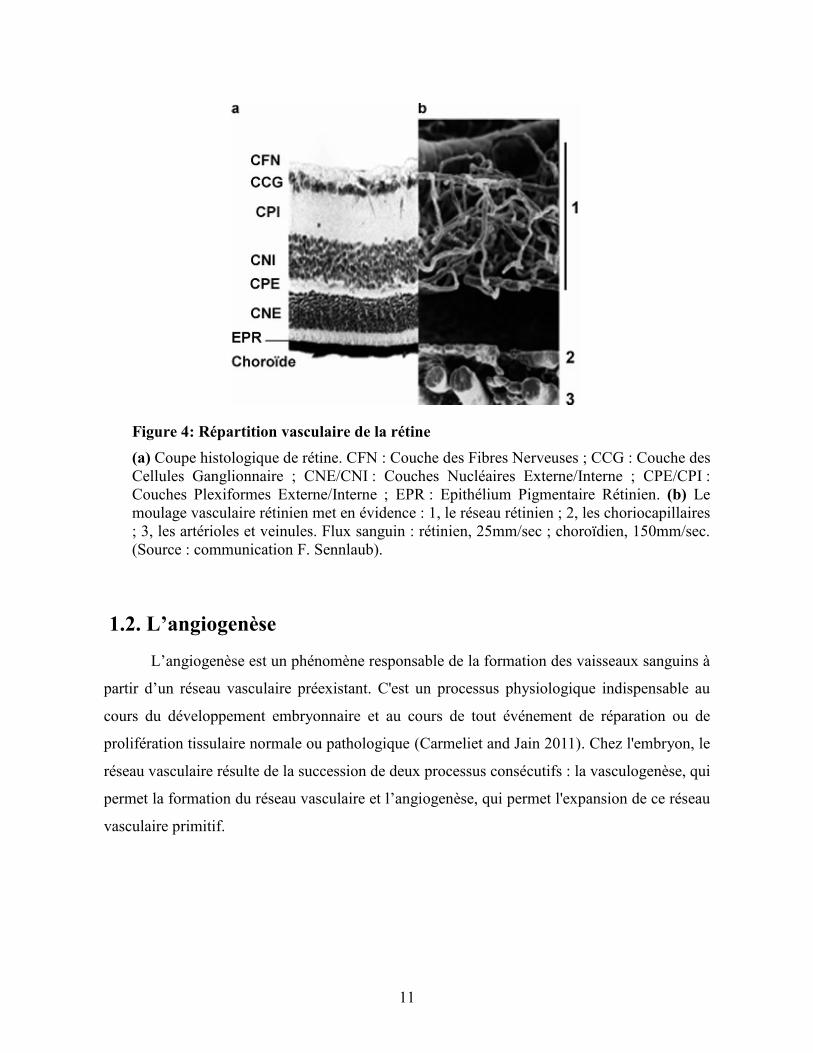
Figure 4: Répartition vasculaire de la rétine
(a) Coupe histologique de rétine. CFN : Couche des Fibres Nerveuses ; CCG : Couche des
Cellules Ganglionnaire ; CNE/CNI : Couches Nucléaires Externe/Interne ; CPE/CPI :
Couches Plexiformes Externe/Interne ; EPR : Epithélium Pigmentaire Rétinien. (b) Le
moulage vasculaire rétinien met en évidence : 1, le réseau rétinien ; 2, les choriocapillaires
; 3, les artérioles et veinules. Flux sanguin : rétinien, 25mm/sec ; choroïdien, 150mm/sec.
(Source : communication F. Sennlaub).
1.2. L’angiogenèse
L’angiogenèse est un phénomène responsable de la formation des vaisseaux sanguins à
partir d’un réseau vasculaire préexistant. C'est un processus physiologique indispensable au
cours du développement embryonnaire et au cours de tout événement de réparation ou de
prolifération tissulaire normale ou pathologique (Carmeliet and Jain 2011). Chez l'embryon, le
réseau vasculaire résulte de la succession de deux processus consécutifs : la vasculogenèse, qui
permet la formation du réseau vasculaire et l’angiogenèse, qui permet l'expansion de ce réseau
vasculaire primitif.

12
Figure 5: Vasculogenèse et angiogenèse
Formation du réseau vasculaire : étapes de vasculogenèse et d’angiogenèse. D’après de (Curtis
et al. 2012) .
1.2.1. La vasculogenèse
La vasculogenèse est un processus embryonnaire par lequel des cellules
hémangioblastiques se différencient en précurseurs de cellules endothéliales (angioblastes), afin
de former des ébauches de vaisseaux sanguins primitifs (Risau and Flamme 1995). Une fois ce
réseau formé, de nouvelles cellules endothéliales peuvent constituer de nouveaux capillaires par
bourgeonnement ou par division du réseau vasculaire préexistant à travers un processus
d'angiogenèse (Figure 5).

13
1.2.2. L’angiogenèse
L’angiogenèse est un processus peu fréquent chez les adultes, toutefois il est impliqué dans
certaines conditions physiologiques, notamment lors du cycle menstruel, la reproduction, la
cicatrisation ainsi que la régénération tissulaire (Carmeliet 2000; Otrock et al. 2007).
L'angiogenèse peut se dérouler selon trois mécanismes (Figure 6) :
• Bourgeonnement : c'est le phénomène le plus courant et le mieux caractérisé. Cette étape
dépend relativement du VEGF qui participe à la migration, la prolifération et la survie
des cellules endothéliales;
• Élargissement : C’est un phénomène basé sur l’élargissement et la séparation des
vaisseaux sanguins déjà existants;
• Séparation des vaisseaux sanguins : Les cellules poussent à l’intérieur des vaisseaux
pour former des canaux des vaisseaux séparés.
Figure 6: Mécanisme d’angiogenèse

14
La formation des vaisseaux peut avoir lieu par bourgeonnement, par élargissement et séparation
des vaisseaux sanguins formés (intussusception) ou par septation des vaisseaux déjà formés pour
créer des canaux vasculaires séparés. Adapté de (Carmeliet 2000).
1.2.2.1 L’extension du réseau vasculaire
L’angiogenèse bourgeonnante est de loin le mécanisme le plus étudié. Elle est caractérisée par
les étapes suivantes :
• Vasodilatation et perméabilité vasculaire
L'angiogenèse est associée à une vasodilatation induite par l'action du monoxyde d'azote (NO),
et dépend en grande partie du VEGF. Le monoxyde d'azote permet la dilatation des réseaux
existants afin de permettre la formation de nouveaux vaisseaux et le VEGF permet la
perméabilité vasculaire et la redistribution des molécules intercellulaires de type Vascular
Endothelial-cadherin (VE-cadhérine) et Platelet/Endothelial Cell Adhesion Molecule
(PECAM). Le VEGF est le facteur clé impliqué dans la plupart des évènements
morphogénétiques de l’angiogenèse et contrôle la perméabilité, la migration, la prolifération et
la survie des cellules endothéliales (Eliceiri et al. 1999; Silvestre and Levy 2002). La
perméabilité vasculaire est également augmentée par l'angiopoïétine (Ang2). Ce dernier
fragilise les vaisseaux en déstabilisant les contacts intercellulaires et permet la migration des
cellules endothéliales.
• Dégradation de la matrice extracellulaire
L'étape de la dégradation de la matrice extracellulaire implique plusieurs protéases notamment
l'activateur du plasminogène urokinase (uPA) et les métalloprotéinases MMPs (Matrix
Metalloproteinases). Ces molécules sont capables de dégrader la matrice extracellulaire et la
membrane basale permettant ainsi la formation d’un espace pour la migration des cellules
endothéliales. Durant cette étape, il y a une libération et activation des facteurs pro-
angiogéniques tels que le VEGF et le fibroblast growth factor 2 (FGF2) qui sont séquestrés à
l'intérieur de la matrice et qui sont primordiales pour les étapes suivantes (Carmeliet 2000; Jain
2003).
15
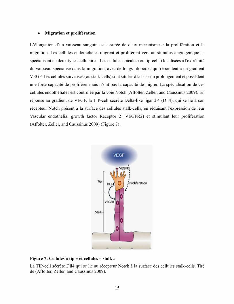
• Migration et prolifération
L’élongation d’un vaisseau sanguin est assurée de deux mécanismes : la prolifération et la
migration. Les cellules endothéliales migrent et prolifèrent vers un stimulus angiogénique se
spécialisant en deux types cellulaires. Les cellules apicales (ou tip-cells) localisées à l'extrémité
du vaisseau spécialisé dans la migration, avec de longs filopodes qui répondent à un gradient
VEGF. Les cellules suiveuses (ou stalk-cells) sont situées à la base du prolongement et possèdent
une forte capacité de proliférer mais n’ont pas la capacité de migrer. La spécialisation de ces
cellules endothéliales est contrôlée par la voie Notch (Affolter, Zeller, and Caussinus 2009). En
réponse au gradient de VEGF, la TIP-cell sécrète Delta-like ligand 4 (Dll4), qui se lie à son
récepteur Notch présent à la surface des cellules stalk-cells, en réduisant l'expression de leur
Vascular endothelial growth factor Receptor 2 (VEGFR2) et stimulant leur prolifération
(Affolter, Zeller, and Caussinus 2009) (Figure 7) .
Figure 7: Cellules « tip » et cellules « stalk »
La TIP-cell sécrète Dll4 qui se lie au récepteur Notch à la surface des cellules stalk-cells. Tiré
de (Affolter, Zeller, and Caussinus 2009).

16
• Maturation et survie des vaisseaux sanguins
Pendant la maturation du réseau vasculaire, le tube néoformé acquiert une lumière et recrute des
cellules accessoires afin d'élaborer une matrice extracellulaire. Les cellules accessoires sont les
péricytes et les cellules des muscles lisses qui participent à l’inhibition de la prolifération et la
migration des cellules endothéliales. Les péricytes produisent l'angiopoiétine-1 (Ang-1) et -2
(Ang-2) qui se lient aux cellules endothéliales afin de stabiliser les nouveaux vaisseaux
(Maisonpierre et al. 1997).
1.2.2.2 La régulation de l'angiogenèse
L’angiogenèse est un processus complexe qui implique plusieurs voies de signalisation
afin de contrôler étroitement l’expression de facteurs pro-angiogéniques et anti-angiogéniques.
Parmi les régulateurs clés de l'angiogenèse on retrouve le VEGF (Byrne, Bouchier-Hayes, and
Harmey 2005), le facteur de croissance dérivé des plaquettes (PDGF), le FGF (Presta et al.
2005), le facteur de croissance épidermique (EGF) (Geiger and Peeper 2009), les Matrix
metalloproteinases (MMP) et leurs inhibiteurs Tissue inhibitors of metalloproteinases (TIMPs)
(Laka, Gondi et Rao, 2005) ainsi que les lysophospholipides (sphingosine-1-phosphate (S1P) et
les acides lysophosphatidiques (LPA)) (English et al. 2002) (Figure 8).

17
Figure 8: Balance angiogénique
L’angiogenèse est contrôlée par des facteurs pro- et anti-angiogéniques. Adapté de (Hanahan
and Folkman 1996).
a) Les principaux activateurs de l'angiogenèse
Plusieurs facteurs pro-angiogéniques agissent au niveau des différentes étapes de
l’angiogenèse en modulant l’activité des cellules endothéliales (perméabilisation, prolifération,
différenciation, migration). Cinq facteurs sont exposés ci-dessous à savoir :
• Vascular Endothelial Growth Factor (VEGF)
Le VEGF a été identifié en 1983 par l’équipe de Senger, dans le surnageant d’une lignée de
cellules tumorales (Senger et al. 1983). L’équipe avait mis en évidence que cette molécule
induisait une augmentation de la perméabilité vasculaire, d’où son premier nom de facteur de
perméabilité vasculaire (VPF). La séquence du VEGF a été découverte en 1989 par l'équipe de
Ferrada et Henzel à partir du VEGF purifié provenant d’un milieu conditionné de cellules
hypophysaires (Ferrara and Henzel 1989).
Le VEGF appartient à une famille de glycoprotéines constituée de six homodimères (VEGF A,
B, C, D, E) (Ferrara and Henzel 1989) jouant un rôle crucial dans l’angiogenèse en intervenant
dans la formation des vaisseaux (Shams and Ianchulev 2006) et du facteur de croissance

18
placentaire (PIGF) (Koch et al. 2011). Chaque isoforme est générée à partir d’un épissage
alternatif. Les différentes isoformes du VEGF peuvent se lier à trois récepteurs : le VEGFR1
(ou fms-related tyrosine kinase-1; Flt- 1), le VEGFR2 (ou fetal liver kinase 1; ou Flk-1 kinase
insert domain-containing receptor ; KDR) et le VEGFR3 (ou fms-related tyrosine kinase-4; Flt-
4).
Elles peuvent également se lier à des cofacteurs : les neuropilines 1 et 2 (NP1 et NP2). Le VEGF
A et B sont principalement impliqués dans la perméabilité et l'épanchement vasculaire impliqué
dans l'angiogenèse. Le VEGF C et D sont impliqués dans la lymphangiogenèse.
Le VEGF stimule la prolifération des cellules endothéliales et leur migration via de nombreuses
voies incluant les protéines kinases (MAK), extracellular signal-regulated kinases (ERK),
mitogen-activated protein-kinases p38 (MAPK p38) et C-junN-terminal kinase (JNK) et les
membres de la famille RhoGTPase (Papetti and Herman 2002). Il agit comme facteur de survie
des cellules endothéliales via l'activation de la voie de la kinase phosphatidyl-inositol 3
(PI3K)/RAC-alpha serine/threonine-protein kinase (AKT) et via son association avec l'integrine
αvβ3 et l'activation de kinase d'adhésion focale (Milkiewicz et al. 2006) (Figure 9).

19
Figure 9: Famille des VEGFs et leurs récepteurs
La liaison des différents membres de la famille VEGF aux récepteurs VEGFR1 et VEGFR2
induit l'angiogenèse, tandis que le VEGFR3 induit la lymphangiogenèse et il est
occasionnellement impliqué dans l'angiogenèse. Les co-récepteurs NP-1 et NP-2 peuvent se lier
aux récepteurs du VEGF. Tiré de (Ellis and Hicklin 2008).
Le VEGF joue un rôle primordial dans la néovascularisation rétinienne et choroïdienne. Des
études ont montré qu’il est exprimé dans l’œil au niveau des cellules gliales de Müller, les
cellules endothéliales, les astrocytes, les cellules ganglionnaires et l’EPR (Aiello et al. 1995;
Pierce et al. 1995; Stone et al. 1996). Il est impliqué dans plusieurs pathologies oculaires telles
que la dégénérescence maculaire liée à l’âge, l’occlusion veineuse rétinienne et la rétinopathie
diabétique (Shams and Ianchulev 2006).

20
• Facteur de croissance dérivé des plaquettes (PDGF)
Le PDGF est une glycoprotéine sécrétée principalement par les plaquettes et joue un rôle crucial
dans l’angiogenèse. Ce facteur est impliqué dans la prolifération, la croissance et la survie des
péricytes et des cellules musculaires lisses afin de stabiliser le réseau vasculaire (Lindahl et al.
1997). Des travaux récents l’ont mis en évidence dans les cellules endothéliales, les fibroblastes
et les macrophages (Siegbahn et al. 1990). Le PDGF est un dimère qui donne naissance à trois
isoformes AA, AB, BB. Il a trois récepteurs à activité tyrosine kinase : le PDGFR-αα, le
PDGFR-αβ et le PDGFR-ββ (Westermark and Heldin 1987). Sa voie de signalisation active les
MAP-Kinases ainsi que la voie PI3K/AKT, favorisant la maturation et la stabilisation des
néovaisseaux (Yamazaki et al. 2009; Mellgren et al. 2008).
• Angiopoiétines 1 et 2 (Ang-1 et Ang-2)
Les Ang-1 et Ang-2 sont sécrétés principalement par les cellules murales (péricytes et cellules
musculaires lisses) ainsi que les cellules endothéliales (Jain 2003). L'Ang-1 joue un rôle dans la
croissance et le maintien du vaisseau sanguin. Il se lie à son récepteur Tie-2, présent à la surface
des cellules endothéliales et induit la voie de PI3K/AKT en activant la sous unité p85 de PI3
kinase et par la suite la protéine kinase AKT (Kim et al. 2000). L'Ang-1 induit la survie des
cellules endothéliales en plus de stabiliser les interactions entre les cellules murales et les
cellules endothéliales en recrutant des péricytes.
L’Ang-2 est un antagoniste de la même affinité que l'Ang-1, il permet d’augmenter la
perméabilité vasculaire. Il se lie au récepteur Tie-2 induisant ainsi une inhibition de la
phosphorylation du récepteur Tie-2. L’Ang-2 déstabilise l'endothélium en induisant le
détachement de la matrice et des péricytes (Gardner et al. 1997), rendant le réseau vasculaire
plus susceptible à répondre aux facteurs pro-angiogéniques. En absence de VEGF, l’Ang-2
déstabilise les vaisseaux en dégradant des réseaux vasculaires.
• Facteurs de croissance des fibroblastes (FGF)
La famille des FGFs comprend 23 membres, qui se lient aux récepteurs à activité tyrosine kinase
(FGFR1, R2, R3, R4). Le FGF1 et le FGF2 jouent un rôle important dans plusieurs phénomènes

21
physiologiques ainsi que pathologiques comme le développement embryonnaire, l'angiogenèse,
la vasculogenèse et la cicatrisation (Presta et al. 2005; Bottcher and Niehrs 2005; Grose and
Dickson 2005; Ortega et al. 1998).
Le FGF1 est exprimé principalement par les cardiomyocytes et les cellules de muscles lisses. Il
induit l'angiogenèse en activant les voies mitogéniques. Le FGF 2 est un polypeptide de 18 kDa,
qui se présente sous cinq isoformes produites par un seul ARNm. Il est sécrété principalement
par les cellules myocardiques, les cellules endothéliales et les cellules musculaires lisses. Il
induit la prolifération et la migration des cellules endothéliales à travers l’activation de la voie
de signalisation MAP kinase p38. Il est aussi capable d'activer la voie de signalisation tyrosine
kinase en activant la phosphorylation des phospholipases C (PLC) et l'expression des gènes
impliqués dans le cycle cellulaire (Bikfalvi 2003).
• Transforming growth factor β (TGF-β)
Le TGF-β est une cytokine multifonctionnelle générée sous forme de précurseur dimérique clivé
par des protéases. Il est secrété par plusieurs types cellulaires, incluant les cellules endothéliales
et les cellules murales. Le TGF-β peut lier deux types de récepteurs à activité sérine/thréonine
kinase, les récepteurs de types I aussi appelés activin receptor-like kinase (ALKs) tels que ALK5
et les récepteurs de type II tels que TGF-β type (TβRII). Le TGF-β régule la maturation des
vaisseaux sanguins via l'activation des récepteurs ALK5 et ALK1 (Goumans et al. 2002). Cette
cytokine joue un rôle complexe dans l'angiogenèse car elle peut à la fois activer ou inhiber
l'angiogenèse (Pepper 1997). À faible concentration elle contribue à augmenter la régulation du
VEGF et des protéases comme (uPA) à forte concentration elle stimule la reconstruction de la
membrane basale et la différenciation des cellules mésenchymateuses en cellules murales (Jain
2003).
b) Les inhibiteurs de l'angiogenèse
L'équilibre entre les facteurs pro-angiogéniques et anti-angiogéniques permet de maintenir une
angiogenèse quiescente.

22
• Thrombospondine-1 (TSP-1)
La TSP-1 fait partie d'une large famille de glycoprotéines matricielles. La TSP-1 est un ligand
du récepteur scavenger CD36. Il a été démontré, in vitro, sur des cellules endothéliales que la
liaison du TSP-1 sur son récepteur CD36 empêchait la formation de tube (Jimenez et al. 2000).
Cet effet anti-angiogénique médié par la signalisation du CD36, induit l’expression des facteurs
anti-angiogéniques et inhibe celle des facteurs impliqués dans la prolifération et la migration
(Jimenez et al. 2000). La signalisation du complexe TSP-1/CD36 est initiée par le recrutement
de fyn qui est une protéine tyrosine kinase de la famille src, qui active la protéine kinase p38,
JNK et la caspase 3 induisant l’apoptose (Jimenez et al. 2000). Ce complexe est capable
également de contrôler l’expression du VEGF en inhibant la voie de signalisation AKT (Sun et
al. 2009).
La TSP-1 peut également exercer son action anti-angiogénique de façon indirecte en inhibant
l’activation de MMP-9 bloquant ainsi la dégradation de la matrice et empêche la signalisation
du VEGF (Bikfalvi 2003) (Figure 10).
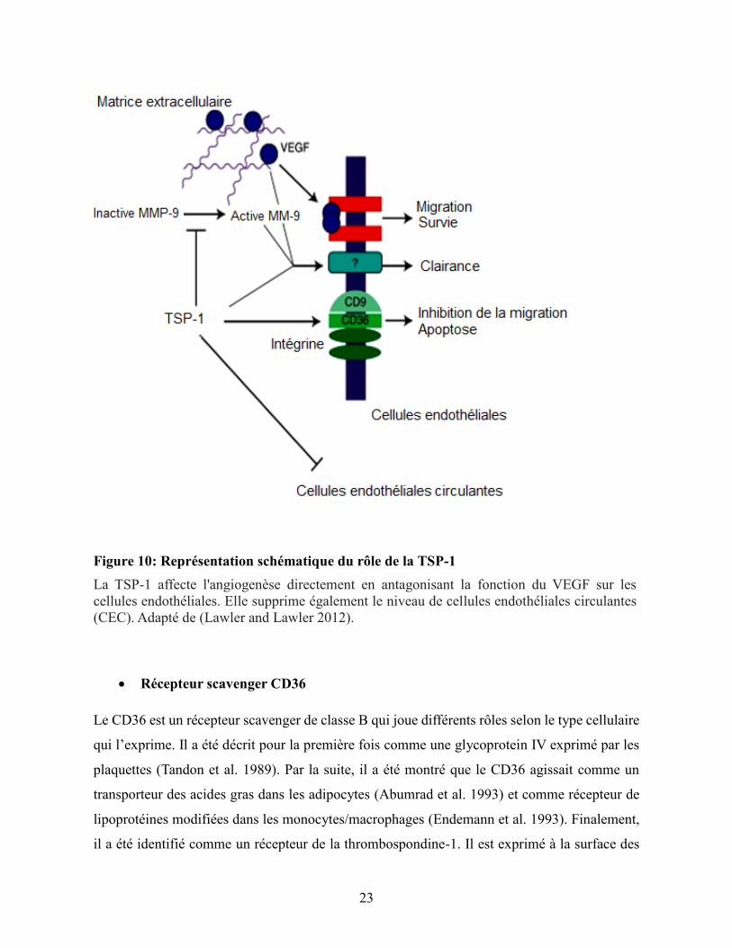
23
Figure 10: Représentation schématique du rôle de la TSP-1
La TSP-1 affecte l'angiogenèse directement en antagonisant la fonction du VEGF sur les
cellules endothéliales. Elle supprime également le niveau de cellules endothéliales circulantes
(CEC). Adapté de (Lawler and Lawler 2012).
• Récepteur scavenger CD36
Le CD36 est un récepteur scavenger de classe B qui joue différents rôles selon le type cellulaire
qui l’exprime. Il a été décrit pour la première fois comme une glycoprotein IV exprimé par les
plaquettes (Tandon et al. 1989). Par la suite, il a été montré que le CD36 agissait comme un
transporteur des acides gras dans les adipocytes (Abumrad et al. 1993) et comme récepteur de
lipoprotéines modifiées dans les monocytes/macrophages (Endemann et al. 1993). Finalement,
il a été identifié comme un récepteur de la thrombospondine-1. Il est exprimé à la surface des
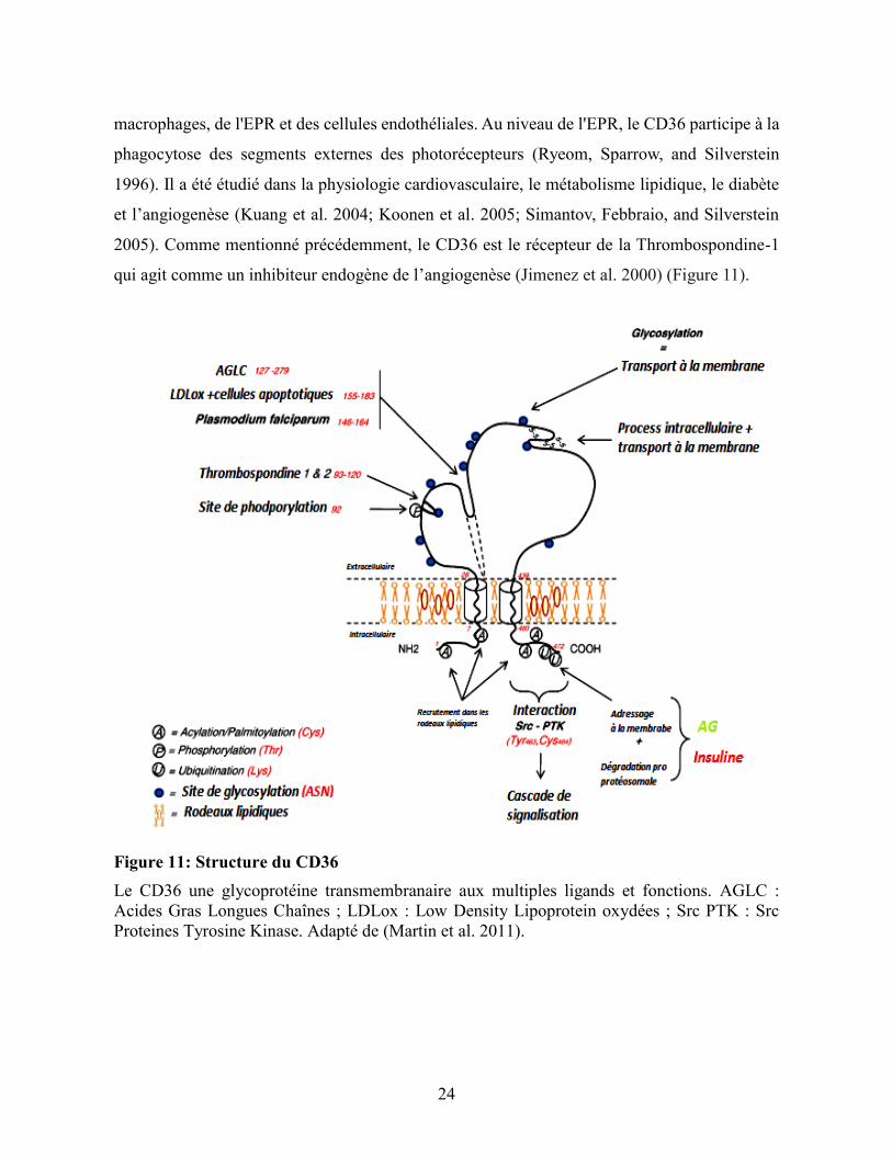
24
macrophages, de l'EPR et des cellules endothéliales. Au niveau de l'EPR, le CD36 participe à la
phagocytose des segments externes des photorécepteurs (Ryeom, Sparrow, and Silverstein
1996). Il a été étudié dans la physiologie cardiovasculaire, le métabolisme lipidique, le diabète
et l’angiogenèse (Kuang et al. 2004; Koonen et al. 2005; Simantov, Febbraio, and Silverstein
2005). Comme mentionné précédemment, le CD36 est le récepteur de la Thrombospondine-1
qui agit comme un inhibiteur endogène de l’angiogenèse (Jimenez et al. 2000) (Figure 11).
Figure 11: Structure du CD36
Le CD36 une glycoprotéine transmembranaire aux multiples ligands et fonctions. AGLC :
Acides Gras Longues Chaînes ; LDLox : Low Density Lipoprotein oxydées ; Src PTK : Src
Proteines Tyrosine Kinase. Adapté de (Martin et al. 2011).

25
• Le facteur dérivé de l’épithélium pigmentaire (PEDF)
Le PEDF est une glycoprotéine qui appartient à la superfamille des inhibiteurs de protéinases à
sérine " sérine protease inhibiteur" aussi appelé serpines. Il a été isolé à partir des cellules de
l’EPR de fœtus humain (Tombran-Tink and Johnson 1989). Il est sécrété par différents organes
tels que : Le foie, l’estomac, les ovaires et le cerveau (Bilak et al. 2002; Browne et al. 2006;
Cheung et al. 2006). C'est une protéine qui montre diverses activités telles que des effets anti-
angiogéniques, neurotrophiques, neuroprotecteurs et anti-tumorales.
Le PEDF module l'angiogenèse en inhibant directement la prolifération et la migration en
régulant le récepteur VEGFR et la survie des cellules endothéliales. Il active également la voie
de signalisation Fas ligand (FasL) induisant l’apoptose (Aurora et al. 2010).
• L’angiostatine
L'angiostatine fait partie des nombreux fragments protéolytiques issus du clivage du
plasminogène de la matrice extracellulaire par des protéases. L'angiostatine a été initialement
identifiée à partir de sérum et d'urine de souris portant des tumeurs pulmonaires de lewis LLC
(O'Reilly et al. 1994). Il a été démontré que l’angiostatine pouvait interagir avec des sites
membranaires présent à la surface des cellules endothéliales tels que les -ATP synthases et
les 3- intégrines. Ces interactions régulent négativement l'angiogenèse en inhibant
principalement la prolifération et la migration des cellules endothéliales (Moser et al. 1999;
Tarui et al. 2002).
• L’endostatine
C’est une protéine qui a été identifiée dans les cellules endothéliales hémangiomateuses et qui
joue un rôle important dans l'inhibition de l'angiogenèse. Elle correspondant au fragment C-
terminal du collagène de type XVIII (O'Reilly et al. 1994). Elle régule positivement des facteurs
anti-angiogéniques et négativement de nombreux facteurs pro-angiogéniques, entre autres
l'ensemble de la signalisation induite par le VEGFA en interférant avec le VEGFR2 (Kim et al.
2002). D'autre part l'endostatine peut se lier à une intégrine inhibant la voie de signalisation
ERK 1/2 (Sudhakar et al. 2003). Des études in vivo ont montré que cette molécule inhibe la

26
croissance des tumeurs primaires et prévient la formation de nouveaux vaisseaux ainsi que la
croissance des métastases (O'Reilly et al. 1997).
• Les neurotrophines
Les neurotrophines appartiennent à une famille de facteurs de croissance qui régule la survie
cellulaire neuronale, le guidage axonal, la différenciation cellulaire et la neuroprotection. Cette
famille inclut : Le nerve growth factor (NGF), le brain derived neurotrophic factor (BDNF), les
neurotrophines NT-3 à NT-7. Les neurotrophines jouent différents rôles, selon le type cellulaire;
leur rôle physiologique principal c'est la croissance neuronale, le maintien de la stimulation
neuronale ainsi que l'inhibition de l'apoptose. Ces facteurs se lient à deux types de récepteurs :
Les récepteurs tropomyosin-receptor-kinase (TRKs) à activité tyrosine kinase et les récepteurs
p75NTR. Dans notre étude nous nous sommes particulièrement intéressés au NGF et au
récepteur p75NTR.
➢ Le récepteur p75NTR
Il fait partie de la famille des récepteurs Fas (Apo-1) et tumor necrosis factor receptor I et II
(TNFR I et II). C’est un récepteur protéique transmembranaire de type I avec une partie
extracellulaire contenant quatre domaines riches en cystéine, une partie transmembranaire et
une partie cytoplasmique qui contient le domaine de la mort cellulaire (Liepinsh et al. 1997).
Initialement, il a été caractérisé comme un récepteur spécifique du NGF. Par la suite il a été
démontré qu’il était capable de lier les autres neurotrophines ainsi que d’autres types de ligands.
Dans la rétine, il est localisé principalement au niveau des cellules gliales de Müller et dans les
prolongements des cellules ganglionnaires (Hu, Yip, and So 1998). Physiologiquement, il est
peu exprimé par les cellules endothéliales quiescentes. Cependant, suite à l'activation et
l'augmentation de la prolifération des cellules endothéliales, son expression peut augmenter
(Caporali et al. 2008). Le p75NTR peut se dimériser ou agir en co-récepteur associé au récepteur
TRK. Selon le type cellulaire, les voies de signalisation peuvent induire l’apoptose ou de survie
cellulaire. Le p75NTR est un récepteur qui ne possède pas d'activité catalytique propre, ainsi il
a besoin de recruter des protéines adaptatrices intracellulaires menant à l'activation de diverses
voies de signalisation. Il peut induire l’apoptose via le recrutement de molécules adaptatrices
qui se fixent au domaine Chopper (Coulson et al. 1999; Coulson et al. 2000), ou au domaine de

27
mort, activant la GTPase Rac qui active à son tour la kinase JNK. Par la suite, JNK active le
facteur de transcription c-jun, p53, et les protéines pro-apototiques à domaine Bad (Bcl-2
Antagonist of cell Death) et Bim (Bcl-2-Interacting Mediator of cell death), suivi par le
relargage du cytochrome c dans le cytosol qui stimule à son tour les caspases 9, 6 et 3 (Nykjaer,
Willnow, and Petersen 2005) (Figure 12).
Figure 12: Représentation schématique des voies de signalisation du p75NTR
Adapté de (Molloy, Read, and Gorman 2011).
Le p75NTR induit soit la mort ou la survie cellulaire. La voie de survie cellulaire est induite
principalement par l'activation du facteur NF-κB (Hamanoue et al. 1999). D'une part le NF-κB
induit la transcription de clAP-1 et clAP-2 des inhibiteurs de l'apoptose, et d'autre part, il peut
bloquer la voie JNK qui induit l'apoptose. Il peut aussi induire la survie cellulaire en activant
directement la voie de PI3K/AKT généralement associé au récepteur TRK.

28
➢ Le NGF
Le NGF est le premier facteur neurotrophique découvert dans les années cinquante par l'équipe
S Cohen et R Levi-Montalcini, il joue un rôle essentiel dans le développement, la survie et la
différentiation des cellules neuronales (Cohen, Levi-Montalcini, and Hamburger 1954). De plus,
des études ont montré son implication dans le guidage axonal, la plasticité synaptique et la
différenciation cellulaire. In vitro, des études ont démontré que le NGF induit la croissance des
fibres nerveuses lors de l'implantation d'un sarcome de souris dans un embryon de poulet
(Cohen, Levi-Montalcini, and Hamburger 1954). In vivo, l'utilisation d'un anticorps dirigé contre
le NGF provoque la privation des facteurs endogènes et cause un déficit au niveau de
l'innervation sympathique. En plus de son rôle essentiel dans la survie et la différentiation des
cellules neuronales, le NGF est aussi impliqué dans l'angiogenèse.
Des études ont démontré que le NGF, via le récepteur TRK-A, avait un effet pro-angiogénique
qui augmentait la tumorigenèse des cellules cancéreuses pancréatiques et ovariennes (Tacconelli
et al. 2004). Dans la rétine, le NGF est sécrété principalement par les cellules de Müller et de
l'EPR (Wang et al. 2016) (Figure 13).
Figure 13: différentes sources de synthèses du NGF
Le NGF peut être synthétisé par différentes cellules inflammatoires ainsi que des cellules
structurales. Tiré de (Advenier 2006).

29
➢ Le pro-NGF
Le NGF est une protéine traduite à partir de deux transcrits pour produire deux pré-pro-NGF de
tailles de 34 et 27 kDa. Le pré-pro-NGF peut être soumis à un clivage dans le réticulum
endoplasmique et donner deux isoformes pro-NGF de 32 et 25 kDa (Fahnestock et al. 2001;
Buttigieg, Kawaja, and Fahnestock 2007). Par la suite, le pro-NGF subit des modifications post-
traductionnelles au niveau du C et N-terminales pour générer le NGF mature (Darling et al.
1983).
Durant longtemps, le pro-NGF était considéré comme une forme intermédiaire dans la synthèse
du NGF mature sans aucun rôle. Par la suite, de nombreuses études ont démontré que le pro-
NGF est sécrété majoritairement par les cellules de la prostate, les follicules capillaires, les
neurones sympathiques (Chen, Dicou, and Djakiew 1997; Delsite and Djakiew 1999; Yardley
et al. 2000; Smith et al. 2002). De plus, l’équipe de Shanab a démontré que le pro-NGF se lie
sélectivement au récepteur p75NTR et induit l’apoptose des cellules endothéliales rétiniennes
(Shanab et al. 2015).
La liaison du pro-NGF au p75NTR est associée au recrutement de la sortiline qui est un co-
récepteur appartenant à la famille des protéines VPS10 « Vacuolar carboxy-peptidase Sorting
receptor-10 » (Willnow, Petersen, and Nykjaer 2008). Cette famille participe généralement aux
transferts protéiques à travers le réseau Golgien, pour la maturation des protéines qui seront
exprimées à la membrane plasmique (Willnow, Petersen, and Nykjaer 2008). La sortiline est
une protéine transmembranaire de type I, exprimée en grande partie dans le système nerveux
central au cours du développement chez l’humain (Nielsen et al. 2001). De nombreux travaux
sont actuellement en cours afin de déterminer sa cascade de signalisation. Des résultats semblent
suggérer que la sortiline joue un rôle dans la signalisation pro-apoptotique impliquant les
protéines adaptatrices NRIF et NRAGE (Linggi et al. 2005; Bertrand et al. 2008).
1.3. L’inflammation et l'angiogenèse
L'angiogenèse est souvent précédée par l'inflammation qui fait intervenir les mécanismes de la
prolifération, la migration et le recrutement des cellules inflammatoires. Ces dernières
interviennent à plusieurs étapes de l’angiogenèse et sont capables de libérer plusieurs facteurs

30
angiogéniques (VEGF, PDGF, TGF). De plus, les médiateurs inflammatoires comme la
prostaglandine E, interleukine 6 (IL-6), et IL-1 augmentent l'expression du VEGF-A (Dvorak
2002).
1.3.1. Les macrophages
Décrits la première fois par Elie Metchnikov en 1883 les macrophages sont des acteurs
nécessaires à l'homéostasie au niveau tissulaire de l'organisme. Leur principal rôle se définie
dans la défense de l'hôte, la réparation et la régulation de l'immunité (Mechnikov 1988). Ils font
partie du système mononucléaire qui regroupe également les cellules dendritiques et les
précurseurs de la lignée myéloïde. Les monocytes sont les cellules circulantes dans le sang qui
représentent le réservoir périphérique des macrophages. Il existe cependant des macrophages
résidents dans certains tissus tels que le foie (les cellules de Kupffer), la peau (les cellules de
Langerhans) ou encore le système nerveux central (la microglie) (Dheen, Kaur, and Ling 2007;
Ingber 2007; Sheth and Bankey 2001). Lors du processus inflammatoire, les monocytes sont
recrutés au site inflammatoire et se différencient en macrophages. Pendant la réponse
immunitaire les macrophages peuvent avoir différentes fonctions. Leurs principales propriétés
sont la phagocytose (FCR, Complement Receptor 1 (CR1) récepteur mannose) (Zhao et al.
2015) (Griffin et al. 1975), la présentation des antigènes (CD80 et CD86) et la sécrétion des
chimiokines et des cytokines pro- (ex : IL-1a et b, IL-6, TNFα) et anti-inflammatoire (IL-10 et
le TGFβ).
1.3.1.1. Polarisation des macrophages
La polarisation des macrophages M1/M2 est un phénomène très complexe qui fait intervenir de
nombreux facteurs immunomodulateurs. Plusieurs études ont montré que les macrophages
passent d'un état polarisé à un autre, en réponse à leur environnement.
1.3.1.1.1. Les macrophages de types M1
Les macrophages M1 sont impliqués dans l'initiation de la réponse inflammatoire et
l’élimination des pathogènes. Ils sont caractérisés par une activité microbicide et tumoricide

31
augmentées. Une réponse inflammatoire soutenue engendre des dommages tissulaires
importants, qui peuvent générer plusieurs pathologies comme la polyarthrite rhumatoïde.
La stimulation des monocytes avec du LPS/interferon gamma (IFN-γ) ou du granulocyte
macrophage colony-stimulating factor (GM-CSF) ainsi que du TNF-α peuvent polariser les
macrophages en type M1 (Gensel and Zhang 2015) (John C Gensel 2014). Cette stimulation
active la voie de signalisation des facteurs de transcription STAT1 et NF-κB qui, à leur tour,
activent le promoteur des gènes codant les sous-unités p35 et p40 de l’IL-12.
Les macrophages M1 sont caractérisés par une production de niveau élevé d'IL-1β, IL-12, IL-
18, l'IL-23, TNF-α, et CXCL10 (Ley et al. 2006). Fonctionnellement, ils peuvent induire une
réponse cytotoxique suite à l'augmentation de l'enzyme iNOS responsable de la production de
NO. En plus de leur effet pro-inflammatoire, il a été démontré que les M1 avaient également un
effet anti-angiogénique. Ils secrètent des facteurs comme le TNF-α et le NO, capables d’induire
la mort cellulaire de façon indirecte via l’activation des protéines du complément. Le rôle du
TNF-α dans l'angiogenèse est complexe; In vivo, le TNF-α est capable d'induire la nécrose des
tumeurs transplantées chez la souris (Carswell et al. 1975). Une étude clinque a démontré la
capacité du TNF-α à induire sélectivement la mort cellulaire des vaisseaux sanguins intra-
tumoraux suite à son administration locale en association à la chimiothérapie (Lejeune, Ruegg,
and Lienard 1998). Toutefois, une étude in vivo du modèle d’une souris transgénique déficient
en TNF-α démontre que le TNF-α stimule l'angiogenèse (Keffer et al. 1991). D'autres études in
vitro ont démontré son effet prolifératif (Kaiser and Polk 1997; Montesano et al. 2005). L'IL-12
est une cytokine fortement exprimée par les M1. Elle a une action anti-tumorale et anti-
métastatique et des études ont démontré sa capacité à inhiber l’angiogenèse dans un modèle de
néovascularisation cornéenne chez la souris en activant la voie de signalisation de IFN qui est
une protéine à action anti-proliférative et anti-angiogénique. L'IFN augmente l'expression de la
protéine anti-angiogénique inducible protéine 10 (IP-10) (Voest et al. 1995) (Figure 14).
1.3.1.1.2. Les macrophages de types M2
Il a été montré que la stimulation de monocytes avec différentes interleukines, l'IL-4, IL-10, IL-
13 ou des corticostéroïdes induisaient une polarisation des monocytes en macrophages de type
M2 (activation dite alternative) (Gensel and Zhang 2015). La caractérisation des macrophages

32
M2 a révélé qu’ils pouvaient se diviser en sous-classes : M2a, M2b et M2c. Ces trois sous-types
partagent certaines propriétés fonctionnelles dont une augmentation de l’expression de
l'arginase-1 et de l'IL-10, une diminution de l’IL-12 et l’IL-23, et conservent toujours leur
propriété anti-inflammatoire (Sica and Mantovani 2012; Murray and Wynn 2011). En plus de
leur différence dans la production des cytokines/interleukines, les macrophages M2 se
distinguent des macrophages M1 par la présence, à leur surface, des récepteurs de mannose
(CD206) et par le métabolisme de l'arginine. Le rôle des macrophages M2 est décrit comme
intervenant dans la réparation des tissus et le retour à l'homéostasie (Lumeng, Bodzin, and
Saltiel 2007). En fait, les M2 sont capables de métaboliser l'arginine en urée et ornithine, un
précurseur des polyamines (impliquées dans la division et la croissance cellulaire) et de la
proline un constituant du collagène (Hesse et al. 2001; Mosser 2003).
Récemment, une autre classe de macrophages M2 a été identifiée associée aux tumeurs, les
Tumor-Associated Macrophages (TAMs). Les TAMs sont des macrophages qui favorisent
l’angiogenèse dans plusieurs types de cancers. Des études ont conclu que les TAMs sont des
cellules qui favorisent l’angiogenèse et la progression de la tumeur. Parmi ces recherches, les
travaux de Shojaei et al., et de Gorelik E et al., ont démontré que dans un modèle de cancer
pancréatique les TAMs et les neurotrophiles étaient la source principale des facteurs pro-
angiogéniques (Gorelik et al. 1982; Shojaei et al. 2008). Ces macrophages sécrètent des
métalloprotéases et plusieurs facteurs de croissance (l’EGF, TGF-β, bFGF, PDGF) qui
favorisent la prolifération cellulaire (Solinas et al. 2010; Solinas et al. 2009) (Figure 14).

33
Figure 14: Polarisation des macrophages
Les macrophages se constituent d’une population hétérogène formée de deux classes principales
: M1 et M2, qui se distinguent par le type de cytokines produites et des marqueurs membranaires
distincts. Tiré de (Solinas et al. 2009).

34
1.4. Pathologie de l’œil
La néovascularisation choroïdienne (NVC) est la formation de nouveaux vaisseaux qui
traversent la membrane choriocapillaire à travers l’espace sous-rétinien. Les pathologies
oculaires liées à la NVC se traduisent, le plus souvent, par une malvoyance ou une perte d’acuité
visuelle conduisant ainsi à une diminution de la qualité de vie. Citons quelques pathologies
oculaires liées à la NVC :
Dans les conditions dégénératives, on retrouve la dégénérescence maculaire, la myopie forte,
les stries angioides. Dans les conditions inflammatoires ou infectieuses on retrouve la choroïdite
multifocale, le pseudo-histoplasmose, la sarcoïdose. Dans les tumeurs choroïdiennes on a le
mélanome et l'hémangiome choroïdien.
1.4.1. Dégénérescence maculaire liée à l'âge (DMLA)
La DMLA constitue la première cause de malvoyance après 50 ans dans les pays industrialisés.
Cette maladie affecte la rétine et plus précisément la macula, elle cause une baisse progressive
de la vision aboutissant à une perte de la vision centrale. C’est une maladie multifactorielle
associant des facteurs de risques environnementaux et génétiques (Suner et al. 2004). On
distingue deux formes de la DMLA :
• La forme sèche ou atrophique se traduit par une atrophie de la macula, associée à une
accumulation de drusen qui est des dépôts jaunâtres entre la choroïde et l'EPR;
• La forme humide ou néovasculaire représente 20% de la DMLA, elle correspond à
l’apparition anormale des vaisseaux au niveau de la choroïde. Ces vaisseaux laissent
échapper des fluides et du sang causant la formation d'une poche sous la macula,
entrainant ainsi une perte rapide de la vision centrale (Figure 15).

35
Figure 15: Développement de la néovascularisation
La forme humide de la DMLA est due à l'apparition anormale de néovaisseaux sous la rétine.
Ces néovaisseaux traversent la membrane de Brüch et s'étendent au niveau de l'espace sous
rétinien entrainant un saignement et une accumulation du liquide entre la membrane de Brüch
et l'EPR. Tiré de (http://www.scienceofamd.org/treat/)
1.4.2. Myopie forte
Constitue la première cause de baisse de vision chez les personnes de moins de 50 ans et la
deuxième cause de néovascularisation choroïdienne après la DMLA (Cohen et al. 1996). C'est
une maladie évolutive qui se définie par une distension excessive du globe oculaire. Les causes
de cette maladie ne sont pas bien comprises, mais elles sont liées à des anomalies biomécaniques
ou à des facteurs héréditaires. Les anomalies biomécaniques supposent que sous la pression
interne de l'œil la sclère s'étend et cause un étirement de la rétine qui provoquera par la suite une
dégénérescence rétinienne (Hotchkiss and Fine 1981; Avila et al. 1984) .
1.4.3. Hémangiome choroïdien
C’est un hématome vasculaire (une tumeur vasculaire bénigne), localisé principalement au
niveau de la choroïde. Deux types peuvent être signalés, l'hémangiome sporadique et
l'hémangiome diffus.

36
L'hémangiome sporadique est caractérisé par un décollement de la rétine exsudative et un
œdème maculaire. L'hémangiome diffus s'inscrit, le plus souvent, dans le syndrome de Sturge
Weber Krabbe, il est caractérisé par une dilatation vasculaire conjonctivale et épisclérale (Ruby
et al. 1992).
1.5. Traitements
Plusieurs traitements de la néovascularisation choroïdienne peuvent être décrits, citons entre
autres : La photocoagulation au laser, la thérapie photodynamique, la chirurgie, les traitements
médicamenteux. Cependant jusqu'à présent il n’y a malheureusement aucun traitement capable
de conserver la fonction visuelle centrale.
1.5.1. La photocoagulation au laser
Le traitement au laser (argon vert ou krypton rouge) a pour but de cicatriser et coaguler les
néovaisseaux par une élévation thermique afin de prévenir toute perte additionnelle de la vision.
Cette technique ne peut être utilisée que pour les vaisseaux extra-fovéolaires (ce qui représente
la minorité des cas), car elle entraine une destruction des photorécepteurs ainsi que l'apparition
d'un scotome central définitif conduisant à une baisse d'acuité visuelle centrale sévère. On peut
également constater un décollement de la rétine exsudative, un décollement de la choroïde ainsi
qu'une baisse de la vision périphérique.
1.5.2. La thérapie photodynamique
C’est une thérapie qui vise à détruire les néovaisseaux. Elle consiste à administrer en
intraveineux une substance photosensibilisante, la vertéporfine (visudyne). Cette substance
diffusante dans le réseau vasculaire est activée par exposition à un laser (690nm) dans la zone
pathologique qui aura été préalablement identifiée par angiographie (Pieramici et al. 2006;
Barbazetto et al. 2003). Cette exposition produit un effet photochimique détruisant les
néovaisseaux ciblés. Toutefois, ce traitement présente des effets secondaires tels que les
décollements de la rétine et les hémorragies sous-rétiniennes.
37
1.5.3. La chirurgie
La chirurgie de la région maculaire a beaucoup progressé au cours de ces dernières années.
Cependant, elle est souvent accompagnée d’un scotome central, un décollement rétinal et une
prolifération vitro-rétinienne. On retrouve deux différents types de chirurgie : La chirurgie des
membranes néovasculaires sous rétiniennes et la chirurgie translocation rétinienne. La première
est indiquée principalement lorsque la membrane néovasculaire est située dans l'espèce sous
rétinien en avant de l'EPR; elle consiste à faire une incision au niveau de la rétine, par la suite,
et à l'aide d'une pince coudée le chirurgien pourrait retirer les vaisseaux sanguins sous rétiniens.
La seconde consiste en un raccourcissement scléral temporal supérieur suivi par décollement
rétinien temporal supérieur et maculaire.
1.5.4. La radiothérapie
Utilisée dans de nombreuses maladies de la NVC, c'est une thérapie susceptible d'inhiber la
NVC dans la rétine. Elle utilise soit une irradiation par photons gamma ou par électrons produits
par un accélérateur linéaire, soit une irradiation par photons produits par un cyclotron. En 1993,
l’équipe de Chakravarthy a montré une diminution de la néovascularisation à travers une étude
concernant le traitement des patients atteints de la NVC avec des faibles doses de radioactivité
5Gy-36Gy (Chakravarthy, Houston, and Archer 1993). D'autres études, cependant ont démontré
des résultats très variables et contradictoires surtout à cause de la technique d'irradiation (type
de rayonnement, doses, fractionnement, étalement) (Martin et al. 1997; Spaide et al. 1998).
Signalons que l’atteinte de la choroïde, une neuropathie optique, un glaucome sont les
principaux effets indésirables de cette thérapie.
1.5.5. Les médicaments anti-angiogéniques
Ces médicaments visent à réduire substantiellement et immédiatement la présence de fluides
dans la rétine. Comme avancé plus haut, le VEGF est un puissant facteur pro-angiogénique
puisqu’ il induit la prolifération des cellules endothéliales ainsi que la perméabilité vasculaire.
Les anti-VEGF occupent une place cruciale dans le traitement de la NVC. Depuis 2006, c'est le
traitement principalement prescrit. Les autres mentionnés plus haut sont relégués au second
plan.

38
1.5.5.1. Pegaptanib
Le pegaptanib (Macugen) est le premier médicament anti-angiogénique, découvert en 2005, et
considéré plus performant que la photodynamique; il permet de prévenir la perte visuelle sévère.
Le pegaptanib est un oligonucléotide qui inhibe spécifiquement le VEGF-A (Fajnkuchen and
Cohen 2008).
Afin de confirmer son efficacité dans le traitement de la CNV suite à la DMLA, une étude
clinique contrôlée randomisée, à double issu et multicentrique a été menée (Gragoudas et al.
2004). Elle a été faite sur deux groupes, un groupe témoin et l’autre recevait le pagaptanib. On
a constaté que 70% des patients ont perdu moins de 15 lettres d'acuité visuelle, contre 50% du
groupe contrôle (placébo). Cependant, des effets secondaires apparaissent; les patients étaient
plus susceptibles d'avoir de l'inflammation au niveau de la chambre antérieure, opacité au niveau
du corps vitré ainsi qu'un décollement de la rétine.
1.5.5.2. Ranibizumab
Le ranibizumab est un fragment d'anticorps monoclonal humanisé dirigé contre le VEGF-A, qui
se lie à grande affinité aux isoformes VEGF121, VEGF110, VEGF165 (Kaiser 2006). Sa liaison
empêche celle de VEGF-A à son récepteur VEGF-R1. Afin de démontrer l'efficacité du
ranibizumab trois études randomisées en double aveugle, contrôlées ont été effectuées. L’étude
Marina (Rosenfeld et al. 2006), l’étude Anchor (Brown et al. 2006) et l'étude Pier (Regillo et al.
2008). Ces études ont comparé l'efficacité de deux doses soit 0,3mg ou 0,5mg. La posologie
recommandée est celle de 0,5mg, par injection intravitréenne, une fois par mois. Elle peut même
être tous les trois mois si l'injection chaque mois n'est pas possible. Sur le plan local, les effets
secondaires sont limités.
1.5.5.3. Bevacizumab
En 2004, le bévacizumab a été indiqué dans le traitement des cancers colorectaux métastatiques.
Par la suite, en 2007 il a été approuvée au Canada et a été utilisée pour le traitement de la forme
humide de la DMLA. C'est un anticorps humanisé dont le fragment FAB (Fragment anti-
binding) maturé est le ranibizumab. Le bévacizumab a été largement utilisé à cause de son faible
coût, son efficacité et sa plus longue demi-vie qui permet de diminuer la fréquence d'injection.

39
L'étude CATT (Comparison of Age-related macular degeneration Treatments Trials) menée au
États-Unis sur 1208 patients atteints de DMLA humide, a confirmé que le ranibizumab a la
même efficacité que le bevacimab ainsi que les mêmes effets secondaires (Comparison of Age-
related Macular Degeneration Treatments Trials Research et al. 2012). Dans l'ensemble, la
thérapie anti-angiogénique est bien tolérée, malgré les effets secondaires cardiovasculaires ou
rénaux (hypertension artérielle, protéinurie) liés en partie à la production de NO.
1.6. Microparticules (MPs)
1.6.1. Définition
La vésiculation membranaire et la génération de microparticules (MPs) sont des phénomènes
ubiquitaires finement régulés. Décrites la première fois par Wolf en 1967, les LMPs ont été
longtemps considérées comme des débris cellulaires dépourvues d'activité biologique (Wolf
1967). Elles sont des fragments membranaires (0,1-1 µm), qui se libèrent dans le milieu
environnant suite à l'activation ou l'apoptose des cellules (Figure 16). Chaque cellule soumise à
un stress est capable de produire des microparticules, qui auront des caractères spécifiques de la
cellule émettrice (Martinez et al. 2005). Ainsi, elles constituent une réserve de bio-effecteurs
circulants impliqués dans différents processus tels que la coagulation, l'inflammation et
l'angiogenèse (Mostefai, Andriantsitohaina, and Martinez 2008). La majorité des MPs détectées
dans le plasma proviennent principalement des cellules endothéliales et des cellules circulantes
telles que les plaquettes, les leucocytes et les érythrocytes. Elles sont anucléées et contiennent
un cytosquelette et une membrane riche en phosphatidylsérine (PSer).

40
Figure 16: Analyse en microscopie électronique à balayage de microparticules provenant
des cellules endothéliales
Cellules endothéliales quiescentes (a-c) ou stimulées par TNF-α (d-f). D’après (Combes et al.
1999).
Les MPs partagent des similitudes avec les corps apoptotiques et les exosomes. La mort
cellulaire par apoptose est un processus important pour le maintien de l'homéostasie d’un
organisme multicellulaire. L'apoptose cause la fragmentation de la cellule qui forme, par la suite,
des corps apoptotiques. Ces derniers contiennent du matériel nucléaire, une membrane riche en
phosphatidylsérine de taille supérieure à celle des MPs (Hristov et al. 2004) (Figure 17).

41
Exosomes Microparticules Corps
apoptotiques
Taille ˂ 0,1 µm 0,1à 1 µm 1 à 4µm
Centrifugation 100000g 20000g 16000g
Origine Corps
multivesiculaire
Membrane plasmatique Fragments
cellulaires
Production Activation
cellulaire
Activation cellulaire et/ou
apoptose précoce
Apoptose avancée
Externalisation
phosphatidylsérine
-/+ +++ +++
Marqueurs
protéiques
TSG101
CD63
Antigènes de la cellule
d'origine
Histones
Tableau I : Caractéristiques des différents types de vésicules extracellulaires
Adapté de (Mause and Weber 2010)
Les exosomes sont libérées dans le milieu extracellulaire après fusion des endosomes
multivésiculaires avec la membrane plasmique (Figure 17). Leur taille, inférieure à celles des
MPs, varie entre 0,1 à 1 µm. Ils sont d'origine endosomale et sont pauvres en PSer, mais enrichis
en tétraspanines (Tableau I).

42
Figure 17: Schéma représentatif de la libération des microvésicules membranaires dans
l'espace extracellulaire
A: Libération des exosomes suite au trafic d’endocytose intracellulaire et excrétion de
microvésicules de la membrane plasmique. Corps multivésiculaire (MVB), intraluminale
microvésicule (ILV). B: l'excrétion de microvésicules de la membrane plasmique et la
production des corps apoptotiques provenant de cellules apoptotiques. D'après (Mathivanan, Ji,
and Simpson 2010)

43
1.6.2. Formation des microparticules
Les MPs sont émises par la cellule sous l’effet d’un stress provoquant le réarrangement des
phospholipides membranaires. En conditions physiologiques, les phospholipides sont distribués
de façon asymétrique au sein de la bicouche lipidique, sur le feuillet externe on retrouve la
phosphatidylcholine et la sphingomyeline, sur le feuillet interne, les phosphatidylsérines et les
phosphatidyléthanolamines.
Le stimulus induit un influx calcique dans le cytosol entraînant une réorganisation du
cytosquelette (activité protéolytique des calpaînes) et modulant l’activité des transporteurs
transmembranaires des phospholipides : flippase, floppase et scramblase. Dans la cellule au
repos, ces transporteurs assurent l’asymétrie des phospholipides membranaires, avec une
externalisation précoce de la phosphatidylsérine. La flippase maintient la distribution des
phospholipides au repos, la floppase est directement responsable de la perturbation de
l'asymétrie des cellules activées et la scramblase permet l'externalisation des phosphatidylsérine
au cours de l'apoptose ou de l'activation cellulaire (Hugel et al. 2005). D'autres protéines de
signalisation sont impliquées dans la libération des MPs, particulièrement les petites protéines
G de la famille RhoA (ROCK-I et ROCK-II); lors du bourgeonnement ces molécules sont
activées par des caspases. Le ROCK-I activé par la caspase 3 favorise le réarrangement du
cytosquelette en régulant les interactions entre l'actine et la myosine. Le ROCK-II via la caspase
2 est impliqué également dans la libération des MPs (Sebbagh et al. 2001; Coleman et al. 2001).
D’autre part, Il a été démontré que le tumor nercrosis factor-related apoptosis inducing ligand
(TRAIL) et son récepteur le TRAIL-R2 sont impliqués dans la libération des MPs endothéliales
par l'activation de NF-κB (Zauli and Secchiero 2006). L’ensemble de ces événements complexes
entraîne un bourgeonnement de la membrane et le détachement des vésicules membranaires
(Figure 18).

44
Figure 18: Formation des microparticules
D'après (Martinez et al. 2011)
1.6.3. Composition des microparticules
Physiologiquement, les principales MPs circulantes sont d’origines plaquettaire, leucocytaire et
occasionnellement d'origines érythrocytaire et endothéliale. Elles peuvent être retrouvées
également dans le liquide cérébrospinal, la salive et l'urine. La composition protéique et
lipidique des MPs varie selon l’origine du stimulus et le type de la cellule émettrice (Freyssinet
and Toti 2010). Elles expriment à leur surface une panoplie de substances bioactives et de
récepteurs et renferment aussi des enzymes, des cytokines, des facteurs de transcription, des
ARN messagers et des micro ARN (Morel et al. 2008; Patz et al. 2013).

45
Les MPs plaquettaires expriment le CD41 (glycoprotéine GpIIb/IIIa) et le CD61; les MPs
endothéliales expriment le CD144, le CD146 et le Cd31+/CD41- (Jimenez et al. 2001); quant
aux MPs leucocytaires, elles expriment le CD45, le CD11a et le CD11b et finalement les MPs
lymphocytaires expriment le CD4, le CD8 et le CD20 (Yuana, Bertina, and Osanto 2011; Burnier
et al. 2009).
1.6.4. Fonctions des microparticules
1.6.4.1. Interaction entre les microparticules et les cellules cibles
La communication intercellulaire est basée soit sur une cascade de signalisation qui sollicite un
contact cellulaire direct soit sur une interaction à distance. Les MPs sont perçues comme des
médiateurs de communication intercellulaire. Elles peuvent interagir avec les cellules cibles par
l’action de leur ligand exprimé à la surface en transférant des récepteurs de surface ou des
antigènes délivrant des protéines (Forlow, McEver, and Nollert 2000). Les MPs peuvent
également induire l'apoptose avec la présence de Fas ligand (Fas-L) à leur membrane, ainsi elles
sont capables de modifier le phénotype et la reprogrammation des fonctions cellulaires
permettant un partage de propriétés spécifiques et d'information cellulaire (Tesse et al. 2005).
Finalement, elles peuvent soit fusionner ou s'internaliser avec les cellules cibles et libérer par
suite leur contenu qui peut contenir entre autres de l'ARNm (Deregibus et al. 2007).
1.6.4.2. Activité pro-coagulante
Les MPs peuvent moduler plusieurs fonctions biologiques entre autres, la coagulation,
l'inflammation et différentes fonctions vasculaires. Elles varient soit à la baisse ou à la hausse
permettant ainsi le diagnostic de certaines maladies. En ce sens, l'équipe de Castaman a montré
une diminution de la coagulation causée par la réduction des MPs plaquettaires, montrant ainsi
leur propriété pro-coagulante (Castaman et al. 1997). Les MPs expriment à leur surface des
phosphatidylsérines de même que le facteur tissulaire qui est un facteur majeur déclenchant la
coagulation. Cela permet de constituer une surface catalytique pour le complexe d'activation de
la prothrombinase, pour ensuite former de la thrombine. Ces microparticules adhèrent à
46
l’endothélium et facilitent l'adhésion du fibrogène. En conséquence, un taux élevé de MPs est
associé à des pathologies cardiovasculaires à risque accru de thrombose.
1.6.4.3. Activité pro inflammatoire
Les MPs participent à la réponse inflammatoire vasculaire en ciblant l'endothélium. De
nombreux groupes ont déjà montré que les microparticules diminuaient l'expression de NO en
réduisant l'activité de l’eNOS; d'autres études ont montré que les MPs plaquettaires transportent
de l'acide arachidonique causant ainsi une augmentation de l'expression de la cyclooxygénase
endothéliale de type 2 sur des cellules endothéliales (Meziani et al. 2006). Ces MPs augmentent
aussi la libération des cytokines endothéliales pro-inflammatoires (IL6, IL8) (Mesri and Altieri
1998) ainsi que des molécules d'adhésion (ICAM-1, VCAM-1 et E-sélectine) (Wang et al. 2011).
Ces dernières permettent la liaison des neurotrophines et des leucocytes, pouvant être impliqués
dans le développement de l'athérosclérose. D'autre part, les MPs sont une source de la
phospholipase A2 qui conduit à la production d'un puissant médiateur qui est l'acide
lysophosphatidique (LPA) connu comme un puissant facteur pro-agrégeant et pro-
inflammatoire, qui se retrouve en abondance dans les lésions athéroscléroses (Rousseau et al.
2015).
1.6.4.4. L’angiogenèse et les microparticules
Les MPs sont capables d'intervenir au niveau des trois étapes de l'angiogenèse à savoir : le
recrutement et la différenciation des cellules endothéliales, la dégradation de la matrice et
finalement la prolifération (Taraboletti et al. 2002). Ces effets sont évidemment différentiels
selon l'origine des microparticules. Des recherches ont démontré que les MPs contiennent des
MMPs qui favorisent la formation du tube par des cellules endothéliales et régulent l'activité
protéolytique, étape impliquée dans la migration des cellules. Les MPs endothéliales portent à
leurs surfaces les MMP2 et MMP9 impliquées dans la dégradation de la matrice extracellulaire
(Taraboletti et al. 2002). Les MPs plaquettaires induisent in vitro l'angiogenèse et stimulent la
revascularisation post-ischémique, cependant les MPs endothéliales ainsi que celles produites à
partir des lymphocytes ont un effet anti-angiogénique.

47
1.6.5 Les différentes origines des microparticules
1.6.5.1 Les microparticules plaquettaires
Les microparticules plaquettaires (MPPs) sont des vésicules présentes dans le sang et générées
à partir de la membrane plasmique plaquettaire lors d’activation cellulaire ou d’apoptose.
Plusieurs agonistes physiologiques (Thrombine, collagène…) ou non physiologiques (Ca2+,
ionophore) peuvent réguler la production des MPPs. La protéine kinase A joue un rôle central
dans l’externalisation des phosphatidylsérine à la surface de la membrane plasmique (Yan et al.
2009). Ces microparticules expriment à leurs surfaces toute une gamme de phospholipides,
lipides oxydés et protéines d’origine plaquettaire.
Les MPPs ont été identifiées comme les plus abondants dans la circulation (Horstman and Ahn
1999). Elles sont de véritables effecteurs qui modulent l’homéostasie, l’inflammation et le tonus
vasculaires. Chez des sujets sains, on retrouve un niveau élevé de ces microparticules, toutefois
elles n’expriment pas à leurs surfaces des marqueurs d’activation et leur demie vie indique
qu’elles sont rapidement éliminées (Flaumenhaft 2006; Rand et al. 2006). Plusieurs études ont
montré l’intérêt des MPPs pour le pronostique et le diagnostic de certaines maladies. En effet
un niveau très élevé des MPPs a été identifié dans certaines pathologies comme la maladie de
Crohn, les maladies cardiovasculaires ainsi que les pathologies auto-immunes (Mallat et al.
2000; Italiano, Mairuhu, and Flaumenhaft 2010). D’autre part, l’augmentation de ces
microparticules est proportionnelle à l’activation plaquettaire diffuse, suivie par la formation
d’un thrombus. Owens et Mackman les ont identifiées comme des marqueurs spécifiques de
l’athérosclérose (Owens and Mackman 2011).
• Contribution des MPPs dans la formation du thrombus
L’effet pro-coagulant des MPPs est médié principalement par leurs surfaces riches en PSer après
l’activation cellulaire. Lorsque la PSer est exposée cela permet de recruter principalement le FT
qui est un puissant stimulus prothrombotique qui favorise une surface catalytique pour
l’assemblage des complexes enzymatiques de la coagulation et permet de soutenir une activité
prothrombinase ainsi que la liaison au facteur VIIa/Xa. Ce processus aboutit par un thrombus,
initialement localisé au site de production des MPPs (Morel et al. 2004). In vitro, ces propriétés

48
dites pro-coagulantes peuvent être mesurées dans des systèmes enzymatiques reconstituant des
complexes prothrombinase et ténase de la coagulation (Gilbert et al. 1991).
Chez les patients atteints du syndrome de Scott, la production de MPPs est quasi inexistante à
cause d’un défaut d’exposition de la PSer à la surface plaquettaire engendré par une déficience
de l’enzyme scramblase (Budaj et al. 2012). Cela conduit à une absence d’assemblage des
complexes enzymatiques de la coagulation et par la suite un phénotype hémorragique associé
(Budaj et al. 2012).
Ces microparticules ont fait l’objet d’un essai clinique de phase III aux États-Unis qui semble
très prometteur. Les microparticules lyophilisées fabriquées par la société Cyprex Bioscience
(CyplexTM), possèdent les mêmes propriétés hémostatiques que les plaquettes natives testées
chez des personnes saines ainsi que des patients thrombocytopénies dans la phase I et II (Nasiri
2013).
• Les MPPs et la communication cellulaire
Les microparticules sont perçues comme étant des « cargos » transporteurs de signaux. Celles
d’origine plaquettaire sont aussi capables de communiquer avec les cellules soit directement
(fusion/endocytose), soit par l’intermédiaire d’un ligand exprimé à la surface afin de transmettre
les signaux bioactifs (Baj-Krzyworzeka et al. 2002). Janowska-Wieczorek et al ont rapporté que
des cellules souches progénitrices hématopoïétiques recouvertes de MPPs expriment de
nouveau récepteur plaquettaire comme CXCR4, CD41, CD62 et PAR-1 ce qui faciliterait la
greffe. En effet, ils ont démontré que, chez des souris irradiées, la transplantation de cellules
mononucléaires de la moelle osseuse recouvertes de MPPs accélérait la greffe, suggérant ainsi
un nouveau rôle des MPPs dans le processus de transplantation (Janowska-Wieczorek et al.
2001). D’autre part, le transfert de l’antigène CD41 (intégrine αIIbβ3) augmente leur adhérence
au fibrinogène, une molécule clé dans la formation de caillot sanguin (Baj-Krzyworzeka et al.
2002).
De nombreuses données suggèrent que les plaquettes jouent un rôle essentiel dans la modulation
de l’immunité innée et adaptative (Semple and Freedman 2010), toutefois leurs mécanismes
d’action doivent être élucidés. Sprague et son équipe ont démontré que, suite aux signaux
déclencheurs, les MPPs via le CD154 sont capables d’activer les cellules immunitaires

49
adaptatives ainsi que la production d’anticorps (Sprague et al. 2008) . Dans l’arthrite rhumatoïde
(RA), il a été rapporté qu’une accumulation des plaquettes dans les articulations faisait
augmenter le nombre des MPs plaquettaires (Knijff-Dutmer et al. 2002; Choy and Panayi 2001).
La RA est une maladie inflammatoire des articulations provoquée par des frottements anormaux
entre les os et les tendons articulaires. Une des hypothèses qui pourrait expliquer la cause de
cette maladie serait le dérèglement du système immunitaire. L’équipe de Boilard confirme le
rôle crucial que les MPPs pourraient jouer dans la maladie de RA et montre que le récepteur de
collagène glycoprotéine VI, et spécifiquement sa chaine gamma, joue un rôle clé dans la
génération des MPPs (Boilard et al. 2010).
• Les MPPs et l’angiogenèse
Les MPPs favorisent la prolifération, la survie, la migration ainsi que la formation des tubes
endothéliales, suggérant ainsi leur capacité à induire la formation de nouveaux vaisseaux. En
effet l’injection intra-myocardique de MPPs augmente la formation de nouveau capillaire dans
le muscle cardiaque après ischémie (Kim, Song, et al. 2004). Selon Brill, cet effet est médié par
les facteurs de croissances suivants : VEGF, EGF, et PDGF qui activent la voie de signalisation
PI3K, Src kinase et ERK (Brill et al. 2005). Bien que le rôle des MPPs dans le développement
du cancer soit inconnu, il est bien établi que certaines tumeurs sont capables d’activer les
plaquettes et induire l’agrégation plaquettaire. Dans le cancer gastrique le niveau de MPPs était
un meilleur prédicteur de métastases que les concentrations plasmatiques de l’IL-6, Rantes et le
VEGF (Kim et al. 2003).
1.6.5.2 Les microparticules endothéliales
Dans la circulation, on retrouve également des microparticules d’origines endothéliales (EMPs)
mais en quantité inférieure à celle des plaquettes. Leur présence a aussi été identifiée dans le
plasma humain, dans l’urine, au niveau des lésions inflammatoires comme les plaques
d’athérosclérose et dans l’humeur vitrée de la chambre antérieure de l’œil (Chahed et al. 2010).
Elles sont un indice de plusieurs maladies entre autres l’insuffisance rénale, les maladies
artérielles coronariennes, le diabète de type 2, la maladie de Crohn, l’insuffisance cardiaque,
l’hypertension artérielle etc…(Chironi et al. 2009). On peut les identifier par différents
50
antigènes tels que l’endogline (CD105), les progéniteurs hématopoïétiques (CD34), le melCAM
ou melanoma cell adhesion molecule (CD146), l’E-selectine (CD62E), l’ICAM-1 ou
intracellular adhesion molecule 1 (CD54). Parmi les activateurs puissants de leur libération, il y
a le TNF-α (Doeuvre and Angles-Cano 2009).
• Les EMPs et la coagulation
Le rôle des microparticules endothéliales dans la coagulation est bien documenté. Elles peuvent
agir directement, en déclenchant la formation de la trombine de façon dépendante du FT
(Combes et al. 1999), ou indirectement en se liant à d’autres types cellulaires comme les
monocytes ainsi que les plaquettes et transférer par la suite leur FT. Il a été démontré in vitro,
que les EMPs peuvent interagir avec la lignée monocytaire THP-1 (humain acute monocytic
leukemia cell line) entrainant la génération du FT à leurs surfaces, qui est à l’origine de l’activité
pro-coagulante (Sabatier et al. 2002). D’autre part, les EMPs sont aussi capables de générer la
plasmine et développer une activité fibrolytique et protéolytique par la présence de
métalloprotéinases matricielles (MMP) (Lacroix et al. 2012), ce qui leur confère un rôle dans la
dissolution du caillot.
• Les EMPs et l’angiogenèse
Les EMPs activent les métalloprotéinases qui sont impliquées dans la dégradation de la matrice
extracellulaire ainsi que la production de facteurs de croissance impliqué dans l'angiogenèse
(Lacroix et al. 2007). Elles sont capables également de faire un transfert horizontal d'ARNm au
niveau des cellules endothéliales et d'activer des facteurs angiogéniques. In vivo, il a été
démontré que les EMPs provenant de muscles ischémiques sont capables de favoriser la
prolifération endothéliale in vitro et la vasculogenèse post-natale (Leroyer et al. 2009). De plus,
la présence de la cadhérine T sur les EMPs serait responsable d’interaction hémophilique et
l'activation de la voie d'AKT via la phosphorylation de cette dernière (Mezentsev et al. 2005).
Toutefois, l'effet angiogénique des EMPs est controversé par une autre étude qui a révélé que
des EMPs isolées à partir des cellules ombilicales favorisaient l'angiogenèse à faible
concentration et l'inhibaient à forte concentration (Taraboletti et al. 2002).

51
1.6.5.3 Les microparticules leucocytaires
Les leucocytes sont des cellules produites dans la moelle osseuse et présentes dans le sang, la
lymphe et d’autres organes. Ils regroupent les neutrophiles, éosinophiles, basophiles,
lymphocytes, monocytes/macrophages. Les leucocytes produisent aussi des microparticules, qui
se trouvent dans la circulation et jouent un rôle important aussi bien dans un contexte
physiologique que physiopathologique (Hargett and Bauer 2013). On les retrouve dans le liquide
synovial lors de l’arthrite rhumatoïde et au sein de la plaque d’athérome lors de l’athérosclérose
(Leroyer et al. 2007).
Dans le processus de coagulation, les MPs leucocytaires présentent à leurs surfaces le FT et
PSer, ce qui leur confère un rôle clé dans la thrombose. Des travaux ont démontré qu’une
augmentation du taux de MP leucocytaires pourrait constituer une valeur prédictive
d'athérosclérose chez les sujets asymptomatiques (Leroyer et al. 2007). Elles peuvent avoir un
effet pro-inflammatoire en augmentant l'expression de l’IL-6 et l’IL-8 par les cellules
endothéliales et leucocytaires (Mesri and Altieri 1999). Les MP leucocytaires agissent sur
l'adhésion des monocytes en induisant l'expression des molécules d'adhésion comme CD11a à
la surface des monocytes et les ICAM-1 sur les cellules endothéliales (Nomura et al. 2001; Mesri
and Altieri 1999). D'autre part, l'équipe de Koppler a démontré la capacité des MP leucocytaires
à diminuer la réponse inflammatoire des leucocytes par la production des cytokines anti-
inflammatoires comme le TGF-B1 (Koppler et al. 2006). Lorsque ces microparticules sont
internalisées par les monocytes ou les lymphocytes B, elles sont capables d'induire un profil
anti-inflammatoire.
Il est bien connu qu’il existe un lien entre les MPs et le syndrome d'apnée obstructive (SAOS).
Le syndrome d'apnée obstructive du sommeil se définie par une occlusion des voies aériennes
supérieures pendant le sommeil. En effet, l'hypoxie ainsi que les micro-éveils interrompent le
sommeil et influencent la fonction endothéliale. L’équipe de Jenkinson a montré une
augmentation significative des MPs leucocytaires associée à la dysfonction endothéliale
(Jenkinson et al. 1999).

52
1.6.5.4 Les microparticules monocytaires
Les microparticules dérivées de monocytes (mMPs) jouent un rôle clé dans la coagulation,
l'inflammation et la fonction endothéliale ; elles expriment également à leurs surfaces le FT et
le ligand de glycoprotéine-1 de la sélectine P (PSGL-1) à des niveaux élevés. Toutefois, elles
restent peu étudiées comparativement à celles d'origine plaquettaire et endothéliale.
• Les mMPs et la coagulation
Les mMPs peuvent être soit pro-coagulante (Aleman et al. 2011; Del Conde et al. 2005) ou anti-
coagulante (Robinson, Worfolk, and Tracy 1992; Satta, Freyssinet, and Toti 1997). Elles
activent les plaquettes via le PSGL-1, et capables de fusionner avec celles-ci pour ensuite
transférer le FT à la membrane plaquettaire, favorisant ainsi une coagulation optimale par les
plaquettes activées. En 2010, l'équipe de Li a démontré que les MPs produites dans les maladies
à partir de macrophage dérivé de monocyte humain exposé à la fumée de tabac, sont hautement
pro-coagulantes et contribuent à l'hypercoagulabilité pathologique des fumeurs (Li et al. 2010).
Le facteur tissulaire dans les monocytes est dépendant du TNF-α et de l’IL-1β (Khan et al.
2010). Les mMPs contiennent du TNF-α et IL1-β ce qui confirme qu’elles induisent une boucle
de rétroaction positive jouant un rôle thrombotique (Wen et al. 2014).
L'effet anti-inflammatoire des mMPs est médié par la thrombomoduline (TM) qui est un
cofacteur qui transforme la thrombine, enzyme coagulante, en une enzyme anti-coagulante. Au
cours des premières étapes de l'inflammation, l'expression du FT est régulée à la hausse, par la
suite et grâce au TM, les propriétés pro-coagulantes diminuent (Satta, Freyssinet, and Toti
1997). L'effet anti-inflammatoire des mMPs est médié aussi par l'inhibiteur du facteur tissulaire
qui régule en suite la production de la thrombine et la formation de la fibrine par laquelle il
inhibe l'activité pro-coagulante du FT (Bajaj, Ghosh, and Bajaj 2007).
• Les mMPs et l'inflammation
Les MPs peuvent non seulement médier la communication inter-cellulaire mais également agir
dans la stimulation autocrine. La plupart des MPs montrent un effet pro-inflammatoire. Cette
action se fait de façon autocrine sur les monocytes/macrophages en libérant le TNF-α et IL-6

53
activés par la voie du NF-κB (Bardelli et al. 2012). Un autre effet autocrine des mMPs a été
démontré à travers l'augmentation de la production des radicaux de l'oxygène (O2-) (Bardelli et
al. 2012). De leur part, Mastronardi et al. ont mis en évidence que les mMPs augmentent la
production du monoxyde d’azote (NO) par les cellules endothéliales via la PI3K kinase et
ERK1/2 (Mastronardi et al. 2011). Il est bien connu que le NO peut réagir avec le O2- pour
former le peroxynitrite (ONOO-), ce dernier à des concentrations élevées peut induire l’apoptose
et la nécrose (Novo and Parola 2008).
Il a été démontré que sur les cellules épithéliales de la lignée alvéolaire (A549) ainsi que la
lignée de cellules épithéliales bronchiques (BEAS-2B), les mMPs induisent la translocation du
NF-κB dans le noyau, activant la production de l'IL-8 et la protéine chimioattractive monocyte
chemoattractant protein (MCP-1) (Cerri et al. 2006). Ces cytokines pro-inflammatoires recrutent
ensuite des leucocytes vers le site de l'inflammation. Sur des cellules endothéliales les mMPs
sont capables d’activer la voie de signalisation intracellulaire par la phosphorylation de ERK1/2
et la dégradation de IkB-α conduisant ainsi la translocation de NF-κB et la production des
molécules ICAM-1, VCAM-1 et la E-sélectine (Wang et al. 2011).
Les mMPs peuvent avoir aussi des effets anti-inflammatoires via l'inhibition des voies de
signalisation (Neri et al. 2011). Sur la lignée immortalisée monocytaire Mono Mac-6 (MM6) et
la lignée monoblastique THP-1, les mMPs permettraient le renforcement des jonctions serrées
des cellules endothéliales. Elles induisent également la production de la protéine PPAR-γ qui
inhibe l'activité de NF-κB inhibant ainsi la voie de la signalisation et la réduction des molécules
d'adhésion sur les cellules cibles (Wen et al. 2014; Bardelli et al. 2012).
• Les mMPs et l'angiogenèse
Peu de travaux ont été portés sur le rôle des mMPs dans la vascularisation. Chez des patients
atteints de cancer du poumon, le niveau de microparticules monocytaires est augmenté. Le
facteur tissulaire n’est pas seulement l'activateur normal de la coagulation mais aussi l’activateur
de la coagulation dans les maladies malignes. Sawada et son équipe ont signalé l'importance du
FT dans le cancer bronchique non à petites cellules (NSCLC). Des études ont suggéré que les
mMPs exprimant le FT réguleraient la croissance et l'angiogenèse tumorale dans le NSCLC

54
(Sawada et al. 1999). Certaines études confirment aussi que les macrophages sont capables
d’infiltrer les tumeurs et d’activer les cellules tumorales (Vitale et al. 2015).
1.6.5.5 Microparticules lymphocytaires
Dans la circulation, on retrouve également des microparticules d'origine lymphocytaires (LMPs)
qui sont augmentées dans certaines pathologies. Des niveaux élevés de LMPs ont été détectés
chez les patients : diabétiques, atteints d'un infarctus de myocarde ou infectés par le VIH (Martin
et al. 2004). Des études ont démontré que les LMPs réduisent l'expression du eNOS et
augmentent l'expression de la caveoline-1. Il a été aussi démontré que les LMPs de patients
diabétiques ou de patients HIV étaient capables d'altérer l'expression du eNOS (Martin et al.
2004). Dans ce cadre, une étude portée sur l’effet des LMPs sur l’endothélium s’avère
prometteuse et pertinente.
• LMPs et l’angiogenèse
Les MPs générées à partir des lymphocytes T activées par la phytohémagglutinine et l'ester de
phorbol et stimulées avec l'actinomycine D (induction de l’apoptose) expriment le morphogène
Sonic hedgehog (MPShh+) qui favorise l'angiogenèse. En revanche, les MP obtenues à partir de
lymphocytes T traitées uniquement avec l'actinomycine D n’expriment pas le Sonic hedgehog
(MPShh-) (Martinez et al. 2006). La composition différente des LMPs peut expliquer les
différences observées sur les cellules cibles. Le traitement in vitro des cellules endothéliales de
la veine ombilicale humaine avec les LMPs de type MPShh+ augmente la prolifération et
l’adhésion intracellulaire par l’augmentation des Rho kinase et Sonic hedgehog (Soleti et al.
2009). Ces effets étaient associés à une augmentation de l'expression de plusieurs facteurs pro-
angiogéniques (VEGF et IL-1β). D'autre part, le traitement par les MPShh+ dans un modèle
d'ischémie de souris, augmente la néovascularisation et une amélioration du débit sanguin dans
les régions ischémiques. Ces effets évoqués par les MPShh+ étaient attribuables à l'interaction du
ligand Sonic hedgehog et son récepteur (patched/smoothened receptors) et associés à une
augmentation de monoxyde d'azote, favorisant ainsi l'angiogenèse (Soleti et al. 2009).

55
Notre laboratoire a étudié l’effet des LMPs dans l’angiogenèse grâce à différents modèles
pathologiques. On a démontré, in vitro et ex vivo, que les LMPs diminuaient la prolifération des
cellules endothéliales de la rétine (HREC) (Yang et al. 2012), de la veine du cordon ombilical
humain (HUVEC) (Yang et al. 2008) et d'anneaux aortiques. Par la suite, on a montré in vivo
l’action anti-angiogénique des LMPs dans des modèles expérimentaux de néovascularisation
cornéenne (Yang et al. 2008), de rétinopathie de prématuré (ROP) (Yang et al. 2012) et de cancer
du poumon (Lewis lung carcinoma cells) (Yang et al. 2010).
Selon le type cellulaire, l'effet des LMPs peut être médié par différentes voies de signalisation.
Dans le modèle de rétinopathie du prématuré la diminution de l'angiogenèse est médiée par
l'augmentation du stress oxydatif via les espèces radicalaires oxygénées (ROS) et l'activité de la
nicotinamide adenine dinucleotide phosphate oxydase (NADPH oxydase ou NOX). Les LMPs
augmentent plus précisément l'expression des sous-unités de NOX : gp91phox, p22 phox, p47 phox
(Figure 19). De plus, elles diminuent l'expression du complexe VEGF/VEGFR2 ainsi que
l'expression d’ERK1/2 et augmentent l'expression du récepteur anti-angiogénique CD36 (Yang
et al. 2008).

56
Figure 19: Schéma du mécanisme d’induction de l’angiogenèse par les LMPs.
L'effet anti-angiogénique des LMPs a été démontré dans plusieurs modèles de cancer. Les
travaux antérieurs du laboratoire ont démontré que les LMPs diminuaient la prolifération et la
viabilité des cellules endothéliales dans un modèle murin de cancer pulmonaire (Lewis lung
carcinoma). Ils ont montré dans ce modèle que les LMPs agissaient sur la voie du
VEGF/VEGFR2 via le récepteur de Low-Density Lipoprotein receptor (LDLR). Ils ont

57
également montré dans le modèle de rétinoblastome, que les LMPs diminuaient l'expression du
proto-oncogene spleen tyrosine kinase (SYK), une protéine surexprimée durant ce cancer
induisant la survie cellulaire. Il a été mis en évidence que les LMPs activaient la voie de
signalisation p53 et p21 augmentant l’expression de la caspase 3 (Qiu et al. 2015). D’autres
études réalisées sur des cellules de l'épithélium bronchique ont montré que les LMPs
augmentaient le stress oxydatif via l’activation de la voie MAPK p38, et induisaient une
apoptose dépendante de la caspase 3 (Figure 20) (Qiu et al. 2014).
Figure 20: Schéma du mécanisme de destruction de l’épithélium bronchique et alvéolaire
par les LMPs.
D'après (Qiu et al. 2014)

58
Objectifs de la thèse
Les données de la littérature et les travaux antérieurs du laboratoire ont démontré le pouvoir
anti-angiogénique des LMPs. Dans ce contexte, nous nous sommes intéressés à étudier la
contribution que pourrait apporter les LMPs dans le contrôle de la néovascularisation
choroïdienne.
Notre travail s’organisera autour des objectifs suivants :
1) Développer un modèle de croissance vasculaire choroïdien et étudier les mécanismes
associés aux effets anti-angiogéniques des LMPs.
2) Étudier l'effet anti-angiogénique des LMPs dans un modèle in vivo de NVC induite au
laser. Nous nous intéresserons à étudier comment les LMPs modulent l’environnement
inflammatoire associé à la NVC.
La mise en évidence de l’effet anti-angiogénique des LMPs sur le réseau vasculaire choroïdien
et la compréhension des mécanismes associés permettra de déterminer le potentiel thérapeutique
des LMPs dans le cas des NVC.

59
Résultats
Cette thèse comporte deux articles:
A) p75 Neurotrophin Receptor Participates in the Choroidal Antiangiogenic and
Apoptotic Effects of T-Lymphocyte-Derived Microparticles
Publié dans le journal Investigative Ophthalmology &Visual Science 2013.
B) Lymphocytic Microparticles Modulate Angiogenic Properties of Macrophages in
Laser-induced Choroidal Neovascularization
Publié dans le journal Scientific Reports 2016.

60
A) Article 1
p75 Neurotrophin Receptor Participates in the Choroidal
Antiangiogenic and Apoptotic Effects of T-Lymphocyte-Derived
Microparticles

61
p75 Neurotrophin Receptor Participates in the Choroidal Antiangiogenic and Apoptotic
Effects of T-Lymphocyte-Derived Microparticles
Houda Tahiri, 1, 2 Chun Yang, 2 François Duhamel, 1 Samy Omri, 3 Emilie Picard,4 Sylvain
Chemtob,1, 2, 3 and Pierre Hardy 1, 2
1 Department of Pharmacology, Faculty of Medicine, Université de Montréal, Montréal, QC,
Canada; 2 Department of Pediatrics, and Ophthalmology, Research Center of CHU
Sainte-Justine, Montréal QC, Canada; 3 Department of Ophthalmology, Hôpital Maisonneuve-
Rosemont Research Center, Montréal, QC, Canada; 4 Université Pierre et Marie Curie et
Université Descartes, Paris, France; INSERM, CRC UMRS872 team 17, Paris, France
.

62
Abstract
Background: Choroidal neovascularization (CNV) is a major cause of vision loss in which
choroidal vessels penetrate the retinal pigment epithelium (RPE)—an important source of
growth factors, including nerve growth factor (NGF), whose activation via the p75NTR receptor
promotes apoptosis and inhibits angiogenesis. We previously demonstrated that human T-
lymphocyte-derived microparticles (LMPs) significantly inhibit angiogenesis in several models
of ocular neovascularization. We herein investigated how LMPs modulate pro and
antiangiogenic microenvironments during choroidal angiogenesis.
Methods: Antiangiogenic effects of LMPs were investigated using a rat model of choroidal
angiogenesis. The impact of LMPs on expression of major angiogenic factors was assessed by
real-time qPCR. To determine whether p75NTR signalling was implicated in LMPs-induced
activities, we used a specific antibody and shRNA targeting p75NTR. Cellular apoptosis was
determined via evaluation of activated caspase-3 and annexin V binding.
Results: LMPs time-dependently inhibited choroidal angiogenesis by more than 64% after 48
hours of treatment. Removal of the RPE from choroidal explants abolished the antiangiogenic
effects of LMPs. mRNA levels of pigment epithelium-derived factor (PEDF) and NGF were
significantly increased following LMPs treatment of intact, but not RPE-removed choroids.
Downregulation of PEDF and p75NTR significantly blocked the antiangiogenic effects of
LMPs. Finally, induction of choroidal endothelial cell apoptosis by LMPs was dependent on
p75NTR.
Conclusions: We demonstrate for the first time that LMPs markedly inhibit choroidal
angiogenesis via mechanisms that are dependent on the integrity of the RPE and that are largely
mediated by the PEDF and proapoptotic activities of p75NTR.
Keywords: Choroidal neovascularization, Lymphocytic mcroparticles, Neurotrophins, PEDF,
p75NTR.

63
Introduction
Choroidal neovascularization (CNV) triggered by multi-etiological factors is a major cause of
vision loss. It represents the growth of new blood vessels from the choriocapillaris into the
retinal pigment epithelium (RPE), a specialized monolayer of cells that secretes antiangiogenic
factors, such as pigment epithelium-derived factor (PEDF), and proangiogenic factors, such as
vascular endothelial growth factor (VEGF) (Klein et al. 2004; Grossniklaus and Green 2004).
The balance of antiangiogenic and proangiogenic factors is crucial for the physiological stability
of the choroid (Davies, Eubanks, and Powers 2003; Gehlbach et al. 2003).
Whereas anti-VEGF therapies are the current standard treatment for CNV (Folk and
Stone 2010; Gragoudas et al. 2004), they do not address the underlying cause of this pathology
and are increasingly associated with drug resistance. Promising therapeutic approaches include
treatments directed against the vascular and extravascular components of CNV, such as glia and
RPE cells (Takahashi et al. 2000; Spaide 2006); therapies that target inflammatory and apoptotic
mediators may also prove beneficial and warrant further investigation (Hisatomi et al. 2012;
Benny et al. 2010; Cooney et al. 2006; Mori et al. 2002; Lima et al. 2006; Boosani et al. 2009).
Human T lymphocyte-derived microparticles (LMPs) possess strong antiangiogenic
properties in vivo (Yang et al. 2008; Yang et al. 2010; Yang et al. 2012) and exert numerous
actions on a variety of cells and tissues (Martin et al. 2004; Distler et al. 2005; Huber et al.
2007). We have established that LMPs strongly suppress in vivo corneal neovascularization and
tumor angiogenesis (Yang et al. 2008; Yang et al. 2010), and limit neovascularization during
the vasoproliferative phase of ischemic retinopathy (Yang et al. 2012). The antiangiogenic
effects of LMPs are linked to their antiVEGF properties and to their ability to increase
expression of the CD36 antiangiogenic factor. However, to date, the regulation of cellular
apoptosis by LMPs is largely unknown.
The neurotrophins nerve growth factor (NGF) and brain-derived neurotrophic factor
(BDNF) comprise a family of secreted growth factors that have been extensively studied for
their actions on the nervous system. Neurotrophins are known to regulate angiogenesis through
autocrine and paracrine mechanisms, which are mediated by 2 structurally unrelated cell surface
receptors: the common neurotrophin receptor p75NTR and the tropomyosin kinase receptors
(Trks) (Caporali and Emanueli 2009; Reichardt 2006; Frade and Barde 1998). Studies have
shown that p75NTR plays a critical role in triggering apoptosis of endothelial cells and vascular

64
progenitor cells and in suppressing neovascularization (Wang et al. 2000; Caporali et al. 2008).
Notably, a role of LMPs in mediating intercellular cross talk and apoptosis has been suggested
(Distler et al. 2006; Distler et al. 2005). Thus, p75NTR may contribute to the inhibitory actions
of LMPs during choroidal angiogenesis.
In the present study, we investigated whether LMPs modulate pro and antiangiogenic
microenvironments during choroidal angiogenesis. We hereby demonstrate that LMPs
significantly inhibit choroidal angiogenesis through mechanisms that are dependent on the
structural integrity of the RPE and that are largely mediated by the antiangiogenic factor PEDF
and proapoptotic activities of p75NTR.
Materials and Methods
Animals
Newborn and adult Sprague-Dawley rats were purchased from Charles River (St. Constant, QC,
Canada). All animal experiments were performed according to the ARVO Statement for the Use
of Animals in Ophthalmic and Vision Research and were approved by the Animal Care
Committee of CHU Sainte-Justine (Montreal, QC, Canada).
LMPs Production
LMPs were generated as described previously.(Yang et al. 2008) Human CEM T cells were
purchased from ATCC (Manassas, VA) and cultured with X-VIVO medium (Cambrex,
Walkersville, MD). Briefly, CEM T cells were treated with 0.5 μg/mL actinomycin D for 24
hours. A supernatant was obtained by centrifugation at 750 g for 15 min, then 1500 g for 5 min
to remove cells and large debris. LMPs from the supernatant were washed after 3 centrifugation
steps (50 min at 12,000 g) and recovered in PBS. Washing medium from the last supernatant
was used as a control.
Cell Culture
Rat brain microvascular endothelial cells (RBMVEC) were obtained from Cell Applications Inc
(San Diego, CA), and cultured in rat brain endothelial cell growth medium (Cell Applications
Inc.), supplemented with 10% fetal bovine serum and 1% penicillin/streptomycin.
Primary RPE Culture
Primary RPE cultures were obtained as described previously from 9-day-old rat pups (Wang,
Koutz, and Anderson 1993; Geisert et al. 2002). Animals were sacrificed, eyes enucleated, and
65
maintained overnight at room temperature in Dulbecco's Modified Eagle's Medium (DMEM;
Invitrogen) and then incubated for 45 min with 2 mg/mL trypsin/collagenase I at 37 °C. After
trypsin inhibition with DMEM containing 10% fetal calf serum (FCS), the RPE layer was
harvested. RPE cells were plated in 8-well LabTek (Nunc, Thermo Fisher Scientific Inc, NY) at
a rate of RPE from 1 eye per well in DMEM containing 10% FCS and 1%
penicillin/streptomycin. To evaluate whether mediators secreted by RPE cells inhibit capillary
formation, conditioned medium from confluent RPE cells in the presence or absence of LMPs
(30 µg/mL, 48 hours) was collected and used to treat choroidal explants.
RNA Isolation and Quantitative Real-Time PCR (qPCR)
Total RNA was extracted from choroidal tissue and RPE cells using an RNA extraction kit
(Qiagen, Mississauga, ON). DNase-treated RNA was then converted into cDNA using M-MLV
reverse transcriptase (Invitrogen). Quantitative analysis of gene expression was performed using
an ABI Prism 7700 sequence detection system and the SYBR Green Master Mix Kit (BioRad).
For each sample, reactions were performed in duplicate, and threshold cycle numbers were
averaged. Gene expression was normalized to 18S, and the percentage of reduction was
calculated according to a previously described formula.(Buckhaults et al. 2001) PCR primers
targeting rat VEGF-A, VEGFR-1, VEGFR-2, PEDF, nerve growth factor (NGF), p75NTR, and
tropomyosin-receptor-kinase (TrkA) were synthesized by Alpha DNA (Montreal, Quebec,
Canada) based on the following sequences: VEGF-A, forward 5’-
ACGAAAGCGCAAGAAATCCC-3’ reverse 5’-TTAACTCAAGCTGCCTCGCC-3’;
VEGFR-1, forward 5'-GACTGCTGACCTGCGAAGCC-3', reverse 5'-AGGAATCTCAC
GGGGCTCGG-3'; VEGFR-2, forward 5'-TGGATGAGCGCTGTGAACGC-3', reverse 5'-
ACTTGGCCAAAGGCACCACG-3'; PEDF, forward 5’-AAGAGTGCTTCCAGAATTG TG-
3’, reverse5’-CCCAGTTGTTAATCTCCTGAAGG-3’; NGF, forward 5'-GGACTC
ACAGGAGCAAGCGC- 3', reverse 5'-GCCCTTGATGTCCGTGGCTG-3'; p75NTR, forward
5'-TGCACAGCGACAGTGGCATC-3', reverse 5'-TCCTCACGCTTGGTCAGGGG-3'; TrkA,
forward 5'-GAATGATGTGGGCCGGGCTG-3', reverse 5' –GAAGAACCA
GCGCAGGGACG-3'; 18S, forward 5’-CCTGCGGCTTTAAATTTGACTCA-3’, reverse 5’-
GCTATCAATCTGTCAATCCTGTC-3’.

66
Preparation of Choroidal Explants and RPE-Removed Choroidal Explants
Choroidal explants were prepared from adult Sprague Dawley rats as previously described
(Lameynardie et al. 2005). Eyes were rapidly isolated under aseptic conditions. Preparation of
choroidal explants was performed under a stereomicroscope with an optic fiber light source in
a sterile vertical laminar flow hood. Blood vessels and connective and fatty tissue were removed
from the exterior of the eyeballs in a Petri dish containing DMEM. After removing the cornea,
lens, corpus vitreum, and retina, the posterior segment containing the sclera and choroid was
sectioned into 1- to 2-mm sections and placed in growth factor-reduced basement membrane
matrix (Matrigel; BD Biosciences).
RPE-removed choroidal explants were prepared according to a previously described
procedure (Defoe and Easterling 1994). In a Petri dish containing 1X Hank’s balanced Salt
Solution (HBSS, Invitrogen), the blood vessels, connective and fatty tissue were removed from
the exterior of the eyeballs. Eyes were then incubated at 37 °C for 45 min with 2% diapase
II (neutral protease; Roche) in HBSS. After removing the cornea, lens, corpus vitreum, and
retina, the RPE layer was peeled off from the choroid with fine forceps. The posterior segment
containing the sclera and choroid without the RPE was sliced into 1- to 2-mm sections and
placed in growth factor reduced basement membrane matrix. The effectiveness of the RPE
removal was assessed histologically.
Treatment of Choroidal Explants and Measurement of Angiogenesis
Choroidal explants were cultured at 37°C in 5% CO2 for 3 days. The culture medium
was changed on day 4 and explants were treated with saline or 50 µg/mL of LMPs (or indicated
concentrations) until day 6. In some experiments, choroidal explants were pre-incubated with
the following antibodies for 3 hours before LMPs treatment: 45 µg/mL anti-p75NTR (Millipore,
USA), 45 µg/mL anti-PEDF (Santa Cruz Biotechnology Inc.), and 45 µg/mL anti-rat IgG
(SigmaAldrich). Photographs were taken daily from day 4 to day 6 using an Axiovert 200M
inverted microscope (Zeiss). The angiogenic response was determined by measuring the area
covered by vessels (neovessel formation) using Image-Pro Plus software, and presented as a
percentage of that in control groups (set as 100%).
Immunohistochemical Staining of Choroidal Explants
The choroidal explants were fixed in cold 4% paraformaldehyde for 1 hour followed by rinsing
with phosphatebuffered saline (PBS, pH 7.4). Explants were then permeabilized in 1.0% Triton

67
X100 and blocked in 10% normal goat serum. The rabbit anti-activated caspase-3 (Asp175)
(1:200 in PBS; Cell Signaling Technology) with secondary antibody Alexa Fluor 488 goat anti-
rabbit IgG (1:500; Invitrogen), or annexin V conjugated to green-fluorescent FITC dye (FITC
annexin V, 1:200, Invitrigen) were used to detect apoptotic cells. Phycoerythrin-conjugated
anti-rat CD31 (PECAM-1, 1:200, BD Pharmingen) and mouse monoclonal to RPE65 (ab13826,
1:100, Abcam) with secondary antibody 594 Fab goat anti-mouse (1:500, Invitrogen) were used
to specifically identify endothelial and RPE cells respectively. Alexa-488-conjugated phalloidin
(1:200, Invitrogen) was used to determine the cell morphology. Cell nuclei were stained with
Dapi (1:5000; Invitrogen). Explants were then mounted onto glass slides with the epithelium
facing upwards using Vectashield mounting medium (Vector Laboratories, Burlingame, CA).
Images were taken using fluorescence microscopy (Eclipse E800; Nikon).
Preparation of Short Hairpin RNAs (shRNAs) and Lentivirus Production
Lentiviral pLKO.1 vector expressing shRNA against rat p75NTR and control pLKO.1 vector
containing scramble shRNA were obtained from OpenBiosystems (Huntsville, AL). Infectious
lentiviruses (LV. shRNA-p75NTR) were generated by transiently cotransfecting plasmids
encoding proteins essential for viral assembly and shRNA encoding plasmids into 293FT cells
(Invitrogen), as previously described (Dull et al. 1998).
Western Blot Analysis
RBMVEC cells were collected 24 hours after infection with lentiviruses. Extraction of soluble
proteins and fractionation by SDS-PAGE was performed as described previously (Yang et al.
2008). An anti-p75NTR antibody (1:1000) was used to reveal the protein levels of p75NTR. β-
actin was used as a loading control. Proteins were visualized using the ECL Western blotting
detection system (PerkinElmer, Inc.). Densitometry values were measured in terms of pixel
intensity by Fluorchem software.
Statistical Analysis
All experiments were repeated at least 3 times. Values are presented as means ± SEM. Data
were analyzed by one-way ANOVA followed by post-hoc Bonferroni tests for comparison
among means. Statistical significance was set at P < 0.05.

68
Results
LMPs Suppress Neovessel Sprouting From Cultured Choroidal Explants
We previously reported that LMPs inhibit pathophysiological ocular neovascularization in
animal models of inflammatory corneal neovascularization and oxygen-induced retinopathy
(Yang et al. 2008; Yang et al. 2012). In the present study, we investigated the effects of LMPs
on neovessel sprouting from cultured choroidal explants. Our results demonstrate that LMPs
treatment (50 µg/mL) significantly reduced neovessel formation by 44% and 64% after 24 and
48 hours of treatment, respectively (Figs. 1A, 1B).
Inhibition of Choroidal Neovessel Formation by LMPs is Largely Dependent on the RPE
The importance of the RPE in the development of the choroidal vasculature has been described
(Sakamoto et al. 1995); however, its role in mediating the inhibitory effects of LMPs on
choroidal angiogenesis has not been investigated. We explored this premise using normal
choroidal explants and explants in which the RPE had been removed. Of note, RPE-deficient
choroidal explants lacked the characteristic hexagonal shape of RPE cells (Fig. 2A). Compared
with control, LMPs did not significantly suppress neovessel sprouting from RPE-deficient
explants. Conversely, conditioned medium from LMPs-stimulated RPE primary cells caused a
75% reduction in the neovessel area (Figs. 2B, 2C).
LMPs Increase mRNA Levels of PEDF, NGF, and p75NTR in Choroidal Explants
RPE cells secrete numerous growth factors that are essential for maintaining the choroidal
vasculature. To elucidate the molecular program contributing to the LMPs-induced suppression
of choroidal angiogenesis, we assayed choroidal explants for expression of major pro and
antiangiogenic factors by real-time qPCR. Relative to control, LMPs significantly upregulated
the mRNA levels of the neurotrophins PEDF (3.4-fold), NGF (2-fold), and p75NTR (2.6-fold).
However, we did not detect any significant changes in the mRNA levels of VEGF-A, VEGFR-
1, VEGFR-2, and TrkA (Fig. 3A). Removal of the RPE from choroidal explants caused a 70%
decrease in NGF expression (P < 0.001). Upon LMPs treatment, mRNA levels of NGF and
p75NTR were markedly increased in RPE-deficient explants (P < 0.05) compared with control.

69
In RPE primary cell cultures, LMPs upregulated the expression of NGF (P < 0.05, Fig. 3B);
however, expression of p75NTR was undetectable (data not shown).
Blockade of p75NTR and PEDF Promotes Neovessel Formation in Choroidal Explants
and Abrogates Effects of LMPs
Based on our data showing increased expression of PEDF and p75NTR in LMPs-treated
explants, we questioned the specific involvement of these factors in the antiangiogenic activities
of LMPs. Blockade of PEDF and p75NTR signaling with functionally blocking antibodies
significantly attenuated the LMPs-mediated inhibition of choroidal angiogenesis (Fig. 4A).
Similarly, downregulation of p75NTR with shRNA targeting p75NTR as indicated by a 66%
reduction of mRNA level in RPE-removed choroidal explant (Fig. 4B) and 49% decrease of
protein expression in endothelial cells (Fig. 4C) significantly prevented neovessel formation in
LMPs-treated choroidal explants with RPE compared with shRNA-scrambled control. Again,
LMPs did not significantly suppress neovessel sprouting from RPE-deficient explants with or
without shRNA-p75NTR (Fig. 4D).
p75NTR Mediates LMP-induced Apoptosis of Choroidal Microvascular Cells
Because p75NTR is known to induce apoptosis in many cell systems, we evaluated whether it
could induce apoptosis of choroidal microvascular cells. Apoptosis was assessed after 48 hours
LMPs treatment via immunoreactivity to activated caspase-3 and annexin V binding. Compared
with IgG-pretreated explants, LMPs treatment caused a 5.5-fold increase in apoptosis, an effect
that was significantly abrogated in Abp75NTR-pretreated explants (Figs. 5A, 5B).

70
Discussion
The pathogenic mechanisms underlying CNV are complex and largely unknown. Accumulating
evidence indicates that inflammatory and neovascular events contribute significantly to the
development of this pathology. We previously demonstrated that LMPs can serve as tools for
suitable therapeutic intervention in animal models relevant to human disease, including
inflammatory and ischemia-induced ocular neovascularization (Yang et al. 2008). We herein
report for the first time that LMPs significantly and time-dependently suppress choroidal
angiogenesis. Our results show that the antiangiogenic actions of LMPs are dependent on the
integrity of the RPE and are mediated by the apoptotic activities of the neurotrophin receptor
p75NTR (Fig. 6).
The integrity of RPE cells is critical for proper vision and for maintenance of the
choroidal vasculature (Sakamoto et al. 1995; Bazan 2008). In our studies, removal of the RPE
abolished the antiangiogenic actions of LMPs, suggesting that factors secreted by the RPE are
involved in this mechanism (Fig. 2). Of note, RPE cells secrete numerous growth factors,
including neurotrophins (Kolomeyer, Sugino, and Zarbin 2011). which are necessary for RPE
cell survival (Caporali et al. 2008). Neurotrophins act via 2 different transmembrane receptors
the Trk and p75NTR receptors (Aloe, Tirassa, and Bracci-Laudiero 2001; Casaccia-Bonnefil,
Gu, and Chao 1999; Sofroniew, Howe, and Mobley 2001). It is well established that the overall
levels of neurotrophins determine the balance between cell survival and apoptosis during
development (Chao 2003). Alterations in the levels of neurotrophins and their receptors also
have profound effects on the function of NGF-sensitive cells (eg, retinal cells and Müller cells)
(Sofroniew, Howe, and Mobley 2001), on the visual system (Caminos et al. 1999), and on
neuron development and survival (Bibel and Barde 2000). Herein, we show that LMPs
significantly increase choroidal expression of PEDF, NGF, and p75NTR. Moreover, the
induction of NGF by LMPs was blunted in RPE-depleted choroids, indicating that the integrity
of the RPE is important in this process (Fig. 3).
PEDF is a potent endogenous inhibitor of angiogenesis (Chen et al. 2006). Secreted by
RPE cells, PEDF can inhibit the growth of blood vessels in the eye induced in a variety of ways
(Barnstable and Tombran-Tink 2004). Previous reports have demonstrated that activation of
p75NTR inhibits neovascularization (Caporali et al. 2008). Using functionally blocking
antibodies against PEDF and p75NTR, or using shRNA-mediated knockdown of p75NTR,

71
LMPs treatment resulted in a partial, albeit significant reduction in choroidal angiogenesis. This
observation was confirmed using combination of anti-PEDF and anti-NGF antibodies resulted
in a more significant suppression of the antiangiogenic effect of LMPs on CNV (Fig. 4).
Therefore, our results suggest that inhibition of choroidal angiogenesis by LMPs is significantly
mediated by PEDF and p75NTR. Regulation of apoptosis is multifaceted and comprises
numerous mediators. It is well established that endothelial cell apoptosis plays a major role
during vascular development, remodeling, and regression (Pollman, Naumovski, and Gibbons
1999; Walsh, Smith, and Kim 2000), and that the balance between pro and anti-apoptotic factors
is critical for regulating ocular neovascularization (Davies, Eubanks, and Powers 2003; Wang,
Sorenson, and Sheibani 2005). We provide evidence that LMPs act via p75NTR to induce
microvascular cell apoptosis and inhibit choroidal angiogenesis (Fig. 5). In endothelial cells,
p75NTR is known to induce apoptosis via ligand-dependent and independent mechanisms, as
well as suppress the VEGF-A/Akt/eNOS/NO pathway (Caporali et al. 2008). It has also been
reported that PEDF can stimulate apoptosis in retinal endothelial cells (Sheikpranbabu et al.
2009). Although many findings suggest a receptoric nature of PEDF action, the receptor
responsible for its antiangiogenic effect still remains to be identified (Marciniak, Butwicka, and
Nowak 2006). In addition, several antiangiogenic factors, including thrombospondin-1 and
endostatin, exert their inhibitory effects by triggering endothelial cell apoptosis via
downregulation of bcl-2 expression (Dhanabal et al. 1999; Jimenez et al. 2000). Whether these
downstream signaling mediators are implicated in the apoptotic effects of LMPs remains to be
determined.
LMPs are membrane-derived vesicles released from cells during activation or cell death.
They can serve as mediators of intercellular cross-talk (Distler et al. 2006). Very little is known
about the role of LMPs during apoptosis; however, macrophages are said to undergo apoptosis
after phagocytosing microparticles (Huber et al. 2007). Our results show that LMPs induce
apoptosis via mechanisms requiring activation of p75NTR and caspase-3 (Figs. 5A, 5B). This
is in line with observations by others demonstrating that p75NTR promotes endothelial cell
apoptosis via caspase-3 activation (Caporali et al. 2008). Whether LMPs transfer caspase-3 to
other cells thus contributing to the induction of endothelial cell dysfunction remains to be
elucidated.
72
In summary, we demonstrate that LMPs are important candidates for antiangiogenic
therapy. We identify p75NTR and PEDF as crucial mediators of the inhibitory actions of LMPs
on choroidal angiogenesis. Examination of the role of the RPE and the downstream signaling
mechanisms triggered by activation of p75NTR by LMPs may unveil key pathways for
pharmacologic intervention of CNV. Therapeutic strategies involving apoptotic clearance of
excessive inflammatory or endothelial cells also represent a novel clinical approach. Findings
also suggest that effective treatments to counter CNV should consider preservation of RPE
integrity.
Acknowledgments:
The authors wish to thank Carmen Gagnon and Bupe R. Mwaikambo for invaluable technical
and editing assistance, respectively. Sylvain Chemtob holds a Canada Research Chair (Vision
Science) and the Leopoldine Wolfe Chair in translational research in age-related macular
degeneration.
Grants:
Houda Tahiri is a recipient of awards from the CHU Ste-Justine’s Foundation, Fellowship from
the Eye Network Fonds de la recherche en santé du Québec (Réseau Vision) and a Canada
Graduate Scholarships Master’s Award from the Canadian Institutes of Health Research.
François Duhamel is a recipient of doctoral scholarships from the Fonds de la recherche en santé
du Québec (FRSQ), from the CHU Ste-Justine’s Foundation and a Banting & Best Canada
Graduate Scholarship from the Canadian Institutes of Health Research.
This work is supported by a grant from the Canadian Institutes of Health Research (MOP-
86631).
Author contributions statements:
H.T designed experiments, prepared all figures (1-6) and wrote main manuscript text.
C.Y designed experiments and edited main manuscript text.
F.D help for prepared figure 4.
F.D, S.O, S.C edited the manuscript.
P.H designed experiments and edited the manuscript.

73
References
Aloe, L., P. Tirassa, and L. Bracci-Laudiero. 2001. 'Nerve growth factor in neurological and non-neurological diseases: basic findings and emerging pharmacological prospectives', Curr Pharm Des, 7: 113-23.
Barnstable, C. J., and J. Tombran-Tink. 2004. 'Neuroprotective and antiangiogenic actions of PEDF in the eye: molecular targets and therapeutic potential', Prog Retin Eye Res, 23: 561-77.
Bazan, N. G. 2008. 'Neurotrophins induce neuroprotective signaling in the retinal pigment epithelial cell by activating the synthesis of the anti-inflammatory and anti-apoptotic neuroprotectin D1', Adv Exp Med Biol, 613: 39-44.
Benny, O., K. Nakai, T. Yoshimura, L. Bazinet, J. D. Akula, S. Nakao, A. Hafezi-Moghadam, D. Panigrahy, P. Pakneshan, and R. J. D'Amato. 2010. 'Broad spectrum antiangiogenic treatment for ocular neovascular diseases', PLoS One, 5.
Bibel, M., and Y. A. Barde. 2000. 'Neurotrophins: key regulators of cell fate and cell shape in the vertebrate nervous system', Genes Dev, 14: 2919-37.
Boosani, C. S., N. Nalabothula, V. Munugalavadla, D. Cosgrove, V. G. Keshamoun, N. Sheibani, and A. Sudhakar. 2009. 'FAK and p38-MAP kinase-dependent activation of apoptosis and caspase-3 in retinal endothelial cells by alpha1(IV)NC1', Invest Ophthalmol Vis Sci, 50: 4567-75.
Buckhaults, P., C. Rago, B. St Croix, K. E. Romans, S. Saha, L. Zhang, B. Vogelstein, and K. W. Kinzler. 2001. 'Secreted and cell surface genes expressed in benign and malignant colorectal tumors', Cancer Res, 61: 6996-7001.
Caminos, E., E. Becker, D. Martin-Zanca, and E. Vecino. 1999. 'Neurotrophins and their receptors in the tench retina during optic nerve regeneration', J Comp Neurol, 404: 321-31.
Caporali, A., and C. Emanueli. 2009. 'Cardiovascular actions of neurotrophins', Physiol Rev, 89: 279-308. Caporali, A., E. Pani, A. J. Horrevoets, N. Kraenkel, A. Oikawa, G. B. Sala-Newby, M. Meloni, B. Cristofaro,
G. Graiani, A. S. Leroyer, C. M. Boulanger, G. Spinetti, S. O. Yoon, P. Madeddu, and C. Emanueli. 2008. 'Neurotrophin p75 receptor (p75NTR) promotes endothelial cell apoptosis and inhibits angiogenesis: implications for diabetes-induced impaired neovascularization in ischemic limb muscles', Circ Res, 103: e15-26.
Casaccia-Bonnefil, P., C. Gu, and M. V. Chao. 1999. 'Neurotrophins in cell survival/death decisions', Adv Exp Med Biol, 468: 275-82.
Chao, M. V. 2003. 'Neurotrophins and their receptors: a convergence point for many signalling pathways', Nat Rev Neurosci, 4: 299-309.
Chen, L., S. S. Zhang, C. J. Barnstable, and J. Tombran-Tink. 2006. 'PEDF induces apoptosis in human endothelial cells by activating p38 MAP kinase dependent cleavage of multiple caspases', Biochem Biophys Res Commun, 348: 1288-95.
Cooney, M. M., W. van Heeckeren, S. Bhakta, J. Ortiz, and S. C. Remick. 2006. 'Drug insight: vascular disrupting agents and angiogenesis--novel approaches for drug delivery', Nat Clin Pract Oncol, 3: 682-92.
Davies, M. H., J. P. Eubanks, and M. R. Powers. 2003. 'Increased retinal neovascularization in Fas ligand-deficient mice', Invest Ophthalmol Vis Sci, 44: 3202-10.
Defoe, D. M., and K. C. Easterling. 1994. 'Reattachment of retinas to cultured pigment epithelial monolayers from Xenopus laevis', Invest Ophthalmol Vis Sci, 35: 2466-76.
Dhanabal, M., R. Ramchandran, M. J. Waterman, H. Lu, B. Knebelmann, M. Segal, and V. P. Sukhatme. 1999. 'Endostatin induces endothelial cell apoptosis', J Biol Chem, 274: 11721-6.
Distler, J. H., L. C. Huber, S. Gay, O. Distler, and D. S. Pisetsky. 2006. 'Microparticles as mediators of cellular cross-talk in inflammatory disease', Autoimmunity, 39: 683-90.

74
Distler, J. H., L. C. Huber, A. J. Hueber, C. F. Reich, 3rd, S. Gay, O. Distler, and D. S. Pisetsky. 2005. 'The release of microparticles by apoptotic cells and their effects on macrophages', Apoptosis, 10: 731-41.
Dull, T., R. Zufferey, M. Kelly, R. J. Mandel, M. Nguyen, D. Trono, and L. Naldini. 1998. 'A third-generation lentivirus vector with a conditional packaging system', J Virol, 72: 8463-71.
Folk, J. C., and E. M. Stone. 2010. 'Ranibizumab therapy for neovascular age-related macular degeneration', N Engl J Med, 363: 1648-55.
Frade, J. M., and Y. A. Barde. 1998. 'Nerve growth factor: two receptors, multiple functions', Bioessays, 20: 137-45.
Gehlbach, P., A. M. Demetriades, S. Yamamoto, T. Deering, E. J. Duh, H. S. Yang, C. Cingolani, H. Lai, L. Wei, and P. A. Campochiaro. 2003. 'Periocular injection of an adenoviral vector encoding pigment epithelium-derived factor inhibits choroidal neovascularization', Gene Ther, 10: 637-46.
Geisert, E. E., Jr., H. J. Abel, L. Fan, and G. R. Geisert. 2002. 'Retinal pigment epithelium of the rat express CD81, the target of the anti-proliferative antibody (TAPA)', Invest Ophthalmol Vis Sci, 43: 274-80.
Gragoudas, E. S., A. P. Adamis, E. T. Cunningham, Jr., M. Feinsod, D. R. Guyer, and Vegf Inhibition Study in Ocular Neovascularization Clinical Trial Group. 2004. 'Pegaptanib for neovascular age-related macular degeneration', N Engl J Med, 351: 2805-16.
Grossniklaus, H. E., and W. R. Green. 2004. 'Choroidal neovascularization', Am J Ophthalmol, 137: 496-503.
Hisatomi, T., S. Nakao, Y. Murakami, K. Noda, T. Nakazawa, S. Notomi, E. Connolly, H. She, L. Almulki, Y. Ito, D. G. Vavvas, T. Ishibashi, and J. W. Miller. 2012. 'The regulatory roles of apoptosis-inducing factor in the formation and regression processes of ocular neovascularization', Am J Pathol, 181: 53-61.
Huber, L. C., A. Jungel, J. H. Distler, F. Moritz, R. E. Gay, B. A. Michel, D. S. Pisetsky, S. Gay, and O. Distler. 2007. 'The role of membrane lipids in the induction of macrophage apoptosis by microparticles', Apoptosis, 12: 363-74.
Jimenez, B., O. V. Volpert, S. E. Crawford, M. Febbraio, R. L. Silverstein, and N. Bouck. 2000. 'Signals leading to apoptosis-dependent inhibition of neovascularization by thrombospondin-1', Nat Med, 6: 41-8.
Klein, R., T. Peto, A. Bird, and M. R. Vannewkirk. 2004. 'The epidemiology of age-related macular degeneration', Am J Ophthalmol, 137: 486-95.
Kolomeyer, A. M., I. K. Sugino, and M. A. Zarbin. 2011. 'Characterization of conditioned media collected from cultured adult versus fetal retinal pigment epithelial cells', Invest Ophthalmol Vis Sci, 52: 5973-86.
Lameynardie, S., C. Chiavaroli, P. Travo, R. P. Garay, and N. Pares-Herbute. 2005. 'Inhibition of choroidal angiogenesis by calcium dobesilate in normal Wistar and diabetic GK rats', Eur J Pharmacol, 510: 149-56.
Lima, E. Silva R., S. Kachi, H. Akiyama, J. Shen, S. Aslam, Y. Yuan Gong, N. H. Khu, M. C. Hatara, A. Boutaud, R. Peterson, and P. A. Campochiaro. 2006. 'Recombinant non-collagenous domain of alpha2(IV) collagen causes involution of choroidal neovascularization by inducing apoptosis', J Cell Physiol, 208: 161-6.
Marciniak, K., A. Butwicka, and J. Z. Nowak. 2006. '[PEDF: an endogenous factor displaying potent neuroprotective, neurotrophic, and antiangiogenic activity]', Postepy Hig Med Dosw (Online), 60: 387-96.
75
Martin, S., A. Tesse, B. Hugel, M. C. Martinez, O. Morel, J. M. Freyssinet, and R. Andriantsitohaina. 2004. 'Shed membrane particles from T lymphocytes impair endothelial function and regulate endothelial protein expression', Circulation, 109: 1653-9.
Mori, K., P. Gehlbach, A. Ando, D. McVey, L. Wei, and P. A. Campochiaro. 2002. 'Regression of ocular neovascularization in response to increased expression of pigment epithelium-derived factor', Invest Ophthalmol Vis Sci, 43: 2428-34.
Pollman, M. J., L. Naumovski, and G. H. Gibbons. 1999. 'Endothelial cell apoptosis in capillary network remodeling', J Cell Physiol, 178: 359-70.
Reichardt, L. F. 2006. 'Neurotrophin-regulated signalling pathways', Philos Trans R Soc Lond B Biol Sci, 361: 1545-64.
Sakamoto, T., H. Sakamoto, T. L. Murphy, C. Spee, D. Soriano, T. Ishibashi, D. R. Hinton, and S. J. Ryan. 1995. 'Vessel formation by choroidal endothelial cells in vitro is modulated by retinal pigment epithelial cells', Arch Ophthalmol, 113: 512-20.
Sheikpranbabu, S., R. Haribalaganesh, E. Banumathi, N. Sirishkumar, K. J. Lee, and S. Gurunathan. 2009. 'Pigment epithelium-derived factor inhibits advanced glycation end-product-induced angiogenesis and stimulates apoptosis in retinal endothelial cells', Life Sci, 85: 719-31.
Sofroniew, M. V., C. L. Howe, and W. C. Mobley. 2001. 'Nerve growth factor signaling, neuroprotection, and neural repair', Annu Rev Neurosci, 24: 1217-81.
Spaide, R. F. 2006. 'Rationale for combination therapies for choroidal neovascularization', Am J Ophthalmol, 141: 149-56.
Takahashi, T., T. Nakamura, A. Hayashi, M. Kamei, M. Nakabayashi, A. A. Okada, N. Tomita, Y. Kaneda, and Y. Tano. 2000. 'Inhibition of experimental choroidal neovascularization by overexpression of tissue inhibitor of metalloproteinases-3 in retinal pigment epithelium cells', Am J Ophthalmol, 130: 774-81.
Walsh, K., R. C. Smith, and H. S. Kim. 2000. 'Vascular cell apoptosis in remodeling, restenosis, and plaque rupture', Circ Res, 87: 184-8.
Wang, N., C. A. Koutz, and R. E. Anderson. 1993. 'A method for the isolation of retinal pigment epithelial cells from adult rats', Invest Ophthalmol Vis Sci, 34: 101-7.
Wang, S., P. Bray, T. McCaffrey, K. March, B. L. Hempstead, and R. Kraemer. 2000. 'p75(NTR) mediates neurotrophin-induced apoptosis of vascular smooth muscle cells', Am J Pathol, 157: 1247-58.
Wang, S., C. M. Sorenson, and N. Sheibani. 2005. 'Attenuation of retinal vascular development and neovascularization during oxygen-induced ischemic retinopathy in Bcl-2-/- mice', Dev Biol, 279: 205-19.
Yang, C., C. Gagnon, X. Hou, and P. Hardy. 2010. 'Low density lipoprotein receptor mediates anti-VEGF effect of lymphocyte T-derived microparticles in Lewis lung carcinoma cells', Cancer Biol Ther, 10: 448-56.
Yang, C., B. R. Mwaikambo, T. Zhu, C. Gagnon, J. Lafleur, S. Seshadri, P. Lachapelle, J. C. Lavoie, S. Chemtob, and P. Hardy. 2008. 'Lymphocytic microparticles inhibit angiogenesis by stimulating oxidative stress and negatively regulating VEGF-induced pathways', Am J Physiol Regul Integr Comp Physiol, 294: R467-76.
Yang, C., W. Xiong, Q. Qiu, Z. Shao, D. Hamel, H. Tahiri, G. Leclair, P. Lachapelle, S. Chemtob, and P. Hardy. 2012. 'Role of receptor-mediated endocytosis in the antiangiogenic effects of human T lymphoblastic cell-derived microparticles', Am J Physiol Regul Integr Comp Physiol, 302: R941-9.
76
Legends
Figure 1. (A) Representative images of neovessels arising from choroidal explants incubated
after 24 and 48-hour treatment with 50 g/mL of LMPs. Bar, 400µm. (B). The area of neovessel
formation in choroidal explants was quantified using Image-Pro Plus software, and normalized
to control (n = 7 per group). Values are depicted as means ± SEM of 5 separate experiments. *P
< 0.05, ***P 0.001, vs control.
Figure 2. (A) Representative images of fluorescent phalloidin-stained choroidal explants with
and without the retinal pigment epithelium (RPE). (B) Representative images of neovessels
sprouting from RPE-removed choroidal explants cultured in normal medium (control), with 50
µg/mL of LMPs, with RPE-conditioned culture medium (cult.med.), or RPE-conditioned culture
medium plus 30 µg/mL LMPs for 48 hours. Bar, 400µm. (C) Histogram representing the relative
neovessel area of RPE-removed choroidal explants for each condition vs control choroidal
explants without LMPs treatment (neovessel area for control choroidal explants at 48 hours was
set at 100%). Values are mean ± SEM of 5 separate experiments. **P < 0.01, ***P 0.001 vs
LMPs treatment; +P < 0.05, difference in LMPs treatment between RPE-removed choroidal
explants and choroidal explants.
Figure 3. (A) mRNA levels of VEGF-A, VEGFR-1, VEGFR-2, PEDF, NGF, p75NTR, and
TrkA in choroidal explants after 48-hour treatment of LMPs. mRNA expression was assessed
using real-time qPCR and presented as fold change in gene expression in the LMPs-treated
group compared with the control group (choroidal explants without LMPs treatment), *P < 0.05,
**P <0.01, ***P < 0.001 vs control. (B) RPE-removed choroidal explants and primary culture
RPE cells were treated with or without LMPs for 48 hours and processed for real-time qPCR.
mRNA levels of NGF and p75NTR were subsequently assessed and presented as fold change
over mRNA levels in the control group ***P < 0.001 vs control choroidal explant without LMPs
treatment; +P < 0.05 vs RPE-removed choroidal explants without LMPs; *P < 0.05 vs control
without LMPs treatment.

77
Figure 4. (A) Choroidal explants were pretreated with antibodies against p75NTR or PEDF
followed by incubation with or without LMPs (50 µg/mL) for 48 hours. The choroidal neovessel
area was quantified and normalized to control. (B) The RPE-removed choroidal explants were
infected with lentiviral-scrambled shRNA (control) or lentirival-shRNA-p75NTR for 24 hours.
The p75NTR mRNA was quantified by real-time PCR and presented as percentage of that in
control group. (C) Western blot and accompanying histogram showing knockdown of p75NTR
expression by lentirival-shRNA-p75NTR in rat brain microvascular endothelial cells
(RBMVEC). (D) Choroidal explants with or without RPE were infected with lentirival-shRNA-
p75NTR followed by LMPs treatment for 48 hours and the neovascular area was quantified and
normalized to control. *P < 0.05, ***P 0.001 vs control; +P 0.05, ++P < 0.01, +++P 0.001
vs LMPs group.
Figure 5. (A) High magnification micrographs of choroidal endothelial cells stained with CD31
antibody (red) and apoptotic cells using either annexin V binding (green) or
immunohistochemical staining of activated caspase-3 (green) and Dapi (blue) in choroidal
explants with or without 50 µg/mL of LMPs plus an IgG isotype control or antibody against
p75NTR (Ab-p75NTR). Magnification: ×200. Bar, 30µm. Arrows point to either the annexin
V- or activated caspase 3-positive cells (green). (B) Quantitative assessment of apoptotic cells
in choroidal explant sections. The average percentage of annexin V- and caspase-3–positive
cells was presented as mean ± SEM from 6 individual choroidal explants per condition. **P <
0.01 vs control Ab-IgG, +P < 0.05 vs Ab-IgG + LMPs;
Figure 6. Summary of the signaling pathways altered by LMPs during the choroidal
neovascularization. LMPs, via the upregulation of PEDF and NGF in RPE, activate p75NTR to
induce choroidal endothelial cells apoptosis and suppress choroidal neovascularization.
LMPs, human T-lymphocyte-derived microparticles; RPE, retinal pigment epithelium; PEDF,
pigment epithelium-derived factor; NGF, nerve growth factor; EC, endothelial cell

78
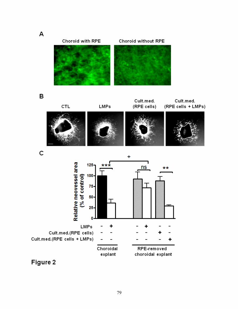
79

80

81
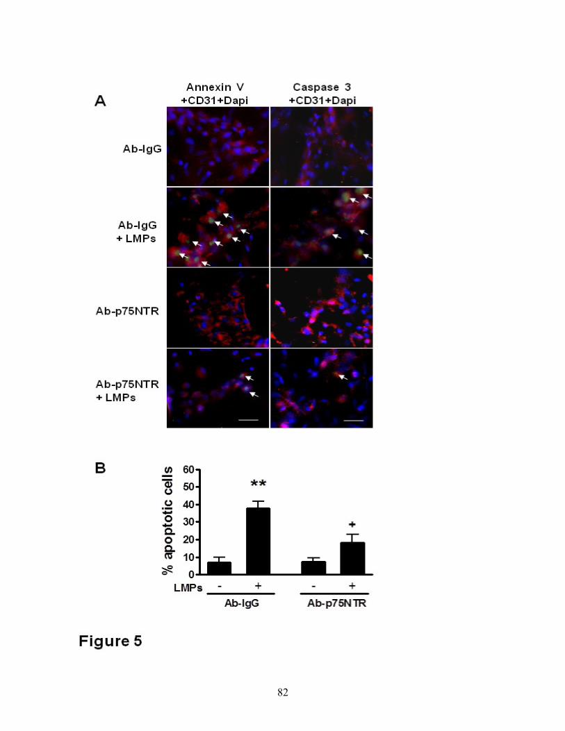
82

83

84
B) Article 2
Lymphocytic Microparticles Modulate Angiogenic Properties of
Macrophages in Laser-induced Choroidal Neovascularization

85
Lymphocytic Microparticles Modulate Angiogenic Properties of Macrophages in Laser-
induced Choroidal Neovascularization
Houda Tahiri1,3, Samy Omri2, Chun Yang3, François Duhamel1, Suzanne Samarani4, Ali
Ahmad4, Mark Vezina5, Martin Bussières6, Elvire Vaucher7, Przemyslaw Sapieha2,8, Gilles
Hickson9, Karim Hammamji8, Réjean Lapointe10, Francis Rodier10,11, Sophie Tremblay13,14,
Isabelle Royal10, Jean-François Cailhier10, Sylvain Chemtob1,2,3,8,12, and Pierre Hardy1,3,12
1Department of Pharmacology, Université de Montréal, Montréal, QC, Canada; 2Research
Center Hôpital Maisonneuve-Rosemont, Université de Montréal, Montréal, QC, Canada;
3Research Center CHU Sainte-Justine, Université de Montréal, Montréal, QC, Canada;
4Departments of Microbiology and Immunology, Université de Montréal, Montréal, QC,
Canada; 5Charles River Laboratories, Senneville, Montreal, QC, Canada ; 6V&O Services,
Saint-Lazare, QC, Canada ; 7School of Optometry, Université de Montréal, Montréal, QC,
Canada; 8Department of Ophthalmology, Université de Montréal, Montréal, QC, Canada;
9Department of Pathology and Cell Biology, Université de Montréal, Montréal, QC, Canada;
10Institut du Cancer de Montréal, CRCHUM – Centre de Recherche du Centre Hospitalier de
l’Université de Montréal and Department of Medicine, Université de Montréal, Montréal, QC,
Canada; 11Department of Radiology, Radio-Oncology and Nuclear Medicine, Université de
Montréal, Montréal, QC, Canada;12Department of Pediatrics, Université de Montréal, Montréal,
QC, Canada, 13 University of British Columbia, Vancouver, BC, Canada, 14Centre for Molecular
Medicine and Therapeutics, Vancouver, BC, Canada

86
Abstract:
Pathological choroidal neovascularization (CNV) is the common cause of vision loss in patients
with age-related macular degeneration (AMD). Macrophages possess potential angiogenic
function in CNV. We have demonstrated that human T lymphocyte-derived microparticles
(LMPs) exert a potent antiangiogenic effect in several pathological neovascularization models.
In this study, we investigated the alteration of proangiogenic properties of macrophages by
LMPs treatment in vitro and in vivo models. LMPs regulated the expression of several
angiogenesis-related factors in macrophages and consequently stimulated their antiangiogenic
effects evidenced by the suppression of the proliferation of human retinal endothelial cells in
co-culture experiments. The involvement of CD36 receptor in LMPs uptake by macrophages
was demonstrated by in vitro assays and by immunostaining of choroidal flat mounts. In
addition, ex vivo experiments showed that CD36 mediates the antiangiogenic effect of LMPs in
murine and human choroidal explants. Furthermore, intravitreal injection of LMPs in the mouse
model of laser-induced CNV significantly suppressed CNV in CD36 dependent manner. The
results of this study suggested an ability of LMPs to alter the gene expression pattern of
angiogenesis-related factors in macrophages, which provide important information for a new
therapeutic approach for efficiently interfering with both vascular and extravascular components
of CNV.
Keywords: Angiogenesis, Choroidal neovascularization, CD36 receptor, macrophages, T
lymphocyte-derived microparticles (LMPs).
Classifications: Experimental, extracellular microvesicles; vascularization, macrophages
polarization; pathophysiology.

87
Introduction:
Lymphocyte-derived microparticles (LMPs) are small membrane microvesicles released
from human T lymphocytes during apoptosis (Carpintero et al. 2010; Martin et al. 2004). We
have established that LMPs exert strong antiangiogenic effect in vivo on corneal
neovascularisation, tumor neovascularization (Yang et al. 2008; Yang et al. 2010), and limit
neovascularization during the vasoproliferative phase of ischemic retinopathy (Yang et al.
2012). Ex vivo, LMPs suppress microvessel sprouting in aortic ring (Yang et al. 2008) and
choroidal explants (Tahiri et al. 2013). These effects depended upon on the integrity of the
retinal pigment epithelium (RPE) and involved release of pigment epithelium-derived growth
factor (PEDF) and p75 neurotrophin receptor (p75NTR) (Tahiri et al. 2013). However, the role
of LMPs in choroidal neovascularization (CNV) secondary to age-related macular degeneration
(AMD) remains unknown.
Age-related macular degeneration (AMD) is a major cause of legal blindness in older
patients of industrialized countries (Grassmann, Fauser, and Weber 2015). The neovascular
form of AMD is characterized by the formation of subretinal choroidal neovascularization, and
it critically depends on local production of vascular endothelial growth factor (VEGF). Anti-
VEGF therapy is currently the new treatment used for treating CNV, but it is plagued by
unwanted side effects and/or insurmountable complications (Sene, Chin-Yee, and Apte 2015).
Therefore, there is an urgent need for identifying alternative approaches to treat CNV. Recent
studies have suggested that immune vascular interactions play an important role in regulating
angiogenesis (Nakao et al. 2005). In mouse models of AMD, macrophages and microglia
(tissue-resident macrophages), recruited to the subretinal space, play a crucial role in choroidal
blood vessel growth (Combadiere et al. 2007; Huang et al. 2013). Blocking VEGF signaling
inhibits the infiltration of macrophages and microglia in the murine laser-induced
photocoagulation model (Huang et al. 2013). Moreover, activation of the interferon-β signaling
pathway in retinal macrophages and microglia cells reduces the inflammation and limits the
development of CNV lesions (Luckoff et al. 2016). These data suggest that modulation of
macrophages and microglia activity may become an attractive therapeutic tool for the treatment
of neovascular AMD.
Recent studies have shown that macrophages comprise an extremely heterogeneous
lineage, displaying a combination of antiangiogenic and proangiogenic functions (Mantovani,
88
Sica, and Locati 2005). In response to microenvironmental signals, macrophages may undergo
classical M1 activation (antiangiogenic) or, alternatively, M2 activation (proangiogenic) (Hume
2015). Macrophages express a panoply of cell surface receptors, including scavenger receptors
such as CD36 (Fadok et al. 1998), low-density lipoprotein receptors (LDLr) (Whitman et al.
2002), phosphatidylserine receptors and mannose receptors (Wilkinson and El Khoury 2012).
The expression of different scavenger receptors in polarized cells contributes to various
pathologies.
CD36 is a transmembrane receptor that mediates diverse physiological as well as
pathological processes, such as metabolism, angiogenesis and atherosclerosis (Febbraio, Hajjar,
and Silverstein 2001). Besides being expressed in macrophages, CD36 is also expressed on
many other mammalian cell types including microvascular endothelial, microglial and retinal
pigment epithelial cells (Cho 2012). We demonstrated previously that LMPs upregulate CD36
protein levels on endothelial cells (Yang et al. 2008). Given the important role of CD36 in
macrophages, we investigated whether LMPs modulate the angiogenic microenvironment by
altering macrophage phenotype, and whether CD36 is involved in the LMPs-induced
antiangiogenic effects.
Results
LMPs dose-dependently inhibit macrophage cell proliferation
We have previously reported that LMPs exert not only a strong inhibitory effect on proliferation
of endothelial and cancer cells (Yang et al. 2008; Yang et al. 2015), but also have a pro-apoptotic
effect on certain types of cells (Qiu et al. 2014; Tahiri et al. 2013; Yang et al. 2015). Here we
observed that LMPs significantly reduced the proliferation of macrophages (RAW 264.7) in a
dose-dependent manner (Figure 1A). The FACS analysis further revealed that concentrations of
LMPs below 20µg/ml did not significantly induce macrophage cell death, although these LMPs
doses significantly suppressed cell growth (Figure 1B-C, P ˃0.05). Similar results were
generated by using pan caspase (FAM-VAD-FMK) fluorescent green probe for detecting active
caspases in the LMPs-treated macrophages (data not shown). Thus, we chose the concentration
of LMPs at 10µg/ml for the following in vitro studies. Similar effects of LMPs were observed
on bone marrow-derived macrophages (BMDM) from femurs of C57BL/6 mice and on human

89
macrophages differentiated from HL60 cells (Supplementary Figures S1, S2). Thus, we chose
the concentration of LMPs at 10µg/ml for the following in vitro studies.
LMPs modulate polarization of macrophages
Macrophages have the ability to adapt to their microenvironment by undergoing phenotypic and
functional changes. They have been broadly characterized by their polarization state according
to the M1/M2 classification system (Martinez, Helming, and Gordon 2009). M1 macrophages
produce high levels of IL-12, whereas M2 macrophages produce high levels of IL-10. The ratio
of IL-12 to IL-10 production has been effectively used to distinguish M1 and M2 macrophages
(Martinez et al. 2008). To determine whether LMPs modulate the polarization of mice
macrophages (RAW264.7), we first quantified the mRNA levels of IL-10 and IL-12 in
macrophages after 24 hours of LMPs treatment. LMPs significantly increased the mRNA level
of IL-12 in macrophages; however, they had no effect on the expression of IL-10 (Figure 2A).
In order to confirm the effects of LMPs, we performed flow cytometric analysis to detect several
other specific markers for M1 or M2 macrophages. In addition to IL-12, CD80 and CD86 also
serve as biomarkers for M1 macrophages (Davis et al. 2013; Zhang, Choksi, et al. 2013) while
the mannose receptor C type 1 (CD206) and Arginase-1 (ARG) are commonly used as
biomarkers for M2 macrophages(Martinez, Helming, and Gordon 2009), as for IL-10. LMPs
treatment significantly increased the number of macrophages expressing IL-12, CD80, or CD86
while it reduced significantly the numbers of macrophages expressing CD206, or ARG (Figure
2B, 2C). Taken together, these data suggest that LMPs drive macrophages into M1 type.
LMPs regulate angiogenic factors expression in macrophages
Macrophages possess potential angiogenic functions. They are capable of releasing angiogenic
cytokines (e.g. VEGF) and are known to contribute to inflammatory neovascularization, tumor
angiogenesis and choroidal neovascularization (Barbera-Guillem et al. 2002; Oh et al. 1999;
Tsutsumi et al. 2003). To ascertain the LMPs-induced antiangiogenic activity of macrophages,
proliferation of human retinal endothelial cells (HREC) was assessed after cells were co-
cultured with macrophages or LMPs-pretreated macrophages. As shown in Figure 3A, the
proliferation rate of HREC was significantly decreased by LMPs-pretreated macrophages
(approximately by 65%), whereas macrophages alone had non-significant effects on endothelial

90
cell proliferation. To answer whether LMPs directly modulate the expression of angiogenesis-
related factors in macrophages, we performed a gene expression analysis to define temporal
changes in a panel of angiogenesis-related factors in LMPs-treated macrophages (Supplemental
Table 1). LMPs downregulated the expression of proangiogenic factors such as VEGFa, insulin-
like growth factor (IGF), fibroblast growth factor (FGF) etc., but upregulated the expression of
brain specific angiogenesis inhibitor 1 (Bai1), thrombospondin-1 (TSP-1), the antiangiogenic
factors (Figure 3B). We further performed the quantitative RT-PCR to confirm the expression
of key angiogenic factors (highly expressed) in the choroidal tissue (Dawson et al. 1999) . LMPs
significantly reduced the expression of VEGFa, FGF but increased pigment epithelium-derived
factor (PEDF) and TSP-1 expression (Figure 3C).
Blockade of CD36 decreases LMPs uptake by macrophages and abrogates the effect of
LMPs on gene expression in macrophages
We have shown that LMPs are taken up by endothelial and epithelial cells via receptor-mediated
endocytosis and phagocytosis, respectively (Yang et al. 2012; Qiu et al. 2014). Moreover, it is
known that the membrane scavenger receptor CD36 proteins are highly expressed on
macrophage membranes (Nakata et al. 1999). Here we observed that CD36 is co-localized with
LMPs when macrophages were incubated with DiI-LMPs (Figure 4A). Blockade of CD36 on
macrophages with anti-CD36 antibody significantly prevented LMPs uptake (Figure 4B),
suggesting that uptake of LMPs into macrophages largely depends on scavenger receptor CD36.
The inhibition of CD36 consequently abrogated the effect of LMPs on VEGFa and TSP-1
expression in macrophages (Figure 4C). Moreover, LMPs-treated macrophages exhibited strong
antiangiogenic effects in the choroidal explant experiments in which the neovascularization of
the RPE-free choroidal explants was decreased by 40%. This LMPs-provoked antiangiogenic
effect of macrophages was attenuated by the pre-treatment of CD36 –specific antibody (Figure
4D).
LMPs suppress laser-induced choroidal neovascularization (CNV) in vivo
We have demonstrated that LMPs exert a strong antiangiogenic effect on several in vivo
neovascularisation animal models (Yang et al. 2008; Yang et al. 2010; Yang et al. 2012). We
extended our investigation of the in vivo effect of LMPs using a laser-induced CNV mouse

91
model. Following intravitreal injections of LMPs, we observed a significant decrease of the
choroidal neovascularization areas (Figure 5).
LMPs modulate angiogenesis-related gene expression in macrophages in vivo
Emerging data support that macrophages contribute to pathological neovascularization (Apte et
al. 2006; Huang et al. 2013; Jawad et al. 2013). To verify whether modulation of the angiogenic
activity of macrophages contributes to the LMPs-mediated reduction of CNV, we first
investigated the uptake of LMPs by macrophages in vivo (Figures 6A-B). Fluorescent confocal
microscopy revealed the presence of macrophages (IBA-1+ cells) in the CNV areas (Lectin-
positive, in green), and LMPs co-localized with retinal macrophages after intravitreal injections
of Dil-LMPs (in red) (Figure 6B). Immunofluorescence staining also shown the expression of
IL-10 and IL-12 in macrophages in the CNV areas, and LMPs induced expression of IL-12 in
macrophages (Figure 6C). To have a better understanding of the expression pattern of
angiogenesis-related factors involved in this process, laser capture microdissection was
performed to collect tissues in the CNV regions, the key factors were analyzed by quantitative
PCR. Consistent with in vitro studies, LMPs significantly induced expression of IL-12 and TSP-
1 but decreased the expression of VEGFa and IL-10 (Figure 6D).
Antiangiogenic effect of LMPs is attenuated in CD36 Knockout mice
Since CD36 has been proven to be involved in the uptake of LMPs by macrophages, and LMPs
are able to directly modulate macrophages gene expression, we surmised that the antiangiogenic
effects of LMPs on laser-induced CNV could be significantly abolished in the CD36-deficient
mice (CD36-/- mice). As expected, no significant differences were observed between the areas
of CNV of control CD36-/- mice and LMPs-treated CD36-/- mice (Figure 7, P ˃0.05). These
results suggested that LMPs-mediated attenuation of CNV is dependent on CD36 expression.
Therefore, it is most likely that both macrophages and endothelial cells play important roles in
the LMPs-induced antiangiogenic effects. Further studies would be required to investigate the
relative role of each cell type.

92
Discussion
Macrophages are components of the immune system that play major roles in the pathological
angiogenesis of the retina and the choroidal tissue (Jawad et al. 2013). In the present study, we
report for the first time that lymphocytic microparticles are capable of altering the expression of
key angiogenesis-related factors in macrophages, and that the scavenger receptor CD36 plays a
crucial role in the mediation of these effects.
LMPs are membrane-derived vesicles derived from human apoptotic T cells (Yang et al. 2008).
We have demonstrated that LMPs can act as mediators of intercellular cross-talk and
consequently induce a variety of cellular responses, including inhibition of proliferation of
endothelial cells, and induction of apoptosis in airway epithelial and cancer cells (Qiu et al.
2014; Yang et al. 2012). Current study revealed that LMPs suppress cell proliferation of both
mouse and human macrophages in a dose-dependent manner, and have no pro-apoptotic effects
on these cells (Figure 1 and Supplemental Figures S1, S2). However, other studies indicate that
LMPs derived from apoptotic Jurkat cells (human T lymphocyte) can induce macrophages to
undergo apoptosis (Huber et al. 2007). These divergent effects of LMPs could be due to the
differences in their apoptotic stimuli, and/or in their parental cells.
Macrophages are immune cells with potential angiogenic functions. Emerging evidence
suggests that macrophages have the ability to undergo two different activation pathways,
classical and alternate ones, in response to different microenvironmental stimuli (Apte et al.
2006). Classicallyactivated pro-inflammatory macrophages possessing an antiangiogenic
phenotype are termed M1 macrophages (Apte et al. 2006). Conversely, macrophages displaying
a proangiogenic alternative phenotype are named M2 (Park and Barbul 2004). M2 macrophages
are capable of releasing angiogenic cytokines (e.g. VEGF, TNF-alpha, IL-10) and are known to
contribute to wound healing and tissue repair as well as to pathological neovascularization (Oh
et al. 1999; Tsutsumi et al. 2003). Several markers have been used to identify M1 and M2
macrophages, such as the ratio of IL-10 and IL-12, CD206, CD80, CD86, and ARG (Mantovani
et al. 2004; Stein et al. 1992; Davis et al. 2013; Zhang, Choksi, et al. 2013). LMPs treatment
promoted differentiation of macrophages into M1 phenotype, evidenced by the increased
expression of IL-12, CD80, CD86 and decreased expression of CD206 and ARG in LMPs-

93
treated cells (Figure 2). Similar results were obtained in human primary macrophages following
a LMPs treatment (Supplemental Figures S3 and S4). Although the LMPs-treated macrophages
exhibit M1 phenotype, they do not express more TNF-alpha, a major inflammatory factor
(Supplemental Figure S5). Most interestingly, LMPs dramatically modulate the expression of a
panel of angiogenesis-related factors in macrophages (Figure 3B). The expression of key
angiogenic factors such as proangiogenic factors, VEGFa and FGF, was decreased, while the
expression of antiangiogenic factors, pigment epithelium-derived factor (PEDF) and TSP-1 was
increased (Figure 3C). These changes of expression of angiogenesis-related factors in LMP-
treated macrophages had significant impact on endothelial cell proliferation as well as on CNV
of choroidal explants from mouse and human (Figure 4D and Supplemental Figure S6). Taken
together, these results suggested that LMPs have a strong effect on modulating the angiogenic
proprieties of macrophages. To investigate whether the antiangiogenic effect observed in this
study is LMPs specific, we repeated the experiments of cell viability assay, FACS analysis of
polarization of macrophages and ex vivo CNV assay using different controls such as endothelial
cell derived microparticles (EMPs), and human T lymphocytes (CEM T cells, the parent cells
of LMPs). These study revealed T lymphocytes have no effect on macrophages viability, and
EMPs have no significant effect on macrophages polarization (data not shown). Moreover,
opposite to the effect of LMPs, EMPs increased the retinal endothelial cell growth and exhibited
pro-angiogenic effects in the ex vivo choroidal angiogenesis assay (Supplemental Figure S7).
Nonetheless, without optimal control, we cannot exclude the possibility that microparticles
derived from other apoptotic cells may be able to modulate macrophage function and possess
antiangiogenic effects.
Previously, we have demonstrated that LMPs are taken up by target cells through receptor-
mediated endocytosis or phagocytosis (Yang et al. 2012; Qiu et al. 2013). The reason for
investigating CD36 involvement in mediating the effect of LMPs on macrophages is due to the
facts that CD36 is highly expressed in macrophages, and it plays an important role in
angiogenesis (Silverstein and Febbraio 2009). The following observations supported our
conclusion that CD36 does play a pivotal role in mediating the effects of LMPs: 1) CD36
colocalized with LMPs in macrophages (Figure 4A); 2) blockade of CD36 prevented LMPs
uptake by macrophages (Figure 4B); 3) inhibition of CD36 in macrophages altered the effect of

94
LMPs on the expression of angiogenesis-related factors (Figure 4C); 4) blockade of CD36 in
macrophages also attenuated the inhibitory effect of LMPs in choroidal angiogenesis assays
(Figure 4D); 5) the antiangiogenic effect of LMPs was completely abolished in CD36 KO mice
(Figure 7). Nonetheless, CD36 is a multifunctional membrane receptor which can act as an
antiangiogenic or proangiogenic factor depending on the microenvironment and its ligands (Ren
et al. 2011; Febbraio, Hajjar, and Silverstein 2001; Dawson et al. 1997; Mwaikambo et al. 2008).
We reported previously that LMPs upregulated CD36 expression in immortalized human
microvascular endothelial cells, we also observed the upregulated expression of CD36 in LMPs-
treated RAW 264.7 (Supplemental Figure S8). Thus, CD36 may mediate downstream events
following LMPs ingestion and act as an antiangiogenic effector as well.
The mechanisms by which LMPs influence macrophage activity following their interaction with
CD36 on the surface of macrophages have not been well defined. Whereas based on published
reports(Silverstein et al. 2010; Rahaman et al. 2006; Chen et al. 2008; Kennedy et al. 2011) and
our observations, we surmised that there are two possible CD36-dependent signal transduction
pathways may proceed in macrophages. Firstly, CD36 function as traditional signal transduction
receptor initiating a signaling cascade conferred upon LMPs binding. This is because LMPs lose
their membrane asymmetry and express anionic phospholipids, such as phosphatidylserine on
their surface (Silverstein et al. 2010). Thus LMPs become the endogenous CD36 ligands. The
lipids of LMPs binding to CD36 mediates the activation of the C-Jun N-terminal kinase (JNKs).
The JNKs belong to the conserved and ubiquitous signaling network of mitogen-activated
protein kinases (MAPKs), and they are essential mediators of relevant pro-inflammatory
functions in macrophages and microglia (Waetzig et al. 2005). Particularly, JNK1 and JNK2
positively regulate IL-12 production in macrophages (Utsugi et al. 2010; Utsugi et al. 2009).
Relevantly, we observed that JNK1 expression was increased in LMPs-treated macrophages
(Supplemental Figure S8), which indicated a possibility that LMPs activate the CD36-dependent
MAP kinases JNKs and consequently induce IL-12 expression. Nonetheless, the function of
JNKs in LMPs-induced antiangiogenic effect in macrophages need be further verified.
Secondly, we contemplated that CD36 facilitates the internalization of bioactive components
from LMPs that participate in the initiation of a transcriptional program that includes
upregulation of its own gene as well as those of other critical genes involved in the function of

95
macrophages. This is because we observed CD36 expression was significantly increased by
LMPs treatment (Supplemental Figure S8). Moreover, we recently analyzed the microRNAs
sequences in the LMPs, and found several abundant miRNAs possess potential regulatory
effects on macrophages activity according to the reported (Zhang, Zhang, et al. 2013; Freilich,
Woodbury, and Ikezu 2013; Cobos Jimenez et al. 2014). The precise role of these miRNAs in
LMPs remains to be determined. Taken together, in macrophages, LMPs might directly bind to
CD36 and activate its downstream signaling resulted in an increasing expression of IL-12, the
marker for M1 phenotype. Meanwhile, transfer the components of LMPs into macrophages
may be mediated by CD36, and these active components consequently regulate genes expression
including CD36, and the resulting cytokines production causes the antiangiogenic effect. In such
a way, LMPs modulated the angiogenic properties of macrophages which may related to the
suppression of CNV.
Neovascularization plays a key role in the pathophysiology of several diseases of the retina such
as retinopathy of prematurity (ROP), diabetic retinopathy and choroidal neovascularization
associated with age-related macular degeneration (AMD). LMPs possess strong antiangiogenic
effect through direct targeting of vascular endothelial cells, and this effect was confirmed in ex
vivo and in vivo ROP models (Yang et al. 2008; Yang et al. 2010; Yang et al. 2012). As
previously reported, LMPs had no significant anti-angiogenic effect on mouse RPE-free
choroidal angiogenesis (Figure 4C), and the lack of anti-angiogenic effect of LMPs was also
observed on human RPE-free choroidal explants (Supplemental Figure S6). The laser-induced
murine model of CNV is widely used to mimic wet AMD by compromising Bruch's membrane
to induce CNV formation in the subretinal region, allowing visualization and evaluation of the
morphologic changes of experimental CNV. It has been a valuable tool to study the diverse
components of complex CNV lesions (Campos et al. 2006). In line with the anti-angiogenic
effects of LMPs observed in other pathological angiogenesis-related models (Yang et al. 2008;
Tahiri et al. 2013; Yang et al. 2010; Yang et al. 2012), intravitreal injection of LMPs
significantly suppressed laser-induced CNV (Figure 5). In this model, LMPs uptake by
macrophages led to an increased IL-12 expression (Figure 6) suggesting that LMPs are capable
of reprograming macrophage functions in vivo. In addition, LMPs-induced expression changes
of key angiogenic factors in the CNV areas may contribute to the overall CNV suppression.

96
In conclusion, LMPs promoted macrophages polarization toward antiangiogenic phenotype, and
the resulting antiangiogenic activity of macrophages was confirmed on both murine and human
choroidal explants as well as in vivo CNV murine model. Moreover, CD36 played a critical role
in mediating the effect of LMPs on macrophages. This study highlighted the ability of LMPs to
alter the gene expression pattern of angiogenesis-related factors in macrophages, and provided
important information for a new therapeutic approach to efficiently interfere with both vascular
and extravascular components of CNV.
Methods
LMPs production
Human CEM T cells were purchased from American Type Culture Collection (ATCC) and
grown in X-VIVO medium (Cambrex). LMPs were generated as described previously (Yang et
al. 2008). Briefly, CEM T cells were treated with 0.5 μg/ml actinomycin D (Sigma-Aldrich) for
24 hours. A supernatant was obtained by centrifugation at 750 g for 15 min, then 1500 g for 5
min to remove cells and large debris. LMPs from the supernatant were washed after 3
centrifugation steps (50 min at 12,000 g) and recovered in PBS. Washing medium from the last
supernatant was used as a control (CTL) unless otherwise noted. Up to now, there is no proper
control for microparticles due to their heterogeneous components and unclarified
pathophysiological roles. However, this last washing medium ensures that the effects seen are
due to LMPs and not due to any soluble factor present in the preparation, which has been widely
used as the control vehicle for each individual microparticle according to the published
literatures (Benameur, Soleti, et al. 2010; Martin et al. 2004; Vasina et al. 2011). LMPs were
characterized with annexin V (BD bioscience) staining by fluorescence-activated cell sorting
(FACS) analysis. The concentrations of LMPs were determined using the Bio-Rad protein
assay. A lipophilic fluorescent stain Dil (1,1'-Dioctadecyl-3,3,3',3'-
Tetramethylindocarbocyanine Perchlorate) was added to CEM T cells 24h before actinomycin
D treatment. The process generated fluorescent Dil-labelled LMPs (Dil-LMPs).
Cell culture
RAW 264.7 macrophages (murine cell line) were purchased from ATCC, and cultured in
Dulbecco's Modified Eagle's Medium (Gibco, Life Technologies), supplemented with 10% FBS
(Gibco, Life Technologies) and 1% penicillin/streptomycin.

97
Proliferation and apoptosis assays
Macrophages (RAW 264.7) were incubated for 24h with indicated concentrations of LMPs, then
cell proliferation was evaluated by [3H]-thymidine incorporation assay. In co-culture
experiments, human retinal epithelial cells (HREC) were seeded in the lower compartment of
the Transwell unit (0.4μm pore, Mississauga, ON) and macrophages or LMPs-pretreated
macrophages (24 h pre-treatment of LMPs) were placed on the Transwell inserts. After 24 hours
of co-incubation, HREC were assayed for proliferation rate. The relative cell proliferation rate
is presented at a percentage of control (set as 100%). For apoptosis assay, the RAW 264.7 cells
at approximately 60% confluence were treated with 10 or 20 μg/ml of LMPs for 24 hours
followed by staining with reagents from a Vybrant apoptosis assay kit (Molecular Probes,
Invitrogen, Carlsbad, CA) according to the manufacturer’s protocol. Apoptosis was determined
by flow cytometry using FacsCalibur (BD Biosciences) and expressed as apoptotic rate (the
percentage of apoptotic cells over the total number of cells). We observed that LMPs at 10 μg/ml
concentration has no effect on macrophages viability, and used this LMPs dose in the subsequent
in vitro studies.
Quantitative RT-PCR and PCR array
Total RNA was extracted from RAW 264.7 cells and choroidal tissues using an RNA extraction
kit (Qiagen, Mississauga, ON). DNase-treated RNA was then converted into cDNA using M-
MLV reverse transcriptase (Invitrogen). Quantitative analysis of gene expression was performed
on a Stratagene Mx3000p sequence detection system with SYBR Green Master Mix Kit
(BioRad). Each sample was analyzed in triplicate and threshold cycle numbers were averaged.
Gene expression was normalized to 18S, and the percentage of change was calculated according
to a previously described formula (Buckhaults et al. 2001). PCR primers were synthesized by
Alpha DNA (Montreal, Quebec, Canada) based on the following sequences: IL-10, forward 5’-
TGGCCACACTTGAGAGCTGC-3’, reverse 5’-TTCAGGGATGAAGCGGCTGG-3’; IL-12,
forward 5'-CAACATCAAGAGCAGTAGCAG-3', reverse 5'-
TACTCCCAGCTGACCTCCAC-3'; VEGFa, forward 5'-TGCAGGCTGCTGTAACGATG-3’,
reverse 5'- GAACAAGGCTCACAGTG
ATTTTCT-3'; PEDF, forward 5'-TCGAAAGCAGCCCTGTGTT-3', reverse 5'-
AATCACCCGACTTCAGCAAGA-3'; FGF forward 5'-CAACCGGTACCTTGCTATGA-3',
reverse 5' -TCCGTGACCGGTAAGTATTG-3', TSP-1, forward 5’-

98
AACAAAGGACCTCCAAGCTATCTG-3’, reverse 5’-GGGAGGCCGCTTCAGC-3’; 18S
rRNA, forward 5’-CCTGCGGCTTTAAATTT GACTCA-3’, reverse 5’-
GCTATCAATCTGTCAATCCTGTC-3’.
For the SABioscience Mouse Angiogenesis array (PAMM-024), total RNAs from cells
were isolated according to manufacturer’s instructions by using RNeasy Microarray Tissue Mini
Kit (SABioscience, Qiagen). 1 μg of RNA was used for RT-PCR array which was carried out
on Stratagene model Mx3000p with SYBR Green (SABiosciences). The data was analyzed
using the RT2 Profiler PCR array data analysis software (SABiosciences).
Flow cytometry analysis
For intracellular staining of IL-10, IL-12, and Arginase-1 (ARG), cells were treated with
Brefeldin A and permeabilized using the Fixation & Permeabilization kit (eBioscience)
according to the recommended protocols. The treated cells were then incubated with following
first antibodies against mouse IL-10-APC (JES5-16E3), IL-12-PE (C17.8, eBioscience),
Arginase-1 (PA5-22009; Fisher Scientific) respectively, followed by staining with secondary
antibody goat anti-rabbit IgG-FITC (Santa Cruz). For extracellular staining of the macrophage
receptors, cells were incubated with the following antibodies: anti-mouse CD206-APC
(C068C2, Biolegend), CD86-PE (GL1), CD80-PE (16-10A1, eBioscience) for 30 min on ice
and washed three times with PBS containing 0.05% bovine serum albumin. Cells were re-
suspended in 2% paraformaldehyde and then subjected to flow cytometry analysis using
FacsCalibur (BD Biosciences).
Detection of CD36 expression and macrophage uptake of LMPs
RAW 264.7 were seeded onto coverslip in 12 well plates. Next day, cells were incubated with
10 μg/ml Dil-LMPs for 4h. After LMPs treatment, macrophages were fixed and nuclei were
stained with DAPI (1:5000; Invitrogen). The expression of CD36 was detected with anti-CD36-
FITC (1:100, Serotec, Oxford, UK). Images were taken using a laser scanning confocal
microscope (Zeiss LSM 510). To investigate whether CD36 is involved in the uptake of LMPs,
macrophages were seeded into 96-well plates, and pretreated with anti-CD36 antibodies (15
μg/mL) to block CD36 receptors (Cayman Chemical, Ann Arbor, MI) or with isotype-matched
control antibodies (Sigma) for 3 hours followed by 4 hours DiI-LMPs incubation. The DiI
fluorescence intensity was measured using spectrofluorometer and presented as mean
fluorescence intensity (MFI).
99
Animals
C57BL/6J and CD36 knockout mice (CD36 KO) were purchased from the Jackson Laboratory
(Bar Harbor, Maine, USA). For the CNV model, mice were anesthetized using intraperitoneal
administration of ketamine hydrochloride/xylazine. Their pupils were dilated with 1%
tropicamide. A coverslip was placed over the cornea, and Argon laser photocoagulation
(400mW 0.05s) was performed to rupture Bruch’s membrane at four locations in each eye (Tobe
et al. 1998; Seo et al. 1999). Mice were randomly grouped to receive intravitreal injections on
day 1 and day 7 with the last wash-treated from LMPs (control group), LMPs (50 μg) (treatment
group) or Dil-LMPs (50 μg). The 50μg of LMPs is the optimal dose seen in our previous in vivo
and ex vivo experiments. Mice were sacrificed 14 days after the laser photocoagulation, and the
eyes were rapidly enucleated. The choroids were prepared for immunofluorescence and RT-
PCR analysis. All procedures in this study were approved by the Maisonneuve-Rosemont
Hospital Animal Welfare Committee, and are in accordance with the guidelines of the Canadian
Council on Animal Care and the Guide for the Care and Use of Laboratory Animals published
by the US National Institutes of Health.
Preparation of retinal pigment epithelium (RPE)-free choroidal explants
The RPE-removed choroidal explants were prepared according to a previously described
procedure (Tahiri et al. 2013). Mice eyes were dissected in a Petri dish containing 1X Hank’s
balanced Salt Solution (HBSS, Invitrogen). The blood vessels, connective and fatty tissues were
removed from the exterior of the eyeballs. The eyes were then incubated at 37°C for 45 min
with 2% dispase II (neutral protease; Roche) in HBSS. After removing the anterior segment,
retina and sclera, the RPE layer was peeled off from the choroid with fine forceps. The choroid
without RPE was sliced into 1~2-mm sections and placed in growth factor–reduced basement
membrane matrix. The effectiveness of the RPE removal was histologically evaluated (Tahiri et
al. 2013).
Treatment of choroidal explants and measurement of neovascularization
Choroidal explants were cultured at 37°C in 5% CO2 for 4 days. The culture medium was
changed on day 5 and explants were incubated with saline, 50µg/mL of LMPs or co-cultured
with LMPs-pretreated macrophages, CD36 antibody pre-treated macrophages, or CD36 pre-
treated+ LMPs treated macrophages in a Transwell for 48 hours. For the pre-treatments,
macrophages were pre-incubated with CD36 specific antibody 45 μg/mL for 3 hours, or/and 24

100
hours of LMPs treatment. Photographs of individual explants were taken before and after the
treatment using an Axiovert 200M inverted microscope (Zeiss). The neovessel areas were
determined using Image-Pro Plus software.
Immunohistochemistry staining of choroidal flat-mounts
The choroidal flat-mounts were prepared as described previously (Omri et al. 2011). In brief,
the eyes of mice that received an intravitreal injection of control vesicles, LMPs, or DiI-LMPs
(n≥3 for each group) were enucleated and fixed in 4% PFA. After the cornea, lens and retina
were removed, the choroid-sclera was permeabilized in 1.0% Triton X100 and blocked in 10%
normal goat serum. A first antibody against the ionized calcium-binding adapter molecule 1
(Iba1) (1:500; Wako Chemicals, USA) and second antibodies the Alexa-448 -conjugated goat
anti-rabbit or Alexa-594 conjugated goat anti-rabbit (1:1000; Invitrogen) were used to
specifically identify macrophages. FITC-conjugated lectin was used to stain endothelial cells
(1:100; Sigma-Aldrich, St. Louis, MO); Rhodamine Phalloidin (R415; 1:400; Santa Cruz
Biotechnology, Santa Cruz, CA) was used for RPE cells. Antibody against mouse IL-10-APC
(1:1000) and against mouse IL-12-PE clone (1:1000) were used to detect IL-10 and IL-12,
respectively. Cell nuclei were stained with DAPI (1:5000; Invitrogen). Labeled flat-mounts
were examined with a laser scanning confocal microscope (Zeiss LSM 510).
Laser-capture microdissection
The mice eyes were snap frozen in optimal cutting temperature compound (OCT, Fisher).
Sections of 16µm were dissected with a Zeiss Observer microscope equipped with Palm
MicroBeam device for laser-capture microdissection. Total RNAs were extracted from these
dissected tissues as described (Chabrat, Doucet-Beaupre, and Levesque 2015).
Statistical analysis
All experiments were performed in duplicate or triplicate and repeated independently at least 3
times. Values are presented as means ± SEM. Data were analyzed by one-way ANOVA
followed by post-hoc Bonferroni tests for comparison of means. Statistical significance was set
at P < 0.05.
References
Apte, R. S., J. Richter, J. Herndon, and T. A. Ferguson. 2006. 'Macrophages inhibit neovascularization
in a murine model of age-related macular degeneration', PLoS Med, 3: e310.
101
Barbera-Guillem, E., J. K. Nyhus, C. C. Wolford, C. R. Friece, and J. W. Sampsel. 2002. 'Vascular
endothelial growth factor secretion by tumor-infiltrating macrophages essentially supports tumor
angiogenesis, and IgG immune complexes potentiate the process', Cancer Res, 62: 7042-9.
Benameur, T., R. Soleti, C. Porro, R. Andriantsitohaina, and M. C. Martinez. 2010. 'Microparticles
carrying Sonic hedgehog favor neovascularization through the activation of nitric oxide pathway
in mice', PLoS One, 5: e12688.
Buckhaults, P., C. Rago, B. St Croix, K. E. Romans, S. Saha, L. Zhang, B. Vogelstein, and K. W. Kinzler.
2001. 'Secreted and cell surface genes expressed in benign and malignant colorectal tumors',
Cancer Res, 61: 6996-7001.
Campos, M., J. Amaral, S. P. Becerra, and R. N. Fariss. 2006. 'A novel imaging technique for
experimental choroidal neovascularization', Invest Ophthalmol Vis Sci, 47: 5163-70.
Carpintero, R., L. Gruaz, K. J. Brandt, A. Scanu, D. Faille, V. Combes, G. E. Grau, and D. Burger. 2010.
'HDL interfere with the binding of T cell microparticles to human monocytes to inhibit pro-
inflammatory cytokine production', PLoS One, 5: e11869.
Chabrat, A., H. Doucet-Beaupre, and M. Levesque. 2015. 'RNA Isolation from Cell Specific
Subpopulations Using Laser-capture Microdissection Combined with Rapid Immunolabeling', J
Vis Exp.
Chen, K., M. Febbraio, W. Li, and R. L. Silverstein. 2008. 'A specific CD36-dependent signaling
pathway is required for platelet activation by oxidized low-density lipoprotein', Circ Res, 102:
1512-9.
Cho, S. 2012. 'CD36 as a therapeutic target for endothelial dysfunction in stroke', Curr Pharm Des, 18:
3721-30.
Cobos Jimenez, V., A. M. Willemsen, E. J. Bradley, F. Baas, A. H. van Kampen, and N. A. Kootstra.
2014. 'Next-generation sequencing of microRNAs in primary human polarized macrophages',
Genom Data, 2: 181-3.
Combadiere, C., C. Feumi, W. Raoul, N. Keller, M. Rodero, A. Pezard, S. Lavalette, M. Houssier, L.
Jonet, E. Picard, P. Debre, M. Sirinyan, P. Deterre, T. Ferroukhi, S. Y. Cohen, D. Chauvaud, J.
C. Jeanny, S. Chemtob, F. Behar-Cohen, and F. Sennlaub. 2007. 'CX3CR1-dependent subretinal
microglia cell accumulation is associated with cardinal features of age-related macular
degeneration', J Clin Invest, 117: 2920-8.
Davis, M. J., T. M. Tsang, Y. Qiu, J. K. Dayrit, J. B. Freij, G. B. Huffnagle, and M. A. Olszewski. 2013.
'Macrophage M1/M2 polarization dynamically adapts to changes in cytokine microenvironments
in Cryptococcus neoformans infection', MBio, 4: e00264-13.
Dawson, D. W., S. F. Pearce, R. Zhong, R. L. Silverstein, W. A. Frazier, and N. P. Bouck. 1997. 'CD36
mediates the In vitro inhibitory effects of thrombospondin-1 on endothelial cells', J Cell Biol,
138: 707-17.
Dawson, D. W., O. V. Volpert, P. Gillis, S. E. Crawford, H. Xu, W. Benedict, and N. P. Bouck. 1999.
'Pigment epithelium-derived factor: a potent inhibitor of angiogenesis', Science, 285: 245-8.
Fadok, V. A., M. L. Warner, D. L. Bratton, and P. M. Henson. 1998. 'CD36 is required for phagocytosis
of apoptotic cells by human macrophages that use either a phosphatidylserine receptor or the
vitronectin receptor (alpha v beta 3)', J Immunol, 161: 6250-7.
Febbraio, M., D. P. Hajjar, and R. L. Silverstein. 2001. 'CD36: a class B scavenger receptor involved in
angiogenesis, atherosclerosis, inflammation, and lipid metabolism', J Clin Invest, 108: 785-91.
Freilich, R. W., M. E. Woodbury, and T. Ikezu. 2013. 'Integrated expression profiles of mRNA and
miRNA in polarized primary murine microglia', PLoS One, 8: e79416.
Grassmann, F., S. Fauser, and B. H. Weber. 2015. 'The genetics of age-related macular degeneration
(AMD) - Novel targets for designing treatment options?', Eur J Pharm Biopharm.
Huang, H., R. Parlier, J. K. Shen, G. A. Lutty, and S. A. Vinores. 2013. 'VEGF receptor blockade
markedly reduces retinal microglia/macrophage infiltration into laser-induced CNV', PLoS One,
8: e71808.

102
Huber, L. C., A. Jungel, J. H. Distler, F. Moritz, R. E. Gay, B. A. Michel, D. S. Pisetsky, S. Gay, and O.
Distler. 2007. 'The role of membrane lipids in the induction of macrophage apoptosis by
microparticles', Apoptosis, 12: 363-74.
Hume, D. A. 2015. 'The Many Alternative Faces of Macrophage Activation', Front Immunol, 6: 370.
Jawad, S., B. Liu, Z. Li, R. Katamay, M. Campos, L. Wei, H. N. Sen, D. Ling, F. Martinez Estrada, J.
Amaral, C. C. Chan, R. Fariss, S. Gordon, and R. B. Nussenblatt. 2013. 'The role of macrophage
class a scavenger receptors in a laser-induced murine choroidal neovascularization model', Invest
Ophthalmol Vis Sci, 54: 5959-70.
Kennedy, D. J., S. Kuchibhotla, K. M. Westfall, R. L. Silverstein, R. E. Morton, and M. Febbraio. 2011.
'A CD36-dependent pathway enhances macrophage and adipose tissue inflammation and impairs
insulin signalling', Cardiovasc Res, 89: 604-13.
Luckoff, A., A. Caramoy, R. Scholz, M. Prinz, U. Kalinke, and T. Langmann. 2016. 'Interferon-beta
signaling in retinal mononuclear phagocytes attenuates pathological neovascularization', EMBO
Mol Med, 8: 670-8.
Mantovani, A., A. Sica, and M. Locati. 2005. 'Macrophage polarization comes of age', Immunity, 23:
344-6.
Mantovani, A., A. Sica, S. Sozzani, P. Allavena, A. Vecchi, and M. Locati. 2004. 'The chemokine system
in diverse forms of macrophage activation and polarization', Trends Immunol, 25: 677-86.
Martin, S., A. Tesse, B. Hugel, M. C. Martinez, O. Morel, J. M. Freyssinet, and R. Andriantsitohaina.
2004. 'Shed membrane particles from T lymphocytes impair endothelial function and regulate
endothelial protein expression', Circulation, 109: 1653-9.
Martinez, F. O., L. Helming, and S. Gordon. 2009. 'Alternative activation of macrophages: an
immunologic functional perspective', Annu Rev Immunol, 27: 451-83.
Martinez, F. O., A. Sica, A. Mantovani, and M. Locati. 2008. 'Macrophage activation and polarization',
Front Biosci, 13: 453-61.
Mwaikambo, B. R., F. Sennlaub, H. Ong, S. Chemtob, and P. Hardy. 2008. 'Genetic ablation of CD36
induces age-related corneal neovascularization', Cornea, 27: 1037-41.
Nakao, S., T. Kuwano, C. Tsutsumi-Miyahara, S. Ueda, Y. N. Kimura, S. Hamano, K. H. Sonoda, Y.
Saijo, T. Nukiwa, R. M. Strieter, T. Ishibashi, M. Kuwano, and M. Ono. 2005. 'Infiltration of
COX-2-expressing macrophages is a prerequisite for IL-1 beta-induced neovascularization and
tumor growth', J Clin Invest, 115: 2979-91.
Nakata, A., Y. Nakagawa, M. Nishida, S. Nozaki, J. Miyagawa, T. Nakagawa, R. Tamura, K.
Matsumoto, K. Kameda-Takemura, S. Yamashita, and Y. Matsuzawa. 1999. 'CD36, a novel
receptor for oxidized low-density lipoproteins, is highly expressed on lipid-laden macrophages
in human atherosclerotic aorta', Arterioscler Thromb Vasc Biol, 19: 1333-9.
Oh, H., H. Takagi, C. Takagi, K. Suzuma, A. Otani, K. Ishida, M. Matsumura, Y. Ogura, and Y. Honda.
1999. 'The potential angiogenic role of macrophages in the formation of choroidal neovascular
membranes', Invest Ophthalmol Vis Sci, 40: 1891-8.
Omri, S., F. Behar-Cohen, Y. de Kozak, F. Sennlaub, L. M. Verissimo, L. Jonet, M. Savoldelli, B. Omri,
and P. Crisanti. 2011. 'Microglia/macrophages migrate through retinal epithelium barrier by a
transcellular route in diabetic retinopathy: role of PKCzeta in the Goto Kakizaki rat model', Am
J Pathol, 179: 942-53.
Park, J. E., and A. Barbul. 2004. 'Understanding the role of immune regulation in wound healing', Am J
Surg, 187: 11S-16S.
Qiu, Q., W. Xiong, C. Yang, X. Dai, X. Dan, Z. Yang, Y. Jiao, Y. Xiang, G. Liu, and P. Hardy. 2014.
'Lymphocyte-derived microparticles induce apoptosis of airway epithelial cells through
activation of p38 MAPK and production of arachidonic acid', Apoptosis, 19: 1113-27.
Qiu, Q., W. Xiong, C. Yang, C. Gagnon, and P. Hardy. 2013. 'Lymphocyte-derived microparticles induce
bronchial epithelial cells' pro-inflammatory cytokine production and apoptosis', Mol Immunol,
55: 220-30.

103
Rahaman, S. O., D. J. Lennon, M. Febbraio, E. A. Podrez, S. L. Hazen, and R. L. Silverstein. 2006. 'A
CD36-dependent signaling cascade is necessary for macrophage foam cell formation', Cell
Metab, 4: 211-21.
Ren, B., J. Hale, S. Srikanthan, and R. L. Silverstein. 2011. 'Lysophosphatidic acid suppresses
endothelial cell CD36 expression and promotes angiogenesis via a PKD-1-dependent signaling
pathway', Blood, 117: 6036-45.
Sene, A., D. Chin-Yee, and R. S. Apte. 2015. 'Seeing through VEGF: innate and adaptive immunity in
pathological angiogenesis in the eye', Trends Mol Med, 21: 43-51.
Seo, M. S., N. Kwak, H. Ozaki, H. Yamada, N. Okamoto, E. Yamada, D. Fabbro, F. Hofmann, J. M.
Wood, and P. A. Campochiaro. 1999. 'Dramatic inhibition of retinal and choroidal
neovascularization by oral administration of a kinase inhibitor', Am J Pathol, 154: 1743-53.
Silverstein, R. L., and M. Febbraio. 2009. 'CD36, a scavenger receptor involved in immunity,
metabolism, angiogenesis, and behavior', Sci Signal, 2: re3.
Silverstein, R. L., W. Li, Y. M. Park, and S. O. Rahaman. 2010. 'Mechanisms of cell signaling by the
scavenger receptor CD36: implications in atherosclerosis and thrombosis', Trans Am Clin
Climatol Assoc, 121: 206-20.
Stein, M., S. Keshav, N. Harris, and S. Gordon. 1992. 'Interleukin 4 potently enhances murine
macrophage mannose receptor activity: a marker of alternative immunologic macrophage
activation', J Exp Med, 176: 287-92.
Tahiri, H., C. Yang, F. Duhamel, S. Omri, E. Picard, S. Chemtob, and P. Hardy. 2013. 'p75 neurotrophin
receptor participates in the choroidal antiangiogenic and apoptotic effects of T-lymphocyte-
derived microparticles', Invest Ophthalmol Vis Sci, 54: 6084-92.
Tobe, T., S. Ortega, J. D. Luna, H. Ozaki, N. Okamoto, N. L. Derevjanik, S. A. Vinores, C. Basilico, and
P. A. Campochiaro. 1998. 'Targeted disruption of the FGF2 gene does not prevent choroidal
neovascularization in a murine model', Am J Pathol, 153: 1641-6.
Tsutsumi, C., K. H. Sonoda, K. Egashira, H. Qiao, T. Hisatomi, S. Nakao, M. Ishibashi, I. F. Charo, T.
Sakamoto, T. Murata, and T. Ishibashi. 2003. 'The critical role of ocular-infiltrating
macrophages in the development of choroidal neovascularization', J Leukoc Biol, 74: 25-32.
Utsugi, M., K. Dobashi, A. Ono, T. Ishizuka, T. Hisada, Y. Koga, Y. Shimizu, T. Kawata, S. Matsuzaki,
H. Aoki, Y. Kamide, and M. Mori. 2010. 'JNK1 and JNK2 differently regulate IL-12 production
in THP-1 macrophage cells', Cytokine, 51: 127-31.
Utsugi, M., K. Dobashi, A. Ono, T. Ishizuka, S. Matsuzaki, T. Hisada, Y. Shimizu, T. Kawata, H. Aoki,
Y. Kamide, and M. Mori. 2009. 'PI3K p110beta positively regulates lipopolysaccharide-induced
IL-12 production in human macrophages and dendritic cells and JNK1 plays a novel role', J
Immunol, 182: 5225-31.
Vasina, E. M., S. Cauwenberghs, M. A. Feijge, J. W. Heemskerk, C. Weber, and R. R. Koenen. 2011.
'Microparticles from apoptotic platelets promote resident macrophage differentiation', Cell
Death Dis, 2: e211.
Waetzig, V., K. Czeloth, U. Hidding, K. Mielke, M. Kanzow, S. Brecht, M. Goetz, R. Lucius, T.
Herdegen, and U. K. Hanisch. 2005. 'c-Jun N-terminal kinases (JNKs) mediate pro-
inflammatory actions of microglia', Glia, 50: 235-46.
Whitman, S. C., D. L. Rateri, S. J. Szilvassy, J. A. Cornicelli, and A. Daugherty. 2002. 'Macrophage-
specific expression of class A scavenger receptors in LDL receptor(-/-) mice decreases
atherosclerosis and changes spleen morphology', J Lipid Res, 43: 1201-8.
Wilkinson, K., and J. El Khoury. 2012. 'Microglial scavenger receptors and their roles in the pathogenesis
of Alzheimer's disease', Int J Alzheimers Dis, 2012: 489456.
Yang, C., C. Gagnon, X. Hou, and P. Hardy. 2010. 'Low density lipoprotein receptor mediates anti-
VEGF effect of lymphocyte T-derived microparticles in Lewis lung carcinoma cells', Cancer
Biol Ther, 10: 448-56.

104
Yang, C., B. R. Mwaikambo, T. Zhu, C. Gagnon, J. Lafleur, S. Seshadri, P. Lachapelle, J. C. Lavoie, S.
Chemtob, and P. Hardy. 2008. 'Lymphocytic microparticles inhibit angiogenesis by stimulating
oxidative stress and negatively regulating VEGF-induced pathways', Am J Physiol Regul Integr
Comp Physiol, 294: R467-76.
Yang, C., W. Xiong, Q. Qiu, Z. Shao, D. Hamel, H. Tahiri, G. Leclair, P. Lachapelle, S. Chemtob, and
P. Hardy. 2012. 'Role of receptor-mediated endocytosis in the antiangiogenic effects of human
T lymphoblastic cell-derived microparticles', Am J Physiol Regul Integr Comp Physiol, 302:
R941-9.
Yang, C., W. Xiong, Q. Qiu, H. Tahiri, C. Gagnon, G. Liu, and P. Hardy. 2015. 'Generation of
lymphocytic microparticles and detection of their proapoptotic effect on airway epithelial cells',
J Vis Exp: e52651.
Zhang, Y., S. Choksi, K. Chen, Y. Pobezinskaya, I. Linnoila, and Z. G. Liu. 2013. 'ROS play a critical
role in the differentiation of alternatively activated macrophages and the occurrence of tumor-
associated macrophages', Cell Res, 23: 898-914.
Zhang, Y., M. Zhang, M. Zhong, Q. Suo, and K. Lv. 2013. 'Expression profiles of miRNAs in polarized
macrophages', Int J Mol Med, 31: 797-802.
Funding: Grant
This work was supported by operating grants to P. Hardy from the Fonds de Recherche du
Québec en Santé (FRQS) – Vision Health Research Network and Antoine-Turmel Foundation
(32645) and the Canadian Institutes of Health Research (362383). H. Tahiri and S. Omri were
recipients of a fellowship from the Fonds de Recherche du Québec en Santé (FRQS). F.
Duhamel was recipient of a Canadian Institute of Health Research Frederick Banting and
Charles Best Canada Graduate Scholarship (PhD), a FRQS PhD Scholarship for Health
Professional, a Stars Foundation & CHU Sainte-Justine’s Foundation Scholarship for
Excellence (PhD) and the Hydro-Québec Scholarship for Excellence (PhD). S. Chemtob holds
a Canada Research Chair (Vision Science) and the Leopoldine Wolfe Chair in Translational
Research in age-related macular degeneration.
Acknowledgments
The authors thank Carmen Gagnon and Isabelle Lahaie for technical assistance.
Conflict of Interest
None declared

105
Legends
Figure 1. LMPs dose-dependent inhibition of macrophage proliferation. (A) Indicated
concentrations of LMPs were incubated with macrophages (RAW 264.7) for 24 hours. The
proliferation of macrophages was determined using 3H-thymidine incorporation and values were
presented as a percentage of control (CTL). ***P < 0.001 vs. CTL. (B) Representative results
of flow cytometry analysis of macrophage cell apoptosis after 24-hours of treatment with
indicated concentrations of LMPs, or staurosporine (positive control). FACS analysis was
performed after macrophages were stained with Annexin-FITC and propidium iodide using
Vybrant Apoptosis assay kit. Note that the staurosporin-treated cells undergo apoptosis and
become PI+/Annexin V+ in the later stages of apoptosis. No difference is observed in LMPs-
treated and control macrophages for Annexin V staining. (C) The apoptosis rates were presented
as the percentage of apoptotic cells relative to the total number of cells. Values are means ±
SEM. *P < 0.05 vs. CTL. Mac and Stauro indicate macrophages and staurosporin, respectively.
Figure 2. LMPs altered the expression of M1 and M2 markers of macrophages. (A) The
mRNA expression levels of IL-10 and IL-12 were quantified by quantitative RT-PCR after
macrophages (RAW 264.7) were treated with 10μg/mL of LMPs for 24 hours. The values were
presented as fold changes relative to the control group (CTL) set as 1. **P < 0.01 vs. CTL (B)
Representative FACS analysis of the expression of IL-10, IL-12, CD80, CD86, ARG, and
CD206 in macrophages after 24-hour treatment with 10μg/mL of LMPs. (C) The numbers of
cells expressing IL-10, IL-12, CD80, CD86, ARG, or CD206 were calculated respectively and
presented as a percentage of CTL (set as 100%). ***P < 0.001, **P < 0.01, *P < 0.05 vs. CTL.
Figure 3. LMPs modulated angiogenesis-related factor expression in macrophages. (A)
Cell proliferation of human retinal microvascular endothelial cells (HREC) was assessed after
cells were co-cultured for 24 hours with LMPs, control medium (CTL), macrophages (RAW
264.7 pre-treated with the control medium, Mac), or LMPs pre-treated macrophages
(Mac+LMPs). The relative proliferation rates of HREC were presented as a percentage of CTL.
*P < 0.05, **P < 0.01 vs. CTL. (B) The mRNA levels of a panel of angiogenesis-related factors
in macrophages (RAW 264.7) were evaluated by using a commercial PCR array after treatment
with LMPs (10μg/mL) for 24 hours. The relative mRNA level of each factor was calculated and
expressed as fold change compared to control (CTL). (C) The gene expression of VEGFa, FGF,
PEDF and TSP-1 in LMPs-treated macrophages was quantified by real-time PCR. The relative

106
mRNA levels are presented as fold change relative to control macrophages (CTL). *P < 0.05,
**P < 0.01 vs. CTL.
Figure 4. CD36 was involved in the uptake of LMPs by macrophages and mediated the
effect of LMPs. (A) Representative images of immunohistochemistry staining of CD36 (in
green) and localization of LMPs in macrophages (RAW 264.7) after 4 hours of incubation with
DiI-LMPs (in red). Bar: 25 µm. The merged images on the right indicate the co-localization of
CD36 and LMPs. (B) After a 4h incubation of DiI-LMPs (10µg/ml) with macrophages (control),
the isotype-matched control antibody pre-treated (IgA) macrophages, or anti-CD36 antibody
pre-treated macrophages, the uptake of DiI-LMPs was measured and expressed as percentage
of control (CTL, set as 100%). **P < 0.01 anti-CD36 vs. CTL. (C) RAW 264.7 were pre-treated
with control antibody (IgA) or anti-CD36 before 24 hours LMPs treatment. The mRNA levels
of VEGFa and TSP-1 in the macrophages were determined by quantitative PCR. *P < 0.05, ###P
< 0.001 vs. LMPs+IgA. (D) The RPE-free choroidal explants were cultured for developing new
vessels in the first 5 days, and then explants were incubated for another 48 hours with 50μg/mL
of LMPs, macrophages RAW 264.7(Mac), LMPs-treated Mac, anti-CD36 pre-treated Mac or
anti-CD36 pre-treated + LMPs-treated Mac. The neovascularized areas in each condition were
measured and presented as a percentage of control (explants without any treatment set as 100%).
***P < 0.001 vs. CTL.
Figure 5. LMPs suppressed laser-induced CNV in vivo. (A) Representative images of laser-
induced CNV on choroidal flat-mounts 7 days after the second intravitreal injection of 50μg/mL
of LMPs. The choroidal flat-mounts were stained with FITC-lectin (marker of endothelial cells,
green) and Phalloidin (red). CTL represent the control mice that received intravitreal injections
of the control medium. (B) CNV areas were quantified using computer-assisted semi-
quantitative assay and normalized to control. Bar: 50 µm, *P < 0.05 vs. CTL. Values are means
± SEM from 10 eyes (each eye contains 3 impact laser-induced CNV) for each condition.
Figure 6. LMPs modulated gene expression in macrophages in vivo. (A, B) Representative
images of LMPs uptake by macrophages in vivo. Immunofluorescence staining of mouse
choroidal flat-mounts 7 days after the second intravitreal injections of DiI-LMPs (red) in laser-
induced CNV model. The vessels were stained with FITC-lectin (green) and macrophage with
IBA-1 in grey. Bar: 50 µm in A and 25 µm in B. (C) Representative images of expression of
IL-12 (green) and IL-10 (red) in macrophages in CNV areas after LMPs treatments. Bar: 15 µm.

107
CTL represent the control mice received intravitreal injections of control medium. (D) Relative
mRNA expression level of IL-10, IL-12, VEGFa and TSP-1 in CNV areas. Tissues from the
CNV areas were collected by laser-capture microdissection. The mRNA levels of the genes of
interest were quantified by quantitative RT-PCR. The values were presented as mean fold
changes relative to the control (CTL) values (set as 1). *P < 0.05, ***P < 0.001 vs. CTL.
Figure 7. The antiangiogenic effect of LMPs was attenuated in CD36 knockout mice. (A)
Representative images of choroidal flat-mounts of laser-induced CNV in CD36-/- mice. The
images were taken 7 days after the second LMPs intravitreal injection. Choroidal flat-mounts
were immunostained with FITC-lectin (green) and phalloidin (red). Bar: 50 µm. (B) The CNV
areas were quantified with computer-assisted semi-quantitative assay. Values were presented as
percentage of control (set as 100%). Values are means ± SEM of 6 eyes (each eye containing 3
impact laser-induced CNV) for each condition. P ˃ 0.05 between the two groups.
Author contributions statements:
-H.T prepared all figures (1-7) and wrote main manuscript text.
-S.O prepared in vivo experiments: figures 5-7.
-C.Y designed experiments and edited main manuscript text
-F.D, S.S and A.A prepared flow cytometry experiments: figures 2 and figure S3
-F.D, M.V, M.B, E.V, P.S, G.H, K.H, R.L, F.R, S.T, I.R edited the manuscript.
-J.C, S.C designed experiments and edited the manuscript.
-P.H designed experiments and edited the manuscript.

108
Figure 1

109
Figure 2

110
Figure 3

111
Figure 4

112
Figure 5

113
Figure 6

114
Figure 7

115

116
A

117

118

119

120

121

122

123

124

125
Discussion et perspectives
L'objectif général de ce travail a été d'étudier l’effet anti-angiogénique des microparticules
générées à partir des cellules lymphocytaires dans un modèle de néovascularisation
choroïdienne (NVC).
Malgré de nombreux travaux, la pathogénie et les facteurs de risques de la NVC associés à la
dégénérescence maculaire liée à l’âge (DMLA) restent mal connus. De nos jours, la prise en
charge des patients atteints de la DMLA repose essentiellement sur des stratégies anti-
angiogéniques visant à bloquer l’action du VEGF par des injections intra-vitréennes d’anticorps
anti-VEGF (Avastin, Lucentis) ou des ARN anti-sens/interférences (Macugen). Ces thérapies
ont démontré un effet bénéfique transitoire nécessitant des injections fréquentes provoquant des
risques de décollement de la rétine et des hémorragies. D'autre part, la production de VEGF est
nécessaire pour l'homéostasie de la choroïde (Saint-Geniez, Maldonado, and D'Amore 2006).
Par le passé, l’équipe du Dr Hardy a démontré la capacité des LMPs à inhiber in vitro la
prolifération des cellules endothéliales de la veine de cordon ombilical humain (HUVEC) (Yang
et al. 2008). Par la suite, leur effet anti-angiogénique a été mis en évidence in vivo dans des
modèles expérimentaux de néovascularisation cornéenne (Yang et al. 2008), de rétinopathie de
prématuré (ROP) (Yang et al. 2012) et de cancer du poumon (Lewis lung carcinoma cells) (Yang
et al. 2010). Ces observations suggèrent que l’étude du potentiel anti-angiogénique des LMPs
pourrait offrir une nouvelle perspective thérapeutique dans le traitement de NVC justifiant ainsi
la pertinence scientifique, sociale et économique de cette recherche.
Étudier et comprendre l'effet anti-angiogénique des LMPs
Le premier objectif de ce travail était d’étudier les effets anti-angiogéniques des LMPs,
in vitro, sur des cellules endothéliales choroïdiennes. L’absence de lignées cellulaires
commercialement disponibles au début de ce projet, associée à la difficulté d’isoler des cellules
endothéliales choroïdiennes, nous a poussés à mettre au point un modèle ex vivo d’explant
choroïdien. À partir des yeux de rats, nous avons isolé le complexe EPR, choroïde et sclère en
retirant mécaniquement le segment antérieur de l’œil et la rétine et nous avons retiré la

126
monocouche épithéliale des explants (digestion enzymatique à la dispase). Nous avons ensuite
analysé spécifiquement la croissance de l'aire vasculaire choroïdienne par immunofluorescence
avec un anticorps CD31, une glycoprotéine spécifique à l’adhésion des cellules endothéliales.
Le traitement des explants avec les LMPs n’a montré aucun effet sur la prolifération des cellules
endothéliales choroïdiennes (Article 1; Figure 2C). De façon surprenante, nous avons constaté
que les LMPs n'agissaient pas directement sur les cellules endothéliales choroïdiennes alors
qu'elles inhibaient la prolifération des cellules endothéliales des explants aortiques et des lignées
HREC et HUVEC. Par ailleurs, nos résultats ont montré que les LMPs diminuaient l’expression
du VEGF et du VEGFR-2 dans les lignées HREC et HUVEC alors qu’elles n’avaient pas d’effet
sur la choroïde. Ces divergences ont souligné l’existence d’une hétérogénéité entre le réseau
vasculaire choroïdien et les autres types de cellules endothéliales. La différence de sensibilité
aux LMPs peut s’expliquer par un niveau d’expression différentiel des acteurs impliqués dans
la balance angiogénique. En effet, il a été décrit par Brylla et al., que les profils d’expression de
VEGF-R1 (Flt-1) et VEGF-R2 (Flk-1) variaient entre les microvaisseaux choroïdiens et
rétiniens (Brylla et al. 2003). Leurs travaux ont démontré que le niveau basal de VEGF était
plus important dans les microvaisseaux choroïdiens comparés aux microvaisseaux rétiniens,
suggérant une capacité de stimulation autocrine plus élevée dans la choroïde pouvant balancer
l’effet anti-angiogénique des LMPs.
D’autres hypothèses peuvent étayer la différence d’effet des MPs sur les réseaux
vasculaires choroïdiens et rétiniens. Les MPs pouvant fusionner avec la membrane de la cellule
cible, la fenestration de la choroïde pourrait diminuer la surface d’interaction avec les LMPs.
Cette interaction pouvant se faire également via des récepteurs, l’hétérogénéité de ces récepteurs
exprimée par les 2 réseaux vasculaires pourrait aussi expliquer les différences d’action des
LMPs.
Si les cellules endothéliales de la choroïde révèlent qu’elles peuvent présenter des
spécificités les rendant insensibles aux LMPs, nos travaux antérieurs et la littérature démontrent
que l’action anti-angiogénique observée sur d’autres lignées endothéliales semble spécifique
aux MP lymphocytaires. En effet, en fonction du type cellulaire qui les produit, les
microparticules semblent avoir des effets opposés. De nombreux travaux ont montré que les
microparticules d'origines endothéliales avaient un effet pro-angiogénique en agissant sur la
matrice extracellulaire via l’activation des MMPs, sur la prolifération et la migration via la voie

127
de signalisation AKT et l’augmentation de l'expression de NOS endothéliale (eNOS) (Deregibus
et al. 2007; Taraboletti et al. 2002; Benameur, Tual-Chalot, et al. 2010). Des effets similaires
ont été démontrés avec des microparticules d'origine plaquettaire sur des cellules endothéliales
HUVEC (Kim, Song, et al. 2004). Gemma Arderieu et al., ont montré un effet pro-angiogénique
des microparticules endothéliales dans un modèle d’ischémie en augmentant la vascularisation
post-ischémique (Arderiu, Pena, and Badimon 2015). Ces travaux soutiennent que les
caractéristiques fonctionnelles des microparticules dépendent de l’origine des cellules qui les
produisent.
Les premiers résultats nous ont permis de mettre en évidence, pour la 1ere fois, l’effet
des LMPs sur la vascularisation choroïdienne qui est largement dépendant de l’EPR. Cela
suggère que l’induction des voies de signalisation requière soit une fusion soit l’implication de
récepteurs sur les cellules endothéliales choroïdiennes.
L'implication de l'EPR dans l'effet des LMPs
Bien que nous n’ayons pas observé d’effet anti-angiogénique sur la choroïde, de façon
intéressante nous avons constaté que lorsque l’EPR était présent sur les explants, les LMPs
inhibaient la prolifération des cellules choroïdiennes (Article 1; Figure 2C). Nous avons ainsi
montré que l’effet anti-angiogénique des LMPs sur les explants choroïdiens était médié par
l’EPR. L’EPR peut sécréter de nombreux facteurs tels que des facteurs anti-angiogéniques
(PEDF, TSP-1) (Farnoodian et al. 2015), des facteurs impliqués dans la prolifération des
progéniteurs rétiniens (VEGF, FGF et IGF-1) (Simo et al. 2006), ainsi que des facteurs
neurotrophiques (NGF, BDNF et CNTF) (Frade, Bovolenta, and Rodriguez-Tebar 1999). Nos
études sur les cultures primaires d’EPR de rat ont mis en évidence que les LMPs induisaient
l’augmentation de l’expression du NGF. Or, ce facteur peut activer les voies de signalisation
apoptotique (Broadhead, Dass, and Choong 2009; Allington et al. 2001) (Article 1; Figure 3B).
L'effet des LMPs médié par le récepteur p75NTR
Nous avons constaté également que les LMPs étaient capables d’induire
l’augmentation de l’expression du P75NTR dans les explants choroïdiens dépourvus d’EPR.
128
Caporali et al., ont démontré qu’une surexpression du récepteur p75NTR pouvait conduire à
l'inhibition de l'angiogenèse (Caporali et al. 2008; Han et al. 2008). Le récepteur p75NTR ne
possède pas d'activité catalytique propre; sa signalisation se fait par le recrutement de différentes
protéines adaptatrices intracellulaires NRAGE qui participe à l’activation de la voie apoptotique
(Park et al. 2000; Salehi et al. 2000). Cette voie active l’apoptose mitochondriale par la libération
du cytochrome C qui active les caspases 9, 6 et 3 (Nykjaer, Willnow, and Petersen 2005; Salehi,
Xanthoudakis, and Barker 2002).
Dans nos expériences, nous avons mis en évidence l'apoptose des cellules
endothéliales dans des explants choroïdiens traités avec les LMPs par immunofluorescence avec
un anticorps anti-caspase 3 clivée (Article 1, Figure 5). Nos résultats sont en accords avec
différentes études démontrant que le p75NTR est capable d’induire la mort des cellules
endothéliales (Kim, Li, et al. 2004; Salehi et al. 2000) .
Ce dernier résultat nous a permis de comprendre que bien que les LMPs n’aient pas
une action directe sur la choroïde, elles augmentent leur sensibilité aux facteurs pro-
apoptotiques. L’ensemble de nos résultats suggèrent ainsi que l’effet anti-angiogènique des
LMPs observé dans notre modèle d’explant choroïdien passe par une action apoptotique du NGF
et du PEDF, sécrétés par l’EPR, et agissant via le récepteur p75NTR présent sur les cellules
endothéliales de la choroïde. Des études ont montré que le pro-NGF, qui est le précurseur du
NGF, a une activité neurotrophique similaire au NGF mature. Il se lie avec une forte affinité au
complexe protéique p75NTR et à la sortiline afin d’induire l’apoptose. Plusieurs études ont
montré l’implication du pro-NGF dans des conditions pathologiques entrainant la mort de
cellules neuronales, telles que les lésions de la moelle épinière et la maladie d’Alzheimer
(Fahnestock et al. 2001; Chao and Bothwell 2002). L’utilisation d’inhibiteurs du pro-NGF a
montré une diminution de la mort neuronale. Nous avons constaté que les LMPs ne modulent
pas le pro-NGF que ce soit dans les cellules de l’EPR et l’endothélium choroïdien. Il sera
pertinent d’étudier la modulation du pro-NGF dans les macrophages, autre type cellulaire
impliqué dans le développement de la DMLA.
Les conditions de stimulation des cellules qui produisent les MPs peuvent également
moduler leurs effets. En effet, il a été démontré que les MPs produites par des lymphocytes
traitées avec la staurospaurine ou l’etoposide, induisaient une augmentation de l’activité de la
caspase 3 dans la lignée macrophagique RAW246.7 (Distler et al. 2005). En revanche, ces

129
mêmes MPs lymphocytaires stimulées avec l’actinomycine D n’induisaient pas d’apoptose chez
ces macrophages (Distler et al. 2005). Nos résultats ont démontré que nos LMPs obtenues après
stimulation par l’actinomycine D n’induisaient pas d’apoptose que ce soit dans des lignées de
RAW246.7 ou des cultures primaires BMDM confirmant ainsi ceux décrits dans la littérature
(Article 2; Figure 1B, S1).
Les MP endothéliales (EMP) peuvent également présenter des effets opposés
dépendamment de leur stimulation. Il a été décrit que des EMP générées à partir d’une
stimulation au tétrabioptérine (THB4) induisaient une augmentation de l'adhésion monocytaire
alors qu’un traitement au calcium n’avait pas d’effet sur cette adhésion (Huber et al. 2002). Ces
différents résultats démontrent que les effets des MPs varient également en fonction des
conditions de production.
NVC induite au laser : comment l’action des LMPs sur l’environnement inflammatoire
module l’angiogenèse.
Après avoir démontré l’effet anti-angiogénique des LMPs ex-vivo sur des explants
choroïdiens, nous avons étudié et confirmé leur effet anti-angiogénique dans un modèle in vivo
de NVC induite au laser. En effet, nous avons montré que les injections intravitréennes de LMPs
diminuaient la NVC induite au laser, confirmant nos résultats observés dans les précédents
modèles.
Contrairement aux études in vitro et ex vivo, le modèle de NVC induite au laser
s’accompagne d’une réponse inflammatoire qui se traduit par un relargage de cytokines pro-
inflammatoires par le tissu endommagé et un recrutement de cellules macrophagiques (Lopez
et al. 1991; Grossniklaus et al. 2002). Afin d’avoir une vision d’ensemble de l’action anti-
angiogénique des LMPs, nous avons étudié la capacité des LMPs à moduler l’inflammation et
son impact sur l'angiogenèse dans un modèle in vivo de NVC induite au laser (Article 2; Figure
5).

130
Les LMPs modulent la polarisation des macrophages
L’infiltration des macrophages et des microglies dans l’espace sous rétinien est décrite
au cours du vieillissement, ainsi que dans l’inflammation associée aux rétinopathies et aux NVC
pathologiques. Plusieurs études ont démontré le rôle bénéfique des macrophages dans le modèle
de NVC (Ambati et al. 2003; Lobov et al. 2005; Apte et al. 2006). Les macrophages, en réponse
aux cytokines présentes dans leur environnement peuvent se polariser en macrophages de type
M1 avec une activité anti-angiogénique, ou de type M2 avec une action pro-angiogénique. Dans
ce contexte, nous nous sommes intéressés à étudier la capacité des LMPs à moduler la
polarisation des macrophages. Nous avons constaté que le traitement de différentes lignées
(RAW264,7, HL60) et cultures primaires (BMDM) de macrophages avec différentes doses de
LMPs ne diminuaient pas leur prolifération à faible dose et n’induisaient pas de mort cellulaire.
Nos résultats ont démontré que les LMPs induisaient une augmentation de l'expression
de marqueurs M1 tels que l’Il-12, CD80, CD86 et une diminution de l’expression de marqueurs
M2 tels que l’arginase-1 et le CD206 (Article 2; Figure 2). Ces résultats nous ont permis de
conclure que les LMPs polarisaient les macrophages en type M1. De façon intéressante nous
avons constaté que le traitement des macrophages avec des EMPs n’induisait pas de changement
de polarisation, soulignant ainsi la spécificité de nos LMPs dans le changement de polarité des
macrophages.
L’étude de l’effet polarisant des MPs a émergé ces dernières années mais reste peu
documentée. Vasina et al., ont décrit pour la première fois que des MPs issues de plaquettes
apoptotiques induisaient la polarisation de monocytes en macrophages de type M2 (Vasina et
al. 2011). Les travaux de Ma et al., ont mis en évidence que des MPs d’origine tumorale
polarisaient les macrophages en type M2 (Ma et al. 2016). Nos travaux montrent pour la
première fois que des MPs issues de lymphocytes apoptotiques peuvent induire une polarisation
de type M1. Ces résultats suggèrent que la composition des MPs, due au type cellulaire dont
elles proviennent, définissent l’effet polarisant sur les macrophages.

131
Les LMPs modifient l’action angiogénique des macrophages
Pour mieux définir comment les LMPs pouvaient moduler la NVC, nous avons étudié
la réponse angiogénique des macrophages après stimulation par les LMPs.
Nos résultats ont montré une diminution de la prolifération des HREC et des cellules
endothéliales choroïdiennes après une co-culture avec des macrophages prétraités avec les
LMPs. Afin d’identifier les facteurs impliqués dans la réponse anti-angiogénique des
macrophages, nous avons réalisé un criblage des gènes angiogéniques à l’aide d’un PCR array
(angiogenese RT2 profiler PCR array, Qiagen). Nous avons noté une diminution des facteurs
pro-angiogéniques, principalement le VEGFa, le FGF et l’IGF et une augmentation des facteurs
anti-angiogéniques tels que le PEDF et le TSP-1 (Article 2, Figure 3).
Nous avons ainsi mis en évidence que les LMPs pouvaient reprogrammer l’expression
des gènes impliqués dans la balance angiogénique en faveur des gènes anti-angiogéniques. Il
est intéressant de noter que dans le modèle de NVC induite au laser, nous avons montré par
qPCR une diminution du VEGF et une augmentation de TSP-1 (Article 2; Figure 6). Ces
résultats confirment également le fait que la polarisation des macrophages en M1 est associée
avec un effet anti-angiogénique.
L’interaction LMPs - CD36 contrôle la réponse angiogénique des macrophages
Les microparticules contiennent une panoplie de récepteurs et de protéines. Dans notre
recherche d’identification des récepteurs macrophagiques pouvant interagir avec les LMPs,
nous nous sommes intéressés au récepteur scavenger CD36. Plusieurs travaux ont démontré le
rôle du CD36 dans l’inflammation et l’angiogenèse.
La neutralisation des récepteurs CD36 à la surface des macrophages avec des anticorps
anti-CD36 a diminué de moitié la quantité de LMPs taguées (fluorochrome) dans les
macrophages (Article 2; Figure 4B). Ces résultats ont démontré que les macrophages
internalisaient les LMPs de façon dépendante du CD36. Nous avons montré par la suite que ces
macrophages préalablement incubés avec des anticorps anti-CD36 et stimulés par les LMPs,
n’exerçaient plus d’effet anti-angiogénique sur la choroïde. Nos résultats ont ainsi démontré que
l’effet anti-angiogénique des macrophages induit par les LMPs était dépendant du CD36.
132
Nos résultats suggèrent que l’internalisation des LMPs médiée par le CD36 favorise la libération
du contenu des MPs (ARN, ADN, proteines) dans le milieu intracellulaire des macrophages
activant notamment la reprogrammation des gènes anti-angiogéniques.
L’activation des voies de signalisation dépendante du CD36 par les LMPs est également
envisageable. La variété de protéines membranaires présentes sur les MPs fait que l’étude de
ces mécanismes d’action est complexe. La compréhension du mécanisme sous-jacent à l’effet
anti-angiogénique du macrophage stimulé par les LMPs reste à élucider.
Le défi de chaque thérapie est de cibler les cellules endommagées et de préserver celles
qui sont saines. Dans le cas des stratégies anti-VEGF, le VEGF ciblé est une molécule
primordiale assurant le maintien de la choroïde et la survie des photorécepteurs. Le blocage de
cette voie de signalisation peut provoquer, à terme, des altérations du réseau vasculaire existant.
L’utilisation de la thérapie locale avec des LMPs pourrait être pertinente dans le cas de la DMLA
humide, car leurs actions anti-prolifératives et pro-apototiques n’agissent que sur les cellules
prolifératives (cellules endothéliales) et n’ont pas d’effet sur les cellules quiescentes (cellule de
l’EPR) ou neuronales (RGC). L’action anti-angiogénique des LMPs peut également s’avérer
utile dans le traitement des cancers (Yang et al. 2014).
D’autre part, dans un contexte de développer une stratégie LMPs, il est intéressant de noter que
leur production industrielle est peu couteuse et nécessite qu’une prise de sang du patient pour
extraire ses lymphocytes. Après les avoir fait proliférer en cultures et stimuler pour obtenir les
LMPs, ces derniers pourront être injectés au patient. Une des limitations sera l’historique
médical du patient atteint de DMLA et s’assurer que celui-ci ne souffre pas d’autres maladies
pouvant affecter son système immunitaire. Cela ouvre la porte à un possible traitement
personnalisé.
Perspectives
Prochain intérêt dans l’étude des mécanismes d’action des LMPs
Nous avons mis en évidence que les LMPs polarisaient les macrophages en M1 qui sont
décrits comme pro-inflammatoire. À long terme, avec un traitement prolongé des LMPs, cette

133
inflammation induite par les LMPs pourrait être délétère pour le tissu, la rétine dans le cas de la
DMLA. Ce serait intéressant d’identifier et d’isoler les principes actifs responsables de l’effet
anti-angiogéniques. On soupçonne que ce principe actif peut être un micro ARN car tout
récemment notre laboratoire a étudié le contenu des LMPs et constaté une quantité abondante
de micro ARN. Dans la littérature, Philipp Diehl et al., ont présenté les MPs comme les
principaux transporteurs des micro ARN dans la circulation (Diehl et al. 2012). D’autres études
ont démontré que les micro ARN étaient capables de polariser les macrophages (Liu and
Abraham 2013). Ces différents travaux confortent notre intérêt pour l’identification des
différents micro ARN présents dans les LMPs et leurs effets sur la polarisation et l’angiogenèse.
Ce principe actif pourrait ensuite être inclus dans un vecteur comme le liposome pour son
administration. Le liposome est une vésicule artificielle formée par des bicouches lipidiques
concentriques d’une forme sphérique, leur taille varient de 20 à 1000 nm2. On peut aussi
surexprimer à la membrane des liposomes des protéines présentes à la surface des LMPs comme
l’IFITM1 (Interferon-induced transmembrane1) qui joue un rôle primordial dans l’effet anti-
prolifératif de l’interféron-gamma. Le design de ces liposomes permettrait de mimer l’action
des microparticules en contrôlant les effets non désirés. En effet, on pourrait réduire le potentiel
inflammatoire en éliminant les composés pro-inflammatoires exprimés par les LMPs tels que
des molécules d’adhésion ICAM-3 (intracellular adhesion molcule-3) et des molécules
impliquées dans la migration comme le NCF-3 (Neutrophil cytosolic factor 3) (Miguet et al.
2006).
L’implication d’autres origines de microparticules dans la NVC
Il est connu que suite à un stress, n’importe quelle cellule peut produire des microparticules. Il
serait intéressant d’étudier les microparticules d’origine de l’EPR et leur implication dans la
NVC. En effet, une étude in vivo a démontré une corrélation entre l’augmentation de ces MPs
en réponse au stress oxydatif et une réduction de l’activité de métalloprotéinases matricielles,
l’épaississement de la membrane de Brûch et l’augmentation du recrutement des macrophages
(Pons et al. 2010).
134
Conclusion
Les données présentées dans cette thèse démontrent l'effet anti-angiogénique des LMPs
dans un modèle de NVC. Ce travail a mis en évidence différents mécanismes par lesquels les
LMPs peuvent contrôler la NVC. Parmi ceux-là, nous avons montré que les LMPs induisaient
l’activation de la voie NGF/P75NTR et l’augmentation de l’expression du facteur anti-
angiogénique PEDF dans le complexe EPR/choroïde. Nous avons également montré que
l’internalisation des LMPs via le CD36 agissait sur la polarisation des macrophages en les
orientant vers un profil anti-angiogénique.
Les traitements actuels visent principalement à ralentir et non à empêcher la formation
des nouveaux vaisseaux. À date les traitements les plus efficaces visent à bloquer
spécifiquement l’action du VEGF. De façon intéressante, nous avons démontré que l’action anti-
angiogénique des LMPs était indépendante du VEGF. Nos résultats nous permettent de penser
qu’une stratégie anti-angiogénique utilisant des LMPs pourrait être utilisée en complément ou
non des stratégies anti-VEGF existantes. Ce potentiel élément thérapeutique pourrait ainsi
diminuer l'intervale des injections et augmenter l'efficacité du traitement à long terme.
135
Bibliographie
Abumrad, N. A., M. R. el-Maghrabi, E. Z. Amri, E. Lopez, and P. A. Grimaldi. 1993. 'Cloning of a rat adipocyte membrane protein implicated in binding or transport of long-chain fatty acids that is induced during preadipocyte differentiation. Homology with human CD36', J Biol Chem, 268: 17665-8.
Advenier, C. 2006. 'NGF une neurotrophine impliquée dans l'allergie', 2004. http://www.respir.com/doc/abonne/base/ngf.asp.
Affolter, M., R. Zeller, and E. Caussinus. 2009. 'Tissue remodelling through branching morphogenesis', Nat Rev Mol Cell Biol, 10: 831-42.
Aiello, L. P., J. M. Northrup, B. A. Keyt, H. Takagi, and M. A. Iwamoto. 1995. 'Hypoxic regulation of vascular endothelial growth factor in retinal cells', Arch Ophthalmol, 113: 1538-44.
Aleman, M. M., C. Gardiner, P. Harrison, and A. S. Wolberg. 2011. 'Differential contributions of monocyte- and platelet-derived microparticles towards thrombin generation and fibrin formation and stability', J Thromb Haemost, 9: 2251-61.
Allington, C., I. L. Shamovsky, G. M. Ross, and R. J. Riopelle. 2001. 'Zinc inhibits p75NTR-mediated apoptosis in chick neural retina', Cell Death Differ, 8: 451-6.
Aloe, L., P. Tirassa, and L. Bracci-Laudiero. 2001. 'Nerve growth factor in neurological and non-neurological diseases: basic findings and emerging pharmacological prospectives', Curr Pharm Des, 7: 113-23.
Ambati, J., A. Anand, S. Fernandez, E. Sakurai, B. C. Lynn, W. A. Kuziel, B. J. Rollins, and B. K. Ambati. 2003. 'An animal model of age-related macular degeneration in senescent Ccl-2- or Ccr-2-deficient mice', Nat Med, 9: 1390-7.
Apte, R. S., J. Richter, J. Herndon, and T. A. Ferguson. 2006. 'Macrophages inhibit neovascularization in a murine model of age-related macular degeneration', PLoS Med, 3: e310.
Arderiu, G., E. Pena, and L. Badimon. 2015. 'Angiogenic microvascular endothelial cells release microparticles rich in tissue factor that promotes postischemic collateral vessel formation', Arterioscler Thromb Vasc Biol, 35: 348-57.
Aurora, A. B., D. Biyashev, Y. Mirochnik, T. A. Zaichuk, C. Sanchez-Martinez, M. A. Renault, D. Losordo, and O. V. Volpert. 2010. 'NF-kappaB balances vascular regression and angiogenesis via chromatin remodeling and NFAT displacement', Blood, 116: 475-84.
Avila, M. P., J. J. Weiter, A. E. Jalkh, C. L. Trempe, R. C. Pruett, and C. L. Schepens. 1984. 'Natural history of choroidal neovascularization in degenerative myopia', Ophthalmology, 91: 1573-81.
Baj-Krzyworzeka, M., M. Majka, D. Pratico, J. Ratajczak, G. Vilaire, J. Kijowski, R. Reca, A. Janowska-Wieczorek, and M. Z. Ratajczak. 2002. 'Platelet-derived microparticles stimulate proliferation, survival, adhesion, and chemotaxis of hematopoietic cells', Exp Hematol, 30: 450-9.
Bajaj, M. S., M. Ghosh, and S. P. Bajaj. 2007. 'Fibronectin-adherent monocytes express tissue factor and tissue factor pathway inhibitor whereas endotoxin-stimulated monocytes primarily express tissue factor: physiologic and pathologic implications', J Thromb Haemost, 5: 1493-9.
Barbazetto, I., A. Burdan, N. M. Bressler, S. B. Bressler, L. Haynes, A. D. Kapetanios, J. Lukas, K. Olsen, M. Potter, A. Reaves, P. Rosenfeld, A. P. Schachat, H. A. Strong, A. Wenkstern, Group Treatment of Age-Related Macular Degeneration with Photodynamic Therapy Study, and Group Verteporfin in Photodynamic Therapy Study. 2003. 'Photodynamic therapy of subfoveal

136
choroidal neovascularization with verteporfin: fluorescein angiographic guidelines for evaluation and treatment--TAP and VIP report No. 2', Arch Ophthalmol, 121: 1253-68.
Barbera-Guillem, E., J. K. Nyhus, C. C. Wolford, C. R. Friece, and J. W. Sampsel. 2002. 'Vascular endothelial growth factor secretion by tumor-infiltrating macrophages essentially supports tumor angiogenesis, and IgG immune complexes potentiate the process', Cancer Res, 62: 7042-9.
Bardelli, C., A. Amoruso, D. Federici Canova, L. Fresu, P. Balbo, T. Neri, A. Celi, and S. Brunelleschi. 2012. 'Autocrine activation of human monocyte/macrophages by monocyte-derived microparticles and modulation by PPARgamma ligands', Br J Pharmacol, 165: 716-28.
Barnstable, C. J., and J. Tombran-Tink. 2004. 'Neuroprotective and antiangiogenic actions of PEDF in the eye: molecular targets and therapeutic potential', Prog Retin Eye Res, 23: 561-77.
Bazan, N. G. 2006. 'Survival signaling in retinal pigment epithelial cells in response to oxidative stress: significance in retinal degenerations', Adv Exp Med Biol, 572: 531-40.
Bazan, N. G. 2008. 'Neurotrophins induce neuroprotective signaling in the retinal pigment epithelial cell by activating the synthesis of the anti-inflammatory and anti-apoptotic neuroprotectin D1', Adv Exp Med Biol, 613: 39-44.
Benameur, T., R. Soleti, C. Porro, R. Andriantsitohaina, and M. C. Martinez. 2010. 'Microparticles carrying Sonic hedgehog favor neovascularization through the activation of nitric oxide pathway in mice', PLoS One, 5: e12688.
Benameur, T., S. Tual-Chalot, R. Andriantsitohaina, and M. C. Martinez. 2010. 'PPARalpha is essential for microparticle-induced differentiation of mouse bone marrow-derived endothelial progenitor cells and angiogenesis', PLoS One, 5: e12392.
Benny, O., K. Nakai, T. Yoshimura, L. Bazinet, J. D. Akula, S. Nakao, A. Hafezi-Moghadam, D. Panigrahy, P. Pakneshan, and R. J. D'Amato. 2010. 'Broad spectrum antiangiogenic treatment for ocular neovascular diseases', PLoS One, 5.
Bertrand, M. J., R. S. Kenchappa, D. Andrieu, M. Leclercq-Smekens, H. N. Nguyen, B. D. Carter, F. Muscatelli, P. A. Barker, and O. De Backer. 2008. 'NRAGE, a p75NTR adaptor protein, is required for developmental apoptosis in vivo', Cell Death Differ, 15: 1921-9.
Bibel, M., and Y. A. Barde. 2000. 'Neurotrophins: key regulators of cell fate and cell shape in the vertebrate nervous system', Genes Dev, 14: 2919-37.
Bikfalvi, A. 2003. '[Tumor angiogenesis]', Bull Cancer, 90: 449-58. Bilak, M. M., S. P. Becerra, A. M. Vincent, B. H. Moss, M. S. Aymerich, and R. W. Kuncl. 2002.
'Identification of the neuroprotective molecular region of pigment epithelium-derived factor and its binding sites on motor neurons', J Neurosci, 22: 9378-86.
Boilard, E., P. A. Nigrovic, K. Larabee, G. F. Watts, J. S. Coblyn, M. E. Weinblatt, E. M. Massarotti, E. Remold-O'Donnell, R. W. Farndale, J. Ware, and D. M. Lee. 2010. 'Platelets amplify inflammation in arthritis via collagen-dependent microparticle production', Science, 327: 580-3.
Boosani, C. S., N. Nalabothula, V. Munugalavadla, D. Cosgrove, V. G. Keshamoun, N. Sheibani, and A. Sudhakar. 2009. 'FAK and p38-MAP kinase-dependent activation of apoptosis and caspase-3 in retinal endothelial cells by alpha1(IV)NC1', Invest Ophthalmol Vis Sci, 50: 4567-75.
Bottcher, R. T., and C. Niehrs. 2005. 'Fibroblast growth factor signaling during early vertebrate development', Endocr Rev, 26: 63-77.
Broadhead, M. L., C. R. Dass, and P. F. Choong. 2009. 'Cancer cell apoptotic pathways mediated by PEDF: prospects for therapy', Trends Mol Med, 15: 461-7.

137
Brown, D. M., P. K. Kaiser, M. Michels, G. Soubrane, J. S. Heier, R. Y. Kim, J. P. Sy, S. Schneider, and Anchor Study Group. 2006. 'Ranibizumab versus verteporfin for neovascular age-related macular degeneration', N Engl J Med, 355: 1432-44.
Browne, M., V. Stellmach, M. Cornwell, C. Chung, J. A. Doll, E. J. Lee, J. L. Jameson, M. Reynolds, R. A. Superina, L. P. Abramson, and S. E. Crawford. 2006. 'Gene transfer of pigment epithelium-derived factor suppresses tumor growth and angiogenesis in a hepatoblastoma xenograft model', Pediatr Res, 60: 282-7.
Brylla, E., G. Tscheudschilsuren, A. N. Santos, K. Nieber, K. Spanel-Borowski, and G. Aust. 2003. 'Differences between retinal and choroidal microvascular endothelial cells (MVECs) under normal and hypoxic conditions', Exp Eye Res, 77: 527-35.
Buckhaults, P., C. Rago, B. St Croix, K. E. Romans, S. Saha, L. Zhang, B. Vogelstein, and K. W. Kinzler. 2001. 'Secreted and cell surface genes expressed in benign and malignant colorectal tumors', Cancer Res, 61: 6996-7001.
Budaj, M., Z. Poljak, I. Duris, M. Kasko, R. Imrich, M. Kopani, L. Maruscakova, and I. Hulin. 2012. 'Microparticles: a component of various diseases', Pol Arch Med Wewn, 122 Suppl 1: 24-9.
Burnier, L., P. Fontana, B. R. Kwak, and A. Angelillo-Scherrer. 2009. 'Cell-derived microparticles in haemostasis and vascular medicine', Thromb Haemost, 101: 439-51.
Buttigieg, H., M. D. Kawaja, and M. Fahnestock. 2007. 'Neurotrophic activity of proNGF in vivo', Exp Neurol, 204: 832-5.
Byrne, A. M., D. J. Bouchier-Hayes, and J. H. Harmey. 2005. 'Angiogenic and cell survival functions of vascular endothelial growth factor (VEGF)', J Cell Mol Med, 9: 777-94.
Caminos, E., E. Becker, D. Martin-Zanca, and E. Vecino. 1999. 'Neurotrophins and their receptors in the tench retina during optic nerve regeneration', J Comp Neurol, 404: 321-31.
Campos, M., J. Amaral, S. P. Becerra, and R. N. Fariss. 2006. 'A novel imaging technique for experimental choroidal neovascularization', Invest Ophthalmol Vis Sci, 47: 5163-70.
Caporali, A., and C. Emanueli. 2009. 'Cardiovascular actions of neurotrophins', Physiol Rev, 89: 279-308. Caporali, A., E. Pani, A. J. Horrevoets, N. Kraenkel, A. Oikawa, G. B. Sala-Newby, M. Meloni, B. Cristofaro,
G. Graiani, A. S. Leroyer, C. M. Boulanger, G. Spinetti, S. O. Yoon, P. Madeddu, and C. Emanueli. 2008. 'Neurotrophin p75 receptor (p75NTR) promotes endothelial cell apoptosis and inhibits angiogenesis: implications for diabetes-induced impaired neovascularization in ischemic limb muscles', Circ Res, 103: e15-26.
Carmeliet, P. 2000. 'Mechanisms of angiogenesis and arteriogenesis', Nat Med, 6: 389-95. Carmeliet, P., and R. K. Jain. 2011. 'Molecular mechanisms and clinical applications of angiogenesis',
Nature, 473: 298-307. Carpintero, R., L. Gruaz, K. J. Brandt, A. Scanu, D. Faille, V. Combes, G. E. Grau, and D. Burger. 2010.
'HDL interfere with the binding of T cell microparticles to human monocytes to inhibit pro-inflammatory cytokine production', PLoS One, 5: e11869.
Carswell, E. A., L. J. Old, R. L. Kassel, S. Green, N. Fiore, and B. Williamson. 1975. 'An endotoxin-induced serum factor that causes necrosis of tumors', Proc Natl Acad Sci U S A, 72: 3666-70.
Casaccia-Bonnefil, P., C. Gu, and M. V. Chao. 1999. 'Neurotrophins in cell survival/death decisions', Adv Exp Med Biol, 468: 275-82.
Castaman, G., L. Yu-Feng, E. Battistin, and F. Rodeghiero. 1997. 'Characterization of a novel bleeding disorder with isolated prolonged bleeding time and deficiency of platelet microvesicle generation', Br J Haematol, 96: 458-63.
Cerri, C., D. Chimenti, I. Conti, T. Neri, P. Paggiaro, and A. Celi. 2006. 'Monocyte/macrophage-derived microparticles up-regulate inflammatory mediator synthesis by human airway epithelial cells', J Immunol, 177: 1975-80.

138
Chabrat, A., H. Doucet-Beaupre, and M. Levesque. 2015. 'RNA Isolation from Cell Specific Subpopulations Using Laser-capture Microdissection Combined with Rapid Immunolabeling', J Vis Exp.
Chahed, S., A. S. Leroyer, M. Benzerroug, D. Gaucher, A. Georgescu, S. Picaud, J. S. Silvestre, A. Gaudric, A. Tedgui, P. Massin, and C. M. Boulanger. 2010. 'Increased vitreous shedding of microparticles in proliferative diabetic retinopathy stimulates endothelial proliferation', Diabetes, 59: 694-701.
Chakravarthy, U., R. F. Houston, and D. B. Archer. 1993. 'Treatment of age-related subfoveal neovascular membranes by teletherapy: a pilot study', Br J Ophthalmol, 77: 265-73.
Chao, M. V. 2003. 'Neurotrophins and their receptors: a convergence point for many signalling pathways', Nat Rev Neurosci, 4: 299-309.
Chao, M. V., and M. Bothwell. 2002. 'Neurotrophins: to cleave or not to cleave', Neuron, 33: 9-12. Chen, K., M. Febbraio, W. Li, and R. L. Silverstein. 2008. 'A specific CD36-dependent signaling pathway
is required for platelet activation by oxidized low-density lipoprotein', Circ Res, 102: 1512-9. Chen, L., S. S. Zhang, C. J. Barnstable, and J. Tombran-Tink. 2006. 'PEDF induces apoptosis in human
endothelial cells by activating p38 MAP kinase dependent cleavage of multiple caspases', Biochem Biophys Res Commun, 348: 1288-95.
Chen, Y., E. Dicou, and D. Djakiew. 1997. 'Characterization of nerve growth factor precursor protein expression in rat round spermatids and the trophic effects of nerve growth factor in the maintenance of Sertoli cell viability', Mol Cell Endocrinol, 127: 129-36.
Cheung, L. W., S. C. Au, A. N. Cheung, H. Y. Ngan, J. Tombran-Tink, N. Auersperg, and A. S. Wong. 2006. 'Pigment epithelium-derived factor is estrogen sensitive and inhibits the growth of human ovarian cancer and ovarian surface epithelial cells', Endocrinology, 147: 4179-91.
Chironi, G. N., C. M. Boulanger, A. Simon, F. Dignat-George, J. M. Freyssinet, and A. Tedgui. 2009. 'Endothelial microparticles in diseases', Cell Tissue Res, 335: 143-51.
Cho, S. 2012. 'CD36 as a therapeutic target for endothelial dysfunction in stroke', Curr Pharm Des, 18: 3721-30.
Choy, E. H., and G. S. Panayi. 2001. 'Cytokine pathways and joint inflammation in rheumatoid arthritis', N Engl J Med, 344: 907-16.
Cobos Jimenez, V., A. M. Willemsen, E. J. Bradley, F. Baas, A. H. van Kampen, and N. A. Kootstra. 2014. 'Next-generation sequencing of microRNAs in primary human polarized macrophages', Genom Data, 2: 181-3.
Cohen, S., R. Levi-Montalcini, and V. Hamburger. 1954. 'A Nerve Growth-Stimulating Factor Isolated from Sarcom as 37 and 180', Proc Natl Acad Sci U S A, 40: 1014-8.
Cohen, S. Y., A. Laroche, Y. Leguen, G. Soubrane, and G. J. Coscas. 1996. 'Etiology of choroidal neovascularization in young patients', Ophthalmology, 103: 1241-4.
Coleman, M. L., E. A. Sahai, M. Yeo, M. Bosch, A. Dewar, and M. F. Olson. 2001. 'Membrane blebbing during apoptosis results from caspase-mediated activation of ROCK I', Nat Cell Biol, 3: 339-45.
Combadiere, C., C. Feumi, W. Raoul, N. Keller, M. Rodero, A. Pezard, S. Lavalette, M. Houssier, L. Jonet, E. Picard, P. Debre, M. Sirinyan, P. Deterre, T. Ferroukhi, S. Y. Cohen, D. Chauvaud, J. C. Jeanny, S. Chemtob, F. Behar-Cohen, and F. Sennlaub. 2007. 'CX3CR1-dependent subretinal microglia cell accumulation is associated with cardinal features of age-related macular degeneration', J Clin Invest, 117: 2920-8.
Combes, V., A. C. Simon, G. E. Grau, D. Arnoux, L. Camoin, F. Sabatier, M. Mutin, M. Sanmarco, J. Sampol, and F. Dignat-George. 1999. 'In vitro generation of endothelial microparticles and possible prothrombotic activity in patients with lupus anticoagulant', J Clin Invest, 104: 93-102.

139
Comparison of Age-related Macular Degeneration Treatments Trials Research, Group, D. F. Martin, M. G. Maguire, S. L. Fine, G. S. Ying, G. J. Jaffe, J. E. Grunwald, C. Toth, M. Redford, and F. L. Ferris, 3rd. 2012. 'Ranibizumab and bevacizumab for treatment of neovascular age-related macular degeneration: two-year results', Ophthalmology, 119: 1388-98.
Cooney, M. M., W. van Heeckeren, S. Bhakta, J. Ortiz, and S. C. Remick. 2006. 'Drug insight: vascular disrupting agents and angiogenesis--novel approaches for drug delivery', Nat Clin Pract Oncol, 3: 682-92.
Coulson, E. J., K. Reid, M. Baca, K. A. Shipham, S. M. Hulett, T. J. Kilpatrick, and P. F. Bartlett. 2000. 'Chopper, a new death domain of the p75 neurotrophin receptor that mediates rapid neuronal cell death', J Biol Chem, 275: 30537-45.
Coulson, E. J., K. Reid, G. L. Barrett, and P. F. Bartlett. 1999. 'p75 neurotrophin receptor-mediated neuronal death is promoted by Bcl-2 and prevented by Bcl-xL', J Biol Chem, 274: 16387-91.
Curtis, C. D., R. B. Davis, K. G. Ingram, and C. T. Griffin. 2012. 'Chromatin-remodeling complex specificity and embryonic vascular development', Cell Mol Life Sci, 69: 3921-31.
Darling, T. L., P. E. Petrides, P. Beguin, P. Frey, E. M. Shooter, M. Selby, and W. J. Rutter. 1983. 'The biosynthesis and processing of proteins in the mouse 7S nerve growth factor complex', Cold Spring Harb Symp Quant Biol, 48 Pt 1: 427-34.
Davies, M. H., J. P. Eubanks, and M. R. Powers. 2003. 'Increased retinal neovascularization in Fas ligand-deficient mice', Invest Ophthalmol Vis Sci, 44: 3202-10.
Davis, M. J., T. M. Tsang, Y. Qiu, J. K. Dayrit, J. B. Freij, G. B. Huffnagle, and M. A. Olszewski. 2013. 'Macrophage M1/M2 polarization dynamically adapts to changes in cytokine microenvironments in Cryptococcus neoformans infection', MBio, 4: e00264-13.
Dawson, D. W., S. F. Pearce, R. Zhong, R. L. Silverstein, W. A. Frazier, and N. P. Bouck. 1997. 'CD36 mediates the In vitro inhibitory effects of thrombospondin-1 on endothelial cells', J Cell Biol, 138: 707-17.
Dawson, D. W., O. V. Volpert, P. Gillis, S. E. Crawford, H. Xu, W. Benedict, and N. P. Bouck. 1999. 'Pigment epithelium-derived factor: a potent inhibitor of angiogenesis', Science, 285: 245-8.
Defoe, D. M., and K. C. Easterling. 1994. 'Reattachment of retinas to cultured pigment epithelial monolayers from Xenopus laevis', Invest Ophthalmol Vis Sci, 35: 2466-76.
Del Conde, I., C. N. Shrimpton, P. Thiagarajan, and J. A. Lopez. 2005. 'Tissue-factor-bearing microvesicles arise from lipid rafts and fuse with activated platelets to initiate coagulation', Blood, 106: 1604-11.
Delsite, R., and D. Djakiew. 1999. 'Characterization of nerve growth factor precursor protein expression by human prostate stromal cells: a role in selective neurotrophin stimulation of prostate epithelial cell growth', Prostate, 41: 39-48.
Deregibus, M. C., V. Cantaluppi, R. Calogero, M. Lo Iacono, C. Tetta, L. Biancone, S. Bruno, B. Bussolati, and G. Camussi. 2007. 'Endothelial progenitor cell derived microvesicles activate an angiogenic program in endothelial cells by a horizontal transfer of mRNA', Blood, 110: 2440-8.
Dhanabal, M., R. Ramchandran, M. J. Waterman, H. Lu, B. Knebelmann, M. Segal, and V. P. Sukhatme. 1999. 'Endostatin induces endothelial cell apoptosis', J Biol Chem, 274: 11721-6.
Dheen, S. T., C. Kaur, and E. A. Ling. 2007. 'Microglial activation and its implications in the brain diseases', Curr Med Chem, 14: 1189-97.
Diehl, P., A. Fricke, L. Sander, J. Stamm, N. Bassler, N. Htun, M. Ziemann, T. Helbing, A. El-Osta, J. B. Jowett, and K. Peter. 2012. 'Microparticles: major transport vehicles for distinct microRNAs in circulation', Cardiovasc Res, 93: 633-44.
Distler, J. H., L. C. Huber, S. Gay, O. Distler, and D. S. Pisetsky. 2006. 'Microparticles as mediators of cellular cross-talk in inflammatory disease', Autoimmunity, 39: 683-90.

140
Distler, J. H., L. C. Huber, A. J. Hueber, C. F. Reich, 3rd, S. Gay, O. Distler, and D. S. Pisetsky. 2005. 'The release of microparticles by apoptotic cells and their effects on macrophages', Apoptosis, 10: 731-41.
Doeuvre, L., and E. Angles-Cano. 2009. '[Cell-derived microparticles unveil their fibrinolytic and proteolytic function]', Med Sci (Paris), 25: 37-44.
Ducasse, A., J. F. Delattre, A. Segal, and A. Burette. 1983. '[The central retinal artery. Macroscopic aspects apropos of 50 dissections]', Bull Mem Soc Fr Ophtalmol, 95: 574-7.
Dull, T., R. Zufferey, M. Kelly, R. J. Mandel, M. Nguyen, D. Trono, and L. Naldini. 1998. 'A third-generation lentivirus vector with a conditional packaging system', J Virol, 72: 8463-71.
Dvorak, H. F. 2002. 'Vascular permeability factor/vascular endothelial growth factor: a critical cytokine in tumor angiogenesis and a potential target for diagnosis and therapy', J Clin Oncol, 20: 4368-80.
Eliceiri, B. P., R. Paul, P. L. Schwartzberg, J. D. Hood, J. Leng, and D. A. Cheresh. 1999. 'Selective requirement for Src kinases during VEGF-induced angiogenesis and vascular permeability', Mol Cell, 4: 915-24.
Ellis, L. M., and D. J. Hicklin. 2008. 'VEGF-targeted therapy: mechanisms of anti-tumour activity', Nat Rev Cancer, 8: 579-91.
Endemann, G., L. W. Stanton, K. S. Madden, C. M. Bryant, R. T. White, and A. A. Protter. 1993. 'CD36 is a receptor for oxidized low density lipoprotein', J Biol Chem, 268: 11811-6.
English, D., D. N. Brindley, S. Spiegel, and J. G. Garcia. 2002. 'Lipid mediators of angiogenesis and the signalling pathways they initiate', Biochim Biophys Acta, 1582: 228-39.
Euler, T., H. Schneider, and H. Wassle. 1996. 'Glutamate responses of bipolar cells in a slice preparation of the rat retina', J Neurosci, 16: 2934-44.
Fadok, V. A., M. L. Warner, D. L. Bratton, and P. M. Henson. 1998. 'CD36 is required for phagocytosis of apoptotic cells by human macrophages that use either a phosphatidylserine receptor or the vitronectin receptor (alpha v beta 3)', J Immunol, 161: 6250-7.
Fahnestock, M., B. Michalski, B. Xu, and M. D. Coughlin. 2001. 'The precursor pro-nerve growth factor is the predominant form of nerve growth factor in brain and is increased in Alzheimer's disease', Mol Cell Neurosci, 18: 210-20.
Fajnkuchen, F., and S. Y. Cohen. 2008. '[Age-related macular degeneration and antivascular endothelial growth factor: a review]', J Fr Ophtalmol, 31: 94-110.
Farnoodian, M., J. B. Kinter, S. Yadranji Aghdam, I. Zaitoun, C. M. Sorenson, and N. Sheibani. 2015. 'Expression of pigment epithelium-derived factor and thrombospondin-1 regulate proliferation and migration of retinal pigment epithelial cells', Physiol Rep, 3.
Febbraio, M., D. P. Hajjar, and R. L. Silverstein. 2001. 'CD36: a class B scavenger receptor involved in angiogenesis, atherosclerosis, inflammation, and lipid metabolism', J Clin Invest, 108: 785-91.
Ferrara, N., and W. J. Henzel. 1989. 'Pituitary follicular cells secrete a novel heparin-binding growth factor specific for vascular endothelial cells', Biochem Biophys Res Commun, 161: 851-8.
Flaumenhaft, R. 2006. 'Formation and fate of platelet microparticles', Blood Cells Mol Dis, 36: 182-7. Folk, J. C., and E. M. Stone. 2010. 'Ranibizumab therapy for neovascular age-related macular
degeneration', N Engl J Med, 363: 1648-55. Forlow, S. B., R. P. McEver, and M. U. Nollert. 2000. 'Leukocyte-leukocyte interactions mediated by
platelet microparticles under flow', Blood, 95: 1317-23. Frade, J. M., and Y. A. Barde. 1998. 'Nerve growth factor: two receptors, multiple functions', Bioessays,
20: 137-45. Frade, J. M., P. Bovolenta, and A. Rodriguez-Tebar. 1999. 'Neurotrophins and other growth factors in
the generation of retinal neurons', Microsc Res Tech, 45: 243-51.

141
Freilich, R. W., M. E. Woodbury, and T. Ikezu. 2013. 'Integrated expression profiles of mRNA and miRNA in polarized primary murine microglia', PLoS One, 8: e79416.
Freyssinet, J. M., and F. Toti. 2010. 'Formation of procoagulant microparticles and properties', Thromb Res, 125 Suppl 1: S46-8.
Gardner, T. W., E. Lieth, S. A. Khin, A. J. Barber, D. J. Bonsall, T. Lesher, K. Rice, and W. A. Brennan, Jr. 1997. 'Astrocytes increase barrier properties and ZO-1 expression in retinal vascular endothelial cells', Invest Ophthalmol Vis Sci, 38: 2423-7.
Gehlbach, P., A. M. Demetriades, S. Yamamoto, T. Deering, E. J. Duh, H. S. Yang, C. Cingolani, H. Lai, L. Wei, and P. A. Campochiaro. 2003. 'Periocular injection of an adenoviral vector encoding pigment epithelium-derived factor inhibits choroidal neovascularization', Gene Ther, 10: 637-46.
Geiger, T. R., and D. S. Peeper. 2009. 'Metastasis mechanisms', Biochim Biophys Acta, 1796: 293-308. Geisert, E. E., Jr., H. J. Abel, L. Fan, and G. R. Geisert. 2002. 'Retinal pigment epithelium of the rat express
CD81, the target of the anti-proliferative antibody (TAPA)', Invest Ophthalmol Vis Sci, 43: 274-80.
Gensel, J. C., and B. Zhang. 2015. 'Macrophage activation and its role in repair and pathology after spinal cord injury', Brain Res, 1619: 1-11.
Gilbert, G. E., P. J. Sims, T. Wiedmer, B. Furie, B. C. Furie, and S. J. Shattil. 1991. 'Platelet-derived microparticles express high affinity receptors for factor VIII', J Biol Chem, 266: 17261-8.
Gorelik, E., R. H. Wiltrout, M. J. Brunda, H. T. Holden, and R. B. Herberman. 1982. 'Augmentation of metastasis formation by thioglycollate-elicited macrophages', Int J Cancer, 29: 575-81.
Goumans, M. J., G. Valdimarsdottir, S. Itoh, A. Rosendahl, P. Sideras, and P. ten Dijke. 2002. 'Balancing the activation state of the endothelium via two distinct TGF-beta type I receptors', EMBO J, 21: 1743-53.
Gragoudas, E. S., A. P. Adamis, E. T. Cunningham, Jr., M. Feinsod, D. R. Guyer, and Vegf Inhibition Study in Ocular Neovascularization Clinical Trial Group. 2004. 'Pegaptanib for neovascular age-related macular degeneration', N Engl J Med, 351: 2805-16.
Grassmann, F., S. Fauser, and B. H. Weber. 2015. 'The genetics of age-related macular degeneration (AMD) - Novel targets for designing treatment options?', Eur J Pharm Biopharm.
Griffin, F. M., Jr., J. A. Griffin, J. E. Leider, and S. C. Silverstein. 1975. 'Studies on the mechanism of phagocytosis. I. Requirements for circumferential attachment of particle-bound ligands to specific receptors on the macrophage plasma membrane', J Exp Med, 142: 1263-82.
Grose, R., and C. Dickson. 2005. 'Fibroblast growth factor signaling in tumorigenesis', Cytokine Growth Factor Rev, 16: 179-86.
Grossniklaus, H. E., and W. R. Green. 2004. 'Choroidal neovascularization', Am J Ophthalmol, 137: 496-503.
Grossniklaus, H. E., J. X. Ling, T. M. Wallace, S. Dithmar, D. H. Lawson, C. Cohen, V. M. Elner, S. G. Elner, and P. Sternberg, Jr. 2002. 'Macrophage and retinal pigment epithelium expression of angiogenic cytokines in choroidal neovascularization', Mol Vis, 8: 119-26.
Hamanoue, M., G. Middleton, S. Wyatt, E. Jaffray, R. T. Hay, and A. M. Davies. 1999. 'p75-mediated NF-kappaB activation enhances the survival response of developing sensory neurons to nerve growth factor', Mol Cell Neurosci, 14: 28-40.
Han, Y., Y. Qi, J. Kang, N. Li, X. Tian, and C. Yan. 2008. 'Nerve growth factor promotes formation of lumen-like structures in vitro through inducing apoptosis in human umbilical vein endothelial cells', Biochem Biophys Res Commun, 366: 685-91.
Hanahan, D., and J. Folkman. 1996. 'Patterns and emerging mechanisms of the angiogenic switch during tumorigenesis', Cell, 86: 353-64.

142
Hargett, L. A., and N. N. Bauer. 2013. 'On the origin of microparticles: From "platelet dust" to mediators of intercellular communication', Pulm Circ, 3: 329-40.
Hartveit, E. 1997. 'Functional organization of cone bipolar cells in the rat retina', J Neurophysiol, 77: 1716-30.
Hesse, M., M. Modolell, A. C. La Flamme, M. Schito, J. M. Fuentes, A. W. Cheever, E. J. Pearce, and T. A. Wynn. 2001. 'Differential regulation of nitric oxide synthase-2 and arginase-1 by type 1/type 2 cytokines in vivo: granulomatous pathology is shaped by the pattern of L-arginine metabolism', J Immunol, 167: 6533-44.
Hisatomi, T., S. Nakao, Y. Murakami, K. Noda, T. Nakazawa, S. Notomi, E. Connolly, H. She, L. Almulki, Y. Ito, D. G. Vavvas, T. Ishibashi, and J. W. Miller. 2012. 'The regulatory roles of apoptosis-inducing factor in the formation and regression processes of ocular neovascularization', Am J Pathol, 181: 53-61.
Ho, G., S. Kumar, X. S. Min, Y. L. Kng, M. Y. Loh, S. Gao, and L. Zhuo. 2009. 'Molecular imaging of retinal gliosis in transgenic mice induced by kainic acid neurotoxicity', Invest Ophthalmol Vis Sci, 50: 2459-64.
Horstman, L. L., and Y. S. Ahn. 1999. 'Platelet microparticles: a wide-angle perspective', Crit Rev Oncol Hematol, 30: 111-42.
Hotchkiss, M. L., and S. L. Fine. 1981. 'Pathologic myopia and choroidal neovascularization', Am J Ophthalmol, 91: 177-83.
Hristov, M., W. Erl, S. Linder, and P. C. Weber. 2004. 'Apoptotic bodies from endothelial cells enhance the number and initiate the differentiation of human endothelial progenitor cells in vitro', Blood, 104: 2761-6.
Hu, B., H. K. Yip, and K. F. So. 1998. 'Localization of p75 neurotrophin receptor in the retina of the adult SD rat: an immunocytochemical study at light and electron microscopic levels', Glia, 24: 187-97.
Huang, H., R. Parlier, J. K. Shen, G. A. Lutty, and S. A. Vinores. 2013. 'VEGF receptor blockade markedly reduces retinal microglia/macrophage infiltration into laser-induced CNV', PLoS One, 8: e71808.
Huber, J., A. Vales, G. Mitulovic, M. Blumer, R. Schmid, J. L. Witztum, B. R. Binder, and N. Leitinger. 2002. 'Oxidized membrane vesicles and blebs from apoptotic cells contain biologically active oxidized phospholipids that induce monocyte-endothelial interactions', Arterioscler Thromb Vasc Biol, 22: 101-7.
Huber, L. C., A. Jungel, J. H. Distler, F. Moritz, R. E. Gay, B. A. Michel, D. S. Pisetsky, S. Gay, and O. Distler. 2007. 'The role of membrane lipids in the induction of macrophage apoptosis by microparticles', Apoptosis, 12: 363-74.
Hugel, B., M. C. Martinez, C. Kunzelmann, and J. M. Freyssinet. 2005. 'Membrane microparticles: two sides of the coin', Physiology (Bethesda), 20: 22-7.
Hume, D. A. 2015. 'The Many Alternative Faces of Macrophage Activation', Front Immunol, 6: 370. INCA. 2015. 'Santé visuelle et soins oculaires: la perspective canadienne'.
http://www.cnib.ca/fr/participez/participer-a-un-evenement/Mois-de-la-sante-visuelle/Documents/National-Vision-Health-Report-French.pdf.
Ingber, A. 2007. 'Langerhans cell receptors', Dermatol Clin, 25: 559-62, ix. Italiano, J. E., Jr., A. T. Mairuhu, and R. Flaumenhaft. 2010. 'Clinical relevance of microparticles from
platelets and megakaryocytes', Curr Opin Hematol, 17: 578-84. Jain, R. K. 2003. 'Molecular regulation of vessel maturation', Nat Med, 9: 685-93.
143
Janowska-Wieczorek, A., M. Majka, J. Kijowski, M. Baj-Krzyworzeka, R. Reca, A. R. Turner, J. Ratajczak, S. G. Emerson, M. A. Kowalska, and M. Z. Ratajczak. 2001. 'Platelet-derived microparticles bind to hematopoietic stem/progenitor cells and enhance their engraftment', Blood, 98: 3143-9.
Jawad, S., B. Liu, Z. Li, R. Katamay, M. Campos, L. Wei, H. N. Sen, D. Ling, F. Martinez Estrada, J. Amaral, C. C. Chan, R. Fariss, S. Gordon, and R. B. Nussenblatt. 2013. 'The role of macrophage class a scavenger receptors in a laser-induced murine choroidal neovascularization model', Invest Ophthalmol Vis Sci, 54: 5959-70.
Jenkinson, C., R. J. Davies, R. Mullins, and J. R. Stradling. 1999. 'Comparison of therapeutic and subtherapeutic nasal continuous positive airway pressure for obstructive sleep apnoea: a randomised prospective parallel trial', Lancet, 353: 2100-5.
Jimenez, B., O. V. Volpert, S. E. Crawford, M. Febbraio, R. L. Silverstein, and N. Bouck. 2000. 'Signals leading to apoptosis-dependent inhibition of neovascularization by thrombospondin-1', Nat Med, 6: 41-8.
Jimenez, J. J., W. Jy, L. M. Mauro, L. L. Horstman, and Y. S. Ahn. 2001. 'Elevated endothelial microparticles in thrombotic thrombocytopenic purpura: findings from brain and renal microvascular cell culture and patients with active disease', Br J Haematol, 112: 81-90.
Kaiser, G. C., and D. B. Polk. 1997. 'Tumor necrosis factor alpha regulates proliferation in a mouse intestinal cell line', Gastroenterology, 112: 1231-40.
Kaiser, P. K. 2006. 'Antivascular endothelial growth factor agents and their development: therapeutic implications in ocular diseases', Am J Ophthalmol, 142: 660-8.
Keffer, J., L. Probert, H. Cazlaris, S. Georgopoulos, E. Kaslaris, D. Kioussis, and G. Kollias. 1991. 'Transgenic mice expressing human tumour necrosis factor: a predictive genetic model of arthritis', EMBO J, 10: 4025-31.
Kennedy, D. J., S. Kuchibhotla, K. M. Westfall, R. L. Silverstein, R. E. Morton, and M. Febbraio. 2011. 'A CD36-dependent pathway enhances macrophage and adipose tissue inflammation and impairs insulin signalling', Cardiovasc Res, 89: 604-13.
Khan, M. M., Y. Liu, M. E. Khan, M. L. Gilman, S. T. Khan, M. Bromberg, and R. W. Colman. 2010. 'Upregulation of tissue factor in monocytes by cleaved high molecular weight kininogen is dependent on TNF-alpha and IL-1beta', Am J Physiol Heart Circ Physiol, 298: H652-8.
Kim, H. K., K. S. Song, J. H. Chung, K. R. Lee, and S. N. Lee. 2004. 'Platelet microparticles induce angiogenesis in vitro', Br J Haematol, 124: 376-84.
Kim, H. K., K. S. Song, Y. S. Park, Y. H. Kang, Y. J. Lee, K. R. Lee, H. K. Kim, K. W. Ryu, J. M. Bae, and S. Kim. 2003. 'Elevated levels of circulating platelet microparticles, VEGF, IL-6 and RANTES in patients with gastric cancer: possible role of a metastasis predictor', Eur J Cancer, 39: 184-91.
Kim, H., Q. Li, B. L. Hempstead, and J. A. Madri. 2004. 'Paracrine and autocrine functions of brain-derived neurotrophic factor (BDNF) and nerve growth factor (NGF) in brain-derived endothelial cells', J Biol Chem, 279: 33538-46.
Kim, I., H. G. Kim, J. N. So, J. H. Kim, H. J. Kwak, and G. Y. Koh. 2000. 'Angiopoietin-1 regulates endothelial cell survival through the phosphatidylinositol 3'-Kinase/Akt signal transduction pathway', Circ Res, 86: 24-9.
Kim, Y. M., S. Hwang, Y. M. Kim, B. J. Pyun, T. Y. Kim, S. T. Lee, Y. S. Gho, and Y. G. Kwon. 2002. 'Endostatin blocks vascular endothelial growth factor-mediated signaling via direct interaction with KDR/Flk-1', J Biol Chem, 277: 27872-9.
Klein, R., T. Peto, A. Bird, and M. R. Vannewkirk. 2004. 'The epidemiology of age-related macular degeneration', Am J Ophthalmol, 137: 486-95.

144
Knijff-Dutmer, E. A., J. Koerts, R. Nieuwland, E. M. Kalsbeek-Batenburg, and M. A. van de Laar. 2002. 'Elevated levels of platelet microparticles are associated with disease activity in rheumatoid arthritis', Arthritis Rheum, 46: 1498-503.
Koch, S., S. Tugues, X. Li, L. Gualandi, and L. Claesson-Welsh. 2011. 'Signal transduction by vascular endothelial growth factor receptors', Biochem J, 437: 169-83.
Kolb, H., and L. Dekorver. 1991. 'Midget ganglion cells of the parafovea of the human retina: a study by electron microscopy and serial section reconstructions', J Comp Neurol, 303: 617-36.
Kolomeyer, A. M., I. K. Sugino, and M. A. Zarbin. 2011. 'Characterization of conditioned media collected from cultured adult versus fetal retinal pigment epithelial cells', Invest Ophthalmol Vis Sci, 52: 5973-86.
Koonen, D. P., J. F. Glatz, A. Bonen, and J. J. Luiken. 2005. 'Long-chain fatty acid uptake and FAT/CD36 translocation in heart and skeletal muscle', Biochim Biophys Acta, 1736: 163-80.
Koppler, B., C. Cohen, D. Schlondorff, and M. Mack. 2006. 'Differential mechanisms of microparticle transfer toB cells and monocytes: anti-inflammatory propertiesof microparticles', Eur J Immunol, 36: 648-60.
Kuang, M., M. Febbraio, C. Wagg, G. D. Lopaschuk, and J. R. Dyck. 2004. 'Fatty acid translocase/CD36 deficiency does not energetically or functionally compromise hearts before or after ischemia', Circulation, 109: 1550-7.
Lacroix, R., C. Judicone, P. Poncelet, S. Robert, L. Arnaud, J. Sampol, and F. Dignat-George. 2012. 'Impact of pre-analytical parameters on the measurement of circulating microparticles: towards standardization of protocol', J Thromb Haemost, 10: 437-46.
Lacroix, R., F. Sabatier, A. Mialhe, A. Basire, R. Pannell, H. Borghi, S. Robert, E. Lamy, L. Plawinski, L. Camoin-Jau, V. Gurewich, E. Angles-Cano, and F. Dignat-George. 2007. 'Activation of plasminogen into plasmin at the surface of endothelial microparticles: a mechanism that modulates angiogenic properties of endothelial progenitor cells in vitro', Blood, 110: 2432-9.
Lameynardie, S., C. Chiavaroli, P. Travo, R. P. Garay, and N. Pares-Herbute. 2005. 'Inhibition of choroidal angiogenesis by calcium dobesilate in normal Wistar and diabetic GK rats', Eur J Pharmacol, 510: 149-56.
Lawler, P. R., and J. Lawler. 2012. 'Molecular basis for the regulation of angiogenesis by thrombospondin-1 and -2', Cold Spring Harb Perspect Med, 2: a006627.
Lejeune, F. J., C. Ruegg, and D. Lienard. 1998. 'Clinical applications of TNF-alpha in cancer', Curr Opin Immunol, 10: 573-80.
Leroyer, A. S., T. G. Ebrahimian, C. Cochain, A. Recalde, O. Blanc-Brude, B. Mees, J. Vilar, A. Tedgui, B. I. Levy, G. Chimini, C. M. Boulanger, and J. S. Silvestre. 2009. 'Microparticles from ischemic muscle promotes postnatal vasculogenesis', Circulation, 119: 2808-17.
Leroyer, A. S., H. Isobe, G. Leseche, Y. Castier, M. Wassef, Z. Mallat, B. R. Binder, A. Tedgui, and C. M. Boulanger. 2007. 'Cellular origins and thrombogenic activity of microparticles isolated from human atherosclerotic plaques', J Am Coll Cardiol, 49: 772-7.
Ley, R. E., P. J. Turnbaugh, S. Klein, and J. I. Gordon. 2006. 'Microbial ecology: human gut microbes associated with obesity', Nature, 444: 1022-3.
Li, M., D. Yu, K. J. Williams, and M. L. Liu. 2010. 'Tobacco smoke induces the generation of procoagulant microvesicles from human monocytes/macrophages', Arterioscler Thromb Vasc Biol, 30: 1818-24.
Liepinsh, E., L. L. Ilag, G. Otting, and C. F. Ibanez. 1997. 'NMR structure of the death domain of the p75 neurotrophin receptor', EMBO J, 16: 4999-5005.
Lima, E. Silva R., S. Kachi, H. Akiyama, J. Shen, S. Aslam, Y. Yuan Gong, N. H. Khu, M. C. Hatara, A. Boutaud, R. Peterson, and P. A. Campochiaro. 2006. 'Recombinant non-collagenous domain of
145
alpha2(IV) collagen causes involution of choroidal neovascularization by inducing apoptosis', J Cell Physiol, 208: 161-6.
Lindahl, P., B. R. Johansson, P. Leveen, and C. Betsholtz. 1997. 'Pericyte loss and microaneurysm formation in PDGF-B-deficient mice', Science, 277: 242-5.
Linggi, M. S., T. L. Burke, B. B. Williams, A. Harrington, R. Kraemer, B. L. Hempstead, S. O. Yoon, and B. D. Carter. 2005. 'Neurotrophin receptor interacting factor (NRIF) is an essential mediator of apoptotic signaling by the p75 neurotrophin receptor', J Biol Chem, 280: 13801-8.
Liu, G., and E. Abraham. 2013. 'MicroRNAs in immune response and macrophage polarization', Arterioscler Thromb Vasc Biol, 33: 170-7.
Lobov, I. B., S. Rao, T. J. Carroll, J. E. Vallance, M. Ito, J. K. Ondr, S. Kurup, D. A. Glass, M. S. Patel, W. Shu, E. E. Morrisey, A. P. McMahon, G. Karsenty, and R. A. Lang. 2005. 'WNT7b mediates macrophage-induced programmed cell death in patterning of the vasculature', Nature, 437: 417-21.
Lok, C. 2011. 'Vision science: Seeing without seeing', Nature, 469: 284-5. Lopez, P. F., H. E. Grossniklaus, H. M. Lambert, T. M. Aaberg, A. Capone, Jr., P. Sternberg, Jr., and N.
L'Hernault. 1991. 'Pathologic features of surgically excised subretinal neovascular membranes in age-related macular degeneration', Am J Ophthalmol, 112: 647-56.
Luckoff, A., A. Caramoy, R. Scholz, M. Prinz, U. Kalinke, and T. Langmann. 2016. 'Interferon-beta signaling in retinal mononuclear phagocytes attenuates pathological neovascularization', EMBO Mol Med, 8: 670-8.
Lumeng, C. N., J. L. Bodzin, and A. R. Saltiel. 2007. 'Obesity induces a phenotypic switch in adipose tissue macrophage polarization', J Clin Invest, 117: 175-84.
Ma, R., T. Ji, D. Chen, W. Dong, H. Zhang, X. Yin, J. Ma, X. Liang, Y. Zhang, G. Shen, X. Qin, and B. Huang. 2016. 'Tumor cell-derived microparticles polarize M2 tumor-associated macrophages for tumor progression', Oncoimmunology, 5: e1118599.
Maisonpierre, P. C., C. Suri, P. F. Jones, S. Bartunkova, S. J. Wiegand, C. Radziejewski, D. Compton, J. McClain, T. H. Aldrich, N. Papadopoulos, T. J. Daly, S. Davis, T. N. Sato, and G. D. Yancopoulos. 1997. 'Angiopoietin-2, a natural antagonist for Tie2 that disrupts in vivo angiogenesis', Science, 277: 55-60.
Mallat, Z., H. Benamer, B. Hugel, J. Benessiano, P. G. Steg, J. M. Freyssinet, and A. Tedgui. 2000. 'Elevated levels of shed membrane microparticles with procoagulant potential in the peripheral circulating blood of patients with acute coronary syndromes', Circulation, 101: 841-3.
Mantovani, A., A. Sica, and M. Locati. 2005. 'Macrophage polarization comes of age', Immunity, 23: 344-6.
Mantovani, A., A. Sica, S. Sozzani, P. Allavena, A. Vecchi, and M. Locati. 2004. 'The chemokine system in diverse forms of macrophage activation and polarization', Trends Immunol, 25: 677-86.
Marciniak, K., A. Butwicka, and J. Z. Nowak. 2006. '[PEDF: an endogenous factor displaying potent neuroprotective, neurotrophic, and antiangiogenic activity]', Postepy Hig Med Dosw (Online), 60: 387-96.
Marks, M. S., and M. C. Seabra. 2001. 'The melanosome: membrane dynamics in black and white', Nat Rev Mol Cell Biol, 2: 738-48.
Martin, C., M. Chevrot, H. Poirier, P. Passilly-Degrace, I. Niot, and P. Besnard. 2011. 'CD36 as a lipid sensor', Physiol Behav, 105: 36-42.
Martin, P., M. Mauget, J. P. Gerard, C. Chiquet, D. Milea, F. Koenig, V. Bourret, and I. Sentenac. 1997. '[Radiotherapy of macular lesions in age-related macular degeneration (AMD): initial results of a study inducted in Lyon, France]', Cancer Radiother, 1: 227-33.

146
Martin, S., A. Tesse, B. Hugel, M. C. Martinez, O. Morel, J. M. Freyssinet, and R. Andriantsitohaina. 2004. 'Shed membrane particles from T lymphocytes impair endothelial function and regulate endothelial protein expression', Circulation, 109: 1653-9.
Martinez, F. O., L. Helming, and S. Gordon. 2009. 'Alternative activation of macrophages: an immunologic functional perspective', Annu Rev Immunol, 27: 451-83.
Martinez, F. O., A. Sica, A. Mantovani, and M. Locati. 2008. 'Macrophage activation and polarization', Front Biosci, 13: 453-61.
Martinez, M. C., F. Larbret, F. Zobairi, J. Coulombe, N. Debili, W. Vainchenker, M. Ruat, and J. M. Freyssinet. 2006. 'Transfer of differentiation signal by membrane microvesicles harboring hedgehog morphogens', Blood, 108: 3012-20.
Martinez, M. C., A. Tesse, F. Zobairi, and R. Andriantsitohaina. 2005. 'Shed membrane microparticles from circulating and vascular cells in regulating vascular function', Am J Physiol Heart Circ Physiol, 288: H1004-9.
Martinez, M. C., S. Tual-Chalot, D. Leonetti, and R. Andriantsitohaina. 2011. 'Microparticles: targets and tools in cardiovascular disease', Trends Pharmacol Sci, 32: 659-65.
Mastronardi, M. L., H. A. Mostefai, R. Soleti, A. Agouni, M. C. Martinez, and R. Andriantsitohaina. 2011. 'Microparticles from apoptotic monocytes enhance nitrosative stress in human endothelial cells', Fundam Clin Pharmacol, 25: 653-60.
Mathivanan, S., H. Ji, and R. J. Simpson. 2010. 'Exosomes: extracellular organelles important in intercellular communication', J Proteomics, 73: 1907-20.
Mause, S. F., and C. Weber. 2010. 'Microparticles: protagonists of a novel communication network for intercellular information exchange', Circ Res, 107: 1047-57.
Mechnikov, II. 1988. 'Immunity in infective diseases. By Il'ia Il'ich Mechnikov, 1905', Rev Infect Dis, 10: 223-7.
Mellgren, A. M., C. L. Smith, G. S. Olsen, B. Eskiocak, B. Zhou, M. N. Kazi, F. R. Ruiz, W. T. Pu, and M. D. Tallquist. 2008. 'Platelet-derived growth factor receptor beta signaling is required for efficient epicardial cell migration and development of two distinct coronary vascular smooth muscle cell populations', Circ Res, 103: 1393-401.
Mesri, M., and D. C. Altieri. 1998. 'Endothelial cell activation by leukocyte microparticles', J Immunol, 161: 4382-7.
Mesri, M., and D. C. Altieri. 1999. 'Leukocyte microparticles stimulate endothelial cell cytokine release and tissue factor induction in a JNK1 signaling pathway', J Biol Chem, 274: 23111-8.
Mezentsev, A., R. M. Merks, E. O'Riordan, J. Chen, N. Mendelev, M. S. Goligorsky, and S. V. Brodsky. 2005. 'Endothelial microparticles affect angiogenesis in vitro: role of oxidative stress', Am J Physiol Heart Circ Physiol, 289: H1106-14.
Meziani, F., A. Tesse, E. David, M. C. Martinez, R. Wangesteen, F. Schneider, and R. Andriantsitohaina. 2006. 'Shed membrane particles from preeclamptic women generate vascular wall inflammation and blunt vascular contractility', Am J Pathol, 169: 1473-83.
Miguet, L., K. Pacaud, C. Felden, B. Hugel, M. C. Martinez, J. M. Freyssinet, R. Herbrecht, N. Potier, A. van Dorsselaer, and L. Mauvieux. 2006. 'Proteomic analysis of malignant lymphocyte membrane microparticles using double ionization coverage optimization', Proteomics, 6: 153-71.
Milkiewicz, M., E. Ispanovic, J. L. Doyle, and T. L. Haas. 2006. 'Regulators of angiogenesis and strategies for their therapeutic manipulation', Int J Biochem Cell Biol, 38: 333-57.
Molloy, N. H., D. E. Read, and A. M. Gorman. 2011. 'Nerve growth factor in cancer cell death and survival', Cancers (Basel), 3: 510-30.

147
Montesano, R., P. Soulie, J. A. Eble, and F. Carrozzino. 2005. 'Tumour necrosis factor alpha confers an invasive, transformed phenotype on mammary epithelial cells', J Cell Sci, 118: 3487-500.
Morel, N., O. Morel, L. Petit, B. Hugel, J. F. Cochard, J. M. Freyssinet, F. Sztark, and P. Dabadie. 2008. 'Generation of procoagulant microparticles in cerebrospinal fluid and peripheral blood after traumatic brain injury', J Trauma, 64: 698-704.
Morel, O., F. Toti, B. Hugel, and J. M. Freyssinet. 2004. 'Cellular microparticles: a disseminated storage pool of bioactive vascular effectors', Curr Opin Hematol, 11: 156-64.
Mori, K., P. Gehlbach, A. Ando, D. McVey, L. Wei, and P. A. Campochiaro. 2002. 'Regression of ocular neovascularization in response to increased expression of pigment epithelium-derived factor', Invest Ophthalmol Vis Sci, 43: 2428-34.
Moser, T. L., M. S. Stack, I. Asplin, J. J. Enghild, P. Hojrup, L. Everitt, S. Hubchak, H. W. Schnaper, and S. V. Pizzo. 1999. 'Angiostatin binds ATP synthase on the surface of human endothelial cells', Proc Natl Acad Sci U S A, 96: 2811-6.
Mosser, D. M. 2003. 'The many faces of macrophage activation', J Leukoc Biol, 73: 209-12. Mostefai, H. A., R. Andriantsitohaina, and M. C. Martinez. 2008. 'Plasma membrane microparticles in
angiogenesis: role in ischemic diseases and in cancer', Physiol Res, 57: 311-20. Murray, P. J., and T. A. Wynn. 2011. 'Protective and pathogenic functions of macrophage subsets', Nat
Rev Immunol, 11: 723-37. Mwaikambo, B. R., F. Sennlaub, H. Ong, S. Chemtob, and P. Hardy. 2008. 'Genetic ablation of CD36
induces age-related corneal neovascularization', Cornea, 27: 1037-41. Nakao, S., T. Kuwano, C. Tsutsumi-Miyahara, S. Ueda, Y. N. Kimura, S. Hamano, K. H. Sonoda, Y. Saijo,
T. Nukiwa, R. M. Strieter, T. Ishibashi, M. Kuwano, and M. Ono. 2005. 'Infiltration of COX-2-expressing macrophages is a prerequisite for IL-1 beta-induced neovascularization and tumor growth', J Clin Invest, 115: 2979-91.
Nakata, A., Y. Nakagawa, M. Nishida, S. Nozaki, J. Miyagawa, T. Nakagawa, R. Tamura, K. Matsumoto, K. Kameda-Takemura, S. Yamashita, and Y. Matsuzawa. 1999. 'CD36, a novel receptor for oxidized low-density lipoproteins, is highly expressed on lipid-laden macrophages in human atherosclerotic aorta', Arterioscler Thromb Vasc Biol, 19: 1333-9.
Nasiri, S. 2013. 'Infusible platelet membrane as a platelet substitute for transfusion: an overview', Blood Transfus, 11: 337-42.
Neri, T., C. Armani, A. Pegoli, C. Cordazzo, Y. Carmazzi, S. Brunelleschi, C. Bardelli, M. C. Breschi, P. Paggiaro, and A. Celi. 2011. 'Role of NF-kappaB and PPAR-gamma in lung inflammation induced by monocyte-derived microparticles', Eur Respir J, 37: 1494-502.
Newman, E., and A. Reichenbach. 1996. 'The Muller cell: a functional element of the retina', Trends Neurosci, 19: 307-12.
Nielsen, M. S., P. Madsen, E. I. Christensen, A. Nykjaer, J. Gliemann, D. Kasper, R. Pohlmann, and C. M. Petersen. 2001. 'The sortilin cytoplasmic tail conveys Golgi-endosome transport and binds the VHS domain of the GGA2 sorting protein', EMBO J, 20: 2180-90.
Nomura, S., N. N. Tandon, T. Nakamura, J. Cone, S. Fukuhara, and J. Kambayashi. 2001. 'High-shear-stress-induced activation of platelets and microparticles enhances expression of cell adhesion molecules in THP-1 and endothelial cells', Atherosclerosis, 158: 277-87.
Novo, E., and M. Parola. 2008. 'Redox mechanisms in hepatic chronic wound healing and fibrogenesis', Fibrogenesis Tissue Repair, 1: 5.
Nykjaer, A., T. E. Willnow, and C. M. Petersen. 2005. 'p75NTR--live or let die', Curr Opin Neurobiol, 15: 49-57.

148
O'Reilly, M. S., T. Boehm, Y. Shing, N. Fukai, G. Vasios, W. S. Lane, E. Flynn, J. R. Birkhead, B. R. Olsen, and J. Folkman. 1997. 'Endostatin: an endogenous inhibitor of angiogenesis and tumor growth', Cell, 88: 277-85.
O'Reilly, M. S., L. Holmgren, Y. Shing, C. Chen, R. A. Rosenthal, M. Moses, W. S. Lane, Y. Cao, E. H. Sage, and J. Folkman. 1994. 'Angiostatin: a novel angiogenesis inhibitor that mediates the suppression of metastases by a Lewis lung carcinoma', Cell, 79: 315-28.
Oh, H., H. Takagi, C. Takagi, K. Suzuma, A. Otani, K. Ishida, M. Matsumura, Y. Ogura, and Y. Honda. 1999. 'The potential angiogenic role of macrophages in the formation of choroidal neovascular membranes', Invest Ophthalmol Vis Sci, 40: 1891-8.
Omri, S., F. Behar-Cohen, Y. de Kozak, F. Sennlaub, L. M. Verissimo, L. Jonet, M. Savoldelli, B. Omri, and P. Crisanti. 2011. 'Microglia/macrophages migrate through retinal epithelium barrier by a transcellular route in diabetic retinopathy: role of PKCzeta in the Goto Kakizaki rat model', Am J Pathol, 179: 942-53.
Ortega, S., M. Ittmann, S. H. Tsang, M. Ehrlich, and C. Basilico. 1998. 'Neuronal defects and delayed wound healing in mice lacking fibroblast growth factor 2', Proc Natl Acad Sci U S A, 95: 5672-7.
Otrock, Z. K., R. A. Mahfouz, J. A. Makarem, and A. I. Shamseddine. 2007. 'Understanding the biology of angiogenesis: review of the most important molecular mechanisms', Blood Cells Mol Dis, 39: 212-20.
Owens, A. P., 3rd, and N. Mackman. 2011. 'Microparticles in hemostasis and thrombosis', Circ Res, 108: 1284-97.
Papetti, M., and I. M. Herman. 2002. 'Mechanisms of normal and tumor-derived angiogenesis', Am J Physiol Cell Physiol, 282: C947-70.
Park, J. A., J. Y. Lee, T. A. Sato, and J. Y. Koh. 2000. 'Co-induction of p75NTR and p75NTR-associated death executor in neurons after zinc exposure in cortical culture or transient ischemia in the rat', J Neurosci, 20: 9096-103.
Park, J. E., and A. Barbul. 2004. 'Understanding the role of immune regulation in wound healing', Am J Surg, 187: 11S-16S.
Patz, S., C. Trattnig, G. Grunbacher, B. Ebner, C. Gully, A. Novak, B. Rinner, G. Leitinger, M. Absenger, O. A. Tomescu, G. G. Thallinger, U. Fasching, S. Wissa, J. Archelos-Garcia, and U. Schafer. 2013. 'More than cell dust: microparticles isolated from cerebrospinal fluid of brain injured patients are messengers carrying mRNAs, miRNAs, and proteins', J Neurotrauma, 30: 1232-42.
Pepper, M. S. 1997. 'Transforming growth factor-beta: vasculogenesis, angiogenesis, and vessel wall integrity', Cytokine Growth Factor Rev, 8: 21-43.
Pieramici, D. J., S. B. Bressler, J. M. Koester, and N. M. Bressler. 2006. 'Occult with no classic subfoveal choroidal neovascular lesions in age-related macular degeneration: clinically relevant natural history information in larger lesions with good vision from the Verteporfin in Photodynamic Therapy (VIP) Trial: VIP Report No. 4', Arch Ophthalmol, 124: 660-4.
Pierce, E. A., R. L. Avery, E. D. Foley, L. P. Aiello, and L. E. Smith. 1995. 'Vascular endothelial growth factor/vascular permeability factor expression in a mouse model of retinal neovascularization', Proc Natl Acad Sci U S A, 92: 905-9.
Pollman, M. J., L. Naumovski, and G. H. Gibbons. 1999. 'Endothelial cell apoptosis in capillary network remodeling', J Cell Physiol, 178: 359-70.
Pons, M., S. W. Cousins, K. G. Csaky, G. Striker, and M. E. Marin-Castano. 2010. 'Cigarette smoke-related hydroquinone induces filamentous actin reorganization and heat shock protein 27 phosphorylation through p38 and extracellular signal-regulated kinase 1/2 in retinal pigment epithelium: implications for age-related macular degeneration', Am J Pathol, 177: 1198-213.
149
Presta, M., P. Dell'Era, S. Mitola, E. Moroni, R. Ronca, and M. Rusnati. 2005. 'Fibroblast growth factor/fibroblast growth factor receptor system in angiogenesis', Cytokine Growth Factor Rev, 16: 159-78.
Qiu, Q., W. Xiong, C. Yang, X. Dai, X. Dan, Z. Yang, Y. Jiao, Y. Xiang, G. Liu, and P. Hardy. 2014. 'Lymphocyte-derived microparticles induce apoptosis of airway epithelial cells through activation of p38 MAPK and production of arachidonic acid', Apoptosis, 19: 1113-27.
Qiu, Q., W. Xiong, C. Yang, C. Gagnon, and P. Hardy. 2013. 'Lymphocyte-derived microparticles induce bronchial epithelial cells' pro-inflammatory cytokine production and apoptosis', Mol Immunol, 55: 220-30.
Qiu, Q., C. Yang, W. Xiong, H. Tahiri, M. Payeur, R. Superstein, A. S. Carret, P. Hamel, B. Ellezam, B. Martin, M. Vezina, P. Sapieha, G. Liu, and P. Hardy. 2015. 'SYK is a target of lymphocyte-derived microparticles in the induction of apoptosis of human retinoblastoma cells', Apoptosis, 20: 1613-22.
Rahaman, S. O., D. J. Lennon, M. Febbraio, E. A. Podrez, S. L. Hazen, and R. L. Silverstein. 2006. 'A CD36-dependent signaling cascade is necessary for macrophage foam cell formation', Cell Metab, 4: 211-21.
Rand, M. L., H. Wang, K. W. Bang, M. A. Packham, and J. Freedman. 2006. 'Rapid clearance of procoagulant platelet-derived microparticles from the circulation of rabbits', J Thromb Haemost, 4: 1621-3.
Regillo, C. D., D. M. Brown, P. Abraham, H. Yue, T. Ianchulev, S. Schneider, and N. Shams. 2008. 'Randomized, double-masked, sham-controlled trial of ranibizumab for neovascular age-related macular degeneration: PIER Study year 1', Am J Ophthalmol, 145: 239-48.
Reichardt, L. F. 2006. 'Neurotrophin-regulated signalling pathways', Philos Trans R Soc Lond B Biol Sci, 361: 1545-64.
Ren, B., J. Hale, S. Srikanthan, and R. L. Silverstein. 2011. 'Lysophosphatidic acid suppresses endothelial cell CD36 expression and promotes angiogenesis via a PKD-1-dependent signaling pathway', Blood, 117: 6036-45.
Risau, W., and I. Flamme. 1995. 'Vasculogenesis', Annu Rev Cell Dev Biol, 11: 73-91. Robinson, R. A., L. Worfolk, and P. B. Tracy. 1992. 'Endotoxin enhances the expression of monocyte
prothrombinase activity', Blood, 79: 406-16. Rosenfeld, P. J., D. M. Brown, J. S. Heier, D. S. Boyer, P. K. Kaiser, C. Y. Chung, R. Y. Kim, and Marina
Study Group. 2006. 'Ranibizumab for neovascular age-related macular degeneration', N Engl J Med, 355: 1419-31.
Rousseau, M., C. Belleannee, A. C. Duchez, N. Cloutier, T. Levesque, F. Jacques, J. Perron, P. A. Nigrovic, M. Dieude, M. J. Hebert, M. H. Gelb, and E. Boilard. 2015. 'Detection and quantification of microparticles from different cellular lineages using flow cytometry. Evaluation of the impact of secreted phospholipase A2 on microparticle assessment', PLoS One, 10: e0116812.
Ruby, A. J., L. M. Jampol, M. F. Goldberg, R. Schroeder, and S. Anderson-Nelson. 1992. 'Choroidal neovascularization associated with choroidal hemangiomas', Arch Ophthalmol, 110: 658-61.
Ryeom, S. W., J. R. Sparrow, and R. L. Silverstein. 1996. 'CD36 participates in the phagocytosis of rod outer segments by retinal pigment epithelium', J Cell Sci, 109 ( Pt 2): 387-95.
Sabatier, F., V. Roux, F. Anfosso, L. Camoin, J. Sampol, and F. Dignat-George. 2002. 'Interaction of endothelial microparticles with monocytic cells in vitro induces tissue factor-dependent procoagulant activity', Blood, 99: 3962-70.
Saint-Geniez, M., A. E. Maldonado, and P. A. D'Amore. 2006. 'VEGF expression and receptor activation in the choroid during development and in the adult', Invest Ophthalmol Vis Sci, 47: 3135-42.

150
Sakamoto, T., H. Sakamoto, T. L. Murphy, C. Spee, D. Soriano, T. Ishibashi, D. R. Hinton, and S. J. Ryan. 1995. 'Vessel formation by choroidal endothelial cells in vitro is modulated by retinal pigment epithelial cells', Arch Ophthalmol, 113: 512-20.
Salehi, A. H., P. P. Roux, C. J. Kubu, C. Zeindler, A. Bhakar, L. L. Tannis, J. M. Verdi, and P. A. Barker. 2000. 'NRAGE, a novel MAGE protein, interacts with the p75 neurotrophin receptor and facilitates nerve growth factor-dependent apoptosis', Neuron, 27: 279-88.
Salehi, A. H., S. Xanthoudakis, and P. A. Barker. 2002. 'NRAGE, a p75 neurotrophin receptor-interacting protein, induces caspase activation and cell death through a JNK-dependent mitochondrial pathway', J Biol Chem, 277: 48043-50.
Saraux, Henri Camille, and Gabriel Renard. 1982. Anatomie et histologie de l'oeil (Masson: Paris ;). Sarna, T. 1992. 'Properties and function of the ocular melanin--a photobiophysical view', J Photochem
Photobiol B, 12: 215-58. Sarthy, V. P., L. Pignataro, T. Pannicke, M. Weick, A. Reichenbach, T. Harada, K. Tanaka, and R. Marc.
2005. 'Glutamate transport by retinal Muller cells in glutamate/aspartate transporter-knockout mice', Glia, 49: 184-96.
Satta, N., J. M. Freyssinet, and F. Toti. 1997. 'The significance of human monocyte thrombomodulin during membrane vesiculation and after stimulation by lipopolysaccharide', Br J Haematol, 96: 534-42.
Sawada, M., S. Miyake, S. Ohdama, O. Matsubara, S. Masuda, K. Yakumaru, and Y. Yoshizawa. 1999. 'Expression of tissue factor in non-small-cell lung cancers and its relationship to metastasis', Br J Cancer, 79: 472-7.
Sebbagh, M., C. Renvoize, J. Hamelin, N. Riche, J. Bertoglio, and J. Breard. 2001. 'Caspase-3-mediated cleavage of ROCK I induces MLC phosphorylation and apoptotic membrane blebbing', Nat Cell Biol, 3: 346-52.
Semple, J. W., and J. Freedman. 2010. 'Platelets and innate immunity', Cell Mol Life Sci, 67: 499-511. Sene, A., D. Chin-Yee, and R. S. Apte. 2015. 'Seeing through VEGF: innate and adaptive immunity in
pathological angiogenesis in the eye', Trends Mol Med, 21: 43-51. Senger, D. R., S. J. Galli, A. M. Dvorak, C. A. Perruzzi, V. S. Harvey, and H. F. Dvorak. 1983. 'Tumor cells
secrete a vascular permeability factor that promotes accumulation of ascites fluid', Science, 219: 983-5.
Seo, M. S., N. Kwak, H. Ozaki, H. Yamada, N. Okamoto, E. Yamada, D. Fabbro, F. Hofmann, J. M. Wood, and P. A. Campochiaro. 1999. 'Dramatic inhibition of retinal and choroidal neovascularization by oral administration of a kinase inhibitor', Am J Pathol, 154: 1743-53.
Shams, N., and T. Ianchulev. 2006. 'Role of vascular endothelial growth factor in ocular angiogenesis', Ophthalmol Clin North Am, 19: 335-44.
Shanab, A. Y., B. A. Mysona, S. Matragoon, and A. B. El-Remessy. 2015. 'Silencing p75(NTR) prevents proNGF-induced endothelial cell death and development of acellular capillaries in rat retina', Mol Ther Methods Clin Dev, 2: 15013.
Sheikpranbabu, S., R. Haribalaganesh, E. Banumathi, N. Sirishkumar, K. J. Lee, and S. Gurunathan. 2009. 'Pigment epithelium-derived factor inhibits advanced glycation end-product-induced angiogenesis and stimulates apoptosis in retinal endothelial cells', Life Sci, 85: 719-31.
Sheth, K., and P. Bankey. 2001. 'The liver as an immune organ', Curr Opin Crit Care, 7: 99-104. Shojaei, F., M. Singh, J. D. Thompson, and N. Ferrara. 2008. 'Role of Bv8 in neutrophil-dependent
angiogenesis in a transgenic model of cancer progression', Proc Natl Acad Sci U S A, 105: 2640-5.
Sica, A., and A. Mantovani. 2012. 'Macrophage plasticity and polarization: in vivo veritas', J Clin Invest, 122: 787-95.
151
Siegbahn, A., A. Hammacher, B. Westermark, and C. H. Heldin. 1990. 'Differential effects of the various isoforms of platelet-derived growth factor on chemotaxis of fibroblasts, monocytes, and granulocytes', J Clin Invest, 85: 916-20.
Silverstein, R. L., and M. Febbraio. 2009. 'CD36, a scavenger receptor involved in immunity, metabolism, angiogenesis, and behavior', Sci Signal, 2: re3.
Silverstein, R. L., W. Li, Y. M. Park, and S. O. Rahaman. 2010. 'Mechanisms of cell signaling by the scavenger receptor CD36: implications in atherosclerosis and thrombosis', Trans Am Clin Climatol Assoc, 121: 206-20.
Silvestre, J. S., and B. I. Levy. 2002. '[Role of matrix proteolysis in angiogenesis]', Journ Annu Diabetol Hotel Dieu: 23-32.
Simantov, R., M. Febbraio, and R. L. Silverstein. 2005. 'The antiangiogenic effect of thrombospondin-2 is mediated by CD36 and modulated by histidine-rich glycoprotein', Matrix Biol, 24: 27-34.
Simo, R., E. Carrasco, M. Garcia-Ramirez, and C. Hernandez. 2006. 'Angiogenic and antiangiogenic factors in proliferative diabetic retinopathy', Curr Diabetes Rev, 2: 71-98.
Smith, P. G., J. D. Warn, J. J. Steinle, D. Krizsan-Agbas, and W. Hasan. 2002. 'Modulation of parasympathetic neuron phenotype and function by sympathetic innervation', Auton Neurosci, 96: 33-42.
Sofroniew, M. V., C. L. Howe, and W. C. Mobley. 2001. 'Nerve growth factor signaling, neuroprotection, and neural repair', Annu Rev Neurosci, 24: 1217-81.
Soleti, R., T. Benameur, C. Porro, M. A. Panaro, R. Andriantsitohaina, and M. C. Martinez. 2009. 'Microparticles harboring Sonic Hedgehog promote angiogenesis through the upregulation of adhesion proteins and proangiogenic factors', Carcinogenesis, 30: 580-8.
Solinas, G., G. Germano, A. Mantovani, and P. Allavena. 2009. 'Tumor-associated macrophages (TAM) as major players of the cancer-related inflammation', J Leukoc Biol, 86: 1065-73.
Solinas, G., F. Marchesi, C. Garlanda, A. Mantovani, and P. Allavena. 2010. 'Inflammation-mediated promotion of invasion and metastasis', Cancer Metastasis Rev, 29: 243-8.
Spaide, R. F. 2006. 'Rationale for combination therapies for choroidal neovascularization', Am J Ophthalmol, 141: 149-56.
Spaide, R. F., D. R. Guyer, B. McCormick, L. A. Yannuzzi, K. Burke, M. Mendelsohn, A. Haas, J. S. Slakter, J. A. Sorenson, Y. L. Fisher, and D. Abramson. 1998. 'External beam radiation therapy for choroidal neovascularization', Ophthalmology, 105: 24-30.
Sprague, D. L., B. D. Elzey, S. A. Crist, T. J. Waldschmidt, R. J. Jensen, and T. L. Ratliff. 2008. 'Platelet-mediated modulation of adaptive immunity: unique delivery of CD154 signal by platelet-derived membrane vesicles', Blood, 111: 5028-36.
Stein, M., S. Keshav, N. Harris, and S. Gordon. 1992. 'Interleukin 4 potently enhances murine macrophage mannose receptor activity: a marker of alternative immunologic macrophage activation', J Exp Med, 176: 287-92.
Stone, J., T. Chan-Ling, J. Pe'er, A. Itin, H. Gnessin, and E. Keshet. 1996. 'Roles of vascular endothelial growth factor and astrocyte degeneration in the genesis of retinopathy of prematurity', Invest Ophthalmol Vis Sci, 37: 290-9.
Sudhakar, A., H. Sugimoto, C. Yang, J. Lively, M. Zeisberg, and R. Kalluri. 2003. 'Human tumstatin and human endostatin exhibit distinct antiangiogenic activities mediated by alpha v beta 3 and alpha 5 beta 1 integrins', Proc Natl Acad Sci U S A, 100: 4766-71.
Sun, J., B. D. Hopkins, K. Tsujikawa, C. Perruzzi, I. Adini, R. Swerlick, P. Bornstein, J. Lawler, and L. E. Benjamin. 2009. 'Thrombospondin-1 modulates VEGF-A-mediated Akt signaling and capillary survival in the developing retina', Am J Physiol Heart Circ Physiol, 296: H1344-51.

152
Suner, I. J., D. G. Espinosa-Heidmann, M. E. Marin-Castano, E. P. Hernandez, S. Pereira-Simon, and S. W. Cousins. 2004. 'Nicotine increases size and severity of experimental choroidal neovascularization', Invest Ophthalmol Vis Sci, 45: 311-7.
Tacconelli, A., A. R. Farina, L. Cappabianca, G. Desantis, A. Tessitore, A. Vetuschi, R. Sferra, N. Rucci, B. Argenti, I. Screpanti, A. Gulino, and A. R. Mackay. 2004. 'TrkA alternative splicing: a regulated tumor-promoting switch in human neuroblastoma', Cancer Cell, 6: 347-60.
Tahiri, H., C. Yang, F. Duhamel, S. Omri, E. Picard, S. Chemtob, and P. Hardy. 2013. 'p75 neurotrophin receptor participates in the choroidal antiangiogenic and apoptotic effects of T-lymphocyte-derived microparticles', Invest Ophthalmol Vis Sci, 54: 6084-92.
Takahashi, T., T. Nakamura, A. Hayashi, M. Kamei, M. Nakabayashi, A. A. Okada, N. Tomita, Y. Kaneda, and Y. Tano. 2000. 'Inhibition of experimental choroidal neovascularization by overexpression of tissue inhibitor of metalloproteinases-3 in retinal pigment epithelium cells', Am J Ophthalmol, 130: 774-81.
Tandon, N. N., R. H. Lipsky, W. H. Burgess, and G. A. Jamieson. 1989. 'Isolation and characterization of platelet glycoprotein IV (CD36)', J Biol Chem, 264: 7570-5.
Tanihara, H., M. Hangai, S. Sawaguchi, H. Abe, M. Kageyama, F. Nakazawa, E. Shirasawa, and Y. Honda. 1997. 'Up-regulation of glial fibrillary acidic protein in the retina of primate eyes with experimental glaucoma', Arch Ophthalmol, 115: 752-6.
Taraboletti, G., S. D'Ascenzo, P. Borsotti, R. Giavazzi, A. Pavan, and V. Dolo. 2002. 'Shedding of the matrix metalloproteinases MMP-2, MMP-9, and MT1-MMP as membrane vesicle-associated components by endothelial cells', Am J Pathol, 160: 673-80.
Tarui, T., M. Majumdar, L. A. Miles, W. Ruf, and Y. Takada. 2002. 'Plasmin-induced migration of endothelial cells. A potential target for the anti-angiogenic action of angiostatin', J Biol Chem, 277: 33564-70.
Tesse, A., M. C. Martinez, B. Hugel, K. Chalupsky, C. D. Muller, F. Meziani, D. Mitolo-Chieppa, J. M. Freyssinet, and R. Andriantsitohaina. 2005. 'Upregulation of proinflammatory proteins through NF-kappaB pathway by shed membrane microparticles results in vascular hyporeactivity', Arterioscler Thromb Vasc Biol, 25: 2522-7.
Tobe, T., S. Ortega, J. D. Luna, H. Ozaki, N. Okamoto, N. L. Derevjanik, S. A. Vinores, C. Basilico, and P. A. Campochiaro. 1998. 'Targeted disruption of the FGF2 gene does not prevent choroidal neovascularization in a murine model', Am J Pathol, 153: 1641-6.
Tombran-Tink, J., and L. V. Johnson. 1989. 'Neuronal differentiation of retinoblastoma cells induced by medium conditioned by human RPE cells', Invest Ophthalmol Vis Sci, 30: 1700-7.
Tout, S., T. Chan-Ling, H. Hollander, and J. Stone. 1993. 'The role of Muller cells in the formation of the blood-retinal barrier', Neuroscience, 55: 291-301.
Tsutsumi, C., K. H. Sonoda, K. Egashira, H. Qiao, T. Hisatomi, S. Nakao, M. Ishibashi, I. F. Charo, T. Sakamoto, T. Murata, and T. Ishibashi. 2003. 'The critical role of ocular-infiltrating macrophages in the development of choroidal neovascularization', J Leukoc Biol, 74: 25-32.
Utsugi, M., K. Dobashi, A. Ono, T. Ishizuka, T. Hisada, Y. Koga, Y. Shimizu, T. Kawata, S. Matsuzaki, H. Aoki, Y. Kamide, and M. Mori. 2010. 'JNK1 and JNK2 differently regulate IL-12 production in THP-1 macrophage cells', Cytokine, 51: 127-31.
Utsugi, M., K. Dobashi, A. Ono, T. Ishizuka, S. Matsuzaki, T. Hisada, Y. Shimizu, T. Kawata, H. Aoki, Y. Kamide, and M. Mori. 2009. 'PI3K p110beta positively regulates lipopolysaccharide-induced IL-12 production in human macrophages and dendritic cells and JNK1 plays a novel role', J Immunol, 182: 5225-31.

153
Vasina, E. M., S. Cauwenberghs, M. A. Feijge, J. W. Heemskerk, C. Weber, and R. R. Koenen. 2011. 'Microparticles from apoptotic platelets promote resident macrophage differentiation', Cell Death Dis, 2: e211.
Vitale, C., M. D'Amato, P. Calabro, A. A. Stanziola, M. Mormile, and A. Molino. 2015. 'Venous thromboembolism and lung cancer: a review', Multidiscip Respir Med, 10: 28.
Voest, E. E., B. M. Kenyon, M. S. O'Reilly, G. Truitt, R. J. D'Amato, and J. Folkman. 1995. 'Inhibition of angiogenesis in vivo by interleukin 12', J Natl Cancer Inst, 87: 581-6.
Waetzig, V., K. Czeloth, U. Hidding, K. Mielke, M. Kanzow, S. Brecht, M. Goetz, R. Lucius, T. Herdegen, and U. K. Hanisch. 2005. 'c-Jun N-terminal kinases (JNKs) mediate pro-inflammatory actions of microglia', Glia, 50: 235-46.
Walsh, K., R. C. Smith, and H. S. Kim. 2000. 'Vascular cell apoptosis in remodeling, restenosis, and plaque rupture', Circ Res, 87: 184-8.
Wang, J. G., J. C. Williams, B. K. Davis, K. Jacobson, C. M. Doerschuk, J. P. Ting, and N. Mackman. 2011. 'Monocytic microparticles activate endothelial cells in an IL-1beta-dependent manner', Blood, 118: 2366-74.
Wang, J., C. He, T. Zhou, Z. Huang, L. Zhou, and X. Liu. 2016. 'NGF increases VEGF expression and promotes cell proliferation via ERK1/2 and AKT signaling in Muller cells', Mol Vis, 22: 254-63.
Wang, N., C. A. Koutz, and R. E. Anderson. 1993. 'A method for the isolation of retinal pigment epithelial cells from adult rats', Invest Ophthalmol Vis Sci, 34: 101-7.
Wang, S., P. Bray, T. McCaffrey, K. March, B. L. Hempstead, and R. Kraemer. 2000. 'p75(NTR) mediates neurotrophin-induced apoptosis of vascular smooth muscle cells', Am J Pathol, 157: 1247-58.
Wang, S., C. M. Sorenson, and N. Sheibani. 2005. 'Attenuation of retinal vascular development and neovascularization during oxygen-induced ischemic retinopathy in Bcl-2-/- mice', Dev Biol, 279: 205-19.
Watanabe, M., and R. W. Rodieck. 1989. 'Parasol and midget ganglion cells of the primate retina', J Comp Neurol, 289: 434-54.
Wen, B., V. Combes, A. Bonhoure, B. B. Weksler, P. O. Couraud, and G. E. Grau. 2014. 'Endotoxin-induced monocytic microparticles have contrasting effects on endothelial inflammatory responses', PLoS One, 9: e91597.
Westermark, B., and C. H. Heldin. 1987. 'Structure and function of platelet-derived growth factor', Acta Med Scand Suppl, 715: 19-23.
Whitman, S. C., D. L. Rateri, S. J. Szilvassy, J. A. Cornicelli, and A. Daugherty. 2002. 'Macrophage-specific expression of class A scavenger receptors in LDL receptor(-/-) mice decreases atherosclerosis and changes spleen morphology', J Lipid Res, 43: 1201-8.
Wilkinson, K., and J. El Khoury. 2012. 'Microglial scavenger receptors and their roles in the pathogenesis of Alzheimer's disease', Int J Alzheimers Dis, 2012: 489456.
Willnow, T. E., C. M. Petersen, and A. Nykjaer. 2008. 'VPS10P-domain receptors - regulators of neuronal viability and function', Nat Rev Neurosci, 9: 899-909.
Witmer, A. N., G. F. Vrensen, C. J. Van Noorden, and R. O. Schlingemann. 2003. 'Vascular endothelial growth factors and angiogenesis in eye disease', Prog Retin Eye Res, 22: 1-29.
Wolf, P. 1967. 'The nature and significance of platelet products in human plasma', Br J Haematol, 13: 269-88.
Yamada, E. 1969. 'Some structural features of the fovea centralis in the human retina', Arch Ophthalmol, 82: 151-9.
Yamazaki, T., H. Sabit, T. Oya, Y. Ishii, T. Hamashima, A. Tokunaga, S. Ishizawa, S. Jie, Y. Kurashige, T. Matsushima, I. Furuta, M. Noguchi, and M. Sasahara. 2009. 'Activation of MAP kinases, Akt and PDGF receptors in injured peripheral nerves', J Peripher Nerv Syst, 14: 165-76.
154
Yan, R., Z. Wang, Y. Yuan, H. Cheng, and K. Dai. 2009. 'Role of cAMP-dependent protein kinase in the regulation of platelet procoagulant activity', Arch Biochem Biophys, 485: 41-8.
Yang, C., C. Gagnon, X. Hou, and P. Hardy. 2010. 'Low density lipoprotein receptor mediates anti-VEGF effect of lymphocyte T-derived microparticles in Lewis lung carcinoma cells', Cancer Biol Ther, 10: 448-56.
Yang, C., B. R. Mwaikambo, T. Zhu, C. Gagnon, J. Lafleur, S. Seshadri, P. Lachapelle, J. C. Lavoie, S. Chemtob, and P. Hardy. 2008. 'Lymphocytic microparticles inhibit angiogenesis by stimulating oxidative stress and negatively regulating VEGF-induced pathways', Am J Physiol Regul Integr Comp Physiol, 294: R467-76.
Yang, C., W. Xiong, Q. Qiu, Z. Shao, D. Hamel, H. Tahiri, G. Leclair, P. Lachapelle, S. Chemtob, and P. Hardy. 2012. 'Role of receptor-mediated endocytosis in the antiangiogenic effects of human T lymphoblastic cell-derived microparticles', Am J Physiol Regul Integr Comp Physiol, 302: R941-9.
Yang, C., W. Xiong, Q. Qiu, H. Tahiri, C. Gagnon, G. Liu, and P. Hardy. 2015. 'Generation of lymphocytic microparticles and detection of their proapoptotic effect on airway epithelial cells', J Vis Exp: e52651.
Yang, C., W. Xiong, Q. Qiu, H. Tahiri, R. Superstein, A. S. Carret, P. Sapieha, and P. Hardy. 2014. 'Anti-proliferative and anti-tumour effects of lymphocyte-derived microparticles are neither species- nor tumour-type specific', J Extracell Vesicles, 3.
Yardley, G., B. Relf, J. Lakshmanan, M. Reinshagen, and G. P. Moore. 2000. 'Expression of nerve growth factor mRNA and its translation products in the anagen hair follicle', Exp Dermatol, 9: 283-9.
Young, R. W., and D. Bok. 1969. 'Participation of the retinal pigment epithelium in the rod outer segment renewal process', J Cell Biol, 42: 392-403.
Yuana, Y., R. M. Bertina, and S. Osanto. 2011. 'Pre-analytical and analytical issues in the analysis of blood microparticles', Thromb Haemost, 105: 396-408.
Zaidi, F. H., J. T. Hull, S. N. Peirson, K. Wulff, D. Aeschbach, J. J. Gooley, G. C. Brainard, K. Gregory-Evans, J. F. Rizzo, 3rd, C. A. Czeisler, R. G. Foster, M. J. Moseley, and S. W. Lockley. 2007. 'Short-wavelength light sensitivity of circadian, pupillary, and visual awareness in humans lacking an outer retina', Curr Biol, 17: 2122-8.
Zauli, G., and P. Secchiero. 2006. 'The role of the TRAIL/TRAIL receptors system in hematopoiesis and endothelial cell biology', Cytokine Growth Factor Rev, 17: 245-57.
Zhang, Y., S. Choksi, K. Chen, Y. Pobezinskaya, I. Linnoila, and Z. G. Liu. 2013. 'ROS play a critical role in the differentiation of alternatively activated macrophages and the occurrence of tumor-associated macrophages', Cell Res, 23: 898-914.
Zhang, Y., M. Zhang, M. Zhong, Q. Suo, and K. Lv. 2013. 'Expression profiles of miRNAs in polarized macrophages', Int J Mol Med, 31: 797-802.
Zhao, X., L. Liu, A. M. Hegazy, H. Wang, J. Li, F. Zheng, Y. Zhou, W. Wang, J. Li, X. Liu, and L. Lin. 2015. 'Mannose receptor mediated phagocytosis of bacteria in macrophages of blunt snout bream (Megalobrama amblycephala) in a Ca(2+)-dependent manner', Fish Shellfish Immunol, 43: 357-63.
