lesson 1: plasma membranes. cell membranes are made from 2 layers (a bilayer) of phospholipids ...
TRANSCRIPT

Lesson 1:Lesson 1:Plasma MembranesPlasma Membranes

Cell membranes are made from 2 Cell membranes are made from 2 layers (a bilayer) of phospholipidslayers (a bilayer) of phospholipids
Phospholipids are Phospholipids are AMPHIPATHICAMPHIPATHIC have a polar have a polar
((hydrophilichydrophilic) ) ““headhead”” andand a non-polar a non-polar
((hydrophobichydrophobic) “tail”) “tail”
In In waterwater, phospholipids , phospholipids formform bilayers bilayers that keep that keep the non-polar regions the non-polar regions from contacting waterfrom contacting water
Would they form the Would they form the same structure in oil?same structure in oil?

Phospholipids are Phospholipids are amphipathicamphipathicHydrophilic headsHydrophilic heads PolarPolar due to phosphate / due to phosphate /
oxygen, and nitrogen atomsoxygen, and nitrogen atoms Form the Form the surfacessurfaces of the of the
phospholipid bilayerphospholipid bilayer Interact well with water and Interact well with water and
each othereach other In contact with watery In contact with watery
cytoplasm and extracellular cytoplasm and extracellular fluidfluid
The The ““breadbread”” in the membrane in the membrane sandwich sandwich
Hydrophobic tailsHydrophobic tails Non-polar Non-polar due to long fatty due to long fatty
acid tails acid tails Form the Form the insideinside of the of the
phospholipid bilayerphospholipid bilayer Interact well with each other, Interact well with each other,
very poorly with watervery poorly with water The The ““cheesecheese”” in the in the
membrane sandwichmembrane sandwich

Diagram of the Plasma MembraneDiagram of the Plasma Membrane
Fluid Fluid mosaic model: much mosaic model: much lateral movement lateral movement in each layerin each layer
(glycoprotein)
Integral protein
Peripheral protein
Be able to draw the fluid mosaic model of plasma membranes including:1. Phospholipids, 2. integral protein channels, 3. other integral proteins, 4. peripheral proteins, 5. glycoproteins, and 6. cholesterol.

How do membranes form?How do membranes form?Membrane Animation

Membrane proteins are diverse in Membrane proteins are diverse in structure, position and function.structure, position and function.
1.1. Hormone binding sitesHormone binding sites2.2. Immobilized enzymesImmobilized enzymes3.3. Cell adhesionCell adhesion4.4. Cell-to-cell communicationCell-to-cell communication5.5. Passive transport channelsPassive transport channels6.6. Active transport pumpsActive transport pumps
Almost 1 in 3 genes code for membrane
proteins!

What do membrane proteins do?What do membrane proteins do?1.1. Hormone binding sitesHormone binding sites2.2. Immobilized enzymesImmobilized enzymes3.3. Cell adhesionCell adhesion4.4. Cell-to-cell communicationCell-to-cell communication5.5. Passive transport channelsPassive transport channels6.6. Active transport pumpsActive transport pumps

What do membrane proteins do?What do membrane proteins do?
1.1. Hormone binding sitesHormone binding sites2.2. Immobilized enzymesImmobilized enzymes3.3. Cell adhesionCell adhesion4.4. Cell-to-cell communicationCell-to-cell communication5.5. Passive transport channelsPassive transport channels6.6. Active transport pumpsActive transport pumps

Cholesterol is found in Cholesterol is found in ANIMALANIMAL cell membranescell membranes
Cholesterol Cholesterol is a fluidity bufferis a fluidity buffer Generally Generally REDUCES fluidity REDUCES fluidity and permeability and permeability
to some solutes.to some solutes.

Determining membrane structure:Determining membrane structure:Davson – Danielli ModelDavson – Danielli Model
Known evidenceKnown evidence Membrane made of Membrane made of
phospholipids and phospholipids and proteins proteins
Stained membrane Stained membrane showed three layersshowed three layers
Correct width (~8 nm)Correct width (~8 nm)
ModelModel Phospholipid bilayer Phospholipid bilayer
with protein coatingwith protein coating
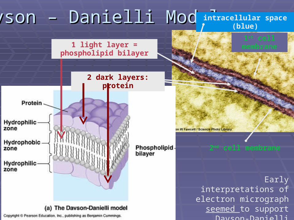
Davson – Danielli ModelDavson – Danielli Model
2nd cell membrane
1st cell membrane
intracellular space (blue)
1 light layer = phospholipid bilayer
2 dark layers: protein
Early interpretations of electron micrograph
seemed to support Davson-Danielli model.

ProblemsProblems with the with the Davson-Danielli ModelDavson-Danielli Model
EvidenceEvidence AmountAmount and and typetype of of
membrane membrane protein varies protein varies widely between cellswidely between cells
Analysis of Analysis of membrane membrane proteinproteins showed lots of s showed lots of hydrophobichydrophobic areas areas
FREEZE FRACTURE FREEZE FRACTURE technique shows inside of technique shows inside of membrane to be membrane to be smooth smooth with bumps (proteins) with bumps (proteins) sticking outsticking out

Current Singer-Nicolson ModelCurrent Singer-Nicolson Model
2nd cell membrane
1st cell membrane
intracellular space (blue)
1 light layer = hydrophobic lipid tails
2 dark layers: polar phosphate heads

Lesson Two:Lesson Two:Membrane TransportMembrane Transport

Materials can move in and out Materials can move in and out of cellsof cells
THROUGH plasma THROUGH plasma membranemembrane
By membrane fusion / By membrane fusion / fissionfission

Diffusion and OsmosisDiffusion and Osmosis
DiffusionDiffusion – the – the passive movement passive movement of particles from of particles from an area of greater an area of greater to lesser to lesser concentration concentration
Due to random, Due to random, continual motion continual motion of particles of particles
Toward dynamic Toward dynamic equilibriumequilibrium

OsmosisOsmosis Osmosis Osmosis – the – the
diffusion of diffusion of water across a water across a selectively selectively permeable permeable membrane membrane (from an area (from an area of lower solute of lower solute to higher solute to higher solute concentration)concentration)

Concentration of solutes (tonicity)Concentration of solutes (tonicity) HypotonicHypotonic – lower concentration of solutes – lower concentration of solutes
COMPARED TO COMPARED TO another solution.another solution. HypertonicHypertonic – higher concentration … – higher concentration … IsotonicIsotonic – equal concentration … – equal concentration …
Osmolarity is a measure of the solute particles in a solution
Can use terms hyposmotic, hyperosmotic, and isosmotic
Difference is that tonicity only refers to solutes that can’t cross
the membrane but osmolarity includes all solutes.

a) Which condition is harmful to both plant and animal cells?a) Which condition is harmful to both plant and animal cells?b) Would putting bacteria in a hypotonic solution be a good way to b) Would putting bacteria in a hypotonic solution be a good way to prevent their growth? Why or why not?prevent their growth? Why or why not?c) When transporting an organ for transplant, what type of solution c) When transporting an organ for transplant, what type of solution should be used for soaking? Why?should be used for soaking? Why?

Passive transport (diffusion) across Passive transport (diffusion) across a membranea membrane
Passive transport does NOT require energy to be Passive transport does NOT require energy to be ““spentspent”” by the cell ( by the cell (NO ATPNO ATP))
Selective permeability Selective permeability -- some particles can move -- some particles can move through the membranethrough the membrane
Different materials can pass through different Different materials can pass through different membranes depending on membrane proteinsmembranes depending on membrane proteins
(simple)
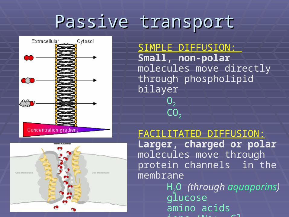
Passive transportPassive transportSIMPLE DIFFUSION: Small, non-polar molecules move directly through phospholipid bilayer
O2
CO2
FACILITATED DIFFUSION: Larger, charged or polar molecules move through protein channels in the membrane
H2O (through aquaporins) glucose
amino acidsions (Na+, Cl-, etc.)

Active transportActive transport Moves particles Moves particles against the concentration against the concentration
gradientgradient (from lower to higher) (from lower to higher) Requires energy Requires energy (ATP) to be used(ATP) to be used

Example of transport in neuronsExample of transport in neurons Neurons are cells that transmit Neurons are cells that transmit
information through electrical information through electrical impulsesimpulses
This is done by movement of ionsThis is done by movement of ions Active transport Active transport at rest:at rest:
Pump keeps a concentration Pump keeps a concentration gradient of Na+/K+ readygradient of Na+/K+ ready
Passive transport Passive transport during impulse:during impulse: Voltage-gated channelsVoltage-gated channels
Na+Na+ K+K+

Facilitated Diffusion Facilitated Diffusion of K+ in neuronsof K+ in neurons
4 polypeptides 4 polypeptides (mostly a-(mostly a-helices) form porehelices) form pore
Placement of polar R-Placement of polar R-groups forms groups forms highly highly selective filterselective filter
Positively charged paddles Positively charged paddles keep gate closed (until keep gate closed (until neuron is depolarized)neuron is depolarized)
Reversal of charge Reversal of charge (voltage) attracts paddles; (voltage) attracts paddles; opens poreopens pore
Top view

Structure and FunctionStructure and Function

Na+ / K+ PumpNa+ / K+ Pump Actively pumps Na+ and K+ Actively pumps Na+ and K+
againstagainst their their concentration concentration gradientsgradients
Phosphate fromPhosphate from ATP ATP helps helps conformational changesconformational changes 3 Na+ allow ATP to bind3 Na+ allow ATP to bind PPii binds, changing shape binds, changing shape
Na+ released, 2 K+ bindNa+ released, 2 K+ bind
Causes PCauses Pii to break off, shape to break off, shape
reverts, K+ releasedreverts, K+ released Cycle continuesCycle continues

VesiclesVesicles (membrane (membrane bubbles) transport bubbles) transport
materials within a cellmaterials within a cell
Fluidity of membrane Fluidity of membrane allows bits to bud off allows bits to bud off from or combine with from or combine with another membraneanother membrane
Little Little ““bubblesbubbles”” of of membrane are called membrane are called vesiclesvesicles
Some animations
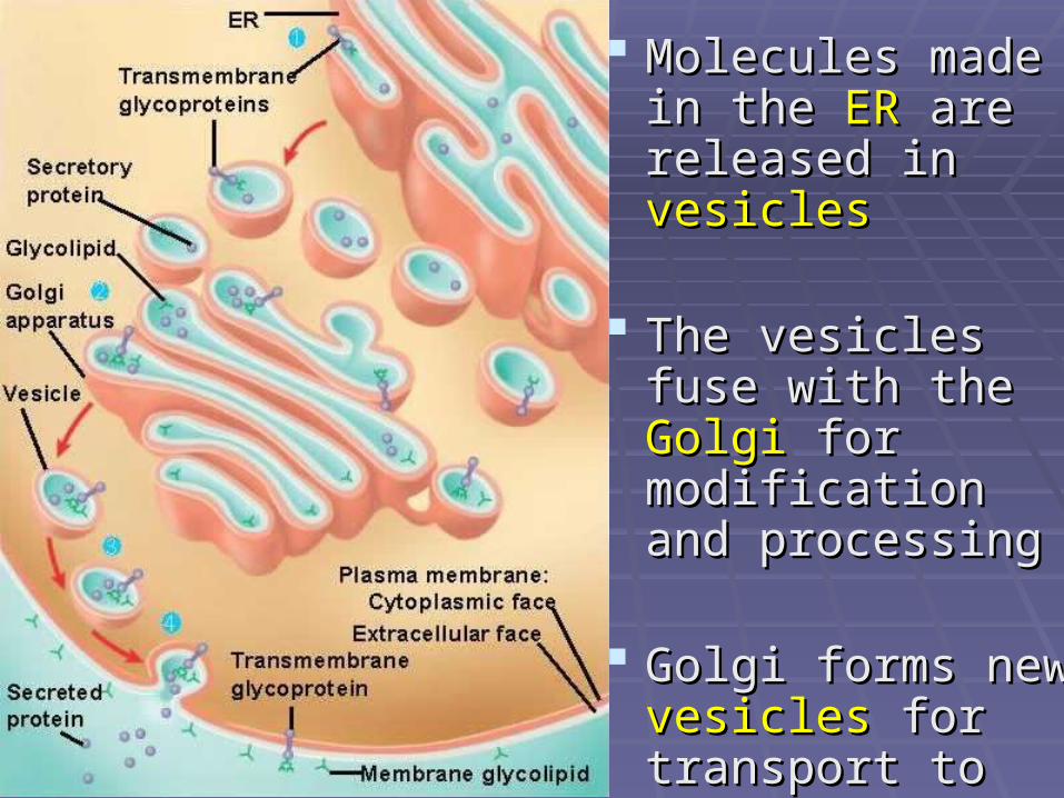
Molecules made in Molecules made in the the ERER are released are released in in vesiclesvesicles
The vesicles fuse The vesicles fuse with the with the GolgiGolgi for for modification and modification and processingprocessing
Golgi forms new Golgi forms new vesiclesvesicles for for transport to the transport to the plasma membraneplasma membrane

Endocytosis and ExocytosisEndocytosis and Exocytosis Endocytosis Endocytosis brings materials brings materials intointo the cell the cell
PinocytosisPinocytosis brings in fluids and small particles brings in fluids and small particles PhagocytosisPhagocytosis combines vesicle with lysosome for digestion (also used by combines vesicle with lysosome for digestion (also used by
phagocytes / macrophages in the immune systemphagocytes / macrophages in the immune system
Exocytosis ejects Exocytosis ejects materials from cellmaterials from cell Can also add proteins to plasma membranes if they are embedded in vesicle Can also add proteins to plasma membranes if they are embedded in vesicle
membranemembrane

Lesson 3:Lesson 3:Cell CycleCell Cycle

The Cell CycleThe Cell CycleAs seen in
eukaryotes

Cell Cycle OverviewCell Cycle Overview Interphase: Interphase:
G1, S, G2 (busy time!)G1, S, G2 (busy time!) Mitosis:Mitosis:
ProphaseProphase – DNA – DNA (chromatin) condenses (chromatin) condenses
MetaphaseMetaphase – – chromosomes align on chromosomes align on equatorequator
AnaphaseAnaphase – chromatids – chromatids separate, move to separate, move to opposite polesopposite poles
TelophaseTelophase – 2 genetically – 2 genetically identical nuclei formidentical nuclei form
Cytokinesis Cytokinesis - cytoplasm - cytoplasm dividesdivides
cytokinesis

Interphase: G1: metabolic reactions, cell growth, cell performs its function(s), transcription and translation of useful genes

Interphase: S : Replication (making a complete copy of DNA)G2: some growth, production of molecules needed for mitosis
At any time during interphase, chloroplasts and mitochondria can reproduce by simple division (binary fission)

DNA vocabulary:Uncondensed DNA is called chromatin.When DNA is condensed, chromosomes are visible.
After S, a chromosome has 2 chromatids (identical). As soon as they separate at the centromere, they will be separate chromosomes.
From chromatin
form, DNA coils and supercoils
into condensed chromosomes

MITOSISMITOSIS

ProphaseProphase: •DNA condenses (supercoils) into chromosomes. •Spindle microtubules assemble•In ANIMALS, centrioles move toward poles•Nuclear membrane breaks down
MITOSIS DETAILS: MITOSIS DETAILS: Does this process of cell division by mitosis
apply to prokaryotes?
Nope! Bacteria have only a single chromosome loop and it does not condense in this way (or line up in pairs, etc.)
Binary fission

MetaphaseMetaphase AnaphaseAnaphase Spindle microtubules Spindle microtubules
attach to centromeresattach to centromeres Chromosomes form a Chromosomes form a
single line single line along the along the equatorequator
Spindle microtubules Spindle microtubules shortenshorten
Sister chromatids split Sister chromatids split into identical into identical chromosomeschromosomes
Chromosomes are Chromosomes are pulled to opposite polespulled to opposite poles

TelophaseTelophase CytokinesisCytokinesis Chromosomes unwindChromosomes unwind Spindle microtubules Spindle microtubules
break downbreak down Nuclear membrane Nuclear membrane
reforms; final productreforms; final product: 2 : 2 genetically identical genetically identical nucleinuclei..
The cytoplasm divides in 2, The cytoplasm divides in 2, forming two cellsforming two cells..
May occur May occur afterafter (or with) (or with) telophasetelophase; ; may not may not happen at all (muscle)happen at all (muscle)!!

Differences in Cytokinesis Differences in Cytokinesis Animal CellsAnimal Cells
Contractile Contractile microfilaments (protein microfilaments (protein belt) forms cleavage belt) forms cleavage furrow furrow (A)(A)
Plant CellsPlant Cells Vesicles build cell plate Vesicles build cell plate (B) (B)
(new cell wall along (new cell wall along equator)equator)

Mitotic IndexMitotic IndexNumber of cell in mitosis Number of cell in mitosis = Mitotic Index= Mitotic Index
Total number of cellsTotal number of cells
E.g. 31 total cellsE.g. 31 total cells
(why these?)(why these?)
8 cells in mitosis8 cells in mitosis
Mitotic index = 8/31Mitotic index = 8/31
MI = 0.26MI = 0.26
1 2 345 6 7
8
910 11
1213 14 15
16 17 18
19 202123 24
25 26
22
27 28 29 3031

a) What is the mitotic index for this image?b) What phase of mitosis is each mitotic cell in?

Mitosis produces genetically Mitosis produces genetically identical identical nucleinuclei
Mitosis animation Mitosis animation with c
ell movie
Chromosomes, made of DNA Chromosomes, made of DNA and protein, contain all the and protein, contain all the genetic information of the cellgenetic information of the cell
Cells divide Cells divide identical sister identical sister chromatids chromatids (doubled during (doubled during replication) into separate, replication) into separate, identical chromosomesidentical chromosomes
One chromosome from identical One chromosome from identical each pair goes to each poleeach pair goes to each pole
Two nuclear envelopes form; Two nuclear envelopes form; one around each set of one around each set of chromosomes chromosomes

Mitosis forms cells for:Mitosis forms cells for: Asexual reproduction. Asexual reproduction. Embryonic developmentEmbryonic development GrowthGrowth Tissue repairTissue repair

MAKING CONNECTIONS:
This is the embryo of a fly (Drosophila melanogaster). Green shows DNA in mitosis and red is a nuclear protein.
Think about how the topics we’ve studied so far relate to this image.
Some ideas:•DNA protein•Surface area : volume•Cells come from pre-existing cells•Stem Cells•Differentiation•Cyclins•Etc.

More review: What would these cells More review: What would these cells do during do during interphaseinterphase??
Pancreatic ExocrinePancreatic Exocrine Copy all chromosomesCopy all chromosomes Build ribosomesBuild ribosomes Multiply mitochondriaMultiply mitochondria Transcribe and translate Transcribe and translate
selected genesselected genes E.g. Hormones and enzymes E.g. Hormones and enzymes
for export for export
Carry out functionsCarry out functions Produce ATP in mitochondria, Produce ATP in mitochondria,
build proteins in rough ER, build proteins in rough ER, package and modify in Golgi, package and modify in Golgi, etc.etc.
Palisade MesophyllPalisade Mesophyll Copy all chromosomesCopy all chromosomes Build ribosomesBuild ribosomes Multiply mitochondria and Multiply mitochondria and
chloroplastschloroplasts Transcribe and translate Transcribe and translate
selected genesselected genes E.g. Enzymes for photosynthesisE.g. Enzymes for photosynthesis
Carry out functionsCarry out functions Convert light energy to chemical Convert light energy to chemical
energy, store energy in energy, store energy in carbohydrates, etc.carbohydrates, etc.

Cyclins: Control of the cell cycleCyclins: Control of the cell cycleCyclins are found in all eukaryotes.
Cyclins are proteins (but NOT enzymes) whose concentration changes in specific parts of the cell cycle.
Cyclins activate CDKs (cyclin-dependent kinases). CDKs are enzymes that regulate the cell cycle through metabolic pathways.

Cancer: Uncontrolled Cancer: Uncontrolled MitosisMitosis Repeated, Repeated, rapid divisionsrapid divisions Ignores body signals (growth Ignores body signals (growth inhibitors)inhibitors) Creates growing group of out-of-Creates growing group of out-of-
control cells (control cells (primary tumourprimary tumour)) Tumours can be in any organ or Tumours can be in any organ or
tissue (though some are more tissue (though some are more common)common)
Secondary tumours Secondary tumours can form can form throughout the body (throughout the body (metastasizemetastasize))
Increased mutations develop from Increased mutations develop from rushed divisionrushed division

PRIMARY TUMOURS: origin of cancerPRIMARY TUMOURS: origin of cancerMutagensMutagens
Agents that cause mutations in DNAAgents that cause mutations in DNA E.g. radiation, free radicals, chemicals, virusesE.g. radiation, free radicals, chemicals, viruses
OncogenesOncogenes Genes that influence cell cycle Genes that influence cell cycle
When in When in normalnormal state, called state, called proto-oncogenesproto-oncogenes
When over-expressed called oncogenes, lead to When over-expressed called oncogenes, lead to cancercancer
Have alleles that make cancer more likely to occur Have alleles that make cancer more likely to occur (fewer mutations needed to cause over-expression)(fewer mutations needed to cause over-expression) E.g. E.g. RasRas helps regulate growth and differentiation; mutated helps regulate growth and differentiation; mutated
form associated with 25% of human cancerform associated with 25% of human cancer

What is the difference between the normal Ras protein (from the proto-oncogene) and the mutant Ras protein (from the oncogene)?
How might this lead to cancer?
If it were possible, would it be helpful to remove
proto-oncogenes from our genome to prevent the
development of cancer?

SECONDARY TUMOURS: SECONDARY TUMOURS: spread of cancerspread of cancerMetastasisMetastasis
Cells break off from Cells break off from primary tumour and lodge primary tumour and lodge in other areas of the in other areas of the body, causing secondary body, causing secondary tumourstumours

Fighting CancerFighting Cancer Some cancer treatments Some cancer treatments
(radiation and some (radiation and some chemotherapy drugs) chemotherapy drugs) target target rapidly dividing cells rapidly dividing cells for for destruction.destruction.
This causes hair to fall out and This causes hair to fall out and damage to the intestinal lining damage to the intestinal lining causing nausea, because causing nausea, because these cells also divide rapidlythese cells also divide rapidly
New treatments try to New treatments try to target target cancer cells more specificallycancer cells more specifically. .