lethal tuberculosis in interleukin-6-deficient …iai.asm.org/content/65/11/4843.full.pdf ·...
TRANSCRIPT

INFECTION AND IMMUNITY,0019-9567/97/$04.0010
Nov. 1997, p. 4843–4849 Vol. 65, No. 11
Copyright © 1997, American Society for Microbiology
Lethal Tuberculosis in Interleukin-6-Deficient Mutant MiceCHRISTOPH H. LADEL,1 CARMEN BLUM,1 ANJA DREHER,1 KURT REIFENBERG,2
MANFRED KOPF,3 AND STEFAN H. E. KAUFMANN1,4*
Department of Immunology1 and Central Animal Facility,2 University of Ulm, Ulm, and Max Planck Institutefor Infection Biology, Berlin,4 Germany, and Basel Institute of Immunology, Basel, Switzerland3
Received 9 April 1997/Returned for modification 13 May 1997/Accepted 15 August 1997
Tuberculosis is a chronic infectious disease which causes major health problems globally. Acquired resis-tance is mediated by T lymphocytes and executed by activated macrophages. In vitro studies have emphasizedthe importance of macrophage activation for mycobacterial growth inhibition. In vivo, the protective hostresponse is focused on granulomatous lesions in which Mycobacterium tuberculosis is contained. A cellular im-mune response of the T helper 1 (Th1) type is considered central for control of tuberculosis. Using interleu-kin-6 (IL-6)-deficient mice, we here demonstrate a crucial role of this pluripotent cytokine in protection againstM. tuberculosis but not against Mycobacterium bovis BCG. Infection with M. tuberculosis was lethal for theIL-6-deficient mice at inocula that were still controlled by IL-6-competent mice. Spleen cells from M. tuber-culosis-infected IL-62/2 mouse mutants produced elevated levels of IL-4 and reduced levels of gamma inter-feron compared to the control levels. Cytofluorometric analyses of spleen cells from M. tuberculosis-infectedmice revealed more-profound alterations in T-cell ratios in IL-62/2 mice than in control mice. We assume thatIL-6 contributes to host resistance by its proinflammatory activity and by its influence on cytokine secretion.
Tuberculosis is a chronic disease, caused by Mycobacteriumtuberculosis, which causes enormous health problems globally(6, 32). In the infected host, mycobacteria are contained withingranulomas where T lymphocytes activate antituberculousmacrophage functions (26, 30). The cross talk between T lym-phocytes, mononuclear phagocytes, and other host cells is me-diated by cytokines. Although Mycobacterium bovis BCG hasbeen widely used as a vaccine, it confers incomplete protectionagainst tuberculosis, at least in adults (9). The insufficient ef-ficacy of M. bovis BCG vaccination has been explained bydifferential activation of T cells and cytokines by M. bovis BCGand M. tuberculosis (18, 45).
According to their cytokine patterns, T helper (Th) cells canbe separated into two groups (for reviews, see references 14and 42). Th2 cells, characterized by potent interleukin-4 (IL-4)and IL-5 production, are primarily responsible for resistanceagainst helminth infections. In contrast, Th1 cells, which pro-duce IL-2 and gamma interferon (IFN-g), are central to de-fense against intracellular bacteria, including M. tuberculosis.
IL-6 is a multifunctional cytokine with at least three majoractivities (1, 34, 54). First, IL-6, together with tumor necrosisfactor (TNF) and IL-1, belongs to the group of proinflamma-tory cytokines which initiate early inflammatory responses (1,2). Second, IL-6 is involved in the promotion of T- and B-cellresponses. Third, it participates in hematopoiesis (5, 34, 54).IL-6 is produced by various cell types, including mononuclearphagocytes, fibroblasts, endothelial cells, B cells, and T cells(54). Although both Th cell types can produce IL-6, it is gen-erally grouped as a Th2 rather than a Th1 cytokine (14).
More-recent attempts to define the role of IL-6 in the anti-infective immune response have emphasized its proinflamma-tory potential during acute infections. For example, IL-6-defi-cient mice rapidly succumb to Listeria monocytogenes infection(12, 35). In contrast, virtually nothing is known about the role
of IL-6 in chronic infectious diseases such as tuberculosis.Phenotypic determination of IL-6 functions in tuberculosis notonly was hampered by the chronicity of disease but was furtherimpeded by chaperoning effects of an anti-IL-6 monoclonalantibody (MAb) which caused paradoxical results (27). Wetook advantage of mouse mutants with a disrupted IL-6 gene(IL-62/2) to overcome these obstacles (35). We found thatIL-62/2 mice were highly susceptible and ultimately suc-cumbed to tuberculosis. These data prove an essential role ofIL-6 in murine tuberculosis.
MATERIALS AND METHODS
Mice. Gene-targeted IL-62/2 mice and their immunocompetent counterparts,IL-61/1 mice, have been described elsewhere (35). The original homozygousIL-62/2 mouse strain on a 129 3 C57BL/6 background was backcrossed toC57BL/6 mice, and the second backcross thereof was used in these experiments.Both IL-62/2 and IL-61/1 mice were derived from this second backcross. The129 and C57BL/6 strains are BCG resistant and sensitive, respectively, and theIL-62/2 and IL-61/1 mice are of mixed bcg genotypes. The IL-62/2 mutantscannot produce IL-6 because of a disrupted IL-6 gene (35). The mice were bredand maintained in our animal facilities at the University of Ulm under specific-pathogen-free conditions. In all experiments, 7- to 10-week-old animals of eithersex were employed. In a given experiment, the mice were age and sex matched.
Bacteria and infections of mice. M. bovis BCG and M. tuberculosis werecultured in Dubos broth base (Difco, Detroit, Mich.) supplemented with Dubosmedium albumin (Difco) after mouse passage. A mid-log-phase culture wasaliquoted and stored at 270°C until use. M. bovis BCG strain Chicago (ATCC27289) was used in the range of 5 3 105 to 5 3 106 live bacteria in phosphate-buffered saline (at a fixed inoculum in a given experiment) for intravenous (i.v.)infection via the lateral tail vein. The exact doses for given experiments areprovided in the figure legends. M. tuberculosis H37Rv was originally provided byJ. K. Seydel (Forschungsinstitut Borstel, Borstel, Germany), and in the differentexperiments concentrations of 5 3 105 to 1 3 106 viable organisms were used fori.v. infection.
Determination of mycobacterial growth. The bacterial loads in liver, spleen,and lung were determined at different times postinfection (p.i.) by plating serialdilutions of organ homogenates obtained by homogenization with a laboratoryblender (Seward Medical, London, United Kingdom). Appropriate dilutionswere plated on Middlebrook agar plates (Difco) supplemented with oleic acid-albumin-dextrose-catalase enrichment (Difco). After 3 to 4 weeks of culture at37°C, CFU were counted.
In vitro stimulation and cytokine production. At the time points indicatedbelow, animals were sacrificed and the spleens were removed. Single-cell sus-pensions were prepared and resuspended in Iscove’s modified Dulbecco’s me-dium (IMDM) (Seromed, Berlin, Germany) supplemented with 10% fetal calfserum (Boehringer Mannheim, Mannheim, Germany), 2 mM glutamine, 100 U
* Corresponding author. Mailing address: Department of Immunol-ogy, University of Ulm, D-89070 Ulm, Germany. Phone: 0731/502-3361, Fax: 0731/502/3367, E-mail: [email protected].
4843
on Septem
ber 2, 2018 by guesthttp://iai.asm
.org/D
ownloaded from

of penicillin-streptomycin (Gibco, Paisley, United Kingdom) per ml, and 1 mg ofindomethacine (Sigma, St. Louis, Mo.) per ml (complete IMDM). Cells wereseeded into 96-well plates (Nunc, Roskilde, Denmark) at a final concentration of2 3 105 per well and stimulated with M. tuberculosis H37Ra lyophilisate (Difco).Positive-control cultures were stimulated with concanavalin A (Sigma) or poke-weed mitogen (Sigma) at 5 mg/ml. After 2 days of culture, supernatants wereremoved, 0.2-mm-pore-size sterile filtered (for M. tuberculosis-infected material),and frozen at 220°C until analyzed. Cytokine concentrations were determined intriplicate.
ELISA. IFN-g concentrations were determined by a double-sandwich enzyme-linked immunosorbent assay (ELISA) using specific MAbs R4-6A2 and AN18-17.24 as described previously (39). Murine recombinant IFN-g (rIFN-g) (gen-erous gift of G. Adolf, Ernst Boehringer-Institut fur Arzneimittelforschung,Vienna, Austria), with a specific activity of 107 U/mg, was diluted in completeIMDM to obtain a standard curve. The detection limit of the ELISA was 0.05 Uof IFN-g per ml. IL-4 concentrations were determined in a sandwich ELISAusing specific MAbs BVD4-1D11 and BVD6-24-G2 as described previously (55).The detection limit was 2 pg of IL-4 per ml, calculated from a standard curve withmurine recombinant IL-4 (rIL-4) (Genzyme, Cambridge, Mass.) with a specificactivity of $107 U/mg. Concentrations of IL-12 were determined in a two-siteELISA using specific MAbs C17.8 and C15.6 (kind gift of G. Trinchieri, WistarInstitute, Philadelphia, Pa.) and murine rIL-12 (kindly provided by S. Wolf,Genetics Institute, Cambridge, Mass.) with a specific activity of 5.6 3 106 U/mgfor standardization as described previously (37). The detection limit was 20 pg ofIL-12 p40/p70 per ml. Determination of TNF activities was done with the TNF-sensitive L929 cell line as described elsewhere (24). Murine recombinant TNF(rTNF) (Genzyme) with a specific activity of $5 3 107 U/mg was used tocalculate a standard curve. The detection limit was 2 pg of TNF per ml. TNF,IFN-g, IL-4, and IL-12 concentrations were determined with SoftmaxPro soft-ware (Molecular Devices, Sunnyvale, Calif.) using four-parameter standardcurve calculations. Optical densities were analyzed with SpectraMax equipment(Molecular Devices).
Cytofluorometry. Spleen cells were stained with the following MAbs: anti-L3T4–phycoerythrin (PE) (clone YTS 191) (MEDAC, Hamburg, Germany),anti-Lyt2–fluorescein isothiocyanate (FITC) (clone YTS 169) (MEDAC), anti-T-cell receptor alpha/beta (anti-TCRab)–biotin (clone H57-597) (kind gift fromR. Kubo), anti-TCRgd–biotin (clone GL-3) (kindly provided by L. Lefrancois),anti-CD3–FITC (clone 145-2C11) (kind gift of J. Bluestone), anti-NK1.1–biotin(clone PK 136) (American Type Culture Collection, Rockville, Md.), and anti-Lyt5(B220)–FITC (clone RA3-6B2) (MEDAC). Biotinylated antibodies weredetected with streptavidin-PE (Gibco) or streptavidin-RED670 (Gibco). Cellswere analyzed with a FACScan (Becton Dickinson, Mountain View, Calif.) usingLYSIS II software (Becton Dickinson).
Histology. Lungs of M. tuberculosis-infected mice were fixed in formalin andparaffin embedded at different times p.i. Sections were cut 3 to 5 mm thick andstained for acid-fast bacilli (AFB) by the Ziehl-Neelsen method and counter-stained with hematoxylin. Slides were analyzed under an AxioPhot microscope(Zeiss, Jena, Germany).
Statistical analysis. To assess significant differences between IL-62/2 andIL-61/1 mice, Student’s t tests were performed. A P value of #0.05 was consid-ered significant.
RESULTS
M. bovis BCG infection in IL-62/2 and IL-61/1 mice. IL-62/2 and IL-61/1 mice were infected i.v. with M. bovis BCG,and bacterial growth in spleens, livers, and lungs was deter-mined at different times p.i. As shown in Fig. 1, the course ofinfection did not differ significantly between IL-6-deficient mu-tants and their immunocompetent wild-type controls. Bothmouse strains controlled infection efficiently, as indicated bythe continuous decline in the numbers of M. bovis BCG or-ganisms in all three organs. In particular, lungs of both IL-62/2
and IL-61/1 mice were free from mycobacteria after day 60 p.i.These results reveal efficient control of M. bovis BCG in theabsence of IL-6, at least under the conditions employed. Thesmall interindividual variations, despite a mixed genetic back-ground in these mice, may suggest a minor impact of thesebackground genes on resistance to BCG in our experimentalsetting.
M. tuberculosis infection in IL-62/2 and IL-61/1 mice. Astrikingly different result was obtained when IL-62/2 and IL-61/1 mice were infected i.v. with M. tuberculosis organisms(Fig. 2). The IL-62/2 mice succumbed to tuberculosis from day50 p.i. onwards (median survival time [MST], 59 days). AllIL-6-deficient mice had died before the end of the 120-day
FIG. 1. Growth of M. bovis BCG in the spleen (A), liver (B), and lung (C) ofIL-62/2 (‚) and IL-61/1 (E) mice. In the experiment whose results are shown,mice were infected with 106 BCG organisms i.v. Organs were removed at theindicated times and homogenized, serial dilutions were plated on Middlebrookagar plates, and CFU were determined 3 to 4 weeks later. The data are meansfor four mice per time point (SD were ,10%) and are from one representativeexperiment repeated twice with similar results.
4844 LADEL ET AL. INFECT. IMMUN.
on Septem
ber 2, 2018 by guesthttp://iai.asm
.org/D
ownloaded from
observation period, whereas all wild-type control animals sur-vived M. tuberculosis infection (MST . 120 days). Already atday 15 p.i., the bacterial load was significantly increased in allthree organs (mean log10 CFU 6 standard deviations [SD] forIL-62/2 and IL-61/1 mice, respectively, were as follows:spleen, 7.47 6 0.11 and 6.20 6 0.35; liver, 6.30 6 0.17 and5.78 6 0.15; and lung, 6.98 6 0.15 and 5.76 6 0.14 [DCFU inall organs was statistically significant at a P of ,0.01; n 5 4mice per group]). At day 60 p.i., 100- to 1,000-fold more M.
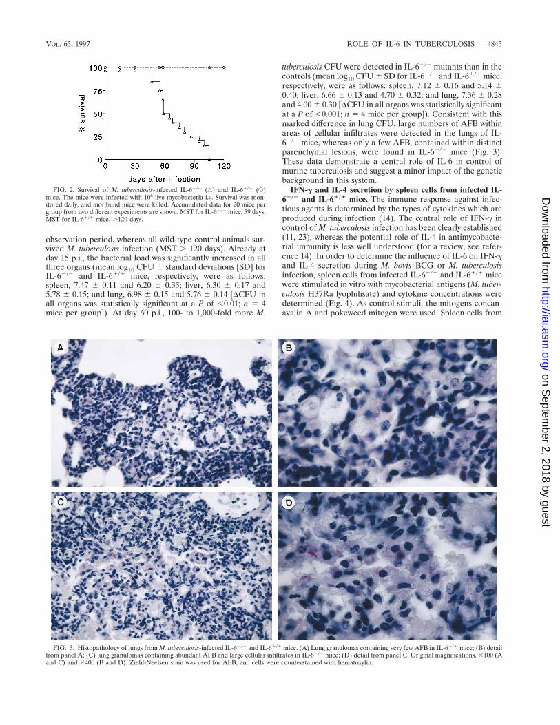
tuberculosis CFU were detected in IL-62/2 mutants than in thecontrols (mean log10 CFU 6 SD for IL-62/2 and IL-61/1 mice,respectively, were as follows: spleen, 7.12 6 0.16 and 5.14 60.40; liver, 6.66 6 0.13 and 4.70 6 0.32; and lung, 7.36 6 0.28and 4.00 6 0.30 [DCFU in all organs was statistically significantat a P of ,0.001; n 5 4 mice per group]). Consistent with thismarked difference in lung CFU, large numbers of AFB withinareas of cellular infiltrates were detected in the lungs of IL-62/2 mice, whereas only a few AFB, contained within distinctparenchymal lesions, were found in IL-61/1 mice (Fig. 3).These data demonstrate a central role of IL-6 in control ofmurine tuberculosis and suggest a minor impact of the geneticbackground in this system.
IFN-g and IL-4 secretion by spleen cells from infected IL-62/2 and IL-61/1 mice. The immune response against infec-tious agents is determined by the types of cytokines which areproduced during infection (14). The central role of IFN-g incontrol of M. tuberculosis infection has been clearly established(11, 23), whereas the potential role of IL-4 in antimycobacte-rial immunity is less well understood (for a review, see refer-ence 14). In order to determine the influence of IL-6 on IFN-gand IL-4 secretion during M. bovis BCG or M. tuberculosisinfection, spleen cells from infected IL-62/2 and IL-61/1 micewere stimulated in vitro with mycobacterial antigens (M. tuber-culosis H37Ra lyophilisate) and cytokine concentrations weredetermined (Fig. 4). As control stimuli, the mitogens concan-avalin A and pokeweed mitogen were used. Spleen cells from
FIG. 2. Survival of M. tuberculosis-infected IL-62/2 (‚) and IL-61/1 (E)mice. The mice were infected with 106 live mycobacteria i.v. Survival was mon-itored daily, and moribund mice were killed. Accumulated data for 20 mice pergroup from two different experiments are shown. MST for IL-62/2 mice, 59 days;MST for IL-61/1 mice, .120 days.
FIG. 3. Histopathology of lungs from M. tuberculosis-infected IL-62/2 and IL-61/1 mice. (A) Lung granulomas containing very few AFB in IL-61/1 mice; (B) detailfrom panel A; (C) lung granulomas containing abundant AFB and large cellular infiltrates in IL-62/2 mice; (D) detail from panel C. Original magnifications, 3100 (Aand C) and 3400 (B and D). Ziehl-Neelsen stain was used for AFB, and cells were counterstained with hematoxylin.
VOL. 65, 1997 ROLE OF IL-6 IN TUBERCULOSIS 4845
on Septem
ber 2, 2018 by guesthttp://iai.asm
.org/D
ownloaded from

infected IL-62/2 and IL-61/1 mice produced comparable con-centrations of IFN-g and IL-4 in response to these nonspecificstimuli (data not shown). By contrast, antigen-specific stimu-lation of splenic cells from M. bovis BCG-infected mice showedthat IL-62/2 mice produced less IFN-g than wild-type controls,in particular at late stages of infection (days 60 to 90 p.i.) (Fig.4A). Conversely, IL-4 production by splenic cells from IL-62/2
mice increased significantly from days 30 to 90 but declinedcontinuously in controls from days 60 to 90. Hence, althoughIL-62/2 and IL-61/1 mice controlled M. bovis BCG infectionequally well, the IL-6 deficiency resulted in decreased IFN-gand increased IL-4 levels. Spleen cells from M. tuberculosis-infected IL-62/2 and IL-61/1 mice produced appreciable lev-els of IFN-g after stimulation with mycobacterial antigens;however, the IL-6 deficiency led to reduced IFN-g secretion(Fig. 4B). Conversely, IL-4 secretion by splenocytes from M.tuberculosis-infected IL-62/2 mice was increased compared tothat of the immunocompetent animals, although IL-4 was de-tectable in cell cultures from both mouse strains. Our findingstherefore suggest that IL-6 deficiency favored IL-4 secretionand impaired IFN-g production during mycobacterial infection.
IL-12 and TNF secretion by spleen cells from M. tuberculo-sis-infected IL-62/2 and IL-61/1 mice. Next, we determinedIL-12 and TNF secretion by spleen cells from M. tuberculosis-infected IL-62/2 and IL-61/1 mice after in vitro restimulationwith mycobacteria (Fig. 5). IL-12 stimulates the developmentof a Th1 immune response, and rIL-12 administration has beenshown to improve resistance against tuberculosis (10, 52). TNFsynergizes with IFN-g in the activation of tuberculostatic mac-rophage functions and is central to resistance against M. tuber-
culosis and M. bovis BCG (24, 28). At the times tested, stimu-lated spleen cells from M. tuberculosis-infected IL-62/2 miceproduced slightly increased IL-12 concentrations compared tothose of IL-61/1 mice (Fig. 5). The level of TNF secretion bystimulated spleen cells from IL-62/2 mice was lower than thatof IL-61/1 controls initially (Fig. 5). TNF secretion by IL-61/1
splenocytes, however, declined after day 30, whereas TNF pro-duction by IL-62/2 cells increased steadily. Accordingly, at day60 p.i., higher TNF levels were produced in the absence of IL-6.
Phenotypes of spleen cells from infected IL-62/2 and IL-61/1 mice. Phenotypic characterization of spleen cells fromIL-62/2 and IL-61/1 mice by microfluorometry revealed dif-ferential effects of M. bovis BCG and M. tuberculosis infections(Table 1). In response to M. bovis BCG, neither the B-cell/T-cell nor the CD4/CD8 T-cell ratio was altered by the IL-6deficiency. Only an initial increase in the NK1.11 populationwas apparent at an early time (day 15 p.i.). In contrast, the IL-6deficiency caused alterations in the lymphocyte ratios duringM. tuberculosis infection. In the IL-62/2 mice, M. tuberculosisinfection decreased the CD4 T-cell population slightly and theCD8 T-cell population more profoundly. As a result, the CD4/CD8 ratio was increased in M. tuberculosis-infected IL-62/2
mice more profoundly than in immunocompetent controls. Inthe IL-61/1 mice, the CD8 T-cell population was enlarged inthe initial phase of tuberculosis. After an initial decline com-pared to B cells in noninfected mice, the proportion of B cellsincreased from days 15 to 60 p.i. in M. tuberculosis-infectedIL-62/2 mice. Finally, M. tuberculosis infection resulted in theexpansion of the NK1.11 population in IL-62/2 mice, whereasthe proportion of NK1.11 cells was only transiently increased
FIG. 4. IFN-g and IL-4 secretion by spleen cells from IL-62/2 and IL-61/1 mice infected with M. bovis BCG (A) or M. tuberculosis H37Rv (B). Spleen cells werecultured with 5 mg of killed and lyophilized M. tuberculosis H37Ra organisms per ml for 2 days. Supernatants were analyzed for IFN-g and IL-4 activities by ELISA.Note that M. tuberculosis-infected IL-62/2 mice died from day 50 onwards. The results are means of triplicates from one representative experiment repeated twice. SDwere #15%. p, significance (P # 0.05 as determined by Student’s t test).
4846 LADEL ET AL. INFECT. IMMUN.
on Septem
ber 2, 2018 by guesthttp://iai.asm
.org/D
ownloaded from

in M. tuberculosis-infected IL-61/1 mice. Thus, the absence ofIL-6 caused more-pronounced changes in T-cell ratios in in-fection with M. tuberculosis than with M. bovis BCG.
DISCUSSION
Our experiments reveal a striking role of IL-6 in tuberculo-sis. M. tuberculosis is an intracellular bacterium which survivesin resting macrophages (8, 30). Macrophage activation by cy-tokines, in particular IFN-g, is considered the principal mech-anism underlying acquired resistance (11, 15, 21–23, 30, 49).However, even activated macrophages fail to fully eradicate M.
tuberculosis (15, 49). Accordingly, even in immune individuals,M. tuberculosis persists and disease generally develops afterreactivation as a consequence of impaired immunity (7, 50).Experimental tuberculosis of mice has provided importantclues into the complexity of the protective host responseagainst tuberculosis (30). Convincing evidence suggests thatdifferent T-cell populations are required for acquisition of pro-tection to mycobacterial infections. This includes CD4, CD8,and g/d T lymphocytes (25, 30, 36, 38, 39, 41, 43). Although thespecific roles of these different T-cell subsets remain incom-pletely understood, the crucial role of the Th1 immune re-sponse is beyond question (14, 30, 31). In contrast, Th2 cells
FIG. 5. IL-12 and TNF secretion by spleen cells from M. tuberculosis-infected IL-62/2 and IL-61/1 mice. Spleen cells were cultured as described in the legend toFig. 4. Supernatants were analyzed by ELISA (for IL-12) or bioassay (TNF). Note that M. tuberculosis-infected IL-62/2 mice died after day 50. The results are meansof triplicates from one representative experiment repeated twice. SD were #15%. p, significance (P # 0.05 as determined by Student’s t test).
TABLE 1. Cytofluorometric analyses of spleen cells from infected IL-62/2 and IL-61/1 micea
Infection typeand mice
Dayp.i.
Spleen cells CD4/CD8ratio
B220/CD3ratioTotal (108) % CD4 % CD8 % CD3 % NK1.1 % B220
NoneIL-61/1 1.1 19.8 12.9 32.5 5.91 59.1 1.54 1.82
IL-62/2 1.0 22.1 13.6 34.4 5.60 57.4 1.62 1.67
M. bovis BCGIL-61/1 15 1.2 23.2 13.1 43.8 4.15 47.3 1.77 1.08
30 1.5 16.5 10.4 29.8 6.89 50.9 1.59 1.7160 2.6 22.2 17.1 35.1 8.23 52.6 1.30 1.5090 1.7 27.7 20.1 54.1 3.56 36.8 1.38 0.68
IL-62/2 15 2.2 17.7 13.5 32.6 8.05 49.5 1.31 1.5230 3.5 13.3 7.5 27.0 3.18 60.7 1.77 2.2560 1.8 18.3 10.8 38.6 2.65 55.2 1.69 1.4390 1.6 22.7 17.7 44.5 5.73 40.9 1.28 0.92
M. tuberculosisIL-61/1 15 2.3 19.2 15.0 33.6 10.1 14.1 1.28 0.42
30 2.0 19.7 18.6 36.2 5.6 40.9 1.06 1.1360 8.8 16.8 8.8 24.7 4.7 42.9 1.91 1.74
120 9.0 12.8 6.4 20.2 4.6 50.2 2.00 2.48
IL-62/2 15 2.0 12.9 5.3 18.3 10.6 33.8 2.45 1.8530 1.5 18.6 4.6 18.6 12.9 51.7 4.01 2.7860 1.3 15.7 4.8 21.7 14.8 50.7 3.30 2.34
a Gated on lymphoid cells by morphological characteristics (forward versus sideward scatter). Note that IL-62/2 mice died after day 51 p.i. (Fig. 2). Results are fromthe experiments depicted in Fig. 4. Similar results were obtained in two experiments.
VOL. 65, 1997 ROLE OF IL-6 IN TUBERCULOSIS 4847
on Septem
ber 2, 2018 by guesthttp://iai.asm
.org/D
ownloaded from

are ineffective or even harmful in tuberculosis, and antibodiesplay a minor role, if any, in protection against tuberculosis.
Consistent with the central contribution of Th1 cells to pro-tection against tuberculosis, IFN-g-mutant mice succumb toinfection with M. tuberculosis (11, 23). These IFN-g-deficientmutants are also more susceptible to BCG vaccination (13, 29).Further, a critical role of TNF alpha (TNF-a) in protectionagainst tuberculosis has been demonstrated (24). This cytokineis produced by activated macrophages and synergizes withIFN-g in macrophage activation (20). Moreover, a role of TNFin necrotic tissue reactions has been established (24, 33). Fi-nally, treatment with rIL-12 has been found to increase resis-tance against tuberculosis (10). Although the great complexityof the immune response which controls tuberculosis stronglysuggests that multiple other cytokines are required, thus farvirtually no information on this issue has been available.
Our data add IL-6 to the list of cytokines which are criticalto resistance against tuberculosis. To our knowledge, this is thefirst report describing an essential role of IL-6 in protectiveimmunity to M. tuberculosis. In vitro, CD4 T cells from M.tuberculosis-infected mice produce high levels of IL-6 duringthe early immune response, and IL-6 induces mycobacterialgrowth inhibition in macrophages (19, 46). Moreover, a previ-ous study found that treatment of mice with anti-IL-6 MAbimpaired protective immunity against Mycobacterium avium(3). However, compelling evidence showing that treatmentwith anti-IL-6 MAb increases serum IL-6 levels, probablythrough chaperoning effects, has been presented (27). Hence,phenotypic characterization of IL-6 functions remained incon-clusive. Consistent with the functional effects of IL-6 in murinetuberculosis, analyses of human pulmonary tuberculosis and oflevels of IL-6 in human plasma point to a role of IL-6 ininfection with M. tuberculosis (17, 40).
IL-6 has a molecular weight of 22,000 to 29,000 and is en-coded by a gene located on chromosome 5 (34, 35). It is apleiotropic cytokine which plays a major role in hematopoiesis,T- and B-cell differentiation, and inflammation (1, 34, 54).Furthermore, IL-6 synergizes with other cytokines in macro-phage activation (19). Therefore, it is possible that differentmechanisms are responsible for the high degree of susceptibil-ity of IL-62/2 mutants to tuberculosis. IL-6, together with IL-1and TNF-a, is a potent proinflammatory cytokine, and it is themajor inducer of the acute-phase response and of neutrophilstimulation in microbial infections (1, 34, 48, 54). Accordingly,the increased susceptibility of IL-62/2 mutants to L. monocy-togenes infection is correlated with impaired blood neutro-philia (12, 35). In other models, IL-6 has been shown to pro-mote differentiation of T cells (51, 53, 54). IL-6 is generallygrouped as a Th2 cytokine, and recently it was found to pro-mote Th2 responses (14, 47, 54). However, it has also beenshown to stimulate cytotoxic T lymphocyte and NK functionsand to favor Th1 cell development (48, 51, 53). In a recentreport, impaired Th1 cell development in IL-62/2 mutant miceinfected with Candida albicans has been described, althoughthe precise mechanisms through which IL-6 contributes to Th1cell differentiation remain to be identified (48). The data inFig. 4 are consistent with an impact of IL-6 in Th1 cell re-sponses in tuberculosis. Taking the published data on biolog-ical activities of IL-6 into account, we consider the possibilitythat IL-6 participates in defense against tuberculosis in theimmunocompetent host by inducing inflammatory responsesand by promoting IFN-g production.
In our experiments, we inoculated mice with an M. tubercu-losis strain of low mouse virulence by the i.v. route (16, 36).Differential virulence of wild-type M. tuberculosis is also con-sidered an important factor in the epidemiology of human
tuberculosis and is thought to influence the efficacy of BCGvaccination (18). It is therefore noteworthy that the IL-62/2
mutants were highly susceptible to the weakly virulent M. tu-berculosis substrain used in our studies but still were capable ofcontrolling the vaccine strain M. bovis BCG efficiently. How-ever, in IL-62/2 mutants, M. bovis BCG apparently also fa-vored IL-4 production and impaired IFN-g secretion. We as-sume that default cytokine secretion in IL-62/2 mutants hadmore-dramatic consequences for control of M. tuberculosisthan for the vaccine strain M. bovis BCG. Similarly, gene dis-ruption mutants with a deficiency in CD8 T cells, g/d T cells,IFN-g, or TNF receptor type I are more susceptible to M.tuberculosis than to M. bovis BCG (11, 13, 23–25, 29, 36, 38,39). It is possible that such differences at least in part contrib-ute to the failure of the M. bovis BCG vaccine to stimulatesatisfactory protection against tuberculosis (6, 9). Although theIL-61/1 and IL-62/2 mice were from the same breedings, thegenetic background between individual mice was heteroge-neous. It is therefore of note that the IL-62/2 mice succumbedto tuberculosis under conditions which were still tolerated bythe IL-61/1 mice.
With the emergence of tuberculosis in human immunodefi-ciency virus-positive individuals and of multidrug-resistantstrains of M. tuberculosis, immunotherapy with recombinantcytokines in adjunct to chemotherapy is currently being con-sidered for treatment of certain cases of tuberculosis (4, 44).Studies using gene disruption mutant mice with defined immu-nodeficiencies may help to define the cytokines which are crit-ical to protective immunity against tuberculosis and thereforemay facilitate the development of new immunologic means forprevention and therapy of tuberculosis.
ACKNOWLEDGMENTS
This work received financial support from the Sonderforschungs-bereich 322 “Lympho-Hamopoese” (project B7) and BMBF project“Mykobakterielle Infektionen.” The Basel Institute of Immunologyhas been founded and is supported by Hoffmann-La Roche, Ltd.,Basel, Switzerland.
We are grateful to K. Tell for MAb production and to G. Szalay forimprovement of the manuscript. We thank G. Adolf, J. Bluestone, L.Lefrancois, G. Trinchieri, and S. Wolf for helpful reagents.
REFERENCES
1. Akira, S., and T. Kishimoto. 1992. IL-6 and NF-IL6 in acute-phase responseand viral infection. Immunol. Rev. 127:26–50.
2. Allen, L.-A. H., and A. Aderem. 1996. Mechanisms of phagocytosis. Curr.Opin. Immunol. 8:36–40.
3. Appelberg, R., A. G. Castro, J. Pedrosa, and P. Minoprio. 1994. Role ofinterleukin-6 in the induction of protective T cells during mycobacterialinfections in mice. Immunology 82:361–364.
4. Bermudez, L. E., and G. Kaplan. 1995. Recombinant cytokines for control-ling mycobacterial infections. Trends Microbiol. 3:22–27.
5. Berna, A., M. Kopf, R. Kulbacki, N. Weich, G. Koehler, and J. C. Gutierrez-Ramos. 1994. Interleukin-6 is required in vivo for the regulation of stem cellsand committed progenitors of the hematopoietic system. Immunity 1:725–731.
6. Bloom, B. R., and C. J. L. Murray. 1992. Tuberculosis: commentary on areemergent killer. Science 257:1055–1064.
7. Bothamley, G. H. 1996. Does immunity to tuberculosis contribute to patho-genesis? Trends Microbiol. 4:95.
8. Clemens, D. L., and M. A. Horwitz. 1995. Characterization of the Mycobac-terium tuberculosis phagosome and evidence that phagosomal maturation isinhibited. J. Exp. Med. 181:257–270.
9. Colditz, G. A., T. F. Brewer, C. S. Berkey, M. E. Wilson, E. Burdick, H. V.Fineberg, and F. Mosteller. 1994. Efficacy of BCG vaccine in the preventionof tuberculosis. Meta-analysis of the published literature. JAMA 271:698–702.
10. Cooper, A. M., A. D. Roberts, E. R. Rhoades, J. E. Callahan, D. M. Getzy,and I. M. Orme. 1995. The role of interleukin-12 in acquired immunity toMycobacterium tuberculosis infection. Immunology 85:423–432.
11. Cooper, A. M., D. K. Dalton, T. A. Stewart, J. P. Griffin, D. G. Russell, andI. M. Orme. 1993. Disseminated tuberculosis in interferon-g gene-disruptedmice. J. Exp. Med. 178:2249–2254.
4848 LADEL ET AL. INFECT. IMMUN.
on Septem
ber 2, 2018 by guesthttp://iai.asm
.org/D
ownloaded from

12. Dalrymple, S. A., L. A. Lucian, R. Slattery, T. McNeil, D. M. Aud, S.Fuchino, F. Lee, and R. Murray. 1995. Interleukin-6-deficient mice arehighly susceptible to Listeria monocytogenes infection: correlation with inef-ficient neutrophilia. Infect. Immun. 63:2262–2268.
13. Dalton, D. K., S. Pitts-Meek, S. Keshav, I. S. Figari, A. Bradley, and T. A.Stewart. 1993. Multiple defects of immune cell function in mice with dis-rupted interferon-g genes. Science 259:1739–1742.
14. Daugelat, S., and S. H. E. Kaufmann. 1996. Role of Th1 and Th2 cells inbacterial infections, p. 66–97. In S. Romagnani (ed.), Chemical immunology:Th1 and Th2 cells in health and disease. S. Karger AG, Basel, Switzerland.
15. Denis, M. 1991. Involvement of cytokines in determining resistance andacquired immunity in murine tuberculosis. J. Leukocyte Biol. 50:495–501.
16. Dunn, P. L., and R. J. North. 1995. Virulence ranking of some Mycobacte-rium tuberculosis and Mycobacterium bovis strains according to their ability tomultiply in the lungs, induce lung pathology, and cause mortality in mice.Infect. Immun. 63:3428–3437.
17. El-Ahmady, O., M. Mansour, H. Zoeir, and O. Mansour. 1997. Elevatedconcentrations of interleukins and leukotriene in response to Mycobacteriumtuberculosis infection. Ann. Clin. Biochem. 34:160–164.
18. Fine, P. E. M. 1989. The BCG story: lessons from the past and implicationsfor the future. Rev. Infect. Dis. 11:S353–S359.
19. Flesch, I. E. A., and S. H. E. Kaufmann. 1990. Stimulation of antibacterialmacrophage activities by B-cell stimulatory factor 2/interleukin 6. Infect.Immun. 58:269–271.
20. Flesch, I. E. A., and S. H. E. Kaufmann. 1993. Role of cytokines in tuber-culosis. Immunobiology 189:316–339.
21. Flesch, I. E. A., and S. H. E. Kaufmann. 1987. Mycobacterial growth inhi-bition by interferon-g activated bone marrow macrophages and differentialsusceptibility among strains of Mycobacterium tuberculosis. J. Immunol. 138:4408–4413.
22. Flory, C. M., R. D. Hubbard, and F. M. Collins. 1992. Effects of in vivo Tlymphocyte subset depletion on mycobacterial infections in mice. J. Leuko-cyte Biol. 51:225–229.
23. Flynn, J. L., J. Chan, K. J. Triebold, D. K. Dalton, T. A. Stewart, and B. R.Bloom. 1993. An essential role for interferon-g in resistance to Mycobacte-rium tuberculosis infection. J. Exp. Med. 178:2249–2254.
24. Flynn, J. L., M. M. Goldstein, J. Chan, K. J. Triebold, K. Pfeffer, C. J.Lowenstein, R. Schreiber, T. W. Mak, and B. R. Bloom. 1995. Tumor ne-crosis factor-a is required in the protective immune response against Myco-bacterium tuberculosis in mice. Immunity 2:561–572.
25. Flynn, J. L., M. M. Goldstein, K. J. Triebold, B. Koller, and B. R. Bloom.1992. Major histocompatibility complex class I-restricted T cells are requiredfor resistance to Mycobacterium tuberculosis infection. Proc. Natl. Acad. Sci.USA 89:12013–12017.
26. Hahn, H., and S. H. E. Kaufmann. 1981. The role of cell mediated immunityin bacterial infections. Rev. Infect. Dis. 3:1221–1250.
27. Heremans, H., C. Dillen, W. Put, J. V. Damma, and A. Billiau. 1992. Pro-tective effect of anti-interleukin (IL)-6 antibody against endotoxin associatedwith paradoxically increased IL-6 levels. Eur. J. Immunol. 22:2395–2401.
28. Hernandez-Pando, R., and G. A. W. Rook. 1994. The role of TNF-a inT-cell-mediated inflammation depends on the Th1/Th2 cytokine balance.Immunology 82:591–595.
29. Kamijo, R., J. Le, D. Shapiro, E. A. Havell, S. Huang, M. Aguet, M. Bosland,and J. Vilcek. 1993. Mice that lack the interferon-g receptor have profoundlyaltered responses to infection with Bacillus Calmette-Guerin and subsequentchallenge with lipopolysaccharide. J. Exp. Med. 178:1435–1440.
30. Kaufmann, S. H. E. 1993. Immunity to intracellular bacteria. Annu. Rev.Immunol. 11:129–163.
31. Kaufmann, S. H. E. 1995. Immunity to intracellular bacteria and protozoa.The Immunologist 3:221–225.
32. Kaufmann, S. H. E., and J. D. A. Van Embden. 1993. Tuberculosis: aneglected disease strikes back. Trends Microbiol. 1:2–5.
33. Kindler, V., A.-P. Sappino, G. E. Grau, P.-F. Piquet, and P. Vassalli. 1989.The inducing role of tumor necrosis factor in the development of bactericidalgranulomas during BCG infection. Cell 56:731–740.
34. Kishimoto, T., S. Akira, and T. Taga. 1992. Interleukin-6 and its receptor: aparadigm for cytokines. Science 258:593–597.
35. Kopf, M., H. Baumann, G. Freer, M. Freudenberg, M. Lamers, T. Kishi-
moto, R. Zinkernagel, H. Bluethmann, and G. Kohler. 1994. Impaired im-mune and acute phase responses in interleukin-6 deficient mice. Nature368:339–341.
36. Ladel, C. H., C. Blum, A. Dreher, K. Reifenberg, and S. H. E. Kaufmann.1995. Protective role of g/d T cells and a/b T cells in tuberculosis. Eur.J. Immunol. 25:2877–2881.
37. Ladel, C. H., C. Blum, and S. H. E. Kaufmann. 1996. Control of natural killercell-mediated innate resistance against the intracellular pathogen Listeriamonocytogenes by g/d T lymphocytes. Infect. Immun. 64:1744–1749.
38. Ladel, C. H., J. Hess, S. Daugelat, P. Mombaerts, S. Tonegawa, and S. H. E.Kaufmann. 1995. Contribution of a/b and g/d T lymphocytes to immunityagainst Mycobacterium bovis bacillus Calmette Guerin: studies with T cellreceptor deficient mutant mice. Eur. J. Immunol. 25:838–846.
39. Ladel, C. H., S. Daugelat, and S. H. E. Kaufmann. 1995. Immune responseto Mycobacterium bovis bacille Calmette Guerin infection in major histocom-patibility complex I and II deficient mutant mice: contribution of CD4 andCD8 T cells to acquired resistance. Eur. J. Immunol. 25:377–384.
40. Law, K., M. Weiden, T. Harkin, K. Tchou-Wong, C. Chi, and W. N. Rom.1996. Increased release of interleukin-1b, interleukin-6, and tumor necrosisfactor-a by bronchoalveolar cells lavaged from involved sites in pulmonarytuberculosis. Am. J. Respir. Crit. Care Med. 153:799–804.
41. Leveton, C., S. Barnass, B. Champion, S. Lucas, B. De Souza, M. Nicol, D.Banerjee, and G. Rook. 1989. T-cell-mediated protection of mice againstvirulent Mycobacterium tuberculosis. Infect. Immun. 57:390–395.
42. Mosmann, T. R., and R. L. Coffman. 1989. TH1 and TH2 cells: differentpatterns of lymphokine secretion lead to different functional properties.Annu. Rev. Immunol. 7:145–173.
43. Muller, I., S. P. Cobbold, H. Waldmann, and S. H. E. Kaufmann. 1987.Impaired resistance against Mycobacterium tuberculosis infection after selec-tive in vivo depletion of L3T41 and Lyt-21 T cells. Infect. Immun. 55:2037–2041.
44. Murray, P. J., A. Aldovini, and R. A. Young. 1996. Manipulation and poten-tiation of antimycobacterial immunity using recombinant bacille Calmette-Guerin strains that secrete cytokines. Proc. Natl. Acad. Sci. USA 93:934–939.
45. Orme, I. M. 1988. Characteristics and specificity of acquired immunologicmemory to Mycobacterium tuberculosis infection. J. Immunol. 140:3589–3593.
46. Orme, I. M., A. D. Roberts, J. P. Griffin, and J. S. Abrams. 1993. Cytokinesecretion by CD4 T lymphocytes acquired in response to Mycobacteriumtuberculosis infection. J. Immunol. 151:518–525.
47. Rincon, M., J. Anguita, T. Nakamura, E. Fikrig, and R. A. Flavell. 1997.Interleukin-6 directs the differentiation of IL-4-producing CD41 T cells.J. Exp. Med. 185:461–469.
48. Romani, L., A. Mencacci, E. Cenci, R. Spaccapelo, C. Toniatti, P. Pucetti, F.Bistoni, and V. Poli. 1996. Impaired neutrophil response and CD41 T helpercell 1 development in interleukin-6 deficient mice infected with Candidaalbicans. J. Exp. Med. 183:1345–1355.
49. Rook, G. A. W., J. Taverne, C. Leveton, and J. Steele. 1987. The role ofgamma-interferon, vitamin D3 metabolites and tumor necrosis factor in thepathogenesis of tuberculosis. Immunology 62:229–234.
50. Stead, W. W. 1967. Pathogenesis of a first episode of chronic pulmonarytuberculosis in man: recrudescence of residuals of the primary infection orexogenous reinfection. Am. Rev. Respir. Dis. 95:729–745.
51. Takai, Y., G. Wong, S. Clark, S. Burakoff, and S. Herrman. 1988. B cellstimulatory factor-2 is involved in the differentiation of cytotoxic T lympho-cytes. J. Immunol. 140:508–512.
52. Trinchieri, G. 1995. Interleukin-12: a proinflammatory cytokine with immu-noregulatory functions that bridge innate resistance and antigen-specificadaptive immunity. Annu. Rev. Immunol. 13:251–276.
53. Uyttenhove, C., P. G. Coulie, and J. Van Snick. 1988. T cell growth anddifferentiation induced by interleukin-HP1/IL-6, the murine hybridoma/plas-macytoma growth factor. J. Exp. Med. 167:1417–1427.
54. Van Snick, J. 1990. Interleukin-6: an overview. Annu. Rev. Immunol. 8:253–278.
55. Yamamoto, S., F. Russ, H. C. Teixeira, P. Conradt, and S. H. E. Kaufmann.1993. Listeria monocytogenes-induced gamma interferon secretion by intesti-nal intraepithelial gamma/delta T lymphocytes. Infect. Immun. 61:2154–2161.
Editor: R. E. McCallum
VOL. 65, 1997 ROLE OF IL-6 IN TUBERCULOSIS 4849
on Septem
ber 2, 2018 by guesthttp://iai.asm
.org/D
ownloaded from