libro de resumenes elafot alicia hernanelafot.ciq.uchile.cl/documentos/lrxielafot.pdf · mariano...
TRANSCRIPT

XI ELAFOT. Córdoba, Argentina. October 1 4, 2012.
XIELAFOT
October 1- 4,
2012Córdoba - Argentina


Comité Organizador Dr. Norman A. García (Chair) Dra. Sonia G. Bertolotti (Vice-Chair) Dra. Marcela Altamirano Dra. Alicia Biasutti Dr. Carlos Chesta Dra. Susana Criado Lic. Daniela Fuentes Dr. Walter Massad Dra. Sandra Miskoski Dra. M. Lorena Gómez Dr. Hernán Montejano Dr. Rodrigo Palacios
Comité Científico Dr. Carlos Previtali (President) Dra. Elsa Abuin Dr. Pedro Aramendía Dra. Teresa Atwars Dra. Ana M. Edwards Dr. Marcelo Gehlen Dr. Daniel Mártire Dra. Alicia Peñeñory
AgradecimientosEl Comité Organizador agradece a los doctorandos y post-doctorandos del Grupo de Fotoquímica de la UNRC por la valiosa colaboración brindada: Dr. Ernesto Arbeloa, Mic. Laura Boiero, Lic. Cecilia Chalier, Lic. Carolina Gambeta, Ing. Eduardo Gatica, Mic. Natalia Gsponer, Lic. Laura Hernández, Dr. José Natera, Lic. Cecila Palacios, Dra. Gabriela Porcal, Mic. Eugenia Reynoso, Mic. Mariel Zalazar y Lic. Claudia Solís.
XI ELAFOT. Córdoba, Argentina. October 1 4, 2012.
2

XI ELAFOT. Córdoba, Argentina. October 1 4, 2012.
Agradecemos a las siguientes instituciones y firmas comerciales por el apoyo económico brindado:
Universidad Nacionalde Río Cuarto
Consejo Nacional deInvestigaciones Científicas y
Técnicas
Agencia Nacional dePromoción Científica
y Tecnológica
Achával Rodríguez 2032Res. Santa Ana (X5010ERH)
Córdoba, Argentina
Lavalle 1634 - Piso 3º "B" C1048AAN Buenos Aires
Argentina
NOBELMULTI S.A.Gallo 1286
Ciudad de Buenos Aires Argentina
.
3

XI ELAFOT. Córdoba, Argentina. October 1 4, 2012.
Agradecemos a las siguientes instituciones por auspiciar el XI ELAFOT:
Universidad Nacionalde Río Cuarto
Facultad de Ciencias Exactas,Fisico Químicas y Naturales
Consejo Nacional deInvestigaciones
Científicas y Técnicas
Agencia Nacional dePromoción Científica
y TecnológicaUniversidad Nacional de Córdoba
4
Índice
Comité Organizador Página 2
Comité Científico Página 2
Agradecimientos Página 2
Apoyo económico Página 3
Auspicios Página 4
Programa sintético Páginas 6-7
Programa extendido Páginas 8-13
Conferencias plenarias (PL) Páginas 14-25
Conferencias invitadas (INV) Páginas 26-37
Presentaciones orales (OP) Páginas 38-55
Presentaciones en pósters (PP) Páginas 56-222
Índice de autores Páginas 223-230
5

XI ELAFOT. Córdoba, Argentina. October 1 4, 2012.
Programa sintético
6

Sund
ay,Sep
t.30
Mon
day,Oct1
Tuesda
y,Oct.2
Wed
nesday,O
ct.3
Thursday,O
ct.4
8.00
–8.45
Registration
8.00
–8.45
8.45
–9.00
Welcome
8.45
–9.00
9.00
9.50
PL1.Juan
C.S
PL1.Juan
C.Scaian
oPL
5.Xa
vier
Allo
nas
PL9.Ca
ssiusStevan
iPL
11.G
ustavo
Argüe
llo9.00
9.50
9.50
10.05
OP14
.Pab
loCo
metto
9.50
10.05
10.05
10.20
INV6.Nan
cyPizarro
OP15
.Ana
Edwards
10.05
10.20
10.20
10.35
OP9.Had
adCa
cier
OP16
.Carmen
Gue
des
10.20
10.35
10.35
10.40
PL2.Anton
ioZano
cco
PL6.Julia
PérezPrieto
10.35
10.40
10.40–11
.10
Coffee
Break
10.40–11
.10
11.10–11
.40
INV1.Ed
uardoLissi
INV3.Ed
gardo
Duran
tini
INV7.Clau
diaLong
oINV10
.Julio
DeLa
Fuen
te11
.10–11
.40
11.40
11.55
OP1.Migue
lNeu
man
11.40
11.55
11.55–12
.10
OP2.Clau
dio
Borsarelli
INV4.Juan
E.Argüe
lloINV8.Mariano
Bossi
INV11
.Carolina
Lorente
11.55–12
.10
12.10–12
.25
OP3.Juan
aSilber
OP6.Ana
Moo
reOP10
.Pau
laCa
regn
ato
OP17
.NataliaPa
cion
i12
.10–12
.25
12.25–15
.00
Free
TimeforLunch
12.25–15
.00
15.00
15.50
PL3.Ed
wardClen
nan
PL7.Pe
terOgilby
PL10
.San
tiNon
ell
15.00
15.50
15.50–16
.40
PL4.Th
orsten
Bach
PL8.JosefB
aade
rDan
ielM
ártire
OP11
.Silvia
Braslavsky
15.50–16
.40
16.40
17.10
Registration
Coffee
Break
16.40
17.10
17.10–17
.40
(17–20
)INV2.
Jean
Cade
tINV5.Ro
drigo
Albuq
uerque
INV9.Th
omas
Dittrich
17.10–17
.40
17.40–17
.55
OP4.Tamara
Benzaq
uen
OP7.Pa
bloGarcía
OP12
.Herná
nRo
drígue
z17
.40–17
.55
17.55–18
.10
OP5.Jazm
inPo
rras
OP8.Cristian
Strassert
OP13
.Alexand
reVieiraSilva
17.55–18
.10
18.10
20.30
Poster
Session
18.10
20.30
PRESEN
TATION
7

XI ELAFOT. Córdoba, Argentina. October 1 4, 2012.
Programa detallado
8

XI Encuentro Latinoamericano de Fotoquímica y Fotobiología
Sunday, Sept 30
17-20 Registration
Monday, 1th
8.00 – 8.45 Registration
8.45 – 9.00 Welcome
SESSION 1. Chairman: Carlos Previtali
9.00 – 9.50 Plenary Talk 1: Juan C. Scaiano (U. Otawa, Canada), Using organic photochemistry to make nanoparticles and nanoparticles todirect organic chemistry.
9.50 – 10.40 Plenary Talk 2: Antonio Zanoco (U. de Chile, Chile),Photophysic of Aryloxazinones and Aryloxazoles
10.40 – 11.10 COFFEE BREAK
11.10 – 11.40 Invited Talk 1: Eduardo Lissi (U. Santiago de Chile, Chile), Evaluación de la asociación soluto – proteína mediante medidas de fluorescencia.
11.40 – 11.55 OP-1: Miguel Neumann (U. de Sao Paulo, Brazil), Photochemistry of tetraphenyldiboroxane and its use as photopolymerization co-initiator
11.55 – 12.10 OP-2: Claudio Borsarelli (U. de Santiago del Estero, Argentina), Biophysical properties and cellular toxicity of covalent cross-linked oligomers of -synuclein formed by photoinduced side-chain tyrosyl radicals
12.10 – 12.25 OP-3: Juana Silber (U. de Río Cuarto, Argentina), ¿Qué ocurre cuando se utiliza un solvente biodegradable en la formación de micelas inversas? Caracterización de sistemas micelares utilizando técnicas fotoquímicas.
9

12.30 – 15.00 FREE TIME FOR LUNCH
SESSION 2. Chairwoman: Alicia Peñéñory
15.00 – 15.50 Plenary Talk 3: Edward Clennan (U. of Wyoming, USA),Type I and II Photooxygenations of Organic Sulfides
15.50 - 16.40 Plenary Talk 4: Thorsten Bach (TU München, Germany), Chirality and Light: Enantioselective Catalysis in Photochemistry
16.40 – 17.10 COFFEE BREAK
17.10 – 17.40 Invited Talk 2: Jean Cadet (Institut Nanosciences & Cryogénie Grenoble, France ), Recent aspects of solar irradiation of cells and human skin: formation and repair of DNA
17.40 – 17.55 OP-4: Tamara Benzanquen (INTEC (U. Litoral) Santa Fe, Argentina), Eficiencias Cuánticas de la Degradación de Atrazina en agua por Foto-Fenton 17.55 – 18.10 OP-5: Jazmín Porras (U de Antioquia, Colombia), Foto-transformación de Clorotalonil Usando Sustancias Húmicas
18.10 – 20.30 POSTER SESSION
Tuesday 2nd
SESSION 3. Chairwoman: María Victoria Encinas
9.00 – 9.50 Plenary Talk 5: Xavier Allonas (U. of Haute Alsace, France),Photocyclic initiating systems for free radical photopolymerization under visible light. Application to holographic recording.
9.50 – 10.40 Plenary Talk 6: Julia Pérez Prieto (U. de Valencia. Spain),Functional Photoactive Nanoparticles
10.40 – 11.10 COFFEE BREAK
11.10 – 11.40 Invited Talk 3: Edgardo Durantini (U.de Río Cuarto), Photodynamic inactivation of microorganisms
11.40 – 12.10 Invited Talk 4: Juan E. Argüello (U. de Córdoba), Study of the Selenide Radical Cation Chemistry, from Synthetic application to the Direct Observation of these Intermediates
10

12.10 – 12.25 OP-6: Ana Moore (Arizona State University), The Photoanode of Photoelectrochemical Cells for the Splitting of Water
12.25 – 15.00 FREE TIME FOR LUNCH
SESSION 4. Chairman: Daniel Mártire
15.00 – 15.50 Plenary Talk 7: Peter Ogilby (Aarhus U., Denmark), Singlet Oxygen: From Single Cells to Gold Nanodiscs, and Beyond (Yes, there is still something new under the sun)
15.50 - 16.40 Plenary Talk 8: Josef Baader (U. de São Paulo, Brazil), On the Efficiency of Electron-Transfer Initiated Organic Chemiluminescence
16.40 – 17.10 COFFEE BREAK
17.10 – 17.40 Invited Talk 5: Rodrigo Albuquerque (U. de São Paulo,Brazil), Diffusion-limited Energy Transfer in Blends of Oligofluorenes with an Anthracene Derivative
17.40 – 17.55 OP-7: Pablo García (U. de Córdoba, Argentina),Asociación y Fotodegradación de Albúmina por complejos diiminos de Cr(III)
17.55 – 18.10 OP-8: Cristian Strassert (Westfälische Wilhelms-U , Germany), Aggregation matters - From planar photosensitizers and electroluminescent materials to organo- and hydrogels
18.10 – 20.30 POSTER SESSION
Wednesday, 3rd
SESSION 5. Chairwoman: Teresa Atvars
9.00 – 9.50 Plenary Talk 9: Cassius Stevani (U. de São Paulo,Brazil ),Fungal bioluminescence: mechanism and application in toxicology
9.50 – 10.20 Invited Talk 6: Nancy Pizarro-Urzua (U Andrés Bello,Chile),Photochemistry of antihypertensive drugs: media and substituent effects.
10.20 – 10.35 OP-9: Hadad Casier (U. of Antioquia, Colombia), Up-conversion and Migration by Energy Transfer: a Mixed Model for Doped Luminescent Solids
10.35 – 11.10 COFFEE BREAK
11

11.10 – 11.40 Invited Talk 7: Claudia Longo (U. of Campinas, Brazil),Photo-electrochemistry and solar energy conversion: application in dye-sensitized solar cells, hydrogen production and water disinfection
11.40 – 12.10 Invited Talk 8: Mariano Bossi (U. Buenos Aires, Argentina), Super-Resolution Imaging with Switchable Fluorophores Based on Oxazine Auxochromes
12.10 – 12.25 OP-10: Paula Caregnato (U. La Plata, Argentina), Variación en las propiedades fotolumiscentes de nanopartículas de silicio modificadas superficialmente con tioles terminales.
12.25 – 15.00 FREE TIME FOR LUNCH
SESSION 6. Chairman: Pedro Aramendía
15.00 – 15.50 Plenary Talk 10: Santi Nonell (U. Ramon Llull, España),Producción de oxígeno singlete codificada genéticamente
15.50 - 16.20 Special Talk: Daniel Mártire (U. La Plata, Argentina)
16.20 – 16.40 OP-11: Silvia Braslavsky, (Max Planck Institut, Germany), Structural volume changes upon triplet state formation of water-soluble porphyrins depend on the resonant effect of the substituents
16.40 – 17.10 COFFEE BREAK
17.10 – 17.40 Invited Talk 9: Thomas Dittrich (Helmholtz Centre Berlin for Materials and Energy, Germany), Investigation of donor-acceptor molecule and quantum dot layer systems by surface photovoltage techniques
17.40 – 17.55 OP-12: Hernán Rodríguez, (U de Buenos Aires, Argentina), Toward highly efficient long-lived excited state generation in crowded constrained environments
17.55 – 18.10 OP-13: Alexandre Vieira Silva (U. of São Paulo, Brazil), Novel Riboflavin Derivatives for Photodynamic Therapy
18.10 – 20.30 POSTER SESSION
12

Thursday, 4th
SESSION 7. Chairman: Enrique San Román
9.00 – 9.50 Plenary Talk 11: Gustavo Argüello (U. de Córdoba, Argentina ), Fotoquímica en fase gaseosa…. y… ¿sólida? .Peroxinitratos y Óxidos de Nitrógeno
9.50 – 10.05 OP-14: Pablo Cometto (U de Córdoba, Argentina),Identificación y determinación del rendimiento de productos para la foto-oxidación troposférica del 3-metil-3-buten-1-ol (331mbo) iniciada por el radical OH
10.05 – 10.20 OP-15: Ana Edwards (Pontificia U. Católica de Chile, Chile), Effect of visible light mediated by Zn Phthalocyanine incorporated to bovine serum albumin (BSA) on HeLa cells
10.20 – 10.35 OP-16: Carmen Guedes, (U. Estadual de Londrina, Brasil), Influência da radiação na produção de carotenoides pela microalga Haematococcus pluvialis
10.35 – 11.10 COFFEE BREAK
11.10 – 11.40 Invited Talk 10: Julio de la Fuente (U. Universidad de Chile,Chile), Photoreduction of 3-Methyl-1H-quinoxalin-2-one derivatives by N-phenylglicine. A mechanistic study.
11.40 – 12.10 Invited Talk 11: Carolina Lorente (U. La Plata, Argentina), Tryptophan photosensitization by pterin
12.10 – 12.25 OP-17: Natalia Pacioni (U. de Córdoba, Argentina), Cambios en la fotofísica de nanopartículas de oro y plata en presencia de un interruptor molecular como estrategia para su diferenciación en mezclas
12.25 END OF THE XI ELAFOT
13

XI ELAFOT. Córdoba, Argentina. October 1 4, 2012.
Conferencias Plenarias (PL)
14

XI ELAFOT. Córdoba, Argentina. October 1 4, 2012.
Using organic photochemistry to make nanoparticles and nanoparticles todirect organic chemistry
Scaiano, Juan C.Department of Chemistry and Centre for Catalysis Research and Innovation,
University of Ottawa, 10 Marie Curie, Ottawa, Ontario, K1N 6N5, Canada [email protected]
Organic photochemistry has proven an excellent tool for the production of nanostructures of gold, silver, copper, cobalt, niobium and other elements, from the corresponding ions in aqueous systems. Among photochemical precursors, ketones are good photosensitizers for nanoparticle synthesis not because of the energy they can absorb or deliver, but rather because of the reducing free radicals they can generate. Thus efficient nanoparticle generation requires a careful selection of substrates and experimental conditions such that free radical generation occurs with high quantum efficiency, and where metal ion precursors do not cause UV screening of the organic photosensitizers. Synthesis strategies based on water-soluble benzoins have proven very versatile.1Beyond organic precursors, hydrogen peroxide has proven a valuable reducing agent for the formation of ultraclean nanoparticles that can later be modified using laser techniques.2 The nanoparticle forming reactions can be interpreted in terms of multisite proton coupled electron transfer (PCeT) reactions.3
Plasmon transitions provide an easy way to deliver energy to metallic nanostructures, that can then be used to control the chemistry and spectroscopy of molecules in their vicinity.A molecule irradiated in the proximity of a metal nanoparticle can be viewed as undergoing transmitter/receiver antenna interactions,4 a process that has also been described as analogous to a lightning rod effect. Thus irradiating the nanoparticle itself can deliver energy to a strategically located organic molecule through plasmon field interactions. While fluorescence and Raman enhancements through these interactions are well established, other forms of plasmon sensitization remain largely unexplored. For example, we have shown that this energy can trigger polymerizations with exceptional spatial resolution,5 a strategy that can be used for imaging applications or for the fabrication of self-assembled nanolasers.
Other examples will include metal nanoparticle catalysis of organic reactions, such as oxidations and reductions, as well as acid/base catalyzed processes. The ‘laser drop’ technique will be discussed in the context of a valuable tool to study the mechanisms of plasmon-mediated photocatalysis.6
The antibacterial properties of silver nanoparticle composites will be briefly discussed, including the long-term goal of producing tissue replacement scaffolds.7
References (1) McGilvray, K. L.; Decan, M. R.; Wang, D.; Scaiano, J. C., J. Am. Chem. Soc.2006, 128, 15980-
15981. (2) McGilvray, K. L.; Granger, J.; Correia, M.; Banks, J. T.; Scaiano, J. C., PCCP2011, 13, 11914-
11918. (3) Scaiano, J. C.; Stamplecoskie, K. G.; Hallett-Tapley, G. L., Chem. Commun.2012, 48, 4798-4808. (4) Pacioni, N. L.; González-Bejar, M.; Alarcón, E.; McGilvray, K. L.; Scaiano, J. C., J. Am .Chem.
Soc.2010, 132, 6298-6299. (5) Stamplecoskie, K. G.; Pacioni, N. L.; Larson, D.; Scaiano, J. C., J. Am. Chem. Soc.2011, 133,
9160-9163. (6) Hallett-Tapley, G. L.; Silvero, M. J.; Gonzalez-Bejar, M.; Grenier, M.; Netto-Ferreira, J. C.; Scaiano,
J. C., J. Phys. Chem. C2011, 115, 10784-10790. (7) Alarcon, E. I.; Udekwu, K.; Skog, M.; Pacioni, N. L.; Stamplecoskie, K. G.; Gonzalez-Bejar, M.;
Polisetti, N.; Wickham, A.; Richter-Dahlfors, A.; Griffith, M.; Scaiano, J. C., Biomaterials2012, 33,4947-4956.
PL1
15

XI ELAFOT. Córdoba, Argentina. October 1 4, 2012. 16
Photophysic of Aryloxazinones and Aryloxazoles
Zanocco, Antonio L.; Lemp Else, Germán Günther.
Universidad de Chile, Fac. de Cs. Químicas y Farmacéuticas, Depto. de QuímicaOrgánica y Fisicoquímica,Sergio Livingstone 1007, Santiago, Chile,
Fluorescent molecules, whose spectra or quantum yields are sensitive to their environment, are valuable in the study of heterogeneous, organized and biological media. Many fluorescent solvatochromic dyes have been developed with this purpose. Aryloxazoles and aryloxazinones are two families of heterocyclic compounds which are widely used in chemistry, industry and medicine. Benzoxazinone and benzoxazole derivatives are compounds exhibiting spectral and photophysical properties of great interest such as broad first absorption band with high molar absorption coefficient values, emission in the red, intense fluorescence in both organic solutions and crystalline state, large dipole moment increase in the excited state, large Stokes shifts, and short fluorescence lifetimes. Several of these compounds are known as photostable highly efficient UV dyes used as organic brightening agents, laser dyes, organic plastic scintillators and optical fibre sensors. Some benzoxazole derivatives are also used as dopants in organic light�emitting diodes,chromophores in nonlinear optical polymers, chemosensors for metal ions or pH sensors. However, most of studies performed up to 2006 havebeen mainly focused on the benzoxazinone and benzoxazole rings and very few researches have been done on fused aromatic oxazoleand oxazinonederivatives.During last years, we have studied the photophysical and photochemicalbehavior of naphthoxazinone and naphthoxazolederivatives.With some exceptions, these compoundshave a photophysicalbehavior comparable to the observed for benzo analogous. In general, naphto-derivatives showhigher fluorescence quantum yields, lower spectral overlapping between the absorption and the emissionspectra, excited singlet lifetime in the order of 1-3 ns. Also,someof them showhighphotochemical stability. In addition, a strong dependence of the emission maxima on the solvent polarity, due to the charge transfer character of the S0 – S1 transition, was found.In this talk, the photophysical behavior of the aforementioned compound is discussed. Furthermore, singlet oxygen generation, white light production and singlet molecular oxygen detection by selected compounds belonging to the series areanalyzed.
Acknowledgements: The financial support of FONDECYT, grants 1050996, 1080410 and 1110636 is gratefully acknowledged.
PL2
16
PL3
17
17

XI ELAFOT. Córdoba, Argentina. October 1 4, 2012. 18
Chirality and Light: Enantioselective Catalysis in Photochemistry
Bach Thorsten1
1DepartmentChemie, TU München, Lichtenbergstr. 4, D-85747 Garching [email protected]
Chirality and Light are two fascinating natural phenomena, which are linked in chemistry by the three-dimensional structure of photochemically accessible compounds. For decades, it has been attempted to produce chiral compounds enantioselectively by photochemical methods but only recently has significant progress been made towards this goal. Our own work in the area commenced with chiral templates, which bind prochiralphotosubstrates by hydrogen bonding and which are required to be used in stoichiometric amounts.[1] This work culminated in the total synthesis of (+)-meloscine (1)[2], in which an enantioselective [2+2] photocycloaddition reaction has been employedin natural product synthesis for the very first time.
NH
O
N
H
(+)-Meloscin (1)
NB
OBr3Al
CF3
3
NH
O
NO
OO
NO
2
O
O
More recently, initial attempts to employ electron transfer or energy transfer for catalytic enantioselective reactions have been successfully extended to sensitized [2+2] photo-cycloaddition reactions.[3]Xanthone2 turned out to be an efficient organocatalyst providing good turnover (10 mol-%) and high enantioselectivities (<90% ee) in intramolecular quinolone [2+2] photocycloaddition reactions. Apart from this approach, we have also looked into the possibility of Lewis-acid mediated enantioselectivity in photochemical reactions. Lewis acid 3was developed for coumarin [2+2] photocycloadditionreactions[4] and is currently being further explored. The presentation discusses the background of the above-mentioned studies and provides the latest results of our research efforts in this area.
References [1] a) T. Bach, H. Bergmann, K. Harms, Angew. Chem. Int. Ed. 2000, 39, 2302-2304; b) T.
Bach, H. Bergmann, B. Grosch, K. Harms, J. Am. Chem. Soc.2002, 124, 7982-7990; c) B. Grosch, C. N. Orlebar, E. Herdtweck, M. Kaneda, T. Wada, Y. Inoue, T. Bach, Chem. Eur. J. 2004, 10, 2179-2189; d) S. Breitenlechner, T. Bach, Angew. Chem. Int. Ed.2008, 47,7957-7959; e) K. A. B. Austin, E. Herdtweck, T. Bach, Angew. Chem. Int. Ed.2011, 50,8416-8419.
[2] a) P. Selig, T. Bach, Angew. Chem.2008, 120, 5160-5162; Angew. Chem. Int. Ed.2008, 47,5082-5084; b) P. Selig, E. Herdtweck, T. Bach, Chem. Eur. J. 2009, 15, 3509-3525.
[3] a) A. Bauer, F. Westkämper, S. Grimme, T. Bach,Nature2005, 436, 1139-1140; b) C.Müller, A. Bauer, T. Bach, Angew. Chem. Int. Ed.2009, 48, 6640-6642; b) C. Müller, M. M. Maturi, A. Bauer, M. C. Cuquerella, M. A. Miranda, T. Bach, J. Am. Chem. Soc.2011,133, 16689-16697.
[4] a) H. Guo, T. Bach, Angew. Chem. Int. Ed.2010, 49, 7782-7785; b) R. Brimioulle, H. Guo, T. Bach, Chem. Eur. J.2012, 18, 7552-7560.
PL4
18
Photocyclic initiating systems for free radical photopolymerization under visible light. Application to
holographic recordingAllonas, Xavier*1; Ley, Christian1; Ibrahim, Ahmad1; Tarzi, Olga2; Chan Yong,
Aurélie3; Carré, Christiane3
1 Laboratory of Macromolecular Photochemistry and Engineering, University of Haute Alsace, 3 rue Alfred Werner - 68093 Mulhouse, FRANCE - [email protected] 2 CIHIDECAR-CONICET, Department of Organic Chemistry, FCEyN-University of Buenos Aires, Pabello´ n 2—Ciudad Universitaria, (1428) Buenos Aires, Argentina
3 CNRS, UMR 6082 FOTON, Enssat, 6 rue de Kerampont, BP 80518, 22305 Lannion, FRANCE
Light induced polymerization reaction is employed in quite different technical applications that have become beneficial to humans. These applications include microelectronics, information technologies, optical fibers, dental materials, printing inks, paints, varnishes, ... In other words, various kinds of polymers can be synthesized by light-induced chemical processes, a technique commonly denoted by the term photopolymerization. A key component of this process is the photoinitiating system, which is responsible of the absorption of light and its conversion into chemical energy. For example, laser direct imaging, graphics arts, holography, and dental materials require irradiation in the visible spectrum to benefit from laser technologies or simply to avoid UV damaging effects on skin. Some dyes absorbing in the visible region have been reported to be photoreducible in the presence of amines. These compounds belong to the families of xanthenes, fluorones, acridines, phenazines, thiazenes, and so on. However, dye/coinitiators systems were not developed significantly in the industry. Very often, dark reactions take place that lead to poor shelf life of the formulation, an effect that was detrimental to their industrial use for a long time. In addition the conversion of the monomer to polymer was generally limited. Indeed, for most of the industrial applications, conversion of more than 60% have to be reached, a goal that is difficult to achieve with conventional dye/coinitiator photoinitiating systems (PIS).
Certain additives improve the polymerization efficiency, leading to the development of the so-called three-component PIS or photocyclic initiating systems [1-3]. The mechanism involved is usually rather complex and is based on chemical secondary reactions. It was reported that different radical intermediates generated during the irradiation and in the subsequent polymerization reaction react with the additive to give new reactive radicals. In this paper, a set of photoinitiating systems (PIS) for free radical photopolymerization was studied using time-resolved spectroscopic experiments, real-time FTIR for holographic recording. It is shown that the efficiency of the photoinitiating system can be drastically increased when a redox additive is added to the conventional dye/coinitiator system by virtue of a photocyclic behaviour. Homogeneous photopolymerization process was found to reach a fast vitrification, limiting the conversion at about 55%. By contrast, holographic recording underlines the differences in photoinitiating system reactivity, allowing diffraction efficiencies close to unity for the most reactive PIS [4].
References [1] J.P. Fouassier, X. Allonas, D. Burget, Prog. Org. Coat., 47, (2003), 16 [2] O. Tarzi, X. Allonas, C. Ley, J.P. Fouassier, J. Polym. Sci., Part A : Polym. Chem.,48(12), (2010) 2594-2603. [3] A. Ibrahim, C. Ley, O.I. Tarzi, J.P. Fouassier, X. Allonas, J. Photopolym. Sci. Techn.23, (2010) 101-108. [4] A. Ibrahim, C. Ley, X. Allonas, O.I.Tarzi, A. Chan Yong, C. Carré, R. Chevallier, Photochem. Photobiol. Sci., 2012, DOI: 10.1039/C2PP25099C.
XI ELAFOT. Córdoba, Argentina. October 1-4, 2012. 19
19
PL5

XI ELAFOT. Córdoba, Argentina. October 1 4, 2012. 20
Functional Photoactive Nanoparticles Pérez-Prieto, Julia
1 Instituto Ciencia Molecular, Universidad de Valencia, C/ Catedrático José Beltrán, 2, 46980 Paterna, Valencia, Spain
Spherical metal or semiconductor nanoparticles (NPs) are smart systems that exhibit unique properties, such as a high surface-to-volume ratio and size-dependent properties. They can be capped with a considerable number of ligands, which have an anchoring atom at one end with affinity for the NP surface and the other end provides the NP periphery with the hydrophobicity or hydrophilicity needed to give rise to stable organic or aqueous NP colloidal solutions, respectively. Additionally, the ligands can be used to introduce functionality at the NP periphery. In this case, the NP would act as a 3D-scaffold which makes it possible to provide a high local concentration of a functional moiety, such as fluorophores, photosensitisers, antioxidants, etc. It should also be taken into account that the organic capping can exert an encapsulating role, establishing specific interactions with nearby analytes or facilitating the interaction of analytes with the NP surface or the functional moieties at the periphery. Photoactive NPs usually exhibit a broad-absorption spectrum and some of them have Stokes, or even anti-Stokes, emission, depending on the intrinsic composition of the NP. All these properties can be advantageously used for molecular recognition, bioimagen, drug-delivery, among others. In this lecture I will comment on systems recently developed by our research group showing the versatility of spherical photoactive NPs.
Acknowledgements: Financial support from the Spanish MICINN (CTQ2011-27758) is acknowledged.
References [1] A. P. Alivisatos, J. Phys. Chem. 1996, 100, 13226; R. Sardar, A. M. Funston, P. Mulvaney, R. W. Murray, Langmuir, 2009, 25, 13840 ; T. K. Sau, A. Pal, M.C. Daniel, D. Astruc, Chem. Rev. 2004, 104, 293. [2] R. E. Galian, M. de la Guardia, J. Pérez-Prieto, J. Am. Chem. Soc., 2009, 131.982 [3] S. Pocoví-Martínez, M. Parreño-Romero, S. Agouram, J. Pérez-Prieto, Langmuir, 2011, 27, 5234. [4] J. Aguilera-Sigalat; J. M. Casas-Solvas; M. C. Morant-Miñana; A. Vargas-Berenguel; R. E. Galian; J. Pérez-Prieto, Chem.Comun., 2012, 573.
PL6
20
XI ELAFOT. Córdoba, Argentina. October 1 4, 2012. 21
Singlet Oxygen: From Single Cells to Gold Nanodiscs, and Beyond
(Yes, there is still something new under the sun)Ogilby, Peter R.
Center for OxygenMicroscopy and Imaging, ChemistryDepartment, Aarhus University, Aarhus, Denmark, [email protected]
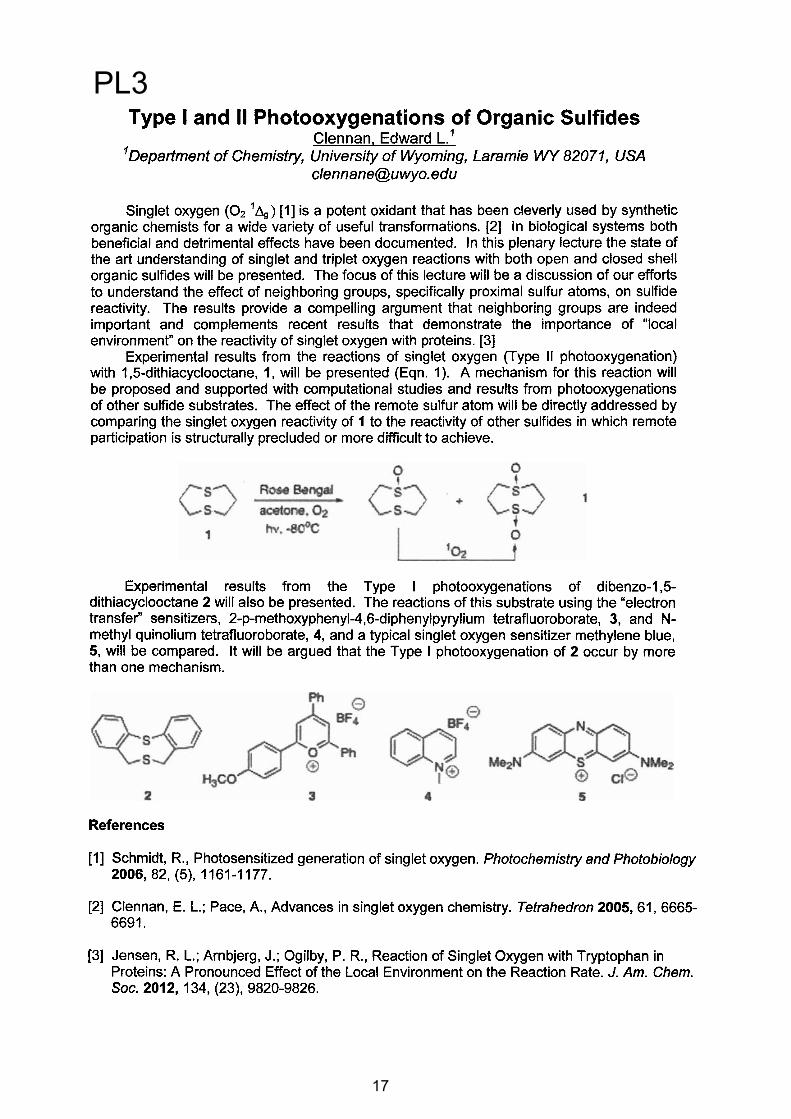
Singlet oxygen, the lowest excited electronic state of molecular oxygen, is a “mature citizen” that has been studied for many years from a wide range of perspectives. Among other things, singlet oxygen has a unique chemistry that results in the oxygenation of many organic molecules. In this way, it plays important roles in biology, particularly in mechanisms of cell signaling and cell death.Singlet oxygen is commonly produced in a photosensitized process wherein light is absorbed by a given molecule (the so-called sensitizer) followed by energy transfer from the excited state sensitizer to ground state oxygen.
We have a multi-faceted program in which the behavior of singlet oxygen is examined in a wide range of systems under a variety of conditions. I will briefly describe our latest work on (a) methods to selectively control the photosensitized production of singlet oxygen in single mammalian cells, (b) monitoring the response of cells to singlet oxygen, (c) the use of two-photon sensitizer excitation to impart spatial, temporal and spectral selectivity in singlet oxygen production, and (d) the use of the electric fields associated with nanoparticle surface plasmons to enhance radiative transitions in oxygen.
Our results indicate that there is still much to be gained from studies of singlet oxygen.
There is still something new under the sun !
PL7
21
XI ELAFOT. Córdoba, Argentina. October 1 4, 2012. 22
On the Efficiency of Electron-Transfer Initiated Organic Chemiluminescence
Baader, Wilhelm Josef
Instituto de Química da Universidade de São Paulo Av. Prof. Lineu Prestes, 748, Butantã, São Paulo, SP, Brazil, [email protected]
The emission of visible light by living organisms (bioluminescence - BL) as well as light emission originated from chemical transformations (chemiluminescence - CL), are long-known phenomena and several reaction mechanisms are discussed to rationalize excited state formation. Many efficient BL and CL transformations are believed to occur with the involvement of electron transfer and electron back-transfer steps, where chemiexcitation finally occurs by radical pair or biradical annihilation.[1]
Initially, a brief introduction to the main known general chemiexcitation mechanisms will be given, including (i) the unimolecular decomposition of 1,2-dioxetanes and 1,2-dioxetanones, (ii) the activated decomposition of cyclic peroxides by appropriate chemiluminescence activators, according to the intermolecular Chemically Initiated Electron Exchange Luminescence (CIEEL) mechanism and (iii) the induced decomposition of phenoxy-substituted 1,2-dioxetanes, following the intramolecular version of the CIEEL mechanism.
In the main part of the contribution recent results of mechanistic studies on electron-transfer initiated CL systems will be presented and it will be shown that intramolecular CIEEL systems can be highly efficient, whereas the intermolecular CIEEL is generally of low efficiency. Subsequently, the peroxyoxalate system, the only intermolecular CIEEL system with proven high quantum yields, will be discussed, specifically with respect to its chemiexcitation step.[2, 3] Thereafter, results will be presented which indicate the occurrence of an intramolecular electron transfer in the first step of the induced decomposition of properly substituted 1,2-dioxetanes.[4] Additionally, it will be shown, using different experimental approaches, that also the electron back-transfer in this transformation is an intramolecularprocess. Finally, recent results on the low efficient catalyzed decomposition of 1,2-dioxetanones, including data with up to now unknown derivatives, will be presented and an explanation for the low efficiency in excited state generation of this system be given.
Acknowledgements: Financial support by The USP Research Consortium for Photochemical Technology (NAP-PhotoTec), Fundação de Amparo À Pesquisa do Estado de São Paulo,(FAPESP), Coordenação de Aperfeicoamento de Pessoal de Nível Superior (CAPES) and Conselho Nacional de Pesquisa (CNPq) is gratefully acknowledged.
References [1] Baader W. J., Stevani C. V., Bastos E. L., “Chemiluminescence of Organic Peroxides”, in: The Chemistry of Peroxides, Chapter 16, p. 1211, ed. Rappoport, Z., Wiley & Sons Ltd, Chichester, 2006.[2] Ciscato, L. F. M. L., Augusto, F. A., Weiss, D., Bartoloni, F. H., Albrecht, S., Brandl, H., Zimmermann, T., Baader, W. J.; ARKIVOC, 2012, 391.[3] Ciscato, L. F. M. L.; Bartoloni, F. H.; Bastos, E. L.; Baader, W. J. J. Org. Chem. 2009, 74,8974.[4] Ciscato, L. F. M. L.; Bartoloni, F. H.; Weiss, D.; Beckert, R.; Baader, W.J. J. Org. Chem. 2010, 75, 6574.
PL8
22
XI ELAFOT. Córdoba, Argentina. October 1-4, 2012.
Fungal bioluminescence: mechanism and application in toxicology
Stevani, Cassius V.Departamento de Química Fundamental, Instituto de Química, Universidade de São
Paulo, CP 26077, 05599-970 São Paulo, SP, Brasil. [email protected]
Although fungal bioluminescence has been reported since ancient times, the chemical pathways involved in light emission, the identity of the substrate and enzymes involved and the biological function of bioluminescence remain unsolved [1]. Likewise the bacterial and firefly bioluminescence, whose study enabled the use of luc and lux reporter genes and harnessed the development of the toxicological bioassay Microtox®, utilized by environmental protection agencies, industries and universities, the comprehension of fungal bioluminescence has the potential to generate a similar assay, provide information about the mechanism of bioluminescence and its use as a tool in Molecular Biology.
In this work will be presented the results obtained by our group in last ten years on: a) the identification and obtention of new cultures of Brazilian bioluminescent fungi [2-5], b) the investigation of the bio- and chemical mechanism of light emission [6,7], and c) the development of a toxicological fungal-based luminescent assay using the species Gerronema viridilucens [8-10].
Acknowledgements: FAPESP, IQ-USP, NAP-PhotoTech (the USP Research Consortiumfor Photochemical Technology)
References
1. D. E. Desjardin, A. G. Oliveira, C. V. Stevani. Fungi bioluminescence revisited. Photochem. Photobiol. Sci. 7: 170-182 (2008).
2. D. E. Desjardin, M. Capelari, C. V. Stevani. A new bioluminescent Agaric from São Paulo, Brazil. Fungal Divers.18: 9-14 (2005).
3. D. E. Desjardin, M. Capelari, C. V. Stevani. Bioluminescent Mycena species from São Paulo, Brazil. Mycologia 99: 317-331 (2007).
4. D. E. Desjardin, B. A. Perry, D. J. Lodge, C. V. Stevani, E. Nagasawa. Luminescent Mycena: new and noteworthy species. Mycologia 102: 459-477 (2010).
5. M. Capelari, D. E. Desjardin, B. A. Perry, T. Asai, C. V. Stevani. Neonothopanus gardneri: a new combination for a bioluminescent Agaric from Brazil. Mycologia 106:1433-1440 (2011).
6. A. G. Oliveira, C. V. Stevani. The enzymatic nature of fungal bioluminescence. Photochem. Photobiol. Sci. 8: 1416-1421 (2009).
7. A. G. Oliveira, D. E. Desjardin, B. A. Perry, C. V. Stevani. Evidence that a single bioluminescent system is shared by all known bioluminescent fungal lineages. Photochem. Photobiol. Sci. 11: 848-852 (2012).
8. L. F. Mendes, E. L. Bastos, D. E. Desjardin, C. V. Stevani. Influence of culture conditions on mycelial growth and bioluminescence of Gerronema viridilucens. FEMS Microbiol. Lett. 282: 132-139 (2008).
9. L. F. Mendes, C. V. Stevani. Evaluation of metal toxicity by a modified method based on the fungus Gerronema viridilucens bioluminescence in agar medium. Environ. Toxicol. Chem. 29: 320-326 (2010).
10. L. F. Mendes, E. L. Bastos, C. V. Stevani. Prediction of metal cation toxicity to the bioluminescent fungus Gerronema viridilucens. Environ. Toxicol. Chem. 29: 2177-2181(2010).
23
PL9
23

XI ELAFOT. Córdoba, Argentina. October 1 4, 2012. 24
PRODUCCIÓN DE OXÍGENO SINGLETE CODIFICADA GENÉTICAMENTE
Santi Nonell*, Rubén Ruiz-González*, Cristiano Viappiani# y Cristina Flors&
*Institut Quimic de Sarria, Universitat Ramon Llull, Barcelona, España. Vía Augusta 390, 08017 Barcelona. e-mail: [email protected]
# Dipartimento di Fisica, Università degli Studi di Parma, Italia. Parco area delle scienze 7A, 43100 Parma, e-mail: [email protected]
& IMDEA Nanociencia, C/ Faraday 9, Ciudad Universitaria de Cantoblanco, 28049 Madrid,
e-mail: [email protected]
Las proteínas fluorescentes (FPs) están adquiriendo una enorme popularidad como sondas codificables genéticamente para observar la dinámica intracelular, la expresión de proteínas y las interacciones proteína-proteína. Sin embargo, su uso en microscopía de fluorescencia se encuentra limitado por la fotodegradación del cromóforo y el daño fotoquímico inducido sobre el medio biológico. La producción de formas reactivas de oxígeno (ROS), especialmente el oxígeno en estado electrónico excitado singlete O2(a1
g), ha sido sugerida para explicar estos efectos.
Por otra parte, la fotosensibilización de O2(a1g) codificada genéticamente podría utilizarse
para estudiar la función de proteínas mediante la técnica de fotoinactivación asistida por cromóforos (chromophore-assisted light inactivation, CALI). Igualmente se ha planteado el desarrollo de mutantes con el propósito específico de generar O2(a1
g) para destruir células con una selectividad inigualable.
En esta presentación se expondrá el trabajo realizador por nuestro grupo en el área de las proteínas fotosensibilizantes.
AgradecimientosEste proyecto ha sido financiado por el Ministerio de Economía y Competitividad de España (Proyectos CTQ2010-20870-C03-01 and RYC-2011-07637). y por la Royal Society (International Joint Projects 2008/R3) y el EPSRC Life Sciences Interface Program del Reino Unido (EP/F042248/1). RRG agradece a la Generalitat de Catalunya y al Fondo Social Europeo la concesión de una beca predoctoral. Agradecemos también el apoyo técnico proporcionado por John White y Laurie Cooper.
PL10
24
XI ELAFOT. Córdoba, Argentina. October 1 4, 2012. 25
Fotoquímica en fase gaseosa….y…¿sólida? Peroxinitratos y Óxidos de Nitrógeno
Argüello, Gustavo A.
INFIQC – Dptode FísicoQuímica, Facultad de CienciasQuímicas, UniversidadNacional de Córdoba, [email protected]
La degradación atmosférica de compuestos hidrocarbonados, y la emisión de los nuevos compuestos hidroclorofluoro-, o hidrofluorocarbonados produce radicales peróxido, que en presencia de contaminantes primarios como el NO2 pueden llevar a la formación de peroxinitratos. Presentaremos resultados obtenidos en nuestros laboratorios donde fotolizando cloro molecular, se inicia el mecanismo equivalente al de degradación atmosférica, que permitió el estudio de nuevos peroxinitratos, derivados tanto de precursores hidrocarbonados como hidrofluorocarbonados. Se dará también una recopilación general del mecanismo de formación de estas “especies reservorio”. Por otro lado, se comentarán los últimos experimentos llevados a cabo en la fotoquímica en matrices de baja temperatura para la dilucidación del mecanismo general de oxidación de óxido nítrico para dar dióxido de nitrógeno. La aparente sencillez de esta reacción (que ha venido estudiándose desde el siglo XVIII y cuya ley de velocidad data de 1918) todavía da lugar a nuevos descubrimientos, como la existencia de un isómero, hasta ahora sólo postulado teóricamente, de N2O4.
PL11
25

XI ELAFOT. Córdoba, Argentina. October 1 4, 2012.
Conferencias Invitadas (INV)
26
26
XI ELAFOT. Córdoba, Argentina. October 1 4, 2012. 27
Evaluación de la asociación soluto – proteína mediante medidas de fluorescencia
Elsa Abuin y Eduardo Lissi
Facultad de Química y Biología, Universidad de Santiago de Chile
Los cambios en la intensidad, longitud de onda y/o polarización de la fluorescencia (del soluto o la proteína) proveen un modo relativamente para estimar la isoterma de adsorción, la distancia entre desactivante y fluoróforo y las propiedades del micro-entorno del cromóforo. Sin embargo, estas evaluaciones no son siempre correctamente llevadas a cabo. En la presente charla nos centraremos en dos aspectos:
1. Las ventajas del método desarrollado en nuestro laboratorio basado en resultados obtenidos a distintas concentraciones de proteína; y
2.La posibilidad de obtener valores errados si no se parte de un modelo adecuado.
Estos dos aspectos serán discutidos en base a resultados obtenidos empleando albúmina como sistema modelo.
INV1
27

XI ELAFOT. Córdoba, Argentina. October 1 4, 2012. 28
Recent aspects of solar irradiation of cells and human skin: formation and repair of DNA
Jean Cadet
Institut Nanosciences & Cryogénie, CEA/Grenoble, 38054 Grenoble, France
The photo-induced formation of base damage to DNA is strongly implicated in the etiology of most skin cancers as the result of exposure to solar radiation and/or UVA photons provided by lamps in tan booths. It is now well documented that the UVB component of solar light is mostly responsible for the formation of bipyrimidine photoproducts within cellular DNA. Indirect support for the major biological role played by the latter photoproducts is provided by the observation of CC to TT tandem mutations that are considered as a molecular signature of the deleterious effects of UVB photons in targeted genes such as p53. Three main classes of photoproducts including cis-syn cyclobutadipyrimidines (P<>Ps), pyrimidine (6-4) pyrimidone adducts (6-4PPs) and related Dewar valence isomers (DewPPs) may be generated at each of the four main bipyrimidine sites (TT,TC,CT and CC sequences) giving rise to a total of 12 possible tandem lesions. Interestingly, most of the latter photoproducts can be singled out as modified dinucleoside monophosphates after DNA extraction from UVB-irradiated cells and subsequent suitable enzymic digestion. The resulting mixture that includes photoproducts and overwhelming normal nucleosides is then subjected to a sensitive HPLC-tandem mass spectrometry analysis, allowing the unambiguous and accurate measurement of several bipyrimidine photoproducts at a dose of UVB radiation as low as 0.2 kJ.m-2. Thus, cyclobutadithymine (T<>T) and a lesser extent 6-4TC and T<>C are detected as the main UVB photoproducts in the DNA of human fibroblasts, keratinocytes and skin. It may be noted that DewPPs are barely detectable, in fact only at CC sites. This is strongly indicative of the poor efficacy for UVB radiation to induce the photoisomerization of the 6-4PP precursors. The situation is totally different when cells are exposed to solar light. Thus, it was shown that the UVA component of solar light is able to partly convert initially UVB-generated 6-4TC and 6-4TT into related Dewar valence isomers. Another interesting observation deals with the UVA-induced formation of T<>T, and to a lesser extent of T<>C, in the DNA of cells and human skin. The specific formation of Pyr<>Pyr, at the exclusion of 6-4TT, may be accounted predominantly by direct excitation of the pyrimidine bases. UVA photons are also able to photo-oxidize cellular DNA through excitation of still unknown endogenous photosensitizers. This was established using a modified comet assay that allows the detection in addition to strand breaks, of oxidized pyrimidine bases and modified purine residues as DNA repair glycosylase-sensitive sites. Thus, singlet oxygen that is generated by a type II photosensitization mechanism was found to be the main contributor to UVA-mediated formation of 8-oxo-7,8-dihydroguanine, an ubiquitous DNA oxidation product. Relevant information on the DNA repair of bipyrimidine photoproducts in cells and human skin was gained from HPLC-MS/MS measurements. It may be pointed out as a striking result that 6-4PP and DewPP are much better substrates for nucleotide excision repair enzymes than Pyr<>Pyr, the cyclobutane dimers at CT and CC being more efficiently repaired that their homologues at TT and TC sites.
Reference:Cadet J., Mouret S., Ravanat J.-L. and Douki T. (2012) Photo-induced damage cellular DNA : Direct and photosensitized reactions, Photochem. Photobiol. doi: 101111:j.1751.1097.2012.01200.x.
INV2
28

XI ELAFOT. Córdoba, Argentina. October 1-4, 2012.
Photodynamic inactivation of microorganismsEdgardo N.Durantini
Departamento de Química, Facultad de CienciasExactasFísico-Químicas y Naturales, UniversidadNacional de Río Cuarto, Agencia Postal Nro 3, X5804BYA Río
Cuarto, Córdoba, Argentina, E-mail [email protected]
The antimicrobial chemotherapy field is in constant changes, because of the great variety of pathogenic species found and their rapid evolutionary changes. The great successes in the war against microorganisms are probably coming to the end. For this reason, it is imperative the development of new drugs and therapies. An innovative method includes a non-oncologic application of photodynamic therapy, named photodynamic inactivation (PDI) of microorganisms. Essentially, PDI is based on the administration of a photosensitizer, which is preferentially accumulated in the microbial cells. The subsequent irradiation with visible light, in the presence of oxygen, specifically generates a cascade of biochemical events that produce cell damages leading to inactivation of the microorganisms.
In these systems, two oxidative mechanisms can occur after photoactivation of the photosensitizer. In the type I photosensitization pathway, the photosensitizer interacts with biomolecules to produce free radicals, while in the type II mechanism, singlet molecular oxygen is produced as the main species responsible for cell inactivation. Depending on the experimental conditions, these mechanisms can take place simultaneously and the ratio between the two processes is influenced by the photosensitizer, substrate and the nature of the medium.
In vitro studies have shown that Gram-positive bacteria are susceptible to the photosensitizing action of a variety of photosensitizers. In contrast, the presence of lipopolysaccharides renders the outer membrane of Gram-negative species with a strong negative charge that makes it impermeable to neutral or anionic compounds. Furthermore, fungal cell walls have a relatively thick layer of -glucan and chitin that leads to a permeability barrier intermediate between Gram-positive and Gram-negative bacteria.This inconvenient can be resolved using positively charges photosensitizers. The presence of cationic groups appears to promote a tight electrostatic interaction with negatively charged sites at the outer surface of the Gram-negative bacteria, increasing the efficiency of the photodynamic activity.
Most of PDI studies have been carried out adding the photosensitizer to cell suspensions. In this procedure, after treatment traces of the photosensitizer can remain in the medium, leading to an undesired remnant photodynamic effect. An alternative to avoid this inconvenient is represented by photosensitizers immobilized on polymeric supports. Also, this procedure could allowing the re-utilization of the photodynamic polymer.
PDI has the advantage over other therapies in that it has selectivity not only because the photosensitizer can be targeted to localized microbial infections, but also the irradiated light can be accurately delivery to the affected area. Practical applications of PDI could involve the elimination of microbial cells growing as localized foci of infection, in liquid media and in biological fluids. Also, photoactive films could be used to form permanent antimicrobial surfaces activated by visible light to maintain aseptic conditions.
Acknowledgements:CONICET, FONCYT-ANPCYT, SECYT-UNRC
29
INV3
29

Study of the Selenide Radical Cation Chemistry, from Synthetic application to the Direct Observation of these
Intermediates.Argüello, Juan E.1; Bouchet, Lydia M. 1; Oksdath-Mansilla, Gabriela1;
Peñéñory, Alicia. B. 1
1Instituto de Investigaciones en Fisicoquímica de Córdoba (INFIQC), Departamento de Química Orgánica, Facultad de Ciencias Químicas,
Universidad Nacional de Córdoba, Córdoba, Argentina.e-mail: [email protected]
Organic selenium compounds have been suggested to have antioxidant properties due to the rather low one-electron reduction potentials of the corresponding radical cations. Some mechanistic aspects and synthetic potentials of photoinduced electron transfer (PET) activation of organoselenium substrates have been explored by Pandey et al.1 These interesting studies have been useful in initiating various synthetic reactions. However, direct evidence of the mediation of selenide radical cations or of other electrophilic selenium and carbocations species is further needed.
We use PET as a tool for radical cation generation, two examples will be discussed.
First, the intramolecular PET reaction in the phthalimide system 1 where selenium containing heterocycles products 2 and 3 are found (eq. 1). In this system, product distribution and photophysical properties of different phthalimides with variable distance between the selenium atom and the phthalimide moiety will be also presented.
Second, the generation of radical cations of ArSeR (5) and its derivatives by intermolecular PET in acetonitrile solution, using different photosensitizers will be discussed (eq 2). The nature of the photoinduced step was confirmed by the observation of the semireduced form of the sensitizer. Thus, the PhSeR radical cations were observed for the first time by transient absorption spectroscopy. The monomeric form of PhSeMe.+ shows a maximum at 500 nm; however, another band at 640 nm was observed for the former, attributed to a -dimmer between the selenide radical cation an a neutral molecule, similar to its periodic neighbor PhSMe.+.2
The influence of electron donating and electron acceptors in the pendant phenyl moiety as well as the steric effect of the alkyl substituent will be discussed in order to explain the experimental observations.
References
1 Pandey, G.; Gadre, S. R. Acc. Chem. Res. 2004, 37, 201 201.2 Yokoi, H.;Hatta, A.; Ishiguro, K.; Sawaki, Y. J. Am. Chem. Soc. 1998, 120, 12728 12733.
30
INV4
30

������������� ���� ���������� ������ ��������������� � ����������������� � �� ������� �
������������� ������ �����������������������������!"�����������������
���������������� ��������������������������������� ���������������!"�� ���������$���������%����'�+��;�<��<��=�<�>�[>;�
����������\��]�$�\���^���^\���_�[���������������`��>�����!�{{��$�\���^��|����\
$'=������������?��������\$^_���`�?{��=���=����������������=���������?������=����|=�� �=��� �}�=����=���{��?��������|�������� �� ����{����}�����==������^��~�?�����?������������ ����=����� |����� �}�=����}��� ������ �==�{����{�=���������������=�����}������������ =���==��������������{$^���� �������==�{��������������=� �� ����| ���� $^ ���{� \���� �����? ���������_ �����~� �? � ����� �������|�������� ����|�==�{���$ �̂������{������������ �������� �{� ~��������������������==�{������������{��=�����=����{���=�{����`�{��=��� �����������������?����������=����������`��?�������{��{��������������� ���
���� ~���������������~���| ����� �� �� �?����� ����}����������~����������������?�������� �{�� ������������������� �==�{���=��=���������� ��=��{��������? ���������������������������������������������{�����������{���������� ������=����������~�����������=��� ���}���}�������==�{���\����=�������������_�
������ ������������������� ���� �{�=�����}����{���~��������������������� ������ ��������{����������==�{���=���������}� ���=�����������==�{���=��=������������ �����������{{�����=����������������� \�{�� �����'=�������_������� ������==�{����� ����� �=����������������������� ����{��}������$^{��=���\��������_�
����� �����������������{�=����������=����������!�� ����==�{���~�����'=�������������{�����������=��=� �=�?����!���������� \��'=�������������_� �̂�{�������������
^��$^{��=����� ������� �?�����?���������~�������� ���������������������� �� ���� ��{������ �� ���=� ������� �������� ������ �� �����'������ ��� ���=�����==��{���������� ���{������������{�=�����}����{�����������~��� ������ �==�{����
��"��� �� � ���# �����=��� ��{{��� ���� ��� ������|������� ���� ����� \�����=� �|���|�����������_ �� ���� ��� ������ �=���=� ���� ����� \�����=� ��� ����_ ��� ���������?�=`��~�� �� �
$ � � �� �
��������������������������!���}���>� ��>�~�'>���%'''�"��������������������������������������!�����������>��^\>��^��>�$%'����������
�
���������� ������������������������������������31
INV5
31
XI ELAFOT. Córdoba, Argentina. October 1-4, 2012.
Photochemistry of antihypertensive drugs: media and substituent effects.
Pizarro, N.1; García, C.1; Cabezas, K.1; Morales, J.2; Günther, G.3
1 Universidad Andrés Bello, Departamento de Ciencias Químicas, Av. República 275, Santiago, Chile, E-mail: [email protected]
2 Universidad de Chile, Facultad de Ciencias Químicas y Farmacéuticas, Depto. de Ciencias y Tecnología Farmacéutica, Santiago, Chile.
3 Universidad de Chile. Facultad de Ciencias Químicas y Farm., Depto. de Química Orgánica y Fisicoquímica, Santiago, Chile.
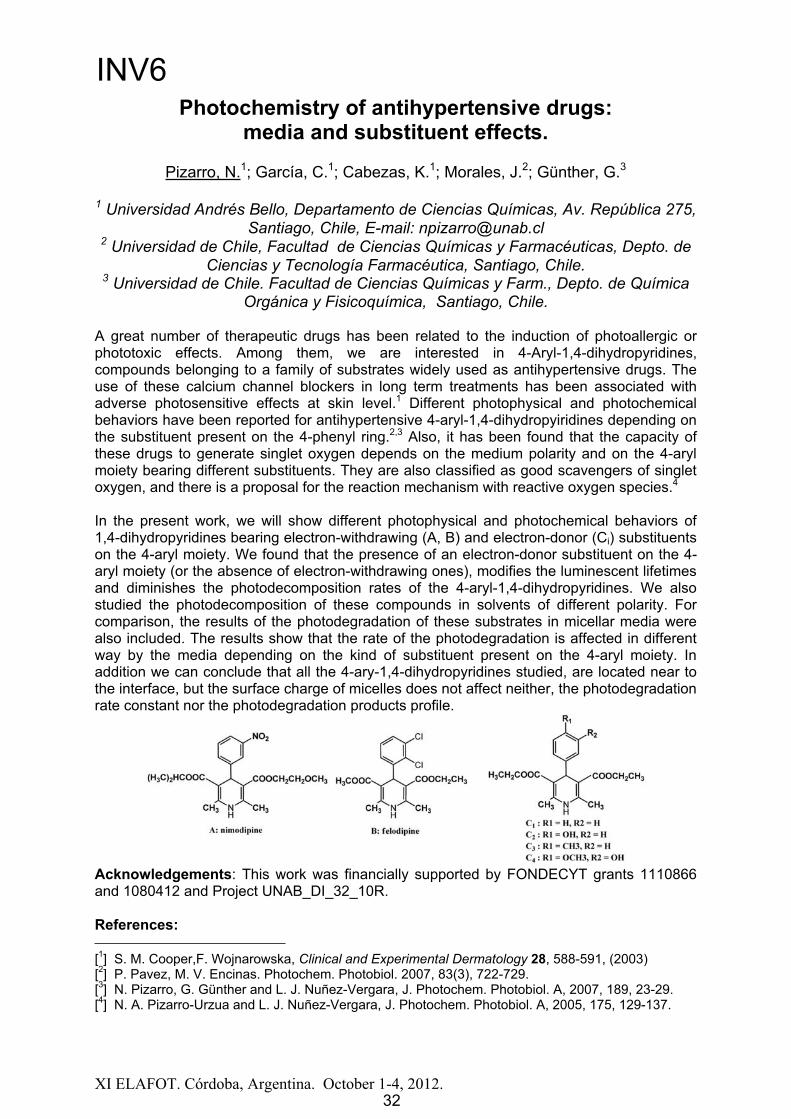
A great number of therapeutic drugs has been related to the induction of photoallergic or phototoxic effects. Among them, we are interested in 4-Aryl-1,4-dihydropyridines, compounds belonging to a family of substrates widely used as antihypertensive drugs. The use of these calcium channel blockers in long term treatments has been associated with adverse photosensitive effects at skin level.1 Different photophysical and photochemical behaviors have been reported for antihypertensive 4-aryl-1,4-dihydropyiridines depending on the substituent present on the 4-phenyl ring.2,3 Also, it has been found that the capacity of these drugs to generate singlet oxygen depends on the medium polarity and on the 4-aryl moiety bearing different substituents. They are also classified as good scavengers of singlet oxygen, and there is a proposal for the reaction mechanism with reactive oxygen species.4
In the present work, we will show different photophysical and photochemical behaviors of 1,4-dihydropyridines bearing electron-withdrawing (A, B) and electron-donor (Ci) substituents on the 4-aryl moiety. We found that the presence of an electron-donor substituent on the 4-aryl moiety (or the absence of electron-withdrawing ones), modifies the luminescent lifetimes and diminishes the photodecomposition rates of the 4-aryl-1,4-dihydropyridines. We also studied the photodecomposition of these compounds in solvents of different polarity. For comparison, the results of the photodegradation of these substrates in micellar media were also included. The results show that the rate of the photodegradation is affected in different way by the media depending on the kind of substituent present on the 4-aryl moiety. In addition we can conclude that all the 4-ary-1,4-dihydropyridines studied, are located near to the interface, but the surface charge of micelles does not affect neither, the photodegradation rate constant nor the photodegradation products profile.
Acknowledgements: This work was financially supported by FONDECYT grants 1110866 and 1080412 and Project UNAB_DI_32_10R.
References:
[1] S. M. Cooper,F. Wojnarowska, Clinical and Experimental Dermatology 28, 588-591, (2003) [2] P. Pavez, M. V. Encinas. Photochem. Photobiol. 2007, 83(3), 722-729. [3] N. Pizarro, G. Günther and L. J. Nuñez-Vergara, J. Photochem. Photobiol. A, 2007, 189, 23-29. [4] N. A. Pizarro-Urzua and L. J. Nuñez-Vergara, J. Photochem. Photobiol. A, 2005, 175, 129-137.
32
INV6
32
XI ELAFOT. Córdoba, Argentina. October 1-4, 2012.
Photo-electrochemistry and solar energy conversion:application in dye-sensitized solar cells, hydrogen
production and water disinfectionLongo, Claudia; Santos, Reginaldo S.; Oliveira, Bruno H.; Silva, Everson T.G.;
Rapelli, Rúbia M.; Oliveira, Haroldo.G.
Institute of Chemistry, University of Campinas–UNICAMP, PO Box 6154, 13083-970, Campinas, SP, Brazil., E-mail: [email protected]
Solar energy conversion has been largely investigated, motivated by its academic relevance, as well as technological and environmental concerns. In the Institute of Chemistry-UNICAMP we investigate semiconductor oxides (TiO2 Fe-doped TiO2, ZnO, WO3) for application in dye-sensitized solar cells, for water disinfection (by photocatalytic removal of organic pollutants) and also for water splitting to produce “solar hydrogen”.
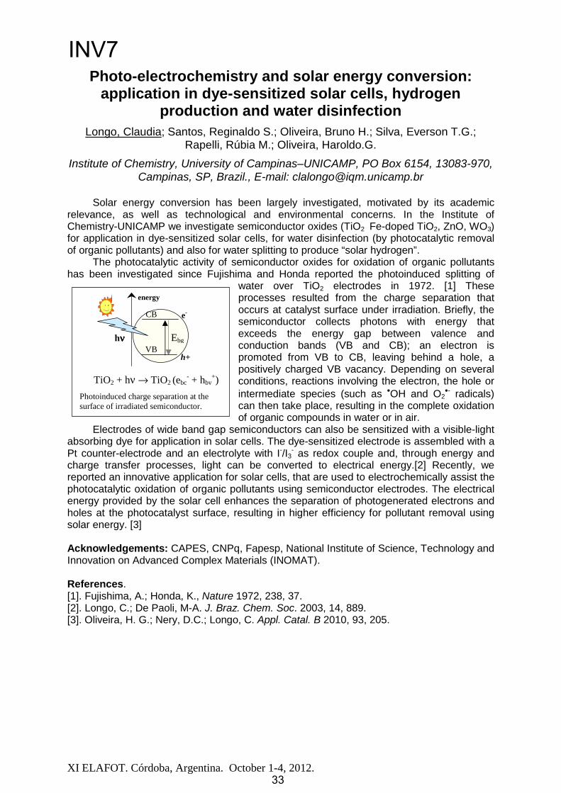
The photocatalytic activity of semiconductor oxides for oxidation of organic pollutants has been investigated since Fujishima and Honda reported the photoinduced splitting of
water over TiO2 electrodes in 1972. [1] These processes resulted from the charge separation that occurs at catalyst surface under irradiation. Briefly, the semiconductor collects photons with energy that exceeds the energy gap between valence and conduction bands (VB and CB); an electron is promoted from VB to CB, leaving behind a hole, a positively charged VB vacancy. Depending on severalconditions, reactions involving the electron, the hole or intermediate species (such as •OH and O2
•- radicals) can then take place, resulting in the complete oxidation of organic compounds in water or in air.
Electrodes of wide band gap semiconductors can also be sensitized with a visible-light absorbing dye for application in solar cells. The dye-sensitized electrode is assembled with a Pt counter-electrode and an electrolyte with I-/I3
- as redox couple and, through energy and charge transfer processes, light can be converted to electrical energy.[2] Recently, we reported an innovative application for solar cells, that are used to electrochemically assist the photocatalytic oxidation of organic pollutants using semiconductor electrodes. The electrical energy provided by the solar cell enhances the separation of photogenerated electrons and holes at the photocatalyst surface, resulting in higher efficiency for pollutant removal using solar energy. [3]
Acknowledgements: CAPES, CNPq, Fapesp, National Institute of Science, Technology and Innovation on Advanced Complex Materials (INOMAT).
References. [1]. Fujishima, A.; Honda, K., Nature 1972, 238, 37. [2]. Longo, C.; De Paoli, M-A. J. Braz. Chem. Soc. 2003, 14, 889. [3]. Oliveira, H. G.; Nery, D.C.; Longo, C. Appl. Catal. B 2010, 93, 205.
�
�
�
�
�
�����TiO2 + hν → TiO2 (ebc- + hbv
+)
Photoinduced charge separation at the surface of irradiated semiconductor.�
CB
VB h+
e-
Ebghνννν
energy
33
INV7
33

XI ELAFOT. Córdoba, Argentina. October 1 4, 2012. 34
Super-Resolution Imaging with Switchable Fluorophores Based on Oxazine Auxochromes
Bossi, Mariano1; Raymo, Françisco2; Petriella, Marco1; Deniz, Erhan2;Subramani Swaminathan2
1 INQUIMAE, FCEyN, Universidad de Buenos Aires, Pabellón 2, Ciudad Universitaria, 1428 Buenos Aires, Argentina,
[email protected] 2 Laboratory for Molecular Photonics, Department of Chemistry, University of Miami,
1301 Memorial Drive, Coral Gables, Florida, 33146-0431, United States [email protected]
Far-field fluorescence microscopy is a widely applied and powerful tool for biological imaging. Its unique selectivity and sensitivity, along with the advantage of being non-invasive, is only hampered by a spatial resolution limited by diffraction to about half of the wavelength of the light (~200 nm). Thus, important processes and subcellular compartments remain unresolved in conventional microscopies. Recent emerging techniques have overcome the diffraction barrier, such as STED, PALM, STORM, etc.1 Their common ground is a time sequential discrimination of close objects or features that are otherwise unresolved or blurred within the resolution (PSF) of the microscope. The key is then to be able to switch on and off the signal of the markers, using distinct molecular states of the fluorophores.
One of the strategies is based upon the detection and localization of single molecules.2The markers, initially all in a dark state, are stochastically switched on as a sparse subset to ensure that they are resolved with a conventional wide-field microscope (i.e. there is only one emitter within a PSF). The position of each emitter can be precisely obtained from an image with an accuracy of ~PSF/ nPH, where nPH is the amount of detected photons. Then, those markers are switched off so new ones can be switched on and the process is repeated. The superresolution image is reconstructed by mapping the position of a large number of localized events. The recording scheme outlines the critical role of the photophysical properties of the probes (photostability, reliable control of signal modulation, etc.) as well as the importance of finding adequate alternative switching mechanisms at the molecular level.
In this talk we will present alternative strategies to achieve fluorescence switching based on novel molecular assemblies containing a [1,3]-oxazine3 as the two-states molecular building-block responsible for the transformation. In particular, we have successfully applied two different triggering events, based on the photochromic and halochromic properties of oxazine respectively, to achieve images with a spatial resolution beyond the diffraction limit. In the first one, photoisomerization of the oxazine is induced by direct irradiation of the chromophore.4 In the second one, the changes are indirectly triggered by a proton uncaging of an auxiliary.5 In both cases, the changes in the molecular structure of the oxazine is exploited to induce a large bathochromic shift of the absorption and emission spectra of the pendant fluorophore, and this ultimate effect is used for fluorescence switching. We will focus on the implementation of those strategies, and discuss the advantages and drawbacks of both processes.
1 Hell S. W.; Nat. Methods 2009, 6, 24 – 32. 2 Betzig, E. et al.; Science 2006, 313, 1642–1645. 3 Tomasulo M., Sortino S., Raymo F. M.; Org. Lett., 2005, 7, 1109–1112. 4 Deniz E., Tomasulo M., Cusido J., Yildiz I., Petriella M., Bossi M., Sortino S., Raymo F.M.; J. Phys. Chem. C,2012, 116, 6058–6068. 5 Swaminathan S., Petriella M., Deniz E., Cusido J., Baker J. D., Bossi M., Raymo F. M., submitted.
INV8
34

XI ELAFOT. Córdoba, Argentina. October 1 4, 2012. 35
D D
A A
+
-
Au
A A
D D+
-TiO2
D: diphenylamineA: dicyano,
cyanoacrylic acid
ITO
S
S
S
SS
S
S
S
SS
SS
S
S
S
S S
S
S
S
S
S
S
S
SS
SS
SS
SS
S
S
S
S
S
S
S
SS
S
S
S
SS
SS
S
S
S
SS
S
S
S
SS
SS
S
S
S
S S
S
S
S
SS SS
S
S
S
S
SS SS
S
S
S
S
SS
SS
S
S
S
S
SS
SS
S
S
S
SS
S
S
S
SS
SS
S
S
S
S
S
S
S
S
SS
SS
S
S
S
S
S
S
S
S S
S
S
S
S
S
S
S
SS
SS
DD DD
AA AA
++
--
Au
AA AA
DD DD++
--TiO2
D: diphenylamineA: dicyano,
cyanoacrylic acid
ITO
S
S
S
SS
S
S
S
SS
SS
S
S
S
S S
S
S
S
S
S
S
S
SS
SS
SS
SS
S
S
S
S
S
S
S
SS
S
S
S
SS
SS
S
S
S
SS
S
S
S
SS
SS
S
S
S
S S
S
S
S
SS SS
S
S
S
S
SS SS
S
S
S
S
SS
SS
S
S
S
S
SS
SS
S
S
S
SS
S
S
S
SS
SS
S
S
S
S
S
S
S
S
SS
SS
S
S
S
S
S
S
S
S S
S
S
S
S
S
S
S
SS
SS
Schematic of charge separation in adsorbed donor-acceptor spiro compound / substrate systems (left) and schematic of a mono-layer of quantum dots with dithiol surfactants and defect states (right).
Investigation of donor-acceptor molecule and quantum dot layer systems by surface photovoltage techniques
Th.Dittrich1; S.Fengler1, E.Zillner1, J.Rappich1, X.Zhang1, L.Otero2,F.Fungo2, L.Macor2, M.Gervaldo2, D.Heredia2, C.-Y.Lin3, L.-C.Chi3,
C.Fang3, S.-W.Lii3, K.-T.Wong3
1 Helmholtz Centre Berlin for Materials and Energy, Hahn-Meitner-Platz 1, D-14109 Berlin, Germany, [email protected]
2 Departamento de Química, Universidad Nacional de Río Cuarto, Agencia Postal 3, X5804BYA, Río Cuarto, Argentina, [email protected]
3 Department of Chemistry, National Taiwan University, Taipei 106, Taiwan, [email protected]
Donor-acceptor molecule and quantum dot (QD) layer systems are of great interest for applications in optoelectronics, photovoltaics and photocatalysis due to their potential for engineering of optical and surface electronic properties. For example, dye sensitized solar cells were realized with donor-acceptor spiro compounds [1] and hetero-junctions between TiO2 and PbS based QD layers are suitable for efficient QD solar cells [2]. Spectral and time dependent surface photovoltage (SPV) techniques were used to study processes of charge separation across interfaces in layer systems containing donor-acceptor spiro compounds with dicyano or cyanoacrylic acceptor and diphenylamine donor groups [3] or containing CdSe QDs with TOP/OA, pyridine or dithiol surfactants [4,5]. Layers were prepared by dip coating or electrochemically. Intramolecular charge transfer and charge separation by electron injection were distinguished. Results were interpreted from point of view of directed molecule adsorption. A strong influence of the donor group on charge separation was observed. It was shown that the adsorption dependence of optical transitions of donor-acceptor spiro compounds can be well studied by SPV. It was found that electronic defect states at QD surfaces are generated by successive surfactant exchange. Information about the energetic distribution and density of defect states at surfaces of QDs was obtained by random walk simulations of SPV transients within the frame of an isolated QD approximation.
Acknowledgements: Th.D., J.R., X.Z., L.O., D.H., F.F., and M.G. are grateful to the DAAD (416-PPP-Proalar) and CONICET for financial support.
References [1] D. Heredia, J. Natera, L. Otero, F. Fungo, C.-Y. Lin, K.-T. Wong, Organic Letters 12 (2010) 12. [2] A. G. Pattantyus-Abraham, I. J. Kramer, A. R. Barkhouse, X. Wang, G. Konstantatos, R. Debnath, L. Levina, I. Raabe, M. K. Nazeeruddin, M. Grätzel, E. H. Sargent, ACS nano 4 (2010) 3374. [3] L. Macor, M. Gervaldo, F. Fungo, L. Otero, Th. Dittrich, C.-Y. Lin, L.-C. Chi, F.-C. Fang, S.-W. Lii, K.-T. Wong, C.-H. Tsaid, C.-C. Wu, RSC Advances 2 (2012) 4869.[4] E. Zillner, Th. Dittrich, Phys. Stat. Solidi RRL 5 (2011) 256 [5] E. Zillner, S. Fengler, P. Niyamakom, F. Rauscher, K. Köhler, T. Dittrich, JPCC, accepted for publication.
INV9
35

XI ELAFOT. Córdoba, Argentina. October 1 4, 2012. 36
Photoreduction of 3-Methyl-1H-quinoxalin-2-one derivatives by N-phenylglicine. A mechanistic study.
De la Fuente, Julio R.1; Cañete, Alvaro2; Aliaga Christian1; Jullian, Carolina1; Saitz, Claudio1; Bernazar, Luan1; Carathanassis, Natalia1;
Bobrowski, Krzysztof3; Kciuk Gabriel3; Szreder Tomaz3.
1 Fac. Cs. Qcas. Y Farm., Universidad de Chile, [email protected] 2 Depto. Qca. Fac. Química, Pontificia Universidad Católica de Chile.
3 lnstitute of Nuclear Chemistry and Technology, Warsaw, Poland.
Many studies devoted to the pharmacological properties of quinoxalin-2-one derivatives have been published during the last decades. Several of these works locate the quinoxalin-2-one moiety inside of protein pockets, suggesting interactions with potentially electron donor amino-acid residues. However, there are not reports concerning to the transient species generated by electron transfer from the amino-acids residues, or about the mechanism of potential radical reactions between these species.
The photoreduction 3-methyl-quinoxalin-2-ones derivatives by N-phenylglicine are efficient process showing several isosbestic points during the photoreduction. GC-mass analysis (CI and EI) of the samples of photoreaction shows the formation of two main products generated by the addition of amino-acid fragments to the quinoxalin-2-one scaffold. The proportion of these products depends of the substituent in the position 7 of quinoxalin-2-ones, as suggested by the isosbestic points.
The molecular ions of these photoproducts which differ only in the substituent mass, as shown below, demonstrate that the photoreduction mechanism is the same for all of the substituted quinoxalin-2-ones.
Substituent CH3O CH3 F H CF3 CNSubst. Mass 31 15 19 1 69 26 Product 1 (M + H+) m/z 298 282 286 268 336 293 Product 2 (M + H+) m/z 310 294 298 280 348 305
Laser flash photolysis and pulse radiolysis experiments results show that the photoreduction is initiated by a single electron transfer followed by a proton transfer, generating Ph-NH-CH2• radicals, which account for the products formation.
Acknowledgements: FONDECYT N° 1100121, D.I. Universidad de Chile and to K. B. at INCT, Warsaw Poland.
INV10
36

XI ELAFOT. Córdoba, Argentina. October 1-4, 2012.
Tryptophan photosensitization by pterinVirginie Rahal,2 Mariana P. Serrano,1 Patricia Vicendo,2 Esther Oliveros,2 Andrés H.
Thomas,1 Carolina Lorente1
1 Instituto de Investigaciones Fisicoquímicas Teóricas y Aplicadas (INIFTA), Fac. Cs. Exactas, UNLP, CCT La Plata-CONICET.
CC 16, Suc. 4, (1900) La Plata, Argentina. E-mail: [email protected]
2 Laboratoire des IMRCP, UMR CNRS/UPS 5623, Université Paul Sabatier (Toulouse III), 118, route de Narbonne, F-31062
Toulouse cédex 9, France.
Pterins belong to a family of heterocyclic compounds present in a wide range of living systems and participate in relevant biological functions. Under UV-A excitation (320 400nm), pterins can fluoresce, undergo photooxidation and generate reactive oxygen species (ROS).1 Pterin (Ptr), the parent compound of oxidized or aromatic pterins, acts as photosensitizer through both type I (electron abstraction) and/or type II (production of singlet molecular oxygen (1O2)) mechanisms. Moreover, Ptr photoinduces DNA damage2 and
- - -monophosphates (dGMP, dAMP)3,4 via electron transfer processes. Tryptophan (Trp), an esencial aminoacid, it is known as a target for oxidation by 1O2.5 Given its structural similarity with guanine and its low redox potential, Trp may be also a potential target for pterin photosensitized mediated oxidation.To evaluate the capability of Ptr to photosensitize tryptophan, aqueous solutions containing both compounds were exposed to UV-A irradiation (320-400 nm) under different experimental conditions. The photochemical reactions were followed by UV/VIS spectrophotometry, HPLC, and an enzymatic method for H2O2 determination. In addition,mass spectrometry, fluorescence quenching and electronic paramagnetic resonanceexperiments were performed.Mechanistic analysis indicates that the Ptr-sensitized oxygenation/oxidation of Trp does not involve exclusively 1O2 as oxidation agent. By contrast, an electron transfer process plays a fundamental role in the photodegradation of Trp. In this mechanism, the excitation of Ptr is followed by an electron transfer from Trp molecule to the Ptr triplet excited state, leading to the formation of the corresponding ion radicals (Ptr and Trp ). In the following step, the electron transfer from Ptr to O2 regenerates Ptr and forms the superoxide anion. The latter,may disproportionate with its conjugated acid (HO2 ) to form H2O2 or react with Trp to regenerate Trp.
References1 Lorente, C.; Thomas, A. H.; Acc. Chem. Res. 2006, 39, 395-4022 Ito, K.; Kawanishi, S. Biochemistry 1997, 36, 1774-1781.3 Petroselli, G.; Dántola, M. L.; Cabrerizo, F. M.; Capparelli, A. L.; Lorente, C.; Oliveros, E.; Thomas, A. H.; J. Am. Chem. Soc. 2008, 130, 3001 3011.4 Petroselli, G.; Erra-Balsells, R.; Cabrerizo, F. M.; Lorente, C.; Capparelli, A. L.; Braun, A. M.; Oliveros, E.; Thomas, A. H.; Org. Biomol.Chem. 2007, 5, 2792 27995 Pattison, D. I., Suryo Rahmanto A., Davies M. J., Photochem. Photobiol. Sci., 2012, 11, 38-53
N
NHN
N
O
NH2
NH2O N
OH
3Ptr*
1Ptr*
h
ISC
.-ISC
O2
1O2
O2
O2
H2O2
H+
Trp.+
Products
O2
O2
.-
.-
Ptr
Trp
Ptr
37
INV11

XI ELAFOT. Córdoba, Argentina. October 1 4, 2012.
Presentaciones Orales (OP)
38
XI ELAFOT. Córdoba, Argentina. October 1 4, 2012. 39
Photochemistry of tetraphenyldiboroxane and its use as photopolymerization co-initiator
Neumann, Miguel G.; Santos, Willy G.; Schmitt, Carla C. Instituto de Química de São Carlos, Universidade de São Paulo, Brazil
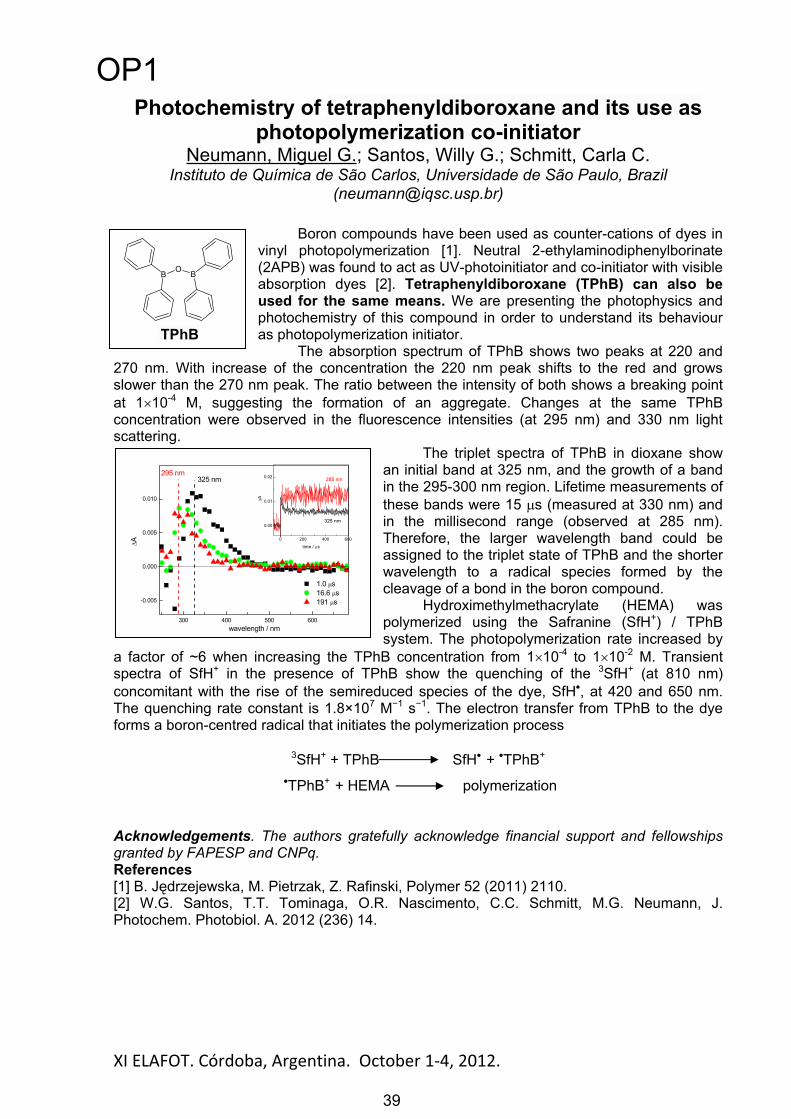
Boron compounds have been used as counter-cations of dyes in vinyl photopolymerization [1]. Neutral 2-ethylaminodiphenylborinate (2APB) was found to act as UV-photoinitiator and co-initiator with visible absorption dyes [2]. Tetraphenyldiboroxane (TPhB) can also be used for the same means. We are presenting the photophysics and photochemistry of this compound in order to understand its behaviour as photopolymerization initiator.
The absorption spectrum of TPhB shows two peaks at 220 and 270 nm. With increase of the concentration the 220 nm peak shifts to the red and grows slower than the 270 nm peak. The ratio between the intensity of both shows a breaking point at 1 10-4 M, suggesting the formation of an aggregate. Changes at the same TPhB concentration were observed in the fluorescence intensities (at 295 nm) and 330 nm light scattering.
The triplet spectra of TPhB in dioxane show an initial band at 325 nm, and the growth of a band in the 295-300 nm region. Lifetime measurements of these bands were 15 s (measured at 330 nm) and in the millisecond range (observed at 285 nm). Therefore, the larger wavelength band could be assigned to the triplet state of TPhB and the shorter wavelength to a radical species formed by the cleavage of a bond in the boron compound.
Hydroximethylmethacrylate (HEMA) was polymerized using the Safranine (SfH+) / TPhB system. The photopolymerization rate increased by
a factor of ~6 when increasing the TPhB concentration from 1 10-4 to 1 10-2 M. Transient spectra of SfH+ in the presence of TPhB show the quenching of the 3SfH+ (at 810 nm) concomitant with the rise of the semireduced species of the dye, SfH , at 420 and 650 nm. The quenching rate constant is 1.8×10⁷ M 1 s 1. The electron transfer from TPhB to the dye forms a boron-centred radical that initiates the polymerization process
3SfH+ + TPhB SfH + TPhB+
TPhB+ + HEMA polymerization
Acknowledgements. The authors gratefully acknowledge financial support and fellowships granted by FAPESP and CNPq. References [1] B. J drzejewska, M. Pietrzak, Z. Rafinski, Polymer 52 (2011) 2110. [2] W.G. Santos, T.T. Tominaga, O.R. Nascimento, C.C. Schmitt, M.G. Neumann, J. Photochem. Photobiol. A. 2012 (236) 14.
BO B
TPhB
300 400 500 600
-0.005
0.000
0.005
0.010
0 200 400 600
0.00
0.01
0.02
A
time / s
325 nm
285 nm
A
wavelength / nm
1.0 s 16.6 s 191 s
325 nm295 nm
OP1
39

Abstract
XI ELAFOT. Córdoba, Argentina. October 1-4, 2012.
Biophysical properties and cellular toxicity of covalent cross-linked oligomers of αααα-synuclein formed by
photoinduced side-chain tyrosyl radicals
Borsarelli, Claudio D.1; Falomir-Lockhart, Lisandro J.2; Ostatná, Veronika3; Fauerbach, Jonathan A.4; Hsiao, He-Shuan2; Urlaub, Henning2; Paleček, Emil3;
Jares-Erijman, Elizabeth A.4; Jovin, Thomas M.2
1Laboratorio de Cinética y Fotoquímica, Centro de Investigaciones y Transferencia de Santiago del Estero (CITSE-CONICET), UNSE. [email protected]
2Max Planck Institute for Biophysical Chemistry, Goettingen, Germany 3Institute of Biophysics, Academy of Sciences of the Czech Republic
4 Departamento de Química Orgánica, Facultad de Ciencias Exactas y Naturales,CIHIDECAR CONICET-UBA
Alpha-synuclein (αS), a 140 amino acid presynaptic protein, is the major component of the fibrillar aggregates (Lewy bodies) observed in dopaminergic neurons of patients affected by Parkinson’s disease. It is currently believed that non-covalent oligomeric forms of αS, arising as intermediates in its aggregation, may constitute the major neurotoxic species. However, attempts to isolate and characterize such oligomers in vitro, and even more so in living cells, have been hampered by their transient nature, low concentration, polymorphism, and inherent instability.
In this work, we describe the preparation and characterization of low molecular weight covalently bound oligomeric species of αS obtained by cross-linking via tyrosyl radicals generated by blue-light photosensitization of the cationic metal coordination complex ruthenium (II) tris-bipyridine, Ru(bpy)3
2+, in the presence of persulfate anion, S2O82-.
Numerous analytical techniques were used to characterize the αS oligomers: biochemical (anion-exchange chromatography, SDS-PAGE and Western blotting); spectroscopic (optical: UV/Vis absorption, steady state and dynamic fluorescence, and dynamic light scattering); mass spectrometry; and electrochemical. Light-controlled protein oligomerization was mediated by –C–C– bonds between Tyr residues to form dityrosine (diTyr) as covalent bridges, with a predominant involvement of Y39 residue.
The diverse oligomeric species exhibited a direct effect on the in vitro aggregation behavior of wild-type monomeric αS, decreasing the total yield of amyloid fibrils inaggregation assays monitored by thioflavin T (ThioT) fluorescence and light scattering, and
by atomic force microscopy (AFM). Compared to the unmodified monomer, the photoinduced covalent oligomeric species demonstrated increased toxic effects on differentiated neuronal-like SH-SY5Y cells. The results highlight the importance of protein modification induced by oxidative stress in the initial molecular events leading to Parkinson´s disease.
Acknowledgement: CDB thanks the Alexander von Humboldt Foundation of Germany for Georg Forster fellowship to visit the Laboratory of Cellular Dynamics, Max Planck Institute for Biophysical Chemistry, Goettingen, Germany.
ααααS
Non-covalentpre-aggregates
Non-covalentoligomers
Maturefibrils
Unfolding/pre-aggregation
Ru(bpy)32+
hνννν + S2O8
2-
SO42- + Ru(bpy)3
3+ + SO4••••-
Side-chainTyr radical
(ααααS)••••
Off-pathwayoligomers
diTyr covalentoligomers
(ααααS)n
cytotoxic
40
OP2
40

¿Qué ocurre cuando se utiliza un solvente biodegradable en la formación de micelas inversas? Caracterización de
sistemas micelares utilizando técnicas fotoquímicas. Durantini, Andrés M.1; Falcone, R. Dario1, Silber, Juana J.1, Correa N. Mariano1
1Departamento de Química. Universidad Nacional de Río Cuarto. Agencia Postal # 3. (X5804ALH) Río Cuarto. ARGENTINA. Email: [email protected]
Las micelas inversas (MIs) son agregados que se obtienen cuando se disuelven moléculas de surfactantes en un solvente orgánico de baja polaridad. Dichos agregados son, en general, esféricos y capaces de disolver una cantidad apreciable de agua en su interior.[1]En los últimos años se ha comenzado a investigar otros solventes polares que reemplacen al agua para formar lo que se denominan micelas inversas no acuosas.[2]- [5] En este marco, es interesante para nosotros estudiar la formación de MIs no contaminantes utilizando el solvente biodegradable lactato de etilo (LE). Con tal fin, se estudió la posible formación de estos agregados utilizando la técnica de dispersión dinámica de la luz (DLS). Además, para conocer cuál es la estructura del solvente LE encapsulado en estos sistemas micelares, se utilizó la molécula prueba 4-aminoftalimida (4-AP) y se estudio su comportamiento fotofísico mediante espectroscopia UV-visible, de emisión en estado estacionario y resuelta en el tiempo (TRES).
O
OO
S
O
O
O-O
Na+
AOT
O
OO
S
O
O
O-O
Na+
AOT
N
N
H
O
O
H
H
4-AP
Para evaluar la formación de MIs inversas utilizando LE se comenzó a analizar diferentes sistemas utilizando tres solventes LE, agua y n-heptano con AOT como surfactante. Por DLS se encontró que LE solo se encapsula en presencia de agua en el sistema. Los estudios utilizando 4-AP demostraron que en el sistemas micelar (LE:Agua/AOT/n-heptano) a WAgua = 5 y variando el WLE (WLE=[LE]/[AOT]), 4-AP sensa un medio estructurado y la presencia de las moléculas de agua únicamente en la interfaz micelar, quedando las moléculas de LE ubicadas en el corazón polar de las MIs. Sin embargo cuando el WAgua = 10, las moléculas de agua comienzan a ubicarse en el corazón polar y las moléculas de LE se desplazan a la interfaz micelar generando un microambiente más fluido. Con estos estudios, es evidente que el lugar de confinamiento de LE en las MIs depende de la cantidad de agua encapsulada. Cuando WAgua=10 el LE se ubica en el corazón polar del agregado mientras que, a bajos valores de WAgua (WAgua=5) cuando solo hay agua unida, LE se encuentra en el corazón polar de los agregados. Los estudios TRES confirman la ubicación de LE en el interior de la micela inversa y descartan que LE este soluble en el solvente orgánico externo. Estos resultados tendrían implicancia en el uso de estos nuevos sistemas organizados como nanoreactor.
1 P.L. Luisi, M. Giomini, M.P. Pileni, B.H. Robinson, Biochimica et Biophysica Acta, 947, 209, 1988.2 Durantini, A. M.; Falcone, R. D.; Silber, J. J.; Correa, N. M. ChemPhysChem, 10, 2034, 2009.3 Falcone, Silber, Correa PCCP,11, 11092, 2009.4 Andrés M. Durantini, R. Dario Falcone, Juana J. Silber, N. Mariano Correa. J. Phys. Chem. B 115, 5894–5902, 2011.5 N. M. Correa, J. J. Silber, R.E. Riter, N. E. Levinger Chem. Rev. 2012, in press dx.doi.org/10.1021/cr200254q.
XI ELAFOT. Córdoba, Argentina. October 1-4, 2012. 41
OP3
41

XI ELAFOT. Córdoba, Argentina. October 1-4, 2012.
Eficiencias Cuánticas de la Degradación de Atrazina en agua por Foto-Fenton
Benzaquén, Tamara B.1, Isla, Miguel A.1,2 y Alfano, Orlando M.1,2.1INTEC(UNL-CONICET), Ruta Nacional N°168, Santa Fe, Argentina. E-Mail:
[email protected], UNL, 3000 Santa Fe, Argentina.
La atrazina, es uno de los herbicidas más comúnmente utilizados a nivel mundial. Debido a su relativamente alta solubilidad en agua y biorresistencia, la atrazina entra con facilidad y persiste en el medio ambiente acuático [1]. Los efectos cancerígenos y tóxicos de este herbicida se han estudiado ampliamente [2]. Además, se sabe que es una sustancia prohibida o regulada en varios países [3].En este trabajo, se ha estudiado la degradación y mineralización del herbicida atrazina enagua (en su formulado comercial) mediante el proceso foto-Fenton. Con el fin de evaluar laseficacias de la degradación del herbicida y comparar los resultados obtenidos, se calcularonlas eficiencias cuánticas ( ) del proceso bajo diferentes condiciones experimentales. Se han evaluado tanto (i) la eficiencia cuántica aparente, [4] como (ii) la eficiencia cuántica absoluta, [4],[5]. Para cuantificar la mineralización completa del herbicida, se midió la conversión del carbono orgánico total (TOC). De esta forma, también se calcularon las eficiencias cuánticas de mineralización [6].El dispositivo experimental en el que se realizó la fotodegradación fue un reactor isotérmico, bien agitado y con reciclo. Este reactor de placas planas y sección circular, fue irradiado aambos lados con dos lámparas UV.Se ha encontrado que cuando las concentraciones iniciales de la sal férrica y delperóxido de hidrógeno se incrementaron, se obtuvieron los valores más altos de . En cambio, la aumentó cuando la relación molar peróxido de hidrógeno/atrazina (R)aumentó y la concentración de hierro férrico disminuyó. Este comportamiento se debe a que
depende fuertemente de la concentración de la sal férrica inicial, a diferencia de, que no varía cuando se incrementa la concentración de hierro.
Por otro lado, las eficiencias cuánticas relativas de mineralización mostraron una mayor sensibilidad respecto a las eficiencias cuánticas de degradación de atrazina. Dos factores pueden ser utilizados para explicar este comportamiento: (i) los tiempos de reacción considerados para las eficiencias de degradación y mineralización son bastante diferentes y (ii) las eficiencia cuántica de mineralización se informó como una eficiencia "relativa", tomando como base la eficiencia máxima que puede alcanzarse de TOC. Es interesante aclarar que la atrazina es resistente a una mineralización completa por la reacción de foto-Fenton. No obstante, se alcanzó una conversión de TOC del 40%, siendo 62,5% del valor máximo que puede alcanzarse debido a la estabilidad del anillo triazínico [7],[8]. El producto final de la degradación, ácido cianúrico, es biodegradable y presenta una toxicidad insignificante [9],[10]; en consecuencia, el ácido cianúrico se podría eliminar posteriormenteutilizando un tratamiento biológico.Referencias[1] Krysova H., Jirkovsky J., Krysa J., Mailhot G., Bolte M., (2003). Appl. Catal., B, 40(1),1 12.[2] Cimino-Reale G., Ferrario D., Casati B., Brustio R., Diodovich C., Collotta A., Vahter M., Gribaldo L.,
(2008). Toxicol. Lett., 180(1),59-66.[3] Ackerman, F. (2007). Int. J. of Occup. and Env. Health, 13(4),437-445.[4] Salaices, M. Serrano, B. de Lasa, H. I. (2002). Chem. Eng. J, 90(3), 219.[5] Cabrera, M., Alfano, O., Cassano, A. (1994). Ind. and Eng. Chem. Research., 33(12), 3031 3042.[6] Satuf M.L., Brandi R., Cassano A., Alfano O. (2007).Ind. and Eng. Chem. Research.,46(1), 43-51.[7] Pelizzetti, E., Maurino, V., Minero, C., Carlin, V., Pramauro, E., Zerbinati, O., Tosato, M.L., (1990).
Environ. Sci. Technol.,24(10),1559-1565.[8] Konstantinou, I.K. and Albanis, T.A., (2003). Appl. Catal., B, 42, 319 335.[9] Chan C.Y., Tao S., Dawson R., Wong P.K. (2004). Environ. Pollut., 131(1), 45 54.
[10] Lapertot M, Pulgarin C, Fernandez-Ibañez, Maldonado, M. I., Pérez-Estrada L., Oller I. Gernjak W., Malato S. (2006). Water Res. ,40(5),1086 1094.
42
OP4
42

XI ELAFOT. Córdoba, Argentina. October 1-4, 2012.
Foto-transformación de Clorotalonil Usando Sustancias Húmicas
Porras, Jazmín1,2*; Fernández, Jhon Jairo1, Torres, Ricardo1, Richard, Claire2
1 Universidad de Antioquia, A.A. 1226 Colombia, [email protected]*2 Université Blaise Pascal, A.A. 63171 France, [email protected]
Las sustancias húmicas (SH) bajo luz ultravioleta pueden generar radicales hidroxilo(OH*), oxígeno singlete (1O2), estados excitados tripletes (3SH), entre otras [1]. Esta propiedad puede ser usada para la transformación de contaminantes en sistemas acuosos.
El Clorotalonil (2,4,5,6-tetracloroisoftalonitrilo) es uno de los fungicidas mas usados al rededor el mundo, particularmente en Colombia es usado para el control de hongos en una amplia gama de cultivos. En aguas superficiales, el Clorotalonil (CT) es resistente a la hidrólisis, la fotólisis y volatilización [2]. Se han realizado diferentes investigaciones acerca de la fotoquímica de este fungicida centrándose principalmente en la cinética de fotodegradación [3,4]. En este trabajo se evaluó la fotólisis indirecta del Clorotalonil en un sistema acuoso usando diferentes sustancias húmicas como fotosensibilizadores; un ácido húmico y fúlvico extraídos de un material carbonoso (AHMC y AFMC) en comparación con un ácido húmico y fúlvico del suelo Elliot (AHE y AFE); este último obtenido del banco de referencia de International Humic Substances Society. Para la irradiación con luz policromática fue usado un dispositivo equipado con 6 lámparas ultravioleta emitiendo entre 300-450 nm con un máximo de emisión a 350 nm. En la tabla se muestra la velocidad experimental para las diferentes SH utilizando aire y argón.
Muestra Velocidad experimentalAire Argón
CT sin SH 4,0 x 10⁻¹¹ 4,0 x 10⁻⁹
CT+AHMC 3,0 x 10⁻¹⁰ 1,8 x 10⁻⁸
CT+AFMC 5,0 x 10⁻¹⁰ 3,2 x 10⁻⁸
CT+AHE 1,5 x 10⁻¹⁰ 9,7 x 10⁻⁸
CT+AFE 3,5 x 10⁻¹⁰ 1,6 x 10⁻⁸
Adicionalmente, también fueron evaluados parámetros como la longitud de onda, la cantidad de oxígeno y la concentración inicial de CT.
Acknowledgements:Los autores desean agradecer el apoyo financiero recibido de la Universidad de Antioquiapara el proyecto "Fotodegradación de contaminantes orgánicos en sistemas acuosos asistida por sustancias húmicas (CODI-2011)". Jazmín Porras da las gracias al Departamento Colombiano Administrativo de Ciencia, Tecnología e Innovación (Colciencias) por su beca doctoral.
References[1] L. Cavani, S. Halladja, A. Ter Halle, G. Guyot, G. Corrado, C. Clavatta, A. Boulkamh, C. Richard, Environment Science & Technology, 43 (2009) 4348.[2] S. Monadjemi, M. El Roz, C. Richard, A. Ter Halle, Environment Science & Technology,45 (2011) 9582.[3] V. Sakkas, D. Lambropoulou, T. Albanis, Chemosphere, 48 (2002) 939.[4] G. Penuela, D. Barcelo, Journal of Chromatography A, 823 (1998) 81.
43
OP5
43

XI ELAFOT. Córdoba, Argentina. October 1 4, 2012. 44
The Photoanode of Photoelectrochemical Cells for the Splitting of Water
Moore, Ana L.; Meggiatto, Jackson D.; Kodis, Gerdenis; Llansola, Manuel J., Méndez; Dalvin D.; Sherman, Benjamin D.; Moore, Thomas A.; Gust,
Devens Department of Chemistry and Biochemistry, Center for Bioenergy and
Photosynthesis, and Center for Bio-Inspired Solar Fuel Production, Arizona State University, Tempe, AZ 85287, USA, [email protected]
Schemes that couple solar energy conversion to the oxidation of water and the subsequent use of the reducing equivalents for the synthesis of energy-rich compounds such as hydrogen or reduced carbon based fuels, is the objective of this study.1-3 We are constructing tandem, two junction photochemical cells using Grätzel-type photoelectrodes that model aspects of photosystems I and II (PSI and PSII) of plants. The photoanode model of PSII contains a mimic of the donor side (water oxidizing side) of PSII reaction centers. In PSII, tyrosine Z (TyrZ) mediates charge transport between the photo-oxidized primary donor (P680•+) and the oxygen-evolving complex (OEC). The oxidation of TyrZ by P680•+ likely occurs with the transfer of the phenolic proton to a hydrogen-bonded histidine residue (PCET). This coupling of proton and redox chemistry is thought to poise the TyrZ oxidation potential between those of P680•+ and the OEC. We have prepared several bioinspired systems consisting of a high oxidation potential porphyrin (PF10
•+/PF10, ~1.60 V vs. NHE, a model of P680) that is covalently attached to different benzimidazole-phenol pairs (BiP) that mimic the TyrZ-His190 of PSII. Electrochemical studies show that the BIP•+/BIP couple is chemically reversible with a midpoint potential of ~1.25 V vs. NHE and is therefore thermodynamically capable of water oxidation. When BiP-PF10 constructs are attached to TiO2 or SnO2nanoparticles and excited with visible light, they undergo photoinduced electron transfer. Electrons are injected into the semiconducting metal oxides and the corresponding holes are localized in the phenol (BiP•+-PF10-TiO2
•–). EPR provides a clear spectroscopic picture of these processes.4 Transient absorption studies of triads containing electron acceptors such as tetracyano porphyrins to replace the semiconductor have been used to characterize the kinetic parameters of the multistep electron transfer process.5
Acknowledgements: This work is supported as part of the Center for Bio-Inspired Solar Fuel Production, an Energy Frontier Research Center funded by the US Department of Energy, Office of Science, Office of Basic Energy Sciences under Award DE-SC0001016.
References [1] M. Hambourger, G. F. Moore, D. M. Kramer, D. Gust, A. L. Moore and T. A. Moore, Chemical Society Reviews, 38, (2009) 25–35.[2] D. Gust, T. A. Moore and A. L. Moore, Acc. Chem. Res., 42, (2009) 1890–1898.[3] Y. Zhao, J. R. Swierka, J. D. Megiatto, B. Sherman, W. J. Youngblood, D. Qina, D. M. Lentza, A. L. Moore, T. A. Moore, D. Gust and T. E. Mallouk, Proc. Natl. Acad. Sci. U.S.A., (2012) [DOI:10.1073/pnas 1118339109][4] G. F. Moore, M. Hambourger, M. Gervaldo, O. G. Poluektov, T. Rajh, D. Gust, T. A. Moore and A. L. Moore, J. Am. Chem. Soc., 130, (2008) 10466–10467. [5] J. D. Megiatto, A. Antoniuk-Pablant, B. D. Sherman, G. Kodis, M. Gervaldo, T. A. Moore, A. L. Moorea and D. Gusta, Proc. Natl. Acad. Sci. U.S.A., (2012) [DOI: 10.1073/pnas.1118348109]
OP6
44

XI ELAFOT. Córdoba, Argentina. October 1 4, 2012. 45
Asociación y Fotodegradación de Albúmina por complejos diiminos de Cr(III)
Coronel, Consuelo1; Velo, Alejandra1; Jazmín Silvero1; Garcia, Pablo Facundo1; Llorens, Candelaria2; Cabanillas, Ana2; Argüello, Gerardo
Aníbal11INFIQC, Ciudad Universitaria Córdoba, Argentina, [email protected]
2CIBICI, Ciudad Universitaria Córdoba, Argentina
En los últimos años, se han estudiado en profundidad la fotoquímica de complejos metálicos en pos de su posible uso en Terapia Fotodinámica (PDT= PhotoDynamic Therapy). El estudio de la asociación de los complejos metálicos a proteínas de transporte, tiene su importancia en que se podrían utilizar a éstas macromoléculas como transportadoras al interior celular. Por otro lado las proteínas comprenden aproximadamente un 68% del peso total de las células y tejidos y, se ha demostrado, que son los principales centros para la fotooxidación en sistemas biológicos. Las proteínas juegan un rol clave en las funciones biológicas de los organismos vivos, por lo que cualquier fotosensibilización en la proteína puede conducir a la muerte celular. Estudios anteriores en el grupo de investigación, demostraron que los complejos polipiridínicos de Cr(III) tienen la capacidad de fotodegradar en forma irreversible ADN plasmídico, siendo ésta más eficiente en ausencia de oxígeno.1 La albúmina sérica bovina (BSA= Bovine Serum Albumin) es bien conocida y ha sido ampliamente estudiada por su capacidad de transportar numerosos sustratos (Cu2+, Ni2+, aspirina, ibuprofeno, diazepam, etc). En este trabajo presentamos el mecanismo de asociación y la fotodegradación de BSA en presencia de complejos fenantrolínicos de Cromo (III) sustituidos en la posición 5 (fenilo, metilo y Cloro), usando distintas técnicas espectroscópicas. Los ensayos muestran que los complejos de cromo se asocian espontáneamente con la proteína con una constante del orden de 105 M-1. La excitación de la proteína a 280nm lleva a la excitación de los residuos triptófano y tirosina, por lo que observamos la emisión de ambos aminoácidos; mientras que si excitamos a 295nm solo se observa la emisión del triptófano. Resultados experimentales en este sentido, indicarían que ambos aminoácidos estarían involucrados en el proceso de asociación a la albúmina. Esto se observa para otros complejos de Cr(III) y albúmina.2 Se realizaron estudios de Fotodegradación excitando el complejo metálico con LEDs a 472nm, y se analizaron los resultados a distintos tiempos de fotólisis por espectrofluorometría y electroforesis en gel de acrilamida, observándose que la fotodegradación es más efectiva en ausencia de oxígeno, del mismo modo que lo observado para cadenas de ADN. Esto demostraría la fotodegradación se da a través de una transferencia electrónica directa entre la proteína y el complejo (mecanismo de fotooxidación Tipo I). Dado que muchas células cancerosas, por su metabolismo, sufren de hipoxia, que nuestro complejo degrade proteínas a través de transferencia electrónica, es sumamente positivo.
Agradecimientos: Se agradece a CONICET, ANPCyT-FONCyT, y SeCyT UNC por los fondos recibidos.
Referencias1. J. Toneatto, G. Lorenzatti, A.M. Cabanillas, G. A. Argüello, Novel photocleavage properties of [Cr(NN)3]3+ complexes to DNA, J. Inorg. Biochem. (2010)2. J. Toneatto, G.A. Argüello, New advances in the study on the interaction of [Cr(phen)2(dppz)]3+ complex with biological models; association to transporting proteins., J. Inorg. Biochem.(2010)
OP7
45
XI ELAFOT. Córdoba, Argentina. October 1 4, 2012. 46
Aggregation matters - From planar photosensitizers and electroluminescent materials to organo- and hydrogels
Strassert, Cristian A.; Grüner, Malte; Sanning, Jan; Stegemann, Linda; De Cola, Luisa; Allampally, Naveen K.; Mauro, M.; Wegner, D.; Ewen, Pascal
Physikalisches Institut and Center for Nanotechnology (CeNTech) Westfälische Wilhelms-Universität Münster
Heisenbergstr. 11, 48149 Münster (Germany) E-mail: [email protected]
Aggregation of planar chromophores greatly affects their photophysical performance. While phthalocyanine derivatives usually tend to form inactive stacks, Pt(II) complexes can show efficient emission from both monomers and aggregates.
It is possible to avoid the aggregation of phthalocyaninates by axially binding them to the surface of aluminosilicates. We have recently designed a multifunctional zeolite L-based hybrid material able to target, label and photoinactivate pathogenic and antibiotic resistant bacteria.[1] A highly green luminescent dye was inserted into the channels of zeolite L nanocrystals for imaging and labeling the cells. The outer surface was functionalized with a Si(IV) phthalocyanine derivative that forms toxic singlet oxygen upon red light irradiation, and with amino groups for targeting the living microorganisms. Currently, we aim to extend these concepts to targeted, fully biodegradable platforms, a prerequisite for biomedical applications.
We have recently developed a straightforward one-pot synthesis of neutral, soluble Pt(II) coordination compounds bearing dianionic tridentate ligands. The complexes reached up to 87% photoluminescence quantum yield (PLQY) in thin films, with concentration independent colour and efficiency.[2] Consequently, we demonstrated their suitability as phosphorescent dopants in organic light-emitting diodes (OLEDs). Furthermore, the judicious choice of bulky peripheral substituents allowed us to completely prevent aggregation and to enhance color purity, a critical aspect when designing triplet emitters for electroluminescent devices.[3] On the other hand, we were able to deliberately induce self-assembly into bright nanofibers, which can interlock to yield highly emissive organo- and hydrogels (90% PLQY), thus constituting a versatile building block for luminescent architectures. The unique properties of the filaments arise from the metal-metal to ligand charge-transfer states of the aggregated species, which display strong Pt-Pt interactions. It is therefore possible to trace the self-assembly process with high sensitivity by monitoring the turn-on of the emission upon aggregation.[2] Currently, we investigate the electronic coupling between Pt(II) d-orbitals and electrodes at single molecule level, employing scanning tunnel microscopy and scanning tunnel spectroscopy.
Our results elegantly show how a careful, rational design allow us to fully control the aggregation of planar chromophores, and, consequently, to tune their photophysical and photochemical properties.
Acknowledgements: We acknowledge the Bundesministerium für Bildung und Forschung(So-Light) and the Deutsche Forschungsgemeinschaft (INST 211/622 - INST 211/624 -STR1186/1-1) for financial support.
References [1] Strassert, C. A. et al., Angew. Chem. Int. Ed. 2009, 48, 7928 [2] Strassert, C. A. et al., Angew. Chem. Int. Ed. 2011, 50, 946[3] Mydlak, M. et al., Chem. Mater. 2011, 23, 3659
OP8
46

XI ELAFOT. Córdoba, Argentina. October 1 4, 2012. 47
Up-conversion and Migration by Energy Transfer: a Mixed Model for Doped Luminescent Solids
Hadad, Cacier; Franklin Ferraro Chemical-Physics Group, University of Antioquia Ciudad Universitaria, 2-337, Medellín, Colombia.
E-mail: [email protected]
From a fundamental and technological point of view energy transfer up-conversion in luminescent solids is a very important phenomenon that requires different viewpoints for understanding and modeling. The efficiency of this process increases in the presence of energy migration. In this work a model to study the energy migration effects on the up-conversion process for the doped solids time-dependent luminescence is proposed. The model takes advantage of the Förster-like method to calculate the relevant quantities averages and a microscopic-macroscopic methodology to solve energy transfer mixed schemes. Along with adequately describe the experimental trends reported in the literature for the relevant involved states, the model separates the migration-excited transient population from the initially excited time-dependent population which permits to analyze separately the dynamics of the components. This last aspect is difficult to achieve experimentally and, to our knowledge, has not been reported previously in the theoretical literature.
Acknowledgements: C.H acknowledges the financial support from Universidad deAntioquia.
OP9
47

XI ELAFOT. Córdoba, Argentina. October 1 4, 2012. 48
VARIACION EN LAS PROPIEDADES FOTOLUMINISCENTES DE NANOPARTICULAS DE SILICIO MODIFICADAS SUPERFICIALMENTE CON TIOLES TERMINALES
Caregnato, Paula1; Dell`Archiprete, M.Laura1, Gonzalez, Mónica C.11INIFTA, Departamento de Química, Facultad de Ciencias Exactas, Universidad
Nacional de La Plata (UNLP) C.C.16, Suc. 4, La Plata, Argentina, [email protected].
Las propiedades que presentan las nanopartículas de silicio semiconductor (NP-Si) del tamaño de los pocos nanómetros de ser fotoluminiscentes, biocompatibles y fáciles de funcionalizar superficialmente, las convierte en uno de los nanosemiconductores de mayor utilidad en las aplicaciones biológicas [1].
Los tioles son un grupo de sustancias extraordinariamente eficientes como antioxidantes, protegiendo a las células contra las consecuencias de los daños inducidos por radicales libres. Por oxidación de los tioles a nivel celular, se forman especies del azufre que a su vez pueden oxidar y posteriormente inhibir la actividad de ciertas proteínas y enzimas.
En este trabajo se sintetizaron y derivatizaron con grupos orgánicos tioles, nanopartículas de silicio con un tamaño entre 0.2 y 3.5 nm, de acuerdo a ensayos de microscopia TEM y espectroscopía STM [2, 3, 4]. Las partículas fueron también caracterizadas por espectroscopía UV-Vis, FTIR, Raman y XPS.
La derivatización de la superficie de las NP-Si, se realizó por reacción de silanización con 3-mercaptopropiltrimetoxisilano, de las partículas parcialmente oxidadas [5, 6].
Se ha observado que los espectros de fluorescencia de las nanoparticulas derivatizadas (NPSi–SH) son diferentes, así como los rendimientos cuánticos, en comparación con los correspondientes a las mismas partículas sin modificar, indicando una variación en las propiedades fotoluminiscentes de las partículas por la presencia de los grupos superficiales –RSH.
Por fotólisis UV de las NP-Si se observó la generación de oxigeno singulete, 1O2. Se estudió el efecto de la presencia de los grupos tioles enlazados a la superficie sobre la generación de esta especie reactiva del oxígeno y la producción de otros radicales de interés biológico.
Referencias
[1] Kang Z, Liu Y, Lee S.-Tong (2011) Nanoscale, 3, 777. [2] Llansola Portolés MJ, David Gara PM, Kotler ML, Bertolotti S, San Roman E, Rodrıguez, H.B., Gonzalez, M.C. (2010) Langmuir 26: 10953-10960. [3] Llansola Portolés MJ, Pis Diez R, Dell’Arciprete ML, Caregnato P, Romero JJ, Mártire DO, Azzaroni O, Ceolín M, Gonzalez MC (2012) J. Phys. Chem. C, 116 (20), 11315–11325. [4] Zhang X, Neiner D, Wang S, Louie AY, Kauzlarich SM, (2007) Nanotechnology 18, 095601 (6pp).[5] Lina Xu, Jianhui Liao, Lan Huang, Ning Gu, Haiqian Zhang, Juzheng Liu, (2003) Applied Surface Science, 211, 184-188. [6] Caregnato P, Forbes MDE, Soria DB, Mártire DO, Gonzalez MC, (2010) J. Phys. Chem. C. 114, 5080-5087.
OP10
48

XI ELAFOT. Córdoba, Argentina. October 1 4, 2012. 49
Structural volume changes upon triplet state formation of water-soluble porphyrins depend on the resonant effect of
the substituents [1]
Mikalai M. Kruk1; Braslavsky, Silvia E.21B.I. Stepanov Institute of Physics of National Academy of Sciences,Nezavisimosti
Ave. 68, 220072, Minsk, [email protected] für Bioanorganische Chemie, Postfach 101365, D-45413,
Mülheim/Ruhr, Germany. [email protected]
Laser-induced optoacoustic spectroscopy (LIOAS) studies of the structural volume changes upon photoexcitation of water-soluble tetrapyrrolic compounds in aq. solutions have given information on the changes in the porphyrin-medium interactions upon formation of the triplet state [2-5]. However, the differences between the values of the structural volume changes upon triplet state formation, TV, of the compounds studied so far were too small to conclude any substitution-property correlation. We extended the series to 5,10,15,20-tetrakis-
(4-carboxyphenyl)-porphin (H2TCPP).Upon pH decrease the carboxylic groups undergo
protonation first (pKaCOOH is ca. 5.8-6.0).With this
porphyrin two free base compounds are available, i.e.,molecules bearing weak electron donating COO groups 1 (at alkaline pH) and strong electron accepting COOH group 2 (at acidic pH). The contractions, TV, for all H2O-soluble 5,10,15,20-tetra-aryl-porphyrins studied to date show a linear correlation with
the Hammett resonant R constant (Fig. 1). This indicates that the resonance electronic communication spreading over the -orbitals between the meso-arylrings and the macrocycle determines the structural contraction of the aqueous sphere around the macrocycle in the triplet state.
Fig. 1. Structural contraction TV as a function of R.
Acknowledgments: MK acknowledges the DAAD for financial support of his stay in the Max-Planck-Institut für Bioanorganische Chemie and the Foundation of Fundamental Research of the Republic of Belarus for continuing financial support.
[1] M. M. Kruk, S. E. Braslavsky, Photochem. Photobiol. Sc. 2012, 11, DOI:10.1039/c2pp05368c. Article published as part of the issue in honour of Professor Kurt Schaffner on the occasion of his 80th birthday.
[2] T. Gensch, S. E. Braslavsky, J. Phys. Chem. B, 1997, 101, 101. [3] J. Feitelson, D. Mauzerall, J. Phys. Chem., 1996, 100, 7698. [4] T. Gensch, C. Viappiani, S. E. Braslavsky, J. Am. Chem. Soc., 1999, 121, 10573. [5] M. M. Kruk, S. E. Braslavsky, J. Phys. Chem.A, 2006, 110, 3414.
OP11
49

XI ELAFOT. Córdoba, Argentina. October 1 4, 2012. 50
Toward highly efficient long-lived excited state generation in crowded constrained environments
Rodríguez, Hernán Bernardo;1 Ferreira Machado, Isabel;2 Braslavsky, Silvia;3 Vieira Ferreira, Luís Filipe;2 San Román, Enrique1
1 INQUIMAE / DQIAyQF, Facultad de Ciencias Exactas y Naturales, UBA, Ciudad Universitaria, Pab. II, C1428EHA Ciudad Autónoma de Buenos Aires, Argentina
e-mail: [email protected] 2 Centro de Química-Física Molecular, Instituto Superior Técnico, Universidade
Técnica de Lisboa, Av. Rovisco Pais, 1049-001 Lisboa, Portugal. 3 Max-Planck-Institut für Bioanorganische Chemie, Postfach 101365,
D 45413 Mülheim an der Ruhr, Alemania.
Application of dye-modified materials (nanoparticles, microparticles, thin films) in the fields of photosensitization and photocatalysis requires high dye loadings in order to maximize light absorption. However, under these conditions the proximity of dye molecules leads to the formation of molecular aggregates and statistical traps, which dissipate the excitation energy and lower the population of useful excited states. For that reason, it has been argued that molecular organization is essential to avoid energy wasting. However, using laser-induced optoacoustic spectroscopy (LIOAS), it has been found that xanthene dyes rose bengal and erythrosin B show concentration independent triplet quantum yields when adsorbed on microcrystalline cellulose, though molecular aggregation leads to significant fluorescence quenching in the same concentration range [1]. Furthermore, using reflectance, steady-state fluorescence and laser-induced time resolved spectroscopies and applying lifetime distribution and bilinear regression analyses, it was demonstrated that eosin Y and phloxine B triplets have concentration independent decays in the same environment [2,3]. Further LIOAS experiments confirmed that both dyes have nearly constant triplet quantum yields. Dye-to-dye interactions cannot be excluded because changes in absorption spectra, fluorescence red-shifts and fluorescence quenching were observed on increasing dye concentration. In particular, weak exciton interactions between close-lying molecules explain the formation of statistical traps for singlet excitation. Exciton interactions depend on the magnitude of the monomer transition moment, being much greater for the singlet state. Accordingly, the interaction of close-lying monomers accelerates singlet state radiationless deactivation, whereas triplet excited molecules behave nearly as isolated monomers. On the other side, the independence of triplet quantum yields on concentration can be explained on grounds of compensation between the singlet state deactivation routes. Hypotheses accounting for this behavior are presented. The effect of site heterogeneity and dye-support interactions in the properties of dyes embedded into microcrystalline cellulose is also discussed. Finally, the results are compared with literature data and the relevance of high triplet quantum yields and lifetimes is analyzed from the point of view of practical applications.
Acknowledgements: Part of this work was a result of a scientific and technological cooperation program between MINCyT (Argentina) and FCT (Portugal), project PO/09/32. Funding from FCT, CONICET, UBA and ANPCyT is also acknowledged.
References: [1] E. P. Tomasini, S. E. Braslavsky, E. San Román. Photochem. Photobiol. Sci. 11, 2012, 1010–1017. [2] H. B. Rodríguez, E. San Román, P. Duarte, I. Ferreira Machado, L. F. Vieira Ferreira. Photochem. Photobiol., in press. [3] P. Duarte, D. P. Ferreira, I. Ferreira Machado, L. F. Vieira Ferreira, H. B. Rodríguez, E. San Román.Molecules 17, 2012, 1602-1616.
OP12
50

XI ELAFOT. Córdoba, Argentina. October 1 4, 2012. 51
Novel Riboflavin Derivatives for Photodynamic Therapy
Silva, Alexandre V.1,3; Baptista, Mauricio S.1*; Rivas, Luis2; Orellana, Guillermo3*
1 Institute of Chemistry, University of São Paulo, Prof. Lineu Prestes Ave., 748, Butantã, São Paulo, Brazil, [email protected]; [email protected]
2 Department of Physico-Chemical Biology, Centro de Investigaciones Biológicas (CSIC), 28040 Madrid, Spain
3 Department of Organic Chemistry, Universidad Complutense de Madrid, 28040 Madrid, Spain, [email protected]
Riboflavin, also known as vitamin B2, is present in aerobic organisms [1]. Because of its chemical versatility, this compound is involved in various biological phenomena, mostly redox reactions [1]. This vitamin is also involved in the natural phenomena of photoaging through a photodynamic mechanism [2]. Because riboflavin can photosensitize the formation of reactive oxygen species (ROS), this substance is being used in the treatment of diseases like sepsis [3], blue nevus [4] and blood infections [5]. Nevertheless, the scarce solubility of riboflavin in most solvents (including water) and its remarkable photoinstability [6] are the main drawbacks to use it in photodynamic therapies. Recognizing the need to facilitate vehiculization of this compound while preserving its activity, we have developed synthetic methodologies to obtain riboflavin derivatives with altered physico-chemical properties (Scheme).
N
N
NR2
N O
O
OR1
R1O
R1O
OR1
Riboflavin
N
N
NH
N O
O
OH
HO
HO
OH
R1 = Acetyl; Palmitoyl
R2 = -H; -CH3; -CH2COOH;-(CH2)2N(CH3)3+.Cl-; -(CH2)5N(CH3)3+.Cl-
R1
R2
Scheme. Synthesized riboflavin derivatives; derivatives marked in boldface were not described before in the literature.
These derivatives were obtained in high yield and purity, and their absorption and fluorescence spectra, as well as the quantum yield of singlet molecular oxygen (1O2)production measured by time-resolved laser kinetic spectrometry in the near infrared region, have been determined. Preliminary tests of the effect of the riboflavine-like sensitizers on suspended Leishmania promastigotes under photodynamic conditions with blue LED illumination have shown promising results (cell viability decreases up to 80% with enhanced stability towards 1O2 ). Acknowledgements: Airbus Military/Força Aérea Brasileira, MINECO and FAPESP.
References 1 Massey, V. Biochem. Soc. Trans. 2000, 28, 283. 2 Wondrak, G. T.; Jacobson, N. K.; Jacobson, E. L. Photochem. Photobiol. Sci. 2005, 2, 215. 3 Toyosawa, T.; Suzuki, M.; Kodama, K.; Araki, S. Infec. & Imunn. 2004, 72, 1820. 4 Sato, K.; Sakakibara, N.; Hasegawa, K.; Minami, H.; Tsuji, T. J. Dermatol. Sci. 2000, 23, 22. 5 Wainwright, M. Chem. Soc. Rev. 2002, 31, 128. 6 Edwards, A. M.; Bueno, C.; Saldaño, A.; Silva, E.; Kassab, K.; Polo, L.; Jori, G. J. Photochem. Photobiol. B: Biol. 1999, 48, 36.
OP13
51

Identificación y determinación del rendimiento de productospara la foto-oxidación troposférica del 3-metil-3-buten-1-ol
(331mbo) iniciada por el radical OH
Peirone Silvina, Taconne Raúl, Nieto Jorge, Cometto Pablo, Lane Silvia I.Instituto de Investigaciones en Fisicoquímica de Córdoba (I.N.F.I.Q.C.), Depto. de Fisicoquímica, Facultad de Ciencias Químicas, Universidad Nacional de Córdoba.
[email protected]ón: : Los alcoholes insaturados son emitidos a la atmósfera por numerosas fuentes biogénicas y antropogénicas. En particular el 331mbo fue detectado por primera vez en 1995 proveniente de emisiones de centeno [1]. Para determinar el impacto de estos compuestos sobre la calidad del aire y el cambio climático, es necesario el estudio de la cinética, los mecanismos de reacción y los productos resultantes de su degradación en la atmósfera. El principal proceso de remoción de alcoholes insaturados en fase gaseosa, es iniciado por la adición del radical OH al doble enlace [2].
En este contexto es esencial la obtención de información cinética y mecanística de las reacciones de degradación foto-oxidativa en la atmósfera de los compuestos emitidos, como así también de los productos resultantes. Las cámaras de simulación que operan bajo condiciones atmosféricas constituyen una técnica especialmente apropiada para este tipo de estudios de reacciones en fase gaseosa, realizando los experimentos en condiciones cercanas a las de la atmósfera real [3].
Objetivos: Determinar el rendimiento de productos y el mecanismo de la reacción del 331mbo(C(1)H2=C(2)(CH3)CH2CH2(OH)) con el radical OH, en presencia de NOx. Además se propuso realizar la determinación de las constantes de velocidad de las reacciones de los productos primarios con el radical OH.
Metología: El estudio del rendimiento y la cinética de los productos, a 298 K y presión atmosférica, se llevaron a cabo en una cámara de simulación de condiciones atmosféricas de 4500 L,empleando la técnica de micro-extracción en fase sólida con derivatización. Las concentraciones de los reactivos fueron monitoreadas a través de un cromatógrafo de gases con detección por ionización de llama, acoplado a un espectrómetro de masas por impacto de electrones.
Resultados: Se identificaron y cuantificaron como productos primarios formaldehido y 4-hidroxi-2-butanona con un rendimiento de (84 12)% y (69 7)% respectivamente, empleando la técnica de. Los rendimientos obtenidos indican que la reacción del radical OH con 3-metil-3-buten-1-ol procede principalmente por la adición del radical OH a los átomos de carbono del doble enlace del alcohol. Los dos radicales -hidroxialquílico formados C(1)H2(OH)C(2)(CH3)CH2CH2(OH) y C(1)H2C(2)(CH3)(OH)CH2CH2(OH), en condiciones atmosféricas, puede adicionar O2 y en presencia de NO reaccionar para formar los radicales -hidroxialcóxido, C(1)H2(OH)C(2)(O·)(CH3)CH2CH2(OH) y C(1)H2(O·)C(2)(CH3)(OH)CH2CH2(OH) más NO2. La descomposición por ruptura del enlace C(1)-C(2), y la posterior reacción de los radicales formados con O2 daría lugar a los productos mencionados.
Se identificó además la formación de 4-metil-2,3-dihidrofurano como producto primario.
El valor de las constantes de velocidad de las reacciones de formaldehido y 4-hidroxi-2-butanona con el radical OH, determinadas a partir del tratamiento de cuadrados mínimos no lineales son, en unidades de cm3molec-1s-1: (8 3)x10-12 y (13 5) x10-12 respectivamente. Dichos valores están en muy buen acuerdo con los informados en literatura previa: (8,5 1,6) x10-12 [4] y (8,1 1,8) x10-12 [5] para las reacciones de formaldehido y 4-hidroxi-2-butanona con el radical OH, respectivamente.Referencias1- Georg König, Monika Brunda and Hans Puxbaum. Atmospheric Environment, 29, 861-874, 1995.2- H. Levy. Planetary and Space Science 20, 919, 1972.3- J.H. Seinfeld, S.N. Pandis. Atmospheric chemistry and physics. Editorial John Wiley & Sons,
Nueva York, Estados Unidos, 1998.4- IUPAC Subcommitte on Gas Kinetic Data Evaluation for Atmospheric Chemistry 2006 Summary
5- Sara M. Aschmann, Janet Arey, and Roger Atkinson. J. Phys. Chem. A 2000, 104, 3998-4003.
52
OP14
52

XI ELAFOT. Córdoba, Argentina. October 1 4, 2012. 53
Effect of visible light mediated by Zn Phthalocyanine incorporated to bovine serum albumin (BSA) on HeLa cells
Edwards, Ana M.1, Garcia, Angélica M.,1 Muñoz, Marcelo.1 Alarcón, Emilio2, Lissi, Eduardo3
1 Pontificia Universidad Católica de Chile, Vicuña Mackenna 4860, Santiago, Chile, [email protected]
2 Centre for Catalysis Research and Innovation, Department of Chemistry, University of Ottawa, Canada, [email protected].
3 Universidad de Santiago de Chile, Facultad de Química y Biología, [email protected]
Phthalocyanines are promising photosensitizers for photodynamic therapy (PDT) due to their intense absorption in the red region, long triplet lifetimes and large singlet oxygen quantum yields [1]. However, most of them have a strong hydrophobic character which promotes aggregation in high polarity media, with the concomitant decrease in the triplet quantum yield and in the singlet oxygen production [2]. In most cases, a delivery system (delivery vehicle, micro aggregates, etc.) would be required for their incorporation to biological systems, and the effect of the new environment on the photophysical and photochemical properties must be studied [3].
Serum albumins are the most abundant plasma proteins, and their most important biological function is the transport of free fatty acids; however, they can also bind a broad range of molecules, including photosensitizers.
In this contribution we compare the photosensitizing properties of Zn Phthalocyanine (ZnPc) when we use bovine serum albumin (BSA) as delivery system with those observed when we use dipalmitoylphosphatidyl choline (DPPC) liposomes as delivery systems.
The results indicate that the incorporation of ZnPc in DPPC liposomes (ZnPc-DPPC) induce a higher decrease in the aggregation degree of the dye than that induced when ZnPc was incorporated in BSA (ZnPc-BSA). On the other hand, the phototoxicity induced on HeLa cells in culture by irradiation with red light in the presence of ZnPc-BSA is considerably higher than that induced by ZnPc-DPPC.
The role of singlet oxygen in the phototoxicity of ZnPc-DPPC on HeLa cells in culture was demonstrated using 3’-p-aminophenylfluorescein (APF). This was not possible when we used ZnPc-BSA on HeLa cells, neither when we studied ZnPc-BSA in homogeneus media (phosphate buffer saline, PBS). In this case we demonstrated the role of singlet oxygen by studying the effect of sodium azide.
Acknowledgements: The authors acknowledge to CONICYT for financial support (grant AT 24080085)
References 1.- R.Bonnet. Chem. Soc. Rev. 24, 19-33 (1995) 2.- E. Alarcón, A.M. Edwards, A.M. García, M. Muñoz, A. Aspée, C.D. Borsarelli andE. Lissi. Photochem. Photobiol. Sci. 8, 255-263 (2009) 3.- Garcia A , Alarcon E , Muñoz M , Scaiano J, Edwards AM , Lissi E. Photochem. Photobiol. Sci.. 10(4):507-514. (2011)
OP15
53

XI ELAFOT. Córdoba, Argentina. October 1-4, 2012.
Influência da radiação na produção de carotenoides pelamicroalga Haematococcus pluvialis
Fin-Lima, Fernanda de Almeida1; Rodrigues, Helder da Silva1; Andrade, Diva Sousa2; Guedes, Carmen Luisa Barbosa1
1LAFLURPE, Departamento de Química, CCE, Universidade Estadual de Londrina, CEP 86 051-990. Londrina, PR, Brasil. [email protected]
2Laboratório de Microbiologia, IAPAR - Instituto Agronômico do Paraná,CEP 86047-902. Londrina, Paraná, Brasil. [email protected]
Haematococcus pluvialis (H. p.) é uma alga verde, unicelular, encontrada em água doce e representa a principal fonte de obtenção de astaxantina que é um carotenoide de acentuada atividade antioxidante. Os carotenoides absorvem luz e repassam a energia para uso na fotossíntese, atuam no mecanismo de fotoproteção da microalga através dasupressão dos estados tripleto da clorofila evitando a formação de oxigênio singleto (¹O2) via fotossensibilização. Condições específicas, como escassez ou excesso de nutrientes e intensidade de radiação luminosa induzem a produção de carotenoides1. O objetivo deste trabalho foi avaliar diferentes condições de cultivo de H. pluvialis (UTEX 2505) para otimizar a produção de carotenoides na biomassa microalgal. O planejamento experimetal seguiu o delineamento Box-Behnken constituído de 27 ensaios adicionado de uma triplicata no ponto central. O cultivo foi conduzido em meio Bold Basal2 sob agitação constante, fotoperíodo de 12 h; temperatura de 28,0 ±2,0 °C sob irradiação e 22,0 ±2,0 °C na ausência de luz. A fonte de radiação utilizada foram lâmpadas fluorescentes de 25 W (900 lm); 45 W (2645 lm) e 85W (5135 lm). Os fatores avaliados foram concentrações de Nitrato de sódio (25,0; 37,5 e 75,0g L-1), Acetato de sódio (0,0; 0,1 e 0,2% m v-1), Cloreto de sódio (0,0; 0,2 e 0,4% m v-1) efluxo de fótons da radiação fotossinteticamente ativa (100; 200 e 300 ±20 μE m-2 s-1).
Durante 20 dias de cultivo foram diariamente coletadas amostras para determinação de carotenóides totais por espectroscopia de absorção UV-VIS.
A análise estatística indicou interação significativa (p>0,05) da concentração de nitrato de sódio e intensidade de radiação, entretanto, o principal fator para a indução de carotenogênese foi a radiação. Deste modo, a modelagem de superfície de resposta (Fig.1), obtida com auxílio do programa STATISTICA 7.0, indicou o perfil de produção de carotenoides diante dos fatores avaliados.
Conclui-se que a intensidade de radiação luminosa e a fonte de nitrogênio têm papel importante na indução da carotenogênese pela microalga Haematococcus pluvialis cultivada nas condições experimentais apresentadas.Agradecimentos: CAPES, Fundação Araucária e COPEL-Projeto Microalgas.Referências[1] Cardoso, S. L. Quím. Nova, 20, 5, 535-540, 1997.[2] Bold, H.C., Bull. Torrey Bot. Club., 76, 101, 1949.
Figura 1. Cultivo de H. pluvialis a 300 μE m-2 s-1 e superfície de resposta para a produção de carotenoides totais sob o teor de nitrato de sódio e intensidade de radiação luminosa.
O ponto ótimo foi sugerido em 200 μE m-2 s-1 de densidade de fluxo de fótons; concentração de 45,0 g L-1 de NaNO3; 0,1 % m v-1 de CH3COONa e0,2 % m v-1 de NaCl.
54
OP16
54
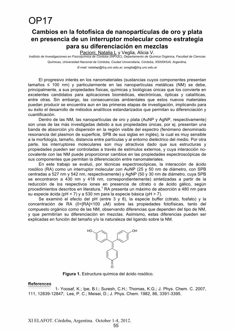
XI ELAFOT. Córdoba, Argentina. October 1-4, 2012.
Cambios en la fotofísica de nanopartículas de oro y plata en presencia de un interruptor molecular como estrategia
para su diferenciación en mezclasPacioni, Natalia L y Veglia, Alicia V.
Instituto de Investigaciones en Fisicoquímica de Córdoba (INFIQC), Departamento de Química Orgánica, Facultad de Ciencias
Químicas, Universidad Nacional de Córdoba, Ciudad Universitaria, Córdoba, X5000HUA, Argentina,
E-mail: [email protected]; [email protected]
El progresivo interés en los nanomateriales (sustancias cuyos componentes presentan tamaños ≤ 100 nm) y particularmente en las nanopartículas metálicas (NM) se debe, principalmente, a sus propiedades físicas, químicas y biológicas únicas que los convierte en excelentes candidatos para aplicaciones biomédicas, electrónicas, ópticas y catalíticas, entre otras. Sin embargo, las consecuencias ambientales que estos nuevos materiales puedan producir se encuentra aun en las primeras etapas de investigación, implicando para su éxito el desarrollo de métodos analíticos estandarizados que permitan su diferenciación y cuantificación.
Dentro de las NM, las nanopartículas de oro y plata (AuNP y AgNP, respectivamente) son unas de las más investigadas debido a sus propiedades únicas, por ej. presentan una banda de absorción y/o dispersión en la región visible del espectro (fenómeno denominado resonancia del plasmon de superficie, SPB de sus siglas en inglés), la cual es muy sensible a la morfología, tamaño, distancia entre partículas y al entorno dieléctrico del medio. Por otra parte, los interruptores moleculares son muy atractivos dado que sus estructuras y propiedades pueden ser controladas a través de estímulos externos, y cuya interacción no-covalente con las NM puede proporcionar cambios en las propiedades espectroscópicas de sus componentes que permitan la diferenciación entre nanomateriales.
En este trabajo se evaluó, por técnicas espectroscópicas, la interacción de ácido rosólico (RA) como un interruptor molecular con AuNP (25 y 50 nm de diámetro, con SPB centradas a 527 nm y 542 nm, respectivamente) y AgNP (50 y 30 nm de diámetro, cuya SPB se encontraron a 430 nm y 418 nm, correspondientemente) sintetizadas a partir de la reducción de los respectivos iones en presencia de citrato o de ácido gálico, según procedimientos descritos en literatura.1 RA presenta un máximo de absorción a 480 nm para su especie ácida (pH < 7) y a 530 nm para la especie básica (pH > 7).
Se examinó el efecto del pH (entre 3 y 8), la especie buffer (citrato, fosfato) y la concentración de RA (0<[RA]<100 μM) sobre las propiedades fotofísicas, tanto del compuesto orgánico como de las NM, observando diferencias que dependen del tipo de NM,y que permitirían su diferenciación en mezclas. Asimismo, estas diferencias pueden serexplicadas en función del tamaño y/o la naturaleza del ligando sobre la NM.
Figura 1. Estructura química del ácido rosólico.
References1- Yoosaf, K.; Ipe, B.I.; Suresh, C.H.; Thomas, K.G.; J. Phys. Chem. C. 2007,
111, 12839-12847; Lee, P. C.; Meisei, D.; J. Phys. Chem. 1982, 86, 3391-3395.
O
HO OH
55
OP17
55

XI ELAFOT. Córdoba, Argentina. October 1 4, 2012. 56
Presentaciones en Pósters (PP)
56

Abstract
XI ELAFOT. Córdoba, Argentina. October 1-4, 2012.
Pt-loaded TiO2 Nanotubes: Very High Photocatalytic Efficiency for Hydrogen Production (~16%) Under UV-B/UV-
C Radiation.Languer, Mariana P. ;1 Feil, Adriano F. ;2 Machado, Guilherme J.;2 Scheffer, Francine R.;1 Migowski, Pedro; 1 Dupont, Jairton;1 Teixeira, Sérgio R.2 and Weibel, Daniel E.11 Instituto de Química, UFRGS. Av. Bento Gonçalves, Nº 9500, Agronomia Porto Alegre-RS, Brasil.
2 Instituto de Física, UFRGS. Av. Bento Gonçalves, Nº 9500, Agronomia Porto Alegre-RS, Brasil.
Photocatalytic hydrogen production via photocatalysts has great potential for solving environmental and energy issues and it has been the focus of increasing research work in the last decade. In particular, heterogeneous photocatalytic reactions on TiO2semiconductors have been attracting much attention because of their potential applications in hydrogen production by water-splitting and environmental clean-up by the so called Advance Oxidative Processes [1, 2]. Although promising results have been reported, a commercially viable catalyst system for this transformation is still missing; even for the high surface area TiO2 nanotubes (NTs). Herein we demonstrate for the first time how to overcome this limitation by loading Pt on TiO2 NTs deposited by DC-magnetron sputtering (DC-MS) deposition method.
For the production of the TiO2 NTs arrays, Titanium (99.6%) foils were anodized and subsequently annealed. Pt was deposited by DC-MS method [3-5]. Hydrogen photogeneration experiments were carried out in a calibrated gas-closed photochemical reactor made of PTFE under continuous magnetic stirring. A high pressure Xe/Hg lamp (150W) was used as light source. The quantification of produced hydrogen was measured by gas chromatography. Actinometry measurements were carried out in the beginning and at the end of a photochemical run, using a wavelength selected 254, 313 and 365 nm. The relative intensities were approximately 1:3:9, respectively.
Figure 1 shows that the rate of hydrogen production for a methanol/water solution (1/8, v/v)strongly increased when Pt was deposited on the TiO2 NTs and that increase is even higher when methanol is present in the water. Apparent hydrogen quantum yields ( H2) for pure TiO2 were between ~0.02 and 0.1%. When Pt was loaded onto the TiO2 NTs, the H2 were: (12 2), (16 2)and (1 0.1)% for 254, 313 and 365 nm of excitation wavelength respectively.
The results presented here are very promising because H2 can be produced at ambient conditions via an efficient, technologically simple and potentially very low-cost process.
Figure 1: Relative hydrogen photogeneration rate obtained from methanol/water solutions 1/8, V/V) and pure water for TiO2 and Pt-loaded TiO2 NTs. Close squares: Pt-loaded TiO2 NTs in methanol/water solution; open squares Pt-loaded TiO2 NTs in pure distilled water and close circles: TiO2 NTs in methanol/water solution.
References:[1] Henderson, M. A., Surf. Sci. Rep. v. 66, p. 185-297, 2011.[2] Fujishima, A.; Zang, X.; Tryk, D. A., Surf. Sci. Rep. v. 63, p. 512-582, 2008.[3] Nam, W.; Han, G. Y., J. Chem. Eng. Jap.,v. 40, p. 266-269, 2007.[4]Esposito, D.V.; Hunt, S. T.; Stottlemyer, A. L.; Dobson, K. D. McCandless, B. E.;
Birkmire, R. W.; Chen, J. G., Angew. Chi., v. 49, p. 9787-9787, 2010.[5]Lee, J. S.; Choi, W.Y., J. Phys. Chem. B, v. 109, p. 7399-7406, 2005.
57
PP1
57

XI ELAFOT. Córdoba, Argentina. October 1 4, 2012. 58
Determinación de parámetros optoacústicos en complejos de Renio (I)
Fabricio Ragone, Hector H. Martinez-Savedra, Gustavo T. Ruiz, Pedro David Gara, Ezequiel Wolcan.
Instituto de Investigaciones Fisicoquímicas Teóricas y Aplicadas (INIFTA, UNLP, CCT La Plata-CONICET), Diag. 113 y 64, Sucursal 4, C.C. 16, (B1906ZAA) La Plata,
Argentina y Centro de investigaciones Ópticas (CIOP) [email protected]
Los complejos de metales del grupo del platino, particularmente Ru(II), Os(II), y Re(I) han despertado un gran interés debido a sus fuertes absorciones en la región visible del espectro, a su buena estabilidad fotoquímica, eficiente luminiscencia y a sus estados excitados de transferencia carga desde el metal y hacia el ligando (MLCT) de vida relativamente larga (generalmente causantes de la luminiscencia). Este interés está basado, en parte, en los esfuerzos dirigidos a diseñar y construir sistemas multicomponentes capaces de llevar a cabo funciones inducidas por la luz y/ó especies redox que sean de utilidad práctica, como por ejemplo en el diseño y el uso de materiales luminiscentes como sondas y sensores.1Nosotros hemos estudiado previamente las propiedades fotofísicas y fotoquímicas de una serie de complejos fac-LS-CO2-Re(CO)3L (con LS = 2-piracina, 2-naftaleno, 9-antraceno, 1-pireno, acetato, 2-anthraquinona y L= 2,2´-bipiridina).2-4 Estos complejos tienen rendimientos cuánticos de emisión que son mucho menores que el de (CH3CN)Re(CO)3(2,2´-bipiridina)+.Por lo tanto, son apropiados para ser estudiados por las técnicas optoacústicas pulsadas, en particular la LIOAS, que se basa en la medición de la onda acústica generada por la expansión del medio, luego de la interacción con un pulso de excitación láser. La técnica proporciona información sobre las vías de decaimiento no radiativas a través de las cuales se desactivan los estados excitados y/o especies transientes generados al absorber la radiación electromagnética. En esta presentación, se compara el comportamiento optoacústico de los complejos fac-LS-CO2-Re(CO)3L con el del complejo (CH3CN)Re(CO)3(2,2´-bipiridina)+.
References
[1] Keefe, M. H.; Benkstein, K. D.; Hupp, J. T. Coord. Chem. Rev. 2000, 205, 201. [2] Guerrero, J.; Piro, O. E.; Wolcan, E.; Feliz, M. R.; Ferraudi, G.; Moya, S. A. Organometallics 2001, 20, 2842-2853. [3] Ruiz, G. T.; Juliarena, M. P.; Lezna, R. O.; Wolcan, E.; Feliz, M. R.; Ferraudi, G. Helv.Chim. Acta 2002, 85, 1261-1275. [4] Wolcan, E.; Torchia, G.; Tocho, J.; Piro, O. E.; Juliarena, P.; Ruiza, G.; Féliz, M. R. Journal of the Chemical Society, Dalton Transactions 2002, 2194-2202.
PP2
58

XI ELAFOT. Córdoba, Argentina. October 1 4, 2012. 59
Fotoquímica de Arilacetamidas en medio heterogéneo. Surfactantes y su efecto en la regioquímica de la reacción.
Iguchi, Daniela; Erra-Balsells, Rosa; Bonesi, Sergio M.
CIHIDECAR-CONICET, Departamento de Química Orgánica, Facultad de Ciencias Exactas y Naturales,Universidad de Buenos Aires, Pabellón II, 3er Piso, Ciudad Universitaria, (1428), Buenos Aires, Argentina, E-mail: [email protected]
El foto reordenamiento de Fries fue descubierto por Anderson y Reese en 1960, y desde entonces, ha sido intensamente estudiado desde los puntos de vista mecanístico y preparativo en fase homogenea. Sin embargo, existen pocos antecedentes en la literatura sobre la misma fotorreacción en medio heterogéneo, por ejemplo, en sistemas acuosos en presencia de surfactantes (micelas), donde los pocos estudios se focalizaron solamente en los aspectos fotofísicos y mecanísticos.
Las aril y heteroaril amidas reaccionan frente a la luz ultravioleta a través del reordenamiento de Fries en fase homogénea y a temperatura ambiente dando como fotoproductos anilina y los regioisómeros orto y para aminofenonas. Durante el proceso fotoquímico se produce la ruptura de la union C - N y se generan en la caja del solvente los radicales intermediarios: radical acilo y radical aminilo. Entonces, si se restringe la libre movilidad de los intermediarios en un entorno micelar, es de esperar una cierta regioselectividad en la formacion de los fotoproductos.
A fin de explorar esta posibilidad con fines preparativos se estudió la reacción fotoquímica de una serie de aril y heteroaril acetamidas en medio heterogéneo con el objeto de analizar si el medio restringido - confinativo tiene un efecto significativo en la regioquímica de la fotorreacción.
Figura 1
Las irradiaciones de los sustratos (ver Fig. 1) se llevaron a cabo en solución acuosa en presencia de una serie de detergentes aniónico (SDS), catiónicos (CTAB, CTAC) y neutros (Brij, Triton X-100) ( exc: 254 nm, a temperatura ambiente, atmósfera (Aire)). Finalizada la fotorreacción, se destruyeron las micelas y los fotoproductos se extrajeron con diclorometano (o cloroformo). Los fotoproductos aislados por métodos cromatográficos se caracterizaron por diferentes métodos espectroscópicos. En la Fig. 1 se muestra el cambio del espectro UV-visible en el tiempo de una solucion de acetanilida irradiada, indicando que la fotorreacción ocurre eficientemente en medio heterogeneo (CTAC 0,05 M). Para todos los sustratos estudiados, se determinaron los rendimientos cuánticos de la fotorreacción ( ) y se compararán con aquellos obtenidos en medio homogéneo (solvente orgánico).
200 250 300 350 400 4500.0
0.4
0.8
1.2
1.6
nm
80 min
0 min
A
HN R
O
hNH O
R
NH2 O
NH2 O
70 - 90%
Extracción consolvente orgánico
H2O
(a) Sustratos estudiados.
(b) Sistema microheterogéneo.
HN
O
R
R = H; Me; MeO; Br; Cl; CN; PhO; COCH3; 2,4-diMeO
O
O
HN
OHN
O
HN
O
Regioselectividad
(c) Seguimiento UV-visible de la irradiación de acetanilida 1,05x10⁻⁴ M en CTAC (0,05 M) en H2O;
exc: 254 nm; atmósfera: aire.
PP3
59

XI ELAFOT. Córdoba, Argentina. October 1-4, 2012.
STUDY OF SODIUM LAURYL ETHER SULFATE (SLES) INDUSTRIAL BY TIME RESOLVED FLUORESCENCE
Friedrich, Leidi Cecilia1,2; Silva, Volnir Oliveira1, Moreira Jr., Paulo Firmino2;Tcacenco, Celize Maia2,3 and Quina, Frank Herbert1
1Instituto de Química, Universidade de São Paulo, CP 26077, 05513970, São Paulo-SP. [email protected]; [email protected]; [email protected]
2Departamento de Eng. Química - Escola Politécnica, Universidade de São Paulo, 05508-000, São Paulo-SP, ; [email protected]
3Departamento de Química, UNIFEO, 06020-190, Osasco-SP,[email protected]
The structure of a detergent or surfactant consists of a hydrophilic part attached to ahydrophobic moiety. Depending on their structure and concentration, the detergent may beadded in aqueous solution to form micelles, which exhibit interesting properties for solubilization and are able to affect the speed of chemical reactions, shifting balance, control regioselectivity and modify the behavior of species photochemically generated [1]. The main objective of this study was to determine the aggregation number of micelles formed bydetergents like sodium lauryl ether sulfate (SLES) as part of a broader investigation of the properties of this important class of industrial detergents.
We used an industrial LESS (Alkopon N, kindly provided by Oxiteno). The number of aggregation was determined by time resolved fluorescence using pyrene fluorescent probesuch as chloride and N-hexadecilpiridíneo as quencher. The fluorescence measurementswere performed on a fluorometer FL-900 Edinburgh Instruments using the excitation (static or pulsed diode laser) at 337 nm.
Fluorescence spectra were recorded and was collected and the decay curves offluorescence of pyrene, fully incorporated into the micelle, in the absence and presence ofvarying concentrations of quencher. The number of aggregation (NAg) was obtained by the technique of time resolved fluorescence, adjusting the Infelta-Tachiya2 equation (Equation 1)to the decay curves of fluorescence intensity of pyrene ( 0=193 ± 1 ns) to for the constant value C, which equals <n>, the average number of suppressors of micelle,
I(t) = I(0) exp (1)
The division of the number of moles <n> supressor added per mol of detergent provides the aggregation number, (NAg), of the micelles (Table 1).
Table 1: Number of aggregation, NAg, the micelles of SLES (cmc = 0,87 mM) at 20º C.[LESS] mM Range <n> NAg
50 0,36-1,33 145±10100 0,38-1,38 140±10
The values of NAg obtained by time resolved fluorescence are reasonable for thisdetergent and confirm that the much lower values (ca. 40) found in the literature3, which were determined by fluorescence doubtful estática3 are incorrect.
Acknowledgements: NAP-PhotoTech, CNPq e LSCP-PQI-EPUSP.
References:[1]Bunton, C.A.; Nome, F.J.; Quina, F.H.; Romsted, L.S. Accts. Chem. Res., 24, 357, 1991.[2]Infelta, P.P; Gratzel, M.; Thomas, J.K. J Phys. Chem., 78, 190, 1974.[3]Aoudia, M.; Al-Haddabi, B.; Al-Harthi, Z.; Al-Rubkhi, A. J Surfact. Deterg., 13, 103, 2010.
60
PP4
60

XI ELAFOT. Córdoba, Argentina. October 1-4, 2012.
Fotogeneración de radicales en un polímero decorado con AlIII(Ftalocianinas) con potencial aplicación en fotocatálisis.
Ruiz, Gustavo T.1; Lappin, A. G.2, Ferraudi, G.21 Instituto de Investigaciones Fisicoquímicas Teóricas y Aplicadas (INIFTA, UNLP,
CCT La Plata-CONICET), Diag. 113 y 64, Sucursal 4, C.C. 16, (B1906ZAA) La Plata, Argentina [email protected]
2 Department of Chemistry, University of Notre Dame, Notre Dame, IN 46556-0579, USA
La incorporación de metalo-ftalocianinas en polímeros organicos mostraron ser buenos catalizadores de reacciones en fase homogénea y heterogenea.1 En general, las propiedades físicas y químicas de ftalocianinas de metales de transición en un entorno polimerico cambian respecto a la ftalocianina libre en solución.2 Recientemente, hemos publicado un trabajo que describe el efecto sobre las propiedades fotoquímicas y fotofísicas de ftalocianinas tretrasulfonadas de Al(III) causado por la unión covalente de éstas en un poly(etilenamida).3 Las cadenas de este poly(HOAlIIItspc), en solución, están asociadas en ovillos esféricos de ~ 150 nm diámetro y los monómeros de AlIII(tspc)-2 se encuentran mayoritariamente como dímeros formando “ -stacks”. El entorno polimerico ejerce un efecto en los tiempos de vida de los radicales reducido, AlIII(tspc•)-3, y oxidado, AlIII(tspc•)-,formados en estas esferulas cuando es atacado por distintos radicales. En este trabajo mostramos la formación indirecta de estos radicales de vida larga, mediante la irradiación continua de soluciones acuosas de poly(HOAlIIItspc) en ausencia de oxígeno. Por este método, se genera el radical (CH3)2C•OH, irradiando con luz de 300 nm soluciones del polímero que contienen acetona y 2-propanol en concentraciones adecuadas. El cambio de color del verde al azul oscuro revela la formación del AlIII(tspc•)-3 en unos 2-3 minutos de irradición producto del ataque del radical 2-propanol. Los rasgos característicos del espectro de absorción del AlIII(tspc•)-3 permanecen inalterables en ausencia de oxígeno por más de 24 hs. El agregado anaeróbico del catalizador negro de Pt, dio lugar a la formación de propano detectado por CG y la consecuente recuperación del color verde de la solución por la regeneración del AlIII(tspc)-2 en el polímero. El radical AlIII(tspc•)-3 también se pudo obtener irradiando con luz de > 500 nm soluciones acuosas deareadas del poly(HOAlIIItspc) y TEOA. En estas condiciones, luego del agregado del catalizador en atmósfera de CO2, se observó la formación de CO monitoreado por CG.
References
1- a) W. M. Brouwer, P. Piett, A. L. German, J. Mol. Catalysis, 1985, 29, 335. b) A. Sivanesan, S. A. JohnElectrochimica Acta, 2008, 53, 6629. 2- S. Thomas, G. Ruiz, G. Ferraudi,Macromolecules, 2006, 39, 6615. 3- G.T. Ruiz, G. Ferraudi, A.G. Lappin J. Photochem. Photobiol. A: Chemistry 2009,206, 1.
61
PP5
61

Abstract
XI ELAFOT. Córdoba, Argentina. October 1-4, 2012.
Photocatalytic degradation of tartrazine induced by Ag-doped TiO2 prepared by ultrasound-mediated synthesis
dos Santos, Lidiaine Maria1, Dias França, Marcela1, Araújo Borges, Karen1, Souza Müller Jr, Paulo1, da Hora Machado, Antonio Eduardo1,2
1Universidade Federal de Uberlândia P. O. Box 563; 38400-902, Instituto de Química, Laboratório de Fotoquímica, Uberlândia, MG, Brasil.
2Universidade Federal de Goiás, Campus Avançado de Catalão; Catalão, GO, Brasil.e-mail: [email protected]
In this communication we present the characterization of a series of Ag-doped TiO2 prepared by ultrasound-mediated synthesis, and a comparative study of the photocatalytic degradation of tartrazine, an azo-dye used as food additive that presents several adverse effects [1],using as photocatalysts the Evonick-Degussa TiO2 P25, a known commercial photocatalyst [2], and the Ag-doped TiO2 with three different percentage of Ag (0.05%, 2.00% and 5.00%)[3]. For the synthesis of the Ag-doped TiO2, titanium tetraisopropoxide was used as precursor. It was dissolved in 2-propanol, under the action of ultrasound. Its hydrolysis,conducted by slow addition of ultrapure water, resulted in the precipitation of TiO2.Subsequently an aqueous solution of AgNO3 was added, also under sonication. The material was dried at 60 °C and subjected to heat treatment at 400 ºC. In the assays, the effluent to be treated (4 L of an aqueous solution containing 42.5 mg.L-1 of tartrazine) was circulated by an annular borosilicate glass reactor. A 400 W high pressure mercury vapor lamp, placed inside the protoreactor was used as radiation font [4]. The photocatalysts were used at the concentration of 100 mg.L-1. In all assays, the initial pH of the reaction medium was fixed in6.90, and the reaction time limited to 120 minutes. Aliquots were collected at 20 minutes intervals and analyzed in terms of total organic carbon (TOC) and discoloration, monitored by absorbance measurements at 428 nm. Our results suggest that the Ag-doped TiO2containing 5.00% m/m of Ag presents a similar mineralization performance to that presented by TiO2 P25: the TOC reduction during the degradation was equal to 44%, 25%, 32% and 40% for respectively for TiO2 P25 and Ag-doped TiO2 (0.05%, 2.00% and 5.00%). This seems to be related to the specific surface area which is directly proportional to thepercentage of added Ag, which is directly related to the amount of reactive sites in the photocatalyst. The band gap was also positively influenced by the amount of Ag into the crystal structure of TiO2, being shifted to lower energies as the doping increases.Acknowledgement: FAPEMIG, CNPq and CAPES.
References:[1]. Pantazaki, A.; Mountoukas, P.; Kostareli, E.; Christodoulou, P.; Kareli, D.; Poliliou, S.; Mourelatos,
C.; Lambropoulou, V.; Lialiaris, T. Cytogenetic evaluation and DNA interaction studies of the food colorants amaranth, erythrosine and tartrazine. Food and Chemical Toxicology v. 48, pp. 2934 2944, 2010.
[2]. Machado, A. E. H.; França, M. D.; Velani, V.; Magnino, G. A.; Velani, H. M. M.; Freitas, F. S.;Müller, P. S.; Sattler, C.; Schmücker, A. Characterization and evaluation of the efficiency of TiO2/zinc phthalocyanine nanocomposites as photocatalysts for wastewater treatment using solar irradiation. International Journal of Photoenergy, v. 2008, pp. 1-12, 2008.
[3]. Machado, A. E. H.; Santos, L. M.; Müller Jr, P. S.; França M. D.; Borges, K. A.; Procedure under protection.
[4]. Oliveira, D. F. M.; Batista, P. S.; Müller Jr, P.S.; Velani, V.; França, M. D.; Souza, D. R.; Machado,A. E. H. Evaluating the effectiveness of photocatalysts based on titanium dioxide in the degradation of the dye Ponceau 4R. Dyes and Pigments, v. 92, No. 1, pp. 563-572, 2012.
62
PP6
62

XI ELAFOT. Córdoba, Argentina. October 1-4, 2012.
Ensamblados de ficocianina sobre nanoestructuras de TiO2como electrodos para celdas DSSC
Enciso, Paula1; Cerdá, María Fernanda1
1 Laboratorio de Biomateriales, Facultad de Ciencias, UdelaR, Iguá 4225, 11400 Montevideo, Uruguay, [email protected]
La ficocianina es un pigmento proteico azulado, accesorio a la clorofila, y es el encargado de la captación de luz solar en la familia de las ficobiliproteínas. Dicho pigmento se encuentra adherido a la superficie citoplasmática de las membranas de los tilacoides de las cianobacterias y de las microalgas. Absorbe en los 620 nm con altos coeficientes de extinción. Esto le confiere interesantes propiedades para captar la luz solar en el rango visible del espectro y transferir esa energía a un material incapaz de absorber en el visible como el dióxido de titanio (TiO2) [1].
Lo que se plantea en la presente propuesta es la elaboración de un electrodo, a partir de un óxido transparente conductor ITO (indium tin oxide) sobre el cual se encuentra un ensamblado de ficocianina adsorbida sobre TiO2 mesoporoso nanoestructurado. De esta manera se obtiene un electrodo con buenas características de captación de energía solar ytransferencia de esta energía, lo cual al completar el circuito con un contra electrodo de platino permite obtener una celda solar de tipo DSSC (dye sensitized solar cell).
Para ello, la ficocianina fue extraída de cápsulas comerciales de Spirulina por centrifugación y filtrado, en presencia de agua desionizada. Por otra parte, el TiO2 fueobtenido a partir de butóxido de titanio IV (97%) en etanol y posterior calcinación a 450ºCpara obtener la estructura química anastasa que resulta más activa fotocataliticamente(fig.1)[2][3].
El ensamblado ITO/TiO2 fue obtenido por calentamiento a 350ºC, y la ficocianina fue adsorbida sobre el TiO2 por simple deposición de la proteína 24hs a 4°C en medio acuoso.
El proceso de ensamblado y la estabilidad del electrodo obtenido fueron seguidos por medidas de voltamperometría cíclica en KI 0.1 M y en [Ru(NH3)6] 1 mM en NaClO4 0.1 M.
Las medidas mostraron la obtención de un electrodo de ITO/TiO2/ficocianina estable en el tiempo.
0 10 20 30 40 50 60 70 80 90
-500
50100150200250300350400450500550600650700750800850900
iInte
nsid
ad (c
ps)
2 grados)
Figura 1. Patrón de difracción de rayos X de la muestra de TiO2 antes de calcinar, estructura química amorfa (en rojo) y después de ser calcinada, estructura química anastasa (en negro).
Acknowledgements: Los autores quisieran agradecer a PEDECIBA, ANII and CSIC(Uruguay)
References
[1] ticles with phycocyanin pigment , Journal of colloid and interface science, vol. 335, no. 2, pp. 196-202, Jul. 2009.[2] X. Chen, odifications, and applications , Chemical reviews, vol. 107, no. 7, pp. 2891-959, Jul. 2007.[3] O2 - new photochemical processes , Chemical reviews, vol. 106, no. 10, pp. 4428-53, Oct. 2006.
63
PP7
63

XI ELAFOT. Córdoba, Argentina. October 1 4, 2012. 64
N
N
R9
R2
Interaction between novel N-methyl- -carboline derivatives and Bovine Serum Albumin, a typical carrier protein
Rasse-Suriani, Federico A.1; Yañuk, Juan G.1;Garcia-Einschlag, Fernando S.2; González, María M.3; Erra-Balsells, Rosa3; Cabrerizo,
Franco M.1.1IIB-INTECH-CONICET (sede Chascomús), UNSAM, Av. Intendente Marino km. 8,2 CC 164(7130),
Chascomús, Argentina. E-mail: [email protected], UNLP, Diag. 113 y 64 (1900), La Plata, Argentina.
3CIHIDECAR-CONICET, FCEyN, UBA, Pabellón 2, 3° piso, Ciudad Universitaria (1428), Buenos Aires, Argentina.
-carboline ( Cs) alkaloids belong to a family of heterocyclic compounds derived from 9H-pyrido[3,4-b]indole or norharmane (nHo) (Scheme 1). These alkaloids are widely distributed in living systems such as plants [1], insects [2], mammals [3], etc. In mammalian body, Cs occur as normal constituents in plasma, platelets and urine. However, under certain conditions, such as after alcohol intake and smoking, Cs basal levels (~ 0.1 nmol/l) are considerably increased (~ 1 nmol/l) [4].Regarding their biological functions, a variety of significant photo-dependent roles have been described, e.g., in plants Cs could be involved in the defense response (via phototoxic effects).In addition, these alkaloids are able to act as good photosensitizers,e.g., upon UV-A excitation, some Cs are able to induce DNA relaxation [5] or chromosome damage in mammalian cells [6], and also to inactivate viruses [7]. All these properties make interesting the photophysical study of Cs in different microheterogeneous environments.
As itis known, the extent of photodynamic action depends not only on the singlet oxygen production but also on the biodistribution of the probe molecule in the cytoplasmic and mitochondrial membranes, the retention and the nature of the binding inside the cell. Thus, the binding interaction of Cs with different biosystems is important to study the net photodynamic efficiency.
In the present work, we have investigated the pH dependence on the interaction between a family of novel N-methyl- -carboline derivatives(table), recently synthesized in our laboratories, andBovine Serum Albumin (BSA),by fluorescence emission and UV-Vis spectroscopy.Data analysis was performed with a series of matrix techniques for the deconvolution of spectra and concentration profiles. Furthermore, by applying the constrained regression analysis, we obtained two matrices (ST = spectra, and C = concentrations) that have physical meaning and whose matrix product reproduces the experimental matrix elements.
Results may help us to understand the photophysical behavior of these alkaloids in biological environments and to better assess the correlation between alkaloid prototropic forms and their biological activity. Scheme 1 (and Table): Chemical structure of norharmanoand their methylated derivatives.
References: [1].Herraiz, T. et al., Food Chem Toxicol, 2010. 48(3): p. 839-45. - [2].Siderhurst, M.S. et al., J Econ Entomol, 2005. 98(5): p. 1669-78. - [3].Airaksinen, M.M. and I. Kari, Med Biol, 1981. 59(1): p. 21-34. - [4].Spijkerman, R. et al., Eur Neuropsychopharmacol, 2002. 12(1): p. 61-71. - [5].Gonzalez, M.M. et al., Org Biomol Chem, 2010. 8(11): p. 2543-52. - [6].Sasaki, Y.F. et al., Mutat Res, 1992. 269(1): p. 79-95. - [7].Hudson, J.B. et al, Photochem Photobiol, 1986. 43(1): p. 21-6.
Table of -carbolines R2 R9
norharmane H HN2-methyl-norharmane CH3 HN9-methyl-norharmane H CH3N2,N9-dimethyl-norharmane CH3 CH3
PP8
64

XI ELAFOT. Córdoba, Argentina. October 1 4, 2012. 65
"Design" of new zinc phthalocyanine derivatives for use in nonlinear optics
Araujo, D. M. S.1; Gomes, W. R.1; Barbosa Neto, N. M.3; Machado, A. E. H.1,2,
1Universidade Federal de Uberlândia, Instituto de Química, Laboratório de Fotoquímica e Ciência dos Materiais, Uberlândia, MG, Brasil. [email protected]
2Universidade Federal de Goiás, Campus Catalão, Depto. de Química, Catalão, GO, Brasil.
3Universidade Federal de Uberlândia, Instituto de Física, Uberlândia, MG, Brasil.
The present study aims the proposition, by "Molecular Engineering", of derivatives of Zinc Phthalocyanine (ZnPc) with desirable characteristics for application in photonic technologies. Three possible derivatives, based on the introduction of peripheral groups in Zinc Phthalocyanine were studied were proposed to encompass a range of different "push-pull" groups.
Figure 1: Representation of the compounds under study: (1): -diethylamino- -nitro-zinc phthalocyanine; (2): -(n-butoxy)-nitrophenylethynyl-zinc phthalocyanine; (3): -sulfonyl- -ammonium zinc phthalocyanine.
To characterize these compounds, a theoretical model was employed to evaluate the geometry, electronic structure, singlet and triplet states and their electronic transitions, and an estimate of the first hyperpolarizability, a nonlinear optical (NLO) property.
The analysis of the results revealed an important participation of HOMO and LUMO orbitals in the first electronic transition, and that the overlap between these orbitals suggest the occurrence of intramolecular electron transfer from the donor to acceptor groups. The compound (2) presented a red shifted transition ( MAX = 744 nm) when compared to the other two, evidencing an effective participation of the added substituents in the electronic delocalization. For all derivatives, the dipole moment estimated for the first excited state is higher than the value in the ground state. The derivative (2) presented the highest gap in the dipole moment ( μ), with S1 about 6% larger than S0. The compound (3) presented the smallest values of polarizability ( ) and first hyperpolarizability ( ) despite of having the highest values of dipole moment. Compound (1) presented intermediate values for these properties between the studied compounds. The results suggest derivative (2) as promising for application in technologies based on NLO. However, further studies are necessary to reveal more precisely what is the real applicability of this derived in photonic technologies.
Acknowledgement: The authors thank to FAPEMIG, CNPq and CAPES for financial support and research grants.
(2)(1)
(3)
PP9
65

XI ELAFOT. Córdoba, Argentina. October 1-4, 2012.
Photochemical study of a new thioxanthone derivativePinto, Leticia F.A.1; Cavalheiro, Carla C.S.1, Neumann, Miguel G.1
1 Instituto de Química de São Carlos, Universidade de São Paulo, São Carlos, [email protected]
Thioxanthone and its derivatives have a wide range of application, such as type II photoinitiators for free-radical polymerization or as antitumoric, antiparasitic, and anticarcinogenic agents. The synthesis of thioxanthone derivatives has recently gained interest in photochemistry because of their good absorption characteristics and their high photoinitiation efficiency in the near-UV range. In this study thioxanthen-11-one-12,12-dioxide (1) was synthetized and characterized. Photophysical properties: fluorescence and phosphorescence emission spectra and fluorescence quantum yield of (1) (Φf = 0.06) were determined. The phosphorescence lifetime found was of 800 ms for compound (1) at 77 K in methylcyclohexane, suggesting a π–π* nature of the lowest triplet state.[1]
S
O
O O
(1)
Structure of thioxanthen-11-one-12,12-dioxide
300 400 500 600 700
100
200
300
400
500
77 K295 K
λ (nm)
Inte
nsity
(u.
a.)
0
500
1000
1500
2000
0 1000 2000 3000 4000
0
1000
2000
3000
4000
Inte
nsity
(a.
u.)
Time (ms) Intensity (u.a.)
Fluorescence and Phosphorescence spectra of 1 in MCH Inset: phosphorescence decay.
Laser flash photolysis was used to investigate the transients of (1) in acetonitrile, methanol and methylcyclohexane (MCH) degassed solutions. The transient spectra show an absorption peak at 530 nm, corresponding to the triplet state. The lifetime of the triplet state of (1) was found to be 6.0 µs, 8.0 µs, and 9.0 µs in acetonitrile, methanol and MCH, respectively. The rate constants for the quenching of the triplet in MCH by 2-propanol, ethyl-4-dimethylaminobenzoate and transtilbene were 6.2×10⁸, 4,2×10⁹ and 5,7×10⁹ L mol-1 s-1, respectively. The quenching rate constants in other solvents like methanol and acetonitrile have similar values.[2]
300 400 500 600 700 800
0.00
0.02
0.04
0.06
0 1000 2000 3000 4000
0
1000
2000
3000
4000
Inte
nsity
(a.
u.)
Time (ms)
Δ Abs
orba
nce
λ (nm)
Transient absorption spectra of (1) in MCH, recorded at (�) 2.4 , (�) 8.8, (▲) 26.0 and (∆) 102.0 µs after excitation (355 nm). Inset: decay of the transient at 530 nm in MCH.
Acknowledgements: The authors would like to thank CNPq and Fapesp for the financial support.
References [1] D.K. Balta, N.Cetiner, G. Temel, Z. Turgut, N. Arsu. J. Photochem. Photobiol. A: Chem. 2008, 199, 316. [2] J.C. Netto-Ferreira, E.S.L. da Silva, N.C. Lucas, J. Photochem. Photobiol. A: Chem. 2011, 225, 135.
66
PP10
66
XI ELAFOT. Córdoba, Argentina. October 1 4, 2012. 67
DERIVADOS ANTRAQUINÓNICOS AISLADOS DE Heterophyllaea pustulata: POSIBLE MECANISMO DE
ACCIÓN COMO AGENTE ANTIBACTERIANOComini, Laura1; Garagiola, Betania1; Páez, Paulina2; Albesa, Inés2; Núñez
Montoya, Susana1; Cabrera, José Luis1.1Farmacognosia, Dpto. de Farmacia (IMBIV-CONICET). FCQ-UNC. (5000). Córdoba.
Argentina. E-mail: [email protected] y Microbiología, Dpto. de Farmacia. FCQ-UNC. (5000). Córdoba. Argentina.
E-mail: [email protected]
Heterophyllaea pustulata Hook. f. (Rubiáceas) es un arbusto que habita en el NO argentino, sobre la Cordillera de Los Andes entre los 2500 y 3000 m. s. m. Se conoce popularmente como “cegadera”, en referencia a la pérdida de visión que provoca en los animales que la ingieren y se exponen directamente a la luz solar [1]. A partir de las hojas y tallos de esta especie vegetal fueron aisladas e identificadas nueve antraquinonas (AQs) mayoritarias: soranjidiol, 1-metil éter de soranjidiol, rubiadina, 1-metil éter de rubiadina, damnacantal, damnacantol, heterofilina, pustulina y 5,5’-bisoranjidiol [2]. Se ha demostrado previamente que éstos derivados son agentes fotosensibilizantes (FS) Tipo I y/o Tipo II, con generación de anión superóxido (O2
.-) y/o oxígeno singlete (1O2), respectivamente. Numerosos FS fotodinámicos naturales han resultado bioactivos y con potencial aplicación como antimicrobianos en la terapia fotodinámica antimicrobiana (TFDA). La TFDA implica el uso de FS y luz visible o ultravioleta, que promueve la generación de especies reactivas del oxígeno (ERO), tales como O2
.- y/o 1O2, induciendo daño celular que conlleva a la muerte de los microorganismos, revirtiendo la infección [3].
En esta oportunidad se evaluó la actividad antibacteriana de estas AQs midiendo la concentración inhibitoria mínima (CIM) mediante el método de macrodilución en caldo [4], sobre distintas bacterias Gram positivas: Staphylococcus epidermidis ATCC 12228, Staphylococcus epidermidis 688 y Enterococcus faecalis ATCC 29212. Para los compuestos que mostraron actividad antibacteriana, se procedió a evaluar, si el mecanismo de acción involucra un aumento en los niveles de O2
•- y si este efecto es potenciado en presencia de luz. La producción de O2
•- se determinó, tanto en oscuridad como bajo irradiación actínica (380-480 nm), mediante un ensayo fotobiológico indirecto [5], que evalúa la reducción de Nitroblue Tetrazolium (NBT) a azul de formazán, por acción del O2
•- producido por las bacterias en presencia de un agente oxidante (AQs). La suspensión bacteriana se incubó con cada AQ y NBT (0,1 % P/V) a 37 ºC, por triplicado y a distintos tiempos. Se midió la absorbancia del azul de formazán generado en el interior de las bacterias por espectrofotometría UV-Visible ( = 575 nm).
Según los resultados obtenidos, rubiadina y soranjidiol poseen una CIM entre 125 y 250 μg/ml sobre las tres bacterias ensayadas y 1-metil éter de rubiadina sólo mostró inhibición sobre Staphylococcus epidermidis ATCC 12228 con una CIM de 125 μg/ml.
Respecto a la producción de O2•-, se pudo observar que, a excepción de rubiadina en S.
epidermidis 688, para todos los casos ensayados hubo un incremento en la producción de esta ERO y la producción fue mayor en condiciones de irradiación.
En base a los resultados mencionados, se puede considerar el uso de estas AQs como potenciales candidatos para la TFDA.
Bibliografía: [1] Aguirre D.H.; Neumann R.A. - Med. Vet. 18, 487-490, 2001. [2] Núñez Montoya S.C.; Agnese A.M.; Cabrera J.L. - Nat. Prod. J. 69, 801-803, 2006. [3] Comini L.R.; Núñez S.C.; Sarmiento M.; Cabrera J.L.; Argüello G.A. - J. Photochem. Photobiol. A. Chem. 188,185-191, 2007. [4] Ferraro M. - Methods for dilution susceptibility tests for bacteria that grow aerobically, fifth ed., Clinical and Laboratory Standards Institute (CLSI). 2005. [5] Carbonare M.D.; Pathak M.A. - J. Photochem. Photobiol. B: Biol. 14, 105-124, 1992.
PP11
67

XI ELAFOT. Córdoba, Argentina. October 1-4, 2012.
Mesoporous silica nanoparticle as a delivery system for novel phthalocanines
López Zeballos, Noelia1; García Vior, María C.2; Awruch Josefina2; Dicelio, Lelia E.11INQUIMAE/ Departamento de Química Inorgánica, Analítica y Química Física.
Facultad de Ciencias Exactas y Naturales, UBA, Pabellón II, C1428EHA, Buenos Aires, Argentina.
2Departamento de Química Orgánica. Facultad de Farmacia y Bioquímica, UBA, Junín 956, 1113, Buenos Aires Argentina.
E-mail: [email protected]
Key Words: silica nanoparticles, phthalocyanines, photodynamic therapy.IntroductionOver the last few years, photodynamic therapy (PDT) has emerged as an alternative to chemo and radiotherapy for the treatment of various diseases including cancer. It involvesthe use of light and photosensitizers (PS) that accumulate in the tumor tissue. Photodynamic sensitizers, such as zinc phthalocyanines (ZnPc), are drugs that can transfer their energy from their triplet excited state to neighboring oxygen molecules when activated by light of a specific wavelength. Singlet oxygen (1O2) and other cytotoxic reactive oxygen species are formed and lead to the destruction of cancer cells by both apoptosis and necrosis. PS described so far in the literature present several disadvantages. Mainly, they are hydrophobic or have a limited solubility in water and therefore aggregate in aqueous media such as blood which leads to the modification of their photophysical properties and particularly to the decrease of 1O2 quantum yield. Moreover, PS are required to accumulate selectively in tumorcells. In order to address these issues, PS have been encapsulated in nanoparticles (NPs). Therefore, NPs have been prepared to improve the efficiency of PDT. Polymeric, metallic, or silica-based nanoparticles have been described. Among the variety of NPs, silica-basednanomaterials have very recently emerged as promising vectors for PDT applications. Theyare chemically inert and the silica matrix porosity is not susceptible to swell or change with a varying pH. Also, particles size, shape, porosity and mono-dispersibility can be easily controlled during their preparation. The surface modification with specific biomolecules for tumor-cell targeting is another possibility offered by silica nanoparticles. [1]Aim of this studyIncorporation of different lipophilic ZnPcs in mesoporous silica nanoparticles (MSN) in order to evaluate their photophysical properties, as well as the size, shape and the release of them through a period of time. Results
ConclusionsMSN with and without ZnPcs were obtained. No significant changes in the shape of the absorption spectra with respect to the phthalocyanines in THF were observed. References[1] P. Couleaud, V. Morosini, C. Frochot, S. Richeter, L. Raehma and J. Durand. Nanoscale, 2010, 2, 1083 1095.
550 600 650 700 750 800
0,00,20,40,60,81,00,00,20,40,60,81,00,00,20,40,60,81,0
/ nm
3-THF 3-MSN
Abs
orba
nce
Rel
ativ
e
2-THF 2-MSN
1-THF 1-MSN
620 640 660 680 700 720 740 760 780 800
0,00,20,40,60,81,00,00,20,40,60,81,00,00,20,40,60,81,0
/ nm
3-THF 3-MSN
Fluo
resc
ence
Rel
ativ
e In
tens
ity /a
.u.
2-THF 2-MSN
1-THF 1-MSN
1. 2,9(10),16(17),23(24)-tetrakis-(1-adamantylsulfanyl) phthalocyaninatozinc (II) 2. 2,9(10),16(17),23(24)-tetrakis-[(2-dimethylamino)ethylsulfanyl] phthalocyaninatozinc (II) 3. 1,8(11),15(18),22(25)-tetrakis-[(2-dimethylamino)ethylsulfanyl] phthalocyaninatozinc (II)
SEM images of the empty mesoporuos silica nanoparticles.
68
PP12
68

XI ELAFOT. Córdoba, Argentina. October 1 4, 2012. 69
Análisis comparativo del efecto de la radiación UV-C, UV-A y visible en la atenuación de taquizoitos de Toxoplasma
gondiiYañuk, Juan Gabriel; Cóceres,Verónica; Rasse-Suriani, Federico
A.;Angel,Sergio O.;Cabrerizo, Franco M.IIB-INTECH-UNSAM-CONICET (sede Chascomús). Intendente Marino Km 8,2. CC
164 (7130) Chascomús, Buenos Aires, Argentina. E-mail:[email protected]
Toxoplasma gondii es un parásito intracelular obligado ampliamente distribuido en humanos y animales de sangre caliente. Este parásito es el responsable de la toxoplasmosis, infección que puede producir abortos o considerables daños al feto, inflamación de la retina y otras patologías. El desarrollo de una vacuna contra la infección en humanos es un tema prioritario debido a la relevancia de la enfermedad en algunas regiones del mundo como Sudamérica, y a la falta de drogas efectivas para su tratamiento [1]. Las vacunas basadas en parásitos atenuados vivos confieren mejor protección, comparada con aquellas en las que se utilizan taquizoítos inactivados [2], ya que estas simulan la infección natural desencadenado una respuesta inmune adecuada (respuesta inmune celular) [3,4].
Entre las alternativas para la atenuación de taquizoítos se encuentra la utilización de radiación UV-C [5]. La radiación UV se clasifica, según su energía decreciente en UV-C, UV-B y UV-A, y todas ellas presentan, en mayor o menor medida, la capacidad de inducir daños en los organismos vivos. Típicamente, existen dos vías o mecanismos a través de los cuales la radiación UV induce daño en los sistemas vivos:(i) el daño directo (absorción de la radiación por biomoléculas) que tiene lugar bajo radiación UV-C y UV-B, y (ii) el daño indirecto, mediado por la absorción de la radiación por otros cromóforos endógenos o exógenos (denominados fotosensibilizadores) que tiene lugar bajo irradiación UV-A y/o visible.
En este trabajo se presentan los resultados obtenidos en el estudio comparativo del efecto atenuador de distintos tipos de radiación (UV-C, UV-A y visible) sobre taquizoítos de T. gondii. En el caso de las irradiaciones con luz UV-A y visible, se evaluó no sólo el efecto de los fotosensibilizadores endógenos del parásito sino también el de fotosensibilizadores exógenos derivados de porfirinas. Luego de cada tratamiento se evaluó la capacidad invasiva y replicativa del parásito. Desde el punto de vista experimental, la cuantificación de estos procesos se realizó invadiendo monocapas celulares (células Vero) con los taquízoitos previamente tratados. Al cabo de 16 horas de incubación a 37°C en una atmósfera de 5 % de CO2, se procedió a contar el número de vacuolas por campo y la cantidad de parásitos por vacuola. Además se verificó la integridad del núcleo y otras estructuras celulares utilizando microscopia de fluorescencia. Referencias:[1]Jongert, E., Roberts, C. W., Gargano, N., Förster-Waldl, E., Förster-Wald, E., & Petersen, E., 2009. Vaccines against Toxoplasma gondii: challenges and opportunities. Memórias do InstitutoOswaldo Cruz, 104(2), 252-266. [2]Waldeland H., Frenkel J.K., 1983. Live and killed vaccines against toxoplasmosis in mice. JParasitol, 1983, 69: 60-65. [3]Suzuki Y., Remington J.S., 1990. The effect of anti-IFN-antibody on theprotective effect of LyT-2+ immune T cells against toxoplasmosis in mice.J Immunol, 1990,144:1954–1956. [4] Denkers E.Y. T-Lymphocyte-dependent effector mechanisms of immunity to Toxoplasma gondii.Microb Infect. 1999, 1:699–708. [5] Grimwood, B. G., Infective Toxoplasma gondii trophozoites attenuated by ultraviolet irradiation. Infect Immun, 1980, 28(2), 532-535.
PP13
69

XI ELAFOT. Córdoba, Argentina. October 1 4, 2012. 70
Determinación del mecanismo de oxidación atmosférica de C6F13CH2C(O)H en presencia de NO2
Diana P. Henao1, Fabio E. Malanca1, Malisa S. Chiappero2 y Gustavo A. Argüello1
1INFIQC (CONICET) – Dpto de Fisicoquímica – Facultad de Ciencias Químicas – Universidad Nacional de Córdoba. e-mail : [email protected]
2 Depto de Química, FCEyN. UNMdP, Funes 3350 – Nivel +2 (B7600AYL).
Los alcoholes fluoroteloméricos (FTOHs) son una clase de alcoholes fluorados que se han sugerido como fuentes de ácidos perfluoroalquil carboxílicos (PFCAs) [1]. Dichos alcoholes son volátiles y se han detectado en el aire en diversos lugares (Norte América, Ártico, Europa, Japón, fuera de la costa al occidente de África) [2] y tienen un tiempo de vida atmosférico (aproximadamente 10-20 días) que les permite ser transportados desde su fuente a lugares distantes [3]. Estos compuestos pueden oxidarse en la atmósfera para dar lugar a la formación de aldehídos teloméricos fluorados (CxF2x+1CH2CHO), aldehídos perfluorados (CxF2x+1C(O)H) y ácidos perfluorocarboxílicos [4].
La foto-oxidación del aldehído fluorotelomérico 6:2 (C6F13CH2C(O)H) iniciada por radicales OH ó átomos de cloro ha sido estudiada por Chiappero y col [5], determinando que el principal producto es perfluoroheptanal (C6F13C(O)H). Según el mecanismo planteado es posible la formación de peroxinitratos en lugares con alta concentración de NO2. En este trabajo se presentan los resultados obtenidos en la foto-oxidación del (C6F13CH2C(O)H) en presencia de NO2 iniciada por átomos de cloro.
La fotólisis se llevó a cabo en un balón de 5L con tres lámparas negras ( >360 nm) y se obtuvieron los espectros infrarrojos de fracciones de la muestra a distintos tiempos de fotólisis (0, 30, 50 y 75 min) en una celda de paso óptico largo (paso óptico: 9 m, resolución de 2 cm-
1). Los espectros obtenidos muestran la desaparición de los picos correspondientes a C6F13CH2C(O)H (1751) y NO2 (1602); la aparición de CF2O (1956, 774), C6F13C(O)H (1775), así como otros picos que se agruparon según su variación con la presión en dos posibles sustancias (1849, 1749, 792) y (1697, 825) que se corresponderían en principio con especies RC(O)OONO2 y RONO2, respectivamente. Todas las unidades se dan en cm-1.
Con el objetivo de obtener los espectros de ambas especies puras, se recogieron los productos en trampas a la temperatura del aire líquido, se destiló en vacío y se analizaron las fracciones resultantes de la evaporación de la mezcla a distintas temperaturas. Luego de la separación, se hicieron evidentes nuevas bandas en cada sustancia (1849, 1749, 1362, 1301, 1252, 1215, 1153, 792 cm-1) y (1697, 1362, 1290, 1252, 1215, 1153, y 825 cm-1). La comparación con valores disponibles en bibliografía, y los datos cinéticos que llevan a su formación sugieren que la estructura de ambas es C6F13CH2C(O)OONO2 y C6F13CH2ONO2.En base a los resultados obtenidos se propone un mecanismo de reacción.
Referencias[1] Chiappero, M. S.; Argüello, G. A.; Hurley, M. D.; Wallington, T. J. J. Phys. Chem. A. 2010,114, 6131-6137 [2] Stock. N. L.; Lau, F. K.; Ellis, D. A.; Martin, J. W.; Muir, D. C. G.; Mabury, S. A.; Environ.Sci. Technol. 2004, 38, 991-996 [3] Ellis, D. A.; Martin, J, W.; Mabury, S, A.; Hurley, M. D.; Andersen. M. P. S.; Wallington, T. J. Environ. Sci. Technol. 2003, 37, 3816-3820 [4] Ellis, D. A.; Martin, J, W.; De Silva, A. O.; Mabury, S, A.; Hurley, M. D.; Andersen. M. P. S.; Wallington, T. J. Environ. Sci. Technol. 2004, 38, 3316-3321 [5] Chiappero, M. S.; Argûello, G. A.; Hurley, M. D.; Wallington, T. J. J. Phys. Chem. A. 2010,114, 6131-6137
PP14
70

XI ELAFOT. Córdoba, Argentina. October 1-4, 2012.
Poly(alkyl methacrylate) Thin Films Relaxation Probed by Nile Red Rotational Dynamics
Araoz, Beatriz1; Carattino, Aquiles; Aramendía, Pedro F.2
1 INQUIMAE-DQIAQF-FCEN-UBA, Pab. II-Ciudad Universitaria, Buenos Aires, [email protected]
2 INQUIMAE-DQIAQF-FCEN-UBA, Pab. II-Ciudad Universitaria, Buenos Aires [email protected]
In the present work we study the rotational dynamics of Nile Red (NR) embedded in
poly(alkyl methacrylates), methyl-, PMMA; ethyl-,PEMA and butyl-, PBMA, thin polymer films
by ensemble and single molecule (SM) polarized fluorescence measurements at 296 K.
Polarized fluorescence microscopy is a powerful tool to study molecular orientation of
fluorescent probes [1], [2]. It gives information about local friction by measuring temporal
fluctuation of SM fluorescence anisotropy. Because excited state lifetime is shorter than the
rotational characteristic time, dye reorientational movements are studied in the ground state,
between successive excitations. In many cases, fluorescence detection is affected by
photobleaching processes; to overcome this problem, we propose an analytical solution of
the rotational diffusion equation including photobleaching, under linearly polarized excitation,
which we apply to the ensemble measurements to derive rotational diffusion coefficients and
photobleaching rates.
Experiments were conducted in a wide field microscope on probes containing NR in
PMMA, PEMA and PBMA thin polymer films of 25 and 200 nm thickness at 296 K. In the
sample preparation process, solvent evaporates and the probe keeps immobilized in the
matrix; therefore, no translational diffusion is observed. The detected movements result from
the confinement that generates small and high fluctuations, due to thermal motion and large
conformational cage changes. Anisotropy time traces of individual molecules show different
dynamics: as expected for a polymer environment, the system is heterogeneous both,
temporally and spatially. SM anisotropy fluctuations were larger and more frequent in PBMA
films and in PEMA of 25 nm thickness than in PEMA 200 nm and PMMA films. It can be
explained by polymer chain relaxation dependence on film thickness [3]. Ensemble
measurements show a similar rotational diffusion coefficient in all the samples and faster
photobleaching rate in the thinner films.
References
[1] D. Woll, A. Deres, F. C. D. Schryver, H. Uji-i, J. Hofkens, Chem. Soc. Rev., vol. 38, no. 2, 313-318, 2009.
[2] T. Ha, J. Glass, T. Enderle, D. S. Chemla, S. Weiss, Phys. Rev. Lett., vol. 80, no. 10, 2093-2096, 1998.
[3] R. Priestley, M. Mundra, N. Barnett, L. Broadbelt, J. Torkelson, Aust. J. Chem, vol. 60, 765-771, 2007.
71
PP15
71

XI ELAFOT. Córdoba, Argentina. October 1 4, 2012. 72
NIR-Fluorescent sensor for Zn2+ derived from a tricarbocyanine
Cecilia Samaniego Lopez, Guillermo O. Menéndez, Carla C. Spagnuolo y Elizabeth Jares-Erijman.
CIHIDECAR, Departamento de Química Orgánica, FCEyN, Universidad de Buenos Aires. 3er piso, Pabellón II, Ciudad Universitaria, Cdad.
Autónoma de Buenos Aires (1428), [email protected]
This work is dedicated to the memory of Eli Jares in acknowledgment of her unparalleled passion, her inexhaustible support, her endless inspiration and her invaluable leadership.
Tricarbocyanines are fluorescent molecules with absorption and fluorescence emission in the near infrared región (650-950 nm) with excellent water solubility and outstanding dyes for bioanalytical applications[1]. Especially, chlorotricarbocyanine 1 is versatile precursor due to the simple substitution reaction that undergo with a convenient nucleophile. A variety of residues could be introduced by substituing the chlorine atom, in particular receptor fragments for certain analytes like cations, anions or neutral molecules that serves as ligands for specific labeling of biomolecules or cellular structures in vivo in fluorescence microscopy studies[2]. In this work we present the synthesis and characterization of a sensor 2 for cation Zn2+
derived from tricarbocyanine 1 upon reaction with di(2-picolil)ethylenediamine (DPEN). We discuss the optical properties of the compound and describe the sensing performance and selectivity by means of fluorescence steady-state and time-resolved techniques.
Cambios en losespectros de absorción con el agregado de Cambios en los espectros de emisión con el agregado de ( )0 Eq Zn2+, ( ) 4 Eq Zn2+. agregado de ( ) 0 Eq Zn2+, ( ) 4 Eq Zn2+. Exc = 650nm
References: 1. Lee, H., Akers, W., Bhushan, K., Bloch, S., Sudlow, G., Tang, R., Achilefu, S.; Bioconjugate Chem. 2011, 22, 777-784 2. Ornelas, C., Lodescar, R., Durandin, A., Canary, J.W., Pennell, R., Liebes, L.F., Weck, M.; Chem. Eur. J. 2011, 17,
3619-3629.
N+ N
HN
-O3SSO3-
N
N
N
2
N+ N
-O3SSO3-
1
Cl
DPENDMF, Et3N
400 500 600 700 800 9000.00
0.05
0.10
0.15
Abs
orba
ncia
Longitud de onda (nm)700 750 800 850 900
0
20
40
60
80
100
Inte
nsid
ad (U
A)
Longitud de onda (nm)
PP16
72

XI ELAFOT. Córdoba, Argentina. October 1 4, 2012. 73
Comparación entre excitación pulsada y continua en espectroscopía de lente térmica de doble haz aplicada a la
detección de Cromo Barreiro, Nadia 1; Slezak, Verónica 1;Peuriot, Alejandro1; Franko, Mladen2
1 Centro de Investigaciones en Láseres y Aplicaciones CEILAP (CITEDEF-CONICET), Juan Bautista de La Salle 4397, B1603ALO Villa Martelli, Argentina,
[email protected] School of Environmental Sciences, University of Nova Gorica, Vipavska 13, P.O.
Box 301, 500-SI, Nova Gorica, Slovenia.
El Cr(VI) es un agente potencialmente carcinogénico mientras que el Cr(III) es un micronutriente esencial para los humanos [1]. Por este motivo, es de gran interés su monitoreo en muestras ambientales. Dado que el contenido típico de cromo en aguas superficiales se encuentra entre 0,3 y 6 ng/ml [2], es importante el uso de una técnica sensible que permita la detección y diferenciación del Cr(VI) y Cr(III). En este trabajo se elige la espectroscopia de lente térmica de dos haces con focos no coincidentes ya que permite alcanzar bajos límites de detección. En primera instancia se hace una experiencia estanca midiendo muestras con cromo disuelto en agua destilada y en agua con acetona (1:1) utilizando una excitación pulsada en 443 nm y un diodo rojo como haz de prueba. Dado que estas muestras presentan baja señal, se realizan dos nuevos experimentos en las que se mide (Cr(VI)) en un complejo violaceo con sym – difenilcarbazida que presenta mayor absorbancia que el cromo en forma aislada. La primera experiencia se realizó con un láser de bombeo pulsado de colorante (cumarina 540A) y una muestra líquida estanca en una cubeta. La segunda experiencia utiliza como haz de bombeo un láser de Argon-ion continuo, modulado mecánicamente y sintonizado en 514 nm. En este caso, la muestra se inyectó al sistema utilizando el método de análisis de inyección de flujo (FIA) que permite utilizar menores cantidades de Cr(VI) que en el caso estanco y además reduce los tiempos de medición. Luego de estudiar la optimización el sistema FIA, se alcanzó un límite de detección de 0,2 ng/ml. Finalmente se evalúan las ventajas y desventajas de las experiencias realizadas.
AgradecimientosAgradecemos al Dr. Francisco Manzano por su gran colaboración en el uso del láser de
N2 y el de colorante, al Ing. Francisco González y a Andrea Pereyra por su ayuda en el diseño del montaje de la cubeta y al Dr. Renato Saavedra y el Lic. Richard Gómez del Centro de Óptica y Fotónica de la Universidad de Concepción, Chile, por sus enriquecedoras sugerencias. Este trabajo fue parcialmente financiado por el Ministerio de Defensa a través del PIDDEF 10/11. Agradecemos al MINCyT por proveer los fondos para la colaboración binacional Argentina- Eslovenia que dio origen a este trabajo.
Referencias[1] A. Madzgalj, M. L. Baesso and M. Franko, Eur. Phys. J. Specpecial Topics, 153, 503-506 (2008)[2] H. J. M. Bowen, Environmental Chemistry of the Elements (Academic Press, London, 1979)
PP17
73
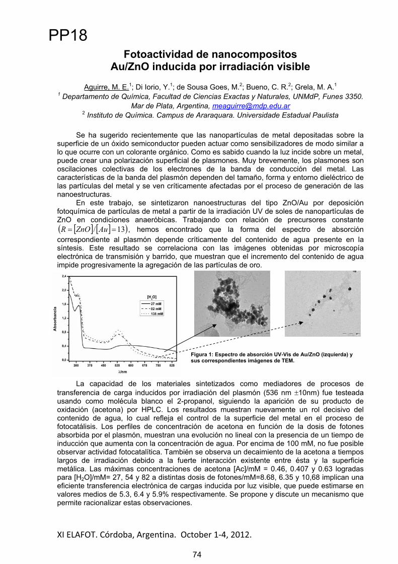
XI ELAFOT. Córdoba, Argentina. October 1 4, 2012. 74
Fotoactividad de nanocompositos Au/ZnO inducida por irradiación visible
Aguirre, M. E.1; Di Iorio, Y.1; de Sousa Goes, M.2; Bueno, C. R.2; Grela, M. A.11 Departamento de Química, Facultad de Ciencias Exactas y Naturales, UNMdP, Funes 3350.
Mar de Plata, Argentina, [email protected] Instituto de Química. Campus de Araraquara. Universidade Estadual Paulista
Se ha sugerido recientemente que las nanopartículas de metal depositadas sobre la superficie de un óxido semiconductor pueden actuar como sensibilizadores de modo similar a lo que ocurre con un colorante orgánico. Como es sabido cuando la luz incide sobre un metal, puede crear una polarización superficial de plasmones. Muy brevemente, los plasmones son oscilaciones colectivas de los electrones de la banda de conducción del metal. Las características de la banda del plasmón dependen del tamaño, forma y entorno dieléctrico de las partículas del metal y se ven críticamente afectadas por el proceso de generación de las nanoestructuras.
En este trabajo, se sintetizaron nanoestructuras del tipo ZnO/Au por deposición fotoquímica de partículas de metal a partir de la irradiación UV de soles de nanopartículas de ZnO en condiciones anaeróbicas. Trabajando con relación de precursores constante
13/ AuZnOR , hemos encontrado que la forma del espectro de absorción correspondiente al plasmón depende críticamente del contenido de agua presente en la síntesis. Este resultado se correlaciona con las imágenes obtenidas por microscopía electrónica de transmisión y barrido, que muestran que el incremento del contenido de agua impide progresivamente la agregación de las partículas de oro.
La capacidad de los materiales sintetizados como mediadores de procesos de transferencia de carga inducidos por irradiación del plasmón (536 nm 10nm) fue testeada usando como molécula blanco el 2-propanol, siguiendo la aparición de su producto de oxidación (acetona) por HPLC. Los resultados muestran nuevamente un rol decisivo del contenido de agua, lo cual refleja el control de la superficie del metal en el proceso de fotocatálisis. Los perfiles de concentración de acetona en función de la dosis de fotones absorbida por el plasmón, muestran una evolución no lineal con la presencia de un tiempo de inducción que aumenta con la concentración de agua. Por encima de 100 mM, no fue posible observar actividad fotocatalítica. También se observa un decaimiento de la acetona a tiempos largos de irradiación debido a la fuerte interacción existente entre ésta y la superficie metálica. Las máximas concentraciones de acetona [Ac]/mM = 0.46, 0.407 y 0.63 logradas para [H2O]/mM= 27, 54 y 82 a distintas dosis de fotones/mM=8.68, 6.35 y 10,68 implican una eficiente transferencia electrónica de cargas inducida por luz visible, que puede estimarse en valores medios de 5.3, 6.4 y 5.9% respectivamente. Se propone y discute un mecanismo que permite racionalizar estas observaciones.
Figura 1: Espectro de absorción UV-Vis de Au/ZnO (izquierda) y sus correspondientes imágenes de TEM.
PP18
74

XI ELAFOT. Córdoba, Argentina. October 1 4, 2012. 75
Photodynamic effect of tetrasubstituted zinc phthalocyanines on macrophages infected with Leishmania
amazonensis and Leishmania braziliensisSilva, E. P. O1; Ferreira, V. T. P¹, Cardoso, M. A. G¹, Mittmann, J¹,
Beltrame Jr, M¹1 Instituto de Pesquisa e Desenvolvimento, Universidade do Vale do Paraíba, 2911
Shishima Hifumi Avenue, São José dos Campos, SP, Brazil, 12244-000, [email protected]
Cutaneous leishmaniasis (CL) is an infectious disease caused by genus Leishmania protozoa, occurring in 88 countries, being that 90% of cases have occurred in just six countries: Iran, Syria, Saudi Arabia, Afghanistan, Peru and Brazil [1]. Even though, there are various anti-parasitic drugs available, the incidence rate has not decreased. Leishmania has been acquiring a high resistance level against the usually prescribed drugs, therefore research of alternative treatment methods are necessary, such as photodynamic therapy (PDT) [2]. The phtoactivity of the zinc phthalocyanines (PcZns): Zinc(II)-phthalocyanine (1), Zinc(II)-tetranitro-phthalocyanine (2), Zinc(II)-tetramino-phthalocyanine (3) and Zinc(II)-tetrasulfonate-phthalocyanine (4) were investigated on macrophage cell line (J774A1, ATCC) infected with L. amazonensis or L. braziliensis. A murine macrophages cell line J774 and J774-infected were incubated with PcZns at different concentrations (1-10μM) for 3 hours and then subjected with LED irradiation at 660nm and fluency of 50J.cm-2 (25mV) in a continuous wave mode. The cellular viability was determined by MTT assay 20 hours after PDT. The results demonstrated no cytotoxicity at 1μM in the dark for all PcZns tested. However, concentrations up to 1μM, only PcZn 1 has shown cytotoxicity in the dark in J774 infected with L. braziliensis. After LED treatment no changes in the cellular viability from control were observed. Meanwhile, there was a significant decrease in the viability after 20 hours post-irradiation with all PcZns to all concentrations, whereas, the concentration which showed the best activity was 10μM. At this concentration, the cell viability of uninfected J774 decreased to 37.7% (1), 45.7% (2), 1.7% (3)and 0.2% (4) for J774 cells only. For cells infected with L. amazonensis the viability decrease were 35.9% (1), 51.9% (2), 30.44% (3) and 2.9% (4), and for cells infected with L. braziliensisthe viabilities were 77.8% (1), 44.4% (2), 6.2% (3) and 1.6% (4). The photoactivity compared between PcZns showed decrease activity in the following order: 4 > 3 > 1> 2; which means that 4 has the strongest PDT activity and 2 has the weakest one. The reasons to photoactivity potency differences between PcZns are as followed [3-5]: (a) the tetra-substituents polarity, since the 4 has the highest polarity and the best water-solubility; (b) the PcZns intracellular distribution; (c) the immunomodulation caused by either Leishmania infection or PDT. The PDT with all PcZns promoted significant reduction in cell viability. Although the PcZns-PDT provide promising results, further studies are needed to understand its mechanism of action.
Acknowledgements: This work was sponsored by the Brazilian funding agency FAPESP. The authors also wish to thank Juliana Ferreira (Lab. Terapia Fotodinâmica – IP&D/UNIVAP) for the LED equipment used.
References[1] Akilov, O. E. et al. Exp Dermatol, 16 (8): 651, 2007. [2] Peloi, L. S. et al. Exp Parasitol, 128 (4): 353, 2011. [3] Durmu , M. et al. Dyes Pigment, 91: 163, 2011. [4] Rosenkranz, A. A. et al. Immunol Cell Biol, 78: 452, 2000. [5] Akilov O. E, Kosake S, Hasan T. Proc SPIE, 7380:73803G, 2009.
PP19
75

XI ELAFOT. Córdoba, Argentina. October 1 4, 2012. 76
Study of energy transfer process in solution and in the solid state using derivative polyfluorene-blue emitter as host and
polyfluorene-polythiophene-green emitter copolymer as guest
Quites, F. J. and Atvars, T.D.Z.
Instituto de Química, Unicamp, Caixa Postal (P.O. Box) 6154, Campinas, 13083-862, SP, Brasil (Brazil), [email protected]
The energy transfer in solutions and in films containing poly[(9,9-dioctylfluorenyl-2,7-diyl)-alt-co-(9,9-di-{5’-pentanyl}-fluorenyl-2,7-diyl)] (PFP) as a host and the poly[(9,9-dihexylfluorenyl-2,7-diyl)-alt-co-(bithiophene)] (F8T2) as guest was studied. The relative weight ratio of F8T2 was changed from 0.075 to 75 wt%. The photoluminescence of the PFP emission in solution and in films is centered at 420-470 nm and 425-480 nm, respectively and of the F8T2 copolymer is in the range of 448-473 nm in solution and in 440-481 nm film. There is a strong overlap between PFP blue emission and the F8T2 absorption. When the amount of F8T2 increases in the solution, there is a resonant energy transfer process which may be described by the Stern-Volmer model, with a Stern-Volmer constant Ksv = 144.440 mol-1 L. The Förster ratio was also evaluated as 4.2 nm and the energy transfer constant is 1.99 x 10⁹
s. The efficiency of the Förster resonant energy transfer process (FRET) was determined in solid state using the lifetime decays of donor in the absence ( 0) and the presence of F8T2 acceptor ( ) and it increases from the 21-27 % in solution to 27-52 % in the solid state, depending on the concentration. The higher value find out the solid state can be attributed the closer contact between the PFP donor ad F8T2 acceptor. Nevertheless, due to the phase separation process, FRET is not complete for higher concentrated blends. The photophysical data was also analyzed together the morphology using scanning electron microscopy and laser confocal fluorescence microscopy. In the solid state depending of the amount of F8T2 the energy transfer process is inefficient: for the blend with 0.075 wt% of F8T2 the PL spectrum presented higher intensity of emission both PFP and F8T2 (covering almost the full visible spectrum). The blends produced in this work can to present interesting characteristics to use in the with-light-emitting-diodes (WLEDs). Studies are progressing to employ these materials in WLEDs.
Acknowledgements: To FAPESP, INEO and CNPq for the financial support and fellowships.
PP20
76

XI ELAFOT. Córdoba, Argentina. October 1 4, 2012. 77
The study on the interaction between bovine serum albumin and tetrasubstituted zinc phthalocyanines
Silva, E. P. O1; Paiva, L. P¹, Simioni, A. R¹, Beltrame Jr, M¹1 Instituto de Pesquisa e Desenvolvimento, Universidade do Vale do Paraíba, 2911
Shishima Hifumi Avenue, São José dos Campos, SP, Brazil, 12244-000, [email protected]
Phthalocyanines (Pcs) are molecules with strong absorption in the red light spectrum (670-780nm), have been shown as a potential agent in the treatment of carcinogenic photosensitive processes [1-2]. However, due to their high insolubility in most common solvents, changes in their molecular structure or the use of delivery systems, such as liposomes, polymeric micelles and serum proteins are required [3-4]. Serum albumin is the most abundant plasma protein (about 0.63mM) and binds reversibly to a large range of endogenous and exogenous compounds, such as fatty acids, drugs and metal ions in the bloodstream [2]. The drug-protein interaction not only affects the absorption, distribution, metabolism and excretion properties, but also interferes with drug stability and toxicity during chemotherapy [2]. In this study, the interaction of zinc phthalocyanines (PcZns): Zinc(II)-phthalocyanine (1), Zinc(II)-tetranitro-phthalocyanine (2), Zinc(II)-tetramino-phthalocyanine (3) to bovine serum albumin (BSA) has been studied for UV-absorption in combination with fluorescence quenching study. The Stern-Volmer quenching constant (Ksv), the quenching rate constant of biomolecular reaction (Kq),the biding constant (K) and the number of binding sites (n) of ZnPcs and BSA were evaluated. Results are showed in Table 1.
Table 1: Stern-Volmer quenching constants and binding parameters of Pc-BSA at 296K, pH 7.4
System Ksv (104 M-1) Kq (1012 M-1 s-1) K (104 M-1) n1-BSA 0.76 0.76 8.10 1.76
2-BSA 1.43 1.43 8.57 1.76
3-BSA 1.17 1.17 8.35 1.75
These results suggest that all PcZns studied quenches the fluorescence intensity of BSA mainly through a static quenching mechanism. The binding constants of the interaction between PcZns and BSA decreased in following order: 2 > 3 > 1 at 296K, which means that 2has the strongest ability to bind with BSA and 1 the weakest ability. This results is believed to be related the high volume of nitro grups present in 2, promoted the biding process of 2-BSA [2]. The comparison of the binding parameters of the interaction between PcZns and BSA indicated that substitution by nitro grups in benzene ring cloud enhance the binding affinity. The experimental results indicate that all phthalocyanines could bind to BSA and may be used as a delivery system for PDT.
Acknowledgements: The authors wish to thank Deborah Dibbern Brunelli (FAPESP grant No. 2000/03186-8) (Chemistry Departament – ITA) for the fluorescence analysis.
References[1] Machado, A. H. A. et al. Photomed Laser Surg, 28(S1): 143, 2010.[2] Yu, X et al. Spectrochim Acta A: Mol Biomol Spectrosc, 78: 1335, 2011. [3] Jian-Dong, H at al. J Inorg Biochem, 100: 951, 2006. [4] He, W et al. Bioorg Med Chem, 13: 1845, 2005.
PP21
77

XI ELAFOT. Córdoba, Argentina. October 1 4, 2012. 78
Application of coordination compounds [Zinc(II)(salicylideneminate)]in OLEDs
Germino, J. C.1, Atvars, T. D. Z.1, Santana, A. M.2,Ramos, J. R.2,Fonseca, F. J.3, dos Santos, G.3, Pereira, N. M. de S.2
1Universidade Estadual de Campinas, Instituto de Química, Campinas, SP, Brasil, E-mail: [email protected]
2Universidade Federal de Mato Grosso, Instituto de Física, Cuiabá, MT, Brasil 3Universidade Estadual de São Paulo, Escola Politécnica, São Paulo,SP, Brasil
The Schiff condensation occurs by a classic reaction, where a primary amine performs a nucleophilic attack on carbonyl group of an aldehyde or ketone, resulting in imine group (C=N)[1]. Recent researches using Zn(II) salicylideneminates in electronic devices have shown interesting results in luminescence, electroluminescence and semiconducting properties, allowing the use of these materials in the manufacture of Organic Light-Emitting Diodes (OLEDs). These devices are based on fluorescent materials, as electroluminescent layer, electron injector layer or hole injector layer [2-3]. Some results from the literature showed that these properties are enhanced when these Zn(II) coordination compounds are added to luminescent semiconducting copolymers, resulting in a host-guest system [3-4]. News alicylidenes, as well as their Zn(II) coordination compounds, were successfully synthesized, with high purity and good yields (75-95%).These new salicylidenes and theirZn(II) coordination compounds were characterize by NMR 1H and 13C, FT-IR, melting point, thermal analysis (TGA/DTA), UV-Vis,steady state and time-resolved fluorescence. Electroluminescent devices composed by ITO/PEDOT:PSS/PVK-Zn(II) compounds or ligands/Alwas preliminary studied by recording the electroluminescence signal as well as their luminance and current/voltage curves. Finally, it was obtained best results from the Zn(II) coordination compounds with respect to the free ligands.
Acknowledgements:Authors acknowledge INEO, FAPESP and CNPqfor financial supports and fellowships.
References
[1] F. Carey and R. J. Sundberg. Advanced Organic Chemistry - Part A: Structure and Mechanism, Fifth Edtion, Springer, Nova York, 646-649 (2007).
[2] M. G. Kaplunov, I. K. Yakushchenko, S. S. Krasnikova, A. P. Pivovarov and I. O. Babashova.Elect.and Photo. Devices, 42, 563-565 (2007);
[3] S. S. Krasnikova, I. K. Yakushchenko, S. N, Shamaev and M. G. Kaplunov. Mol. Cryst. Liq. Cryst., 468, 439-445 (2007);
[4] R. C. Evans, P. Douglas, J. C. Winscon. Coord.Chem. Rev., 250, 2093-2126 (2006).
PP22
78
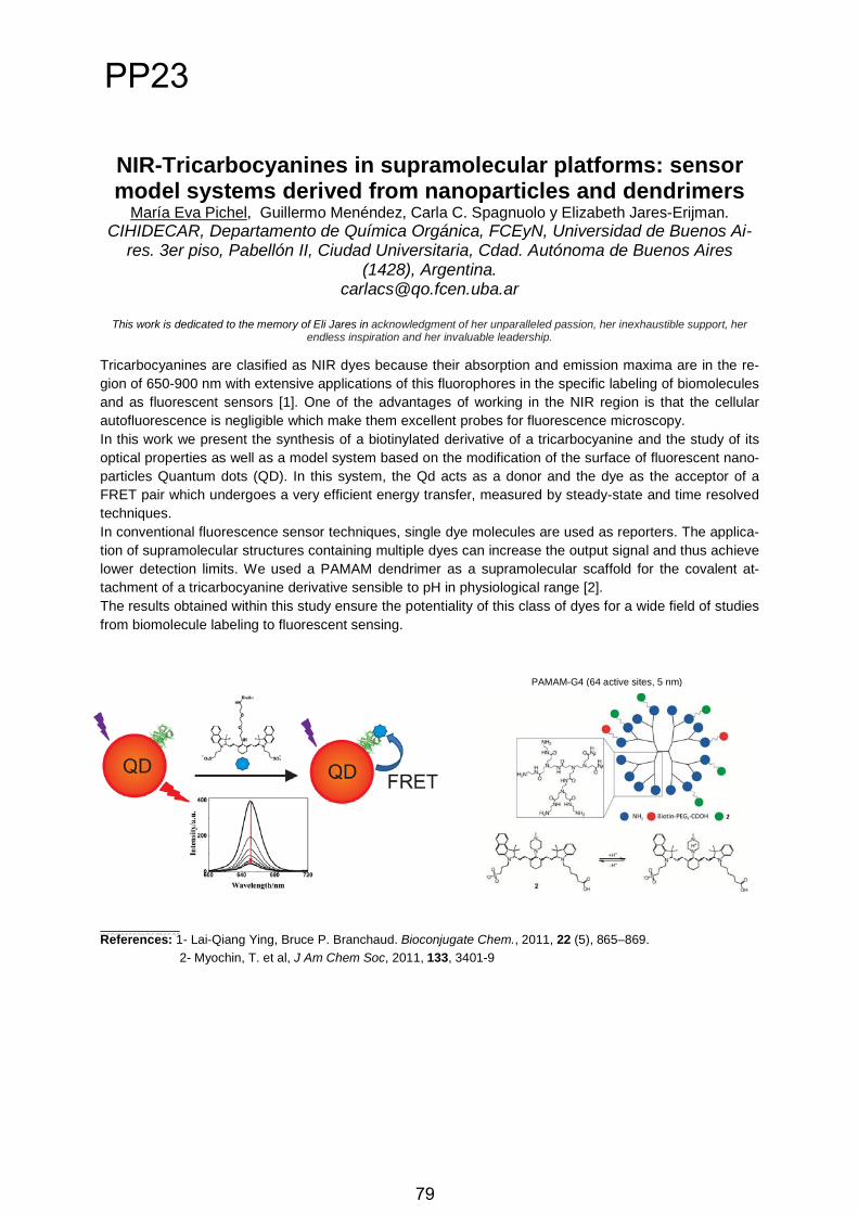
NIR-Tricarbocyanines in supramolecular platforms: sensor model systems derived from nanoparticles and dendrimers�
María Eva Pichel, Guillermo Menéndez, Carla C. Spagnuolo y Elizabeth Jares-Erijman.�CIHIDECAR, Departamento de Química Orgánica, FCEyN, Universidad de Buenos Ai-
res. 3er piso, Pabellón II, Ciudad Universitaria, Cdad. Autónoma de Buenos Aires (1428), Argentina.
This work is dedicated to the memory of Eli Jares in acknowledgment of her unparalleled passion, her inexhaustible support, her endless inspiration and her invaluable leadership.
Tricarbocyanines are clasified as NIR dyes because their absorption and emission maxima are in the re-gion of 650-900 nm with extensive applications of this fluorophores in the specific labeling of biomolecules and as fluorescent sensors [1]. One of the advantages of working in the NIR region is that the cellular autofluorescence is negligible which make them excellent probes for fluorescence microscopy. In this work we present the synthesis of a biotinylated derivative of a tricarbocyanine and the study of its optical properties as well as a model system based on the modification of the surface of fluorescent nano-particles Quantum dots (QD). In this system, the Qd acts as a donor and the dye as the acceptor of a FRET pair which undergoes a very efficient energy transfer, measured by steady-state and time resolvedtechniques. In conventional fluorescence sensor techniques, single dye molecules are used as reporters. The applica-tion of supramolecular structures containing multiple dyes can increase the output signal and thus achieve lower detection limits. We used a PAMAM dendrimer as a supramolecular scaffold for the covalent at-tachment of a tricarbocyanine derivative sensible to pH in physiological range [2]. The results obtained within this study ensure the potentiality of this class of dyes for a wide field of studies from biomolecule labeling to fluorescent sensing.
� �References: 1- Lai-Qiang Ying, Bruce P. Branchaud. Bioconjugate Chem., 2011, 22 (5), 865–869. 2- Myochin, T. et al, J Am Chem Soc, 2011, 133, 3401-9
PAMAM-G4 (64 active sites, 5 nm)
79
PP23
79

XI ELAFOT. Córdoba, Argentina. October 1-4, 2012.
STUDY OF THE DYE PENETRATION FOR TOPICAL FORMULATIONS
Tosato, Maira1; Orallo, Dalila2; Churio, Sandra3; Martin, Airton A.4; Dicelio, Lelia5
1,5Universidad de Buenos Aires- INQUIMAE, Ciudad Universitaria, 1428- Buenos Aires- Argentina, [email protected], [email protected]
2,3Universidad Nacional de Mar del Plata- Facultad de Ciencias Exactas, Dean Funes 3350 - Mar del Plata- Argentina, [email protected]
4Universidade do Vale do Paraíba- Laboratório de Espectroscopia Vibracional Biomédica, Av Shishima Hifumi, 2211- Sao José dos Campos-Sao Paulo- Brasil,
Mycosporine like amino acids (MAA) were isolated from a marine red alga and were identified as porphyra-334 and shinorine. MAA are photoprotector molecules and antioxidants [1], suggesting potential application in prevention and therapeutic treatment of diseases related to the production of free radicals and UV irradiation of the skin which acts as a natural barrier to topical products. We investigated the effects of polymers gels (Poloxamer 407 and Pluronic ® F-127) as in vitro delivery systems [2] of MAA in pig skin. These triblock copolymers are biocompatible, have low toxicity and may be used as carriers for medical and pharmaceutical drug delivery [3-5].
The polymers gels were prepared according to the method described by Schmolka [6]. Appropriate amounts of PL-127 and PO 407 to yield 20, 27 and 30% (w/w) gels were slowly added to cold Milli-Q water (4 - 5º C) with constant stirring during 40 min. Dispersions were left overnight to ensure complete dissolution and clear solutions formed. Rheological measurements dependent on the temperature of the gels were carried out to determine the viscosity at different shear rates.
In vitro release studies of the MAA were carried out using pig skin by confocal Raman spectroscopy. Raman spectra were obtained by with a 785 nm laser excitation in different depths for the low (900-1800 cm-1) frequencies showing alterations in lipids and protein peaks. The skin was used without the topical formulations as a reference.
References
[1] Coba, F.; Aguilera, J.; Herrera, E. et al. Antioxidant activity of mycosporine like amino acids isolated from three red macroalgae and one marine lichen. J Appl Phycol 21, 167-169, 2009.[2] Zatz, J. L. Skin Permeation Fundamentals and Applications 1st ed. United States: Allured Publishing corp, 1993.[3] Johnson, T.P; Miller, S. Toxicologica evaluation of poloxamer vehicles for intramuscular uses, Parenter Drug Assoc 39: 83-88, 1985. [4] Moreira, T.S.; Pierre, M.B. et al. Influence of oleic acid on the rheology and in vitro release of Lumiracoxib from poloxamer gels. J. Pharm Pharmaceut Sci 13(2), 286-302, 2010 [5] Antunes, F.E; Ranieri,G.A. et al. Gels of Pluronic F127 and nonionic surfactants from rheological characterization to controlled drug permeation [6] Schmolka IR. Artificial skin. I. Preparation and properties of pluronic F-127 gels for treatment of burns. J Biomed Mater Res 6: 571–582, 1972.
80
PP24
80
XI ELAFOT. Córdoba, Argentina. October 1 4, 2012. 81
Fotogeneración e Inhibición de Especies Reactivas de Oxígeno por Vitamina B2 y Serotonina. Un estudio cinético.
Blasich, Néstor1; Gutiérrez, Lihuel1, Vázquez, Gabriela1; Haggi, Ernesto1;Criado, Susana2; Miskoski, Sandra2; Ferrari, Gabriela3; Montaña,
Paulina3; García, Norman2
1UARG. Univ. Nac. Pat. Aust., 9000 R. Gallegos, Argentina. [email protected] 2 Dto. De Qca. Univ. de Nac. Río Cuarto, 5800 Río Cuarto, Argentina
3Area de Qca. Fca.-INQUISAL, Univ. Nac. de San Luis, 5700 San Luis, Argentina
Algunos fotoprocesos provocados por sensibilizadores endógenos como la vitamina B2(Riboflavina, Rf), cuando suceden sobre sustratos de relevancia biológica, incrementan el daño oxidativo a nivel subcelular, en mamíferos y particularmente en seres humanos. Estos procesos son provocados por especies reactivas de oxígeno (ROS), y su estudio resulta de sumo interés en tres aspectos: a) La propia posibilidad de fotogeneración de ROS; b) La actividad antioxidante de sustratos biológicos relevantes y c) El daño resultante debido a la eventual generación de ROS.
En el caso que nos ocupa, el sustrato de interés biológico escogido fue la Serotonina (Sero), sintetizada por ciertas neuronas a partir de triptófano y de gran importancia funcional en el organismo humano. El cerebro la utiliza para fabricar una conocida hormona: la melatonina.[1]
Se abordó el estudio de la fotoestabilidad de Sero y Rf y el posible rol como generadores o desactivadores de ROS de Sero y compuestos relacionados, mediante una investigación cinético-mecanística.
Se trabajó a pH 7, bajo condiciones donde solamente Rf absorbe luz. Para modelar estructuralmente al neurotransmisor se emplearon Triptamina (Trpa) y 5 Hidroxi Indol (OH In) (los tres compuestos denominados Der-IN en lo sucesivo). La irradiación con luz visible de Der-IN + Rf induce procesos degradativos en Der-In y en muy menor grado en la propia Rf. Se postulan dos vías mecanísticas: la principal, es una inhibición de 3Rf* mediada por transferencia de electrones desde Der-In. Esto produce Rf / RfH y el catión radicalario Der-In. Estas especies reaccionarían formando fenóxidos y radicales alfa-amino. Posteriores reacciones comprenderían la formación del ión superóxido y radical OH. El segundo mecanismo ocurre por transferencia de energía desde 3Rf* al oxígeno disuelto que genera oxígeno molecular singlete (O2(1
g)), que a su vez interactúa con los Der-IN. Los resultados obtenidos sugieren que la degradación de Sero, sensibilizada con Rf,
ocurre vía ROS y por procesos mediados por radicales no oxigenados. Los Der-In estudiados, desactivan al O2(1
g) de modo predominantemente físico, lo que constituye una propiedad deseable de cualquier antioxidante, dado que por este mecanismo de protección prácticamente no se degrada/elimina al desactivante. Sero ejerce fotoprotección sobre el triptófano –tomado como típico blanco oxidable de relevancia biológica– a través de la desactivación combinada de O2(1
g) y 3Rf*. Esta última especie es la responsable de la generación de ROS.
Agradecimientos: Al Consejo Nacional de Investigaciones Científicas y Técnicas, y a las Secretarías de Ciencia y Técnica de las UNPA, UNRC y UNSL, todos de Argentina, por el apoyo recibido.
Referencias[1] P. M . Whitaker-Azmitia, Handbook of Behavioral Neuroscience, Volume 21, 2010.
PP25
81

XI ELAFOT. Córdoba, Argentina. October 1 4, 2012. 82
Inactivación fotodinámica de Trypanosoma cruzi por el tratamiento con una aminoporfirina
Martinez, M. del Carmen1; Randazzo, Leticia1; Mora, S. Jimena2; Álvarez, M. Gabriela2; Milanesio, M. Elisa2; Durantini, Edgardo N.2, Fukuda, Haydeé1,3, Batlle Alcira3, Lombardo, Elisa1,3.
1 Departamento de Química Biológica, FCEyN, Universidad de Buenos Aires, CABA, Argentina.2Departamento de Química, FCEFQYN, Universidad Nacional de Río Cuarto, Río Cuarto, Córdoba, Argentina. 3 Centro de Investigaciones sobre Porfirinas y Porfirias, CIPYP (UBA-CONICET), CABA, Argentina.
Los fotosensibilizadores derivados de porfirinas ejercen un importante efecto citotóxico mediado por la generación de especies reactivas de oxígeno, las cuales se generan por la irradiación con luz visible. En general, el oxígeno molecular singlete, O2(1
g), es una de las principales especies reactivas que producen la pérdida de funcionalidad celular. Sabiendo que concentraciones altas de hemina ejercen un efecto inhibitorio sobre el crecimiento deTrypanosoma cruzi, en este trabajo evaluamos el efecto in vitro de una porfirina sintética, 5,10,15,20-tetrakis[4-(3-N,N-dimetilaminopropoxi)fenil]porfirina (A4) sobre el parásito en los estadios epi- y tripomastigote. La porfirina A4 fue seleccionada debido a que contiene cuatro grupos aminos separados del macrociclo tetrapirrólico por una cadena alifática, la cual le confiere mayor movilidad para interaccionar con la membrana celular.
Los espectros de absorción UV-visible y de emisión de fluorescencia en N,N-dimetilformamida muestran las bandas típicas de las porfirinas base libre en estado monomérico. En este medio, el rendimiento cuántico de producción de O2(1
g) es de 0,74. Los estudios in vitro mostraron para epimastigotes un valor de IC50 de 10,04 ± 0,50 μM, similar al del benznidazol tomado como droga de referencia (7,40 ± 0,45 μM). Sobre tripomastigotes el efecto de la porfirina se evaluó con y sin una irradiación de 15 minutos luego del agregado de la droga. Los parásitos no irradiados mostraron un IC50 de 11,64± 0,48 μM mientras que luego de la irradiación, este valor resultó ser menor de 11,6 nM. Establecida la efectividad de A4 como potente agente tripanomicida, pensamos en su utilización en los bancos de sangre para tratar la sangre contaminada, minimizando así la propagación de la enfermedad de Chagas por transfusiones.
Luego de establecer un método sensible y eficiente para la cuantificación de A4 por espectrofotometría de fluorescencia, investigamos la estabilidad de esta porfirina en distintos medios, pH’s y los tiempos de exposición a la luz. Observamos que luego del tratamiento de la sangre entera con A4 e irradiación, los glóbulos rojos no se dañan y ni la porfirina ni ningún tipo de metabolito fluorescente derivado de ella, fueron detectados en el plasma ys glóbulos rojos. Descartada la interacción de A4 con la albúmina, que podría enmascarar su fluorescencia, postulamos que la porfirina posiblemente sea fagocitada por macrófagos después de ejercer su acción tripanocida. La citotoxicidad de A4 fue ensayada sobre células Vero (células epiteliales de riñón de mono) y arrojó para la concentración 50% citotóxica (CC50) valores de 267,4 μM y 22,1 μM para células tratadas en oscuridad o irradiadas durante 15 minutos, respectivamente. Este estudio nos permitiría postular a la porfirina A4 como un interesante candidato, con acción antiparasitaria, para tratar la sangre contaminada con Trypanosoma cruzi.
PP26
82
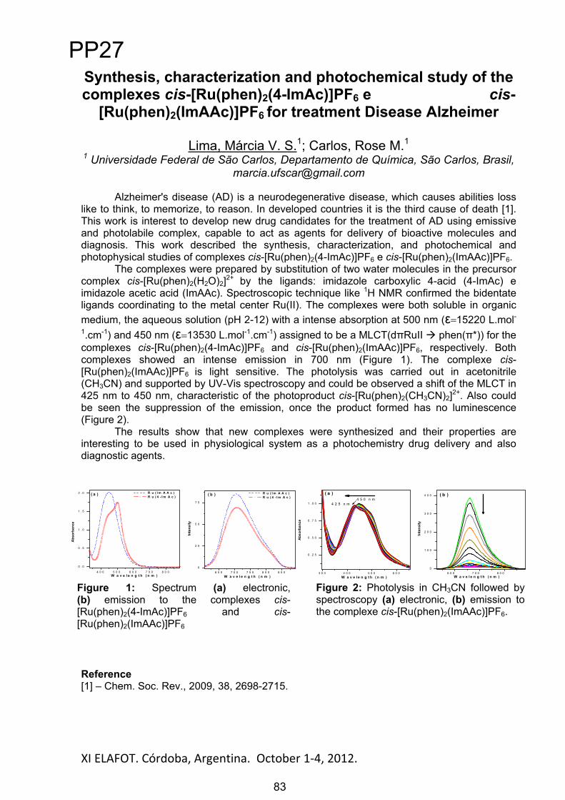
XI ELAFOT. Córdoba, Argentina. October 1 4, 2012. 83
Synthesis, characterization and photochemical study of the complexes cis-[Ru(phen)2(4-ImAc)]PF6 e cis-
[Ru(phen)2(ImAAc)]PF6 for treatment Disease Alzheimer
Lima, Márcia V. S.1; Carlos, Rose M.11 Universidade Federal de São Carlos, Departamento de Química, São Carlos, Brasil,
Alzheimer's disease (AD) is a neurodegenerative disease, which causes abilities loss like to think, to memorize, to reason. In developed countries it is the third cause of death [1]. This work is interest to develop new drug candidates for the treatment of AD using emissive and photolabile complex, capable to act as agents for delivery of bioactive molecules and diagnosis. This work described the synthesis, characterization, and photochemical and photophysical studies of complexes cis-[Ru(phen)2(4-ImAc)]PF6 e cis-[Ru(phen)2(ImAAc)]PF6.
The complexes were prepared by substitution of two water molecules in the precursor complex cis-[Ru(phen)2(H2O)2]2+ by the ligands: imidazole carboxylic 4-acid (4-ImAc) e imidazole acetic acid (ImAAc). Spectroscopic technique like 1H NMR confirmed the bidentate ligands coordinating to the metal center Ru(II). The complexes were both soluble in organic medium, the aqueous solution (pH 2-12) with a intense absorption at 500 nm ( 15220 L.mol-1.cm-1) and 450 nm ( 13530 L.mol-1.cm-1) assigned to be a MLCT(d RuII phen( *)) for the complexes cis-[Ru(phen)2(4-ImAc)]PF6 and cis-[Ru(phen)2(ImAAc)]PF6, respectively. Both complexes showed an intense emission in 700 nm (Figure 1). The complexe cis-[Ru(phen)2(ImAAc)]PF6 is light sensitive. The photolysis was carried out in acetonitrile (CH3CN) and supported by UV-Vis spectroscopy and could be observed a shift of the MLCT in 425 nm to 450 nm, characteristic of the photoproduct cis-[Ru(phen)2(CH3CN)2]2+. Also could be seen the suppression of the emission, once the product formed has no luminescence (Figure 2).
The results show that new complexes were synthesized and their properties are interesting to be used in physiological system as a photochemistry drug delivery and also diagnostic agents.
Reference[1] – Chem. Soc. Rev., 2009, 38, 2698-2715.
Figure 2: Photolysis in CH3CN followed by spectroscopy (a) electronic, (b) emission to the complexe cis-[Ru(phen)2(ImAAc)]PF6.
Figure 1: Spectrum (a) electronic, (b) emission to the complexes cis-[Ru(phen)2(4-ImAc)]PF6 and cis-[Ru(phen)2(ImAAc)]PF6
3 0 0 4 0 0 5 0 0 6 0 0
0 . 2 5
0 . 5 0
0 . 7 5
1 . 0 0
Abs
orba
nce
W a v e l e n g t h ( n m )
( a )
4 2 5 n m4 5 0 n m
W a v e l e n g t h ( n m )6 0 0 7 0 0 8 0 0
0
1 0 0
2 0 0
3 0 0
4 0 0 ( b )
Inte
nsity
4 0 0 5 0 0 6 0 0 7 0 0 8 0 0
0 . 0
0 . 5
1 . 0
1 . 5
2 . 0
6 5 0 7 0 0 7 5 0 8 0 0 8 5 0
0
2 5
5 0
7 5
( b )( a )
Abs
orba
nce
W a v e l e n g t h ( n m )
R u ( I m A A c )R u ( 4 - I m A c )
Inte
nsity
W a v e l e n g t h ( n m )
R u ( I m A A c )R u ( 4 - I m A c )
PP27
83

XI ELAFOT. Córdoba, Argentina. October 1 4, 2012. 84
Photoinduced electron transfer reaction of Mn (I) complexes Marchi, R. C.1*; De Aguiar, I.1; Carlos, R. M.1
1 Departamento de Química, Universidade Federal de São Carlos, São Carlos, Brasil, * [email protected]
Amino acids (aa) for aromatic active redox reactions such as tryptophan and tyrosine play essential roles in the reactions of electron transfer (ET) in biological medium acting as intermediaries between redox metalloproteins, which occur at distances from 3.5 to 10 Å. Therefore, developing a molecular system able to simulate these reactions has implications for both fundamental (understanding the biological system) and practical (fuel production, transport and storage of energy using water and sunlight as an energy source). The strategy we have adopted to investigate these photochemical reactions is to produce the amino acid radical (Y•-) with a reducing agent (RA) in the excited states and monitor kinetically the reactivity of the radical (Y•-) produced by spectroscopic techniques following the reaction:
{RA-Y)} { RA+-Y•-)}.
In this work we choose a series of manganese complexes to act as reducing agent to Y. The UV-vis electronic absorption spectrum of fac-[Mn(CO)3(Phen-OPh)(L)]+, L = imidazole, bromide, triflate show absorption in the UV region (260 and 305 nm) and a broad band in the region 350-460 nm attributed to transitions LLCT (phen, phen, *) and MLCT (Mn3dt2g phen, *), respectively. The photochemical reaction of the complex fac-[Mn(CO)3(Phen-OPh)(Br)]+ was monitored by 1H NMR and UV-vis. In 1H-NMR after irradiation of a solution of the complex in CD2Cl2 observed expansion of the signals indicating the formation of the species paramagnetic {Mn2+(CO)3(Br)(phen-OPh)}* and UV-vis after irradiation of complex solution CH3CN was observed the formation the band due to MLCT indicating an effective charge transfer (Mn 3dt2g phen, *). It is expected that when the compound is fac-[Mn(CO)3 (Phen-OPh)(imadazole)]+ those observations, the formation of paramagnetic species and the formation of the band due to MLCT, are also observed, since the conditions used to complex with Br be the same complex with imidazole. The intermolecular electron transfer reaction with methyl viologen as an electron accpetor is shown in Figure 1:
Firgure 1: Formation of reduced methylviologen, MV.- (blue).
Likewise, it is anticipated that electron transfer reaction is observed when L = imidazole. Furthermore, it is expected that ET reaction can also occur in other intermolecular systems (presence of mioglobima and cytochrome C). These experiments show that the phenolic ring can act as electron acceptor in redox reactions of aa tyrosine.
Acknowledgements: Fapesp,CNPq and Capes References [1] Inorg. Chem., 2008, 47, 24, 11519-11526 [2] J. Am. Chem. Soc., 2001, 123, 3181.
400 500 600 700 800
0.0
0.3
0.6
0.9
1.2
1.5
1.8
Abs
orbâ
ncia
/ (nm)
PP28
84

XI ELAFOT. Córdoba, Argentina. October 1 4, 2012. 85
THE DEVELOPMENT AND SIMULATION OF MATRICES OF LIGHT-EMITTING DIODES AS RADIATIVE ENERGY SOURCES FOR
PHOTO-BIO-REACTORS.I. Niizawa 1,2; J. M. Heinrich1,2; I. Ferrero1; F. A. Botta1,2; A. R. Trombert1; H. A. Irazoqui1,2.1Grupo de Innovación en Ingeniería de Bioprocesos (GiiB) – Facultad de Bioquímica y Ciencias Biológicas (FBCB-UNL). Ciudad Universitaria, Santa Fe (3000), Argentina. 2Instituto de Desarrollo Tecnológico para la Industria Química (INTEC-CONICET). e-mail: [email protected]
Microalgae study has grown exponentially over the last decades. This is due to their multiple applications found in several areas of industrial interest. There is a large number of intracellular metabolites that have attracted the attention of many companies and research groups. For example, lipids for biofuels production, carotene and other pigments for use in pharmaceutical, etc[1].
The design, operation, simulation and optimization of a photo-bio-reactor (PBR) for microalgae production, requires the study of the algal growth on a smaller scale (bench or laboratory) and at pilot plant, in order to obtain accurate information about key aspects regarding the reactor configuration and the operating conditions affecting these microorganisms growth. Since microalgae are photosynthetic organisms, the photon density at each position within the volume of the algal culture is one of the most important factors that influence their growth kinetics[2].
Beer-Lambert's law predicts the energy loss experienced by a light beam while passing through an algal suspension due to absorption (by microalgal pigments) but ignoring scattering (by microalgae as particles and air bubbles), which could lead to errors in predicting light intensity profile in PBRs. The radiative energy transfer equation (RTE) includes both effects[3].However, its analytical solution is possible only in simple cases. Due to the inherent complexity of our system, which consists in an algal suspension, with a bubbling air stream as a means of supplying the culture with carbon dioxide, and also to remove oxygen from it, we chose a stochastic Monte Carlo algorithm for the computation of the local photon density, based on a physical model of the events that may occur to photons travelling through the suspension.
The objective of this work is to develop a radiant field model inside a PBR to predict the spectral photon density at each position, as a function of microalgae concentration; and to evaluate the effect of light quality (i.e. wavelength range) and intensity over microalgal growth. The model was tested in a cylindrical glass PBR, illuminated with light emitting diodes (LEDs). LEDs are a very attractive artificial light source, because of their small size, narrow emission profile; longer life; lower power consume; and low generation of heat[2].
In this work, a computational model that predicts the photon spectral density at different positions inside a microalgal culture is developed. The suspensions are modeled as pseudo-continuum media, with centers of absorption and scattering uniformly distributed.
In order to study the effect of light quality and of the spectral photon density on the synthesis of chlorophylls and on the growth of microalgae, different LED matrices were tested and simulated to assess their performance as light sources. On the basis of the information gathered, we proposed a kinetic model for algae growth that considers the effect of biomass concentration and that of the chlorophylls content on the microalgae growth rate.
References
1. Biodiesel from microalgae. Yusuf Chisti. Biotechnology Advances 25 (2007) 294–306. 2. Light requirements in microalgal photobioreactors: an overview of biophotonic aspects. Ana P. Carvalho; Susana O. Silva; José M. Baptista; F. Xavier Malcata. Appl Microbiol Biotechnol (2011) 89:1275–1288. 3. J.-F. Cornet, C. G. Dussap and J.-B. Gros. Capítulo:”Kinetics and energetics of photosynthetic microorganisms in photobioreactors”, del libro: “Bioprocess and Algae Reactor Technology, Apoptosis”. Editor: Springer Berlin / Heidelberg (1998).
PP29
85

XI ELAFOT. Córdoba, Argentina. October 1 4, 2012. 86
Synthesis, characterization and luminescent properties of new Eu-MOFs
Gomez, Germán1; Bernini, María Celeste1; Brusau, Elena1; Narda, Griselda1; Massad, Walter2
1 UNSL, FQBF, INTEQUI-Química Inorgánica. San Luis. [email protected] 2 UNRC, Fac. de Cs. Exactas, Fco-Qcas y Nat., Dpto de Química. Río Cuarto.
Traditional inorganic and organic luminescent materials have been extensively explored. Mixed oxides containing rare – earths ions i.e., are well – known for their narrow emission and high colour purity. In turn, the interest in organic luminescent materials has been mainly motivated by their applications in organic light emitting diodes (OLEDs) [1]. Metal-organic frameworks (MOFs) [2] are certainly very promising as multifunctional luminescent materials because both inorganic and organic moieties can provide the platforms to generate luminescence. Different organic linkers such as carboxylates, have been useful to construct MOFs based in lanthanides (Ln-MOFs). Recently, Ln-MOFs have received special attention due to their unusual coordination characteristics and exceptional optical and magnetic properties arising from 4f electrons [3]. In the present work, the luminescent properties of three new Eu(III) – based MOFs (Eu-MOFs) –[Eu2(C4H4O4)3(H2O)2].H2O (I),[Eu2(C6H8O4)3(H2O)2] (II) and [Eu2(C10H8O4)3(H2O)] (III)– are reported. The hybrid compounds have been synthesized under different hydrothermal conditions and characterized by single-crystal XRD, thermal analysis and FTIR. Luminescent measurements show that the three Eu-MOFs emit red light, upon irradiation with UV-Vis light source. The characteristic signals are observed in the 590-700 nm region, corresponding to the 5D0
7FJ (J=1–4) transitions. The 5D07F2 emission is a typical electric dipole transition and strongly varies with the local
symmetry of the Eu3+ ion, while the 5D07F1 one, corresponds to a parity-allowed magnetic
dipole transition, which is practically independent of the host material. Therefore, the intensity ratio (R) of 5D0
7F2 to 5D07F1 is sensitive to the symmetry around the Eu3+ ion. The R
values of 4.5 and 4.0 for Eu-MOF II and III, respectively, are higher than the value of 3 for Eu-MOF I indicating that the Eu3+ occupies a site of higher symmetry in the latter one. These values are lower than those reported by Gao et al. for Eu3+ hybrid materials [4]. The luminescence decay profiles are fitted with single exponentials and the lifetimes are in the ms range, which is in agreement with the Eu3+ room temperature luminescence decay time [4].
Acknowledgements: Authors thank to CONICET (PIP 2008-01360), SECyT-UNSL, SECyT-UNRC. References: [1] Guo, H. et al. Adv. Mater. 22, 4190, 2010. [2] Biradha, K. et al. J. Crys.Growth Des. 11, 875, 2011 [3] Cui, Y. et al. Chem.Rev. 112, 1126, 2012. [4] Guo, X. et al. J. Photochem. and Photobiol. A: Chem. 200, 318, 2008.
PP30
86
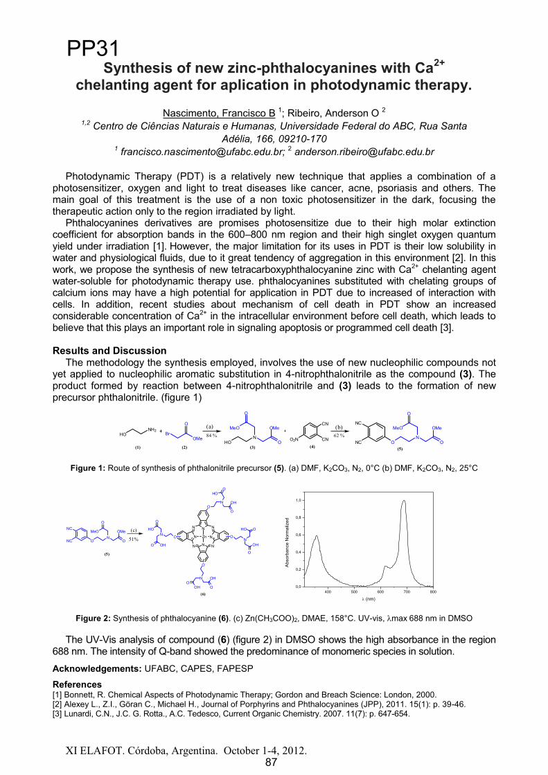
XI ELAFOT. Córdoba, Argentina. October 1-4, 2012.
Synthesis of new zinc-phthalocyanines with Ca2+
chelanting agent for aplication in photodynamic therapy.
Nascimento, Francisco B 1; Ribeiro, Anderson O 2
1,2 Centro de Ciências Naturais e Humanas, Universidade Federal do ABC, Rua Santa Adélia, 166, 09210-170
1 [email protected]; 2 [email protected]
Photodynamic Therapy (PDT) is a relatively new technique that applies a combination of a photosensitizer, oxygen and light to treat diseases like cancer, acne, psoriasis and others. The main goal of this treatment is the use of a non toxic photosensitizer in the dark, focusing the therapeutic action only to the region irradiated by light.
Phthalocyanines derivatives are promises photosensitize due to their high molar extinction coefficient for absorption bands in the 600 800 nm region and their high singlet oxygen quantum yield under irradiation [1]. However, the major limitation for its uses in PDT is their low solubility in water and physiological fluids, due to it great tendency of aggregation in this environment [2]. In this work, we propose the synthesis of new tetracarboxyphthalocyanine zinc with Ca2+ chelanting agentwater-soluble for photodynamic therapy use. phthalocyanines substituted with chelating groups of calcium ions may have a high potential for application in PDT due to increased of interaction with cells. In addition, recent studies about mechanism of cell death in PDT show an increased considerable concentration of Ca2+ in the intracellular environment before cell death, which leads to believe that this plays an important role in signaling apoptosis or programmed cell death [3].
Results and DiscussionThe methodology the synthesis employed, involves the use of new nucleophilic compounds not
yet applied to nucleophilic aromatic substitution in 4-nitrophthalonitrile as the compound (3). The product formed by reaction between 4-nitrophthalonitrile and (3) leads to the formation of new precursor phthalonitrile. (figure 1)
Figure 1: Route of synthesis of phthalonitrile precursor (5). (a) DMF, K2CO3, N2, 0°C (b) DMF, K2CO3, N2, 25°C
NC
NC ON
O
MeO
O
OMeN
NN
N
N
NN N
Zn
ON OH
O
HOO
ON
OH
O
HO O
O
N
OHO
OH
O
ONHO
O
OHO
(5)
(6)
(c)
51%
Figure 2: Synthesis of phthalocyanine (6). (c) Zn(CH3COO)2, DMAE, 158°C. UV-vis, max 688 nm in DMSO
The UV-Vis analysis of compound (6) (figure 2) in DMSO shows the high absorbance in the region 688 nm. The intensity of Q-band showed the predominance of monomeric species in solution.
Acknowledgements: UFABC, CAPES, FAPESP
References[1] Bonnett, R. Chemical Aspects of Photodynamic Therapy; Gordon and Breach Science: London, 2000.[2] Alexey L., Z.I., Göran C., Michael H., Journal of Porphyrins and Phthalocyanines (JPP), 2011. 15(1): p. 39-46.[3] Lunardi, C.N., J.C. G. Rotta., A.C. Tedesco, Current Organic Chemistry. 2007. 11(7): p. 647-654.
400 500 600 700 8000,0
0,2
0,4
0,6
0,8
1,0
Abs
orba
nce
Nor
mal
ized
(nm)
O2N
CN
CN
NC
NC ON
O
MeO
O
OMe
HON
O
MeO
O
OMe(a)HO
NH2Br
OMe
O
(1) (2) (3) (4) (5)
(b)84 % 62 %
87
PP31
87
XI ELAFOT. Córdoba, Argentina. October 1 4, 2012. 88
Terapia fotodinámica basada en ALA en células de adenocarcinoma de pulmón
Teijo, María Julieta1,2; Cassinelli, Jazmín1; Batlle, Alcira1; Fukuda, Haydée1,2
1 Centro de Investigación sobre Porfirias y Porfirinas (CIPYP) - CONICET, Av. Córdoba 2351, Buenos Aires, Argentina, [email protected]
2 Dto. de Química Biológica, Facultad de ciencias Exactas y Naturales, Universidad de Buenos Aires, Intendente Güiraldes 2160, Ciudad Universitaria, Buenos Aires,
Argentina, [email protected]
La Terapia fotodinámica (TFD) es un procedimiento terapéutico clínicamente aprobado para tumores superficiales y obstructivos, mínimamente invasivo, que ejerce una citotoxicidad selectiva hacia células malignas. Se administra un agente fotosensibilizante y se irradia con luz de longitud de onda correspondiente a la banda de absorción de dicho sensibilizante. En presencia de oxígeno, una serie de reacciones fotoquímicas llevan a la muerte de las células tumorales [1]. El ácido 5-aminolevúlico (ALA), es el precursor biológico de la síntesis de porfirinas, acumulando especialmente protoporfirina IX (PpIX), un fotosensibilizante endógeno. En la erradicación de tumores mediante TFD intervienen diferentes procesos de muerte celular [2].
El objetivo del presente trabajo fue determinar la participación de las distintas vías de muerte celular luego de aplicar TFD basada en ALA.
La TFD se llevó a cabo incubando células A549 (adenocarcinoma de pulmón, humanas) con ALA 1mM durante 3h, seguido por una irradiación en un banco de dos tubos fluorescentes. Al cabo de 1h se realizaron los ensayos de muerte celular: estudios morfológicos de microscopía convencional, de fluorescencia y electrónico; citometría de flujo y western blot.
Previamente, se determinó una localización mitocondrial para la PpIX. Asimismo, al irradiar las células en presencia de sondas fluorescentes para mitocondrias y lisosomas se pudo observar liberación de la sonda al citoplasma y disrupción de las organelas analizadas. En base a esto se sugiere que el daño mitocondrial y lisosomal es un indicador de la posible participación de la vía apoptótica intrínseca. Los análisis morfológicos y bioquímicos confirmaron características apoptóticas 1h post-TFD (condensación de cromatina, encogimiento celular, cuerpos apoptóticos) y marcación positiva de AnnexinV-FITC/ioduro de propidio para células apoptóticas (TFD 20 min: 26,8±4,1%). En las dosis de TFD más altas la población de células necróticas aumentó levemente (TFD 20 min: 8,7±1,9%). A fin de estudiar la participación lisosomal en la inducción de apoptosis las células fueron incubadas con el inhibidor de catepsina D, pepstatina A (100 m), irradiadas por 10 min y marcadas con AnnexinV-FITC/ioduro de propidio. El inhibidor no logró reducir significativamente la apoptosis (TFD 10 min: 17,3±4,2%; TFD 10 + Pepstatina A: 21,9±5,1%), sugiriendo que el daño a lisosomas es un efecto secundario y no un iniciador en la muerte celular por TFD.
La reducción de la expresión de procaspasa-3, la liberación dosis-lumínica dependiente de citocromo c y la despolarización de la membrana mitocondrial post-TFD apoyan la hipótesis de la vía apoptótica intrínseca. Sin embargo, en imágenes de microscopía electrónica, puede observarse una marcada desorganización celular y la presencia de vacuolas de doble membrana, lo cual sugiere una vía de autofagia, consistente con el daño a las organelas.
Se concluye que si bien la vía apoptótica intrínseca parece ser la predominante en la muerte celular de células A549 por TFD-ALA, debe tenerse en cuenta la participación de otros procesos simultáneos como necrosis o autofagia. Referencias: [1] Fukuda H, Casas A, Batlle A. Aminolevulinic acid: from its unique biological function to its star role in photodynamic therapy. Int J Biochem Cell Biol. 2005 Feb;37(2):272-6. [2] MacDonald & Dougherty T. Basic principles of photodynamic therapy. J Porphyrins Phthalocyanines 2001, 5:105–29
PP32
88

Estudio de degradación de Vinaza de caña de azúcarpor Fotocatálisis Heterogénea
Segovia, L.P1.; Manfredi, A.P.1,2; Araujo, P.1; Perotti, N.I.1,2
1Facultad de Ciencias Exactas y Tecnología, Universidad Nacional deTucumán, Av. Independencia 1800, [email protected]
2PROIMI - Consejo Nacional de Investigaciones Científicas y Técnicas, Av.Belgrano y Pje. Caseros, San Miguel de Tucumán
Debido al aumento en el precio del petróleo, ha habido un creciente desarrollo decombustibles alternativos y entre ellos los llamados “biocombustibles”, es el caso delbioetanol. Actualmente, en Argentina, se encuentra en vigencia la ley debiocombustibles, en donde se debe incorporar un piso de un 5% de bioetanol en lanafta [1].
Una de las fuentes renovables de producción de etanol es la caña de azúcar,siendo la vinaza el efluente principal de la destilación de etanol a partir de mostosfermentados de melazas. Por cada litro de etanol que se produce, se obtiene comoresiduo entre 10 y 12 litros de vinaza, con elevada carga contaminante.
El uso de las TAO’s (Tecnologías Avanzadas de Oxidación) como proceso detratamiento de efluentes no convencional, ha alcanzado un amplio desarrollo en elárea de la purificación de agua y se basa en procesos fisicoquímicos que producennuevas especies químicas [2]. Dentro de estas tecnologías, la FotocatálisisHeterogénea presenta un desarrollo apreciable pero su implementación en reactoresde escala industrial no ha progresado del mismo modo.
En este trabajo se presenta un estudio experimental de la degradaciónfotocatalítica de la materia orgánica presente en la vinaza mediante el uso desuspensiones de óxido de titanio en lote en presencia de luz UV-A a distintos tiempos.Se siguió el curso de la degradación a través de medidas de Demanda Química deOxígeno (DQO).
Los resultados preliminares indican una reducción de la carga orgánica de 64% enun tiempo de 12 horas con el tratamiento propuesto, evidenciándose que no se tratade una degradación térmica sino de un efecto fotocatalítico.
Referencias1 Ley 26.093 <http://www.infoleg.gov.ar/infolegInternet/anexos/115000-
9999/116299/norma.htm> [consultado 25 de Junio de 2012].2 W.H. Glaze, Environmental Science Technology, 21, 1987, 224-230.
89
PP33
89

XI ELAFOT. Córdoba, Argentina. October 1 4, 2012. 90
Spectroscopic Characterization of Origanum vulgare leaves. Development of Non-destructive Methods for Quality
Assessment
Mendes de Novo, Johanna; Iriel, Analia; Lagorio, María G.
Departamento de Química Inorgánica, Analítica y Química Física/ INQUIMAE. FCEyN, UBA. Buenos Aires, Argentina. Ciudad Universitaria, Pabellón 2, 1er piso,
E-mail: [email protected]
Origanum vulgare is a relevant herb for flavoring food and for pharmaceutical purposes used both fresh and dried. Development of non-destructive optical methodologies to infer its moisture, plant physiological state and content of active substances is of practical importance. Folowing this objective, we have studied the spectroscopic behaviour of fresh and dried leaves of this plant. UV-VIS-NIR reflectance spectroscopy, steady-state fluorescence and pulse amplitude modulated chlorophyll fluorometry (PAM) were used in this study.
Spectroscopy measurements were performed on samples with different water content ranging from 90% to 6%.
Reflectance values in the NIR were successfully correlated with the water content in origanum leaves, allowing non-destructive determination of humidity even through the plastic film packaging of commercial samples.
Fluorescence spectra presented important emissions in the blue, in the green and in the red-far red regions. The experimental ratio of fluorescence maxima blue/green (due to phenolic compounds) remained practically constant upon leaves drying. The fluorescence ratio red/far-red (due to chlorophyll-a) increased about twice, while the ratio green/far red augmented around 4 times. The variations in these ratios have been interpreted in terms of light re-absorption processes and in relation to the enhancement of emission due to fluorophore cristalization upon drying.
The photosynthetic parameters obtained from PAM measurements declined sharply with the water content.
Origanum leaves were also studied by light microscopy. Peltate trichomes and hairs have been observed at the leaf surface (Figure1). Upon excitation at 370 nm, peltate glands do not emit any fluorescence while the hairs emit blue-green fluorescence. A pronounced enhacement of the hairs emission occurs upon leaves drying. During this process a change in the hairs stucture, probably due to cristalization of fluorophores inside them, takes place.
Figure 1. Origanum vulgare leaf surface observed by light microscopy. Magnification 40 . A . Peltate trichomes, B. Hairs
The commercial product, (dried portions of leaves and flowering tops) was also characterized spectroscopically and by light microscopy.
Acknowledgements: University of Buenos Aires (Project UBACyT 20020100100814). UBA fellowship (JMN).
A
B B
A
PP34
90

XI ELAFOT. Córdoba, Argentina. October 1 4, 2012. 91
A comparative study of the photodynamic properties of two isosteric alkyl substituted phthalocyanines
Gauna G. A. 1, Marino J.2, García Vior M. C.1, Roguin L.P.2, Awruch J.1
1Departamento de Química Orgánica. Facultad de Farmacia y Bioquímica, UBA, Junín 956, 1113 Buenos Aires, Argentina.
E-mail: jawruchffyb.uba.ar 2Instituto de Química y Fisicoquímica Biológicas (UBA-CONICET), Facultad de
Farmacia y Bioquímica, Junín 956, 1113 Buenos Aires, Argentina. E-mail: [email protected]
Phthalocyanines (Pcs) are important dyes used in medicinal field as promising candidates for photodynamic therapy (PDT) in cancer treatment. It has been reported that peripheral substituents, the nature of the central metal ion as well as the axial ligand coordinated to the metal center of phthalocyanines, change their physical, chemical, and biological properties. Zinc(II) phthalocyaninates showing improved photophysical properties have been developed during the last years. In addition, attempts have been made both to increase the uptake of the dye by the targeted cells and to improve subcellular localization within the cells.. The lipophilicity of two novel isosteric zinc(II) phthalocyanines as compared with their analogs:2,9(10),16(17),23(24)-tetrakis[(2-trimethylammonium)ethoxy]phthalocyaninatozinc(II) tetraiodide (Pc11) and 2,9(10),16(17),23(24)-tetrakis[(2-trimethylammonium)ethylsulfanyl]ph-thalocyaninatozinc(II) tetraioidide Pc13 [1] was increased in order to obtain a better photodynamic behavior.Thus, 2,9(10),16(17),23(24)-tetrakis[(N-butyl-N-methylammonium)-ethylsulfanyl]phthalocyaninatozinc(II) tetraiodide (P6) and 2,9(10),16(17),23(24)-tetrakis[(N-dibutyl-N-methylammonium)ethoxy]phthalocyaninatozinc(II) tetraiodide (P7) were synthesized and their photophysical parameters and photobiological potentials were evaluated on human nasopharynx KB carcinoma cells The photobiological studies showed a better phototoxic effect for the sulfur-linked cationic phthalocyanine Pc6 (IC50= 1.45 1 M) with respect to the oxygen-linked phthalocyanine Pc7(IC50= 10.5 2 M). This result could be explained by the higher cellular uptake obtained for 6, which was mainly localized within lysosomes. After irradiation, the production of a greater amount of ROS by phthalocyanine Pc6 led to a more effective cell death. Besides, Pc6 was more photoactive in vitro than Pc13 [2], since the IC50 values obtained under similar experimental conditions were two-fold higher for Pc6 than for Pc13 (IC50 = 2.7 0.6 M).ConclusionsIt is possible to consider a structure-activity relationship of alkylthio peripheral substituted zinc(II) phthalocyanines for photobiological purposes. Therefore, further photobiological studies, as well as search for improved phthalocyanine structures, are already in progress.
Acknowledgements: Agencia de Promoción Científica y Tecnológica, Argentina. Consejo nacional de Investigaciones Científicas y Técnicas, Argentina. Universidad de Buenos Aires, Argentina.
References [1] J. Marino, M.C. Garcia Vior, L.E. Dicelio, L. P. Roguin, J. Awruch. Photodynamic effects of isosteric water-soluble phthalocyanines on human nasopharynx KB carcinoma cells. Eu. J. Med. Chem., 2010, 45, 4129-4139. [2] . G. A. Gauna, J. Marino, M. C. García Vior, L. P. Roguin, J. Awruch. Synthesis and comparative photodynamic properties of two isosteric alkyl substituted zinc(II) phthalocyanines. Eu. J. Med. Chem., 2011, 46, 5532-5539.
PP35
91
XI ELAFOT. Córdoba, Argentina. October 1 4, 2012. 92
Detección fotoacústica de amoníaco con un resonador diferencial mediante láseres de CO2 CW y TEA.
Vallespi, Arturo1; Parisi, Marián1; Peuriot, Alejandro1; Slezak, Verónica1;González, Francisco1; Pereyra, Andrea1; Santiago, Guillermo2.
1 CEILAP-CITEDEF-UNIDEF-MINDEF, San Juan Bautista de La Salle 4397 (B1603ALO) Villa Martelli (Bs. As. - Argentina), [email protected]
2 Laboratorio Láser, Facultad de Ingeniería, Universidad de Buenos Aires, Paseo Colón 850, 1063 Bs. As., Argentina, [email protected]
Siendo el amoníaco uno de los productos industriales más utilizados, su detección a nivel de trazas es importante en relación con la contaminación del medio ambiente. Con este fin, y dado que esta molécula presenta una fuerte banda de absorción en la región alrededor de 10 m, es interesante estudiar la aplicación de la técnica fotoacústica por medio de láseres de CO2 sintonizables en dicha región del IR medio y comparar los resultados en base a un láser CW (continuo) y TEA (pulsado). Para ello se pone a prueba una innovadora celda diferencial de doble resonador, construida en polipropileno, cuya frecuencia de resonancia en el segundo modo longitudinal es 1204 Hz con un micrófono diferencial acoplado a ambos resonadores. Las características propias de los micrófonos diferenciales permitirían minimizar los ruidos provenientes de fuentes lejanas, como ruidos ambientales y calentamiento local de las ventanas de la celda. El material elegido es adecuado para disminuir los efectos de adsorción debidos a la polaridad de la molécula de amoníaco. Se realizó un estudio de adsorción, registrando la señal fotoacústica a intervalos regulares, que muestra que se comete un error menor al 5% al cabo de 50 minutos, por lo que al trabajarse en régimen de flujo lento, para evitar ruidos debido a turbulencia, se pueden descartar los efectos de la adsorción en las mediciones. La evolución temporal de la muestra sigue un modelo de adsorción monocapa basado en la teoría de Langmuir. Se obtiene la curva de calibración del amoníaco para ambos sistemas, pulsado y contínuo, en régimen de flujo. Con el láser pulsado se obtiene un límite de detección de 118 ppbV para una fluencia de 278 mJ/cm2,representando este resultado una mejora respecto de trabajos previos con otros resonadores acústicos [1]. Se estudia también la mejora de la relación señal ruido en el sistema basado en el láser de CO2 CW y se comparan ambos sistemas.
Referencias
[1] CO2 LASER-BASED PULSED PHOTOACOUSTIC AMMONIA DETECTION. 16th International Conference on Photoacoustic and Photothermal Phenomena (ICPPP 16), Mérida, Mexico. 2011.
PP36
92

XI ELAFOT. Córdoba, Argentina. October 1 4, 2012. 93
The ß-Scaffold of the LOV Domain of the Brucella Light-Activated Histidine Kinase is a Key Element for Signal
Transduction
Rinaldi, Jimena1; Gallo, Mariana1; Klinke, Sebastián1; Paris, Gastón1;Bonomi, Hernán R.1; Bogomolni, Roberto A.2; Cicero, Daniel O.1;
Goldbaum, Fernando A.1
1 Fundación Instituto Leloir, IIBBA-CONICET, Buenos Aires, Argentina.2 Department of Chemistry and Biochemistry, University of California, Santa Cruz,
USA.
Light–oxygen–voltage (LOV) domains are blue-light-activated signalling modules present in a wide range of sensory proteins. LOV three-dimensional structures show the chromophore FMN non-covalently bound to the protein by hydrogen bonding and hydrophobic interactions, and the sulphur atom of a conserved Cys near the C4 carbon of FMN. Light absorption by the FMN cofactor results in formation of a covalent bond between the sulphur atom from the Cys residue and the C4 carbon from FMN. In most of the LOV domains this covalent bond breaks spontaneously in darkness completing the photocycle.
The genomes from bacteria belonging to the Brucella genus contain a gene sequence that encodes a 463 amino acid sensory box kinase containing three domains: a LOV domain at the N-terminus (the sensory domain), a histidine kinase at the C-terminus (the output domain) and a PAS domain located between the sensory and output domains (LOV-HK). This full-length bacterial photosensory protein, expressed in E. coli and affinity purified, has a molecular weight of 55 kDa, binds FMN as a chromophore, and shows a typical LOV domain absorption spectrum with broad absorption bands in the blue (450nm) and UV-A regions (370 nm).
In previous work we have shown that light modulates the virulence of the pathogenic bacterium Brucella abortus through LOV-HK. One of the striking characteristic of BrucellaLOV-HK is the fact that the protein remains activated upon light sensing, without recovering the basal state in the darkness. In contrast, the light state of the isolated LOV domain slowly returns to the dark state. To gain insight into the light activation mechanism, we have characterized by X-ray crystallography and solution NMR spectroscopy the structure of the LOV domain of LOV-HK in the dark state and explored its light-induced conformational changes. The LOV domain adopts the / PAS (PER-ARNT-SIM) domain fold and binds the FMN cofactor within a conserved pocket. The domain dimerizes through the hydrophobic -scaffold in an antiparallel way. Our results point to the -scaffold as a key element in the light activation, validating a conserved structural basis for light-to-signal propagation in LOV proteins.
Finding out the interacting partner surface of the LOV domain -scaffold in Brucella LOV-HK will reveal further details of the molecular mechanisms of light activation. To answer this question, we are trying to solve the crystallographic structure of the different domains that build up the whole LOV-HK protein. Several constructs are being studied at the moment (HK, LOV-J -PAS, LOV-J , point mutants, etc), including complex structures with the two putative response regulators of this light activated two-component system.
PP37
93

XI ELAFOT. Córdoba, Argentina. October 1 4, 2012. 94
Efecto de la Terapia Fotodinámica con Riboflavina en células de carcinoma escamoso
Juarez, Andrea V1; Boetto, Néstor1, Torres, Alicia1, Pons, Patricia1
1 Centro de Microscopía Electrónica, Facultad de Ciencias Médicas, Universidad Nacional de Córdoba, [email protected]
La Terapia Fotodinámica (TFD) es una modalidad terapéutica basada en la fotooxidación de materiales biológicos inducido por la activación lumínica de un fotosensibilizador (FS) localizado selectivamente en células neoplásicas. Tras la irradiación de dichas células con una luz de adecuada longitud de onda y en dosis suficiente se induce la producción de especies reactivas del oxígeno (ROS) que promueven la muerte celular [1, 2] .La Riboflavina (RF) es un eficiente fotosensibilizador por lo que podría ser utilizado en TFD, ya que su activación puede conducir a la fotooxidación de sustratos y a la consiguiente generación de radicales intermedios (mecanismo tipo I) o a la generación de oxígeno singlete (mecanismo tipo II) [3, 4]. El objetivo de este trabajo fue evaluar la respuesta de células de carcinoma escamoso (SCC-13) a la TFD mediada por RF y por un ester derivado (RFe) determinando el tipo de muerte celular.
Con la finalidad de constatar la generación de ROS por RF y RFe a 444nm se utilizó 3OH piridina y 9,10-dimetilantraceno (DMA), drogas que se descomponen ante el 1O2. En presencia de RF e iluminada por 30 seg la señal de absorbancia de 3OH piridina decayó 28% mientras que DMA con RFe iluminada 2seg disminuyó 78,7% demostrando que ambos fotosensibilizadores irradiados con la lámpara empleada generan 1O2. Células SCC13 fueron incubadas en medio DMEM, con RF o RFe (50μM, 2h) e irradiadas con una lámpara de LED`s de alta potencia ( 444nm, 37mW/cm2). Se estudió la viabilidad celular por la técnica de rojo neutro y la proliferación celular por incorporación de Bromodeoxiuridina. Para evaluar los cambios en la morfología nuclear típicos de apoptosis se realizó la tinción nuclear con Hoechst 33342. Los estudios morfológicos se realizaron por microscopia óptica de alta resolución (MOAR) y microscopia electrónica (ME). Los niveles de ROS intracelulares se detectaron con 2,7-dihidro dicloro fluoresceína (H2DCFDA). Estadística ANOVA-Tuckey.
La viabilidad de células sometidas a TFD con ambas RF disminuyó respecto a los controles en forma dosis de luz dependiente. El tratamiento fotodinámico con ambos Fs generó una disminución en la tasa de proliferación celular. La fluorescencia nuclear demostró que el tratamiento fotodinámico genera condensación de la cromatina y fragmentación nuclear característicos del proceso apoptótico. Por MOAR y ME, los núcleos celulares presentaron condensación y marginación de la cromatina en la periferia nuclear, además se observaron numerosas células con formación de cuerpos apoptóticos. Células sometidas a TFD con dosis lumínicas mayores, presentaron vacuolas citoplasmáticas, alteraciones de las organelas y a menudo ruptura de la membrana plasmática concomitante con un proceso necrótico. RFe generó mayor producción de ROS en la línea celular en comparación con RF. En ausencia de activación lumínica RF y RFe no presentaron ningún efecto citotóxico detectable. Estos resultados sugieren que ambos compuestos estudiados podrían ser eficaces fotosensibilizadores en la aplicación de TFD.
1. Dolmans, D.E., D. Fukumura, and R.K. Jain, Photodynamic therapy for cancer. Nat Rev Cancer, 2003. 3(5): p. 380-7.
2. Gilaberte, Y., et al., Terapia fotodinámica en dermatología. Actas Dermosifiliogr. , 2006(97): p. 83-102.
3. Edwards, A.M. and E. Silva, Effect of visible light on selected enzymes, vitamins and amino acids. J Photochem Photobiol B, 2001. 63(1-3): p. 126-31.
4. Silva, E., et al., Riboflavin-sensitized photoprocesses of tryptophan. J Photochem Photobiol B, 1994. 23(1): p. 43-8.
PP38
94
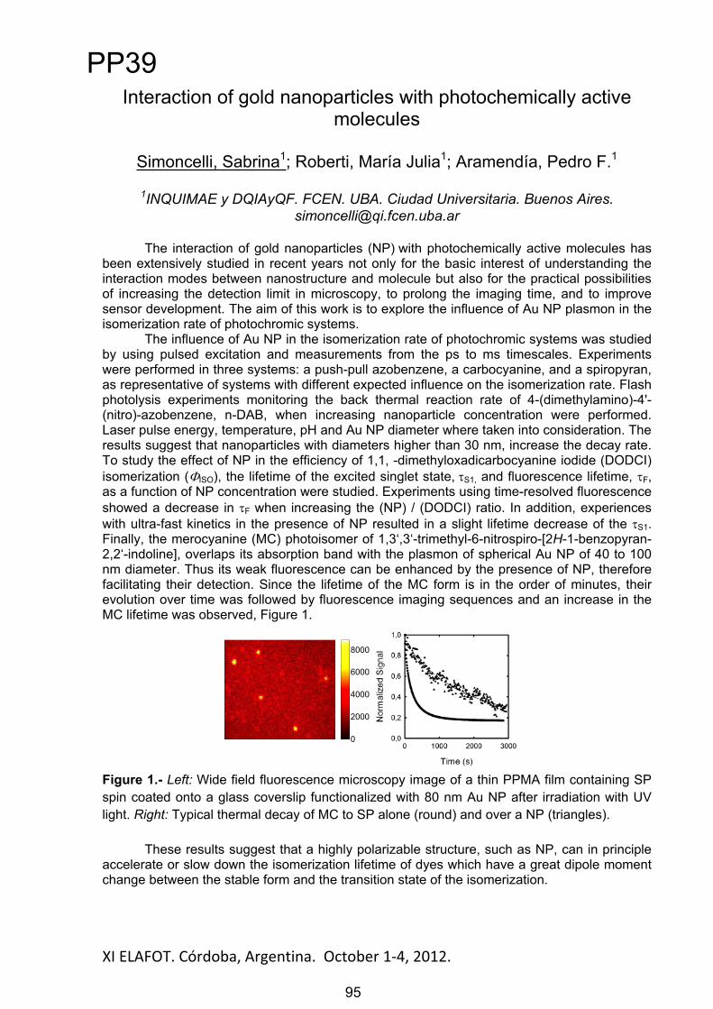
XI ELAFOT. Córdoba, Argentina. October 1 4, 2012. 95
Interaction of gold nanoparticles with photochemically active molecules
Simoncelli, Sabrina1; Roberti, María Julia1; Aramendía, Pedro F.1
1INQUIMAE y DQIAyQF. FCEN. UBA. Ciudad Universitaria. Buenos [email protected]
The interaction of gold nanoparticles (NP) with photochemically active molecules has been extensively studied in recent years not only for the basic interest of understanding the interaction modes between nanostructure and molecule but also for the practical possibilities of increasing the detection limit in microscopy, to prolong the imaging time, and to improve sensor development. The aim of this work is to explore the influence of Au NP plasmon in the isomerization rate of photochromic systems.
The influence of Au NP in the isomerization rate of photochromic systems was studied by using pulsed excitation and measurements from the ps to ms timescales. Experiments were performed in three systems: a push-pull azobenzene, a carbocyanine, and a spiropyran, as representative of systems with different expected influence on the isomerization rate. Flash photolysis experiments monitoring the back thermal reaction rate of 4-(dimethylamino)-4'-(nitro)-azobenzene, n-DAB, when increasing nanoparticle concentration were performed. Laser pulse energy, temperature, pH and Au NP diameter where taken into consideration. The results suggest that nanoparticles with diameters higher than 30 nm, increase the decay rate. To study the effect of NP in the efficiency of 1,1, -dimethyloxadicarbocyanine iodide (DODCI) isomerization ( ISO), the lifetime of the excited singlet state, S1, and fluorescence lifetime, F,as a function of NP concentration were studied. Experiments using time-resolved fluorescence showed a decrease in F when increasing the (NP) / (DODCI) ratio. In addition, experiences with ultra-fast kinetics in the presence of NP resulted in a slight lifetime decrease of the S1.Finally, the merocyanine (MC) photoisomer of 1,3‘,3‘-trimethyl-6-nitrospiro-[2H-1-benzopyran-2,2‘-indoline], overlaps its absorption band with the plasmon of spherical Au NP of 40 to 100 nm diameter. Thus its weak fluorescence can be enhanced by the presence of NP, therefore facilitating their detection. Since the lifetime of the MC form is in the order of minutes, their evolution over time was followed by fluorescence imaging sequences and an increase in the MC lifetime was observed, Figure 1.
Figure 1.- Left: Wide field fluorescence microscopy image of a thin PPMA film containing SP spin coated onto a glass coverslip functionalized with 80 nm Au NP after irradiation with UV light. Right: Typical thermal decay of MC to SP alone (round) and over a NP (triangles).
These results suggest that a highly polarizable structure, such as NP, can in principle accelerate or slow down the isomerization lifetime of dyes which have a great dipole moment change between the stable form and the transition state of the isomerization.
0
2000
4000
6000
8000
PP39
95

XI ELAFOT. Córdoba, Argentina. October 1 4, 2012. 96
Fotodegradabilidad del herbicida Hidrazida Maleica bajo condiciones simil-medioambientales naturales
Pajares, Adriana M.1, Bregliani, Mabel M.1, Boiero, Laura2,Montenegro, Mariana2, García, Norman A.3
1 UARG-UNPA, 9400 Río Gallegos, Argentina. [email protected] 2 Facultad Regional Villa María, UTN, 5900 Villa María, Argentina
3 Dto. de Química-UNRC, 5800 Río Cuarto, Argentina
La hidracida maleica (HM) (1,2-dihidropiridazine-3,6-diona) es un herbicida de acción fitorreguladora, de amplia difusión en el mundo entero por su empleo en numerosos cultivos [1]. Fue sintetizado por primera vez en 1947 y si bien en un principio se lo presentó como selectivamente tóxico para plantas e inofensivo para animales superiores, en la década del `60 se le atribuyeron propiedades carcinogénicas y mutagénicas, que continúan siendo evaluadas en la actualidad [2]. No obstante, como se dijo, se lo sigue empleando, profusamente.Continuando con la línea de trabajo de evaluación de la fotodegradabilidad de contaminantes acuáticos en condiciones simil-medioambientales, naturales, y artificiales [3], presentamos un estudio cinético y mecanístico de la fotodegradación de HM, promovida por luz visible, sensibilizada por vitamina B2 (Rf, Riboflavina), por ácidos húmicos y por el colorante sintético Rosa de Bengala (RB). Dado el carácter antimicrobiano del herbicida, en algunas condiciones de trabajo, también hemos evaluado la evolución del mismo luego de someterlo a fotólisis sensibilizada. Sintéticamente, HM sufre fotodegradación mediada por especies reactivas de oxígeno (ROS), especialmente oxígeno singulete molecular (O2(1
g)), generado por transferencia de energía desde el triplete excitado de menor energía de los fotosensibilizadores. Paralelamente, en competencia con este proceso, en el caso de Rf, se observa una transferencia de energía electrónica desde HM al mencionado estado triplete de la vitamina, que en ambiente aeróbico produce otras ROS, en especial ión superoxido. La velocidad de fotodegradación del herbicida es moderada a pH 7 y se incrementa significativamente a pH 9, debido al aumento de la contribución de desactivación reactiva, frente a la desactivación física de O2(1
g) por HM.De acuerdo a los datos cinéticos obtenidos HM se presenta como un contaminante degradable en condiciones medioambientales naturales y altamente degradable bajo condiciones artificiales sustentables.
Agradecimientos: Agradecemos el apoyo económico del Consejo Nacional de Investigaciones Científicas y Técnicas, de la Agencia Nacional de Promoción Científica y Tecnológica, de las Secretarías de Ciencia y Técnica de las Universidades Nacionales de Río Cuarto, de la Patagonia Austral y de la Universidad Tecnológica Nacional, todos de Argentina.
Referencias[1] Tomlin, The Pesticide Manual, British Crop Protection Council and The Royal Society of Chemistry, London, UK, 1994. [2] S. S. Epstein et al., Nature, 215, 1388-1390 (1967). [3] F. Amat-Guerri and N. A. García Chemosphere. 59, 1067-1082 (2005)
PP40
96

XI ELAFOT. Córdoba, Argentina. October 1 4, 2012. 97
ANTIINFALMATORIOS NO-ESTEROIDES COMO INHIBIDORES DE EPECIES REACTIVAS DE OXÍGENO Y SU
POSIBLE ROL EN EL RETARDO DE ENFERMEDADES NEURODEGENERATIVAS
Purpora Rebeca1, Pajares Adriana M.1, Massad Walter2, García Norman A.2
1 F Ingeniería, UNPSJB, 9000 C. Rivadavia, Argentina [email protected] Dto. Química-UNRC, 5800 Río Cuarto, Argentina
Se han reportado evidencias que fuertemente sugieren una acción retardante de la enfermedad de Alzheimer (EA), en personas que con fines analgésicos, son tratadas con antiinflamatorios no esteroides (AINEs) por períodos prolongados [1]. Paralelamente, es conocido el hecho de que ese stress oxidativo, generalmente acompañado por un exceso de producción y actividad de especies reactivas de oxígeno (ROS), de relativamente alto poder oxidativo en el organismo humano, es un factor muy importante para el desencadenamiento y progreso de la EA [2]. Aunque los dos enunciados anteriores han sido comprobados en la práctica, aun subsisten dudas acerca de una relación directa EA-AINEs [3]. No obstante se ha sugerido que los AINEs podrían actuar como inhibidores de ROS, resultando de esa manera un factor atenuante del desarrollo de la EA. En este contexto resulta interesante el estudio y la caracterización detallada de ciertos AINEs como desactivantes de ROS. Hemos abordado esta investigación, evaluando el efecto de los AINEs sobre ROS fotogeneradas en agua por la vitamina B2, endógena en humanos, en presencia de luz visible. Esta combinación bien puede modelar un escenario biológico natural. Hemos dirigido la investigación sobre Diflunisal (DFN, un derivado del ácido salicílico) Indometacín (IMT, un derivado indólico) y Diclofenac (DCF, derivado anilínico). Los tres AINEs mencionados han sido escogidos debido a que los respectivos compuestos-padre poseen propiedades como interceptores de ROS, de acuerdo con abundante información bibliográfica [4]. Mediante un estudio cinético y mecanístico sistemático, empleando espectrofotometría de absorción, fluorescencia estacionaria y resuelta en el tiempo, detección polarográfica de oxígeno disuelto, detección IR de fosforescencia de oxígeno singulete (O2(1
g)) resuelta en el tiempo y utilizando reacciones auxiliares con inhibidores específicos, se comprobó la interacción de las especies oxígeno singulete (O2(1
g)), radical OH, ion radical superóxido y H2O2 con DFN, IMT y DCF. Las interacciones mencionadas son en la mayor parte de los casos degradativas, de acuerdo a los resultados que arrojan las determinaciones de consumo de oxígeno disuelto sobre los sistemas fotoirradiados. Las constantes de velocidad para la reacción O2(1
g)-AINEs son el orden de 107 M-1s-1.
La conclusión general es que los AINEs estudiados poseen capacidades entre moderadas a altas para la intercepción de ROS y constituyen aceptables candidatos para una efectiva protección antioxidativa.
Agradecimientos: Agradecemos el apoyo económico de CONICET, ANPCyT, SECyT-UNRC y FI-UNPSJB, todos de Argentina.
Referencias[1] P. L. McGeer and E. G. McGeer, Neurobiol. Aging, 28, 639-647 (2007)[2] C. Behl and B. Moosmann, Free Rad. Biol. Med., 33, 182-191(2002) [3] M. M. Abdalla et al., I. J. Biol. Macromol., 51, 56-63 (2012) [4] F. Wilkinson et al., J. Phys. Chem. Ref. Data 24, 663-678 (1995)
PP41
97

XI ELAFOT. Córdoba, Argentina. October 1 4, 2012. 98
Fluorescence of Petroleum Asphaltenes
López, Rosmari Marisa; Mangani, Adriana del Luján, Gutiérrez, María Isela
Departamento de Química, Facultad de Ciencias Naturales, Universidad Nacional de la Patagonia San Juan Bosco, 9000 Comodoro Rivadavia, Argentina, E-mail:
Petroleum asphaltenes are defined by solubility: asphaltenes precipitate when petroleum or petroleum fractions are contacted with an excess of an alkane, usually n-pentane or n-heptane, and the precipitate is soluble in an aromatic solvent such as benzene or toluene. Asphaltenes comprise the most polar fraction of crude oil and consist of polyaromatic condensed rings with short aliphatic chains and heteroatoms such as nitrogen, oxygen, sulfur, and various metals [1].
The asphaltene used in this study was the n-pentane insoluble fraction of a heavy crude oil from San Jorge Gulf basin (well B-483 from Cañadón Perdido field) with a density of 0.9248 ± 0.0001 g/mL at 24 ºC (API degree 21.5). The asphaltene content, obtained from crude oil based on the ASTM 2007 D method modified, was 4.5 %. For comparison purposes, samples of crude oil from different origins and its asphaltene fraction were also studied.
Solutions of asphaltenes in benzene, toluene and dichloromethane were characterized using absorption, different modes of fluorescence spectroscopy (conventional, excitation, synchronous and total fluorescence spectra, EEM) and scanning electron microscope analyses. In addition, the fluorescence quantum yield ( F) was obtained in toluene and dichloromethane by a indirect method, using quinine sulphate in 1.0 N sulphuric acid as standard ( F= 0.546) [2]. The spectra were taken at concentrations lower than 10.0 mg/L to avoid asphaltene aggregation. Fluorescence emission spectra was recorded at an excitation wavelength of 340 nm. The scanning electron microscope images were taken in the low-vacuum mode.
The profiles of the excitation and absorption spectra were similar, suggesting that the basic absorbing chromophores were the same. All the emission spectra obtained consist of a structureless broad band in the visible region, from ca. 350 to 650 nm. The fluorescence quantum yields ( exc= 340 nm) at room temperature of the asphaltene fraction in toluene and dichloromethane were 0.051 ± 0.003 and 0.049 ± 0.001, respectively. The profiles of the EEM show that the emission maxima is centered near 480 nm (excitation around 330 nm).
The present results suggest that fluorescence spectroscopy is a technique capable of providing useful information from samples of crude oils and asphaltenes from the San Jorge Gulf basin, and, as already reported by several authors, offers a sensitive method for analyzing different oils [3, 4].
Acknowledgement: CIUNPAT-UNPSJB for financial and fellowship support.
References:
[1]. Buenrostro-Gonzalez, E.; Groenzin, H.; Lira-Galeana, C.; Mullins, O. C. Energy & Fuels, 15, 972-978, 2001.
[2]. Melhuish, W.H.. J. Phys. Chem., 65, 229 (1961) [3]. Pantoja, P. A;, López-Gejo, J.; Le Roux, G. A. C.; Quina, F. H.; Nascimento, C. A. O. Energy &
Fuels, 125, 3598-3604, 2011.[4]. Sarma, A. K.; Ryder, A. G. Energy & Fuels, 20, 783-785, 2006.
PP42
98

XI ELAFOT. Córdoba, Argentina. October 1-4, 2012.
Estudio de las interacciones entre guanosina y aminoácidos bajo fotosensibilización con Riboflavina en
presencia de luz visible. M. Paulina Montaña1, Gabriela Ferrari1, Eduardo Gatica2, José Natera2,
Walter Massad3 y Norman A. García3
1Area de Química Física – INQUISAL (CONICET-Universidad Nacionalde San Luis), 5700 San Luis, Argentina, [email protected]
2Facultad de Agronomía y Veterinaria. Universidad Nacional de Río Cuarto. 5800 Río Cuarto, Argentina
3Departamento de Química. Universidad Nacional de Río Cuarto.5800 Río Cuarto, Argentina
Los ácidos nucleicos y las proteínas ocupan ambientes comunes en organismos vivos, y sus interacciones en presencia de agentes oxidantes pueden provocar importantes daños y enfermedades [1]. Si bien la luz visible no es altamente agresiva para la mayoría de los ambientes biológicos bajo radiación directa, es fácilmente transmitida a las células donde puede interaccionar con fotosensibilizadores endógenos como la Vitamina B2 o Riboflavina (Rf). Esta vitamina está involucrada en la fotooxidación de residuos de ADN y proteínas, a pesar de que ella misma se degrada, generando diferentes especies reactivas de oxígeno (EROs) [2]. Guanosina (Gse) y sus derivados son subproductos de la oxidación del ADN en las células, y son susceptibles de la acción de las EROs tales como oxígeno molecular singulete (O2
1g) y anión radical superóxido (O2
-), por lo que pueden ser usados como compuestos modelo para el estudio de las fotooxidaciones [3]. Por lo antes expuesto, se eligió investigar las interacciones de Gse como residuo oxidable de ADN y los aminoácidos (AAs) histidina (His), triptofano (Trp) y tirosina (Tir) como residuos oxidables de proteínas, en presencia de un fotogenerador endógeno de EROs como Rf. Se eligieron estos AAs porque interactúan con el estado triplete excitado de Rf (3Rf*) y con O2(1
g), producido desde 3Rf*,en este último caso por mecanismos muy diferentes entre sí: una interacción (quenching)totalmente química para His, una combinación de quenching físico y químico para Trp; y una interacción exclusivamente física para Tir [4].
Una interpretación mecanística basada en los datos cinéticos obtenidos en este trabajo y en los reportes de bibliografía pueden resumirse como sigue: Tir a pH 7 exhibe un efecto protector en la fotooxidación de la mezcla Tir-Gse debido al quenching físico de O2(1
g) por el AA. Se observa el mismo efecto para los sistemas Trp-Gse e His-Gse a pH 7, donde se atribuye al quenching de 3RF* por Gse en detrimento de la vía mediada por O2(1
g), la cual se sabe que es el mecanismo dominante en la oxidación sensibilizada por Rf para ambos aminoácidos. Estos resultados podrían ser de interés en el marco de las interacciones fotoinducidas entre proteínas y ácidos nucleicos, como es el caso de la adición covalente de residuos de AAs a moléculas de ARN/ADN en membranas. Asimismo, estas interacciones entre residuos oxidables de ácidos nucleicos con AAs oxidables deben describirse individualmente a partir del comportamiento de las mezclas de los respectivos compuestos.
Agradecimientos: Al Consejo Nacional de Investigaciones Científicas y Técnicas, y a las Secretarías de Ciencia y Técnica de la UNRC y UNSL por el apoyo recibido.
Referencias
[1] Frôlich I & Riederer P. Drug Res. 45: 443–449, 1995. [2] Joshi PC & Keane TC. Biochem. Biophys. Res. Comm. 400: 729-733, 2010. [3] Cadet J, Anselmino C, Douki T & Voituriez L. J. Photochem. Photobiol. B: Biol. 15: 277-298, 1992. [4] Straight RC & Spikes JD. Photosensitized oxidation of biomolecules. Vol. 4, CRC Press, Boca Raton, FL, 1985. Davies MJ & Truscott RJW. J. Photochem. Photobiol. B: Biol. 63: 114-125, 2001. Bertolotti SG, García NA & Argüello GA. J. Photochem. Photobiol. B: Biol. 10: 57-70, 1991.
99
PP43
99

XI ELAFOT. Córdoba, Argentina. October 1-4, 2012.
Theoretical study of possible sensitizers for Dye-Sensitized Solar Cells based on phthalocyanine
Gomes, W.R.; Araújo, D.M.S.; Machado, A. E. H.
Universidade Federal de Uberlândia, Instituto de Química/Laboratório de Fotoquímica e Ciência de Materiais - Av. João Naves de Ávila, 2121 Bl. 5K
CEP 38408-100 Uberlândia, MG, Brasil, E-mail: [email protected]
The present communication deals with the use of molecular design based on quantum mechanical of three metal-phthalocyanines: (a) (2,9-bis(diethylamino)-16,23-dicarboxy) zinc (II) phthalocyanine, ZnPc, (b) (2,9-bis(diethylamino)-16,23-dicarboxy) chloro-aluminium (III) phthalocyanine, AlClPc, and (c) (2,9-bis(diethylamino)-16,23-dicarboxy) bis(pyridine) Ruthenium (II) phthalocyanine, Ru(py)2Pc, aiming its application as sensitizers for Dye-Sensitized Solar Cells (DSSC).
Full geometry optimization was performed for each molecule using Gaussian 09 in DFT calculations applying the B3LYP hybrid density functional and the LANL2DZ basis set. Unrestricted calculations were performed for open-shell structures. Excitation energies were calculated using TD-DFT with the same parameters above described. These calculations were performed simulating the presence of DMSO under the IEFPCM self-consistent reaction field (SCRF) approach. The charge distribution analysis was carried out using NBO 3.1, available in Gaussian 09.
The analysis of the energy levels between the dye in the first singlet or triplet excited state and the conduction band (CB) level of the semiconductor oxide (TiO2) shows that those compounds present the LUMO energy above the TiO2 CB level. This suggests that the electron transfer from the dye to the semiconductor oxide, resulting in the (dye)+. cation radical and [(TiO2)n]
-., is energetically favorable. Comparing the frontier orbital (SOMO and) energies of the cation-radical with respect to the redox potential of the electrolyte (in this
case, the I/I3- potential), the three compounds present energy sufficiently below this potential, which implies that (dye)+. should be reduced to its original state by the I/I3
- redox pair. In the analysis of charge distribution, five different sites were considered (carboxyl group, diethylamino group, macrocycle, metal ion and axial groups bound to metal ion) on thestructure of the compounds studied, in the S1 and T1, and for (dye)+.. The comparison of the charge distributions shows that a positive charge density tends to be distributed along the molecule after the charge transfer, due to extensive electronic conjugation and, also possibly, to the electronegativity of diethylamino groups. This efficient charge redistribution can favor the regeneration of the neutral molecule from the cation-radical, and hinder the recombination between the electron transferred to the conduction band of semiconductor and the semi-oxidized dye.
Acknowledgements: To CNPq, CAPES and FAPEMIG.
100
PP44
100
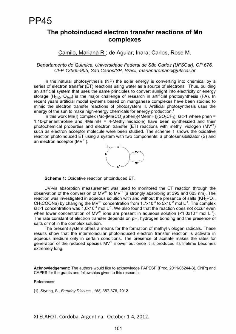
XI ELAFOT. Córdoba, Argentina. October 1 4, 2012. 101
The photoinduced electron transfer reactions of Mn complexes
Camilo, Mariana R.; de Aguiar, Inara; Carlos, Rose M.
Departamento de Química, Universidade Federal de São Carlos (UFSCar), CP 676, CEP 13565-905, São Carlos/SP, Brasil, [email protected]
In the natural photosynthesis (NP) the solar energy is converting into chemical by a series of electron transfer (ET) reactions using water as a source of electrons. Thus, building an artificial system that uses the same principles to convert sunlight into electricity or energy storage (H2(g), O2(g)) is the major challenge of research in artificial photosynthesis (FA). In recent years artificial model systems based on manganese complexes have been studied to mimic the electron transfer reactions of photosystem II. Artificial photosynthesis uses the energy of the sun to make high-energy chemicals for energy production.1
In this work Mn(I) complex (fac-[Mn(CO)3(phen)(4MeImH)](SO3CF3), fac-1 where phen = 1,10-phenanthroline and 4MeImH = 4-Methylimidazole) have been synthesized and their photochemical properties and electron transfer (ET) reactions with methyl viologen (MV2+)such as electron acceptor molecule were been studied. The scheme 1 shows the oxidative reaction photoinduced ET using a system with two components: a photosensibilizator (S) and an electron acceptor (MV2+).
Scheme 1: Oxidative reaction phtoinduced ET.
UV-vis absorption measurement was used to monitored the ET reaction through the observation of the conversion of MV2+ to MV•+ (a strongly absorbing at 395 and 603 nm). The reaction was investigated in aqueous solution with and without the presence of salts (KH2PO4,CH3COONa) by changing the MV2+ concentration from 1.7x10⁻³ to 5x10⁻² mol L-1. The complex fac-1 concentration was 1,0x10⁻⁴ mol L-1. We also found that the reaction does not occur even when lower concentration of MV2+ ions are present in aqueous solution (<1,0x10⁻³ mol L-1).The rate constant of electron transfer depends on pH, hydrogen bonding and the presence of salts or not in the complex solution.
The present system offers a means for the formation of methyl viologen radicals. These results show that the intermolecular photoinduced electron transfer reaction is activate in aqueous medium only in certain conditions. The presence of acetate makes the rates for generation of the reduced species MV•+ slower but once it is produced its lifetime becomes extremely long.
Acknowledgement: The authors would like to acknowledge FAPESP (Proc. 2011/06244-3), CNPq and CAPES for the grants and fellowships given to this research.
References:
[1]. Styring, S., Faraday Discuss., 155, 357-376, 2012.
PP45
101

XI ELAFOT. Córdoba, Argentina. October 1 4, 2012. 102
STUDY OF IODIDE AS SELECTIVE QUENCHER OF TRIPLET EXCITED STATES OF PTERIN AND LUMAZINE
M. Paula Denofrio1,2, Carolina Lorente2, Peter R. Ogilby3 and Andrés H. Thomas2
1 IIB-INTECH-UNSAM-CONICET (sede Chascomús). Intendente Marino Km 8,2. CC 164 (7130) Chascomús, Buenos Aires, Argentina. E-mail: [email protected]
2 INIFTA, Departamento de Química, Facultad de Ciencias Exactas, Universidad Nacional de La Plata, CCT-La Plata, CONICET. Casilla de Correo 16, Sucursal 4,
1900- La Plata, Argentina.
3 Center for Oxygen Microscopy and Imaging, Department of Chemistry, University of Aarhus, DK-8000 Århus, Denmark.
It has been demonstrated that pteridines are able to act as efficient sensitizers in photoinduced oxidations. These processes have been well characterized using 2-aminopteridine-4(3H)-one (pterin; denoted Ptr) and pteridine-2,4(1,3H)-dione(lumazine; denoted Lum) as photosensitizers and the nucleotides 2 -deoxyguanosine5 -monophosphate (dGMP) and 2 -deoxyadenosine 5 -monophosphate (dAMP) as oxidizable targets.(1-5)
It is interesting to establish the nature, singlet (1S*) or triplet (3T*), of the sensitizer excited states involved in such processes. In this regard, the development and optimization of routine techniques that allow distinguishing from which excited state the photosensitization occurs becomes relevant. A good example is the use of certain species which, under controlled working conditions, can deactivate selectively an excited electronic state.(6)
In this work, we present results of the selective deactivation of triplet excited states of Ptr and Lum by iodide anion. The method was applied to the reactions of dGMP and dAMP photosensitization previously studied. At low iodide concentrations, triplet states are deactivated, whereas singlet states do not. Under these conditions the photosensitized oxidation of nucleotides in the presence of pteridines is totally inhibited, demonstrating that the photooxidation process is initiated by pteridine triplet excited states.
(1) G. Petroselli, R. Erra-Balsells, F. M. Cabrerizo, C. Lorente, A. L. Capparelli, A. M. Braun, E. Oliveros, A. H. Thomas. Org. Biomol. Chem. (2007), 5, 2792-2799. (2) G. Petroselli, M. L. Dantola, F. M. Cabrerizo, A. L. Capparelli, C. Lorente, E. Oliveros, A. H. Thomas, J. Am. Chem. Soc. (2008), 130, 3001-3011. (3) M. P. Denofrio, S. Hatz, C. Lorente, F. M. Cabrerizo, P. R. Ogilby, A. H. Thomas, Photochem. Photobiol. Sci. (2009), 8, 1539–1549. (4) M. P. Denofrio, A. H. Thomas, C. Lorente, J. Phys. Chem. A (2010), 114,10944–10950. (5) M. P. Denofrio, M. L. Dántola, P. Vicendo, E. Oliveros, A. H. Thomas, C Lorente. Photochem. Photobiol. Sci., (2012), 11, 409-XXX (6) M. S. Kritsky, T. A. Lyudnikova, E. A. Mironov and I. V. Moskaleva, J. Photochem. Photobiol., B,(1997), 39, 43–48.
PP46
102
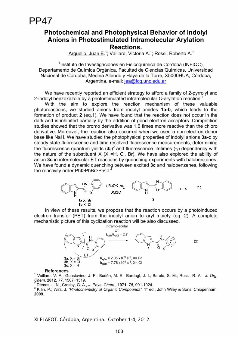
XI ELAFOT. Córdoba, Argentina. October 1 4, 2012. 103
Photochemical and Photophysical Behavior of Indolyl Anions in Photostimulated Intramolecular Arylation
Reactions.Argüello, Juan E.1; Vaillard, Victoria A.1; Rossi, Roberto A.1
1Instituto de Investigaciones en Fisicoquímica de Córdoba (INFIQC), Departamento de Química Orgánica, Facultad de Ciencias Químicas, Universidad Nacional de Córdoba, Medina Allende y Haya de la Torre, X5000HUA, Córdoba,
Argentina. e-mail: [email protected]
We have recently reported an efficient strategy to afford a family of 2-pyrrolyl and 2-indolyl benzoxazole by a photostimulated intramolecular O-arylation reaction.1
With the aim to explore the reaction mechanism of these valuable photoreactions, we studied anions from indolyl amides 1a-b, which leads to the formation of product 2 (eq.1). We have found that the reaction does not occur in the dark and is inhibited partially by the addition of good electron acceptors. Competition studies showed that the bromo derivative was 1.6 times more reactive than the chloro derivative. Moreover, the reaction also occurred when we used a non-electron donor base like NaH. We have studied the photophysical properties of indolyl anions 3a-c by steady state fluorescence and time resolved fluorescence measurements, determining the fluorescence quantum yields ( f)2 and fluorescence lifetimes ( f) dependency with the nature of the substituent X (X =H, Cl, Br). We have also explored the ability of anion 3c in intermolecular ET reactions by quenching experiments with halobenzenes. We have found a dynamic quenching between excited 3c and halobenzenes, following the reactivity order PhI>PhBr>PhCl.3
In view of these results, we propose that the reaction occurs by a photoinduced electron transfer (PET) from the indolyl anion to aryl moiety (eq. 2). A complete mechanistic picture of this cyclization reaction will be also discussed.
N
HN
O
kq3a = 2.05 x10⁹ s-1, X= Brkq3b = 7.76 x10⁸ s-1, X= Cl
IntramolecularET
kqBr/kqCl = 2.7
N
HN
OX X
*
ET
(2)
3a, X = Br3b, X = Cl3c, X = H
References 1 Vaillard, V. A.; Guastavino, J. F.; Budén, M. E.; Bardagí, J. I.; Barolo, S. M.; Rossi, R. A. J. Org. Chem. 2012, 77, 1507 1519. 2 Demas, J. N., Crosby, G. A., J. Phys. Chem., 1971, 75, 991-1024. 3 Klán, P.; Wirz, J. “Photochemistry of Organic Compounds”, 1° ed., John Wiley & Sons, Chippenham, 2009.
PP47
103

XI ELAFOT. Córdoba, Argentina. October 1 4, 2012. 104
Reacciones de Fotosustitución del Anión Difenilamiduro. Estudio de sus Propiedades Fotofisicas.
Bouchet, Lydia M., Barrionuevo Cecilia A., Schmidt, Luciana C., Pierini, Adriana B., Peñéñory, Alicia B. y Argüello, Juan E.
Instituto de Investigaciones en Fisicoquímica de Córdoba (INFIQC), Departamento de Química Orgánica, Facultad de Ciencias Químicas, Universidad
Nacional de Córdoba, Córdoba, Argentina. e-mail: [email protected]
Se conoce que los nucleófilos centrados en nitrógeno pueden reaccionar con diferentes halogenuros de arilo por un mecanismo de sustitución nucleofílica radicalaria unimolecular (SRN1). De hecho este mecanismo fue descubierto explorando las reacciones del anión amiduro (H2N-) con 1-halo-2,4,5-trimetilbenceno y 1-halo-2,3,5-trimetilbenceno.1 Empleandoaniones aril amiduros (ArNH-) se obtienen productos de sustitución tanto en el anillo aromático como sobre el átomo de nitrógeno.2 Y si bien los aspectos fotoquímicos de estos aniones han sido explorados por Pierini y colaboradores, no hay reportes acerca de las propiedades fotofísicas de aniones centrados en nitrógeno. En la presente comunicación se eligió el anión derivado de la N,N-difenilamina (Ph2N-), el cual a pesar de ser un nucleófilo heterocentrado, reacciona mayoritariamente por carbono en reacciones de sustitución radical-nucleófilo. Los productos obtenidos cuando se irradió una mezcla de Ph2N- con halogenuros de alquilo y arilo (iodobenceno, 4-iodoanisol, 4-bromobenzonitrilo, 1-bromonaftaleno y 1-iodoadamantano) fueron mayoritariamente los de sustitución en las posiciones orto y para del anillo fenilo del nucleófilo y en mucha menor proporción el producto de sustitución en nitrógeno (ec. 1).
El anión en estudio es fluorescente y sus propiedades fotofísicas se resumen en la tabla 1, de la cual también se desprende el poder reductor del estado singlete excitado del anión. Esto se ve reflejado en los altos valores de constante de quenching de fluorescencia del anión en presencia de los electrófilos R-X, los cuales se encuentras próximos al límite controlado por la difusión.
Tabla 1: Propiedades fotofísicas del anión difenilamiduro en DMSO a 25°C. max abs max f f f Eox
0S E 1S E ox1S
375 nm 465 nm 0,33 3 ns -0,115eV3 69kcal/mol 2,88eV
En la presente comunicación también se explorará la superficie de energía potencial para la reacción de adición radical-nucleófilo empleando herramientas del modelado molecular para logran un mejor entendimiento de la regioquímica de estas reacciones de foto-sustitución.
Referencias1- (a) Peñéñory, A. B.; Argüello, J. E.; Handbook of Synthetic Photochemistry, Albini, A., Fagnoni, M., Eds., Wiley-VCH: Weinheim. 2010, 10, 319. (b) Rossi, R. A.; Pierini, A. B.; Peñéñory, A. B.; Chem. Rev. 2003, 103, 71. 2- Pierini, A. B.; Baumgartner, M. T.; Rossi, R. A.; Tetrahedron Let. 1987, 28, 4653. 3- Bordwell, F.G.; Zhang, X.; Cheng, J-P; J. Org Chem. 1991, 56, 3216.
+ RX hDMSO
(1)N H
N
RHN
R
H+
+ N
R
+
PP48
104

XI ELAFOT. Córdoba, Argentina. October 1 4, 2012. 105
ESTUDIO FOTOFÍSICO Y FOTOQUÍMICO DEL SISTEMA 3´-HIDROXIFLAVONOL-La(III)
Ferrari, Gabriela V.1; Montaña, M. Paulina1;Pappano, Nora B.2;Debattista, Nora. B.2; García, Norman A.3
1INQUISAL - CONICET-UNSL, 5700 San Luis, Argentina. [email protected] 2Area de Química Física, UNSL 5700 San Luis, Argentina
3Departamento de Química. UNRC, 5800 Río Cuarto, Argentina
3´-hidroxiflavonol es un compuesto perteneciente a la familia de los flavonoides, polifenoles ampliamente difundidos en la naturaleza como metabolitos secundarios de plantas. El estudio de estos compuestos es interesante debido a que presentan poder antioxidante [1], antimicrobiano [2] y habilidad para complejar iones metálicos [3]. Son reconocidos por su acción protectora de medios biológicos en general. Se ha propuesto que la capacidad de los flavonoides para secuestrar iones metálicos puede contribuir a sus propiedades antioxidantes, protegiendo frente a la acción de radicales libres [4]. Riboflavina (Rf) es un conocido pigmento sensibilizador que participa en degradación fotooxidativa de numerosos sustratos naturales [5]. Dado que los flavonoides y Rf pueden localizarse en ambientes biológicos comunes, la interacción de estos potenciales electrodonores y sus complejos metálicos con O2
– y O2(1g) fotogenerados a partir de Rf, es
de interés determinar si dichos sustratos son capaces de favorecer o inhibir la producción de especies reactivas de oxígeno (EROs). Mediante espectrofotometría UV, fluorescencia estacionaria y resuelta en el tiempo, fotólisis de láser flash y detección polarográfica de oxígeno disuelto, se evaluaron las siguientes propiedades fotofísicas y fotoquímicas de 3´-hidroxiflavonol (F) y su complejo con el ión metálico La(III) (F-La): • Rendimiento cuántico de fluorescencia de F y F-La: ca. 10-2 y 10-1 respectivamente, siendo adecuados para su uso como herramienta analítica. • Estados transientes: se obtuvieron espectros de transientes atribuibles a estados 3F*. No se observó señal para F-La dado que el metal produce desactivación (quenching) de 3F*.• Rendimiento cuántico de triplete: se obtuvo un valor límite mínimo para F de 0,13 y nulo para F-La. • Quenching de O2(1
g): F-La y F producen quenching de O2(1g) con constantes de velocidad
totales (kt) de 4x10⁷ y 9x10⁵ M-1s-1 y valores de constantes reactivas (kr), de 7,6x10⁶ y <105 M-
1s-1, respectivamente. • Interacciones con Rf: F desactiva 1Rf* con un valor de kq= 5,45x10¹⁰ M-1 s-1. F-La y F desactivan 3Rf* con kq de 9,4x10⁸ M-1s-1 para F y 1,6x10⁹ M-1s-1 para el complejo. F-La reacciona frente a O2(1
g) y O2•– fotogenerados por Rf, actuando como un eventual protector
antioxidante. El consumo de oxígeno para el sistema Rf + F-La fotoirradiado es inhibido en presencia de NaN3, conocido quencher de O2(1
g) y se retrasa en presencia de superóxido dismutasa (SOD), secuestrante de la especie O2
•–. En cuanto a Rf + F se confirmó la interacción de este flavonoide con O2(1
g). El aumento de velocidad de consumo de oxígeno en presencia de catalasa y SOD indicaría que F probablemente actúe como agente secuestrante de H2O2.En conclusión, las propiedades fotofísicas y fotoquímicas estudiadas muestran a F como potencial fotogenerador de EROs, posiblemente desde 3F*, vía ausente en F-La. Se observó que F-La reacciona frente a O2(1
g) y O2– y puede considerarse como un aceptable
fotoprotector ante la oxidación mediada por O2(1g). F produce quenching de O2(1
g) y muy probablemente actúe como agente secuestrante de H2O2.
Referencias[1] Photochem. Photobiol. 86: 827-834(2010); [2] Folia Microbiol. 54: 516-520(2009); [3] J. Eng. Chem. Data 5: 3080-3083(2010);[4] J. Molec. Struct. 918: 194-197(2009); [5] Chem. Soc. Rev. 11: 15-39(1982).
PP49
105

XI ELAFOT. Córdoba, Argentina. October 1 4, 2012. 106
Nitroanilines as Quencher of Pyrene and 1-Methyl Pyrene Fluorescence.
Oscar F. Silva, 1 Carlos E. Agudelo-Morales, 1 Raquel E. Galian, 1,2
Julia Pérez-Prieto 1
1Instituto de Ciencia Molecular , Universidad de Valencia, Valencia, España, 2Dpto. Química Analítica, Universidad de Valencia, Valencia, España.
E-mail: [email protected]
Nitroanilines are intermediates in the synthesis of dyes, drugs, pesticides, and herbicides. They have been used as fluorescence quenchers of organic chromophores, biomacromolecules, as well as nanomaterials.[1] In fact, some of these fluorescent systems have been used as isomeric aromatic amine probes.[2] In addition, the capacity of nitroanilines as quenchers has been used to study materials, such as fluorescent surfactant aggregates.
It has been suggested that, depending on the nature of the fluorophore, its emission quenching by nitroanilines is static or dynamic, can occur via electron or energy transfer, or can involve formation of transitory charge-transfer complexes or exciplexes (which decay by electron transfer, intersystem crossing, or internal conversion). Therefore, a systematic study on the interaction between a well-known and extensively used fluorophore, such as pyrene, and NAs appeared of interest to gain insight into their role as quenchers. Pyrene has been widely used as a probe due to i) its absorption (strength) features, ii) its long singlet lifetime (> 100 ns), and the information that can be obtained from its fluorescence emission (I3/I1 ratio) and iii) its long-lived triplet excited state (microsecond scale). This chromophore has being used in fluorescent chemosensors that can recognize selectively chemical species in potential analytical applications [3]. In fact, nitrated explosives have been detected by fluorescence quenching of pyrene and related compounds.[4]
We report here on the quenching of pyrene and 1-methylpyrene fluorescence by unsubstituted NAs, methylnitroanilines, and a dinitroaniline in toluene and 1,4-dioxane. Steady-state and time-resolved absorption and fluorescence studies were used to gain insight into the quenching mechanism and the species involved in this process. These studies show the tendency of NAs to establish specific interactions with the pyrene singlet excited state and, depending on their structure, to be adjacent to the fluorophore at the ground state, some forming a ground state complex.
Acknowledgements:We thank MEC (Project CTQ2011-27758, RyC contract to R-E.G, and post-doc contract for O-F. S.)
References [1]- (a) A. Airinei R. I. Tigoianu, E. Rusu, D. O. Dorohoi, Digest J. Nanomat. Biostruct., 2011, 6, 1265; b) H. Li, Y. Li, Nanoscale, 2009, 1, 128. [2]-D. Patra, A. K. Mishra, Sens. Actuators B, 2011, 80, 278. [3]- (a) B. Schazmann, N. Alhashimy, D. Diamond, J. Am. Chem. Soc. 2006, 128, 8607; b) S. H. Lee, S.H. Kim, S. K. Kim, J. H. Jung, J. S. Kim, J. Org. Chem. 2005, 70, 9288; c) K-S. Focsaneanu, J. C. Scaiano, Photochem. Photobiol. Sci. 2005, 4, 817 [4]- M. S. Meany, V. L. McGuffin Anal. Chim Acta 2008, 610, 57.
PP50
106

XI ELAFOT. Córdoba, Argentina. October 1 4, 2012. 107
Mechanism of membrane damage by photosensitization
Bacellar, Isabel O. L.1; Pavani, Christiane1; Sales, Elisa M.2; Itri, Rosangela2; Wainwright, Mark3; Baptista, Mauricio S.1
1 Instituto de Química, Universidade de Sao Paulo, Sao Paulo, Brazil, [email protected], [email protected]
2 Instituto de Física, Universidade de Sao Paulo, Sao Paulo, Brazil, [email protected] John Moores University, Liverpool, England, [email protected]
Photodynamic therapy (PDT) is a clinical modality based on the interaction between light and a photosensitizer (PS), yielding reactive species which can destroy malignant cells. Among these species singlet oxygen (1O2) plays a significant role. Since the cell membrane is considered one of the main targets in PDT, we aimed to study the factors leading to membrane damage. We investigated the effect of different phenothiazinium PSs – methylene blue (MB), toluidine blue (TBO), 1,9-dimethyl methylene blue (DMMB) and a methylene blue derivative (DO15) – in soy lecithin liposomes containing the self-quenched fluorescent probe 5(6)-carboxyfluorescein. Fluorescence increase upon irradiation with red light was used to quantify membrane damage. As presented in Table1, all PSs except for DO15 have similar fluorescence and 1O2 generation quantum yields in ethanol. The lower values observed with DO15 can be attributed to its voluminous side-chains, which would favour non-radiative decay to the ground state. All compounds form aggregates in 3 M NaCl solution, as shown in Table 1. DMMB has the lower monomer to aggregate ratio, which is expected to diminish 1O2 generation. [1] On the other hand, DO15 has the higher tendency to stay in the monomeric form, probably due to the steric effects associated with its bulky terminal rings. Membrane binding percentage was significantly higher for DMMB and DO15 than for MB and TBO. Higher membrane binding in this case is related to more hydrophobic compounds. [2] Membrane damage assays revealed that MB and TBO caused almost no release of the fluorescent probe, whereas DMMB and DO15 showed high membrane damage percentage. Our work showed that membrane binding is the main factor leading to membrane damage for this group of PSs, because it allows ¹O2 generation close to its target (i.e. lipid chain double bonds). [1] In order to further investigate membrane damage by DMMB and DO15, vesicles were damaged in similar levels by 5 hour irradiation and then were analysed by small angle X-ray scattering (SAXS). In the presence of DO15, we observed a decrease in the thickness of the polar headgroup regions as well as in the methyl ends of the hydrophobic chain, showing extensive reorganization of the lipids within the membrane. In the case of DMMB no significant structural modifications were observed. Further studies are necessary to establish a clear relationship between the structural changes monitored by SAXS and photoinduced chemical reactions. These results demonstrate that photophysical efficiency is not the most important factor that must be considered when determining photodynamic efficiency, and that the mechanism of membrane damage may depend on specific PS/membrane interactions.
Table 1. Fluorescence quantum yield ( f) and 1O2 generation quantum yield ( ) in ethanol; percentage of liposome damage (%D) and membrane binding (%L) at [PS] = 15 M; ratio between monomers and aggregate absorbances (M/A) in 3 M NaCl solutions, also at [PS] = 15 M.
MB TBO DMMB DO15 0,52 0,55 ± 0,9 0,59 ± 0,8 0,23 ± 0,7
f /10-2 4 7,3 ± 0,2 4,5 ± 0,5 2,6 ± 0,1 M/A 1,44 1,08 0,122 3,37 %L 1,005 ± 0,004 5,37 ± 0,05 50 ± 1 65,8 ± 0,5 %D 2,9 ± 0,2 3,8 ± 0,5 92 ± 4 95 ± 6
Acknowledgements: the authors thank FAPESP for the financial support.
References: 1. Tardivo, J.P. et al. Photodiag. Photdyn. Ther. 2005, 2,175-191. 2. Wainwright, M.; Giddens, R.M. Dyes Pigments, 2003, 57, 245-257.
PP51
107
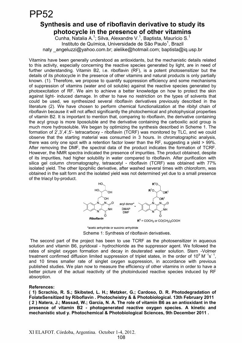
XI ELAFOT. Córdoba, Argentina. October 1-4, 2012.
Synthesis and use of riboflavin derivative to study itsphotocycle in the presence of other vitaminsCunha, Natalia A.1; Silva, Alexandre V.1, Baptista, Maurício S.1
Instituto de Química, Universidade de São Paulo1, Brazilnaty [email protected]; [email protected]; [email protected]
Vitamins have been generally understood as antioxidants, but the mechanistic details related to this activity, especially concerning the reactive species generated by light, are in need of further understanding. Vitamin B2, i.e. riboflavin (RF), is a potent photosensitizer but the details of its photocycle in the presence of other vitamins and natural products is only partially known. (1). Therefore, we propose to quantify suppression efficiency and some mechanisms of suppression of vitamins (water and oil soluble) against the reactive species generated by photoexcitation of RF. We aim to achieve a better knowledge on how to protect the skin against light- induced damage. In other to have no restriction on the types of solvents that could be used, we synthesized several riboflavin derivatives previously described in the literature (2). We have chosen to perform chemical functionalization at the ribityl chain of riboflavin because it will not affect significantly the photochemical and photophysical properties of vitamin B2. It is important to mention that, comparing to riboflavin, the derivative containing the acyl group is more liposoluble and the derivative containing the carboxilic acid group is much more hydrosoluble. We began by optimizing the synthesis described in Scheme 1. The
- tetracarboxy - riboflavin (TCRF) was monitored by TLC, and we could observe that the starting material was consumed in 3 hours. In chromatographic analysis, there was only one spot with a retention factor lower than the RF, suggesting a yield > 99%. After removing the DMF, the spectral data of the product indicates the formation of TCRF. However, the NMR spectra indicated the presence of impurities. The product obtained, despite of its impurities, had higher solubility in water compared to riboflavin. After purification with silica gel column chromatography, tetraacetyl - riboflavin (TCRF) was obtained with 77% isolated yield. The other lipophilic derivative, after washed several times with chloroform, was obtained in the salt form and the isolated yield was not determined yet due to a small presence of the triacyl by-product.
The second part of the project has been to use TCRF as the photosensitizer in aqueous solution and vitamin B6, pyridoxal - hydrochloride as the suppressor agent. We followed the rates of singlet oxygen formation and decay in deuterated water solution. Stern -Volmer treatment confirmed diffusion limited suppression of triplet states, in the order of 109 M- 1s- 1,and 10 times smaller rate of singlet oxygen suppression, in accordance with previous published studies. We plan now to measure the efficiency of other vitamins in order to have a better picture of the actual reactivity of the photoinduced reactive species induced by RF absorption.
References:( 1) Scrachio, R. S.; Skibsted, L. H.; Metzker, G.; Cardoso, D. R. Photodegradation of FolateSensitized by Riboflavin . Photocheistry & & Photobiological. 13th February 2011( 2 ) Natera, J.; Massad, W.; García, N. A. The role of vitamin B6 as an antioxidant in the presence of vitamin B2 - photogenerated reactive oxygen species. A kinetic andmechanistic stud y. Photochemical & Photobiological Sciences, 9th December 2011 .
108
PP52
108

XI ELAFOT. Córdoba, Argentina. October 1 4, 2012. 109
Influence of magnetite concentrations of the physicochemical and photophysical properties of a novel octasubstituted zinc(II) phthalocyanine incorporated into
liposomes
Virginia E. Diz1, Alan Szalai1, Roberto Zysler2, Josefina Awruch3, Lelia E. Dicelio1
1INQUIMAE, Departamento de Química Inorgánica, Analítica y Química Física, Facultad de Ciencias Exactas y Naturales, UBA, Ciudad Universitaria, Pabellón II,
1428 Buenos Aires, Argentina, E-mail:[email protected] 2Centro Atómico Bariloche R8402AGT S. C. de Bariloche, Río Negro, Argentina,
E-mail: [email protected] 3Departamento de Química Orgánica, Facultad de Farmacia y Bioquímica, UBA,
Junín 956, 1113 Buenos Aire, Argentina, E-mail: [email protected]
The photophysical properties of 2,3,9,10,16,17,23,24-octakis[(N,N-dimethylaminoethyl-sulfanyl)]phthalocyaninatozinc(II) (S1), incorporated into nanoparticles of magnetite (Fe3O4)and liposomes of phosphatidylcholine were studied in order to obtain magneto-liposomes as potential photosensitizer carriers for photodynamic therapy. The concentration of magnetite incorporated in LUV liposomes was evaluated in the samples before and after its filtration obtained by using membranes of 50 nm pore diameter. The well-dispersed liposomes were characterized by Dynamic Light Scattering (DLS) and Transmission Electronic Microscopy (TEM). The average diameter for DLS studies was 100 nm; a reproducible unimodal population of LUV liposomes was obtained. The incorporation of magnetite nanoparticles was establish from measurements by Atomic Absorption Spectrophotometry (ASS) and the saturation magnetization hysteresis cur-ves (250K).Fluorescence and singlet molecular oxygen quantum yields were performed in liposomes and in magneto-liposomes. S1 liposomes values were in order of F = 0.13 and = 0.51, whereas S1 in magneto-liposomes were F = 0.04 and = 0.06 respectively.
Acknowledgements: UBACyT 2011-2014 References
Morais, P.C, Neto. K.S, Gravina.P.P, Figueiredo, DaSilva.M.F, Lacava.Z.G.M, Azevedo.R.R, Silva.M and De Cuyper.M. Journal of Magnetism Materials, 2002; 252, 418-420.
M. De Cuyper, M. Joniau, Eur Biophys J, 1988;15:311-319.
Virginia E. Diz, Gabriela Gauna, Cristian Strassert, Josefina Awruch, Lelia Dicelio. Journal of Porphyrins and Phthalocyanines, 2010; 14:278-283.
PP53
109
XI ELAFOT. Córdoba, Argentina. October 1 4, 2012. 110
Photoacoustic studies of electron transfer processes in proteins.
Pedro Maximiliano DAVID GARA1, Gabriel Bilmes2, and Silvia Braslavsky3
1Centro de Investigaciones Ópticas (CIOp), Cno. Parque Centenario e/ 505 y 508 Gonnet. C.C. 3 (1897) Gonnet, and Facultad de Ciencias Exactas, Universidad Nacional de La Plata, La Plata, Argentina. E-mail: [email protected]
2Centro de Investigaciones Ópticas (CIOp), Cno. Parque Centenario e/ 505 y 508 Gonnet. C.C. 3 (1897) Gonnet, and Facultad de Ingeniería, Universidad Nacional de
La Plata. E-mail: [email protected] für Chemische Energiekonversion, Postfach 101365, 45413
Mülheim an der Ruhr, Germany. E-mail: [email protected]
The long-range (10 to 25 Å) charge transfer process in protein complexes is a fundamental process in respiratory and photosynthetic machinery. There are several open questions about the contributions of the environment in these long-range interactions as well as about the influence of the separation between the donor (D) and acceptor (A) partners.
In this work the structural movements in modified proteins due to charge transfer are studied by laser-induced photoacoustic measurements. The systems are CuA centres of cytochrome C oxidase subunit II [1], and two mutants of cytochrome P450 (K97C and Q397C) with a Ru(II)(bpy)3
2+ complex, acting as electron donor upon excitation, attached at different positions to the protein [2,3]*.
Deconvolution methods for signal analysis, in combination with an appropriate model are used for the determination of structural volume changes as a function of the distance between D and A. To analyse the enthalpy-entropy compensation effect and the possible correlation with the Marcus reorganization energy ( ) for the charge recombination process, experiments are performed in buffers of various monovalent cations (Li+, Cs+ and N(CH3)4+).
*Proteins are provided by L. Cheruzel (Department of Chemistry, San Jose State University, San Jose (CA), USA), and L. Abriata and A. J. Vila (Instituto de Biologia Molecular y Celular de Rosario (IBR- CONICET-UNR), Argentina).
References
[1] Luciano A. Abriata, Gabriela N. Ledesma, Roberta Pierattelli, and Alejandro J. Vila. Electronic structure of the ground and excited states of the CuA site by NMR spectroscopy. J. Am. Chem. Soc. 2009, 131, 1939–1946. [2] Ngoc-Han Tran, Ngoc Huynh, Thuba Bui, Yen Nguyen, Phuong Huynh, Mary E. Cooper and Lionel E. Cheruzel. Light-initiated hydroxylation of lauric acid using hybrid P450 BM3 enzymes. Chem. Commun., 2011, 47, 11936–11938. [3] Maraia E. Ener, Young-Tae Lee, Jay R. Winkler, Harry B. Gray, and Lionel Cheruzel. Photooxidation of cytochrome P450-BM3. PNAS, 2010, 107, 18783-18786
PP54
110

XI ELAFOT. Córdoba, Argentina. October 1-4, 2012.
Synthesis of PLGA nanoparticles: Encapsulation of new phthalocyanine derivative for photodynamic therapy use.
Souza, Thalita F. M. de; Ribeiro, Anderson O. 1 Centro de Ciências Naturais e Humanas, Universidade Federal do ABC,
09210-170, Santo André, SP, Brazil,[email protected]; [email protected]
Biodegradable polymers have been used as carriers of drugs. This summary is presented a encapsulation of a new derivative phthalocyanine with potential application in photodynamic therapy. [1]
PLGA 50:50 was synthesized by microwave irradiation, after the preparation of monomers by water remove at 140 ºC. The synthesis of polyesters was confirmed by DSC with Tg 44°C and by infrared spectroscopy: 3800 - 3000 cm-1 (O-H) 3000-2800 cm-1 (C-H),1800-1750cm-1 (C = O). Then, phthalocyanine, synthesized by our group (Figure 1a), was encapsulated in the PLGA nanoparticles by emulsion-evaporation method with sizes around 500nm. In addition, the Zeta potencial values below -20mV, obtained for all formulations, characterize a stable colloidal suspension.[2]
The cell uptake study show (figure 1b) that the nanoparticle 1 have been taken up by the cells and are located inside the cells.
N
NN
N
N
NN N
O
O
O
O
OO
O OO
ZnO
OO
Figure 1. a-) Structure of the new derivative phthalocyanine derivative used in the study; b-)Fluorescence microscopic images of MCF-7 human breast cancer cells with nanoparticle 1.
The results contained in Table 1 show that the photosensitizer loaded in nanoparticles does not suffer changing in its photophysical properties. The degradation of DBPF was employed to determine the generation of singlet oxygen using ZnPc as reference and DMSO as solvent. [3] The comparative results shown that the phthalocyanine encapsulated with polymeric nanoparticles performs better than free phthalocyanine.
Table 1.Physicochemical and Photophysical characterization of nanoparticles and Free Pc.
Nanoparticles Yield (%) Drug loading (%)
Encapsulation efficiency (%)
ABS (nm)
EX (nm)
EM (nm)
Free-Pc - - - 682 616 696 0.661 36 1.4 50 682 615 694 0.792 34 2.5 42 681 615 694 0.823 46 3.8 43 682 613 694 0.87
ABS = maximum wavelength UV-vis absorption, EX = excitation wavelength, EM = maximum wavelength fluorescence emission, = relative efficiency of oxygen singlet with ZnPc as reference.
Acknowledgements: Work supported by UFABC, CNPq, FAPESP (2011/22561-9).
References[1] Kumari, A., et .al, Colloids and Surfaces B: Biointerfaces, 75, 1 (2010).[2] Schaffazick, S. R., et al. Química Nova, 26, 726, (2003).[3] Goslinski, T., et. al. Polyhedron, 30, 1538, (2011).
a b
111
PP55
111

XI ELAFOT. Córdoba, Argentina. October 1-4, 2012.
Synthesis and characterization of phthalocyanines with chiral alcohois for photodynamic therapy use.
Ramos, Aline A.1; Ribeiro, Anderson O.2; Omori, Álvaro T3.1,2,3 CCNH - Universidade Federal do ABC, Av. dos Estados, 5001, Santo André, SP, BRA
[email protected], [email protected],[email protected]
Phthalocyanines (Pc) derivatives are used very successfully as photosensitizers in photodynamic therapy in the treatment of cancer [1], since after being retained by cancerous cells can be activated by radiation of suitable wavelength and cause the formation of reactive species (¹O2, superoxides, peroxides) which causes cells destruction[2].The objective of this work is the synthesis and characterization of new phthalocyanines with chiral substituents in peripheral positions of the ring(scheme 1) and compare their properties such as solubility and aggregation, to improve the
photophysical and photochemical properties of these compounds for use as photosensitizers in Photodynamic Therapy. The characterization was performed by ¹H-NMR, mass spectrometry, absorption spectrum of the UV-vis, calculating
the molar absorptivity coefficient , table 1.Photochemical studies were performed in order to evaluate efficiency and compare properties of the compounds synthesized, these studies were:dimerization constant KD, table 1, fluorescence emission, figure 1, and generation of reactive species,figure 2, to assess this property, we used the indirect method, using the compound diphenylisobenzofuran
(DPBF) as suppressor, the relation between the rate of degradation of suppressor and concentration of standard Pc used was compared when utilizing Pc synthesized.This relation can be used as an indicator of the photosensitizing properties of the novel compound synthesized.The results, table 2, are consistent with the fact that lower aggregation of phthalocyanine results in a higher yield in the generation of reactive oxygen species, and that the geometry of the substituents employees is directly related to the behavior of Pcs synthesized.
Acknowledgements: UFABC, FAPESP e CAPESReferences[1] Lukyanets, Evgevy A.; Nemykin, Victor N. J. Porphyrins Phtalocyanines. 2010, 2.[2] Koman, E. S., Gurny, R., Allemann, E. J. Photochem. Photobio. B: Biology 2002, 66, 89
Table 1. D
Phthalocyanine Maximum absorption (nm) (cm-1 M-1) KD
Pc-rac 684 5,02 6,0. 107
Pc-R 684 4,81 1,56. 106
Pc-S 684 5,01 1,6. 107
Table 2. Degradation rate of DPBF for the compounds Pc- rac, Pc - S and Pc - R and phthalocyanine standard.
Compound absDPBF time (s) concDPBF time Abs (684 nm) concDPBF time /Absorb Reason
Pc rac 0,51308 84 6,1.10-3 0,1490 4,09.10-2 1,28
Pc S 0,49209 84 5,85.10-3 0,1273 4,60.10-2 1,43
Pc - R 0,22869 84 2,7.10-3 0,0479 5,62.10-2 1,75
ZnPc (standard) 0,53786 84 6,4.10--3 0,198 (672 nm) 3,2.10-2 1
O2N
CN
CN
+
OH
CN
CNO
K2CO3
DMF, 7 days
4 - nitrophthalonitrile0,8 mmol
1 - (4 - bromophenyl) ethanol1 mmol
Br
5,8 mmol
modifiedprecursor
Br
Zn(CH3CO2)2
DMAE, 145°C, 8h N N
N
NN
NN
N
Zn
O
O O
O
Br
Br
Br
Br
Phthalocyanine
Chemical Formula: C64H44Br4N8O4ZnMolecular Weight: 1374,11
Scheme 1: Synthetic route
300 400 500 600 700 800 900 1000 1100
0,00
0,05
0,10
0,15
0,20
0,25
absorption
emission
(nm)
Abso
rban
ce
Figure 1: absorption and emission spectra of Pc - rac in DMSO
0
100
200
300
400
500
600
700
800
normalized intensity(a.u.)
684
702
300 400 500 600 700 8000,0
0,2
0,4
0,6
0,8
1,0
Abs
orba
nce
(nm)Figure 2: Absorption spectra in cycles of 6 in 6 seconds for 120 seconds (in DMSO) of Pc-rac and DPBF in the presence of light
112
PP56
112

XI ELAFOT. Córdoba, Argentina. October 1-4, 2012.
Photochemical and photophysical characterization of a new analogue of Hypericin
Andrade, Gislaine Patricia1; Ribeiro, Anderson Orzari21, 2 Centro de Ciências Naturais e Humanas, Universidade Federal do ABC - UFABC-
Santo André, SP, Brazil1 [email protected], 2 [email protected]
Hypericin is a naphtodianthrone present in plants of the genus Hypericum thatgenerates high quantities of singlet oxygen and other reactive oxygen species after irradiation of light with approximately 595nm wavelength [1]. This anthraquinone derivative has shown promising properties for application in Photodynamic Therapy.
In order to study the synthesis and properties of one hypericin analog, the derivative 2,6-dihydroxy-9,10-anthraquinone has been used in the synthesis. In Scheme 1 is shown the synthesis of new analog of hypericin.
O
O
OH
HO
OH OH
HO
OH
SnCl2
HCl, AcOH
O
OH
HO
OHO
O
OHhu
OH OH
Microondas CEM150 W, 10 min
DMF, base
+
O
O
OH
HO
OH
OH
HO
OH
OH
OHO
O
OH
OH OH
HO
+
OH
HO
OHO
O
OH
OH OH
HO
OH
OH
OHO
O
OH
OH OH
HO
+
emodin antrone
2,6-dihidroxi-9,10-antraquinonaemodin
Scheme 1. Synthesis of a new analog of hypericin.
Figure 1a represents the UV-vis spectrum in ethyl acetate with a maximum absorption on 595nm; Figure 1b represents the UV-vis spectra in DMSO, used to calculate the molar absorption coefficient; Figure 1c represents the absorption (595nm) and the fluorescenceemission spectrum (619nm) of the compound; Figure 1d represents the rate of degradation of the DPBF (1,3-diphenylisobenzofuran), which evaluates the quantum yield of singlet oxygen generation of the new analogue synthesized.
300 400 500 600 700 8000,0
0,2
0,4
0,6
0,8
1,0
Abs
orba
nce
(nm)
ab
300 400 500 600 700 8000,0
0,1
0,2
0,3
Absorção
Emissão
( nm)
Abso
rbân
cia
0
50
100
150
200
250
300
350
400
Intensidade normalizada (u.a.)
c
0 50 100 150 200 250 300 350 400
0,2
0,3
0,4
0,5
0,6
0,7
0,8
0,9
1,0
Abso
rbân
cia
Tempo (segundo)
Equation y = A 1*exp(-x/t1) + y0
Adj. R-Square 0,99917Value Standard Error
B y0 0 0B A1 0,96691 0, 00216B t1 252,41922 1, 03292
dFigure 1. a) UV-vis spectrum in ethyl acetate with maximum absorption in 595nm; b) UV-vis spectrum in DMSO; c) absorption (595nm) and emission spectrum of fluorescence (619nm); d) a graph of rate of degradation of the DPBF.
The maximum absorption is in accordance with expected and is close to hypericin (595nm). Absorption and emission graphs allow observing the mirror symmetry between the absorption band and fluorescence. The novel compound has a molar absorptivity ( ) equal to 11,338, confirming the structure is different from hypericin ( = 45,000). Finally, the speed ofdegradation of the DPBF is k = 252.4 s -1, and for hypericin is k = 155.2 s -1.
The results confirm the synthesis of new analog of hypericin. Next steps are in vitrotests with cancer cell lines to verify the application of the compound in Photodynamic Therapy.
Acknowledgements: Universidade Federal do ABC (UFABC), FAPESP, CNPq, CAPES.References: [1] Skalkos, D. et al. Journal of Photochemystry and Photobiology, 2005, 82, 146-151.
[2] Falk, H.,Oberreiter, M.,Meyer, J., Monatshefte für Chemie, 1993, 124, 339-341.
300 400 500 600 700 8000,0
0,1
0,2
0,3
0,4
0,5
0,6
0,7
0,8
Abso
rbân
cia
(nm )
Quantidade em L
30
40
60
80
100
120
140
160
180
200
0 ,0 00 00 0, 00 001 0 ,0 00 02 0, 00 003 0 ,0 000 4 0, 000 05 0, 00 00 6
0 ,1
0 ,2
0 ,3
0 ,4
0 ,5
0 ,6
0 ,7
Absorbância
Con cen tra ção ( mo l. L- 1
)
E q u at io n y = a + b* xA d j. R-S q u a re 0, 99 9 8 5
V al ue S ta n d ar d E rr orB In t er ce p t 0 - -B S lo p e 1 1 33 8 ,9 0 79 1 43 , 24 5 31
113
PP57
113

XI ELAFOT. Córdoba, Argentina. October 1-4, 2012.
Singlet Quantum Yields in the CatalyzedDecomposition of 1,2-Dioxetanones
Bartoloni, Fernando H.1,2; de Oliveira, Marcelo A.1; Augusto, Felipe A.1;Ciscato, Luiz Francisco M. L.1,2; Bastos, Erick L.1; Baader, Wilhelm J.1
1 Departamento de Química Fundamental, Instituto de Química, Universidade de São Paulo, São Paulo, SP, Brazil.
2 Current address: Centro de Ciências Naturais e Humanas, Universidade Federal do ABC, Santo André, SP, Brazil; [email protected]
Chemiluminescence (CL) is the emission of cold light resulting from a chemical reaction [1]. Many CL systems possess cyclic peroxides as high-energy intermediates (HEI) for the generation of excited states. Such is the luciferin/luciferase system of fireflies’bioluminescence, where a 1,2-dioxetanone has been proposed to occur as HEI [2]. This assumption was partially supported by the observation that the activator-catalyzeddecomposition of 1,2-dioxetanones occurs through a Chemically Initiated Electron Exchange Luminescence (CIEEL) mechanism (Scheme 1), with chemiexcitation yields (ΦS) up to 10%,thus explaining the high emission efficiency of bioluminescent systems [1].
Scheme 1. CIEEL mechanism for the bimolecular decomposition of 1,2-dioxetanones, catalyzed by an activator (ACT).
In this work, we redetermined the chemiexcitation quantum efficiency of dimethyl-1,2-dioxetanone (Scheme 1, R1 = R2 = CH3) and found ΦS = 0.1%, a value at least two orders of magnitude lower than originally reported. Furthermore, we synthesized two other 1,2-dioxetanone derivatives and confirmed the low chemiexcitation efficiency (ΦS < 0.1%) of the intermolecular CIEEL-activated decomposition of this class of cyclic peroxides. These results are compared with other chemiluminescent reactions, supporting the general trend that intermolecular CIEEL systems are much less efficient in generating singlet excited states than analogous intramolecular processes (ΦS ≈ 50%) [1], with the noticeable exception of the peroxyoxalate reaction (ΦS ≈ 60%) [1].
Acknowledgements: The authors thank the Fundação de Amparo à Pesquisa do Estado de São Paulo (FAPESP), Conselho Nacional de Desenvolvimento Científico e Tecnológico (CNPq) and Coordenadoria de Aperfeiçoamento de Pessoal de Ensino Superior (CAPES) for financial support.
References[1] Baader, W. J.; Stevani, C. V.; Bastos, E. L. In The Chemistry of Peroxides; Rappoport, Z., Ed.; Wiley & Sons: Chichester, 2006, vol. 2, ch. 16, p. 1211.[2] Koo, J.-Y.; Schmidt, S. P.; Schuster, G. B. Proc. Natl. Acad. Sci. U. S. A. 1978, 75, 30.
O OR1
R2 O+ ACT O O
R1
R2 O
ACT
δ–
δ+
KCTO O
R1
R2 O
ACT
kET
kCAT = KCTkET
kobs= kD + kCAT[ACT]
kCV – CO2
O
R1 R2
ACTkBET
– COR1R2ACT
S1kf
ACT + hν
ΦCL = ΦSΦFLACT
114
PP58
114

Abstract
XI ELAFOT. Córdoba, Argentina. October 1-4, 2012.
SINGLET OXYGEN GENERATION OF A NATURAL ANTHRAQUINONE
Comini, Laura R1; Marioni, Juliana1; Núñez Montoya, Susana C1; Fernández, Ivana2;Rivarola, Viviana2; Garagiola, Betania1; Borsarelli Claudio D3; Cabrera José L1
1Farmacognosia, Dpto. de Farmacia (IMBIV-CONICET). FCQ-UNC. (5000). Córdoba. Argentina. E-mail: [email protected]
2 Dpto. Biol. Mol. Fac. Cs. Exs. FQca y Nat. UNRC. Río IV, Argentina.3Lab. de Cinética y Fotoquímica (LACIFO), (CITSE-CONICET). UNSE, Email :
Many photosensitizers molecules (PS) possess the ability to interact with triplet molecularoxygen and absorb visible light to produce reactive oxygen species (ROS), such as superoxide anion (O2
-) and singlet molecular oxygen (1O2) by means of different mechanisms: electrons transfer (Type I) and energy transfer (Type II), respectively. Because of this property to generate ROS by photoinduction, these compounds are classified asphotodynamic PS [1]. It has been shown that several natural photodynamic PS were bioactive, exhibiting antibiotic, antifungal, antitumoral and antiviral properties, and thus could be used in photodynamic therapy (PDT) [2].
From the aerial parts of Heterophyllaea pustulata Hook f (Rubiáceas, , ten anthraquinone derivatives were isolated [3,4]; and their Type I and/or Type II photosensitizing properties were demonstrated by our research group, as well as theirphotodynamic antibacterial and anticancer activity in vitro of some of them [5,6].
Continuing the phytochemical studies of this vegetal specie, a new anthraquinone was isolated and identified from its roots: lucidin -metil ether (LC), which has demonstrated its ability as PS Type I [7]. In the present work, we report its capability as PS Type II. Therefore, the quantum yield ( ) of 1O2 production was determined using a comparative method based on detection of the NIR phosphorescence dynamic emission of 1O2 produced by a pulsed laser excitation. The third harmonic 355 nm from a Q-switched Nd:YAG (Continuum Minilite II) laser, shooting pulses 5 mJ and half-width of 10 ns, was used to excite the AQ and the reference (perinaphthenone), both dissolved in chloroform. The NIR phosphorescence of 1O2
( max = 1270 nm), previously filtered with a bandpass filter (Spectrogon BP-1260) was obtained in a right angle with a germanium photodiode detector Judson J16TE2-66 G, Peltier cooled to -30ºC. The decay curves of 1O2 phosphorescence vs. incident laser energy were collected with a Tektronix TDS3032B digital oscilloscope and analyzed with the software Microcal Origin 8.0.
The value of 1O2 production for LC was 0.28 ± 0.03, which is in the range of found for other AQs obtained from the aerial parts of this vegetal species. Thus, we confirm itseffect as PS type II. Based on this result and in the fact that this AQ also exhibits Type I photosensitizing properties, this compound could be considered as a good candidate for use in PDT.
References:[1] Whitacre C.; Feyes D.; Satoh T.; Grossmann J.; Mulvihill J.; Mukhtar H.; Oleinick N. - Clinical Cancer Research. 6, 2021-27, 2000.[2] Hudson J.B.; Graham E.A.; Towers G.H.N. - Planta Med. 60, 329-32, 1994.[3] Núñez Montoya, S.C.; Agnese, A.M.; Cabral Pérez, M.; Tiraboschi, I.N.; Cabrera, J.L. -Phytomedicine J. 10, 569-574, 2003.[4] Núñez Montoya, S.C.; Agnese, A.M.; Cabrera J.L.- Nat. Prod. J. 69, 801-803, 2006.[5] Núñez Montoya S.C.; Comini L.R.; Sarmiento M.; Becerra C.; Albesa I.; Argüello G.; Cabrera J.L. -
Photochem. Photobiol J. 78, 77-83, 2005. [6] Comini L.R.; Núñez Montoya S.C.; Sarmiento M.; Cabrera J.L.; Argüello G. - Photochem. Photobiol
J. 188, 185-191, 2007.[7] Fernández I.; Comini L.R.; Farías A.; Konigheim B.S; Núñez Montoya S.C.; Cabrera J.L. XEncuentro Latinoamericano de Fotoquímica y Fotobiología (ELAFOT). La Serena, Chile. 2010.
115
PP59
115

XI ELAFOT. Córdoba, Argentina. October 1 4, 2012. 116
h > 350 nmO2 - CH3CN
R1R2
O
R1R2SS
PhSPh
Ph(1)
(SPy+)
R1 = C6H5, 4-OCH3C6H4, 4-CNC6H4R2 = H, CH3
S S
R1 H 1
2
S
S
R1 H
SS
R1H
+ O2
+ O2
+ H2O
O
R1 H
Mechanistic Aspects of the Photo Removal of Protecting Groups. 1,3-Dithiane Conversion into Carbonyl Group.
Oksdath-Mansilla, Gabriela1; Hajj, Viviane2; Andrada, Diego G.1; Argüello, Juan E.1; Robert, Marc2; Peñéñory, Alicia B.1
1 INFIQC-CONICET, Dpto. Qca. Orgánica, Facultad de Ciencias Químicas, UNC, Argentine, [email protected]
2 Laboratoire d’Electrochimie Moleculaire, UniversitéParis Diderot, Paris, France, [email protected]
Deprotection of 1,3-dithianes and 1,3-dithiolanes of aromatic and aliphatic aldehydes and ketones have been performed under irradiation in presence of a variety of sensitizers. In these reactions, the disulfide is converted into de carbonyl group; however, the operating mechanism as well as the source of oxygen in these reactions is a matter of controversy [1].
We have studied the photorelease of a number of 1,3-dithianes carbonyl protecting group by photosensitized induced oxidation using thiapyrylium cation (Spy+) as sensitizer (eq. 1). The preparative aspect of the reaction has been studied and we have found that the presence of oxygen is essential for a good conversion. In order to establish a complete mechanistic picture of this valuable photoreaction, transient absorption spectroscopy together with quantum chemical exploration at a BHHLYP/6-311++G(d,p) level of theory have been performed. A broad absorption band at 500 nm, assigned to the 1,3-dithiane radical cation was found by laser excitation of the thiapyrylium salt. This intermediate follows a first order kinetic decay which is not affected by the presence of molecular oxygen or water. The energy surface for the Carbon-Sulfur bond fragmentation was considered, the distonic radical cation 1was found as intermediate. Finally, the reactivity between radical cation 1 and water, and the reaction between radical cation 2 and molecular oxygen or superoxide anion will be discussed (Scheme 1).
Scheme 1References [1] (a)Kamata, M.; Otogawa, H.; Hasegawa, E. Tetrahedron Lett. 1991, 32, 7421. (b) Kamata, M.; Kato, Y.; Hasegawa, E. Tetrahedron Lett. 1991, 32, 4349. (c) Epling, G. A.; Wang, Q. Tetrahedron Lett. 1992,33, 5909. (d) Fasanim E.; Freccero, M.; Melle, M.; Albini, A. Tetrahedron, 1997, 53, 2219.
PP60
116

XI ELAFOT. Córdoba, Argentina. October 1 4, 2012. 117
1
SNH
O
O O
2
SN
O
O O
S CH3Acetona
h = 300nmN2, 6h
3 (23%)
SN
O O
S
Estudio Fotoquímico y Fotofísico de Sacarinas N-sustituidas.Síntesis de Heterociclos de Azufre y Selenio.
Oksdath-Mansilla, Gabriela; Argüello, Juan E.; Peñéñory, Alicia B.
INFIQC-CONICET, Dpto. Qca. Orgánica, Facultad de Ciencias Químicas, UNC, [email protected]
El grupo funcional sulfonamida se encuentra presente en numerosos compuestos utilizados en áreas aplicadas como la Agricultura y la Farmacia. Recientemente, se ha mostrado enorme interés en sus derivados cíclicos, las sultamas y benzosultamas, las que también presentan diversas actividades biológicas y usos medicinales como agentes hipoglucémicos y anti-VIH. El ejemplo más destacado de esta familia es el 1,1-dióxido-benzoisotiazol-3-ona, comúnmente conocido como sacarina (1). En consecuencia, el desarrollo de nuevas metodologías de síntesis para la obtención de análogos derivados de la sacarina es un desafío constante [1].
Por otra parte, las reacciones fotoquímicas se han aplicado con éxito en síntesis orgánica [2]. Es conocido que las ftalimidas N-alquil sustituídas exhiben una variada fotoreactividad incluyendo fotoreducciones, fotoadiciones, fotociclizaciones y reacciones de Norrish tipo I y II. Considerando sus propiedades fotofísicas y electrónicas, las ftalimidas se han utilizado en reacciones de transferencia de electrones, constituyendo así una herramienta versátil en el momento de planear la síntesis de heterociclos [3].
A diferencia de la ftalimida, poco se conoce acerca de la fotoquímica de sus análogos sulfonamidas (sacarinas) y de su participación en reacciones de transferencia de electrones [4]. Por tal motivo, se estudiaron las reacciones de fotociclización de sacarinas N-sustituídas con grupos donadores de electrones como azufre y selenio, y su aplicación a la síntesis de heterociclos.
Se observó por ejemplo, que la irradiación a 300nm de una solución del derivado 2 en acetona como solvente origina principalmente el heterociclo 3 en un 23% de rendimiento.
Se analizó además la dependencia del proceso de transferencia de electrones con la separación entre el donador y el aceptor. Asimismo, se realizó un estudio fotoquímico y fotofísico de los distintos derivados por espectroscopia de fluorescencia y por láser flash fotólisis. En el presente trabajo también se discutirá la reactividad de sacarinas N-alquilsustituidas con selenio como grupo donador de electrones, evaluando el alcance sintético de estas reacciones de fotociclización.
Referencias[1] Majundar, K. C; Mondal S.Chem. Rev., 2011, 111, 7749-7773. [2] (a) Griesbeck, A. G.; Mattay, J. Eds. Synthetic Organic Photochemistry, Molecular and Supramolecular Photochemistry, Marcel-Dekker: New York, 2005; Vol 12. (b) Rossi, R. A.; Peñéñory, A. B. Current Organic Synthesis, 2006, 3, 347-51. [3] (a) Griesbeck, A. G.; Hoffmann, N.; Warzecha, K-D. Acc. Chem. Res. 2007, 40, 128-140. (b) Oelgemöller, M.; Griesbeck, A. G. J. Photochem. Photobiol. C.: Photochemistry Reviews 2002, 3, 109-127. [4] (a) Kamigata, N.; Saegusa, T.; Fujie, S.; Kobayashi, M. Chem. Lett., 1979, 9-12. (b) Yoon, U. C.; Koh, Y. S.; Kim, H. J.; Jung, D. Y.; Kim, D. U.; Cho, S. J.; Lee, S. J. Bull. Korean Chem. Soc., 1994, 15, 743-748. (c) Cho, D.W.; Oh, S. W.; Kim, D. U.; Park, H. J.; Xue, J. Y.; Yoon, U. C.; Mariano, P. S. Bull. Korean Chem. Soc., 2010, 31,2453-2458.
PP61
117

XI ELAFOT. Córdoba, Argentina. October 1-4, 2012.
Procesos avanzados de oxidación para la desinfección de agua
Flores, Marina Judith1; Cassano, Alberto Enrique1,2, Labas, Marisol Daniela1,2
1 Instituto de Desarrollo Tecnológico para la Industria Química, UNL CONICET,Güemes 3450, Santa Fe, Argentina.
2 Facultad de Ingeniería y Ciencias Hídricas, UNL, Santa Fe, [email protected]
El agua constituye un factor clave para el desarrollo, su principal importancia radica en el suministro de agua potable y agua para uso doméstico, industrial y para los cultivos agrícolas. Sin embargo, el desarrollo económico y el crecimiento demográfico no sólo ha provocado un aumento en la dependencia de los recursos hídricos, sino que también, en muchas zonas se ha puesto en peligro la calidad del agua. Una de las principales amenazas para la calidad del agua es la contaminación microbiológica.
El elevado potencial de los Procesos de Oxidación Avanzada (POA) para la descontaminación de agua es ampliamente reconocido, en estos procesos se involucran la generación y uso de especies transitorias poderosas, fundamentalmente el radical hidroxilo
a su elevado potencial redox. Aunque existen muchas referencias sobre POAs (UV/H2O2), O3/H2O2, O3/UV) hay escasa información sobre los tratamientos combinados de ácido peracético y UV. El ácido peracético (APA) es un oxidante fuerte, con un amplio poder biocida. Se presenta comercialmente como una mezcla cuaternaria de equilibrio entre el ácido acético, el ácido peracético, el agua y el peróxido de hidrógeno. El mecanismo de principal de acción del APA consiste en atravesar la membrana citoplasmática de la célula, oxidando los componentes y destruyendo el sistema enzimático, permitiendo además, el paso del H2O2 a través de la membrana celular e inhibiendo la enzima catalasa. La desinfección con APA tiene un gran beneficio adicional que es que no produce subproductos de desinfección o lo hace en muy baja cantidad, por tal motivo se lo
[1]. En la molécula de APA, los radicales pueden ser fotoquímicamente producidos por la ruptura del enlace O-O por acción de la luz UV:
La molécula disminuye rápidamente y mientras que la molécula de APA posteriormente puede reaccionar nuevamente con los radicales generados, el peróxido de hidrogeno juega diferentes roles, como participar en la restauración del equilibrio de la mezcla y actuando además como biocida por sí mismo [2].
El objetivo principal de este trabajo es evaluar la eficiencia de desinfección utilizando una solución comercial de ácido peracético y su combinación con radiación UV.
Las experiencias se llevaron a cabo en un reactor batch cilíndrico mezcla perfecta de2000 cm3, la temperatura se mantuvo constante a 20ºC. Este reactor es irradiado con una lámpara Philips TUV de 15 W. Las lámparas utilizadas son de baja presión de vapor de mercurio conocidas como germicidas, que producen la mayor parte de su emisión a 253,7 nm. Durante el desarrollo experimental se evaluaron diferentes dosis de APA en presencia de radiación UV(1, 2, 3, 4, 5, 6, 8 y 10 mg/L APA) a diferentes tiempos de contacto (0 a 10segundos), adoptando E. coli ATCC 8739 como microorganismo de referencia. Las muestras se tomaron cada 1 segundo, con un dispositivo de muestreo diseñado para tal fin.La combinación de los agentes oxidantes UV-APA permitió lograr una inactivación del 99,99% en 7 segundos. No se apreció recrecimiento de los microorganismos en las muestras luego de 24 48 horas de incubación a 37°C en presencia de una concentraciónactiva de desinfectante residual. References[1] Kitis, M., 2004. Disinfection of wastewater with Peracetic acid: a review. Environment International 30, 47-55
[2] Caretti, C, Lubello, C. 2003. Wastewater disinfection with PAA and UV combined treatment: a pilot plant study. Water Research 37, 2365-2371
118
PP62
118

XI ELAFOT. Córdoba, Argentina. October 1-4, 2012.
Utilización de radiación UV germicida para la inactivación de bioaerosoles
Martínez Retamar, M. Eugenia1; Labas, Marisol D.1,2, Brandi, Rodolfo J.1,2
1 Instituto de Desarrollo Tecnológico para la Industria Química, UNL-CONICET,Güemes 3450, Santa Fe (Arg.)
2 Facultad de Ingeniería y Ciencias Hídricas, UNL, Paraje El Pozo, Santa Fe (Arg.)[email protected]
La contaminación del aire en ambientes interiores ha sido reconocida como uno de los principales riesgos para la salud pública [1], vinculándose varios de estos episodios a la presencia de algún contaminante microbiológico [2]. Los bioaerosoles, pequeñas partículas suspendidas en el aire, cargadas con microorganismos o material de origen biológico,resultan responsables, en gran medida, de la carga mundial de enfermedades.
Por ser la utilización de radiación UV germicida uno de los métodos de control de estos microorganismos aerotransportados [3-5], el presente trabajo tiene como objetivo el estudio de la inactivación de microorganismos aerotransportados, mediante la utilización de un fotorreactor UV, diseñado y construido para tal fin.
Para la obtención de datos experimentales, fue utilizado un fotorreactor anular, en cuyo centro se encuentra montada una lámpara UV germicida de 15 W que emite a una longitud de onda de 253,7nm. Por el espacio anular (Vr = 0,67L) circula una corriente del bioaerosol de Escherichia coli, generado con un nebulizador que trabaja a 6 L/min, y al cual se le suma una corriente adicional, libre de microorganismos, para dar lugar a una corriente de entrada al reactor de 33 L/min. A la salida del fotorreactor se encuentra el sistema de captura, conformado por un impinger de vidrio. Durante una corrida experimental, son tomadas muestras mediante acumulación de microorganismos en el impinger, durante tiempos conocidos, antes, durante y después de irradiar la corriente que circula por el fotorreactor. La solución de captura de los diferentes muestreos es plaqueada en Agar EMB, para su posterior recuento, transcurrido un período de incubación en estufa de 24 h a 37ºC.Conociendo las condiciones de experimentación, es posible determinar los porcentajes de inactivación de los microorganismos que atraviesan el fotorreactor debido a la radiación UV. Las corridas experimentales se realizaron a un mismo tiempo de residencia en el reactor (0,02 min), dentro de un rango de humedad relativa que va desde 40% a 75% y de temperatura desde 19ºC a 27ºC. Fueron utilizadas como variables de experimentación las concentraciones iniciales de microorganismos que ingresan al fotorreactor: entre 1x10⁵ y1x109 UFC/m3, y 2 niveles de irradiación UV.
Los valores hallados permiten verificar la efectividad de la radiación UV germicida en lo que respecta a la inactivación de E. coli, bajo las condiciones experimentales estudiadas. Fue posible notar que la inactivación es mayor a mayores niveles de radiación UV, y a mayores concentraciones iniciales de microorganismos que ingresan al reactor (de hasta un 98,9% para un tiempo de residencia de 1,198 segundos).
Referencias[1] CCA, C. p. (2002). Hacia un medio ambiente más sano: Panorama general de los retos ambientales para la salud de la niñez de América del Norte.[2] OSHA, O. S. (1999). OSHA Technical Manual - Section III: Chapter 2: Indoor Air Quality.[3] Davies, A., Pottage, T., Bennett, A., & Walker, J. (2011). Gaseous and air decontamination technologies for Clostridium difficile in the healthcare environment. Journal of Hospital Infection, 77, 199-203.[4] Lin, C. Y., Li, C. S., 2002. Control Effectiveness of Ultraviolet germicidal Irradiation on Bioaerosols. Aerosol Science Technology, 36, 474-478. [5] Memarzadeh, F., Olmsted, R. N., & Bartley, J. M. (2010). Applications of ultraviolet germicidal irradiation disinfection in health care facilities: Effective adjunct, but not stand-alone technology. American Journal of Infection Control , 38 (5), S13-S24.
119
PP63
119
XI ELAFOT. Córdoba, Argentina. October 1-4, 2012.
Contribution of solar UVB to bacterial inactivationOppezzo, Oscar J.1
1Comisión Nacional de Energía Atómica, Departamento de Radiobiología, Av. General Paz 1499, B1650KNA Bs. As., Argentina, [email protected]
Exposure to solar radiation is a potentially useful technique to improve the microbiological quality of water by low-cost procedures, but in practice the efficiency of these procedures seems to be limited. It is generally accepted that the bactericidal effect of sunlight is due to its UVA component, and water treatment with solar radiation is usually conducted using containers made of polyethylene terephthalate (PET), which are opaque to UVB radiation. The objective of this study was to re-evaluate the contribution of different components of solar radiation to the lethal action exerted by this agent on bacteria, in order to establish whether the exclusion of UVB could reduce the efficacy of solar disinfection of water.
Survival curves were obtained for a model organism (Pseudomonas aeruginosaATCC27853) exposed to natural solar radiation under conditions comparable to those expected during water disinfection procedures. Naphthalene filters were used to exclude solar UVB from the incident radiation, and sodium nitrite filters were used to exclude both UVA and UVB simultaneously. The lethal effects of sunlight filtered through sheets of PET or polystyrene were also assayed, since both plastics absorb UVA and visible light similarly, but polystyrene efficiently transmits UVB. Survival curves were analyzed using a model based on the Hit Theory.
The lethal action of sunlight was almost undetectable when UVA and UVB components were excluded, but colony formation was delayed in bacteria exposed under these conditions, suggesting the occurrence of a sub-lethal effect induced by the visible light. Although the amount of solar UVB reaching the ground is small due to absorption and scattering phenomena occurring in the atmosphere, the exclusion of UVB significantly reduced the lethal effect of sunlight on bacteria. According to the model used, when the incident radiation was filtered throughout naphthalene the modification of the inactivation kinetics was comparable to that expected for a 30% reduction in the dose rate. Similar differences in the inactivation kinetics were observed among bacteria shielded with PET or polystyrene sheets during the irradiations.
The obtained results indicate that the influence of UVB on survival of bacteria exposed to sunlight is significant, and should not be neglected. This observation has several practical implications:
# It would be interesting to reconsider the use of PET containers for water disinfection techniques based on solar radiation.
# Absorption of UVB could be a reason for the reduced efficiency of bacterial inactivation due to the presence of dissolved organic matter in water treated with solar radiation.
# The instruments usually employed in solar radiation dosimetry (designed to measure UVA or global irradiance) do not detect changes in UVB irradiance. Probably this changes lead to poor reproducibility occasionally observed in assays performed with natural sunlight.
# Contribution of UVB to the effects of solar radiation, and the eventual interactions of UVA with UVB and visible radiation, should be considered when comparing the effects produced by artificial sources of UV and solar simulators with those of natural radiation.
120
PP64
120

XI ELAFOT. Córdoba, Argentina. October 1-4, 2012.
CH3
O
HO OH
2,4-DHA
CH3
O
OH
2,5-DHA
CH3
O
OH
2,6-DHA
CH3
O
3,5-DHA
HO
OH
OH
HO
CH3HO
OH
O
Fotoquimica vs. comportamiento como matricesUV-MALDI-MS de dihidroxiacetofenonas
Di Stefano, Luciano1; Tarzi, Olga1; Argüello, Juan2; Oksdath-Mansilla, Gabriela2;Erra-Balsells, Rosa1
1 CIHIDECAR-CONICET, FCEN-UBA, Ciudad Universitaria, Pab. II, 1428,Buenos Aires, Argentina, [email protected]
2 INFIQC-CONICET, Dpto. Qca. Orgánica, Facultad de Ciencias Químicas, UNC, [email protected]
Una de las aplicaciones más exitosas de la fotoquímica en estado sólido es la espectrometría de masa UV-MALDI. En esta técnica analítica, la desorción/ionización del analito es inducida por un láser UV y asistida por un fotosensibilizador (matriz).
Si bien los compuestos de uso estándar utilizados como matrices en UV-MALDI-MS son comerciales, se desconoce la mayoría de sus propiedades fotoquímicas y no existe aún un modelo para el proceso fotosensibilizado que ocurre entre la matriz y el analito luego del disparo del láser. Además, dado que no hay reglas acerca de cómo seleccionar la matriz correcta para cada analito, el conocimiento de las propiedades que deben poseer los fotosensibilizadores para desorber eficientemente el analito permitiría simplificar el experimento UV-MALDI y la búsqueda de nuevas matrices. Se sabe que los compuestos comerciales 2,4,6-trihidroxiacetofenona y el ácido 2,5-dihidroxibenzoico son buenas matrices para carbohidratos. Por ello, hemos decidido estudiar las dihidroxiacetofenonas (DHA) que se muestran en el esquema 1, con el objetivo de correlacionar sus propiedades fotoquímicas y fotofísicas con su comportamiento como matrices UV-MALDI. Se obtuvieron los espectros electrónicos de absorción, emisión y excitación de los compuestos y se evaluó su comportamiento fotoquímico en solución en diferentes condiciones de trabajo. Se realizó también un estudio por laser flash fotólisis en diferentes condiciones, mostrando un transiente atribuible a la forma enólica de las dihidroxiacetofenonas. Esto último, junto con la estabilidad fotoquímica y los bajos rendimientos cuánticos de fluorescencia, está de acuerdo con una rápida reacción de Transferencia de Protón Intramolecular en el estado singlete excitado. En la presente comunicación también se explorará la dinámica de esta reacción empleando métodos de modelado molecular (B3LyP/6-31++G(d,p)).
Esquema 1: Compuestos estudiados
Figura 1: Espectros de absorción del transiente de 2,4-DHA ( exc = 355 nm, CH3CN). Inserto: efecto del solvente
300 350 400 450 500 550 600
0,000
0,005
0,010
0,015
0,020
DO
(nm)
0,5 s 1,2 s 2,8 s 7,6 s
121
PP65
121
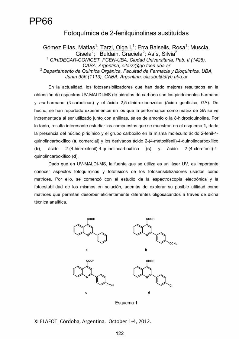
XI ELAFOT. Córdoba, Argentina. October 1 4, 2012. 122
Fotoquímica de 2-fenilquinolinas sustituídas
Gómez Elías, Matías1; Tarzi, Olga I.1; Erra Balsells, Rosa1; Muscia, Gisela2; Buldain, Graciela2; Asís, Silvia2
1 CIHIDECAR-CONICET, FCEN-UBA, Ciudad Universitaria, Pab. II (1428),CABA, Argentina, [email protected]
2 Departamento de Química Orgánica, Facultad de Farmacia y Bioquímica, UBA, Junín 956 (1113), CABA, Argentina, [email protected]
En la actualidad, los fotosensibilizadores que han dado mejores resultados en la
obtención de espectros UV-MALDI-MS de hidratos de carbono son los piridoindoles harmano
y nor-harmano ( -carbolinas) y el ácido 2,5-dihidroxibenzoico (ácido gentísico, GA). De
hecho, se han reportado experimentos en los que la performance como matriz de GA se ve
incrementada al ser utilizado junto con anilinas, sales de amonio o la 8-hidroxiquinolina. Por
lo tanto, resulta interesante estudiar los compuestos que se muestran en el esquema 1, dada
la presencia del núcleo piridínico y el grupo carboxilo en la misma molécula: ácido 2-fenil-4-
quinolincarboxílico (a, comercial) y los derivados ácido 2-(4-metoxifenil)-4-quinolincarboxílico
(b), ácido 2-(4-hidroxifenil)-4-quinolincarboxílico (c) y ácido 2-(4-clorofenil)-4-
quinolincarboxílico (d).
Dado que en UV-MALDI-MS, la fuente que se utiliza es un láser UV, es importante
conocer aspectos fotoquímicos y fotofísicos de los fotosensibilizadores usados como
matrices. Por ello, se comenzó con el estudio de la espectroscopía electrónica y la
fotoestabilidad de los mismos en solución, además de explorar su posible utilidad como
matrices que permitan desorber eficientemente diferentes oligosacáridos a través de dicha
técnica analítica.
N
COOH
N
COOH
OCH3
N
COOH
OH
a b
c
N
COOH
Cl
d
Esquema 1
PP66
122

XI ELAFOT. Córdoba, Argentina. October 1 4, 2012. 123
Degradation of Herbicide 2,4-D employing Ozone and UV Radiation: A complete kinetic scheme
Lovato, María E.1; Gilliard, María B.1, Muchiutti, Ayelén A.2, Ramb, María J.2,Martín, Carlos A.1,2, Cassano, Alberto E.1,2
1 Instituto de Desarrollo Tecnológico para la Industria Química, Colectora de Ruta Nacional Nº 168, Km. 472.5, (3000) Santa Fe, mlovato@santafe-conicet
2 Facultad de Ingeniería y Ciencias Hídricas, Universidad Nacional del Litoral, Ciudad Universitaria, Paraje "El Pozo", (3000) Santa Fe
This work describes the degradation of herbicide 2,4-Dichlorophenoxyacetic Acid (2,4-D) in aqueous solution employing Ozone (O3) based Advanced Oxidation Processes (AOP).
Ozone based methods may involve two different major oxidative species: ozone and •OH radicals. Ozone can also react through the hydroxyl radical generated during its decomposition in water. •OH generation is usually promoted at high pH, with the addition of H2O2, UV irradiation or by reaction with organic compounds itself. Thus, during ozonation in water, both molecular ozone (O3) and hydroxyl radical coexist, having different reactivity and selectivity. The mayor objective was to find a comprehensive reaction pathway for 2,4-D oxidation. Addition of an •OH radical scavenger such as tertiary butanol (t-BuOH) allowed to separately evaluate the contribution of each oxidative species.
In order to perform a comparative study, three sets of experimental runs were carried out: (a) Ozonation reactions (molecular O3+•OH), (b) O3/UV reactions (•OH enhancement), (c) O3+t-BuOH (•OH inhibition). The efficiencies of the different processes were evaluated, taking into account three criteria: (a) percentage conversion of 2,4-D, (b) mineralization rate, and (c) release of chloride ion.
The employed photo-reactor was a cylinder made of Teflon closed in both extremes with two flat, circular windows made of quartz. Two different types of radiation sources were used: (i) Two Philips TUV lamps, 15 W each, (ii) two Heraeus NNI 40/20 lamps, 40 W each. All lamps are low pressure mercury lamps, with one single significant emission wavelength at 253.7 nm. Two different ozone concentrations were utilized (0.1 and 0.23 mM).
In presence of t-BuOH, 2,4-D conversions were lower than those obtained for the same dissolved ozone concentrations, in absence of scavenger. Conversely, the addition of UV radiation improves 2,4-D degradation. For processes involving UV radiation complete dechlorination was found. For the ozonation process, dechlorination ratios were higher than when inhibiting •OH radical reactions. In all cases, TOC removal was significantly lower than the corresponding 2,4-D degradation. The best TOC conversions were reached for O3/UVprocesses, and the poorest results were obtained for O3+t-BuOH.
A reaction mechanism for 2,4-D oxidation was elucidated. Using specific mass spectrometric analysis applied to the degradation reaction of 2,4-D and afterwards, working in the same way, with its first aromatic intermediary (2,4-dichlorophenol) it was possible to identify the components that were included in the mechanistic proposal. When using t-BuOH only the direct reactions of molecular ozone can take place, and some intermediates were not identified. The intermediaries generated from previous compounds that neither have double C=C bonds nor aromatic rings in their molecular structure have not been found.
References E. Brillas et al., Mineralization of 2,4-D by advanced electrochemical oxidation processes, Water Res. 34 (2000) 2253-2262. C. Badellino et al., Oxidation of pesticides by in situ electrogenerated hydrogen peroxide: Study for the degradation of 2,4-dichlorophenoxyacetic acid, J. Hazard. Mater. 137 (2006) 856-864. J. Peller et al., Hydroxyl radical’s role in the remediation of a common herbicide, 2,4-dichlorophenoxyacetic acid (2,4-D), J. Phys. Chem. A 108 (2004) 10925-10933.
PP67
123

XI ELAFOT. Córdoba, Argentina. October 1-4, 2012.
Squaraine Dyes as Optical Sensor for Gliadin Protein in Aqueous Solutions
de Abreu, Marluza Pereira1; Ribeiro, Andresa da Costa2; Scheibel, Joice Maria2;Soares, Rosane Michele Duarte2; da Silveira, Nádya Pesce2; Rodembusch, Fabiano
Severo1; Campo, Leandra Franciscato1
1Laboratório de Novos Materiais Orgânicos2Laboratório de Instrumentação e Dinâmica Molecular
IQ-UFRGS, Av. Bento Gonçalves, 9500, 91501-970, Porto Alegre, RS, Brazil,[email protected]
Squaraine dyes play an important role for proteins investigation due to their photophysical properties [1]. Furthermore, they can form aggregates in certain conditions that can even suppress the emission [1]. Over the last years, this particular feature has been used to detect proteins in solution, since it is well know that these dyes can switch on the fluorescence in the presence of specific peptides. It is worth mentioning thatprotein detection in solution is a very important tool for disease diagnosis, as the celiac disease [2]. In this way, the aim of this work was tostudy the photophysical behaviour of a squaraine dye [1] (SQdye) in presence of gliadin protein. For this purpose, solutions containing gliadinand SQ dye were investigated, using fluorescencespectroscopy. Two solutions were used, water and ethanol:water (6:4) (v:v) in pH 1.2, 6.8 and 9.8. The dye and the protein concentrations were 2.8 and 1.4 μmol·L-1, respectively.
Figure 1 shows the fluorescence emission of SQ dye in water at pH 1.2, 6.8 and 9.8, respectively. It can be observed that the dye is non-fluorescent at all pH values (red line),showing only a small fluorescence band (700 nm) at pH 9.8 ascribed to J-type aggregates. The gliadin in solution switches the dye fluorescence emission. The emission intensity is pH
dependent, increasing the fluorescence intensity from acid to basic pH. This result is probably related to anionic interaction between protein and SQ dye, since in basic media, gliadin can be found as an anionic specie (pI ~ 6.0) affording a negative environment [3].In other words, at pH 9.8 there is an effective interaction between the positive indol core present in the SQ dyes and gliadin. This interaction avoids the SQ dyes aggregation and allows the dye to decay radiactively. This behavior could not be detected inethanol/water solutions, since in absence of protein,the SQ dye is already fluorescent. In addition, nospecific aggregation could be detected. Asconclusion, the SQ dye seemed to be a promising strategy and a potential probe for gliadin detection in water solution, as already observed to the BSA
protein.
Acknowledgements: Conselho Nacional de Desenvolvimento Científico e Tecnológico (CNPq) and the Instituto Nacional de Inovação em Diagnósticos para Saúde Pública (INDI-Saúde) for financial support.
References[1] Pisoni, D.S., Petzhold, C.L., de Abreu, M.P. et al., C. R. Chimie., 2012, 15, 454-462.[2] Sollid, L. M. Annu. Rev. Immunol., 2000, 18, 53-81.[3] Liang, L., Pinier, M., Leroux, J.-C. Et al., Biopolymers, 2009, 91, 169-178.
Figure 1 - Fluorescence spectra in watersolution in different pHs of SQ dye inpresence and absence of gliadin.
124
PP68
124

XI ELAFOT. Córdoba, Argentina. October 1 4, 2012. 125
Influence of cucurbit[n]uril inclusion on photosensitizer mediated protein damage
Fuentealba, Denis1
1 Laboratorio de Química Biológica, Facultad de Química, Pontificia Universidad Católica de Chile, Vicuña Mackenna 4860, Macul, Santiago, Chile,
Cucurbit[n]urils are a family of macrocycles composed of 5-10 units of glycoluril joined by methylene bridges (scheme 1a). These macrocycles have great potential for the encapsulation, transport and delivery of drugs.[1,2] High binding affinities towards cucurbit[n]urils are usually observed for positively charged molecules, such as protonated amines, with a hydrophobic core.[3,4] An interesting molecule to study the formation of a supramolecular complex is the photosensitizer methylene blue. This small molecule is included inside the cavity of cucurbit[7]uril and cucurbit[8]uril forming strong complexes. The photosensitizing properties of these complexes are widely unknown. Additionally, due to the possibility of using cucurbit[n]urils to transport photosensitizers in biological systems, the interactions of these complexes with different cellular components are of importance. In this context, the purpose of this study is to assess the formation of supramolecular aggregates between the photosensitizer-cucurbit[n]uril complex and a protein, and how this supramolecular interaction alters the photochemistry of the molecule (scheme 1b).
Initial results will be presented for the formation and photoactivity of the complexes with proteins.
Scheme 1. (a) Dimensions of the cucurbit[n]uril macrocycle. (b) Cartoon showing the system composed of methylene blue@cucurbit[7]uril complex and human serum albumin.
References
1. Macartney, D. H., Encapsulation of Drug Molecules by Cucurbiturils:Effects on their Chemical Properties in Aqueous Solution. Isr. J. Chem. 2011, 51, 600-615. 2. Walker, S.; Oun, R.; McInees, F. J.; Wheate, N. J., The Potential of Cucurbit[n]urils in Drug Delivery. Isr. J. Chem. 2011, 51, 616-624.3. Lagona, J.; Mukhopadhyay, P.; Chakrabarti, S.; Isaacs, L., The cucurbit[n]uril family. Angew. Chem. Int. Ed. 2005, 44 (31), 4844-4870 4. Lee, J. W.; Samal, S.; Selvapalam, N.; Kim, H.-J.; Kim, K., Cucurbituril Homologues and Derivatives: New Opportunities in Supramolecular Chemistry. Acc. Chem. Res. 2003, 36 (8), 621-630
PP69
125

XI ELAFOT. Córdoba, Argentina. October 1 4, 2012. 126
Effect of the addition of alkanols of different topology to Dipalmitoyl-phosphatidylcholine vesicles in the presence of
gramicidin
Soto-Arriaza, M. A.1*, Olivares-Ortega,C.1,Lissi, E.A.2
1Departamento de Química-Física, Facultad de Química, PontificiaUniversidadCatólica de Chile, 6094411, Av. VicuñaMackenna 4860, Macul,
Santiago, Chile. E-mail: [email protected] 2Departamento de Química, Facultad de Química y Biología, Universidadde Santiago
(USACH), Casilla 40, Correo 33, Santiago, Chile. E-mail: [email protected]
In the present work, we analyse the effect of incorporation of the nonanol family (e.g. 1-Nonanol (1-N), 5-Nonanol (5-N) and 2,6-Dimethyl-4-Heptanol (2,6-DH)) into DPPC LUVs in the presence of different gramicidin concentrations. The principal aim of this work is to study the effect of alkanols solubilization on the physicochemical properties of lipid bilayers in the presence of peptide trans-membrane channels. Fluorescence measurements were carried out at 20°C after direct excitation of the extrinsic probe or by fluorescence resonance energy transfer (FRET) from the tryptophan group of gramicidin. 1-N generates most important changes in the inner part of the bilayer, where it produces an increase of bulk acyl chain mobility. Similarly, 1-N significantly modifies the properties of the hydrophilic-hydrophobic interface region sensed by Laurdan, increasing the polarity of the probe microenvironment and/or increasing the relaxation time of interfacial water molecules. On the other hand, 1-N produces a decrease of PDA fluorescence lifetime, a result that can be explained by a significant amount of water entrance to the inner part of the bilayer. The same behavior was observed when pseudo-first-order quenching rate constants by oxygen were measured. 1-N produces an increase in mobility/solubility of the oxygen in the lipid membrane, an effect that is more noticeable in the deep region of the bilayer sensed by PDA, in the absence and in the presence of 2 mol% of Gr. On the other hand, the addition of three alkanol in the presence of Gr produces a noticeable increase in the water permeability where 1-N show the more important effect respect to branched alkanol. In this context we proposea scheme that represent the effect of 1-nonanol on the water outflow in DPPC LUVs in the absence and in the presence of Gr.
Scheme 1: Representative scheme of lipid bilayer in the presence of gramicidin peptide channel. Models shows water outflow after CaCl2 hypertonic shock in the absence of 1-nonanol (A) and in the presence of 1 mM of 1-nonanol (B). The arrows represent water outflow through peptide channels and membrane bilayer.
Acknowledgements:This work has been supported by Vice Rectoría de Investigación and Facultad de Química of Pontificia Universidad Católica de Chile (MSA). 1.- D.A. Kelkar, A. Chattopadhyay, Modulation of gramicidin channel conformation and organization by hydrophobic mismatch in saturated phosphatidylcholine bilayers, Biochim. Biophys.Acta 1768 (2007) 1103–1113. 2.-M.A. Soto, C.P. Sotomayor, E. Lissi, Effect of gramicidin addition upon the physicochemical properties of dipalmitoylphosphatidyl choline large unilamellar vesicles, J. Photochem. Photobiol.A 152 (2002) 79–93. 3.-C.N. Sobral, M.A. Soto, A.M. Carmona-Ribeiro, Characterization of DODAB/DPPC vesicles, Chem. Phys. Lipids 152 (2008) 38–45.
PP70
126

XI ELAFOT. Córdoba, Argentina. October 1 4, 2012. 127
Interaction of gramicidin with DPPC/DODAB bilayer fragments
Carvalho,C.1, Olivares-Ortega,C.1,2,Soto-Arriaza,M.A.1,2*, Carmona-Ribeiro, A.M.1*
1Departamento de Bioquímica, Instituto de Química, Universidade de São Paulo, CP 26077, CEP 05513-970, São Paulo, SP, Brazil.E-mail: [email protected]
2Departamento de Química-Física, Facultad de Química, PontificiaUniversidadCatólica de Chile, 6094411, Av. VicuñaMackenna 4860, Macul,
Santiago, Chile. E-mail: [email protected]
The interaction between the antimicrobial peptide gramicidin (Gr) and DPPC/DODAB 1:1 large unilamellar vesicles (LV) or bilayer fragments (BF) was evaluated by means of several techniques. The major methods were: 1) Gr intrinsic fluorescence and circular dichroism (CD) spectroscopy; 2) dynamic light scattering for sizing and zeta-potential analysis; 3) determination of the bilayer phase transition from extrinsic fluorescence of bilayer probes; 4) pictures of the dispersions for evaluation of colloidal stability over a range of time and NaCl concentration. For Gr in LV, the Gr dimeric channel conformation is suggested from: 1) CD and intrinsic fluorescence spectra similar to those in trifluoroethanol (TFE); 2) KCl or glucose permeation through the LV/Gr bilayer. For Gr in BF, the intertwined dimeric, non-channel Gr conformation is evidenced by CD and intrinsic fluorescence spectra similar to those in ethanol. Both LV and BF shield Gr tryptophans against quenching by acrylamide but the Stern-Volmer quenching constant was slightly higher for Gr in BF confirming that the peptide is more exposed to the water phase in BF than in LV. The DPPC/DODAB/Gr supramolecular assemblies may predict the behaviour of other antimicrobial peptides in assemblies with lipids.
Scheme 1: Schematic representation of Gr conformations in different media: TFE, ethanol, DPPC/DODAB 1:1 LV or BF.
Acknowledgements:The authors thank the Fundação de Amparo à Pesquisa do Estado de São Paulo (FAPESP), the ConselhoNacional de DesenvolvimentoCientífico e Tecnológico (CNPq) and the International Cooperation Program of CNPq/CONICYT for financial support. CAC is the recipient of a CNPq-PIBIC undergraduate fellowship. We thank Professor Frank Quina for the use of the spectrofluorimeter in his lab.
References 1.-D.A. Kelkar, A. Chattopadhyay, Modulation of gramicidin channel conformation and organization by hydrophobic mismatch in saturated phosphatidylcholine bilayers, Biochim. Biophys.Acta 1768 (2007) 1103–1113. 2.-M.A. Soto, C.P. Sotomayor, E. Lissi, Effect of gramicidin addition upon the physicochemical properties of dipalmitoylphosphatidyl choline large unilamellar vesicles, J. Photochem. Photobiol.A 152 (2002) 79–93. 3.-A.M. Carmona-Ribeiro, Lipid bilayer fragments and disks in drug delivery, Curr. Med. Chem. 13 (2006) 1359–1370. 4.-C.N. Sobral, M.A. Soto, A.M. Carmona-Ribeiro, Characterization of DODAB/DPPC vesicles, Chem. Phys. Lipids 152 (2008) 38–45.
PP71
127

XI ELAFOT. Córdoba, Argentina. October 1 4, 2012. 128
Ruthenium compounds as photosensitizers of singlet oxygen
Maia, P. J. S.1*; Carlos, R. M.1
1 Departamento de Química, Universidade Federal de São Carlos, São Carlos, Brasil, * [email protected]
Singlet oxygen (1O2) was first observed in 1924 and is one of the most active intermediates involved in chemical and biochemical reactions and it has been attract much attention due to their potential applications in biology, environmental, chemical and life science. The main method to produce singlet oxygen is by photosensitized reactions. This photophysical process may occurs by energy transfer of luminescent compounds for the oxygen molecule in its ground state triplet. A strategy that has been studied is the use of transition metal complexes as sensitizers for the activation of the 1O2. Our research group has been working on the development of luminescent mononuclear complexes to activate the singlet oxygen production. Our interest is to obtain complexes with bidentate ligands, for example, phenanthroline, aiming to favor the luminescence of the photosensitizer. With interest focused on ligands that are good acceptors, we prepared the complex cis-[Ru(phen-pery)3]2+ where phen = 1,10-phenanthroline and pery = perylene. The complex was synthesized and characterized by spectroscopic (UV-vis, 1H,13C NMR) and electrochemical (cyclic voltammetry, pulse differential). The photochemical and photophysical properties were investigated by UV-Vis absorption, luminescence emission, transient absorption spectra and DFT/TDDFT calculations. It is expected that the stabilization of the d -orbitals metal by increased -conjugation will be responsible for the drastic change in the energies of the metal d orbitals shiftting the absorption to higher energy, the redox potentials to lower potencials and increase the MLCT emission lifetime of complex. Indeed, the cyclic voltammogram in acetonitrile showed a Ru2+/Ru3+ redox couple at 0.1 V vs Ag/AgCl and three reductive peaks related to the phen-per ligand. The complex exhibits a broad and intense absorption band in the visible region at 550 nm and higher energy absorptions at 290 nm, atributted to MLCT/LLCT (Ru(d ) phen-pery( *) absorptions and LLCT(phen-pery ( *) phen-peyr( *) absorptions respectively. The complex is inert to photochemical reactions and displays intense emission at 640 nm assigned to a mixture of a MLCT (Ru phen-pery) and LLCT (phen phen-pery) electronic transitions. The emission intensity and lifetime of complex spectrum was quenched in air and O2 atmosphere to different extents. This observation is an important indicator that oxygen supply is a crucial factor in the efficacy of photoactivation of oxygen singlet by the complex, and will be of particular significance to practical applications of the complex.
560 580 600 620 640 660 680 700 720 7400
5
10
15
20
25
30
35
40
45
50
Wavelength (nm)
rela
tive
Inte
nsity
(ua)
These results are promise, since the objectives is to use this complex as photosensitizers to generate singlet oxygen.
PP72
128

XI ELAFOT. Córdoba, Argentina. October 1 4, 2012. 129
Lanthanides based Up-conversion Materials
Sorbello, Cecilia1; Gross, Petra2; Jobbágy, Matías1;Strassert, Cristian3; Barja, Beatriz1
1INQUIMAE / DQIAyQF. FCEyN – UBA. Buenos Aires, Argentina 2 Institut für Angewandte Physik- (WWU) Münster, Germany3CeNTech - Institute of Physics (WWU) Münster, Germany
[email protected], [email protected], [email protected], [email protected], [email protected]
Introduction: The process of photon upconversion (UC) is a way to convert long-wavelength excitation radiation into shorter wavelength output radiation. Lanthanide ions commonly have more than one metastable level and long lifetimes (10-6–10-
2s) being the basic requirement for UC [1]. Given that the 4f electrons are shielded by the outer 5s and 5p electrons, radiative processes are much more competitive than multiphonon relaxation in lanthanides compared to other ions.
Experimental: In this work, different amounts of Er(III) and/or Yb(III) lanthanides were doped in CeO2/Gd2O3 matrices (see Table below). The luminescent microparticles (average 150 nm) were synthesized via the homogeneous phase precipitation method through the thermal hydrolysis of urea and the mixed oxides were obtained after heat treatment at
1000oC (5 hs). The characterization of the particles was performed with the powder X-Ray diffraction, IR spectroscopy and high resolution scanning electron microscopy (HRSEM) techniques.
Sample C0E C10E C84E C94E C0EY C10EY C84EY C94EY % CeO2(III) 0 10 84 94 0 10 84 94 % Gd2O3(III) 94 84 10 0 94 84 10 0 % Er2O3(III) 6 6 6 6 5 5 5 5 % Yb2O3(III) 0 0 0 0 1 1 1 1
UC: The samples were illuminated with continuous-wave radiation at a wavelength of 976 nm from a fiber-coupled laser diode (3S Photonics) and the up-converted light was coupled into a transport fiber leading to a spectrometer (Stellarnet EPP2000).
Results and Discussion: The UC emission spectra excited at 976 nm clearly showed the emission bands arising from the emissive 2H11/2 and 4S3/2 (green region- 520-575 nm) and
from the 4F9/2 (red region- 625-725 nm) excited levels to the 4I15/2 ground state of the Er(III) ions (only C0E is shown in the Figure). It was also observed that the addition of Yb(III) ions in the CXEY series slightly enhanced the intensity of the longest wavelength emission when compared with the corresponding CXE samples. At the moment, the dependence of the intensities of the emission maxima on the excitation pump power (10mW- 80mW) is being investigated in terms of the composition and structure of the samples to elucidate what mechanisms are the main responsible for the up converted output light.
Acknowledgements: BCB wants to thank DAAD for financial support.References: [1] M. Haase and H. Schäfer. Angew. Chem. Int. Ed. 2011, 50, 5808 – 5829.
C0EC0E
Figure of UC emission spectra at different incident powers: 10mW (blue), 30mW (red) and 80mW (black)
exc= 976 nm.
PP73
129
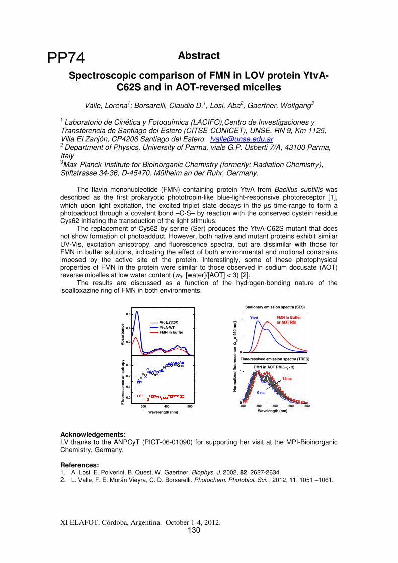
Abstract
XI ELAFOT. Córdoba, Argentina. October 1-4, 2012.
Spectroscopic comparison of FMN in LOV protein YtvA-C62S and in AOT-reversed micelles
Valle, Lorena1; Borsarelli, Claudio D.1, Losi, Aba2, Gaertner, Wolfgang3
1 Laboratorio de Cinética y Fotoquímica (LACIFO),Centro de Investigaciones y Transferencia de Santiago del Estero (CITSE-CONICET), UNSE, RN 9, Km 1125, Villa El Zanjón, CP4206 Santiago del Estero. [email protected] Department of Physics, University of Parma, viale G.P. Usberti 7/A, 43100 Parma, Italy 3Max-Planck-Institute for Bioinorganic Chemistry (formerly: Radiation Chemistry), Stiftstrasse 34-36, D-45470. Mülheim an der Ruhr, Germany.
The flavin mononucleotide (FMN) containing protein YtvA from Bacillus subtillis was described as the first prokaryotic phototropin-like blue-light-responsive photoreceptor [1], which upon light excitation, the excited triplet state decays in the μs time-range to form a photoadduct through a covalent bond –C-S– by reaction with the conserved cystein residue Cys62 initiating the transduction of the light stimulus.
The replacement of Cys62 by serine (Ser) produces the YtvA-C62S mutant that does not show formation of photoadduct. However, both native and mutant proteins exhibit similar UV-Vis, excitation anisotropy, and fluorescence spectra, but are dissimilar with those for FMN in buffer solutions, indicating the effect of both environmental and motional constrains imposed by the active site of the protein. Interestingly, some of these photophysical properties of FMN in the protein were similar to those observed in sodium docusate (AOT) reverse micelles at low water content (w0, [water]/[AOT] < 3) [2].
The results are discussed as a function of the hydrogen-bonding nature of the isoalloxazine ring of FMN in both environments.
450 500 550 600 6500
1
0
1
Time-resolved emission spectra (TRES)
15 ns
Wavelength (nm)
0 ns
Stationary emission spectra (SES)
FMN in AOT RM (w0 <3)
FMN in Bufferor AOT RM
No
rmal
ized
flu
ore
scen
ce (
λλ λλE
x= 45
0 n
m)
YtvA
Acknowledgements: LV thanks to the ANPCyT (PICT-06-01090) for supporting her visit at the MPI-Bioinorganic Chemistry, Germany.
References: 1. A. Losi, E. Polverini, B. Quest, W. Gaertner. Biophys. J. 2002, 82, 2627-2634. 2. L. Valle, F. E. Morán Vieyra, C. D. Borsarelli. Photochem. Photobiol. Sci. , 2012, 11, 1051 –1061.
300 400 500
0.0
0.1
0.2
0.3
Flu
ore
scen
ce a
nis
otr
op
y
Wavelength (nm)
0.2
0.4
0.6
Ab
sorb
ance
YtvA-C62S YtvA-WT FMN in buffer
130
PP74
130
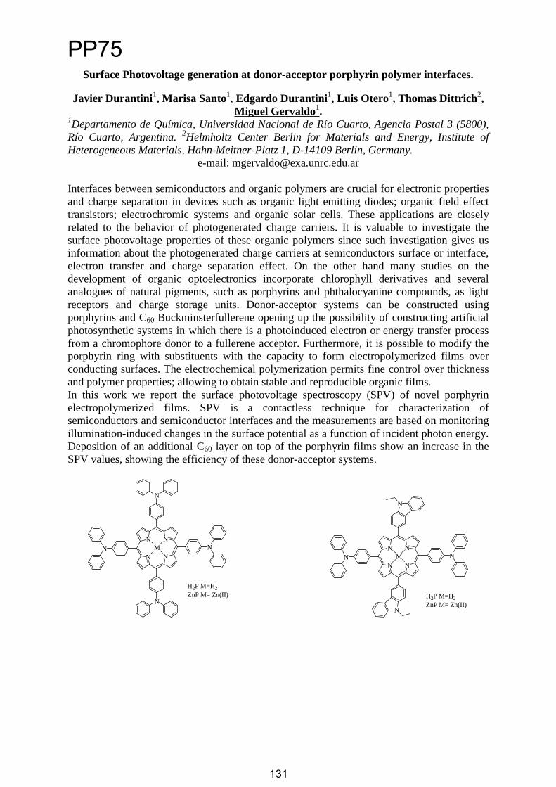
Surface Photovoltage generation at donor-acceptor porphyrin polymer interfaces.
Javier Durantini1, Marisa Santo1, Edgardo Durantini1, Luis Otero1, Thomas Dittrich2,Miguel Gervaldo1.
1Departamento de Química, Universidad Nacional de Río Cuarto, Agencia Postal 3 (5800), Río Cuarto, Argentina. 2Helmholtz Center Berlin for Materials and Energy, Institute of Heterogeneous Materials, Hahn-Meitner-Platz 1, D-14109 Berlin, Germany.
e-mail: [email protected]
Interfaces between semiconductors and organic polymers are crucial for electronic properties and charge separation in devices such as organic light emitting diodes; organic field effect transistors; electrochromic systems and organic solar cells. These applications are closely related to the behavior of photogenerated charge carriers. It is valuable to investigate the surface photovoltage properties of these organic polymers since such investigation gives us information about the photogenerated charge carriers at semiconductors surface or interface, electron transfer and charge separation effect. On the other hand many studies on the development of organic optoelectronics incorporate chlorophyll derivatives and several analogues of natural pigments, such as porphyrins and phthalocyanine compounds, as light receptors and charge storage units. Donor-acceptor systems can be constructed using porphyrins and C60 Buckminsterfullerene opening up the possibility of constructing artificial photosynthetic systems in which there is a photoinduced electron or energy transfer process from a chromophore donor to a fullerene acceptor. Furthermore, it is possible to modify the porphyrin ring with substituents with the capacity to form electropolymerized films over conducting surfaces. The electrochemical polymerization permits fine control over thickness and polymer properties; allowing to obtain stable and reproducible organic films. In this work we report the surface photovoltage spectroscopy (SPV) of novel porphyrin electropolymerized films. SPV is a contactless technique for characterization of semiconductors and semiconductor interfaces and the measurements are based on monitoring illumination-induced changes in the surface potential as a function of incident photon energy. Deposition of an additional C60 layer on top of the porphyrin films show an increase in the SPV values, showing the efficiency of these donor-acceptor systems.
N
NN
N
N
N
N
N M
H2P M=H2
ZnP M= Zn(II)
N
NN
N
N
N
NN
H2P M=H2
ZnP M= Zn(II)
M
131
PP75
131

XI ELAFOT. Córdoba, Argentina. October 1 4, 2012. 132
Photosensitized oxidation of BSA by novel ruthenium polypyridyl complexes with cyano substituents
Mecchia Ortiz, Juan H.1; Morán Vieyra, F. Eduardo2; Mignone, Ricardo A.2 ; Katz, Néstor E.1; Borsarelli, Claudio D.2
1Instituto de Química del Noroeste (INQUINOA-CONICET), UNT.E-mail: [email protected]
2Laboratorio de Cinética y Fotoquímica, Centro de Investigaciones y Transferencia de Santiago del Estero (CITSE-CONICET), UNSE.
Ruthenium (II) polypyridyl complexes (RPC) display excellent properties as both energy- or charge-transfer photosensitizers, which can be modulated with appropriate changes in ligand structure. The well recognized luminescence properties of their metal-to-ligand charge-transfer excited triplet states (3MLCT*) have led to their applications as suitable photoprobes for sensing and/or modification of biomolecules, such as DNA and proteins [1]. Proteins are the most abundant macromolecules in biological environments. In particular, serum albumins are present in submimolar concentration in blood plasma, where their main function is the transport of fatty acids, among others. Recently, we have explored the photosensitized oxidation of both human (HSA) and bovine (BSA) serum albumins by using absorption and emission spectroscopies [2].
In this work, we explored some aspects of the photosensitized oxidation of BSA by a series of novel mononuclear ruthenium complexes: [Ru(bpy)2(Mebpy-CN)](PF6)2 (1),[Ru(bpy)(Mebpy-CN)2](PF6)2 (2), [Ru(Mebpy-CN)3](PF6)2 (3), with (bpy = 2,2’-bipyridine, Mebpy-CN = 4-methyl-2,2 -bipyridine-4 -carbonitrile) in air-saturated phosphate buffer solutions at pH 7.4 and 25 °C. In spite of the low value of the isoelectric point for BSA (pI = 4.8), all the cationic RPC do not bind to BSA, as demonstrated by the lack of changes in either the emission spectra of the complex or of the protein, in solutions containing up to 30 M RPC and 200 M BSA. Under these conditions, the emission anisotropy of the complexes remained unchanged and 0, as can be expected for a free rotor in fluid media. However, continuous irradiation with blue light (460 nm) of air-saturated aqueous solutions of 30 M RPC and 120
M BSA produces only a progressive hyperchromic effect around 300-400 nm, indicating that photolysis modifies BSA but not RPC. The differential absorbance spectra indicated the formation of a new band with maximum at 320 nm. These changes were paralleled with the decrease of almost 50-60% of the fluorescence intensity of BSA, observed by excitation at 295 nm, and with the increase of intensity of a new emission band at 410 nm by excitation at 320 nm. Laser-flash photolysis experiments indicated that the 3MLCT* of all RPC were efficiently quenched by molecular oxygen (kq
O2 = 2-3 109 M-1s-1), and that singlet molecular oxygen (1O2) was produced with quantum yields higher than that of [Ru(bpy)3](PF6)2, as determined by time-resolved NIR phosphorescence detection in aerated acetonitrile.
The above results indicate that the RPC 1-3 are not associated to BSA, but their excited states are capable of producing 1O2 in the bulk solution, which diffuses to the protein molecule to oxidize Trp residues (among other electron-rich aminoacids, e.g. Cys, His, etc), leading to the formation of secondary oxidation products N-formylkynurenine and HPI (3a-hydroxypyrolidinoindole) that give rise to the absorbance at 320 nm and the emission around 410 nm. Therefore, complexes 1-3 are suitable for the photo-oxidation of proteins and subsequently can be easily removed from the modified protein solution by ultrafiltration, dialysis, or ion-exchange chromatography. References: [1] Herman, L.; Ghosh, S.; Defrancq, E.; Mesmaeker, A. K. J. Phys. Org. Chem. 2008, 21, 670-681. [2] Alarcón, E.; Edwards, A. M.; Aspée, A.; Gonzalez-Nilo, D.; Morán, F. E. Borsarelli, C. D.; Lissi. E. A., Poblete, H.; Scaiano, J.C. Photochem. Photobiol. Sci. 2010, 9, 93 – 102.
PP76
132

XI ELAFOT. Córdoba, Argentina. October 1 4, 2012. 133
Photophysical Properties of Surface-Modified Silicon Nanoparticles
Romero, Juan José1; Dell’Arciprete María Laura1, González Mónica1
1INIFTA, Dpto. de Química, FCE, UNLP. CC 16 Suc. 4, (1900) La Plata, Argentina.
Silicon nanoparticles of 1_5 nm size (Si NPs), also known as quantum dots, received great attention, as they combine size-dependent photoluminescence (PL) with the richness of silicon surface chemistry. The momentum requirements which make bulk Si a rather inefficient light emitter are relaxed in the 1_5 nm size silicon crystals as a result of quantum confinement effects. The optical emission properties of these chromophores can be tailored by suitably adjusting the height and width of the potential that confines electrons and holes. In spherically shaped colloidal dots, the band gap and oscillator strength can be tuned by varying the diameter [1]. Silicon in the form of small structures shows a special interest because it is promising for light-emitting optoelectronics, photonics, light emitters in biological labelling, and as photosensitizers of singlet oxygen [2]. In the present work, we report the synthesis of Si NPs by three different routes: A wet chemical method employing reverse micelles [3], an electrochemical technique [4] and using HF etching over porous silicon [5]. In order to stabilize the luminescent properties, the different particles were capped with organic molecules using either thermal silanization or photo-initiated silylation. Naked and protected Si NPs were characterized by XPS, FTIR and TEM. Photophysical properties were evaluated using UV-Vis Absorption Spectroscopy, Fluorescence Spectroscopy and Time Correlated Single Photon Counting (TCSPC). Through Time Resolved Emission Spectrsocopy (TRES) and Time Resolved Emission Anisotropy (TREA) measurements, it was possible to study the nature of the excited state [6].
(a) TEM image, (b) size distribution histogram and (c) Emission-Excitation Matrix (EEM) of Si NPs synthesized using reverse micelles.
References [1] Delley, B.; Steigmeier, E. F. Appl. Phys. Lett. 1995, 67, 2370-2372. [2] Llansola Portolés, M. J.; David Gara, P. M.; Kotler, M. L.; Bertolotti, S.; San Roman, E.;
Rodríguez, H. B.; Gonzalez, M. C. Langmuir 2010, 26, 10953-10960. [3] Rosso-Vasic, M.; Spruijt, E.; Lagen, B. v.; Cola, L. D.; Zuilhof, H. Small 2008, 4, 1835-
1841.[4] Llansola Portolés, M. J.; Rodríguez Nieto, F.; Soria, D. B.; Amalvy, J. I.; Peruzzo, P. J.;
Mártire, D. O.; Kotler, M.; Holub, O.; Gonzalez, M. C. J. Phys. Chem. C 2009, 113, 13694–13702.
[5] Hua, F.; Erogbogbo, F.; Swihart, M. T.; Ruckenstein. E. Langmuir 2006, 22, 4363-4370.
[6] Llansola Portolés, M. J.; Pis Diez, R.; Dell’Arciprete, M. L.; Caregnato, P.; Romero, J. J.; Mártire, D. O.; Azzaroni, O.; Ceolín, M.; Gonzalez, M. C. J. Phys. Chem. C, 2012,116, 11315–11325
PP77
133

Kinetics and Dynamics of Gas Phase PeroxyacidsTaravella Florencia, Arguello Gustavo A & Burgos Paci Maximiliano
INFICQ,[email protected]
Peroxyacids (RC(O)OOH) are a family of chemical compounds widely used in chemistry because of its oxidizing capacity. They have also been proposed as important intermediates in the atmospheric degradation of organic compounds. Beyond their practical uses, peroxyacides exhibit very interesting intramolecular properties, like intramolecular hydrogen bonding and the dissociation energy of the RO-OH bond has not been determined precisely experimentally.
As part of our previous work we have done some experiments studying the kinetics of thermal decomposition of CH3C(O)OOH. These results indicate that the decomposition follows the mechanisms:
CH3C(O)OOH CH3C(O)O + OHCH3C(O)O CH3 + CO2
CH3 + OH CH3OH
The dissociation energy for the peroxy bond was determined to be 41,1 Kcal mol-1.
At the moment we are setting up a laser chemistry laboratory to study the gas phase photodecomposition of hydrogen and fluorinated peroxyacids. The third harmonic of a Nd-YAG laser (Quantel, Brillant B) operated at 10 Hz with 100mJ is used to pump a OPO (GWU) with a signal beam in the wavelength region 400-700nm. The OPO signal is focused on the cell and laser induced fluorescence of the photochemical products is registered using a monochromator and a PMT. The temporal evolution of the PMT signal is averaged using a boxcar integrator. Fresh results using this system will be presentedin the congress.
134
PP78
134
XI ELAFOT. Córdoba, Argentina. October 1 4, 2012. 135
Photodynamic therapy in HeLa and mice fibrosarcoma cells using m-tetrahydroxyphenyl chlorin and 650 nm Light
ETCHEVERRY, María Eugenia 1, GALARZA, Celeste 1, PASQUALE, Miguel Angel2,BIBE, Solange 3, GUTIERREZ Anabela 3, PONZINIBBIO, Carlos 3, POTECA,
Horacio4, GARAVAGLIA, Mario 1,5
1 Departamento de Física, Facultad de Ciencias Exactas, Universidad Nacional de La Plata (UNLP), La Plata, Argentina. [email protected]
2 Instituto de Investigaciones Físicoquímicas Teóricas y Aplicadas (INIFTA) (CCT CONICET La Plata, UNLP y CIC). [email protected]
3 Cátedra de Patología, Facultad de Ciencias Médicas, UNLP. 4Centro Médico Láser, La Plata, Argentina.
1,5 Centro de Investigaciones Opticas (CIOp) (CCT CONICET La Plata, y CIC)
The photodynamic therapy (PDT) consists in the administration of a non toxic photosensitizer (PS) drug and after a certain period of time in which the drug is accumulated in the tumor it is irradiated with visible light, usually a long wavelength red light [1].
In this contribution we present results of PDT employing a 0.8 W LED lamp and a Laser (Medligth FD1, with 0.9 W) both emitting at 650 nm, and (meta-tetra(hydroxyfenyl)) chlorine – Foscan as photosensitizer (PS). HeLa, cells passage 44-60, and fibrosarcoma cells (TMC) obtained from an induced tumor on the flank of BalbC mices were employed. Cell cultured were obtained seeding 2 ml of RPMI medium (Gibco, Invitrogen Corp.) supplemented with 10% FBS, 2g/l bicarbonate, and 100μm/ml streptomycin containing 50000–75000 cells in 3.6 cm Petri dishes. The cultures were incubated overnight in an oven at 37º C in a 5 % carbon dioxide and 97 % humidity atmosphere. Then the medium was replaced by new one supplemented with 2 % FBS with and without PS and incubated for different times (tF). Before PDT treatment cultures were washed and fresh complete medium was added. Cell cultures containing PS were manipulated in black boxes to avoid influence of natural light. PS concentration (cF) in the range 0.05 cF 80 g/ml, tF in the range 90 tF 4320 min and the radiation time in the range 2 tR 18 min were employed. Cell viability before and after PDT treatment was evaluated with standard supra-vital dyes and optical phase contrast and fluorescence microscopy. The effectiveness of PDT treatment was evaluated by cell counting. For some conditions apoptosis and necrosis were investigated.
PS intracellular localization was determined by fluorescence microscopy. It was observed in Golgi apparatus and endoplasmic reticulum and the signal increase with tF and cF.For cF > 40 g/ml and tF > 24 hs, natural toxicity towards both types of cell was observed. After PDT treatment with both light sources at 0.43 W/cm2, for cF < 0.05 g/ml, tF = 2880 min and tR = 10 min, many cells remain viable and the culture recover the normal growing rate. For values 0.1 cF 1.0 g/ml the deleterious effect of the PDT treatment increases with tF and tR.In this case the effect was better appreciated after 24 hs post treatment. It resulted that PDT treatment was about 50% more effective against TMC cells than HeLa cells. Furthermore for both cell phenotypes, staining analysis indicated that cell death mainly occurred by necrosis. For the largest cF values, cell-substrate contact area diminished significantly and formation of vacuoles and cytoplasm contraction could be observed even during irradiation.
The presented results suggest that the proposed irradiation source appears to be useful for in vitro photodynamic studies and in vivo animal models.
Acknowledgements: Authors acknowledge Dr. P. H. González for laboratory facilities.
References [1] A.P. Castano, T.N. Demidova, M.R. Hamblin, Mechanism in photodynamic therapy: part I-photosensitizers, photochemistry and cellular localization. Photodiagnosis and Photodynamic Therapy 1, 279-293 (2004).
PP79
135

XI ELAFOT. Córdoba, Argentina. October 1 4, 2012. 136
Use of PARAFAC Analysis: Correlations between Excitation Emission Matrix Fluorescence and Physical-Chemical
Parameters of Water Quality (Iguazu River, Parana, Brazil)
Sánez, Juan1; Froehner, Sandro1; Fernandes, Cristovão1; Dombroski, Luiz1; Sánez, Patricia2, Knapki, Heloise
1Department of Hydraulics and Sanitation, Federal University of Parana, Curitiba, Paraná, Brazil CEP 81531-980, [email protected]
2Mathematic Department, Center of Technologic Sciences, State University of Santa Catarina, Joinville, Santa Catarina, Brazil CEP 89219-710,
The Barigui River watershed is located in the metropolitan region of Curitiba, state of Parana, southern Brazil. Last decades, due to growth and non planified occupation along the river, in addition to lack of sanitation, the basin suffers a very large inflow of untreated domestic sewage [1]. Evaluation of water quality of fresh waters is a priority task in order to set its availability for different uses, such as drinking water, irrigation, etc. Current programs to monitor the watershed use traditional physical-chemical parameters. Normally, the process is tedious consuming time, human resources and an extensive budget, which is prohibited in developing countries, specially.
Diverse fluorescence centers (fingerprints) in the dissolved organic matter (DOM) of surface waters have the property to fluoresce when excited by light [2]. The excitation emission matrix (EEM) is the fluorescence technique which can aid us to identify those fingerprints [3], being easy to perform the analysis and obtain fast results, at a relatively cheap cost [4]. Diverse studies have correlated those fingerprints with some particular water quality parameter [i.e. 2, 5]. However, the analysis is tedious because the excitation matrix is large, more than 1,000 data per matrix. In that sense, parallel factor analysis (PARAFAC) provides a more easy characterization of those fingerprints. PARAFAC can identify independent fluorescent components, and, later, each component can be correlated to one or several water quality parameters [3, 6].
Our primary objective in this research was to observe if a relationship exists among the components found by the PARAFAC analysis of EEM and water quality parameters. For the case water was collected from six sampling stations located in the Barigui Basin in ten campaigns (2,009 2,010). EEM data was treated following procedure by Stedmond and Bro [3] using a program in N-way tool box in MATLAB® [7]. Different correlations were analyzed between the PARAFAC components and water quality parameters.
References [1] Froehner, S., Martins, R. F., Errera, M. R., (2009). Assessment of fecal sterols in Barigui River
sediments in Curitiba, Brazil. Environ Monit Assess 157, 591–600 [2] Baker, A. 2001. Fluorescence Excitation-Emission matrix characterization of some sewage
impacted rivers. Environ. Sci. Technol. 35, 948-953. [3] Stedmon C. A., Bro, R. (2008), Limnol. Oceanogr.: Methods 6, 572–579 [4] Bell, K. Y., Sánez, J., Wells, M.J.M., (2012). Optimizing Disinfection Pretreatment using
Excitation emission Matrix Fluorescence Spectroscopy. Ozone: Science & Engineering: The Journal of the International Ozone Association, 34:2, 109-114
[5] Sánez, J.M., “Evaluation of Drinking Water Quality and Wastewater Effluents by Fluorescence Excitation Emission Matrix Spectroscopy”, Doctoral Dissertation, Tennessee Technological University, Cookeville,TN (2010).
[6] Hua, B., Veum, K., Yang, J., Jones, J., Deng, B., (2010). Parallel factor analysis of fluorescence EEM spectra to identify THM precursors in lake waters. Environ Monit Assess 161, 71–81
[7] http://www.models.kvl.dk/source.
PP80
136
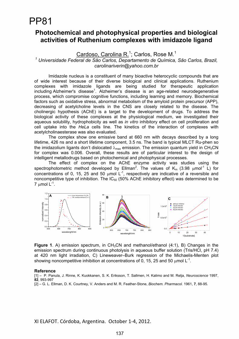
XI ELAFOT. Córdoba, Argentina. October 1 4, 2012. 137
Photochemical and photophysical properties and biological activities of Ruthenium complexes with imidazole ligand
Cardoso, Carolina R.1; Carlos, Rose M.11 Universidade Federal de São Carlos, Departamento de Química, São Carlos, Brazil,
Imidazole nucleus is a constituent of many bioactive heterocyclic compounds that are of wide interest because of their diverse biological and clinical applications. Ruthenium complexes with imidazole ligands are being studied for therapeutic application including Alzheimer's disease1. Alzheimer’s disease is an age-related neurodegenerative process, which compromise cognitive functions, including learning and memory. Biochemical factors such as oxidative stress, abnormal metabolism of the amyloid protein precursor (APP), decreasing of acetylcholine levels in the CNS are closely related to the disease. The cholinergic hypothesis (AChE) is a target to the development of drugs. To address the biological activity of these complexes at the physiological medium, we investigated their aqueous solubility, hydrophobicity as well as in vitro inhibitory effect on cell proliferation and cell uptake into the HeLa cells line. The kinetics of the interaction of complexes with acetylcholinaesterase was also evaluated.
The complex show one emissive band at 660 nm with decays described by a long lifetime, 426 ns and a short lifetime component, 3.5 ns. The band is typical MLCT Ru-phen so the imidazolium ligants don’t dislocated max emission. The emission quantum yield in CH3CN for complex was 0.006. Overall, these results are of particular interest to the design of intelligent metallodrugs based on photochemical and photophysical processes.
The effect of complex on the AChE enzyme activity was studies using the spectrophotometric method developed by Ellman2. The values of Km (3.98 mol-1 L) for concentrations of 0, 15, 25 and 50 mol L-1, respectively are indicative of a reversible and noncompetitive type of inhibition. The IC50 (50% AChE inhibitory effect) was determined to be 7 mol L-1.
500 600 700 800 9000.0
0.2
0.4
0.6
0.8
1.0
1.2A room temperature CH
3CN
77K methanol/ethanol (4:1)
norm
aliz
ed in
tens
ity
wavelenght/nm600 650 700 750 800 850
0
20
40
60
80
B
Emis
sion
inte
nsity
(a.u
.)
wavelength/nm 0 10 20 30 40
0.0
6.0x10⁵
1.2x10⁶
C without inhibitor 15 M inhib itor 25 M inhib itor 50 M inhib itor
1/ra
te
1/[substrate]
Figure 1. A) emission spectrum, in CH3CN and methanol/ethanol (4:1), B) Changes in the emission spectrum during continuous photolysis in aqueous buffer solution (Tris/HCl, pH 7.4) at 420 nm light irradiation, C) Lineweaver–Burk regression of the Michaelis-Menten plot showing noncompetitive inhibition at concentrations of 0, 15, 25 and 50 mol L-1.
Reference[1] – P. Panula, J. Rinne, K. Kuokkanen, S. K. Eriksson, T. Sallmen, H. Kalimo and M. Relja, Neuroscience 1997, 82, 993-997 [2] – G. L. Ellman, D. K. Courtney, V. Anders and M. R. Feather-Stone, Biochem. Pharmacol. 1961, 7, 88-95.
PP81
137

XI ELAFOT. Córdoba, Argentina. October 1-4, 2012.
Interaction of Auramine O with montmorillonite ClaysPoli, Alessandra L.1; Ferreira, Avelardo U.C.1, Neumann, Miguel G.1, Schmitt, C.C.1
1 Instituto de Química de São Carlos, Universidade de São Paulo, São Carlos, [email protected]
Cationic dyes have been intercalated into clay interlayers and various chromatic changes based on the intercalation reactions have been revealed to be induced by the formation of aggregates through intermolecular interactions such as the static interaction with the clay surfaces or by protonation [1,2]. In this work, we report a spectroscopy study on thetime evolution of the absorption of Auramine O by montmorillonite clays and emission properties of the dye in clay suspensions. The fluorescence of Auramine O increases immediately after mixing the dye solution with the suspension of clay due to its adsorption on the external surface of the clays, which restricts the torsional molecular motion of Auramine. At longer times, the dye molecules migrate into the interlamelar region of the clay particles. The aggregation of the dye molecules can be occurring in the interlayer region, leading to the decrease of the fluorescence emission.
0 15000 30000 45000 600000.0
0.2
0.4
0.6
0.8
350 400 450 500 550-0.1
0.0
0.1
0.2
0.3
0.4
0.5
0.6
0.7
0.8
37d20 d10 d7d
5 d4d
1d
Time, min
A41
8 nm
Abs
orba
nce
, nm
0 10 4 h 1 d 4 d 5 d 6 d 7 d 8 d 10 d 20 d 37 d
460 480 500 520 540 560 580 600 620 640 660 680 700
0
500[AuO] = 2,0 x 10 - 5 Mol.L-1
[SAz - 1] = 0,11 g.L-1
Inte
nsid
ade
, nm
0 10 2 h 1 d 2 d 3 d 4 d 5 d 6 d 8 d 10 d 13 d 21 d 37 d
Absorption and Emission spectra of Auramine O in SAz-1 clay suspension.The clays with higher charge density, like SAz-1, show larger particle size that decreases the surface area available for adsorption. On the other hand, SWy-1, having a lower charge density, shows higher surface area available for adsorption of the dye. The dye adsorbed on external surface of SWy-1 induces the flocculation. The difference of the behavior in SAz-1and SWy-1 can be traced to the higher ability of the latter to form clay aggregates. In the case of SWy-1, the AuO+ aggregates are formed in the interparticle spaces of the clay clusters.
Auramine in SWy-1 suspensionThe fluorescence quantum yields ( F) of AuO on the natural montmorillonites SAz-1, SWy-1, Syn-1 and Laponite clays were 0.015, 0.007, 0.016 and 0.017, respectively. These values are higher than the F of AuO in aqueous solution and are of the same order of magnitude of the F found for viscous solvents such as n-hexanol and n-heptanol (0.014 and 0.015).Acknowledgements: The authors would like to thank CNPq and Fapesp for the financial support.References [1] K.Y. Jacobs, R.A. Schoonheydt, J. Colloid Interf. Sci. 220 (1999) 103[2] J. Bujdak, N. Iyi, T. Fujita, Clay Miner. 37 (2002) 121
138
PP82
138

XI ELAFOT. Córdoba, Argentina. October 1 4, 2012. 139
Photosensitizing properties of biopterin and its photoproducts using 2’-deoxyguanosine 5’-
monophosphate as oxidizable target
Serrano, Mariana P.1; Lorente, Carolina 1; Morán Vieyra, Faustino E. 2;Borsarelli, Claudio D. 2; Thomas, Andrés H. 1; Soler, Sofía B. 1
1 Instituto de Investigaciones Fisicoquímicas Teóricas y Aplicadas (INIFTA), Dep. de Química, Fac. de Ciencias Exactas, Universidad Nacional de La Plata (UNLP), CCT-
La Plata-CONICET. C.C. 16, Suc. 4, (1900) La Plata, Argentina.E-mail:[email protected]
2 Laboratorio de Cinética y Fotoquímica (LACIFO). Centro de Investigaciones y Transferencia de Santiago del Estero (CITSE-CONICET) Universidad Nacional de
Santiago del Estero (UNSE). RN 9, Km 1125, Villa El Zanjón. CP 4206, Santiago del Estero, Argentina. E-mail: [email protected]
Pterins belong to a family of heterocyclic compounds of nitrogen, they can exist in living systems in different redox states and may be classified, according to this property, into oxidized (or aromatic) and reduced pterins. Within the latter group, 7,8-dihydropterins and 5,6,7,8-tetrahydropterins are the most important because they are involved in the metabolism of aminoacids[1]. On the other hand, oxidized pterins are not present in mammalians under physiological conditions, but they accumulate in the skin of human beings suffering from vitiligo, a depigmentation disorder [2,3]. In particular, the oxidation of 7,8-dihydrobiopterin (H2Bip), likely via a photochemical process [4], leads to the accumulation of biopterin (Bip) in the skin under pathological conditions. In addition, 6-carboxypterin (Cap), a product of Bip photolysis, has been isolated from the affected tissues, indicating that excited states of pterins are photogenerated in vivo.
This study was aimed to evaluate the photosensitizing properties of oxidized pterins present in the skin and to elucidate the mechanisms involved in the photosensitized oxidation of purine nucleotides by pterins. To this purpose, steady-state and time-resolved experiments in aqueous solution were performed using Bip, Fop and Cap as photosensitizers and the nucleotide 2’-deoxyguanosine 5’-monophosphate (dGMP) as oxidizable target. This compound is a suitable substrate to investigate the behavior of potential photosensitizers because is the main target of photoinduced processes in vivo as part of the DNA molecule [5]. The three pterin derivatives are able to photosensitize dGMP, being Fop the most efficient sensitizer. The reactions proceed through two competing pathways: (1) electron transfer from dGMP to triplet excited-state of pterins (Type I mechanism) and (2) reaction of dGMP with 1O2 produced by pterins (Type II mechanism). Kinetic analysis revealed that the electron transfer pathway is the main mechanism. It was also demonstrated, by laser flash photolysis experiments, that there exists interaction of dGMP with the triplet excited-state of pterins and that dGMP radicals are formed.
References
C. A. Nichol, G. K. Smith and D. S. Duch, Annu. Rev. Biochem., 1985, 54, 729-764. H. Rokos, W. D. Beazley and K. U. Schallreuter, Biochem. Biophys. Res. Commun., 2002, 292, 805–811. K. U. Schallreuter, J. Moore, J. M. Wood, W. D. Beazley, E. M. Peters, L. K. Marles, S. C. Behrens-Williams,
R. Dummer, N. Blau and B. Thöny, J. Invest. Dermatol., 2001, 116, 167–174. Vignoni, F. M. Cabrerizo, C. Lorente, C. Claparols, E. Oliveros and A. H. Thomas Org. Biomol. Chem., 2010,
8, 800–810. G. Petroselli, M. L. Dántola, F. M. Cabrerizo, A. L. Capparelli, C. Lorente, E. Oliveros and A. H. Thomas, J.
Am. Chem. Soc., 2008, 130, 3001–3011.
PP83
139

XI ELAFOT. Córdoba, Argentina. October 1 4, 2012. 140
Does the conformation of gramicidin have an influence in its riboflavin-mediated photo-oxidation?
Soto-Arriaza, M.A.1, Fuentealba, D.1
1 Laboratorio de Química Biológica, Facultad de Química, Pontificia Universidad Católica de Chile, Vicuña Mackenna 4860, Macul, Santiago, Chile,
[email protected]@uc.cl
Gramicidin (Gr) is a hydrophobic polypeptide bearing alternating D- and L- aminoacids. This polypeptide can form ion channels in lipid membranes that can permeate alkaline monovalent cations. The conformation of Gr depends on the solvent. In most alcohols such as ethanol, an intertwined structure with two chains of polypeptides stranded around each other has been observed, while in the less polar solvents trifluoroethanol (TFE) and DMSO a monomeric conformation is observed. The secondary structure in TFE has been proposed to be a helical structure.
The aminoacidic composition of Gr is rich in aromatic aminoacids such as tryptophan (Trp). This characteristic is especially interesting for photochemical studies since tryptophan moieties are usually the target of photosensitized processes. It has been demonstrated in the past that the photosensitizer riboflavin (RF) can induce the photo-oxidation of free Trp and Trp-containing proteins. This effect is potentiated at low oxygen concentration since RF acts preferentially as a type I photosensitizer, thus inducing an electron-transfer process from the target molecule to the RF. The generation of radicals on the protein leads to photo-crosslinking and photo-oxidation of the protein. The conformation of the protein has been suggested to have an important effect on the riboflavin-mediated photo-crosslinking of the protein. In this sense, multimeric proteins are thought to generate more crosslinks than monomeric proteins. In the case of the polypeptide Gr, which possesses a dimeric structure in pure alcohols, the close proximity between the Trp residues could favour the photo-crosslinking of the molecule. The purpose of this study is to observe if the conformation of the molecule has an influence on the photosensitized oxidation mediated by RF. The results show significant Trp decomposition for the systems Trp/RF in aqueous medium and Gr/RF micellar system under irradiation with visible light and at 5% of O2. Additionally, Gr in alcohol and unilamellar vesicles photosensitized by RF show the formation of higher molecular weight aggregates. This result could indicate that crosslinking between Gr monomers occurs.
Acknowledgements This work has been supported by Vice Rectoría de Investigación and Facultad de Química of Pontificia Universidad Católica de Chile.
References 1.- D.W. Urry, M.C. Goodall, J.D. Glickson, D.F. Mayers, The gramicidin A transmembrane channel: characteristics of head-to-head dimerized (L, D) helices, Proc. Natl. Acad. Sci. U. S. A. 68 (1971) 1907–1911. 2.- E. Silva, R. Ugarte, A. Andrade and A.M. Edwards, Riboflavin-sensitized photoprocesses of tryptophan, J. Photochem. Photobiol. B: Biol., 23 (1994) 43-48. 3.- F.R. Svensson, P. Lincoln, B. Nordén, E.K. Esbjörner, Tryptophan orientations in membrane-bound gramicidin and melittin-a comparative linear dichroism study on transmembrane and surface-bound peptides, Biochim. Biophys. Acta 1808(2011) 219-228. 4.- M. Bouchard, D.R. Benjamin, P. Tito, C.V. Robinson, and C.M. Dobson, Solvent Effects on the Conformation of the Transmembrane Peptide Gramicidin A: Insights from Electrospray Ionization Mass Spectrometry, Biophysical J. 78 (2000) 1010–1017. 5.- E. Silva and F. Quina, Photoinduced processes in the eye lens: do flavins really play a role? In Flavins Photochemistry and Photobiology (E. Silva and A. Edwards, Eds) RCS Press, London, England. 6.- D. Fuentealba, B. Friguet and E. Silva, Advanced glycation endproducts induce photocrosslinking and oxidation of bovine lens proteins through type-I mechanism, Photochem. Photobiol., 85 (2009) 185-194
PP84
140

XI ELAFOT. Córdoba, Argentina. October 1 4, 2012. 141
Mechanistic insight of the photodynamic effect induced by a dicationic fullerene C60 derivative on microorganisms
Spesia, Mariana B.; Cormick, M. Paula, Milanesio, M. Elisa, Durantini, Edgardo N.
Departamento de Química, Facultad de Ciencias Exactas Físico-Químicas y Naturales, Universidad Nacional de Río Cuarto, Agencia Postal Nro 3, X5804BYA
Río Cuarto, Córdoba, Argentina, E-mail [email protected]
According with its photochemical properties, fullerene derivatives have been used as efficient agents to mediate photodynamic inactivation (PDI) of various classes of microbial cells [1]. Under aerobic conditions, the 3C60
* can interact with ground state molecular oxygen to form reactive oxygen species. This process can occur by energy transfer from the 3C60
* to produce singlet molecular oxygen, O2(1
g), or by electron transfer to form superoxide anion radical (O2
-). In contrast to O2(1g) generation, the electron transfer type of reaction
preferentially occurs in polar solvents, particularly in the presence of reducing agents such as NADH [2].These pathways are analogous to the two main photochemical reaction types known as type II and type I photochemical mechanisms, respectively.
In previous studies, we have investigated the photodynamic activity of fullerene derivatives with different number of cationic charges as agents to eradicate Gram-negative bacteria [3]. In the present work, the photodynamic mechanism of action induced by N,N-dimethyl-2-(4’-N,N,N-trimethylaminophenyl)fulleropyrrolidinium iodide (DTC60
2+) was investigated on Candida albicans and Escherichia coli cells. First, photogeneration of superoxide anion radical by DTC60
2+
in the presence of NADH was detected using nitro blue tetrazolium method in reverse micelles. In cell suspensions, this dicationic fullerene was an effective photosensitizer, producing a ~5 log decrease of C.albicans cell survival when the cultures were incubated with 10 M photosensitizer and irradiated for 30 min with visible light. Also, the high photodynamic activity of DTC60
2+ was confirmed by growth delay experiments. Thus, C. albicans cells growth was not detected in the presence of 10 M DTC60
2+ and irradiation. Photodynamic mechanism investigations were compared in both C. albicans and E. coli cells. Studies under anoxic conditions indicated that oxygen was required for the photodynamic inactivation of these microorganisms. The photocytotoxicity induced by DTC60
2+ was comparable in D2O with respect to that obtained in aqueous solutions, indicating that the increase in O2(1
g) lifetime did not produce an enhancement of PDI. Furthermore, photoinactivation of microbial cells was negligible in the presence of azide ion, while the addition of mannitol produced a photoprotective effect on the cellular survival. These results indicate that DTC60
2+ has potential as PDI agents and the photocytotoxicity activity can involve the intermediacy of both superoxide anion radical and singlet molecular oxygen.
Acknowledgements: CONICET, FONCYT-ANPCYT, SECYT-UNRC
References [1] S. K Sharma, L. Y Chiang, M. R. Hamblin, Nanomedicine 2011, 6, 1813.[2] Y. Yamakoshi, N. Umezawa, A. Ryu, K. Arakane, N. Miyata, Y. Goda, T. Masumizu, T. Nagano, J. Am. Chem. Soc. 2003, 125, 12803.[3] M. B. Spesia, M. E. Milanesio, E. N. Durantini, Eur. J. Med. Chem. 2008, 43, 853.
N
N+
+
I-I-
DTC602+
PP85
141

XI ELAFOT. Córdoba, Argentina. October 1-4, 2012.
Photodynamic properties of a novel cationic chlorin derivative in solution and in human red blood cells
Ferreyra, Darío D.; Spesia, Mariana B., Milanesio, M. Elisa, Durantini, Edgardo N. Departamento de Química, Facultad de Ciencias Exactas Físico-Químicas y
Naturales, Universidad Nacional de Río Cuarto, Agencia Postal Nro 3, X5804BYA Río Cuarto, Córdoba, Argentina, E-mail [email protected]
Photodynamic therapy is a promising cancer treatment which involves a combination of visible light and a photosensitizer [1]. Recent synthetic activity has led to a great number of potential photosensitizers and among them chlorins are one of the most promising candidates. In chlorin-type photosensitizers the red-shifted absorption band ( max~650 nm, >104 M-1cm-1) allows for deeper light penetration into tissue than in the case of porphyrin
type compounds. The excitation coefficient of chlorins is about ten times higher than that of the corresponding porphyrins. Furthermore, they seem to be highly efficient singlet molecular oxygen, O2(1
g), generators. Such photophysical properties fulfil the requirements for a good photosensitizer and make chlorins promising candidates for applications in PDT.
In this work, meso-tetrakis(9-ethyl-9-methyl-3-carbazoyl)chlorin (Chlor) was synthesized by the reduction of the corresponding porphyrin (Por) using diimide. The absorption spectra of Chlor showed the typical Soret band around 432 nm and a high extinction coefficient for the Q band maximum at 655 nm in N,N-dimethylformamide (DMF). Fluorescence emission spectrum was characterized by a maximum at 654 nm and a weaker band around 722 nm. The fluorescence quantum yield ( F) for Chlor and Por were 0.32 y 0.19 respectively in DMF.
The O2(1g) production was determinate by photodecomposition of 9,10-
dimethylanthracene, in DMF. The result obtained were 0.50 y 0.38 for Chlor and Por respectively. Detection of superoxide anion radical (O2
• ) was followed by the nitro blue tetrazolium method in presence of NADH.
In vitro studies were realized using human red blood cell (HRB) like cellular membrane model [2]. Thus, Chlor was much more effective than the Por to photoinduce HRB cells hemolysis. The membrane fragility was compared after photodynamic treatments. The results indicate that the irradiation of HRB cells in the presence of the photosensitizers produced an increase in the HRB cells osmotic fragility. Additionally the morphology of erythrocytes was observed by microscopy at different photosensitization times. Studies of photodynamic action mechanism showed that photohemolysis of HRB cells was protected in the presence of azide ion, while the addition of mannitol produced a negligible effect on the cellular photodamage, indicating the intermediacy of O2(1
g). Therefore, Chlor derivative bearing four cationic carbazoyl units in the macrocycle represents an interesting molecular architecture for potential phototherapeutic agents.
Acknowledgements: CONICET, FONCYT-ANPCYT, SECYT-UNRC
References [1] M. O. Senge, Photodiagn. Photodyn. Ther. 2012, 9, 170.[2] A. L. Ochoa, T. C. Tempesti, M. B. Spesia, M. E. Milanesio, E. N. Durantini, Eur. J. Med. Chem. 2012, 50, 280.
HN
NNH
NR
R
R´
R´
´R
R´
R´=
R:R C C
C C Chlor
Por
N+
R:R
142
PP86
142

XI ELAFOT. Córdoba, Argentina. October 1-4, 2012.
Photo-protective effect of -carotene- Gum Arabic microencapsules on the riboflavin-mediated photodegradation of
milk
Boiero, Laura1; Mandrioli, Mara2; Rodriguez-Estrada, María Teresa2; Borsarelli, Claudio3; García, Norman A.4; Montenegro, Mariana1
1Dpto. de Química, Facultad Regional Villa María, UTN, Villa María, Córdoba, Argentina. [email protected]
2Dipartimento di Scienze degli Alimenti; Alma Mater Studiorum-Università di Bologna, Bologna, Italia
3 Laboratorio de Cinética y Fotoquímica (LACIFO), Centro de Investigaciones y Transferencia de Santiago del Estero (CITSE-CONICET), UNSE,
4Dpto. de Química, UNRC, Campus Universitario, Río Cuarto, Córdoba, Argentina
Riboflavin (Rf) or vitamin B2 is essential for several biological process, such as lipid metabolism and red blood cell formation, among others. In the daily diet, milk is the most important source of riboflavin. However, after opening the packaging, sunlight and/or artificial light exposition of milk induces a fast degradation of Rf and other components, thus reducing the nutritional quality of milk. These unwanted reactions are initiated by the excited triplet state of the flavin, i.e. 3Rf* which, in presence of molecular oxygen, is an effective generator of singlet molecular oxygen (O2(1
g)), a reactive oxygen species able to oxidize Rf, vitamins A and D, lactoproteins, and lipids [1, 2]. An important goal of the food industry is to develop methods that reduce the detrimental effect of the combination of Rf + O2 + light, without a significant impact on safety and sensory properties of milk. A trivial photo-protection mechanism is to avoid light excitation by using protective packaging material. A second option is to physically quench either 3Rf* or O2(1
g), by means of suitable quenchers [3]. In this work, we explored the photo-protective capability of microcapsules composed by -carotene and Gum Arabic (BCGA) and empty Gum Arabic microcapsules (GA) prepared by spray-dried, on the photooxidation of Rf in commercial milk samples stored under darkness and fluorescent light at 4 C.The photo-stability of Rf in milk samples with and without microcapsules, was analyzed by HPLC with diode array and fluorescence detection. The addition of 1.36 mg/mL GA and BCGA microcapsules decreased the rate of Rf photodegradation by approximately 11% and 30%, respectively. The global photoprotection effect is explained in terms of inner-filter effect of the microcapsules, and quenching of both O2(1
g), and 3Rf* by microcapsules. The results indicated that Rf photodegradation in milk can be strongly reduced by the addition of small amounts of BCGA microcapsules.
Acknowledgements: We thank CONICET, SCyT-UTN, SCyT-UNSE, SCyT-UNRC of Argentina, and RFO 2010 project provided by Alma Mater Studiorum-Università di Bologna for financial support.
References
[1] Edwards, A. M.; Bueno, C.; Saldano, A.; Silva, E.; Kassab, K.; Polo, L.; Jori, G. J. Photochem. Photobiol., B: Biol. 1999, 48, 36-41. [2] Huang, R.; Choe, E.; Min, D. B. J. Food Sci. 2004, 69, C726-C732.[3] Montenegro, M. A.; Nunes, I. l.; Mercadante, A. Z.; Borsarelli, C. D. J. Agric. Food Chem, 2007, 55, 323-329.
143
PP87
143
XI ELAFOT. Córdoba, Argentina. October 1-4, 2012.
Desactivación de Oxígeno Molecular Singulete por Quitosano con Diferente Grado de Deacetilación
Vanden Braber, Noelia1; Boiero, Laura2; Porporatto, Carina1; Massad, Walter3;García, Norman A.3; Montenegro, Mariana1,2
1Inst. A.P. de Ciencias Básicas y Aplicadas, UNVM, Villa María, Córdoba, Argentina. [email protected]
2Dpto. de Química, Facultad Regional Villa María, UTN, Villa María, Córdoba, Argentina.
3 Dpto. de Química, UNRC, Campus Universitario, Río Cuarto, Córdoba, Argentina.
El quitosano (Ch) es un polisacárido formado por unidades de -(1 4)-2-acetamido-D-glucosa y -(1 4)-2-amino-D-glucosa. Constituye un derivado de la quitina, obtenido por deacetilación alcalina de la misma. Este biopolímero ha recibido importante atención para su aplicación en los campos de la biomedicina y la química debido a sus funciones biológicas, tales como, actividad antitumoral, antimicrobiana, antioxidante, antimutagénica y efecto inmuno estimulante [1].
La formación de especies reactivas de oxígeno y en particular oxígeno molecular singulete (O2(1
g)), es una consecuencia inevitable en organismos aeróbicos, con ello se desencadenan una serie de reacciones difíciles de controlar que pueden derivar en estrés oxidativo, responsable de enfermedades cardiovasculares y diabetes, entre otras.
El uso de este polisacárido como antioxidante está en creciente investigación, ya que es el segundo más abundante en la tierra después de la celulosa y no presenta toxicidad alguna [2].
Como una forma de determinar la capacidad antioxidante de Ch, realizamos el estudio de la desactivación de O2(1
g), por Ch con grados de deacetilación (GDA) 70 y 90 a pH 4.7 y 5.8, para evaluar el efecto del número de grupos aminos y el grado de protonación de los mismos. El O2(1
g) fue generado por fotosensibilización con Perinaftenona. Se determinaron las constantes de velocidad para la desactivación total (kt) y reactiva (kr) utilizando detección resuelta en el tiempo de la luminiscencia de O2(1
g) y consumo de oxígeno, respectivamente. Adicionalmente se evaluó el consumo de Ch y la formación de productos de fotooxidación por electroforesis capilar con detección de arreglo de diodos.
Los valores de kt obtenidos indican que la desactivación se debe principalmente a los grupos aminos de la molécula de Ch, siendo los mismos dependientes del pH, indicando un efecto del grado de protonación sobre la eficiencia de desactivación de O2(1
g).Adicionalmente, se observó un leve incremento tanto en los valores de kt como kr, con el aumento en el GDA, indicando que los grupos aminos libres son desactivantes más eficientes de O2(1
g) que los grupos acetamida. Los valores de kr fueron un orden de magnitud menor que los de kt a un mismo pH, sugiriendo que la principal vía de desactivación es la física. Estos resultados estarían indicando que Ch podría actuar como un eficiente antioxidante ya que la fracción oxidada sería mínima.
Agradecimientos: Agradecemos el apoyo económico del Consejo Nacional de Investigaciones Científicas y Técnicas, de las Secretarías de Ciencia y Técnica de las Universidad Nacionales de Río Cuarto, de la Universidad Tecnológica Nacional, y al Instituto de Investigación de la Universidad Nacional de Villa María, todos de Argentina. References
[1] Aranaz, M.; Mengíbar, R.; Harris, I.; Paños, B.; Miralles, N.; Acosta, G.; Galed A. Heras. (2009). Functional Characterization of Chitin and Chitosan. Curr. Chem. Biol., 3: 203-230. [2] Kim K. W.; Thomas R. L. (2007). Antioxidative activity of chitosans with varying molecular weights. Food Chem., 101: 308-313.
144
PP88
144
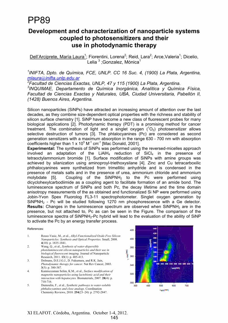
XI ELAFOT. Córdoba, Argentina. October 1-4, 2012.
Development and characterization of nanoparticle systems coupled to photosensitizers and their
use in photodynamic therapy
Dell’Arciprete, María Laura1; Fiorentini, Lorena2; Reid, Lara2; Arce,Valeria1; Dicelio, Lelia 3 ;Gonzalez, Mónica1
1INIFTA, Dpto. de Química, FCE, UNLP. CC 16 Suc. 4, (1900) La Plata, Argentina, [email protected] de Ciencias Exactas, UNLP, 47 y 115 (1900) La Plata, Argentina. 3INQUIMAE, Departamento de Química Inorgánica, Analítica y Química Física, Facultad de Ciencias Exactas y Naturales, UBA, Ciudad Universitaria, Pabellón II, (1428) Buenos Aires, Argentina.
Silicon nanoparticles (SiNPs) have attracted an increasing amount of attention over the last decades, as they combine size-dependent optical properties with the richness and stability of silicon surface chemistry [1]. SiNP have become a new class of fluorescent probes for many biological applications [2]. Photodynamic therapy (PDT) is a promising method for cancer treatment. The combination of light and a singlet oxygen (1O2) photosensitizer allows selective destruction of tumors [3]. The phtalocyanines (Pc) are considered as second generation sensitizers with a maximum absorption in the range 630 - 700 nm with absorption coefficients higher than 1 x 10⁵ M-1 cm-1 [Mac Donald, 2001]. Experimental: The synthesis of SiNPs was performed using the reversed-micelles approach involved an adaptation of the LiAlH4 reduction of SiCl4 in the presence of tetraoctylammonium bromide [1]. Surface modification of SiNPs with amine groups was achieved by silanization using aminoproyl-triethoxysilane [4]. Zinc and Cu tetracarboxilic phthalocyanines were synthetized from trimellitic anhydride and is condensed in the presence of metals salts and in the presence of urea, ammonium chloride and ammonium molybdate [5]. Coupling of the SiNPNH2 to the Pc were performed using dicyclohexylcarbodiimide as a coupling agent to facilitate formation of an amide bond. The luminescence spectrum of SiNPs and both Pc, the decay lifetime and the time domain anisotropy measurements of the as obtained and functionalized Si NP were performed using Jobin-Yvon Spex Fluorolog FL3-11 spectrophotometer. Singlet oxygen generation by SiNPNH2 - Pc will be studied following 1270 nm phosphorescence with a Ge detector. Results: Changes in the luminescence spectrum are observed when SiNPNH2 are in the presence, but not attached to, Pc as can be seen in the Figure. The comparison of the luminescence spectra of SiNPNH2-Pc hybrid will lead to the evaluation of the ability of SiNP to activate the Pc by an energy transfer process.
References:
1. Rosso-Vasic, M., et al., Alkyl-Functionalized Oxide-Free Silicon Nanoparticles: Synthesis and Optical Properties. Small, 2008. 4(10): p. 1835-1841.
2. Wang, Q., et al., Synthesis of water-dispersible photoluminescent silicon nanoparticles and their use in biological fluorescent imaging. Journal of Nanoparticle Research, 2011. 13(1): p. 405-413.
3. Dolmans, D.E.J.G.J., D. Fukumura, and R.K. Jain, Photodynamic therapy for cancer. Nat Rev Cancer, 2003. 3(5): p. 380-387.
4. Kamruzzaman Selim, K.M., et al., Surface modification of magnetite nanoparticles using lactobionic acid and their interaction with hepatocytes. Biomaterials, 2007. 28(4): p. 710-716.
5. Dumoulin, F., et al., Synthetic pathways to water-soluble phthalocyanines and close analogs. Coordination Chemistry Reviews, 2010. 254(23–24): p. 2792-2847.
145
PP89
145

XI ELAFOT. Córdoba, Argentina. October 1-4, 2012.
Porphyrin-fullerene C60 dyads as novel sensitizers for photodynamic therapy
Ballatore, M. Belén; Milanesio, M. Elisa, Durantini, Edgardo N. Departamento de Química, Facultad de Ciencias Exactas Físico-Químicas y
Naturales, Universidad Nacional de Río Cuarto, Agencia Postal Nro 3, X5804BYA Río Cuarto, Córdoba, Argentina, E-mail [email protected]
Porphyrin derivatives have showed great potential as phototherapeutic agents for the treatment of a variety of oncological and non-oncological diseases [1]. In addition, porphyrin-fullerene C60 dyads can efficiently form singlet molecular oxygen, O2(1
g), under certain conditions and give a long-lived radical ion pair upon photoexcitation [2]. These photophysical and redox properties make such dyads very promising photosensitizers for applications in photodynamic therapy.
In this work, a covalently linked carbazoyl porphyrin-fullerene C60 dyad (CBzP-C60) was conveniently synthesized by 1,3-dipolar cycloaddition using 5-(4-carbonylphenyl)-10,15,20-tris[3-(N-ethylcarbazoyl)]porphyrin, N-methylglycine and fullerene C60. This dyad was designed in order to stabilize the photoinduced charge-separated state. This electron transfer process competes with the O2(1
g) production. Also upon oxidation, carbazol (Cbz) moiety can undergo radical cation dimerizations producing dicarbazil units, which conduct to porphyrin electropolymer formation [3]. This property could be used to form permanent photoinactivating surfaces. The spectroscopic properties and photodynamic activity of a CBzP-C60 dyad was compared with 5,10,15,20-tetrakis[3-(N-ethylcarbazoyl)]porphyrin (CBzP) in homogeneous medium bearing photooxidizable substrates. Absorption and fluorescence spectroscopic studies were performed in toluene and N,N-dimethylformamide (DMF). The emission of the porphyrin moiety in the CBzP-C60 dyad is strongly quenched by the attached fullerene C60 moiety. The O2(1
g) productions ( ) were determined using 9,10-dimethylanthracene (DMA). In more polar solvent, the stabilization of charge-transfer state takes place, decreasing the efficiency of porphyrin triplet state formation. Thus, depending on the microenvironment where the sensitizer is localized, these compound could produce a biological photodamage through either a O2(1
g)-mediated photoreaction process or a free radicals mechanism under low oxygen concentration. These results show that molecular dyads, which can form photoinduced charge-separated state, are promising model for phototherapeutic agents with potential applications in cell inactivation by PDT.
Acknowledgements: CONICET, FONCYT-ANPCYT, SECYT-UNRC
References [1] R. R. Allison, C. H. Sibata, Photodiagn. Photodyn. Ther. 2010, 7, 61-75.[2] M. E. Milanesio, M. G. Alvarez, V. Rivarola, J. J. Silber, E. N. Durantini, Photochem. Photobiol. 2005, 81, 891-897.[3] J. Durantini, L. Otero, M. Funes, E. N. Durantini, F. Fungo, M. Gervaldo, Electrochim. Acta 2011, 56, 4126.
146
PP90
146

XI ELAFOT. Córdoba, Argentina. October 1-4, 2012.
Effect of organoclay on different loadings at the kinects of a model dental resin
Terrin, M. M. a, Cavalheiro, C.C.S. a, Horn Jnr, M. A.aa Instituto de Química de São Carlos, Universidade de São Paulo
e-mail: [email protected] Tel.: +55-16-3733-9935
Light cured polymeric composites are commonly used as material in dental restorations.As these composites generally have an inorganic filler, the study of this kind of components are of great interest. Clays and organoclays have brought attention as inorganic fillers in polymer composites because they may improve physical and mechanical properties. The aim of this study is evaluate the effect of the different loadings of organoclay in the mechanical properties of model dental resin[1,2,3].
In this study, composites of BisGMA (34,4%), BisEMA (28,8%), UDMA (28,9%) and TEGDMA (6.9%) with different organoclay loadings (0, 0.5, 2, 5, 7.5, 10 and 15 % w/w), and CQ/EDB (0.5%/0.5% w/w) as an initiation system were made. Those composites correspond to an adequate dental resin model formulation based on commercial selling brands. The composites were the subject of tests involving the evaluation of its kinetic properties through photoinitiated polymerization kinetics. The tests were performed at the temperature of 36,5ºC(body temperature, commonly used in this composites).
The figure bellow shows the preliminary results for this analysis.
Figure 1: Variation of the heat flow vs. time
These preliminary results show that with the increase of clay loadings there s a apparent decrease in speed for the fotopolimerization process. Further analysis of the data obtained are necessary in order to calculate the speed of fotopolimerization and correlate those informations with the different clay loadings.
References[1] Yeh, J.M.; Liou, S.J.; Chang, Y.W.; Journal of Applied Polymer Science, v. 91, p.3489-3496, 2004.[2] Rigoli, I. C., Batista, T., Cavalheiro, C. C. S., Macromolecular Symposia., v. 298, p. 138144, 2010.[3] Rigoli, I. C., Cavalheiro, C. C. S., Neumann, M. G., Journal of Applied Polymer Science,v. 112, p. 679 684, 2009.
147
PP91
147
XI ELAFOT. Córdoba, Argentina. October 1-4, 2012.
Fotodegradación Sensibilizada por Vitamina B2 de Aromatizantes y saborizantes
Carolina Gambetta; Walter Massad y Norman A. García Departamento de Qca. Univ. Nac. de Río Cuarto, 5800 Río Cuarto, Argentina.
Los agentes aromatizantes y saborizantes (Ars) son empleados con gran frecuencia como aditivos alimentarios por su capacidad de mejorar las propiedades organolépticas e incluso de potenciar los sabores dulces. Han cobrado también importancia por su capacidad de actuar como agentes antimicrobianos aumentando la conservabilidad de los alimentos [1]. Debido a que es muy difícil establecer relaciones entre el olor y la estructura química de los Ars, se estudiaron el Maltol (Mt, 3-Hydroxy-2-methyl-4-pyrone) y la Frambinona (Fra, 4-(4-hydroxyphenyl)butan-2-one) que son ejemplos típicos de lo antes mencionado. El Mt se genera en productos de panificación o en alimentos con alto contenido de azúcares por la reacción de Maillard; mientras que la Fraes de ocurrencia natural en el café y en una variedad de frutas entre las que se incluyen frambuesa, arándano y ananá. En este trabajo se realizó un estudio cinético y mecanístico de la degradación directa y sensibilizada de Ars por colorantes sintéticos (Rosa de Bengala, RB) y naturales (Riboflavina, Rf, vitamina del complejo B, presente en una gran variedad de alimentos) en solución acuosa. RB se caracteriza por generar exclusivamente la especie oxígeno singlete molecular (O2(
1Δg)), mientras que Rf en presencia del luz visible da lugar a unas serie de complejas reacciones donde participan los estados excitados de la Rf además de generarsediferentes especies reactivas de oxígeno tales como O2(
1�g) y O2•-.
Se determinaron los rendimientos cuánticos de fotodegradación a 280nm para el Mt (0,45) y el Fra (<0.001) en solución acuosa por el método de actinometría relativa usando Ioduro de potasio como actinómetro [2]. En las experiencias de fotólisis sensibilizada se determinaron las constantes totales (kt) de Ars con O2(
1Δg), empleando RB como sensibilizador, encontrándose valores de kt=1,8 106 M-1s-1 y kt=4,6 106 M-1s-1para el Mt y Fra respectivamente. Las kr medidas son similares a las ktdentro del error experimental. Cuando se irradiaron soluciones de Rf + Ars el efecto de la fotodegradación, relativa a RB fué considerablemente mayor. Los Arsdesactivan al estado singlete excitado de Rf con constantes practicante difusionales(1kq≅ 109 M-1s-1), sin embargo no se espera que la fotodegradación de los Ars por esta vía sea significativa debido a que las concentraciones de AM requeridas para que ocurra este proceso son relativamente altas. En concentraciones sub-mM se encontró que los Arstambién interactúan con el primer estado triplete de Rf con constantes del ordendifusional(3Rf*, 3kq=9.16x10⁸ M-1s-1 para Mt y 2.5x10⁹ M-1s-1 para Fra). En condiciones de sensibilización aeróbica, además de la reacción con el 3Rf* ocurren dos mecanismos de oxidación simultáneos, uno a través de O2(
1Δg) y otro a través del O2•-. A
partir de un análisis cinético, y teniendo en cuenta que las velocidades de fotodegradación de los Ars no se ven afectadas por la presencia de SOD, se estima que la reacción de transferencia de un electrón desde el Ars al 3Rf* es el principal responsable de la degradación de los Ars en presencia de Rf y luz visible.
Agradecimientos:Por el apoyo económico agradecemos a CONICET, ANPCyT, MinCyT-CbaySECyT UNRC, todos de Argentina.
Referencias [1] Zaika, L. L. J. Food Saf. 1988, 9, 97–118. [2] Rahn, R. O. Photochem.Photobiol. 1997, 66, 450–455.
O
O
HO
Maltol (Mt) Aroma pan tostado
OH
O
Frambinona (Fra) Aroma a Caramelo-
frambuesaEstructuras químicas de los Ars
148
PP92
148
XI ELAFOT. Córdoba, Argentina. October 1 4, 2012. 149
Spectroscopic and time-resolved fluorescence emission properties of a cationic and an anionic porphyrin in
biomimetic media and Candida albicans cells
Novaira, Mercedes; Cormick, M. Paula, Durantini, Edgardo N.
Departamento de Química, Facultad de Ciencias Exactas Físico-Químicas y Naturales, Universidad Nacional de Río Cuarto, Agencia Postal Nro 3, X5804BYA Río
Cuarto, Córdoba, Argentina, E-mail [email protected]
In the last years, positively charged porphyrins have attracted considerable attention because of their remarkable ability as phototherapeutic agents. In particular, cationic porphyrin derivatives have been proposed for the treatment and control of microorganisms by photodynamic inactivation (PDI) [1]. Also, porphyrins containing cationic groups are able to interact with DNA bases, inducing DNA lesions upon photoactivation. In general, three binding models have been described for the interaction of cationic porphyrins with DNA, which involve intercalation, outside groove binding and outside binding with porphyrins self-stacking [2]. In biological processes, the solubilization of photosensitizers plays an important role. In this sense, reverse micelles have been frequently used as an interesting model to mimic the water pockets often found in various bioaggregates such as proteins, enzymes and membranes [3].
In the present work, spectroscopic and time-resolved fluorescence emission techniques were used to provide information for the interaction of 5,10,15,20-tetrakis(4-N,N,N-trimethylammoniumphenyl) porphyrin (TMAP4+) and 5,10,15,20-tetrakis(4-sulphonatophenyl) porphyrin (TPPS4-) with different biomimetic media and with Candida albicans cells. In n-heptane/sodium bis(2-ethylhexyl)sulfosuccinate (AOT)/water and benzene/benzyl-n-hexadecyldimethyl ammonium chloride (BHDC)/water reverse micelles interactions were dependent on the micellar interface and the amount of water dispersed in the microemulsion. It was also observed that the DNA binding of cationic porphyrin TMAP4+ led to two lifetimes. In vitroinvestigations showed that TMAP4+ is bound to C. albicans. Fluorescence lifetime measurements and fluorescence microscopic images provided additional insight into the effects of porphyrin uptake by cells. The results reveal a double localization of TMAP4+ inside of C. albicans cells. Thus, a redistribution of TMAP4+ was observed in unwashed cells, probably due to a relocalisation of molecules that were weakly bound to the cells or remained in solution. However, this effect was not found with molecules tightly bound in the cells, after one washing step.
Acknowledgements: CONICET, FONCYT-ANPCYT, SECYT-UNRC References
[1] G. Jori, S.B. Brown, Photochem. Photobiol. Sci. 2004, 5, 403. [2] G. Mez , L. Herényi, J. Habdas, Z. Majer, B. My liwa-Kurdziel, K. Tóth, G. Csík, Biophys. Chem.2011, 155, 36. [3] I. Scalise, E.N. Durantini, J. Photochem. Photobiol. A: Chem. 2004, 162, 105.
HN
N
NH
N
R
R R
R
N+R:
SO3-R:
TMAP4+
TPPS4-
PP93
149

XI ELAFOT. Córdoba, Argentina. October 1 4, 2012. 150
Perfluoroalquil peroxinitratos de cadena lineal. Síntesis fotoquímica y caracterización.
Adriana G. Bossolasco, Jesús A. Vila, Gustavo A. Argüello y Fabio E. Malanca.
INFIQC/CONICET – Dpto. de Fisicoquímica – Fac. de Cs Químicas – UNC– e-mail: [email protected]
La degradación de ciertos compuestos fluorados de uso industrial (hidroclorofluorocarbonados -HCFC-, hidrofluorocarbonados -HFC- e hidrofluoroeteres -HFE-), usados como solventes, agentes espumantes, emulsionantes, etc [1,2]; en ambientes urbanos, en presencia de NO2 y luz solar, puede conducir a la formación de agentes oxidantes tales como radicales peróxidos, peróxidos orgánicos, y peroxinitratos (ROONO2)[2-4].
Los peroxinitratos tienen un papel relevante en el ámbito de la química atmosférica porque actúan como especies reservoria de radicales peróxido (ROO ) y NO2, contribuyendo a la formación de ozono troposférico. Dependiendo de la estructura, sus estabilidades térmicas a temperatura ambiente puede ser desde minutos hasta semanas, y consecuentemente pueden ser transportados desde su fuente de formación a lugares distantes o a mayores alturas. La posible existencia de estas moléculas en la atmósfera conlleva la necesidad de estudiar sus propiedades tantos físicas como químicas, los cuales son objetivos del presente trabajo. Metodología
Los perfluoroalquil peroxinitratos CxF2x+1OONO2 x=2-4 fueron sintetizados mediante la irradiación con luz de = 254 nm a moléculas precursoras conteniendo el fragmento CxF2x+1 (cloruro de perfluoroacetilo C2F5C(O)Cl, para x=2; anhídrido heptafluorobutílico C3F7C(O)OC(O)C3F7, para x=3 y ioduro de nanofluorobutilo C4F9I, para x=4) en presencia de O2 y NO2. La identificación, caracterización y estudios cinéticos se realizaron mediante espectroscopia UV e IR. Resultados
Se determinaron los espectros infrarrojos de los perfluoroalquil peroxinitratos, encontrándose que en general presentan bandas de absorción características a: 1760 cm-1, as NO2; 1240-1230 cm-1,
as C-F; 1000-990 cm-1, O-O; y 790 cm-1, deformación NO2. Los espectros de absorción UV, en tanto, muestran que la sección eficaz de absorción ( ) entre 200 y 330 nm disminuye con la longitud del grupo alquilo, y con la temperatura. El estudio de sus estabilidades térmicas muestra que a medida que aumenta la longitud del grupo alquilo más inestable es el peroxinitrato. Los parámetros cinéticos de Ea (Energía de activación) y A (Factor pre-exponencial) muestran este hecho, y también la dependencia de la constante de descomposición térmica con la presión y la temperatura.
A partir de los resultados obtenidos de estabilidad térmica y secciones de absorción UV, se determinaron los perfiles de tiempo de vida térmico y fotoquímicos, observándose en todos los casos que el proceso de descomposición térmica es el que domina el tiempo de vida de estos peroxinitratos; con valores que van desde horas a semanas en cercanías de la superficie, hasta años a alturas cercanas a la tropopausa.
Referencias
[1] Sulbaek Andersen, M.P.; Hurley, M.D.; Wallington, T.J.; Ball, J.C.; Martin, J.W.; Ellis, D.A.; Mabury, S.A.; Nielsen, O.J. Chem. Phys. Lett. 379 (2003) 28. [2]von Ahsen, S.; Garcia, P.; Willner, H. and Argüello, G. A.; Inorg. Chem. 44 (2005) 5713. [3] Ninomiya Y.; Kawasaki, M .; Guschin , A.; M olina, L . T and Wallington , T.; J. Environ. Sci. Technol. 34 (2000) 2973. [4] Mayer-Figge, A., Zabel, F., Becker, K.H.; J. Phys. Chem. 100 (1996) 6587.
PP94
150

XI ELAFOT. Córdoba, Argentina. October 1 4, 2012. 151
Nanopartículas de silicio conjugadas con ácido fólico para su evaluación en terapia fotodinámica.
Lillo, Cristian1; Rodriguez Sartori, Damián1; Kotler, Mónica2; Gonzalez, Mónica1
1INIFTA, UNLP-CONICET, La Plata diag. 113 y 64 CP1900, [email protected]
2Departamento de Ciencias Biológicas, FCE-UBA
Las partículas de materiales semiconductores como el Si de diámetros nanométricos presentan fotoluminiscencia debido al confinamiento cuántico [1]. En los últimos decenios, estos nanomateriales, más conocidos como QDs han sido extensamente estudiados como agentes luminiscentes para la detección por imágenes en sistemas biológicos, como una alternativa a los colorantes fluorescentes y como agentes terapéuticos contra el cáncer. En este trabajo se describe la síntesis, caracterización y evaluación de las propiedades fotoluminiscentes de nanopartículas de silicio derivatizadas superficialmente con polietilenglicol y con ácido fólico, con el fin de utilizarlos como agentes terapéuticos en sistemas biológicos. Las nanopartículas de silicio de tamaños de entre 1 y 3 nm se sintetizaron por un método top down en micelas invertidas. Las partículas se modificaron superficialmente con alilamina en un fotoreactor Rayonet usando luz UV de =254 nm [2]. La unión del ácido fólico se hizo a través de la formación de un enlace tipo amida entre los grupos carboxilo del ácido fólico y los grupos amino de las nanopartículas derivatizadas con alilamina [3]. Este producto se purifico por diálisis y se caracterizó por diversas técnicas, entre ellas: FTIR, Raman, XPS, matrices de excitación-emisión y anisotropía resuelta en el tiempo.
References [1] M. J. Llansola Portolés, F. Rodriguez Nieto, D. B. Soria, J. I. Amalvy, P.J. Peruzzo, D. O. Mártire, M. L. Kotler, O. Holub, and M. C. Gonzalez. Photophysical properties of blue –emitting silicon nanoparticles. J. Phys. Chem. C. 2009, 113 (31), 13694–13702. [2] Amine-terminated silicon nanoparticles: synthesis, optical propierties and their use in bioimaging. Milena Rosso-Vasic , Evan Spruijt , Zoran Popovi , Karin Overgaag , Barend van Lagen , Bruno Grandidier , Daniel Vanmaekelbergh , David Domínguez-Gutiérrez , Luisa De Colaand Han Zuilhof. J. Mater. Chem., 2009,19, 5926-5933.[3] Folate conjugated fluorescent silica nanoparticles for labeling neoplastic cells. Santra S, Liesenfeld B, Dutta D, Chatel D, Batich CD, Tan W, Moudgil BM, Mericle RA. J. Nanosci. Nanotechnol. 2005,5, 899-904.
PP95
151

XI ELAFOT. Córdoba, Argentina. October 1-4, 2012.
Formación, detección y tiempo de vida del primer triplete de pireno tetrasulfonato
Ahumada, Manuel*; Lissi, Eduardo; Aspée, Alexis; Pino, EduardoFacultad de Química y Biología, Universidad de Santiago de Chile, Santiago, Chile
* E-mail: [email protected]
Nuestro grupo está interesado en el desarrollo de metodologías que permitan medir la velocidad de pasaje de distintos solutos a través de membranas lipídicas empleando liposomas unilaminares como sistemas modelo. Una de estas metodologías se basa en medir el tiempo de vida de una especie triplete generada en el pool acuoso de liposomas en función del tiempo transcurrido luego de adicionar al medio dispersante un desactivador del triplete (quencher). En el presente trabajo hemos estudiado la fotoquímica de pireno tetrasulfonato, una molécula que permite garantizar la formación de la especie triplete sin interacción con la bicapa lipídica en términos de reparto entre liposoma y el medio acuoso.El espectro de absorción triplete-triplete max 440 nm) y su tiempo de vida en ausencia de oxigeno ( sec) han permitido determinar la constante de desactivación por quenchers en medio acuoso. Este modelo se discute en relación a la determinación de velocidades depermeación de quenchers iónicos, y moléculas de distintas hidrofobicidad y tamaño molecular a través de bicapas de liposomas en presencia y en ausencia de poros.
152
PP96
152

Caracterización espectroscópica de sistemas mixtosformados por carbazol, ciclodextrinas y calixarenos
Carranza, Matías E. y Veglia, Alicia V.Instituto de investigación en Físico Química de Córdoba (INFIQC), Departamento de
Química Orgánica. Facultad de Ciencias Químicas. Universidad Nacional de Córdoba. Ciudad Universitaria. Córdoba. C.P.5000. Argentina.
e-mail: [email protected]; [email protected]
Los macrociclos como las ciclodextrinas (CDs) y los calixarenos (CAs) pueden ser empleados para la construcción de dispositivos supramoleculares diseñados para interactuar y producir una señal en presencia de un determinado sustrato, es decir un sensor químico [1]. El carbazol (CZL) forma parte de algunos alcaloides biológicamente relevantes [2]. Este núcleo presenta propiedades fluorescentes que pueden modificarse en presencia de receptores adecuados. Una contribución cooperativa de interacciones no covalentes es responsable de la formación de complejos. El factor principal que dirige el reconocimiento molecular es la relación forma y tamaño entre el receptor macrocíclico (R) y el sustrato interactuante (S).
En este - - - -CD, y los CAs sulfonados CA[6]S y CA[8]S. El efecto individual de CDs sobre las propiedades espectroscópicas del CZL se manifestó en un aumento de la absorbancia (A) máxima a 290 nm
15,4-25,8%) y una exaltación ( -55%) de la emisión fluorescente (F). En tanto que con cantidades crecientes de CA[n]S se observaron pequeños cambios en el espectro de absorción e inhibición (quenching) de la F (-% 47,7-66,8 %) del CZL, a pH = 7. A partir de estas variaciones espectroscópicas se propuso la formación de complejos de inclusión entre el CZL y los receptores macrocíclicos. En esta presentación se detalla el estudio del efecto mixto de CDs y CA[n]S sobre la Abs UV-Vis y la F de CZL. Se comenzó partiendo de un complejo de inclusión CZL-R1 y se fueron agregando concentraciones crecientes de un receptor R2. El incremento de CA[n]S (R2) en una solución del complejo CZL- -CD incrementó la Abs del sistema. Por el contrario, el aumento
-CD en una solución de CZL-CA[n]S disminuyó ligeramente la Abs del sistema mixto. En tanto que la F de CZL- -CD fue inhibida con el agregado de CA[8]S; mientras que no fueafectada al agregar CA[6]S. Por otra parte, al tratar una solución del complejo CZL-CA[8]S con c -CD, se observó una ligera disminución de la F. Por otro lado, la
-CD al complejo CZL-CA[6]S produjo una exaltación de la F. Para este caso, se determinó un valor de constante de asociación aparente experimental (KAP
exp = 7,9 x 10²) para CZL- -CD, el cual fue comparable con un valor teórico (KAP
T = 8,1 x 10²), obtenido de suponer coexistencia de ambos complejos: CZL-CA[6]S y CZL-HP- El análisis de estos resultados llevará a comprobar si todos los sistemas mixtos estudiadospueden interpretarse como coexistencia simultánea de dos complejos o si hay un efecto inhibitorio o cooperativo en la formación de alguno de ellos. En este último caso, se produciríaun complejo ternario. En base a estas conclusiones se podrá proponer el diseño de un sensor mixto para la determinación de CZL.
Referencias:1- Scheneider, H- ,John Wiley & Sons Ltd, England, 2000.2- Sbai, M.; Ait Lyazidi, S.; Lerner, D. A.; Martin, M. A.; del Castillo, B.; Analytica Chimica Acta,1995, 303, 47-55.
153
PP97
153

XI ELAFOT. Córdoba, Argentina. October 1-4, 2012.
Efecto de la presencia de cationes divalentes en la reacción entre Oxígeno Molecular Singulete y Flavonoles.
Sandoval, Catalina1; Lemp, Else1
1Universidad de Chile, Facultad de Ciencias Químicas y Farmacéuticas, Departamento de Química Orgánica y Fisicoquímica, Sergio Livingstone Pohlhammer 1007
Independencia Santiago, [email protected], [email protected].
Los flavonoides, compuestos polifenólicos, son una importante clase de antioxidantes de origen vegetal que se encuentran incorporados al organismo humano. Una multiplicidad de prototipos de sustitución, en los dos anillos de benceno (A y B) de la estructura básica de los flavonoides, se producen en la naturaleza, mientras que los grupos funcionales presentes en el anillo heterocíclico da lugar a flavonoles, flavonas, catequinas, flavononas, antocianidinas e isoflavonas. Estos compuestos muestran una importante actividad biológica, en la prevención de enfermedades cardíacas, en el cáncer, inflamaciones, etc. [1] Muchos de los efectos beneficiosos de los flavonoles han sido relacionados a sus capacidades como aceptores de radicales libres del O2 y desactivadores del 1O2. La capacidad para formar complejos con metales contribuiría a su actividad antioxidante total. Los iones metálicos con actividad redox como cofactores de varias enzimas, son también citotóxicos al generar especies reactivas del oxígeno. Generalmente, la quelación de iones metálicos con flavonoides previenen la generación catalizada por metales, de radicales libres, interviniendo en el estrés oxidativo en moléculas biológicamente activas. [2] Los Flavonoles y los cationes alcalinos térreos pueden formar dos tipos de complejos; externos de baja estabilidad resultante de la interacción metal con el oxígeno del grupo carbonilo del anillo heterociclo y quelante de alta estabilidad donde participa el oxígeno del grupo carbonilo y el átomo de oxígeno del grupo hidroxilo de la posición 3 del anillo heterociclo.[3] Desde hace algunos años, nuestro grupo se ha dedicado a estudiar la reactividad de flavonoles frente al oxígeno molecular singulete en fase homogénea, mezclas metanol-agua y en liposomas, los resultados nos permiten concluir que la reactividad entre flavonoles y el oxígeno excitado es favorecida por la polaridad, la basicidad del medio y la localización estructural en la bicapa lipídica de liposomas. En este trabajo, informamos sobre el efecto de la presencia de los iones divalentes Cu, Mg y Ca, en la reactividad de 3-hidroxiflavona (3HF) y quercitina (Quer) frente al oxígeno molecular singulete. En los espectros UV-vis se observa la aparición de una banda a una longitud de onda cercana a 435 nm (Quer) y 410 nm (3HF), claramente atribuible a la formación de una asociación o complejo, ya que ni el metal ni el flavonoide absorben a esta longitud de onda. Los valores de las constantes de velocidad de reacción química entre los flavonoides y el oxígeno molecular singulete, kr, muestran un aumento en presencia de los iones divalentes, en el caso del ion cobre en una relación [Cu]/ [Quer] de 0,5 el valor de kr aumenta de 9,0 E5 M-1s-1 a 7,5 E6 M-1s-1,en la relación [Mg]/ [Quer] de 2,0 el aumento en el valor de kr es de 9,0 E5 M-1s-1 a 2,7 E7 M-1s-1 . En el caso del ion Ca en una relación [Ca]/[Quer] bastante mayor la variación en la constante de velocidad es cercana a dos ordenes de magnitud. La presencia de los iones divalentes en la reactividad de 3HF con oxígeno molecular singulete, tiene un mayor efecto en el valor de la constante de velocidad que el mostrado por Quercitina. Se agradece el financiamiento de Proyecto Fondecyt Nº 1090267
References1. Procházková, D. et al., Antioxidant and prooxidant properties of flavonoids. Fitoterapia, 2011.
82(4): p. 513-523. 2. Jurasekova Z. et al., A Raman and surface-enhanced Raman scattering (SERS)
investigation of the quercetin interaction with metals: Evidence of structural changing processes in aqueous solution and on metal nanoparticles. Journal of Molecular Structure 918 (2009) 129–137.
3. Roshal A. D. et al., Flavonols and Crown-Flavonols as Metal Cation Chelators. The Different Nature of Ba2+ and Mg2+ Complexes J. Phys. Chem. A 1998, 102, 5907-5914.
154
PP98
154
XI ELAFOT. Córdoba, Argentina. October 1 4, 2012. 155
PC liposomes as carriers of a singlet oxygen quencher
Gabriela Bosio1,2,Jose Luis Alessandrini3, Daniel Mártire1 and Peter Ogilby2
1Instituto de Investigaciones Teóricas y Aplicadas, Universidad Nacional de La Plata,Argentina,2 Center forOxygenMicroscopy and Imaging, Department of Chemistry, Universityof Aarhus, Denmark.3Departamento de Física, Universidad Nacional de La Plata Instituto de
Física de La Plata, Argentina
In this work, three photosensitizers of different hydrophilicity have beenintroduced intophosphatidyl choline liposomes(PCL). The sensitizers employedwereAl(III) Phthalocyanine chloride tetrasulfonic acid (AlPcS4,hydrophilic),Pyropheophorbide a (PPa, hydrophobic) and TPPMeCOO(tetraphenylporphyrin methyl ester, hydrophobic). At the same time threedifferent quenchers were chosen as controls to elucidate where the singletoxygen is produced and diffuses.[1] The quenchers used were BSA (extraliposomal localization), azide (inside and outside localization) and carotene(lipophilic). Small unilamellar liposomes were formed by sonication.Theconcentration of carotene and photosensitizers incorporated into theliposomes were evaluated from absorption and fluorescence spectroscopy,respectively.[2] To characterize the liposomes, the membrane fluidity of purePCL/TRIS or PCL/H2O liposomes and of those containing differentconcentrations -carotene were determined using the fluorescent probepyrene[2] and their side by Dynamic light scattering (DLS).[3]
[1] ] Elsa F. F. da Silva, Brian W. Pedersen, Thomas Breitenbach, RasmusToftegaard, Marina K.Kuimova, Luis G. Arnaut, and Peter R. Ogilby, Irradiation and Sensitizer Dependent Changes in theLifetime of Intracellular Singlet Oxygen Produced in a Photosensitized Process. The Journal of PhysicalChemistry B 2012 116 (1), 445 461
[2]AtanaskaAndreeva and AntoanetaPopova. Integration of carotene molecules in smallliposomes.Journal ofPhysics:ConferenceSeries 253 (2010) 012066.
[3]Fagali N, Catalá A.Fe2+ and Fe3+ initiated peroxidation of sonicated and non sonicated liposomesmade of retinal lipids in different aqueous media.. ChemPhys Lipids. 2009 Jun;159(2):88 94. Epub2009 Mar 24.
PP99
155
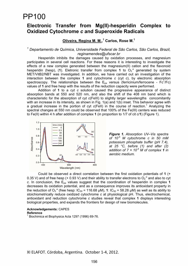
XI ELAFOT. Córdoba, Argentina. October 1 4, 2012. 156
Electronic Transfer from Mg(II)-hesperidin Complex to Oxidized Cytochrome c and Superoxide Radicals
Oliveira, Regina M. M.,1 Carlos, Rose M.1
1 Departamento de Química, Universidade Federal de São Carlos, São Carlos, Brazil, [email protected]
Hesperidin inhibits the damages caused by oxidation processes, and magnesium participates in several cell reactions. For these reasons it is interesting to investigate the effects of a new complex generated between the magnesium(II) cation and the flavonoid hesperidin (hesp), (1). Eletronic transfer from complex 1 to O2
- generated by system MET/VitB2/NBT was investigated. In addition, we have carried out an investigation of the interaction between the complex 1 and cytochrome c (cyt c), by electronic absorption spectroscopy. The relationships between the Epa versus (ferricinium/ferrocene - Fc+/Fc) values of 1 and free hesp with the results of the reduction capacity were performed.
Addition of 1 to a cyt c solution caused the progressive appearance of distinct absorption bands at 550 and 520 nm, and also the shift of the 408 nm band which is characteristic for the absorption of cyt c(FeIII) to slightly larger wavelengths concomitantly with an increase in its intensity, as shown in Fig. 1(a) and 1(b) inset. This behavior agree with a gradual increase in the portion of cyt c(FeII) in the course of reaction.1 Analyzing the spectral changes at 550 nm could be observed that 100% of the Fe(III) centers was reduced to Fe(II) within 4 h after addition of complex 1 (in proportion to 1/7 of cit c/1) (Figure 1).
Figure 1. Absorption UV–Vis spectra of 10-5 M cytochrome c in 50 mM potassium phosphate buffer (pH 7.4), at 25 °C, before (1) and after (2) addition of 7 × 10⁻⁵ M of complex 1 in aerobic medium.
Could be observed a direct correlation between the first oxidation potentials of 1 (+ 0.35 V) and of free hesp (+ 0.93 V) and their ability to transfer electrons to O2
- and also to cyt c. In conclusion, the Epa values suggest that the coordination of hesperidin in complex 1decreases its oxidation potential, and as a consequence improves its antioxidant property in the reduction of O2
- (free hesp: IC50 = 116.68 μM), 1: IC50 = 58.28 μM) as well as its ability to stoichiometrically reduce oxidized cytochrome c at physiological pH. Thus, electrochemical, antioxidant and reduction cytochrome c studies reveal that complex 1 displays interesting biological properties, and expands the frontiers for design of new biomolecules.
Acknowledgements: CAPES Reference 1 Biochimica et Biophysica Acta 1297 (1996) 69-76.
PP100
156
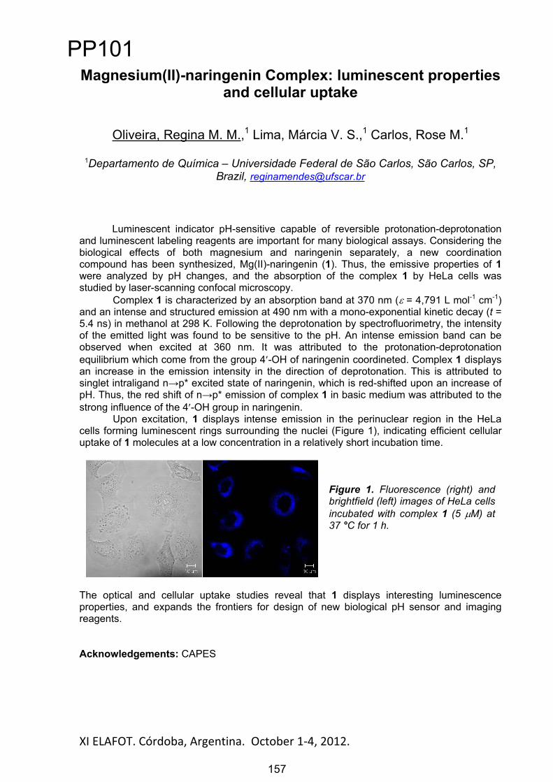
XI ELAFOT. Córdoba, Argentina. October 1 4, 2012. 157
Magnesium(II)-naringenin Complex: luminescent properties and cellular uptake
Oliveira, Regina M. M.,1 Lima, Márcia V. S.,1 Carlos, Rose M.1
1Departamento de Química – Universidade Federal de São Carlos, São Carlos, SP, Brazil, [email protected]
Luminescent indicator pH-sensitive capable of reversible protonation-deprotonation and luminescent labeling reagents are important for many biological assays. Considering the biological effects of both magnesium and naringenin separately, a new coordination compound has been synthesized, Mg(II)-naringenin (1). Thus, the emissive properties of 1were analyzed by pH changes, and the absorption of the complex 1 by HeLa cells was studied by laser-scanning confocal microscopy.
Complex 1 is characterized by an absorption band at 370 nm ( = 4,791 L mol-1 cm-1)and an intense and structured emission at 490 nm with a mono-exponential kinetic decay (t = 5.4 ns) in methanol at 298 K. Following the deprotonation by spectrofluorimetry, the intensity of the emitted light was found to be sensitive to the pH. An intense emission band can be observed when excited at 360 nm. It was attributed to the protonation-deprotonation equilibrium which come from the group 4 -OH of naringenin coordineted. Complex 1 displays an increase in the emission intensity in the direction of deprotonation. This is attributed to singlet intraligand n p* excited state of naringenin, which is red-shifted upon an increase of pH. Thus, the red shift of n p* emission of complex 1 in basic medium was attributed to the strong influence of the 4 -OH group in naringenin.
Upon excitation, 1 displays intense emission in the perinuclear region in the HeLa cells forming luminescent rings surrounding the nuclei (Figure 1), indicating efficient cellular uptake of 1 molecules at a low concentration in a relatively short incubation time.
Figure 1. Fluorescence (right) and brightfield (left) images of HeLa cells incubated with complex 1 (5 M) at 37 °C for 1 h.
The optical and cellular uptake studies reveal that 1 displays interesting luminescence properties, and expands the frontiers for design of new biological pH sensor and imaging reagents.
Acknowledgements: CAPES
PP101
157
XI ELAFOT. Córdoba, Argentina. October 1 4, 2012. 158
Spectroscopic properties of methylene blue in different water-organic solvent systems
Santos, Sandra C. dos1; Santin, Luiza R. R.1, Moreira, Leonardo M.2; Lyon, Juliana P.3;Codognoto, Lúcia4; Bonacin, Juliano A.5; Romani, Ana Paula6; Machado, Antônio E. da
Hora7; de Oliveira, Hueder Paulo M.1
1UFPel - CCQFA. Pelotas, RS, Brasil, [email protected], São João Del Rei, MG, Brasil
3UFSJ - DCN, São João Del Rei, MG, Brasil 4UNIFESP – ICAQF. Diadema - SP, Brasil
5Berian Tecnologia, Ind. e Com. Ltda – NovaNano. São Paulo, SP, Brasil 6DQ - ICEB, UFOP, Ouro Preto, MG, Brasil
7UFU - IQ. Uberlândia, MG, Brasil
The phenothiazines, as methylene blue (MB), are cationic dyes used as a model for phototherapeutic agent [1,2] due to their biological, chemical, photochemical/photophysical properties [3,4]. It is known that the dye self-aggregation in solvent mixtures reduces the ability of singlet oxygen (1O2) formation as well as other reactive oxygen species (ROS), hindering the application of the respective dye as photosensitizer (PS) in photodynamic therapy (PDT) [5,6]. In the present work, it was evaluated the influence of the solvent mixtures on the spectroscopic properties of the MB. Emission spectra were obtained in ethanol-water, acetonitrile-water and glycerol-water systems, as well singlet oxygen quantum yield, lifetime and fluorescence anisotropy. This solvatochromic study demonstrated the appropriate proportion of each solvent in a specific mixture to avoid the phenomenon of self-aggregation. In the excitation and emission spectra, it was observed a decrease in fluorescence intensity in the mixtures in higher concentrations of water. This effect is related to the partial inhibition or more friction in the rotation of substituent groups of the dye [7]. With the addition of organic solvent, the environment becomes favourable to the monomeric form, generating high excitation and emission intensities. The time resolved data and anisotropy was performed and showed that the increase of organic solvent proportion in the solvent system increases the lifetimes values. This may be related to some physico-chemical factors inherent to the chemical system, such as acidity and/or basicity of the system as well as its viscosity. Indeed, regarding the rotational time, there is clearly the influence of viscosity upon this physical property. This also interferes in the processes of deactivation of the excited state of dyes, since this relaxation mechanism becomes slower in more viscous media. With respect to the formation of 1O2, it was observed a decrease in emission intensity at higher proportion of water in solvent mixtures with contribution above 70% of water. It occurs due to formation of dimers in this kind of solvent system. The present data are very auspicious in order to promote a more efficient employment of several photosensitizers in PDT as well as understanding the solvatochromic mechanisms associated to the interaction dye-solvent as model system of the various types of interaction that can occur in a biological medium.
References:[1]. Danziger, R. M.; Bareli, K. H.; Weiss, K. Journal of Physical Chemistry 71, 2633, 1967.[2]. Mellish, K. J.; Cox, R. D.; Vernon, D. I.; Griffiths, J.; Brown, S. B. Photochemistry and Photobiology 75. 392, 2002.[3]. Kobayashi, M.; Maeda, Y.; Hoshi, T.; Okubo, J.; Tanizaki, Y. Journal of the Society of Dyers And Colourists 105, 362, 1989.[4]. Alarcon, E.; Edwards, A. M.; Aspee, A.; Moran, F. E.; Borsarelli, C. D.; Lissi, E. A.; Nilo, D. G.; Poblete, H.; Scaiano, J. C. Photochemical & Photobiological Sciences 9, 93, 2010.[5]. Severino D.; Junqueira H. C.; Gabrielli D. S.; Gugliotti M.; Baptista M. S.; Photochemical Photobiological Sciences 77, 459, 2003.[6]. Junqueira H. C.; Severino D.; Dias L. G.; Gugliotti M., Baptista M. S.; Physical Chemistry Chemical Physics 4, 2320, 2002.[7]. Oliveira, H. P. M.; Junior, A. M.; Legendre, A. O.; Gehlen, M. H. Química Nova 26, 564, 2003.
PP102
158
XI ELAFOT. Córdoba, Argentina. October 1-4, 2012.
Descomposición Térmica y Fotoquímica de nuevos Hidro-Fluoro-Peróxidos.
Matias Berasategui, Maxi Burgos y Gustavo ArgüelloDpto. de Fisicoquímica. Facultad de Ciencias Químicas. Ciudad Universitaria.
CP: 5000. Córdoba, Argentina. [email protected]
Introducción. El trifluorometil fluoroformil peroxidicarbonato (CF3OC(O)OOC(O)F)sintetizado por primera vez en nuestro grupo de trabajo, y el (FC(O)OOC(O)F) [1-2], forman parte de una serie que ha atraído nuestra atención en los últimos años [3-5]. A partir de la reacción de estos con metanol, se llevó a cabo la síntesis y caracterización de nuevas especies peroxidadas, tales como CH3OC(O)OOC(O)F [6] y CH3OC(O)OOC(O)OCF3. En el presente trabajo se pretende estudiar las reacciones de descomposición térmicas y fotoquímicas para estas moléculas en fase gaseosa. Las reacciones también son abordadas desde el punto de vista teórico utilizando la teoría del funcional de densidad.
Resultados y Metodología. Las reacciones de descomposición térmica se llevaron a cabo en un reactor de acero inoxidable de 0,5 L contenido dentro de un sistema de calentamiento capaz de controlar la temperatura con un error de ±1°C, conectado a una celda de acero (23 cm, ventanas de KBr) ubicada en el paso óptico de un espectrofotómetro FTIR (Bruker IFS 28). Este arreglo permitió seguir las concentraciones en función del tiempo de reactivos y productos.
En cuanto a las reacciones fotoquímicas, se llevan a cabo en una celda de cuarzo de 23 cm de paso óptico, y ventana de KBr, ubicada en el paso óptico del espectrofotómetro. Se utiliza una lámpara de baja presión de mercurio de 40 Watts de potencia para iniciar la reacción (Heraeus, Hanau).
Además de la espectroscopia FITR, para la caracterización de los productos, se utilizó un equipo NMR Bruker AVANCE II 400 MHz y un espectrómetro de masas FINNIGAN 3300 F-100. El principal producto encontrado para estas descomposiciones fue el CO2. Para el caso del CH3OC(O)OOC(O)F también se encontró CH2O, HF, CO y HC(O)OH, mientras que para CH3OC(O)OOC(O)OCF3 se encontró CH2O, HC(O)OH, CF2O, HF, CH3OC(O)F y CO.
El mecanismo de las reacciones se estudió utilizando el método de cálculo B3LYP/6-31++G(d,p) implementado en el paquete de programas Gaussian 09. Para corroborar la conexión del estado de transición con los reactivos y productos se utilizó el método de la coordenada intrínseca de reacción (IRC).
Referencias[1] Burgos Paci, M. A.; García P., Malanca, F. E.; Argüello, G. A.; Willner, H. Inorg Chem. 2003, 42,2131-5.[2] Arvia, A. J.; Aymonino, P. J.; Schumacher, H. J. Anal. Asoc. Quím. Arg. 1962, 50, 135 143.[3] Malanca, F. E.; Argüello, G. A.; Staricco, E. H.; Wayne, R. P. J. Photochem. Photobiol. A 117,1998, 163 169.[4] Burgos Paci, M. A.; Argüello, G. A.; Garcia, P.; Willner, H. Int. J. Chem. Kinet. 35, 2003, 15 19.[5] Burgos Paci, M. A.; Argüello, G. A.; García, P.; Willner, H. J. Phys. Chem. A. 2005, 109(33), 7481.[6] Berasategui, M.; Burgos Paci, M. A.; Argüello, G. A. Angew. Chem. 2012, 638 (3-4), 547-552.
159
PP103
159

XI ELAFOT. Córdoba, Argentina. October 1 4, 2012. 160
Fotoinactivacion bacteriana y producción de oxigeno singlete a partir de extractos de origen vegetal e irradiación con laser de 630 y 980 nm
Mamone, Leandro1; Gandara, Lautaro2, Di Venosa, Gabriela3, Rodriguez Lorena4,Batlle Alcira5; Buzzola Fernanda6, Casas Adriana7
1 Centro de Investigaciones sobre Porfirinas y Porfirias (CIPYP)-CONICET. Htal de Clínicas Gral José de San Martín, Av. Cordoba 2351 1er subsuelo, ciudad de Buenos Aires, Argentina, [email protected] Centro de Investigaciones sobre Porfirinas y Porfirias (CIPYP)-CONICET. Htal de Clínicas Gral José de San Martín, Av. Cordoba 2351 1er subsuelo, ciudad de Buenos Aires, Argentina, [email protected] Centro de Investigaciones sobre Porfirinas y Porfirias (CIPYP)-CONICET. Htal de Clínicas Gral José de San Martín, Av. Cordoba 2351 1er subsuelo, ciudad de Buenos Aires, Argentina, [email protected] Centro de Investigaciones sobre Porfirinas y Porfirias (CIPYP)-CONICET. Htal de Clínicas Gral José de San Martín, Av. Cordoba 2351 1er subsuelo, ciudad de Buenos Aires, Argentina, [email protected] Centro de Investigaciones sobre Porfirinas y Porfirias (CIPYP)-CONICET. Htal de Clínicas Gral José de San Martín, Av. Cordoba 2351 1er subsuelo, ciudad de Buenos Aires, Argentina, [email protected] Departamento de Microbiología, Parasitología e Inmunología, Facultad de Medicina, UBA, Paraguay 2155 14º piso, ciudad de Buenos Aires, Argentina [email protected] Centro de Investigaciones sobre Porfirinas y Porfirias (CIPYP)-CONICET. Htal de Clínicas Gral José de San Martín, Av. Cordoba 2351 1er subsuelo, ciudad de Buenos Aires, Argentina, [email protected]
La Terapia Fotodinámica (TFD) es un tratamiento que consiste en la administración de un compuesto fotosensibilizante (FS) que al ser iluminado induce la muerte celular vía generación de especies reactivas del oxigeno, en especial oxigeno singlete. Recientemente, la Terapia Fotodinámica Antimicrobiana (TFDA) se propuso para el tratamiento de infecciones bacterianas superficiales orales o cutáneas, o para el tratamiento de zonas accesibles con fibras ópticas que guían la llegada de la luz al blanco, tales como prótesis ortopédicas o implantes dentales.
Nuestro grupo realizo una colección de 70 extractos de especies vegetales autóctonas de nuestro país. Los mismos fueron testeados en busca de principios fototóxicos para su uso en TFDA.
Se trabajo con las especies bacterianas Staphylococcus epidermidis, Staphylococcus aureus (gram positivas), Escherichia coli y Pseudomonas aeruginosa (gram negativas), creciendo en suspensión o luego de la formación de biofilms.
Se utilizaron diferentes sistemas de irradiación, incluidos dispositivos láser de 630 y 980 nm. La viabilidad bacteriana luego de la TFDA se determinó por recuento de las UFC/ml. En condiciones de crecimiento en suspensión, los extractos de Solanum verbascifolium flor,
Tecoma stans flor y Cissus verticillata raíz fueron fototóxicas para las cepas gram positivas, pero no para las Gram negativas. S. verbascifolium fue la especie más fotoactiva, ya que indujo una reducción de 5 órdenes de magnitud en el número inicial de colonias (2,1 x10⁸ ± 5x10⁸ UFC/ml) luego de la TFD con 0,5 mg/ml del extracto y 3 hs de iluminación (5,2 x10³ ± 0,5x10³ UFC/ml). Con los extractos metanólicos de T. stans flor (0,5 mg/ml) y C. verticillata raíz 0,5 mg/ml se indujeron reducciones de 4 órdenes de magnitud (1,2 x10⁴ ± 2,5x10⁴ UFC/ml y 6,5 x10⁴ ± 3,3 x10⁴ UFC/ml respectivamente), (p< 0,01, Mann-Whitney test).
Los biofilms de Staphylococcus aureus fueron resistentes a la acción de la TFDA, tanto cuando se emplearon los extractos vegetales, como cuando se utilizaron FS de uso actual, como clorina e6 y azul de toluidina.
A su vez, fueron realizados estudios de la producción de oxigeno singlete in vitro, a partir de los extractos fototóxicos mencionados. Se observo correlación entre la producción de oxigeno singlete (dependiente de la irradiación) y la actividad fototóxica.
Estos resultados demuestran el potencial de las especies vegetales como fuente de sustancias fotosensibilizantes de bacterias. Actualmente se están llevando a cabo estudios complementarios de internalización bacteriana intracelular, para determinar la posibilidad de tratar fotodinámicamente células infectadas con bacterias de la cepa S. aureus, sin alterar la viabilidad celular.
PP104
160

XI ELAFOT. Córdoba, Argentina. October 1 4, 2012. 161
Photophysics of 1-Butyl-3-Methylimidazolium Chloride in Aqueous Solution
Mirenda, Martin1,3, Barja, Beatriz C. 1,2, Espinoza, Mariela S.3, Babay, Paola A.3 Gutkowsky, Karin I.3
1 DQIAyQF, 2 INQUIMAE, Facultad de Ciencias Exactas y Naturales, Universidad de Buenos Aires, Ciudad Universitaria, Pab. II, C1428EHA, Buenos
Aires, Argentina 3 Gerencia de Química, CAC, CNEA, Av. Gral. Paz 1499, 1650, San
Martín, Pcia. de Buenos Aires, Argentina, E-mail: [email protected]
Ionic liquids have attracted considerable interest in recent years due to their unique physicochemical properties. One of the most debated issues in the literature concerns the nature of the long tail observed in the absorption spectrum of the imidazolium based ionic liquids, which extends far into the visible region. This long tail, together with a characteristic emission, has been assigned to the presence of aggregated species.[1] However, some authors maintain that the origin of this long tail is due to the presence of impurities, being able to be greatly reduced through different purification processes.[2] Despite the efforts made by several research groups,[3,4] it has not been possible at the moment: a) to obtain a chemical identification of these aggregates or impurities entities and, b) to explain how, in spite of its low concentration, its exerts a remarkable influence on the photophysical properties of these compounds.
In the present work, we carry out studies by irradiation of aqueous solutions of 1-butyl-3-methylimidazolium chloride [BmimCl] with UV-C radiation. We observed the appearance of different absorptions bands between 230 and 350 nm, which are stable at room temperature for several hours. We conducted a detailed characterization of the photo products through UV and IR spectroscopy and HPLC-ESI-MS. The influence of the temperature in the kinetic of the return process – the disappearance of the absorption bands between 230 and 350 nm - is also presented and discussed.
The irradiation of water solutions of [BmimCl] with UV-C light promotes molecular aggregation and the formation of adducts with the solvent molecules. The description and thorough understanding of these phenomena would seem to be a kick start in clarifying the photophysic of the imidazolium based compounds.
References [1] a) Paul, A.; Mandal, P. K.; Samanta, A. J. Phys. Chem. B 2005, 109, 9148–9153. b) Paul, A.; Mandal, P. K.; Samanta, A. Chem. Phys. Lett. 2005, 402, 375–379. [2] Katoh, R. Chem. Lett. 2007, 36, 1256–1257. [3] Earle, M. J.; Gordon, C. M.; Plechkova, N. V.; Seddon; K. R.; Welton, T. Anal. Chem. 2007, 79,758–764. [4] Tang, F.; Wu, K.; Ding, L.; Yuan, J.; Liu, Q.; Nie, L.; Yao, S. Sep. Pur. Tech. 2008, 60, 245–250
PP105
161
XI ELAFOT. Córdoba, Argentina. October 1 4, 2012. 162
Estudio de Bisazopirroles Como Materiales Fotoactivos.
Velásquez, Danny1; Valencia, Cristina1
1 Universidad Nacional de Colombia, Calle 59A No 63 - 20 Medellín, Colombia. [email protected]
Los colorantes tipo azo son una clase de materiales orgánicos fotoactivos muy conocidos por sus excelentes propiedades ópticas, estabilidad química y habilidades para ser procesados por solución, siendo utilizados en la industria textil, almacenamiento óptico de información e industrias de polímeros foto-refractivos [1]. Varios colorantes azo que absorben en el infrarrojo cercano y basado en anillos heterocíclicos de 5 miembros, han mejorado sus efectos moleculares de tipo resonancia debido a la extensión de la conjugación por enlaces N=N de tipo monoazo y bisazo [2].
Recientemente, varios de estos tipos de materiales han sido aplicados como donores en celdas solares de tipo BHJ (Bulk Heterojunction), logrando PCE (Power Conversion Efficiency) entre 1,59%-3,61% [3] utilizando estructuras de tipo Donor-(Puente )-Donor-(Puente )-Donor, en los cuales se ha empleado anillos heterocíclicos de 5 miembros como el pirrol, y anillos conjugados como el benceno y el antraceno. Esto genera la oportunidad de lograr una amplia gama de materiales según los sustituyentes tipo donor que se empleen en la estructura base, la cual se ha logrado demostrar que el pirrol posee buenas propiedades para estas estructuras siendo sustituido en el nitrógeno del anillo pirrólico, con cadenas alquílicas saturadas.
En este trabajo se muestra el estudio teórico del efecto del sustituyente en la estructura base –N=N-Pirrol-N=N-, tanto como en el nitrógeno del pirrol como sustituyentes de tipo donor con características cíclicas, en la disminución de propiedades moleculares como el HOMO-LUMO.
Agradecimientos: Universidad Nacional de Colombia proyecto Darwin-DIME 20101008123.
Referencias.
[1] D. S. Weiss and M. Abkowitz, “Advances in organic photoconductor technology.,” Chemical reviews, vol. 110, no. 1, pp. 479-526, Jan. 2010.
[2] A. T. Slark and J. E. Fox, “The permeability of a disazothiophene dye solute in polymer matrices above the glass transition,” Polymer, vol. 38, no. 12, pp. 2989-2995, Jun. 1997.
[3] J. A. Mikroyannidis, D. V. Tsagkournos, S. S. Sharma, A. Kumar, Y. K. Vijay, and G. D. Sharma, “Efficient bulk heterojunction solar cells based on low band gap bisazo dyes containing anthracene and/or pyrrole units,” Solar Energy Materials and Solar Cells, vol. 94, no. 12, pp. 2318-2327, Dec. 2010.
PP106
162

XI ELAFOT. Córdoba, Argentina. October 1 4, 2012. 163
ESTUDIO DEL COMPORTAMIENTO FOTOQUÍMICO DE SUSTANCIAS SINTÉTICAS TIPO HÚMICAS DE INTERES
AMBIENTAL
Mercado, Donaldo1; Avetta, Paola2; Caregnato, Paula1; Gonzalez, Mónica1; Bianco, Alessandra 2; Valencia, Cristina3.
1INIFTA, Diag.113 y 64. La Plata, Argentina. [email protected] Università degli Studi di Torino. Turín. Italia. [email protected]
3Universidad Nacional de Colombia. Medellín, Colombia. [email protected]
Las sustancias bio-orgánicas solubles (BOS) obtenidas a partir de la biodegradación de la fracción orgánica húmeda de residuos urbanos han mostrado ser prometedores como agentes químicos auxiliares para un gran número de aplicaciones tecnológicas en la industria química y en la remediación ambiental. En este trabajo hemos caracterizado las propiedades ópticas de uno de los extractos obtenidos de la biodegradación aeróbica (Compost) de residuos Verdes durante un periodo de 230 días (BOS-CVT230) cuyo capacidad fotosensitizadora es comparable a las de las sustancias húmicas.
0
0.1
0.2
0.3
0.4
0.5
0.6
0.7
0.8
0.9
1
150 250 350 450 550 650 750 850
Absorbancia(A)
Longiud de Onda (nm)
Espectrode Absorciónde soluciones deCVT230 con diferentes concentraciones
5.08mg/L
13.53mg/L
20.30mg/L
30.45mg/L
El espectro de absorción de soluciones de CVT230 depende de la concentración de húmico, indicando la formación de agregados tipos micelas. Se observa el crecimiento de bandas de absorción en el visible con el aumento de la concentración, tal como se observa en el gráfico. Un análisis de regresión bilineal permite interpretar el espectro de absorción como debido a dos “componentes” con espectros característicos. El componente que crece con la concentración se interpreta como resultante de la formación de agregados. Por otro lado se analizó la matriz de excitación – emisión de una solución 80 ppm de CVT230. El análisis bilineal de la matriz indica la contribución de 4 “familias” de compuestos que pueden atribuirse a compuestos orgánicos del tipo húmico, fúlvicos, triptofánicos o fenólicos.
80
82
84
86
88
90
92
94
96
98
100
0 15 30 45 60 75 90 105 120 135 150
%Abs
Remanen
te
Tiempo (min)
Variaciónde La Abs en el Tiempo
254nm
275nm
400nm
Además, se realizaron estudios de fotoestabilidad de una solución de 150ppm de CVT230 irradiada con lámpara de Hg de alta presión en un reactor con camisa de vidrio a una temperatura de (27 ± 1)
ºC. Los cambios en los BOS con el tiempo de irradiación se siguieron mediante medidas de absorción, fluorescencia, pH, Carbono Orgánico Total y DQO. Estos ensayos muestran la
degradación de los BOS, lo que sugiere la formación de intermediarios reactivos capaces de iniciar la mineralización de estos compuestos. Por ello también se estudió la capacidad de formación de ROS
(Reactive Oxygen Species).
PP107
163
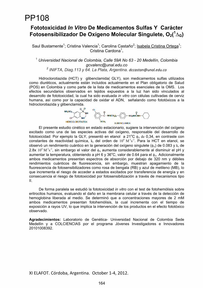
XI ELAFOT. Córdoba, Argentina. October 1 4, 2012. 164
Fototoxicidad In Vitro De Medicamentos Sulfas Y Carácter Fotosensibilizador De Oxígeno Molecular Singulete, O2(1
G)
Saul Bustamente1; Cristina Valencia1; Carolina Castaño2; Isabela Cristina Ortega1;Cristina Cardona1.
1 Universidad Nacional de Colombia, Calle 59A No 63 - 20 Medellín, [email protected]
2 INIFTA, Diag.113 y 64. La Plata, Argentina. [email protected]
Hidroclorotiazida (HCT) y glibenclamida( GLY), son medicamentos sulfas utilizados como diuréticos, actualmente están incluidos actualmente en el Plan obligatorio de Salud (POS) en Colombia y como parte de la lista de medicamentos esenciales de la OMS. Los efectos secundarios observados en tejidos expuestos a la luz han sido vinculados al desarrollo de fototoxicidad, la cual ha sido evaluada in vitro con células cultivadas de cerviz humana, así como por la capacidad de oxidar el ADN, señalando como fototóxicos a la hidroclorotiazida y glibenclamida.
El presente estudio cinético en estado estacionario, sugiere la intervención del oxígeno excitado como una de las especies activas del oxígeno, responsable del desarrollo de fototoxicidad: Por ejemplo la GLY, presentó en etanol a 21oC de 0,34, en contraste con constantes de reactividad química, kr del orden de 104 M-1s-1. Para la HCT en etanol, se observó un rendimiento cuántico en la generación del oxígeno singulete ( ) de 0.083 y kr de 2.8x 10⁴ M-1s-1, sin embargo el valor del aumenta considerablemente al disminuir el pH y aumentar la temperatura, obteniendo a pH 6 y 36oC, valor de 0.64 para el . Adicionalmente ambos medicamentos presentan espectros de absorción por debajo de 320 nm y débiles rendimientos cuánticos de fluorescencia, sin embargo, muestran apagamiento de la fluorescencia de fotosensibilizadores como rosa de bengala (RB) y azul de metileno (MB), lo que incrementa el riesgo de acceder a estados excitados por transferencia de energía y en consecuencia el riesgo de fototoxicidad por fotosensibilización a través de mecanismos tipo II.
De forma paralela se estudió la fototoxicidad in vitro con el test de fotohemólisis sobre eritrocitos humanos, evaluando el daño en la membrana celular a través de la detección de hemoglobina liberada al medio. Se determinó que a concentraciones mayores de 2 mM ambos medicamentos presentan fotohemólisis, la cual incrementa con el tiempo de exposición a rayos UV, lo que implica la intervención de los productos en el efecto fototóxico observado.
Agradecimientos: Laboratorio de Genética- Universidad Nacional de Colombia Sede Medellín y a COLCIENCIAS por el programa Jóvenes Investigadores e Innovadores 20101008392.
PP108
164

XI ELAFOT. Córdoba, Argentina. October 1-4, 2012.
Influence of organic Montmorillonite in photooxidative degradation of PMMA/clay nanocomposites
Valandro, Silvano R.1; Poli, Alessandra, L.1; Neumann, Miguel G1; Schmitt, Carla, C.11Instituto de Química de São Carlos, Universidade de São Paulo,
São Carlos, Brazil [email protected] aim of this work was to study the photooxidative degradation of PMMA/organic Montmorillonite nanocomposites using gel permeation chromatography. A typical 22 factorialdesign was used to study the effect of the different organic-clays and the concentration these clay on the photooxidative degradation of the PMMA/clay nanocomposites. These factors and their studied levels are identified in Table 1 and the statistical analysis of the results was carried out using the software Statistica®. The PMMA and nanocomposites were prepared by photopolymerization in situ. The films were exposed to UV light in an irradiation chamber containing 16 UV lamps (total power 96 W) at 40 °C. The lamps emitted predominantly 254 nm wavelength. For the pure polymer (PMMA), as well as for the nanocomposites (SWy-1-C8/PMMA and SWy-1-C16/PMMA), the changes caused by irradiation of the samples are characterized by a decrease of molecular weights (Table 2).The model for polymer degradation described by Vinu and Madras[1] leads to a correlation between the change of the number –average molecular weight Mn with time and the degradation rate constant kd.This relationship is plotted in Figure 1. It can be seen that the ratio Mno/Mnt shows an assymptotic behaviour, reaching its maximum faster for the pure polymer than for the clay-containing films. The degradation rate coefficients, kd, for PMMA and the nanocomposites films were calculated from the initial slopes of these curves (shown in Figure 1b), using the initial Mn for each system. The values are shown in Table 2, from where it can be seen that kd decreases with increasing concentration of the organic-clay. The results of the statistical analysis are reported in Table 3. It was found that the B factor has more significant effect than A (for a significance level of 95%), this can be interpreted since the SWy-1 organic clays can be considered an stabilizer against UV irradiation. The stabilization mode of the clays may be explained on the ability of SWy-1 not only to scatter the incident light but also to absorb part of the UV light thus minimizing the absorption by PMMA and the degradation of the polymer in the nanocomposites, this happens independently of the organic-cation presence in the Montmorillonite. The photooxidative degradation occurs more slowly (6 times) for the nanocomposite prepared with 5.0 wt% and SWy-C16.
0 20 40 60 80 100 120
0,0
5,0x10⁻⁵
1,0x10⁻⁴
1,5x10⁻⁴
2,0x10⁻⁴
2,5x10⁻⁴
0 5 10 15 20 25 30
0,0
2,0x10⁻⁵
4,0x10⁻⁵
6,0x10⁻⁵ PMMA 1% SWy-1-C8/PMMA 5% SWy-1-C8/PMMA 1% SWy-1-C8/PMMA 5% SWy-1-C16/PMMA
[(1/M
n t)-(1
/Mn o)
]
Irradiation time/h
Figure 1: (a) Variation of [(1/Mnt)/(1/Mn0)] vs. irradiation time for the degradation of PMMA and nanocomposites; (b) blow-up of the initial time.
Table 1:Factors and levels of the 22 factorial design Factors Inferior
level (-) Superior level (+)
(A) Organic-clay SWy-C8 SWy-C16 (B) Concentration 1.0 wt % 5.0 wt %
Table 2: Photooxidative degradation coefficient of PMMA and nanocomposites films
kd(10-6mol g-1h-1)PMMA 2.59 1%SWy-C8/PMMA 1.21 5%SWy-C8/PMMA 0.71 1%SWy-C16/PMMA 1.72 5%SWy-C16/PMMA 0.43
Table 3: Estimated effects of (A) Organic-clay and (B) Concentration
Effects Media 3,329
A 0,871B 3,013
AB 1,505Acknowledgements: FAPESP – 2009/15998-1, CNPq, CAPES
Contribution from the USP Research Consortium for Photochemical Technology. References[1]VINU, R.; MADRAS G., Polymer Degradation and Stability, 2008, 93, 1440
165
PP109
165
ESTUDIO CINÉTICO E IMPLICANCIAS ATMOSFÉRICAS DE LAS REACCIONES DE FOTOOXIDACION DE
HIDROCLOROÉTERES INICIADA POR EL RADICAL OH.Barrera Javier; Aranguren Juan Pablo; Taccone Raúl; Cometto Pablo; Lane
Silvia1.
I.N.F.I.Q.C., Depto. de Fisicoquímica- Facultad de Ciencias Químicas, U.N.C. - C.P.5000, Córdoba, Argentina. E-mail: [email protected]
Introducción: Un gran número de compuestos orgánicos volátiles (COVs) son emitidos a la atmósfera tanto biogénica como antropogénicamente. Por este motivo, en los últimos años, ha crecido la demanda de parámetros cinéticos de las reacciones más importantes que ocurren en la atmósfera, principalmente por el rol que juegan en la contaminación en ambientes cerrados y la formación de aerosoles secundarios.Es ampliamente conocido que la principal vía de remoción de los COVs en la troposfera durante las horas del día es la reacción con el radical OH [1]. La obtención de las constantes de velocidad de las reacciones de este radical con los diferentes compuestos emitidos a la atmósfera es de crucial importancia para estimar el tiempo de residencia de los compuestos en la misma y consecuentemente calcular índices desu impacto ambiental. Objetivos: * Determinar la constante de velocidad en fase gaseosa para las siguientesreacciones: OH + CH3OCHCl2 y OH + CH3OCH2CH2Cl, a fin de ampliar las escasa base de datos cinéticos existente de reacciones del radical OH con hidrocloroéteres (HCEs) y determinar el impacto atmosférico de estos compuestos. * Evaluar la tendencia de reactividad de los HCEs. Metodología: Las constantes de velocidad de las reacciones son determinadas mediante el método relativo, donde en el reactor sólo transcurren las siguientes reacciones: OH + HCE Productos (1) kS OH + Referencia Productos (2) kR
El principio de este método consiste en medir la velocidad de decaimiento de la concentración del HCE, debido a la oxidación inducida por el radical OH, relativa a un compuesto de referencia, cuya constante de velocidad de reacción con el radical OH (kR) es bien conocida.La constante de velocidad se determinó a (298±2) K y a presión atmosférica (750 torr), utilizando aire sintético como gas baño. El dispositivo experimental utilizado pararealizar el estudio cinético consiste en un sistema de vacío convencional, una bolsa colapsable de Teflón de 200L y un cromatógrafo de gas (Claurus 500-Perkin Elemer).Resultados parciales: Los valores de las constantes de velocidad determinadas son,en unidades de cm3 molécula-1 s-1, (9,8±1,7) x10-12, y (0,8±0,1) x10-12 para la reacción del radical OH con CH3OCHCl2 y CH3OCH2CH2Cl, respectivamente. No existen valores anteriores de contantes de velocidad de estas reacciones, siendo también escasos los estudios de cloroéteres con radicales atmosféricos.Por otro lado, a partir de los resultados obtenidos se calcularon parámetros que indican su posible impacto ambiental. Se evaluó el tiempo de vida troposférico, elpotencial de disminución del ozono estratosférico y el potencial de calentamiento global. Los valores obtenidos fueron comparados con compuestos análogos [2].Referencias:[1] B.J. finlayson-Pitts, J.N. Pitts, Jr., Chemestry of the upper and lower atmosphere,Academic Press, N.Y., 2000.[2] P.R Dalmasso, R.A, Taccone,P.M Cometto and S.I Lane. J.Phys. Org. Chem.21,393-396, 2008.
166
PP110
166

XI ELAFOT. Córdoba, Argentina. October 1-4, 2012.
AUTOFLUORESCENCE MICROSCOPY APPLIED TO MORPHOLOGICAL CHARACTERIZATION OF THE
SUGARCANE BAGASSE AND EVALUATION OF THE DISTRIBUTION OF COMPOUNDS INTO THE CELLULOSIC
MATRIX
Chimenez, Tiago1*; Marabezi, Karen1, Curvelo, Antonio1, Gehlen, Marcelo1
1Instituto de Química de São Carlos, USP, São Carlos, SP, Brazil, *[email protected]
The remainder material from sugarcane fermentation is called bagasse. The main product obtained from bagasse is the bio-ethanol [1]. The use of bagasse on the industrial process depends on the knowledge of the inherent morphology and spatial distribution of chemical compounds. The methods based on fluorescence microscopy have been carried out to determine the distribution of lignocellulosic material in plant tissues, within the wood cell wall [2]. However, majority of methodologies only supplies image applications, providing a fluorescence map of scanned region of a given material. We aimed at studying the morphological characterization by mapping of the fluorescence images and component identification by lifetime fluorescence measurements of sugarcane bagasse. The investigation of lignocellulosic materials from whole bagasse, fiber, pith, and the respective samples after bleaching treatment were performed using confocal fluorescence microscope. The images showed a characteristic morphology of non-bleached microfibrils, which has been lost after bleaching treatment. However autofluorescence properties were not observed. The analysis of the decay parameters obtained from the different samples demonstrates a difficult task due to the complexity of the system [3]. In general, the decays of natural and bleached samples have similar multiexponential behavior in time scale. However, the analysis of decay times shows that after bleaching the average decay time increases in about 68% from fiber to bleached fiber, 47% from whole bagasse to bleached bagasse, and 41% from pith to bleached pith. With these issues in mind, it seems that the chemical bleaching process is disrupting and dispersing lignin components although it is also reducing the amount of organic material and intrinsic fluorophoric composition. As a result, the emission intensity of the bleaching samples is still high because the average lifetime increases and most of the remaining chromophores are now partially isolated or dispersed in the cellulose without the effect of excited-state deactivation by self-quenching. Thus, these results indicate that confocal fluorescence microscopy could see significant use to evaluate the efficiency of whitening process of the sugarcane bagasse.
Acknowledgements: The authors thank the support from CNPq, CAPES and FAPESP Brazilian research funds.
References [1] Goldemberg, J. (2007) Ethanol for a sustainable energy future. Science. 315: 808-810. [2] Li, Z., H. Zhai, Y. Zhang, and L. Yu (2012) Cell morphology and chemical characteristics of corn stover fractions. Industrial Crops and Products. 37: 130- 136. [3] Harter, K., A. J. Meixner, and F. Schleifenbaum (2012) Spectro-Microscopy of Living Plant Cells. Molecular Plant. 5: 14-26.
167
PP111
167
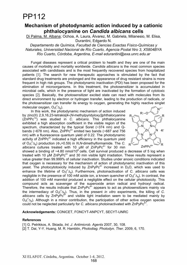
XI ELAFOT. Córdoba, Argentina. October 1-4, 2012.
Mechanism of photodynamic action induced by a cationic phthalocyanine on Candida albicans cells
Di Palma, M. Albana; Ochoa, A. Laura, Álvarez, M. Gabriela, Milanesio, M. Elisa, Durantini, Edgardo N.
Departamento de Química, Facultad de Ciencias Exactas Físico-Químicas y Naturales, Universidad Nacional de Río Cuarto, Agencia Postal Nro 3, X5804BYA
Río Cuarto, Córdoba, Argentina, E-mail [email protected]
Fungal diseases represent a critical problem to health and they are one of the main causes of morbidity and mortality worldwide. Candida albicans is the most common species associated with candidiasis and is the most frequently recovered species from hospitalized patients [1]. The search for new therapeutic approaches is stimulated by the fact that standard drug treatments are prolonged and the appearance of drug resistant strains is more frequent in high risk groups. The photodynamic inactivation (PDI) has been proposed for the elimination of microorganisms. In this treatment, the photosensitizer is accumulated in microbial cells, which in the presence of light are inactivated by the formation of cytotoxic species [2]. Basically, the photosensitizer excited state can react with molecules from its direct environment by electron or hydrogen transfer, leading to the production of radicals, or the photosensitizer can transfer its energy to oxygen, generating the highly reactive singlet molecular oxygen, O2(1
g).In this work, the photodynamic mechanism of action induced
by zinc(II) 2,9,16,23-tetrakis[4-(N-methylpyridyloxy)]phthalocyanine (ZnPPc+4) was studied in C. albicans. This phthalocyanine exhibited a high absorption coefficient in the visible region of the spectrum, characterized by the typical Soret (~374 nm) and Q-bands (~678 nm). Also, ZnPPc4+ emited two bands (~687 and 756 nm) with a fluorescence quantum yield of 0.22. The photodynamic activity of ZnPPc4+ indicated a high efficiency in the quantum yield of O2(1
g) production ( =0.59) in N,N-dimethylformamide. The C. albicans cultures treated with 10 μM of ZnPyPc4+ for 30 min showed a binding of ~4.80 nmol/106 cells. Cell survival produced a decrease of 5 log when treated with 10 μM ZnPyPc4+ and 30 min visible light irradiation. These results represent a value greater than 99.999% of cellular inactivation. Studies under anoxic conditions indicated that oxygen is necessary for the mechanism of action of photodynamic inactivation of this yeast. The photocytotoxicity induced by ZnPyPc4+ increased in D2O, which was used to enhance the lifetime of O2(1
g). Furthermore, photoinactivation of C. albicans cells was negligible in the presence of 100 mM azide ion, a known quencher of O2(1
g). In contrast, the addition of 100 mM mannitol produced a negligible effect on the cellular phototoxicity. This compound acts as scavenger of the superoxide anion radical and hydroxyl radical. Therefore, the results indicate that ZnPyPc4+ appears to act as photosensitizers mainly via the intermediacy of O2(1
g). Thus, in the present in vitro experiments, the killing of C.albicans cells by ZnPyPc4+ and visible light irradiation seem to be mediated mainly by O2(1
g). Although in a minor contribution, the participation of other active oxygen species could not be neglected particularly for C. albicans photoinactivated with ZnPyPc4+.
Acknowledgements: CONICET, FONCYT-ANPCYT, SECYT-UNRC
References [1] G. Petrikkos, A. Skiada, Int. J. Antimicrob. Agents 2007, 30, 108.[2] T. Dai, Y-Y. Huang, M. R. Hamblin, Photodiag. Photodyn. Ther. 2009, 6, 170.
ZnPPc4+
NN
N
NN
N
N
N
R
RR
R
Zn 4I-
R: ON+
168
PP112
168
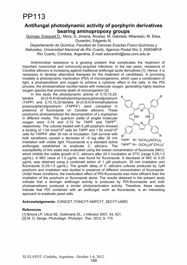
XI ELAFOT. Córdoba, Argentina. October 1-4, 2012.
Antifungal photodynamic activity of porphyrin derivatives bearing aminopropoxy groups
Quiroga, Ezequiel D.; Mora, S. Jimena, Álvarez, M. Gabriela, Milanesio, M. Elisa, Durantini, Edgardo N.
Departamento de Química, Facultad de Ciencias Exactas Físico-Químicas y Naturales, Universidad Nacional de Río Cuarto, Agencia Postal Nro 3, X5804BYA
Río Cuarto, Córdoba, Argentina, E-mail [email protected]
Antimicrobial resistance is a growing problem that complicates the treatment of important nosocomial and community-acquired infections. In the last years, resistance of Candida albicans is increasing against traditional antifungal azole derivatives [1]. Hence, it is necessary to develop alternative therapies for the treatment of candidiasis. A promising modality is photodynamic inactivation (PDI) of microorganisms, which uses a combination of light, a photosensitizer and oxygen to achieve a cytotoxic effect in the cells. In the PDI process, the photosensitizer excited reacts with molecular oxygen, generating highly reactive oxygen species that promote death of microorganism [2].
In this study the photodynamic activity of 5,10,15,20-tetrakis [4-(3-N,N-dimethylaminopropoxy)phenyl]porphyrin (TAPP) and 5,10,15,20-tetrakis [4-(3-N,N,N-trimethylamine propoxy)phenyl]porphyrin (TAPP4+) were compared in presence of fluconazole on Candida albicans. These porphyrins photosensitized the decomposition of L-tryptophan in different media. The quantum yields of singlet molecular oxygen were 0.74 and 0.72 for TAPP and TAPP4+,respectively. The cultures treated with 5 μM porphyrin showed a binding of 1.04 nmol/106 cells for TAPP and 1.50 nmol/106
cells for TAPP4+ after 30 min of incubation. Cell survival with both sensitizers caused a decrease of ~5 log after 30 min irradiation with visible light. Fluconazole is a standard active antifungals established to eradicate C. albicans. The susceptibility of this yeast was evaluated using the lowest concentration of fluconzole (MIC), which inhibits the visible growth of C. albicans after 24 h incubation at 37ºC (range 0.25-1.5 μg/mL). A MIC value of 1.0 μg/mL was found for fluconazole. A decrease of MIC to 0.25 μg/mL was obtained using a combined action of 1 μM porphyrin, 30 min irradiation and fluconazole (0.25-1.5 μg/mL). The growth delay of C. albicans cultures produced by 1μM porphyrin and irradiation was studied in presence of different concentration of fluconazole. Under these conditions, the inactivation effect of PDI-fluconazole was more efficient than the irradiation of the porphyrin or fluconazole alone. The results obtained in the present study indicate that a stronger antifungal activity is produced by PDI-fluconazole and both photosensitizers produced a similar photoinactivation activity. Therefore, these results indicate that PDI combined with an antifungal, such as fluconazole, is an interesting approach to eradicate yeast cells.
Acknowledgements: CONICET, FONCYT-ANPCYT, SECYT-UNRC
References [1] Briona LP, Ukoa SE, Goldmanb DL. J Infection 2007, 54, 521.[2] M. O. Senge, Photodiagn. Photodyn. Ther. 2012, 9, 170.
HN
N
NH
N
R
R R
R
TAPP4+ R= O(CH2)3N+(CH3)3I-TAPP R= O(CH2)3N(CH3)2
169
PP113
169
XI ELAFOT. Córdoba, Argentina. October 1-4, 2012.
Proceso combinado de oxidación-adsorción para la remoción de As en agua. Aplicación del proceso UV/H2O2
Lescano, Maia 1; Zalazar, Cristina1; Brandi, Rodolfo1 1 INTEC (UNL-CONICET). Güemes 3450, (3000) Santa Fe, Argentina,
El arsénico (As) es un elemento ampliamente distribuido en la corteza terrestre. Se encuentra habitualmente presente en la naturaleza combinado formando diversos minerales. A partir de éstos, puede desplazarse hacia el aire, el agua y el suelo. Las actividades humanas (minería, agricultura, fundición de metales, entre otros) pueden movilizar de manera significativa al arsénico desde sus depósitos naturales y generar contaminación ambiental de importancia [1]. Los efectos del arsénico en la salud pueden incluir lesiones en la piel y alteraciones sistémicas cancerosas y no cancerosas. La vía más directa y perjudicial de contacto de este mineral con el hombre es a través de la ingesta en el agua, aunque también puede ser por medio del contacto de la piel con el agua o el suelo que lo contenga. Debido a estos efectos nocivos que produce este tóxico clase A (EPA, 2000), es que la Organización Mundial de la salud (OMS) recientemente ha reducido el máximo nivel permisible del contaminante en agua para consumo de 50 a 10 μg/L.
En agua subterránea, las especies de arsénico inorgánico arsenato (As V) y arsenito (As III) son las más abundantes. El As (III) no sólo es mucho más tóxico que la forma (V) sino que tiene mayor movilidad, y por lo tanto, es más difícil de remover utilizando procesos fisicoquímicos tradicionales [2]. Es de gran importancia, entonces, una etapa de oxidación previa para obtener buenos rendimientos en la remoción del contaminante. Nuestro grupo de investigación estudió detalladamente el proceso avanzado de oxidación UV/H2O2
obteniendo resultados satisfactorios para la oxidación de arsénico en agua [3]. Es por eso que resulta de interés el estudio de la etapa posterior a la oxidativa, que es la de remoción del contaminante y el diseño del equipo combinado oxidación-remoción.
Teniendo en cuenta los diferentes métodos existentes, el proceso de adsorción es considerado como el más económico y fácil de llevar a cabo. En este trabajo se estudian tres materiales disponibles comercialmente (Dióxido de titanio granular, Hidróxido de hierro granular y Alúmina activada) eligiendo, a partir de los resultados obtenidos (isotermas de adsorción, curvas de ruptura), los más eficientes en la remoción del contaminante.
El objetivo principal de este trabajo es, partir de la selección de los adsorbentes a utilizar, diseñar un prototipo de remoción a escala laboratorio acoplándolo al sistema de oxidación previamente estudiado.
Se construyó un dispositivo compuesto por un reactor anular irradiado por una lámpara germicida (λ = 253,7 nm). A la salida del reactor, parte del agua es recirculada al mismo reactor y otra ingresa a la columna de adsorción rellena con el material adsorbente seleccionado. Todo el sistema opera en forma continua. Las experiencias se llevaron a cabo fijando la concentración de arsénico total en 200 µg/L pero variando la relación de especies As (V) / As (III) presentes: 75/125; 50/150; 25/175. La concentración de H2O2 utilizada fue de 6 mg/L.
De acuerdo a los resultados obtenidos, trabajando con un caudal de 0,8 L/min, se logra una concentración menor a 2 µg/L de As (III) a la salida del sistema de reacción y una remoción completa del contaminante a la salida de la columna de adsorción. De esta manera se comprueba que la tecnología combinada de oxidación (UV/H2O2) - adsorción puede ser un proceso factible y eficaz para la remoción de arsénico en agua a pequeña y mediana escala. Referencias[1] Corey, G., Tomasini, R., Pagura, J., 2005. Estudio epidemiológico de la exposición al arsénico a través del consumo
de agua. Provincia de Santa Fe, Argentina. Ente Regulador de Servicios Sanitarios (ENRESS). [2] Yang, H., Lin, W., Rajeshwar, K., 1999. Homogeneous and heterogeneous photocatalytic reactions involving As (III)
and As (V) species in aqueous media. J. Photochem and Photobiol. A: Chemistry, 123, 137-143. [3] M. Lescano, C. Zalazar, A. Cassano, R. Brandi, Arsenic (III) oxidation of water applying a combination of hydrogen
peroxide and UVC radiation, Photochem. Photobiol. Sci. 10 (2011) 1797-1803.
170
PP114
170
XI ELAFOT. Córdoba, Argentina. October 1 4, 2012. 171
Efecto de Campo Magnético en la Fotoreactividad de(2E/2T)Cr(phen)3
3+ y (2E/2T)Cr(5Clphen)33+.
Bazán, Claudia M.1; Gerardo A. Argüello1.
1 INFIQC, Departamento de Fisicoquímica, Facultad de Ciencias Químicas, Universidad Nacional de Córdoba. Ciudad Universitaria, 5000
Córdoba. Argentina. Email: [email protected]
Intensos campos magnéticos (B), mayores a 1 Tesla (T), inducen cambios en los espectros de emisión, tiempos de vida del estado excitado, rendimientos cuánticos de reacción y procesos de quenching. Por ejemplo, B entre 1 y 5 T incrementan la velocidad de relajación del (3CT)Ru(bpy)3
2+ 1 y del (2E/2T)Cr(NN)33+ 2 en un 10% . Estudios de
quenching por transferencia de electrones (TE) de estos estados excitados, demostraron que intensos B inducen tanto aceleraciones como desaceleraciones de estas reacciones.
Se estudio el efecto de B sobre la velocidad de relajación del (2E/2T)Cr(phen)33+ y del
(2E/2T)Cr(5Clphen)33+ (phen=1,10-fenantrolina y 5Clphen= 5-Cl-fenantrolina). Se obtuvieron
las constantes de velocidad de decaimiento (k0= 1/ 0) en soluciones aireadas (buffer fosfato pH=8, NaCl 0,1M, Tº= 25±1 ºC) en ausencia y presencia de B aplicado (B= 0, 2, 4, 6, 8 y 10T) obteniendo a B=10T un incremento de 32% para (2E/2T)Cr(phen)3
3+ y 12 % para (2E/2T)Cr(5Clphen)3
3+. La variación de k0 indicaría que B incrementaría algún proceso no radiativo.
Se estudio el efecto de B sobre las constantes de velocidad de quenching (kq) por TE del (2E/2T)Cr(5Clphen)3
3+ y del (2E/2T)Cr(phen)33+ por aminas (Anilina, p-Anisidina, p-
fenilendiamina, 1-Naftilamina) y del (2E/2T)Cr(phen)33+ por fenoles (fenol, 4-Cl-fenol, 4-Cl-3
metil fenol y 4-Br-2,6 dimetilfenol en soluciones aireadas, buffer Britton-Robinson pH=10, NaCl 0,1M, Tº= 25±1 ºC). De las pendientes de los gráficos de Stern-Volmer en ausencia y presencia de B se calcularon las correspondientes kq, dichas constantes disminuyen en presencia de B=6T en un 10% para el quenching por aminas y en presencia de B=10Tdisminuyen en un 30 % para el quenching por fenoles, dicho efecto de B se explica en términos del acoplamiento del B externo con el momento angular de los estados excitados orbitalmente degenerados.
Agradecimientos: Los autores agradecen por las becas de postgrado otorgadas por CONICET. Éste trabajo es financiado por INFIQC - CONICET, SECyT - U.N.C. y ANPCyT - FONCyT.
Referencias1 Ferraudi, G. and Argüello, G. A., J. Phys. Chem. 92(1988) 1846.
2 Ferraudi, G., Argüello, G. A. and Frink, M., J. Phys. Chem. 91(1987) 64.
PP115
171

XI ELAFOT. Córdoba, Argentina. October 1 4, 2012. 172
Estudios Antibacterianos de 6-MeQ y Derivados Organometálicos,Relativo a la Generación de 1O2(1
g)
Villa, Cristian1; Valencia, Cristina1;Mercado, Donaldo2; Ortega, Isabel1;Montoya, Olga1
1Universidad Nacional de Colombia, Calle 59A No 63 - 20 Medellín, Colombia [email protected]
2INIFTA, Diag.113 y 64. La Plata, Argentina. [email protected]
Dado el eficiente carácter fotosensibilizadorde oxígeno molecular singulete del compuesto 6-metoxiquinolina (6-MeQ) y su potencial uso como bactericida en el tratamiento de aguas residuales contaminadas de bajo caudal, se ha venido trabajando en lograr la incorporación de este fragmento orgánico en una matriz sólida, donde se conserve sucarácterfotosensibilizador.
De forma inicial se ha realizado la síntesis de complejos organometálicosentre el ligando orgánico 6-MeQ y los metales disprosio, itrio, rutenio, cobalto e itrio. En todos los casos se observópara los complejos obtenidos un rendimiento cuántico en la generación de oxígeno molecular singulete, , mayor a 0,9;medidas obtenidas en experimentos en estado estacionario. Hasta el momento se ha logrado la obtención de materiales sólidos para los complejos con los metales rutenio, disprosio y neodimio.
Paralelamente, a estos complejos se les ha realizado la prueba de susceptibilidad antibacteriana por difusión por disco (antibiograma Kirby-Bauer), que correlaciona la susceptibilidad de un microorganismo a un antibiótico determinado. Se observa halo de inhibición de crecimiento bacteriano,para Escherichiacoli, Staphylococcusaureusy Bacilluscereus, al emplear 6-MeQ y los complejos con disprosio e itrio.
El trabajo se extenderá a otros metales como son europio, neodimio, gadolinio,holmio yerbio, metales para los que también se realizarán pruebas bactericidas.
Agradecimientos:Universidad Nacional de Colombia-Sede Medellín DIME-DARWIN 20101008123
PP116
172

300 400 500 600 700-0,04
-0,02
0,00
0,02
0,04
Abs
(nm)
After 9 s Laser pulse RF-Air RF-Argon RF+Trp0,3mM-Argon
Energy and charge-transfer quenching of flavins in AOT-reversed micelles solutions
Perez, Hugo; Valle, Lorena; Morán Vieyra, Faustino and Borsarelli Claudio.Laboratorio de Cinética y Fotoquímica, Centro de Investigaciones y Transferencia de
Santiago del Estero (CITSE-CONICET), UNSE.E-mail: [email protected]
Flavins are widely distributed in the biosphere, serve as cofactors in redox proteins and are commonly applied photosensitizers [1]. Riboflavin (RF), present in free form or as flavinmononucleotide (FMN) and flavin adenine dinucleotide (FAD) in cells, is an important and efficient endogenous cellular photosensitizer [2]. The triplet state of a flavin can be quenched by oxygen and a variety of electron donors, e.g. ascorbic acid, amines or amino acids, e.g. tyrosine or tryptophan [1].
We have studied RF triplet excited state (3RF*) quenching by tryptophan and molecularoxygen in (AOT)-iso-octane-water reverse micelles by laser flash photolysis (LFP) with excitation at 355 nm.
The 3RF* quenching efficiency ( q) by tryptophan is governed by the local concentration of the species at the interface of the micelle, and also by fluidity and water content. The presence of neutral radical of tryptophan (TrN ) was confirmed by two structureless bands located around 510 and 330 nm, which demostrated an electron transfer quenching mechanism [3].
On the other hand, the deactivation of 3RF* by molecular oxygen showed that qdecreases slightly with the water content of the micelle, that is in agreement with RF location on the micelar structure [4]. The high increment of quenching efficient at low internal oxygen micelar concentration is explained by the large constant of quenching (kq ca. 109 M-1 s-1).
Acknowledgements
HP thanks to CONICET for PhD fellowship and supporting.
1 H. Gorner. Oxygen uptake after electrón transfer from amines, amino acids and ascorbic acid to triplet flavins in air saturated aqueous solution. J. Photochem. Photobiol. B. 87, 73 80 (2007).2 L. Chang- Yuan, L. Yan- Yun. Electron transfer oxidation of tryptophan and tyrosine by triplet states and oxidized radicals of flavin sensitizers: a laser flash photolysis study. Biochim. Biophys. Acta. 1571, 71 76 (2002).3 T.B. Melø, M.A. Ionescu, G.W. Haggquist, K.R. Naqvi. Hydrogen abstraction by triplet flavins. I: time-resolved multi-channel absorption spectra of flash-irradiatedriboflavin solutions in water. Spectrochimica. A. 55, 2299 2307 (1999).4 L. Valle, F.E. Morán Vieyra, C.D. Borsarelli. Hydrogen-bonding modulation of excitated-state properties of flavins in a model of aqueous confined environment. Photochem. Photobiol. Sci. 11, 1051-1061 (2012).
0,0 0,2 0,4 0,6 0,8 1,0
0,0
0,2
0,4
0,6
0,8
1,0
W 2 W 3 W 10 Buffer
q
Trpinter (mM)0,0 0,3 0,6 0,9 1,2 1,5
0,0
0,2
0,4
0,6
0,8
1,0
W 3 W 8 W 20 Buffer
q
[O2]Mic mM
173
PP117
173

XI ELAFOT. Córdoba, Argentina. October 1 4, 2012. 174
The Photophysics Properties and Theoretical Studies of a New Ruthenium (II) Perylenediimide Complex
Dos Santos, Edjane R.1; Aguiar, Inara1; Carlos, Rose M.11 Departamento de Química,Universidade Federal de São Carlos,São Carlos/São
Paulo-Brazil, [email protected]
Ruthenium (II) diimine complexes have attracted much attention due to their applications in photophysical. This complexe with Ru(II) coordination center, with strong absorption of visible light and long-lived triplet excited states are useful for applications such as photovoltaics, photocatalysis, luminescent molecular probes and up conversion, etc. This complex is different compared to organic fluorophores which have triplet excited states instead of the singlet excited states, are populated upon photoexcitation of this complex [1-3]. The new complex of [Ru(phen)2(phenBL)](PF6)2 (phen= 1,10-phenanthroline and phenBL= 3,4,9,10-Perylene-Bis(Diaminoimidephen) has been synthesized and characterized by spectroscopic (UV-vis, IR, 1H NMR, Emission and Excitation) and electrochemical (cyclic voltammetry and differential pulse voltammetry) techniques. The spectroscopic studies of this complex is presented based on theoretical and experimental analysis. All calculations were performed with the Gaussian 09 (G09) program package employing the DFT method with Becke’s threeparameter hybrid functional and Lee-Yang-Parr’s gradient corrected correlation functional (B3LYP). The LanL2DZ basis set and effective core potential were used for the Ru atom. TD-DFT were used to provide the UV-vis spectrum. The influence on the coordination of the phenBL to the RuII center could be seen in the IR spectra, the as(CO) stretching shift to (1662 cm-1) compared to the free ligand 1697 cm-1. The cyclic voltammograms showed high potential values for the RuII/RuIII oxidation, ca. 1.48 V. According to TD-DFT and UV-vis spectra, the complex shows a absorption at approximately 420 nm assigned as a MLCT and overlapped * absorption bands at 460, 494 and 533 nm of ligand phenBL. The emission spectrum of the complex presents three high emission bands at 545, 587, 640 (when excited in bands of the ligand at 494 nm), which is very similar with the emission showed for the free ligand; when excited at 393 nm the emission spectrum of the complex presents only one band at 604 nm. The fluorescence studies were carried out in DMSO solution and acetonitrile. The complex also was studied in solid state, and in a polymeric matrix (a film), and could be observed a similar emission spectra. This compound exhibits to be very stable in presence of light and did not present photochemical sensibility. These results show that a new complex exhibits interesting properties to future studies of electrons transfer reactions, due to its advantages of readily tunable excitation/emission wavelengths, low excitation power density and the high up conversion quantum yields.
Acknowledgements: We thank the Fundação Ao Amparo de Pesquisa do Estado de São Paulo (FAPESP).
References [1] L. Flamigni, A. Barbieri, C. Sabatini, B. Ventura and F. Barigellet, Top. Curr. Chem., 2007, 281, 143. [2] S. Campagna, F. Puntoriero, F. Nastasi, G. Bergamini and V. Balzani, Top. Curr. Chem., 2007, 280, 117. [3] N. Armaroli, ChemPhysChem, 2008, 9, 371.
PP118
174

Solvent effect on the Q-band shape of cationic porphyrins
R. N. Sampaio1, E. Piovesan1, M. B. Silva1, M. O. Borges1, A. E. H. Machado2, R. De Paula3, N. M. Barbosa Neto1
1Instituto de Física, Universidade Federal de Uberlândia, Brazil 2Instituto de Química, Universidade Federal de Uberlândia, Brazil
3Universidade Federal do Recôncavo da Bahia
Photophysical properties of porphyrin [1] derivatives have been the target of a huge amount of investigations during the past few decades, motivated by the possibilities of their use in a large variety of applications including light harvesting systems, photodynamic therapy, chemical sensors and others. In general, these studies have the purpose of correlating the porphyrin molecular structure to some photophysical characteristic that can be modified upon substitution of a central ion, the attachment of outlying or axial groups, etc. In particular, cationic porphyrins have been the subject of intense investigations due its applicability and interaction with DNA [2]. In this work we employed an analysis of two new cationic porphyrin derivative, the free-base 5,10,15,20-tetrakis (1,3-dimethylimidazolium-2-yl) porphyrin tetraiodide [(H2-TDMImP)I4], as well as its zinc derivative [(Zn-TDMImP)I4] in a set of solvents at different concentrations [3]. The study was restricted to the first singlet excited state, characterized by the Q-band region of the absorption spectra. It was observed a solvatochromism effect by changing the solvent for both compounds. When varying the concentration, a changing of the in the relative intensity of Q-band, mostly for Qx(1,0) [(H2-TDMImP)I4] is observed. This effect is probably due to distortions in the porphyrin ring caused by solvent interactions with the peripheral cationic groups. Such phenomenology can affect the vibrational levels involved in the light absorption process. The same effect wasn’t observed for the [(Zn-TDMImP)I4] once the central substituent Zn stabilize the porphyrin compound avoiding distortions in the structure. This kind of characterization and the understanding of the processes involved are of huge importance in order to adjust the porphyrin properties aiming specific application. Keywords: cationic porphyrin, solvent effect, absorption.
[1] K. Kalyanasundaram, Photochemistry of Polypyridine and Porphyrin Complexes, Academic Press, San Diego, 1992. [2] S. Mettath, B.R. Munson, R.K. Pandey, DNA interaction and photocleavage properties of porphyrins containing cationic substituents at the peripheral position, Bioconjug. Chem. 10 (1999) 94–102. [3] J.H. Chou, M.E. Kosal, H.S. Nalwa, N.A. Rakow, K.S. Suslick, Applications of porphyrins and metalloporphyrins to materials chemistry, in: K. Kadish, K. Smith, R. Guillard (Eds.), The Porphyrin Handbook, Academic Press, New York, 2000, pp. 43–141. [4] A.E.H. Machado, N.M. Barbosa Neto, W.R. Gomes, D.M.S. Araújo, H.S. Miglio, L. T. Ueno, P. L. Franzen, S. C. Zilio, R. de Paula, J.A.S. Cavaleiro, Synthesis and optical characterization of two tetrasubstituted cationic porphyrin derivatives. Paper submitted to be considered for publication at Journal of Photochemistry and Photobiology A: Chemistry.
Work supported by CAPES, FAPEMIG, CNPq, and INCT/INFo.
[email protected], João Naves de Ávila Avenue, 2121, Santa Mônica, Uberlândia – MG, CEP: 38400-902.
175
PP119
175

XI ELAFOT. Córdoba, Argentina. October 1 4, 2012. 176
Photophysical, Photochemical and Electrochemical properties of Binuclear carbonyl-containing complexes of
Ru and Mn using bi- and tridentate ligands as bridge de Aguiar, Inara1; Lever, Alfred B. P.2; Carlos, Rose M.1
1Universidade Federal de São Carlos, Rod. Washington Luis Km 235, São Carlos, SP, 13565-905,Brasil, [email protected]
2York University, 88, The Pond Rd., Toronto, ON, M3J1P3, Canada
To mimic the photoinduced electron-transfer steps from the manganese cluster to the photoactive P680
+ in photosystem II, a series of new binuclear complexes have been synthesized and characterized by elemental analysis, spectroscopic (EPR, UV-vis, FTIR, 1HNMR) and electrochemical (cyclic voltammetry and spectroelectrochemistry) techniques. The systems studied were [(phen)2Ru(BL)2Mn(CO3)(im)]n+3 and [(BL’)Ru(BL’)Mn(CO)3(phen)]3+,where BL = 4,4-bipyridine (4,4bpy) (I) and pyrazine (pz) (II), BL’ = 4'-(4-pyridyl)-2,2':6',2''-terpyridine (pytpy) (III) and 4'-(4-pyridyl)-2,6-di(2-pyrazinyl)pyridine (pydpp) (IV).
The DFT calculations were carried out using the B3LYP functional and LanL2DZ basis set within the GAUSSIAN (G09) [1]. DFT showing molecular orbital composition of the complexes exhibit the HOMO composed majority by dRu (~60%) and LUMO constituted by BL component (~100%) indicating a favorable photoinduced electron transfer pathway (D*-BL-A D+-BL-A-). In acetonitrile, the complexes I and II present intense absorptions in the visible region (420 nm), assigned as a MLCT (Ru phen, 4,4’bpy) and broad emissions at 590 nm. For complexes III and IV the intense MLCT are observed at 490 nm (Ru pytpy, pydpp) and also two emission bands at 367 and 700 nm, when excited at 290 and 490 nm, respectively. Compared to [Ru(phen)3]2+ [2] the lifetimes of I, II, III were strongly reduced and showed bi-exponential kinetics ( 1 = 1.25 ns, %lum = 70% and 2 = 36 ns, %lum = 30%) assigned to the population of MLCT (Ru phen) and LLCT (4,4bpy) states, while complex IIIexhibited a bi-exponential kinetics ( 1 = 2.6 ns, %lum = 60% and 2 = 25 ns, %lum = 40%) assigned to the population of MLCT (Ru pytpy) and LLCT (pytpy) excited states.
Photophysical characterization of I and III indicates E0-0 2.33 eV (I) and 2.10 eV (II)respectively, which is available energy stored in their respective 3MLCT (metal-to-ligand charge transfer) excited states. The correlation between the electrochemical and spectroscopic measurements show that complex III is a powerful excited-state oxidant (E°(Ru*/2+) = 0.5) and a poor excited-state reductant (E°(Ru2+/*) = -0.72 V compared to -0.84 and 1.26 V for Ru(bpy)3.)The oxidative properties of the excited state in the complexes I, II, III and IV are being exploited in presence of the electron acceptor methylviologen (MV2+). The results show an intermolecular electron-transfer reaction from the excited binuclear complexes to MV2+.
These new complexes are capable of visible-light absorption and consequent one-electron reduction of external electron acceptor molecules. The electron transfer ability and its electrochemical and photochemical properties indicate an interesting system to use in photocatalytic water splitting process.
Acknowledgements: The authors would like to thanks CNPq and CAPES for financial assistance. Computing was carried out by courtesy of the SHARCNET, Ontario, Canada.
PP120
176

XI ELAFOT. Córdoba, Argentina. October 1-4, 2012.
Oxidation of BSA photosensitized by Rose BengalArgañaraz, Natalia Mariel1; Espeche Turbay, M. Beatriz1; Rey, Valentina1; Borsarelli
Claudio D.* 1
Laboratorio de Cinética y Fotoquímica (LACIFO). Centro de Investigaciones y Transferencia de Sgo. del Estero (CITSE-CONICET) Universidad Nacional de Santiago del Estero. Ruta N9, km 1125, Vil la
El Zanjón. 4200 - Sgo. del Estero, Argentina. E-mail:[email protected]
Proteins like other biological macromolecules may be targets to the action of reactive oxygen species (ROS). The oxidation of these substrates produces changes in the functionality and/or alterations of the structure causing further changes at physiological level. As a result, the characterization of protein oxidative damage both by 1O2 (singlet oxygen) or by any other ROS, using model systems in vitro is necessary to evaluate the damage occurring in biological media.
The aim of this work is to study the oxidation of bovine serum albumin (BSA) by 1O2
generated through photosensitization of Rose Bengal (RB) in homogeneous media. We have focused in the analysis of the photo-oxidation that occurs in the adduct
formated using different concentrations of BSA (10 M, 50 M and 100 M) with a fixed concentration of RB (10 M), adduct I (1/1), II (1/5) and III (1/10). This was evaluated using various spectroscopic techniques, i.e. UV-Vis absorption and fluorescence, as well as analytical and biochemical techniques (peroxides and carbonyl determination).
As shown in the graphs, the increase in the concentration of BSA, which participates in the photo-oxidation processes, produce an increase in the oxidation products. This has been associated with increased concentration of carbonyl groups that has been related to oxygen consumption. In this way, it is demonstrated that the type II process (singlet oxygen dependent) play an important role in the photo-oxidation products formation.
We can conclude from our results that the processes of photosensitization in vitro are an appropriate methodology to study protein oxidation, and to interpret the changes that oxidative stress may cause in biological media.
0 5 10 15
100
200
300
1000
2000
IF 410 nm IF 342 nm
Photolysis time (min)
Fluo
res.
Inte
ns. (
a.u.
) 410
nm
Fluo
res.
Inte
ns. (
a.u.
) 342
nm
0 10 20 300
100
200
Adduct I II III[C
arbo
nyl g
roup
s] (
M)
Photolyis time (min)0 10 20 30
0,6
0,9
V0= 9.55 e -2min-1
V0= 6.87 e -2min-1
[o2] R
elat
ive
Time (min)
Adducts I II III
V0= 2.7 e-2min-1
Acknowledgements: Authors gratefully acknowledge CONICET for the financial support to convey this research.
177
PP121
177

Síntesis y propiedades fotofísicas de ternaftalenos y derivados del terrileno
Jimenez, Liliana; Medrano, Carlos; Pierini, AdrianaDpto. de Qca. Orgánica – INFIQC, Universidad Nacional de Córdoba,
Ciudad Universitaria, Córdoba, Argentina. E-mail: [email protected]
Especial interés y atención en el área de fotoquímica y nanotecnología se ha dedicado, en la última década, al desarrollo de estructuras cromofóricas específicas por su posible aplicación en diferentes dispositivos moleculares con aplicación en celdas fotovoltaicas, antenas colectoras de luz (Light-Harvesting Antennae), diodos orgánicos emisores de luz (OLEDs), etc.[1] Un ejemplo particular de estas estructuras es la molécula del perileno ya que presenta excelentes propiedades electrónicas y ópticas.[2] El perileno, debido a su rigidez, posee una fluorescencia característica con un alto rendimiento cuántico, el cual varía de acuerdo a los sustituyentes que posea el policiclo. La síntesis y el manejo de los derivados del perileno son complicados y generalmente poseen bajos rendimientos globales de reacción.[3] Es de nuestro interés estudiar otras vías sintéticas para la obtención de derivados perilénicos y a su vez, analizar y caracterizar las propiedades fotofísicas de estas nuevas estructuras.
En el presente trabajo se muestra un estudio fisicoquímico y sintético de la reacción entre el sustrato 1,4-dibromonaftaleno (1) y el anión del 2-naftol (2), esquema 1. Esta reacción se describe, bajo fotoestimulación, mediante un mecanismo de sustitución nucleofílica por transferencia de electrones, involucrando, por lo tanto, radicales y radicales aniones como intermediarios. A su vez, se continua con la caracterización fotofísica de losproductos obtenidos.
Un segundo objetivo planteado es la obtención de derivados perilénicos (terrileno, 3)mediante ciclaciones intramoleculares oxidativas y, por consiguiente, con la descripción fotofísica de los productos obtenidos. Para este paso se emplearon dos vias sintéticas diferentes, por un lado el empleo de una fuente de electrones como potasio y posterior oxidación, y en paralelo mediante reacciones catalizadas por metales de transición (reacciones de Suzuki).
Referencias[1] - Balzani, V. C., A.; Venturi, M. Molecular Devices and Machines. Concepts and Perspectives for the Nanoworld.; 2nd ed.; Wiley-VCH Verlag GmbH & Co.: Weinheim, 2008.[2] - Gryko, D. T.; Piechowska, J.; Galezowski, M. J. Org. Chem. 2010, 75, 1297. Würthner, F.; Stepanenko, V.; Chen, Z.; Saha-Möller, C. R.; Kocher,N.; Stalke, D. J. Org. Chem. 2004, 69, 7933.[3] - Mikroyannidis, J. A.; Stylianakis, M. M.; Roy, M. S.; Suresh, P.; Sharma, G. D. Journal of Power Sources 2009, 194, 1171. Sotero, P. and Arce, R. J.; Photochem.Photobiol. A: Chem. 2000, 136, 1524. Jiang, W.; Zhou, Y.; Geng, H.; Jiang, S.; Yan, S.; Hu, W.; Wang, Z.; Shuai, Z.; Pei, J. J. Am. Chem. Soc. 2011, 133, 1.
Esquema 13
178
PP122
178

Kinetic characterizagreen states of the
Valle, Lorena1; Borsarelli, CS
1 Laboratorio de Cinética y FoTransferencia de Santiago deVilla El Zanjón, CP4206 Santi2 State Key Laboratory of AgriWuhan 430070 (P.R. China)3Max-Planck-Institute for ChemMülheim an der Ruhr, German
Fluorescent photo-switchaconsidered for applications in dfluorescent proteins with chromchain. In the cyanobacterium Sygreen photoreversible cyanobacstates [1].
We studied the photocycle596) of the above mentioned psteady-state and time-resolvedforward reaction from the red todetermined by steady state illumobtained by laser flash photolindicates the formation of at leas(τrg = 2.6 ms), with absorption mthe red to the green forms, i.e., P
Figure 1: Absorption spectra of the st
† GAF = cGMP phosphodie
Acknowledgements:LV thanks the ANPCyT (PICT-0and Leslie Currell (MPI-CEC) for
References [1] Zhao, K-H et al. Angew. Chem
400 500 600 700 80.0
0.2
0.4
0.6
0.8
Ab
s
λλλλ (nm)
Pg
Pr
Abstract
ation of intermediates between photo-switchable PCB-GAF3 p
Claudio D.1; Zhao, Kai-Hong2; Tag, Kun2; BrSilvia3, Gärtner, Wolfgang3
toquímica (LACIFO),Centro de Investigaciol Estero (CITSE-CONICET), UNSE, RN 9, Kiago del Estero. [email protected] Microbiology. Huazhong Agricultura
mical Energy Conversion, Stiftstrasse 34-36ny.
able proteins are ubiquitous in the biosphdata storage. Of particular interest for in vivo
mophores generated autocatalytically from theynechocystis sp. PCC6803 the gene slr1393 encteriochrome, fluorescent in one of the two
e of a protein consisting of only the GAF3† domrotein and assembled with phycocyanobilin (Pabsorption spectroscopy. The global quantum
o the green state (Pr→Pg) (See Figure 1), Φrg
mination. The lifetime-associated difference specysis and data global fit with a single-exponst one intermediate species (Irg) in the milliseco
maxima at 580 and 610 nm, during the photoco
grg(650nm)
r PIP ⎯→⎯⎯⎯⎯ →⎯hv
table photochromic states, photocycle scheme, and LADS
esterase, adenylyl cyclase and FhlA protein
6-01090) for supporting her visit at the MPI-CEr his permanent technical support.
m. Int. Ed. 2010, 49, 5456 –5458
800500 600
-2
-1
0
1
2
3
Irg
ΔΔ ΔΔA
bs 0
x103
λλλλ (nm
Lifetime-associated differenc
ΔΔ ΔΔA
bs
x10-3
red and protein
raslavsky,
ones y Km 1125,
al University.
6,D-45470.
ere and are o studies are e amino acid ncodes a red-photochromic
main (aa 441-PCB), by both m yield for the
= 0.026, was ctrum (LADS) nential decay
ond time-scale nversion from
for PCB-GAF3
EC, Germany,
700 800
0 10 20 30-2
-1
0
1
2
m)
ce spectrum (LADS)
640 nm
ΔΔ ΔΔA
bs
x10
time (ms)
520 nm
τ τ τ τ = 2.60 ± 0.5 ms
179
PP123
179
XI ELAFOT. Córdoba, Argentina. October 1 4, 2012. 180
Can benzophenone triplet excited states form gold nanoparticles?
Christopher, McTiernan,1Alarcon, Emilio Isaac,1Hallet-Tapley,Geniece,1Netto-Ferreira, Jose Carlos,1,2 and Scaiano, Juan (Tito)1
1Department of Chemistry and Centre for Catalysis Research and Innovation, University of Ottawa, 10, Marie Curie, Ottawa, Ontario, K1N 6N5, Canada
2Departamento de Química, Universidade Federal Rural do Rio de Janeiro, Antiga Rio-São Paulo km 47, Seropédica, 23851-970, Rio de Janeiro, Brazil
Light mediated processes have been widely used in the synthesis of noble metal nanoparticles because of their unique temporal control. In the majority of the cases those protocols involve the generation of intermediates i.e radicals from photo-fragmentation (Norish type I) or intermolecular hydrogen abstraction from the triplet excited state of the photoinitiator to a hydrogen donor molecule.1, 2 Moreover, we have recently reported that the Cl*, generated upon photolysis of AuCl4-, in the presence of H2O2 is the key species in the reduction and sequential formation of AuNP.3In the present contribution we have explored the ability of pure benzophenone triplet excited state to act as an electron donor in the reduction of AuCl4- and the sequential formation of AuNP.
Acknowledgements:This work was supported by NSERC-Canada.
References 1. J. C. Scaiano, J. C. Netto-Ferreira, E. Alarcon, P. Billone, C. J. Bueno Alejo, C.-O. L.
Crites, M. Decan, C. Fasciani, M. González-Béjar, G. Hallett-Tapley, M. Grenier, K. L. McGilvray, N. L. Pacioni, A. Pardoe, L. René-Boisneuf, R. Schwartz-Narbonne, M. J. Silvero, K. Stamplecoskie and T.-S. Wee, Tuning plasmon transitions and their applications in organic photochemistry, Pure Appl. Chem., 2011, 83 (4), 913-930.
2. S. Eutis, in School of Chemistry and Biochemistry College of Science, Georgia Institute of Technology, Georgia, 2006, p. 290.
3. K. L. McGilvray, J. Granger, M. Correia, J. T. Banks and J. C. Scaiano, Opportunistic use of tetrachloroaurate photolysis in the generation of reductive species for the production of gold nanostructures, Phys. Chem. Chem. Phys., 2011, 13 (25), 11914-11918.
PP124
180

XI ELAFOT. Córdoba, Argentina. October 1 4, 2012. 181
Reactivity studies in Sucrose Monoesters Reversed Micelles
Anakenna Ortega, Germán Günther
Laboratorio de Cinética y Fotoquímica, Facultad de Ciencias Químicas y Farmacéuticas, Universidad de Chile, Santiago, Chile. E-mail: [email protected]
In this work the structuration of water promoted by the presence of sucrose moieties in the aqueous pool of reversed micelles was studied. Also, we investigated the effect of water structuration on the generation and reactivity of singlet molecular oxygen.
The structuration of water molecules inside the aqueous pools of reversed micelles formed in chloroform was examined in terms of the behavior of the acid-base equilibrium of pyranine (a known pH fluorescent probe). At high contents of water (large values of R, [H2O]/[surfactant]), the water inside the pool behaves like pure water, and pyranine emission involves protonated and de-protonated excited states. While at low water content (values of R lower than 10), water molecules are less available, because their intense interaction with hydroxyl groups of sucrose. Pyranine equilibrium is almost completely displaced to the protonated form, and only one emission band is observed. This behavior has been reported for other systems of reversed micelles. For example in AOT reversed micelles, the structuration has been observed until R equals 5, however for all sucrose monoesters the structuration is maintained until R 10. [1,2]
The effect of water structuration and the concomitant change in viscosity was analyzed in terms of generation and reactivity of singlet molecular oxygen, O2(1
g). By using steady state measurements the rate constant of 1,3-diphenylisobenzofuran (DPBF) consumption (or singlet oxygen generation rate constant) was determined to be higher in reverse micelles of sucrose esters when compared with neat chloroform. Additionally, the total quenching rate constant of O2(1
g) by the carotenoid astaxanthin (ASTA) and DPBF were evaluated. The results obtained as a function of R, show a different behavior for both quenchers, consequence of their different localization in the micelar interface. The substituted furan, DPBF, has a lower total quenching rate constant in sucrose monoester reversed micelles than in homogeneous solvent.
Finally, time resolved singlet oxygen emission allowed us, from the time emission profile, to determine Rose Bengal triplet state lifetime. Values evaluated in reversed micelles at different R values show a decrease from 4.0 s to 1.5 s (the triplet lifetime determined in water was 2.9 s). This behavior can be explained in terms of media viscosity and oxygen concentration in the micelar system.
Acknowledgements: The authors thank financial support of grant Fondecyt 1080412.
Bibliography [1] Montenegro, M. A.; Nazareno, M. A.; Durantini, E. N.; Borsarelli, C. D., Photochemistry and Photobiology 2002, 75, (4), 353-361. [2] Borsarelli, C.; Braslavsky, S., Journal of Physical Chemistry B 1997, 101, (31),6036-6042.
PP125
181

Fotofísica y fotoquímica de Eosina-Y en micelas inversas y su empleo en procesos de fotopolimerización vinílica.
Arbeloa, Ernesto M.*; Porcal, Gabriela V.; Bertolotti, Sonia G.; Previtali, Carlos M. Departamento de Química Facultad de Ciencias Exactas, Fisicoquímicas y Naturales Universidad Nacional de Río Cuarto Córdoba, Argentina.*[email protected]
Los sistemas formados por colorantes sintéticos en micelas inversas han recibido gran in-
terés en los últimos años. A diferencia de lo que ocurre en solución homogénea, el confinamiento
de un medio organizado permite controlar la eficiencia y cinética de los procesos físico-químicos.
Los colorantes de tipo xanténicos como la Eosina-Y poseen intensa absorción en la zona de los
500-550 nm, y esto posibilita su aplicación para la iniciación de polimerizaciones empleando ra-
diación visible como alternativa al uso del UV o de métodos térmicos más costosos. El proceso se
basa en la transferencia de electrones desde un dador (generalmente, una amina) hacia el colo-
rante previamente excitado, lo cual da origen a radicales que pueden actuar como iniciadores en
presencia de monómeros vinílicos. La factibilidad de este mecanismo depende de la localización
de ambos reactivos y de la capacidad dadora de la amina. La ventaja de emplear microemulsiones
radica en que se pueden obtener nanopartículas poliméricas de diámetros y pesos moleculares
controlados.
En este trabajo se estudió el efecto de las micelas inversas de BHDC (cloruro de bencil-
hexadecil-dimetilamonio) sobre las propiedades espectroscópicas del colorante di-aniónico Eosi-
na-Y (Eos). El estado singlete fue caracterizado a través de los espectros de absorción y emisión,
tiempos de vida y rendimientos cuánticos de fluorescencia. Los resultados, en comparación a los
reportados en medios homogéneos, sugieren que la Eos se ubica en la interfase micelar.
La caracterización del estado triplete fue realizada mediante la técnica de láser flash fotóli-
sis. Se registraron los espectros de absorción de transitorios del colorante en ausencia y en pre-
sencia de trietanolamina (TEOA), soluble en el interior acuoso de la micela. Las bandas obtenidas
pudieron asignarse al estado triplete de la Eos y a su forma semi-reducida. Se determinaron las
constantes de velocidad del proceso de transferencia electrónica a partir de los tiempos de vida
del estado triplete.
Muestras de Eos en BHDC fueron irradiadas en forma estacionaria a 530±10 nm en pre-
sencia de TEOA y acrilamida. Luego de la irradiación las soluciones permanecieron estables y
translúcidas. Se evaluó el efecto del tamaño micelar y de las concentraciones de monómero, colo-
rante y amina sobre las propiedades del polímero. Las partículas de látex obtenidas presentaron
baja dispersión de tamaños con diámetros de 30-40 nm, determinados mediante dispersión diná-
mica de luz. A partir de ensayos de viscosidad se registraron pesos moleculares del orden de 105.
Los estudios realizados demuestran que el sistema Eos/BHDC/amina es apto para la ob-
tención de nanopartículas poliméricas de tamaño controlado, mediante el empleo de radiación
visible.
182
PP126
182

XI ELAFOT. Córdoba, Argentina. October 1-4, 2012.
Studies on the chemiluminescence properties of oxalic peracid derivatives
Augusto, Felipe A., Pradie, Noriberto A., Borin, Antônio C., Bastos, Erick L., Baader, Wilhelm J.
Instituto de Química da Universidade de São Paulo Av. Prof. Lineu Prestes, 748, Butantã, São Paulo, SP, Brazil,
The peroxyoxalate reaction is one of the most important chemiluminescence transformations due to the high emission quantum yields that can be obtained and the vast number of possible analytical applications.[1] However, even fifty years after the discovery of this reaction, the identity of the high energy intermediate (HEI), which is responsible for excited state generation, remains unknown. Oxalic peracid derivatives have been proposed long ago as HEI and although this hypothesis was ruled out,[2] they still have major importance for the formation of these HEI.[1]
In this work, several oxalic peracid derivatives (1 - 6, Scheme 1) have been studied using theoretical calculations and, initially, the calculated energies of these peracids, of the possible cyclic intermediates, including 1,2-dioxetanedione, of the phenols, and of carbon dioxide were compared. All energy calculations and geometries optimizations were made using the density functional M06-2X and the 6-311+G(d,p) basis set. Solvation with ethyl acetate was simulated by the Polarizable Continuum Model. All calculations were done with the Gaussian 09 program.
Scheme 1. Proposed intermediates of the peroxyoxalate system studied.
Table 1. Relative values obtained for Gsolu (in kJ mol-1)for the three different steps studied. In each case, the energy of the peracid was normalized to zero.
intermediateA
phenol + 1,2-dioxetanedione
phenol + 2 carbon dioxide
1 16,1 12,2 -115,5 2 15,6 12,0 -115,7 3 15,8 11,6 -116,1 4 15,4 11,0 -116,7 5 15,7 10,3 -117,4 6 16,3 9,5 -118,2
molecules is an extremely exergonic process, probably due to the formation of two stable molecules and the release of the ring strain. This last process should therefore be responsible for the occurrence of the whole process, even so the other formation of the HEI being highly endergonic.
Acknowledgements: We acknowledge FAPESP, CAPES and CNPq for the financial support.References [1] Ciscato, L. F. M. L., Augusto, F. A., Weiss, D., Bartoloni, F. H., Albrecht, S., Brandl, H., Zimmermann, T., Baader, W. J.; ARKIVOC, 2012, 391-430. [2] Stevani, C. V., Baader, W. J.; J. Phys. Org. Chem., 1997, 10, 593-599.
The values obtained show that, in every case, the formation of the cyclic interme-diate and of the dioxetanedione is endergonic, which can be explained by the formation of the high tensioned four-mem-bered ring.
Also, we can see that the decomposition of the dioxeta-nedione in two carbon dioxide
183
PP127
183

XI ELAFOT. Córdoba, Argentina. October 1 4, 2012. 184
NUEVOS SENSORES FLUORESCENTES DE OXÍGENO MOLECULAR SINGULETE
Renzo P. Zanocco1, Yasser Gidi1, Antonio L. Zanocco1, José Gaete1, Santi Nonell2 y Else Lemp1
1Universidad de Chile, Facultad de CienciasQuímicas y Farmacéuticas, Departamento de QuímicaOrgánica y Fisicoquímica, Casilla 233, Santiago-1,
Santiago, Chile, E-mail: [email protected] d’EnginyeriaMolecular, Institut Químic de Sarrià, Universitat Ramon Llull, Via
Augusta 390, E-08017, Barcelona, España, E-mail: [email protected]
La detección y cuantificación de oxígeno molecular singulete, O2 (1g), en sistemas biológicos, es
un desafío científico trascendente, dada la importancia de los procesos en que interviene en esta especie excitada del oxígeno y la naturaleza compleja de estos sistemas. Las técnicas de detección más desarrolladas involucran el uso de espectroscopia resonancia paramagnética electrónica, espectroscopia de emisión en el infrarrojo cercano y sondas espectrofotométricas, quimioluminiscentes y fluorescentes. Los métodos instrumentales, EPR y espectroscopia de emisión en el IR cercano son frecuentemente utilizados para obtener valiosa información relativa a la reactividad del O2 (1
g) y a la eficiencia con que es producido [1,2]. También se ha utilizado la microscopía para determinar el sitio de generación del O2 (1
g) debido a la resolución espacial que ofrece este método [3]. Sin embargo, el costo de los instrumentos, la relativa complejidad de los métodos de análisis, y el bajo nivel de luminiscencia del O2 (1
g) en el IR cercano, limitan la aplicación estas metodologías en sistemas biológicos y los métodos indirectos basados en el uso de sondas espectrofotométricas, quimioluminiscentes y fluorescentes son preferentemente utilizados. En particular, las sondas fluorescentes tienen una elevada sensibilidad y típicamente favorable selectividad. Por estas razones la síntesis de nuevas sondas con características analíticas optimizadas es de interés para diversos grupos de investigación dedicados a esta área. En nuestro laboratorio, hemos estudiado a la síntesis y el comportamiento fotofísico de nuevas moléculas fluorescentes, derivados furánicos de compuestos heterocíclicos aromáticos y en este trabajo informamos acerca de los cambios observados en la fluorescencia de los compuestos 2-(furan-2-il)benzo[d]tiazol, (E)-2-(2-(furan-2-il)vinil)nafto[1,2-d]oxazol, (E)-2-(2-(5-metilfuran-2-il)vinil) nafto[1,2-d]oxazol y 2-(furan-2-il)nafto[1,2-d]oxazo, cuando se genera O2 (1
g) en el medio. Estos compuestos, tienen rendimientos cuánticos de fluorescencia del orden de 0,02 a 0,07 en solventes polares. Cuando se irradia con luz visible, un sensibilizadores típico de O2 (1
g)como azul de metileno, rosa de bengala o tetrafenilporfirina de zinc, en metanol como solvente, y en presencia de un furil derivado de benzotiazol o naftoxazol, la fluorescencia típica del heterociclo aumenta significativamente. Estas observaciones se han pueden explicar si la fluorescencia del furil derivado del heterociclo está inhibida por un proceso de desactivación intramolecular debido a transferencia de carga desde el anillo furano al compuesto heterocíclico aromático. Cuando en el medio se genera O2 (1
g), la reacción de éste con el anillo furanoformaun intermediario del tipo endoperóxido, que probablemente se reordena generando productos de fragmentación, se pierde la aromaticidad del furano y la desactivación intramolecular deja de operar, apareciendo la fluorescencia típica de los derivados del naftoxazol. Consecuentemente, estas moléculas son promisorios sensores fluorescentes tipo “apagado-encendido” del oxígeno molecular singulete.
Agradecimientos.Los autores agradecen el financiamiento de FONDECYT, proyecto 1120237. Referencias. 1. M. Sang, F. Ma, J. Xie, X.B. Chen, K.B. Wang, X.C. Qin, W.D.Wang, J.Q. Zhao, L.B. Li, J.P.
Zhang, T.Y. Kuang, Biophys. Chem. 2010, 146, 7. 2. A. Jimenez-Banzo, X. Ragas, P. Kapusta, S. Nonell, Photochem. Photobiol. Sci. 2008, 7,
1003.3. M. Johnsen, P. R. Ogilby,J. Phys. Chem. A 2008, 112, 7831.
PP128
184

XI ELAFOT. Córdoba, Argentina. October 1 4, 2012. 185
Estudio fotoquímico de la actividad de Propóleos frente a especies oxidantes y especies reactivas de oxígeno
Tolay Martín1, Nuño Fernando1, Tereschuk M. Laura1, Albarracín Patricia M.1, Criado Susana2, Montaña M. Paulina3 y González, Mariela1.
1FACET. Univ. Nac. de Tucumán. 4000 Tucumán, Argentina [email protected]
2Area de Qca.Fca. – INQUISAL (CONICET-Univ. Nac. de San Luis. 5700 San Luis, Argentina
3Dto. de Qca., Univ. Nac.de Río Cuarto. 5800 Río Cuarto, Córdoba, Argentina.
El propóleos es una mezcla resinosa elaborada por la abeja Apis mellifera [1]. Ha sido destacado en la literatura su capacidad antioxidante entre otras propiedades biológicas [2]. Sin embargo, no fue exploradada su potencialidad como protector frente a especies oxidativas generadas por luz natural en presencia de fotosensibilizadores naturales.En este trabajo se presenta el estudio fisicoquímico de la interacción fotopromovida por luz visible entre la vitamina B2 (riboflavina, Rf) con propóleos Pro6, cosechado en la región de Amaicha del Valle, Tucumán. Asimismo, se evaluó este propóleos y otro de la región de Trancas (Pro6DA) mediante las determinaciones: fenoles totales (Folin – Ciocalteu), fingerprint en UV y su actividad antioxidante (por oxidación de -caroteno y por el método de DPPH). El estudio como protector frente a especies oxidativas generadas por luz natural en presencia de fotosensibilizadores, se basó en la absorción de la radiación por parte de Rf, que produce la interacción de Pro6 con los estados electrónicamente excitados singulete y triplete de la vitamina, procesos que desencadenan una serie de pasos generadores de especies reactivas de oxígeno (EROs)[3]. Se emplearon las técnicas de espectroscopía de absorción y una serie de reacciones clásicas de identificación de EROs. Se estableció que la presencia de Pro6 en soluciones agua-metanol de Rf, bajo irradiación de luz visible, genera las especies oxidantes ión radical superóxido, oxígeno singlete molecular, agua oxigenada y radical oxhidrilo. Con excepción de agua oxigenada, intermediaria en la cadena reactiva, las restantes especies son a la vez desactivadas por Pro6, que actúa como antioxidante de sacrificio, debido a que el proceso de desactivación conlleva la degradación del propio Pro6. Siguiendo el consumo de oxígeno en soluciones fotoirradiadas de Rf-Pro6 y Rf-trolox, con la misma concentración masa/volumen de Pro6 y trolox, observamos velocidades similares de consumo de oxígeno en ambas soluciones, lo que indica la alta capacidad fotoprotectora de Pro6, comparable con la del conocido antioxidante artificial. En cuanto a las otras determinaciones realizadas, se observó que Pro6 posee 455,5mg/g de compuestos fenólicos, fingerprint en UV con una envolvente de 250 a 400 nm, actividad antioxidante según la técnica de -caroteno del 78% y DPPH del 78.39% a 100 g/cm3.Pro6DA posee 255,5 mg/g de fenoles, en UV un máximo a 290 nm, 270sh y 330sh; ensayo de método de -caroteno 57,99% y DPPH del 78.39%.
Agradecimientos: Por el apoyo económico agradecemos a CONICET, ANPCyT, y a las Secretarías de Ciencia y Técnica de las Universidades Nacionales de Tucumán, San Luis y Río Cuarto, todos de Argentina.
References [1] Daugsch, et al., Evid-Based Compl. Alt. 5: 435-441 (2008). [2] Agüero M. B. et al., J. Agric. Food Chem. 58: 194–201 (2010). [3] Montaña, M.P et al., Photochem. Photobiol. 85:1097-1102 (2009).
PP129
185

XI ELAFOT. Córdoba, Argentina. October 1-4, 2012.
Fotoestabilidad de fotoprotectores naturales y sus derivados en sistemas microheterogéneos
Orallo, Dalila E.1; Balan, Mirko1; Carignan, Mario O.2; Carreto, José I.2; Bertolotti, Sonia G.3; Churio, M. Sandra1
1 Departamento de Química, FCEyN, Universidad Nacional de Mar del Plata, Funes 3350, Mar del Plata B7602AYL, Buenos Aires, Argentina. [email protected] 2 Instituto Nacional de Investigación y Desarrollo Pesquero, Paseo Victoria Ocampo Nº1, Escollera Norte, Mar del Plata B7602HSA, Buenos Aires, Argentina. 3 Departamento de Química, FCEyN, Universidad Nacional de Río Cuarto, Ruta Nacional 36 Km 601, Río Cuarto X5804BYA, Córdoba, Argentina.
El estudio de la fotoquímica y fotofísica in vitro de compuestos naturales relacionados con la fotoprotección puede proveer información sobre los mecanismos que utilizarían los organismos vivos para defenderse de los efectos nocivos de las radiaciones y al mismo
tiempo aporta a la evaluación de propiedades relevantes para potenciales aplicaciones de las sustancias en la formulación de nuevas pantallas solares. Una gran variedad de organismos marinos sintetizan o acumulan aminoácidos tipo micosporinas (MAAs), en correlación con su exposición a la radiación UV lo que ha llevado a postular su función fotoprotectora. [1] Por otra parte, los sistemas organizados tales como micelas normales e inversas suelen ser tomados como modelos simplificados de entornos biológicos, por lo que resulta de interés la exploración del comportamiento de los MAAs en estos medios y su comparación con el que presentan en solución acuosa. En este trabajo, se estudia el compuesto shinorine, extraído de algas rojas de la especie Porphyra, [2] y un derivado del mismo, la shinorine dimetil-éster obtenida por síntesis no catalítica a partir de shinorine y metanol (esquema 1). Se busca establecer el efecto micelar
sobre la fotoestabilidad y los caminos de desactivación de los MAAs excitados. En particular, el derivado éster abre la posibilidad de explorar las propiedades de estructuras análogas a las naturales en microentornos de menor polaridad. Se obtuvieron las velocidades de fotodescomposición frente a radiación UV continua y los espectros de emisión de fluorescencia de los compuestos en soluciones micelares aniónicas de dodecil sulfato de sodio (SDS), y catiónicas de cloruro de cetil-trimetilamonio (CTAC). El rendimiento de la fotolisis y la emisión de shinorine y de su derivado en solución acuosa se compararon con los obtenidos en sistemas microheterogéneos. Los resultados indican una variación en las velocidades de fotodegradación, aumentando la fotoestabilidad en presencia de las micelas respecto de la solución acuosa y del derivado éster en comparación con shinorine. El efecto es mayor en los casos de shinorine en CTAC y dimetil-éster en SDS, lo que puede interpretarse en función de las interacciones coulombicas entre las cabezas polares del surfactante y el fotoprotector. Las bandas de emisión fluorescente muestran un aumento en la intensidad y un corrimiento hipsocrómico en SDS y CTAC con respecto al agua pura. Las modificaciones se pueden atribuir a una limitación en la relajación del medio que rodea a la molécula excitada que podría identificarse con la zona de la interfase, comparable en su polaridad con una solución metanólica.
Referencias [1] W.M. Bandaranayake. Nat. Prod. Rep. 15 (1998) 159-172; J. I. Carreto, M. O. Carignan. Mar. Drugs 9 (2011) 387-446. [2] K. Tsujino, K. Yabe and I. Sekikawa. Botanica Marina, 23 (1980) 65-68.
�
���
���
��
�
���
�
� ����� �� � �������� ����������
���������
�� ��������� ��������! ���
186
PP130
186
XI ELAFOT. Córdoba, Argentina. October 1-4, 2012.
Evaluation of the photocatalytical activity of TiO2synthesized under ultrasound and a TiO2/Zinc
phthalocyanine nanocomposite in the mineralization of tartrazine
França, Marcela Dias1; Borges, Karen Araújo1, Dos Santos, Lidiaine Maria1, Müller Jr, Paulo Souza, Machado, Antonio Eduardo Hora1,2
1Universidade Federal de Uberlândia P. O. Box 563; 38400-902, Instituto de Química, Laboratório de Fotoquímica, Uberlândia, MG, Brazil.
2Universidade Federal de Goiás, Campus Avançado de Catalão; Catalão, GO, Brazil. E-mail: [email protected]
In this communication are presented results of a comparative study involving the photocatalytic degradation of tartrazine (Trisodium (4E)-5-oxo-1-(4-sulfonatophenyl)-4-[(4-sulfonatophenyl)hydrazono]-3-pyrazolecarboxylate) - is one dye used in various non-food products, food and medicine, but this dye can cause adverse actions, so has been banned in some countries with Norway, Autralia and Germany, their use is controlled by ANVISA in Brazil. In this study we used the titanium oxide P25 (TiO2 P25), titanium oxide sintetized under ultrasound (Lafot50) [1] and a composite based in the association between Lafot50 and zinc phthalocyanine (Lafot50/Zinc 2,5% m/m) [2,3]. In the essays, the effluent to be treated (4 L of an aqueous solution containing 42,5 ppm of tartrazine) was circulated by an annular borosilicate glass reactor with a flow rate of 1345 mL.min-1. A mercury vapor lamp, high pressure, 400 W, was placed inside. The catalysts studied were used at the concentration of 100 mg.L-1. (Machado et al., 2008; Machado et al., 2004). Assays were performed at pH 6.9 without adjustment, and the reaction time limited to 120 minutes. Aliquots were collected at 20 minutes intervals and analyzed in terms of total organic carbon (TOC). The TOC reduction during the reaction was of 49%, 18%, 30%, and degradation rates were 94%, 56%, and 64% respectively for (TiO2 P25), (Lafot50) and (Lafot50/Zinc). The specific surface area of photocatalysts are 52, 70, 69 g/m2, the increase in the specific area is a very important parameter in the process thus justifying the photocatalytic efficiency of the composite high obtained. The incorporation of zinc phthalocyanine did not result in distortions in the cystal structure, the photocatalysts are only synthesized anatase phase. This suggests that the dye was adsorbed onto the semiconductor surface without compromising its structural integrity.
Acknowledgements: Fapemig, Cnpq and Capes
References [1] Machado, A. E. H. ; Santos, L. M. ; Borges, K. A. ; Batista, P. S. ; Paiva, V. A. B. ; Muller JR., P. S. ; Oliveira, D. F. M. ; Franca, M. D. (2012) . Potential applications for solar photocatalysis: from environmental remediation to energy conversion. Solar Radiation, v. Único, p. 339-378.[2] Machado, A. E. H., França, Marcela D., Velani, Valdemir, Magnino, Gabriel A.,Velani, Hosana M. M., Freitas, Flávio S., Muller Jr, P. S., Sattler, C., Schmuecker, M., Int. J. Photoenerg. (online), 2008 (2008) 482373. [3] Oliveira D. F. M., Batista P. S., Müller Jr P.S., Velani V., França M. D., Souza D. R., Machado A. E. H., Evaluating the effectiveness of photocatalysts based on titanium dioxide in the degradation of the dye Ponceau 4R. Dyes and Pigments, Vol. 92, No. 1, p. 563-572, 2012.
187
PP131
187
XI ELAFOT. Córdoba, Argentina. October 1-4, 2012.
VISIBLE LIGHT SINGLET OXYGEN PRODUCTION BY TETRA(4-CARBOXYPHENYL)PORPHYRIN/SiO2
Diaz-Uribe, Carlos E.1,3; Daza, Martha C.2; Páez-Mozo, Edgar A.3; Martínez O.,Fernando3; Guedes, Carmen L. B.4; Di Mauro, Eduardo4
1Grupo de Investigación en Fotoquímica y Fotobiología, Universidad del Atlántico, Barranquilla, Colombia. [email protected]
2Grupo de Bioquímica Teórica, Universidad Industrial de Santander, Bucaramanga, Colombia. [email protected]
3Centro de Investigaciones en Catálisis, Universidad Industrial de Santander, Bucaramanga, Colombia. [email protected]
4Laboratório de Fluorescência e Ressonância Paramagnética Eletrônica (LAFLURPE), Universidade Estadual de Londrina, Londrina, PR, Brazil.
Clean oxidation reactions promoted by singlet oxygen (1O2) are very attractive in environmental remediation processes [1]. In order to study 1O2 oxidation reactions it is necessary to selectively generate this reactive specie. However in chemical and photochemical singlet oxygen generation, other reactive oxygen species, such as the superoxide anion (O2
-), may be produced [2,3]. Hydrogen peroxide-sodium molybdate has been proposed to produce singlet oxygen with 100% of efficiency [4]; however we have observed that hydroxyl radicals are also produced [5].Production of singlet oxygen by tetra(4-carboxypenyl)porphyrins adsorbed on SiO2 irradiated with visible light ( > 500 nm) was evidenced by EPR spectra of TEMPO formed by oxidation of 2,2,6,6-tetramethyl-4-piperidone (TEMP) with 1O2. Formation of singlet oxygen was also evidenced by the formation of anthraquinone and oxanthrone as oxidation products of anthracene with 1O2. No O2
- was detected.
Acknowledgments: Financial support from Universidad Industrial de Santander (5138 Project) is gratefully acknowledged. Carlos E. Diaz-Uribe a los
suggestions.
References[1] De Rosa, M. C.; Crutchley, R. J.; Coord. Chem. Rev. 2002, 351, 233.[2] Lambert, C. R.; Kochevar, I. E.; J. Am. Chem. Soc. 1996, 118, 3297.[3] Tardivo, J. P.; Del Giglio, A.; Santos de Oliveira, C.; Santesso-Gabrielli, D.; Couto-Junqueira,
H.; Batista-Tada, D.; Severino, D., Turchiello, R.; Baptista, M. S.; Photodiag. Photodyn. Ther. 2005, 2, 175.
[4] Aubry, J. M.; J. Am. Chem. Soc. 1985, 107, 5844.[5] Diaz-Uribe, C. E.; León, F.; Daza, M. C.; Martínez, F.; Rev. Colomb. Química. 2008, 37, 45.
188
PP132
188

XI ELAFOT. Córdoba, Argentina. October 1 4, 2012. 189
RIBOFLAVIN NANOPARTICLES ENCAPSULATION DECREASES DYE PHOTOBLEACHING
Muñoz, Marcelo1; Alarcon, Emilio2, Scaiano, Juan (Tito) 2 Edwards, Ana María1
1 Pontificia Universidad Católica de Chile, Vicuña Mackenna 4860, Santiago, Chile, [email protected]
2 Centre for Catalysis Research and Innovation, Department of Chemistry, University of Ottawa, Canada, [email protected]
Riboflavin is one of the component of the vitamin B complex, and it is essential for living organisms. It is also present in all aerobic cells in its free form and also as flavin mononucleotide (FMN) and flavin adenine dinucleotide (FAD), acting as prosthetic groups of important redox enzymes. At the same time, RF and FMN are photosensitizers, i.e., dyes that can induce photo-modification of compounds that do not directly absorb or are not directly modified by visible light, either by energy transfer to oxygen, generating singlet oxygen (type II mechanism), or by electron transfer generating radical intermediates (type I mechanism) [1].
Riboflavin to Photodynamic Therapy (PDT) is currently limited because of the poor dye photostability. We have demonstrated that photobleaching of riboflavin is complete in only 6 seconds upon 450 nm light irradiation at 40 W/m2 [2].
In the literature there are some reports concerning chemical modifications on the structure of riboflavin to reduce the dye photobleaching. In particular we have recently modified riboflavin incorporating butyl esters groups that reduce photobleaching [3].
In the present contribution we report that the incorporation of riboflavin into the core of Silica nanoparticles reduces the dye photobleaching but there is no modification in the photophysical and photochemical properties.
Acknowledgements: The authors acknowledgement Conicyt Doctoral Fellowship, Ottawa University and Canada Government Leadership fellowship for Americas.
References
[1] M. Muñoz, A. Pacheco, M.I. Becker, E. Silva, R. Ebensperger, A. García, A.E. De Ioannes, Different cell death mechanisms are induced by a hydrophobic flavin in human tumor cells after visible light irradiation, J. Photochem. Photobiol. B 103 (2011) 57–67.
[2] A.M. Edwards, General properties of flavins, in: E. Silva, A.M. Edwards (Eds.), Flavins Photochemistry and Photobiology, Cambridge, 2006.
[3] A.M. Edwards, C. Bueno, A. Saldano, E. Silva, K. Kassab, L. Polo, G. Jori, Photochemical and pharmacokinetic properties of selected flavins, J. Photochem. Photobiol. B 48 (1999) 36–41.
PP133
189

XI ELAFOT. Córdoba, Argentina. October 1-4, 2012.
Fotofísica y fotoquímica de sondas moleculares incorporadas en polímeros nanoparticulados
Solis, Claudia1; Medina, Franco1; Montejano, Hernán1; Chesta Carlos1
1Departamento de Química, Universidad Nacional de Río Cuarto, (5800)Río Cuarto, Argentina, E-mail: [email protected]
El estudio de reacciones que involucran pares radicalarios de bajo peso molecular (neutros o iónicos) en matrices poliméricas es de gran importancia para comprender los mecanismos de degradación de estos materiales, como así también, para entender el fenómeno de transporte de cargas en polímeros fotoconductores y en dispositivos opto-electrónicos. En principio, la factibilidad de generación y posterior combinación de pares radicales en polímeros no solo depende de la naturaleza de las especies reactivas producidas (carga, movilidad, etc.) sino también del efecto ejercido por la matriz polimérica sobre la termodinámica y cinética de estas reacciones.
Aquí se presentan resultados sobre la síntesis y caracterización de los microentornos en partículas de polímeros sintéticos, utilizando sondas moleculares fluorescentes aptas para el estudio de procesos de transferencia de electrones fotoinducidos.
Mediante polimerización térmica de monómeros vinílicos encapsulados en micelas directas de CTAB se sintetizaron distintos tipos de nanopartículas de polímeros. La matriz de las nanopartículas se obtuvo co-polimerizando metilmetacrilato (MMA) entrecruzado con etilenglicol dimetacrilato (EGDMA), e incorporando distintos monómeros funcionalizados (MF), en cuya estructura está incorporado el donador o el aceptor de electrones.
Como MF se usaron monómeros vinílicos que posen unidos covalentemente a grupos aceptores de electrones (9-antracenilmetil metacrilato y 1-pirenilmetil metacrilato) y donadorde electrones (dimetil anilina) en distintas proporciones y combinaciones.
Por este procedimiento de síntesis se obtuvieron nanopartículas esferoides con undiámetro de 10 ± 6 nm, las cuales fueron caracterizadas a través de diversas experiencias mediante las técnicas de DLS, espectroscopias de fluorescencia (estática, dinámica, anisotropía), IR y láser flash fotólisis.
Las partículas obtenidas permiten estudiar la reacción de transferencia de electronesfotoinducida entre los reactivos localizados en forma permanente dentro de las nanoestructuras.
Agradecimientos: Los autores agradecen el financiamiento de CONICET, UNRC y ANPCyT.
190
PP134
190

XI ELAFOT. Córdoba, Argentina. October 1 4, 2012. 191
Estudio termodinámico de la formación de complejos de inclusión entre ariloxazinonas y -ciclodextrina.
Silvana Valdebenito, Antonio L. Zanocco
1Universidad de Chile, Facultad de CienciasQuímicas y Farmacéuticas, Departamento de QuímicaOrgánica y Fisicoquímica, Casilla 233, Santiago-1,
Santiago, Chile, E-mail: [email protected], [email protected]
En las últimas décadas el interés por estudiar la formación de complejos entre ciclodextrinas y moléculas huésped fotoquímicamente activas ha crecido sostenidamente. Con este propósito se han utilizado diversas técnicas experimentales entre las que se incluyen espectrofluorometría, espectroscopia RMN y dicroísmo circular, aunque probablemente la más popular corresponde a las mediciones de fluorescencia en estado estacionario y resuelta en el tiempo.Típicamente, la mayoría de las moléculas estudiadas fluorescen con distinta eficiencia en solventes orgánicos apróticos y emiten débilmente en agua. La adición de ciclodextrinas, que forma complejos de inclusión con las moléculas huésped en solución acuosa, puede producir un significativo aumento en la intensidad de fluorescencia. Esta habilidad de las ciclodextrinas para acomodar moléculas huésped en su cavidad, ha sido utilizado con diversos propósitos, entre otros, controlar las propiedades fotoquímicas y fotofísicas del diversos colorantes orgánicos: aumento en la intensidad de luminiscencia; procesos de transferencia de protones en el estado excitado; procesos de transferencia intramolecular de carga en el estado excitado, procesos de formación de enlace de hidrógeno intermolecular y formación de excímeros. Las ariloxazinonas son compuestos heterocíclicos similares a las cumarinas,que presentan propiedades espectrales y fotofísicas de gran interés, elevada absorbilidad molar en la primera banda de absorción, intensa emisión fluorescente en el rojo, tanto en soluciones orgánicas como en estado cristalino, gran aumento de momento dipolar en el estado excitado, grandes desplazamientos de Stokes, y tiempos de vida de fluorescencia en el rango de 1 a 3 ns. Considerando a estas propiedades, se ha sugerido que este tipo de compuestos pueden emplearse como contadores cuánticos, desplazadores de longitud de onda, concentradores solares fluorescentes, sondas fluorescentes para sistemas biológicos, y colorantes láser. Sin embargo, en nuestro laboratorio hemos encontrado que algunos compuestos de la serie reaccionan eficientemente en el estado excitado con donores de electrones, y que su baja solubilidad en agua limita el desarrollo de aplicaciones en un solvente compatible con el medioambiente.Sobre la base de estas consideraciones es que se ha considerado estudiar la formación de complejos de inclusión con ciclodextrinas. Los resultados obtenidos para los derivados de oxazinona 3-fenil-2H-1,4-benzoxazin-2-ona, 7-amino-3-fenil-2H-1,4-benzoxazin-2-ona y 7-dimetilamino 3-fenil-2H-1,4-benzoxazin-2-ona, observando los cambios en el espectro de fluorescencia en función de la adición de ciclodextrina, muestran que la formación del complejo de inclusión es termodinámicamente favorable (K obtenida a partir de gráficos de Benessi-Hildebrand), que los complejos de inclusión tienen estequiometria 1:1 (gráficos de JOB) y que la energía libre de formación del complejo es determinada por factores entálpicos y/o entrópicos que dependen de la estructura de la molécula huésped.
Agradecimientos: Los autores agradecen el financiamiento de FONDECYT, proyecto 1110636.
PP135
191
XI ELAFOT. Córdoba, Argentina. October 1-4, 2012.
Síntesis y caracterización fotofísica del complejo [P,N-{(C6H5)2(C5H4N)P}Re(CO)3L]CF3SO3
con L: 4-Vinilpiridina
Vega, A.1,3; Gallardo, H.1; Venegas, F.1; Günther, G.2; Chamorro, E.1; Pizarro, N.1
1 Universidad Andres Bello, Departamento de Ciencias Químicas, Av. República 275, Santiago, Chile, E-mail: [email protected] 2 Universidad de Chile. Facultad de Ciencias Químicas y Farm.,
Depto. de Química Orgánica y Fisicoquímica, Sergio Livingstone Pohlhammer 1007, Independencia, Santiago, Chile. E-mail:
[email protected] 3 Centro Para el Desarrollo de la Nanociencia y la Nanotecnología,
CEDENNA, Santiago, Chile.
Debido a sus destacables propiedades luminiscentes a temperatura ambiente, los complejos tricarbonilos de Re(I), ReI(CO)3, han llegado a ser considerados como valiosos fragmentos moleculares para insertarlos en moléculas diseñadas para aplicaciones tecnológicas. En contraste con los complejos de ReI(CO)3 con ligandos N,N-bidentados, sus análogos con ligandos P,N-bidentados han recibido mucha menos atención. Los ligandos tipo fosfinas con un segundo átomo coordinante han demostrado tener un gran impacto en muchas áreas de la química. Recientemente hemos preparado y caracterizado el complejo mononuclear de ReI (a.-): [P,N-{(C6H5)2(C5H4N)P}Re(CO)3Br] [1]. Ahora este complejo ha sido usado como precursor para preparar el derivado (b.-): [P,N-[{(C6H5)2(C5H4N)P}Re(CO)3L]CF3SO3 (L: 4-vinilpiridina), a través del siguiente esquema de síntesis:
Este nuevo complejo (b.-) se puede emplear para la preparación de colorantes poliméricos que pueden enlazarse a nanopartículas de óxidos semiconductores.
Las propiedades fotofísicas de este complejo (espectros electrónicos de absorción y emisión, tiempos de vida de luminiscencia, etc) han sido evaluadas empleando técnicas en estado estacionario y resueltas en el tiempo. Los resultados muestran que la presencia del ligando 4-vinilpiridina modifica completamente las propiedades fotofísicas observadas para el complejo precursor (a.-). La típica banda de absorción MLCT observada para el complejo (a.-) desaparece en el caso de este nuevo compuesto y prácticamente no se encontró espectro de emisión detectable. Sin embargo fue posible evaluar cortos tiempos de vida de luminiscencia y rendimientos cuánticos de generación de oxígeno singulete.
Agradecimientos: Los autores agradecen el financiamiento otorgado por Fondecyt 1120865, Conicyt ACE-03 y UNAB DI-111-12R. AV es miembro de Financiamiento Basal para Centros Científicos y Tecnológicos de Excelencia FB0807. Referencias[1] F. Venegas, N. Pizarro, A. Vega, J. Chil. Chem. Soc., 2011, 56, 682-685.
192
PP136
192

XI ELAFOT. Córdoba, Argentina. October 1 4, 2012. 193
Estudio cinético de la fotodegradación de nuevas 4-aril-1,4-dihidropiridinas en sistemas homogéneos
y microheterogéneos
García, C.1; Cabezas, K.1; Morales, J.2; Günther, G.3; Pizarro, N.1
1 Universidad Andres Bello, Departamento de Ciencias Químicas, Av. República 275, Santiago, Chile, E-mail: [email protected]
2 Universidad de Chile, Facultad de Ciencias Químicas y Farmacéuticas, Depto. de Ciencias y Tecnología Farmacéutica, Santiago, Chile. E-mail: [email protected]
3 Universidad de Chile. Facultad de Ciencias Químicas y Farm., Depto. de Química Orgánica y Fisicoquímica, Sergio Livingstone Pohlhammer 1007, Independencia, Santiago, Chile. E-
mail: [email protected]
Los fármacos antihipertensivos de segunda generación del tipo 4-aril-1,4-dihidropiridinas (A, B), son sustratos fotolábiles que han sido relacionados con efectos secundarios adversos tales como fotosensibilidad de la piel de pacientes expuestos a la luz solar. Se han reportado diferentes comportamientos fotofísicos y fotoquímicos para la familia de 4-aril-1,4-dihidropiridinas antihipertensivas dependientes de los sustituyentes presentes en el anillo 4-fenil. Estos influyen también en la capacidad de estos fármacos para generar una especie reactiva del oxígeno (oxígeno singulete), capacidad que además depende de la polaridad del medio. Por otro lado, estos sustratos han sido clasificados como buenos desactivadores de oxígeno singulete y existe una propuesta acerca del mecanismo de reacción a través del cual conducen a productos de fotooxidación [1].
En este trabajo hemos comparado los parámetros fotofísicos y el comportamiento fotoquímico de 4-aril-1,4-dihidropiridinas con sustituyentes aceptores de de carga (A, B) y electrodonadores (Ci) sobre el grupo 4-arilo. Los resultados muestran que la presencia de grupos electrodonadores sobre el grupo 4-arilo (o la ausencia de grupos electroaceptores), modifica los tiempos de vida de luminiscencia y disminuye las velocidades de fotodegradación de estos compuestos. Las cinéticas de fotodegradación fueron estudiadas en solventes de diferentes polaridades y comparadas con el comportamiento al emplear medios micelares con distintos tipos de detergentes: micelas de dodecil sulfato de sodio (SDS, aniónicas), micelas de cloruro de dodecil-piridinium (DPC, catiónicas) o micelas de mono lauril sucrosa ester (MLS, no iónicas). Los resultados muestran que la velocidad de fotodegradación es afectada de diferente manera por el medio, dependiendo del tipo de sustituyente presente sobre el grupo 4-arilo. Es posible concluir además, que las 4-aril-1,4-dihidropiridinas estudiadas, se incorporan a estos medios micelares ubicándose cerca de la interfase aunque la carga de la superficie no afecta las velocidades de fotodegradación ni los fotoproductos obtenidos.
Agradecimientos: Los autores agradecen el financiamiento otorgado por los proyectos Fondecyt N°1110866, N°1080412 y proyecto interno UNAB_DI_32_10R.Referencias [1] N. Pizarro, G. Günther and L. J. Nuñez-Vergara, J. Photochem. Photobiol. A, 2007, 189, 23-29.
PP137
193

XI ELAFOT. Córdoba, Argentina. October 1-4, 2012.
Propiedades fotofísicas de un complejo binuclear de ReI y 1,10-fenantrolina
Vega, A.1,3; Gallardo, H.1; Venegas, F.1; Günther, G.2; Chamorro, E.1; Pizarro, N.1
1 Universidad Andres Bello, Departamento de Ciencias Químicas, Av. República 275, Santiago, Chile, E-mail: [email protected]
2 Universidad de Chile. Facultad de Ciencias Químicas y Farm., Depto. de Química Orgánica y Fisicoquímica, Sergio Livingstone Pohlhammer 1007, Independencia, Santiago, Chile. E-mail: [email protected]
3 Centro para el Desarrollo de la Nanociencia y la Nanotecnología, CEDENNA, Santiago, Chile.
Los complejos tricarbonilos de Re(I), ReI(CO)3, han despertado mucho interés, y han sido muy estudiados tanto por sus sobresalientes propiedades luminiscentes como por su participación en procesos fotocatalíticos que son una alternativa viable a la producción de energía desde fuentes no renovables. Este tipo de complejos, presenta una intensa emisión luminiscente en la región visible del espectro, además de tener una alta estabilidad fotoquímica. La transición electrónica más característica para los complejos de ReI(CO)3 con ligandos bidentados N,N es del tipo transferencia de carga del metal al ligando (MLCT).
En el presente trabajo se reporta la síntesis y las propiedades fotofísicas de un nuevo complejo binuclear de Renio(I) y 1,10-fenantrolina (phen). La síntesis es similar a la reportada previamente para un análogo [1] y se muestra en el siguiente esquema de reacción (ver esquema 1):
Esquema 1. Síntesis del complejo binuclear de ReI y 1,10-fenantrolina.
El compuesto consta de un catión bimetálico, [(CO)3(phen)Re( -Br)Re(phen)(CO)3]+, constituido por dos fragmentos organometálicos unidos por un puente central bromuro y por un anión, [(CO)3Re( -Br)3Re(CO)3]-, formado por dos unidades fragmentos renio-carbonilo unidos por tres puentes bromuro. Este catión, no lineal, presenta un ángulo Re1-Br1-Re2 con un valor de 119.65(4)°. Las propiedades fotofísicas de este complejo (espectros electrónicos de absorción y emisión, tiempos de vida de luminiscencia, etc) han sido evaluadas empleando técnicas en estado estacionario y resueltas en el tiempo.
Figura 1. Estructura Molecular del catión del complejo binuclear de ReI.
Agradecimientos: Los autores agradecen el financiamiento de Fondecyt 1120865, Conicyt ACE-03 y UNAB DI-111-12R. AV es miembro de Financiamiento Basal para Centros Científicos y Tecnológicos de Excelencia FB0807. Referencias[1] F. Venegas, N. Pizarro, A. Vega, J. Chil. Chem. Soc., 2011, 56, 682-685.
194
PP138
194

XI ELAFOT. Córdoba, Argentina. October 1 4, 2012. 195
Síntesis y caracterización de nanopartículas poliméricas modificadas con sensibilizadores de Oxígeno Singlete
Spada, Ramiro M.1; Gomez, María L.1; Chesta, Carlos A.1; Pizarro Urzua Nancy A.2; Vega Carvallo, Andrés2; Palacios, Rodrigo E.1
1Dpto. Química, Universidad Nacional de Río Cuarto, Ruta Nacional 36 km. 601, Río Cuarto, Cba, CP:5800, Argentina, [email protected]
2 Dpto. Cs. Químicas, Universidad Andres Bello, Av. República 275, 3er Piso, Santiago, Chile.
El diseño y desarrollo de materiales poliméricos que contienen agentes foto-sensibilizadores ha atraído la atención en los últimos años por su aplicación en tecnologías de degradación fotoquímica orgánica; característica atribuida al microambiente polimérico, el cual proporciona diversas funciones tales como: la acumulación de sustratos,1 la estabilización de sensibilizadores foto-excitados y de oxígeno activado molecular (O2),1 y la eficiente transferencia de energía hacia substratos1. En el presente trabajo se presenta la síntesis y caracterización espectroscópica de nanopartículas (NPs) poliméricas dopadas con colorantes foto-sensibilizadores de oxigeno singlete (O2(1
g)). La síntesis de NPs poliméricas se llevó a cabo mediante la técnica de polimerización en mini-emulsión. Siguiendo protocolos reportados en bibliografía1-1 se procedió a la síntesis de NPs escogiendo monómeros afines a los colorantes a incorporar. Se utilizaron como monómeros: ácido metacrílico y glicidil metacrilato. Como agente de entrecruzamiento se empleó trimetacrilato de trimetilpropano. La mini-emulsión se generó con agua y dodecilsulfato de sodio empleando hexadecano como agente estabilizante. La polimerización fue iniciada térmicamente empleando 1,1´-azobis(1-ciclohexanocarbonitrilo). De esta forma se obtuvieron NPs con un radio hidrodinámico promedio de ~200nm determinado por la técnica de dispersión dinámica de luz. La incorporación del colorante sensibilizador a las NPs en distintas concentraciones se realizó mediante la incorporación del mismo en la mezcla pre-polimérica. Las NPs obtenidas fueron purificadas y caracterizadas por espectroscopia de absorción y emisión estacionaria y resuelta en el tiempo. Las medidas de absorción se llevaron a cabo en suspensión y en muestras sólidas utilizando la técnica de reflectancia difusa. La eficiencia de formación de O2(1
g) se llevo a cabo indirectamente mediante estudios espectroscópicos de degradación de moléculas zonda tales como derivados de antraceno sustituidos en la posiciones 9 y 10 por grupos metilos, fenilos o –(CH2)2-COOH. En presencia de O2(1
g) dichas moléculas son oxidadas para dar los correspondiente endoperóxidos, dicho proceso fue monitoreado siguiendo la desaparición de la absorción característica del cromóforo antraceno (entre 340-420 nm)1. Los resultados obtenidos muestran que las propiedades fotofísicas y fotoquímicas de los colorantes incorporados a las NPs poliméricas no se vieron afectas por el proceso de polimerización y que los sistemas desarrollados son eficientes sensibilizadores de O2(1
g),con potencial aplicación en la degradación de sustancias orgánicas. Agradecimientos: MLG, CACH y REP, son miembros de la CIC CONCIET. Se agradece el financiamiento otorgado por parte de: Agencia Nacional de Promoción Cientifica y Tecnológica (PRH 23 PICT 140/08, PICT 2691/11). SECyT UNRC, CONICET. MINCyT Cordoba (PID 2010).
Referencias 1 Shiraishi Y., Koizumi H., Hirai T., J. Phys. Chem. B, 2005, 109, 8580.2 Bhyrappa P., Young J. K., Moore J. S., Suslick K. S., J. Am. Chem. Soc., 1996, 118, 5708.3 Oar M. A., Dichtel W. R., Serin J. M., Frechet J. M. J., Rogers J. E., Slagle J. E., Fleitz P. A., Tan L.-S.,
Ohulchanskyy T. Y., Prasad P. N., Chem. Mater., 2006, 18, 3682.4 Vaihinger, D.; Landfester, K.; Krauter, I.; Brunner, H.; Tovar, G. E. M. Macromol. Chem. Phys. 2002, 203, 1965.5 Pouci, F.; Lemma, F.; Cirillo, G.; Curcio, M.; Parisi, O. I.; Spizzirri, U. G.; Picci, N. Europ. Polym. J. 2009, 45,
1634.6 Moreno M, Monson E, Reddy R, Sensors and Actuators B: Chemical. 2003; 90(1-3),82.
PP139
195

XI ELAFOT. Córdoba, Argentina. October 1 4, 2012. 196
Dispersión de colorantes xanténicos en una matriz flexible de silsesquioxano. Síntesis y Caracterización.
Gomez, María Lorena, Montejano, Hernán A.
Dpto. Química, UniversidadNacional de Río Cuarto, RutaNacional 36 km. 601, Río Cuarto, Cba, CP:5800, Argentina,
[email protected] – [email protected]
Los silsesquioxanos puenteados (SSO)constituyen una familia de materiales híbridos orgánico-inorgánicos, usualmente producidos por la hidrólisis y policondensación de monómeros del tipo (EtO)3Si-R-Si(OEt)3 (donde R es un grupo orgánico y el grupo etoxi puede ser remplazado por otros alcóxidos u otros sustituyentes hidrolizables). Esta reacción genera un polímero inorgánico constituido por uniones Si-O-Si donde cada átomo de Si está unido al grupo orgánico R[1-3]. Una característica notable de algunos de estos materiales es su capacidad de emitir luz blanca (banda ancha del espectro de emisión) cuando son excitados con luz UV[4-5]. En este trabajo se presentan los resultados obtenidos al llevar a cabo la síntesis de películas basadas en un SSO con un grupo dodecilo colgante,dopado con diversos colorantes xanténicos. El SSO se sintetizó a partir de la reacción estequiométrica entre dodecilamina yglicidoxipropiltrimetoxisilano. La hidrólisis y condensación de este precursor se llevó a cabo en THF empleando agua y ácido fórmico como catalizador en la siguiente relación estequiométrica Si:HCOOH:H2O 1:0,1:3. El método de síntesis empleado y los reactivos escogidos permiten obtener películas por evaporación en estufa a 30°C en 24 hs, cuando en general este tipo de materiales se obtienen por evaporación contralada a temperatura ambiente en periodos de entre 10 y 20 días. En la etapa de hidrólisis y condensación se incorporaron diversos colorantes xanténicos, derivados de eosina y rosa de bengala, en distintas proporciones. Una vez encontradas las condiciones de síntesis se obtuvieron películas flexibles, transparentes y de baja densidad óptica. A pesar de que los colorantes son incorporados al material en forma física, no se encontró migración del colorante al medio luego de dejar las películas en contacto con diversos solventes por 7 días. Las películas obtenidas fueron caracterizadas a partir de sus espectros de absorción y emisión. Se observó que las características fotofísicas delos colorantes permanecen intactas,los espectros de absorción y emisión de las películas dopadas son semejantes a los espectros de los colorantes determinados en solución; tampoco se observaron efectos de agregación de los colorantes en la matriz del SSO, aun cuando se emplearon altas concentraciones de los mismos. En algunos casos, fue posible observar transferencia de energía de la matriz al colorante, aumentando su versatilidad en cuanto a las posibles fuentes de excitación.
Cabe destacar que los colorantes incorporados son importantes sensibilizadores de Oxígeno Singlete;de esta manera, las películas obtenidas además de ser materiales de interés en la fabricación de LEDs, materiales de soporte para láseres de estado sólidos, etc, serían de interés en la degradación de diversas sustancias orgánicas. Agradecimientos: Se agradece el financiamiento otorgado por parte de: ANPCyT (PICT 2691/11),SECyT-UNRC (PPI 2012-2015 18/C355) y CONICET (PIP 11220100100284).
Referencias [1]M. L. Gómez, D. P. Fasce, R. J. J. Williams, H. A. Montejano, C. M. Previtali. J. Polym. Sci. Part B: Polym. Phys.2008,46,289.[2] M. L. Gómez, D. P. Fasce, R. J. J. Williams, C. M. Previtali, L. Matejka, J. Plestil, J. Brus. Macromol.Chem. Phys.2008, 209,634.[3] M. L. Gomez, D. P. Fasce, R. J. J. Williams, C. M. Previtali, H. A. Montejano. Macromol.Mater.Eng., 2010, 295, 1042. [4] T. Brankova, V. Bekiari, P. Llanos, Chem. Mater.2003, 15, 1855. [5] L. D. Carlos, R. A. Sá Ferreira, R. N. Pereira, M. Assunção, V. de ZeaBermudez, J. Phys. Chem. B2004, 108, 14924.
PP140
196
XI ELAFOT. Córdoba, Argentina. October 1 4, 2012. 197
Estudios de liberación de fármacos incorporados a hidrogeles durante el proceso de fotopolimerización.
Gallastegui, Antonela;Previtali, Carlos Mario;Gomez, María Lorena
Dpto. Química, Universidad Nacional de Río Cuarto, Ruta Nacional 36 km. 601, Río Cuarto, Cba, CP:5800, Argentina, [email protected]
Los hidrogeles son materiales aptos para aplicaciones farmacéuticas y biomédicas dada su estructura y alta capacidad de hinchamiento, respecto a otros biomateriales poliméricos[1].Esta estructura altamente hidratada y su biocompatibilidad los hacen aplicables en el empleo de lentes de contacto, biosensores, órganos artificiales, y matrices para la liberación de fármacos[1]. Por otra parte, la respuesta de ciertos hidrogeles a diversos estímulos como pH[1], salinidad[1], corriente eléctrica[1], temperatura[1] y antígenos[1], los hizo altamente atractivos desde hace varios años en aplicaciones biomédicas. Los hidrogeles pueden obtenerse por diversas vías de polimerización: térmica, redox e irradiación UV; sin embargo, el empleo de la fotopolimerización, irradiando en el rango visible del espectro electromagnético, para la fabricación de estos hidrogeles presenta la ventaja de poder emplear condiciones de pH y temperatura cercanas a las condiciones fisiológicas; además puede ser factible realizar la polimerización en presencia de materiales biológicamente activos[1]. En este trabajo se presenta un estudio acerca de la liberación de aspirina e ibuprofeno, los que fueron incorporados a los hidrogeles durante el proceso de fotopolimerización,empleando luz visible de 530nm y safranina como sensibilizador. Los hidrogeles estudiados se basan en un agente de entrecruzamiento derivado de un silsesquioxano y están compuestos por acrilamida en un 10% y 2-hidroxietilmetacrilato en un 90%. El silsesquioxano se encuentra funcionalizado con grupos amino y metacrilato (SFAM), por lo que puede actuar tanto como co-iniciador en la reacción de fotopolimerización y como agente de entrecruzamiento, obteniéndose hidrogeles sensibles a los cambios de pH a partir de la generación de las correspondientes especies radicalarias[1].También se presentan aquí los resultados obtenidos al variar la concentración de SFAM, fundamentalmente en la capacidad de hinchamiento de estos hidrogeles a distintos pH y de liberación de fármacos incorporados durante la polimerización. Se sintetizaron muestras conteniendo 0,5; 1 y 2 % de SFAM estudiando el efecto en hinchamiento a distintos pH. Los resultados hallados permiten concluir que se puede controlar la capacidad de hinchamiento de estos hidrogeles con pequeñas variaciones del porcentaje del agente de entrecruzamiento, lo que resulta de importancia para las aplicaciones de estos sistemas en la incorporación y liberación de principios activos. Por otra parte se encontró que estos sistemas son aptos para la liberación controlada de aspirina e ibuprofeno en medio alcalino, siendo mayor la eficiencia de liberación para la aspirina.
Agradecimientos:CMP y MLG son miembros de la CIC-CONCIET. Se agradece el financiamiento otorgado por parte de: ANPCyT (PICT 2691/11), SECyT-UNRC (PPI 2012-2015 18/C355) y CONICET (PIP 11220100100284).
Referencias [1] O. Wichterle, D. Lim, Nature1960, 185, 117.[2] N. A. Peppas, P. Bures, W. Leobandung, Eur. J. Pharm. Biophar.2000, 50, 27.[3]T. Tanaka, D. Fillmore, S. T. Sun, I. Nishio, G. Swislow, A. Shah, Phys. Rev. Lett.1980, 45, 1636.[1]A. J. Grodzindsky, P. E. Grimshaw, Pulsed Self-Regul. Drug Deliv. 1990, 47.[5] T. Tanaka, I. Nishio, S. T. Sun, S. Uenonishio, Science1982, 218, 467.[6] Z. B. Hu, X. M. Zhang, Y. Li, Science1995, 269, 525.[7] T. Miyata, N. Asami, T. Uragami, Nature1999, 399, 766.[8] S. J. Bryant, J. L. Cuy, K. D. Hauch, B. D. Ratner, Biomaterials2007, 28, 2978.
PP141
197
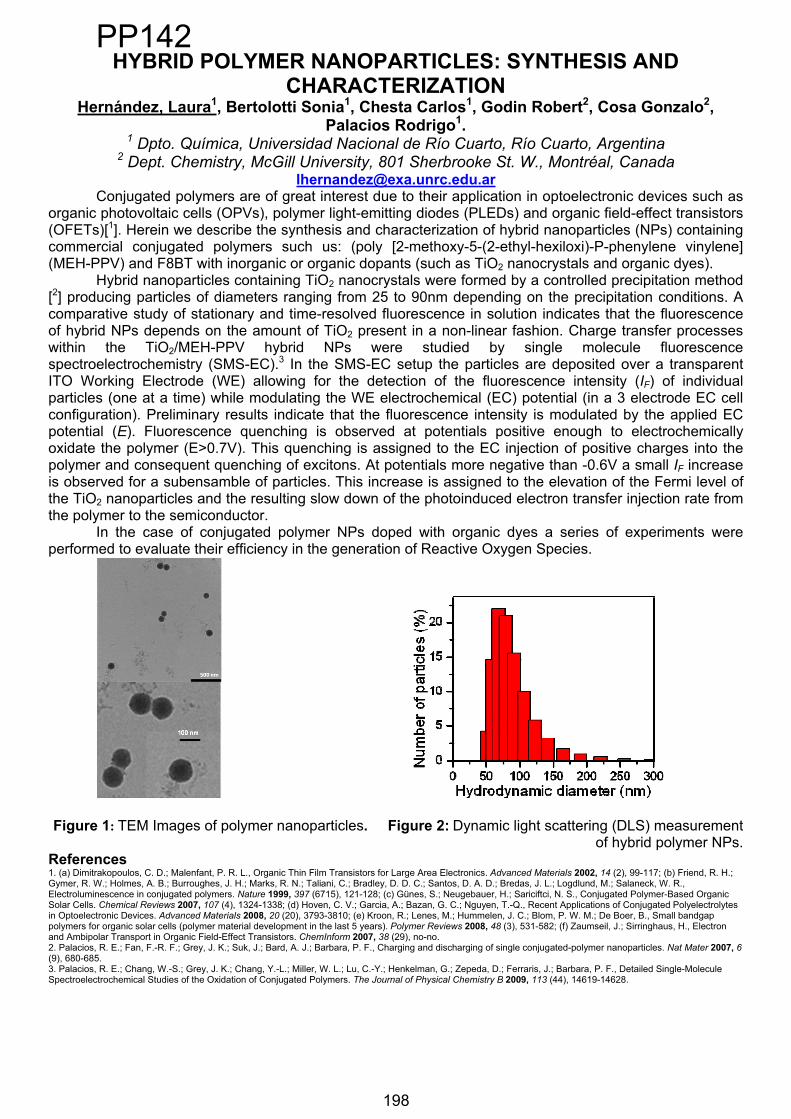
HYBRID POLYMER NANOPARTICLES: SYNTHESIS AND CHARACTERIZATION
Hernández, Laura1, Bertolotti Sonia1, Chesta Carlos1, Godin Robert2, Cosa Gonzalo2,Palacios Rodrigo1.
1 Dpto. Química, Universidad Nacional de Río Cuarto, Río Cuarto, Argentina 2 Dept. Chemistry, McGill University, 801 Sherbrooke St. W., Montréal, Canada
[email protected] polymers are of great interest due to their application in optoelectronic devices such as
organic photovoltaic cells (OPVs), polymer light-emitting diodes (PLEDs) and organic field-effect transistors (OFETs)[1]. Herein we describe the synthesis and characterization of hybrid nanoparticles (NPs) containing commercial conjugated polymers such us: (poly [2-methoxy-5-(2-ethyl-hexiloxi)-P-phenylene vinylene] (MEH-PPV) and F8BT with inorganic or organic dopants (such as TiO2 nanocrystals and organic dyes).
Hybrid nanoparticles containing TiO2 nanocrystals were formed by a controlled precipitation method [2] producing particles of diameters ranging from 25 to 90nm depending on the precipitation conditions. A comparative study of stationary and time-resolved fluorescence in solution indicates that the fluorescence of hybrid NPs depends on the amount of TiO2 present in a non-linear fashion. Charge transfer processes within the TiO2/MEH-PPV hybrid NPs were studied by single molecule fluorescence spectroelectrochemistry (SMS-EC).3 In the SMS-EC setup the particles are deposited over a transparent ITO Working Electrode (WE) allowing for the detection of the fluorescence intensity (IF) of individual particles (one at a time) while modulating the WE electrochemical (EC) potential (in a 3 electrode EC cell configuration). Preliminary results indicate that the fluorescence intensity is modulated by the applied EC potential (E). Fluorescence quenching is observed at potentials positive enough to electrochemically oxidate the polymer (E>0.7V). This quenching is assigned to the EC injection of positive charges into the polymer and consequent quenching of excitons. At potentials more negative than -0.6V a small IF increase is observed for a subensamble of particles. This increase is assigned to the elevation of the Fermi level of the TiO2 nanoparticles and the resulting slow down of the photoinduced electron transfer injection rate from the polymer to the semiconductor.
In the case of conjugated polymer NPs doped with organic dyes a series of experiments were performed to evaluate their efficiency in the generation of Reactive Oxygen Species.
Figure 1: TEM Images of polymer nanoparticles. Figure 2: Dynamic light scattering (DLS) measurement of hybrid polymer NPs.
References 1. (a) Dimitrakopoulos, C. D.; Malenfant, P. R. L., Organic Thin Film Transistors for Large Area Electronics. Advanced Materials 2002, 14 (2), 99-117; (b) Friend, R. H.; Gymer, R. W.; Holmes, A. B.; Burroughes, J. H.; Marks, R. N.; Taliani, C.; Bradley, D. D. C.; Santos, D. A. D.; Bredas, J. L.; Logdlund, M.; Salaneck, W. R., Electroluminescence in conjugated polymers. Nature 1999, 397 (6715), 121-128; (c) Günes, S.; Neugebauer, H.; Sariciftci, N. S., Conjugated Polymer-Based Organic Solar Cells. Chemical Reviews 2007, 107 (4), 1324-1338; (d) Hoven, C. V.; Garcia, A.; Bazan, G. C.; Nguyen, T.-Q., Recent Applications of Conjugated Polyelectrolytes in Optoelectronic Devices. Advanced Materials 2008, 20 (20), 3793-3810; (e) Kroon, R.; Lenes, M.; Hummelen, J. C.; Blom, P. W. M.; De Boer, B., Small bandgap polymers for organic solar cells (polymer material development in the last 5 years). Polymer Reviews 2008, 48 (3), 531-582; (f) Zaumseil, J.; Sirringhaus, H., Electron and Ambipolar Transport in Organic Field-Effect Transistors. ChemInform 2007, 38 (29), no-no. 2. Palacios, R. E.; Fan, F.-R. F.; Grey, J. K.; Suk, J.; Bard, A. J.; Barbara, P. F., Charging and discharging of single conjugated-polymer nanoparticles. Nat Mater 2007, 6(9), 680-685. 3. Palacios, R. E.; Chang, W.-S.; Grey, J. K.; Chang, Y.-L.; Miller, W. L.; Lu, C.-Y.; Henkelman, G.; Zepeda, D.; Ferraris, J.; Barbara, P. F., Detailed Single-Molecule Spectroelectrochemical Studies of the Oxidation of Conjugated Polymers. The Journal of Physical Chemistry B 2009, 113 (44), 14619-14628.
198
PP142
198

XI ELAFOT. Córdoba, Argentina. October 1-4, 2012. 199
PHOTOCATALYTIC ACTIVITY AND CHARACTERIZATION OF A NEW TIO2 PHOTOCATALYST SYNTHESIZED UNDER
ULTRASOUND AND ITS N-DOPED DERIVATIVE
Araújo Borges, Karen1, Dias França, Marcela1, dos Santos,Lidiaine Maria1, Jr Souza Müller, Paulo 1, da Hora Machado, Antonio Eduardo1,2
1Universidade Federal de Uberlândia, Instituto de Química, Laboratório de Fotoquímica e Ciência dos Materiais, Uberlândia, MG, Brasil.
2Universidade Federal de Goiás, Campus Avançado de Catalão; Catalão, GO, Brasil. e-mail: [email protected]
In this communication we present a comparative study about the photocatalytic activity and a partial characterization of two new TiO2 photocatalysts synthesized under ultrasound, the first named LAFOT50 (1) and the second LAFOT50-N (2) [1]. In the assays, aqueous solutions containing 42.5 ppm of tartrazine, a known food dye, were used as model-effluent. It was circulated by an annular borosilicate glass reactor at a flow rate of 1,345 mL.min-1, and irradiated by a 400 W high pressure mercury vapor lamp [2]. The catalysts were used at a concentration of 100 mg.L-1, and the initial pH of the reaction medium was fixed at 6.71. The reaction monitoring was limited to 120 minutes. Aliquots were collected at 20 minutes intervals and analyzed in terms of Total Organic Carbon (TOC) for mineralization, and absorbance measurements at 428 nm for effluent discoloration. The percentage of discoloration was the same (47%) using both photocatalysts. However, the mineralization was equal to 18% and 25%, respectively for (1) and (2). The X-ray diffraction shows that the both catalysts present only anatase crystalline phase. The average crystallite size calculated by using Scherrer's equation was 14 nm and 33 nm, respectively for (1) and (2). A lowering of the band gap observed for LAFOT50-N, enhancing visible light absorption and homogenous distribution of the N dopant, favoring the mobility of charge carriers into the semiconductor oxide [3,4], help to explain this slightly better result observed for LAFOT50-N. Acknowledgement: CAPES,FAPEMIG and CNPq.
[1]. Machado, A. E. H.; Santos, L. M.; Müller Jr, P. S.; França, M. D.; Borges, K. A.; Procedures under protection. [2] Oliveira D. F. M., Batista P. S., Müller Jr, P. S., Velani V., França M. D., Souza D. R., Machado A. E. H., Evaluating the effectiveness of photocatalysts based on titanium dioxide in the degradation of the dye Ponceau 4R. Dyes and Pigments, Vol. 92, No. 1, p. 563-572, 2012. [3] Machado, A. E. H.; Santos, L. M. ; Borges, K. A. ; Batista, P. S. ; Paiva, V. A. B. ; Muller JR., P. S. ; Oliveira, D. F. M. ; Franca, M. D. (2012) . Potential applications for solar photocatalysis: from environmental remediation to energy conversion. In: Solar Radiation, p. 339-378. ISBN 978-953-51-0384-4 [4] Kumar, S. G. & Devi, L. G. (2011). A review on modified TiO2 photocatalysis under VU/visible light: selected results and related mechanisms on interfacial charge carrier transfer dynamics. The Journal of Physical Chemistry A. Vol.115, No.46, pp. 13211-13241.
PP143
199
XI ELAFOT. Córdoba, Argentina. October 1 4, 2012. 200
Aplicación de sustancias bio-orgánicas solubles (SBO) como aditivo en el proceso foto-Fenton a pH neutro para el
tratamientos de aguas contaminadas
Carlos, Luciano1; Gomis, Juan2, Arques, Antonio2,Gonzalez,Mónica1,Mártire, Daniel1
1Instituto de InvestigacionesFisicoquímicasTeóricas y Aplicadas (INIFTA), Diag. 113 y 64, La Plata, [email protected]
2Departamento de IngenieríaTextil y Papelera, UniversidadPolitécnica de Valencia (UPV), Alcoy, España.
En el presente trabajo se muestran resultados parciales del uso de sustancias bio-orgánicas solubles (SBO), extraídas del compost de residuos verdes urbanos, como aditivo en la técnica foto-Fenton a pH neutro. Las SBO han sido previamente caracterizadas en términos de composición, grupos funcionales, peso molecular y tamaño, mostrando estructuras similares a las sustancias húmicas [1,2].La presencia de grupos carboxílicos y fenólicos hace posible su uso como agente quelante del hierro para mantenerlo en suspensión a pH cercanos a 7. A su vez, las SBO contienen una pequeña cantidad de hierro (0.8% (p/p)) aumentando el potencial catalítico de estas sustancias.Para estudiar la aplicabilidad de las SBO en la técnica foto-Fenton se utilizó como contaminante modeloel colorante cristal violeta (CV). Los experimentos fotoquímicos fueron realizados en un reactor anular a 25°C y a pH 7. Una lámpara de Hg de media presión inmersa en una camisa de vidrio fue utilzada como fuente de irradiación ( > 300 nm). La degradación del CV fue analizada siguiendo su decoloración empleando un espectrofotómetro UV-Vis. Los resultados mostraron que al cabo de 3 h de fotólisis de una solución de VC (10 mg/L) a pH 7 en presencia de Fe(II) (5 mg/L) y H2O2 (8.3 mg/L), la absorbancia de la solucion disminuyó solamente un 24% mientras que la adición de 100 mg/L de SBO al sistema anterior disminuyóla absorbancia un 46%. Asimismo, se estudió el efecto en la fotodegradación del CV de cada reactivo en forma independiente. Finalmente, experimentos en presencia de scavengers de radicales HO• y de especies de hierro con altos estados de oxidación, tales como el ion ferrilo (FeO+2) sugieren que el mecanismo de degradación del CV no está mediado principalmente por el radical HO•, sino que involucraría la participación de otras espcies oxidantes.
References
[1] Montoneri E, Mainero D, Boffa V, Perrone DG, Montoneri C. Biochemenergy: A project to turn an urban wastes treatment plant into biorefinery for the production of energy, chemicals and consumer's products with friendly environmental impact. International Journal of Global Environmental Issues 2011, 11, 170-196.
[2] Quagliotto P, Montoneri E, Tambone F, Adani F, Gobetto R, Viscardi G. Chemicals from wastes: Compost-derived humic acid-like matter as surfactant. Environmental Science and Technology 2006, 40, 1686-1692
PP144
200
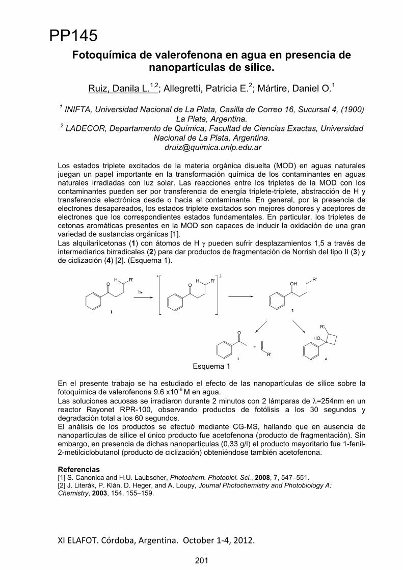
XI ELAFOT. Córdoba, Argentina. October 1 4, 2012. 201
Fotoquímica de valerofenona en agua en presencia de nanopartículas de sílice.
Ruiz, Danila L.1,2; Allegretti, Patricia E.2; Mártire, Daniel O.1
1 INIFTA, Universidad Nacional de La Plata, Casilla de Correo 16, Sucursal 4, (1900) La Plata, Argentina.
2 LADECOR, Departamento de Química, Facultad de Ciencias Exactas, Universidad Nacional de La Plata, Argentina.
Los estados triplete excitados de la materia orgánica disuelta (MOD) en aguas naturales juegan un papel importante en la transformación química de los contaminantes en aguas naturales irradiadas con luz solar. Las reacciones entre los tripletes de la MOD con los contaminantes pueden ser por transferencia de energía triplete-triplete, abstracción de H y transferencia electrónica desde o hacia el contaminante. En general, por la presencia de electrones desapareados, los estados triplete excitados son mejores donores y aceptores de electrones que los correspondientes estados fundamentales. En particular, los tripletes de cetonas aromáticas presentes en la MOD son capaces de inducir la oxidación de una gran variedad de sustancias orgánicas [1]. Las alquilarilcetonas (1) con átomos de H pueden sufrir desplazamientos 1,5 a través de intermediarios birradicales (2) para dar productos de fragmentación de Norrish del tipo II (3) y de ciclización (4) [2]. (Esquema 1).
H R'O
h
H R'O
* 3R'
OH
.
.
O
+
R'
R'
HO
1 2
3 4
Esquema 1
En el presente trabajo se ha estudiado el efecto de las nanopartículas de sílice sobre la fotoquímica de valerofenona 9.6 x10⁻⁶ M en agua.Las soluciones acuosas se irradiaron durante 2 minutos con 2 lámparas de =254nm en un reactor Rayonet RPR-100, observando productos de fotólisis a los 30 segundos y degradación total a los 60 segundos. El análisis de los productos se efectuó mediante CG-MS, hallando que en ausencia de nanopartículas de sílice el único producto fue acetofenona (producto de fragmentación). Sin embargo, en presencia de dichas nanopartículas (0,33 g/l) el producto mayoritario fue 1-fenil-2-metilciclobutanol (producto de ciclización) obteniéndose también acetofenona.
Referencias[1] S. Canonica and H.U. Laubscher, Photochem. Photobiol. Sci., 2008, 7, 547–551. [2] J. Literák, P. Klán, D. Heger, and A. Loupy, Journal Photochemistry and Photobiology A: Chemistry, 2003, 154, 155–159.
PP145
201
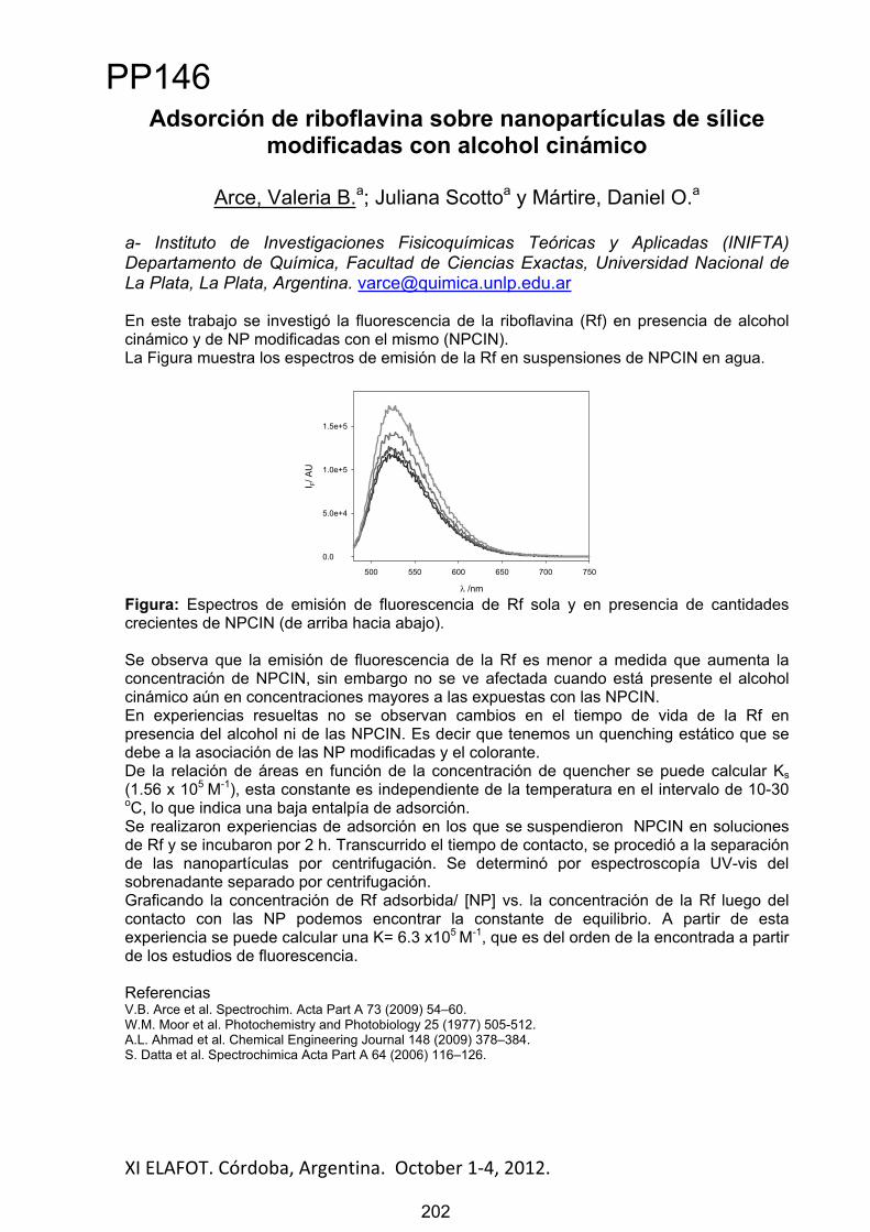
XI ELAFOT. Córdoba, Argentina. October 1 4, 2012. 202
Adsorción de riboflavina sobre nanopartículas de sílice modificadas con alcohol cinámico
Arce, Valeria B.a; Juliana Scottoa y Mártire, Daniel O.a
a- Instituto de Investigaciones Fisicoquímicas Teóricas y Aplicadas (INIFTA) Departamento de Química, Facultad de Ciencias Exactas, Universidad Nacional de La Plata, La Plata, Argentina. [email protected]
En este trabajo se investigó la fluorescencia de la riboflavina (Rf) en presencia de alcohol cinámico y de NP modificadas con el mismo (NPCIN). La Figura muestra los espectros de emisión de la Rf en suspensiones de NPCIN en agua.
/nm
500 550 600 650 700 750
I F/ A
U
0.0
5.0e+4
1.0e+5
1.5e+5
Figura: Espectros de emisión de fluorescencia de Rf sola y en presencia de cantidades crecientes de NPCIN (de arriba hacia abajo).
Se observa que la emisión de fluorescencia de la Rf es menor a medida que aumenta la concentración de NPCIN, sin embargo no se ve afectada cuando está presente el alcohol cinámico aún en concentraciones mayores a las expuestas con las NPCIN. En experiencias resueltas no se observan cambios en el tiempo de vida de la Rf en presencia del alcohol ni de las NPCIN. Es decir que tenemos un quenching estático que se debe a la asociación de las NP modificadas y el colorante. De la relación de áreas en función de la concentración de quencher se puede calcular Ks(1.56 x 10⁵ M-1), esta constante es independiente de la temperatura en el intervalo de 10-30 oC, lo que indica una baja entalpía de adsorción. Se realizaron experiencias de adsorción en los que se suspendieron NPCIN en soluciones de Rf y se incubaron por 2 h. Transcurrido el tiempo de contacto, se procedió a la separación de las nanopartículas por centrifugación. Se determinó por espectroscopía UV-vis del sobrenadante separado por centrifugación. Graficando la concentración de Rf adsorbida/ [NP] vs. la concentración de la Rf luego del contacto con las NP podemos encontrar la constante de equilibrio. A partir de esta experiencia se puede calcular una K= 6.3 x10⁵ M-1, que es del orden de la encontrada a partir de los estudios de fluorescencia.
ReferenciasV.B. Arce et al. Spectrochim. Acta Part A 73 (2009) 54–60. W.M. Moor et al. Photochemistry and Photobiology 25 (1977) 505-512. A.L. Ahmad et al. Chemical Engineering Journal 148 (2009) 378–384. S. Datta et al. Spectrochimica Acta Part A 64 (2006) 116–126.
PP146
202

XI ELAFOT. Córdoba, Argentina. October 1 4, 2012. 203
Fotodegradación sensibilizada de pesticidas fenólicos adsorbidos químicamente a nanopartículas de sílice
Escalada, Juan Pablo a; Arce, Valeria B.b; Criado, Susana c; García, Norman A.c y Mártire, Daniel O.b
a- Unidad Académica Río Gallegos de la Universidad Nacional de la Patagonia Austral, Río Gallegos, Argentina. b- Instituto de Investigaciones Fisicoquímicas Teóricas y Aplicadas (INIFTA) Departamento de Química, Facultad de Ciencias Exactas, Universidad Nacional de La Plata, La Plata, Argentina. c- Departamento de Química. Universidad Nacional de Río Cuarto. Campus Universitario, Río Cuarto, Argentina. E-mail: [email protected]
El uso y la producción de los pesticidas fenólicos: Bromoxynil (BXN, herbicida1) y Diclorofen(DCP, alguicida, funguicida, bactericida¡Error! Marcador no definido.) produce la liberación de estos en el ambiente. En aguas naturales, es de esperar que se adsorban a sólidos en suspensión y/o sedimentos en la columna de agua1. Esto nos motivó a sintetizar nanopartículas de sílice (NP) modificadas con los pesticidas, transparentes a la luz visible, mediante el método de esterificación1. La adsorción química del plaguicida sobre las nanopartículas de 7 nm de diámetro fue corroborada mediante diversas técnicas (RMN de C y Si, FTIR, LDS, determinación de área específica superficial por (BET)). Se suspendieron las NP en soluciones de riboflavina (Rf), un pigmento -fotosensibilizador- que absorbe en la región visible del espectro, y que se encuentra normalmente presente en aguas naturales. La luz natural a través de procesos fotosensibilizados produce distintos tipos de transformaciones sobre estos contaminantes1.Experimentos de desactivación de fluorescencia del sensibilizador muestran que suspensiones de las NP, en el intervalo de concentraciones 0-2,5 g/L de NP, no afectan el espectro de emisión ni el tiempo de vida de fluorescencia, lo que descarta la participación de los estados excitados singlete en el mecanismo de fotosensibilización. Asimismo, mediante la técnica de láser flash-fotólisis, se determinaron las constantes de desactivación de los estados triplete de Rf por las NP, mientras que nanopartículas de sílice sin modificar no mostraron desactivación de estos estados. Esto confirma la reactividad de los estados triplete de Rf con los plaguicidas quimisorbidos. Se determinaron también, las constantes de desactivación de oxígeno singlete por las NP mediante la técnica de fosforescencia resuelta en el tiempo (TRPD) y se midió consumo de oxígeno en las soluciones irradiadas con luz visible.Los resultados obtenidos fueron comparados con experimentos similares realizados con soluciones de los plaguicidas1 a fin de evaluar el efecto de la quimisorción en la fotodegradación sensibilizada de los mismos.
Referencias1 Tomlin, The Pesticide Manual. British Crop Protection Council and The Royal Society of Chemistry. London, UK. (1994) 2 Hansch et al; Exploring QSAR. Hydrophobic, Electronic, and Steric Constants. ACS Prof Ref Book. Heller SR, consult. ed., Washington, DC: Amer Chem Soc p. 108 (1995) 3 Arce et al. J. Phys. Chem. C, 115, 18122–18130. (2011) 4 Haag. Aquatic and Surf. Photochem. Ed. G. Helz, R. Zep and D. Crosby. CRC, Boca ratón (1994) 5Escalada et al. J. Hazard. Mat., 186, 466-472 (2011)
PP147
203

Abstract
XI ELAFOT. Córdoba, Argentina. October 1-4, 2012.
Environmentally-benign Heterogeneous Photoinduced Electron Transfer Substitution of Electron-rich Aromatic
Compounds with Perfluoroalkyl Groups in Water. ARadical-ion Chain Reaction
Barata-Vallejo, Sebastián1, Martin-Flesia, Marina2; Postigo, Al11 Departamento de Química Orgánica, Facultad de Farmacia y Bioquímica, Universidad de Buenos Aires. Junín 954-Buenos Aires, CP 1113-Argentina
Tel.-Fax.: +54 011 4964-8252; e-mail: [email protected] Facultad de Ciencias Exactas, Universidad de Belgrano. Villanueva 1324-CP 1428-
Buenos Aires-Argentina
The photoinduced electron transfer (PET) substitution reaction of electron rich aromatic nuclei, such as N,N-dimethylaniline and N,N-dimethyl-1-naphthylamine, anisol and 2-methoxynaphthalene with perfluoroalkyl Rf groups [1-3] was carried out in water to render the homolytic substitution products resulting from replacement of aromatic H´s for the Rf moiety in good yields. Some mechanistic aspects are discussed, supporting the notion of a PET reaction as opposed to direct photolysis of RfI to generate reacting Rf radicals leading to a classical radical homolytic aromatic substitution reaction. A radical chain mechanism superimposed with a redox process is proposed to account for product formations.
In the Figure below, the propagation steps for the radical perfluorobutylation of aromatics in water, superimposed with a redox mechanism, is shown.
Figure 1: Propagation cycle for the proposed PET chain substitution of electron-rich aromatic compounds by the perfluorobutyl group in water: AH stands for the aromatic amines. AC4F9 being the Rf-substituted amines.[4]
References:
[1]. Barata-Vallejo, S.; Postigo, A. Eur.J.Org.Chem. 2012. DOI: 10.1002/ejoc.201101808[2]. Barata-Vallejo, S.; Postigo, A. J.Org.Chem. 2010,75, 6141-6148[3]. Slodowicz, M.; Barata-Vallejo, S.; Vazquez, A.; Nudelman, N.; Postigo, A. J.Fluor.Chem. 2011.
Elsevier Science Publications. http://dx.doi.org/10.1016/j.jfluchem.2011.10.002. ISSN: 0022-1139
[4] Barata-Vallejo, S.; Martín Flesia, M., Postigo, A. Manuscrito en preparación
204
PP148
204

XI ELAFOT. Córdoba, Argentina. October 1 4, 2012. 205
CHOLESTEROL EFFECT IN VESICLE VECICLE AND CELL-VESICLES FUSION PROCESS
Cuevas, Francisco J1, Valle, Carolina A 2 and Aguilar, Luis Felipe 2
1 Departamento de Ciencias Básicas, Universidad Santo Tomás, Av. Limonares 190,Viña del Mar, [email protected]
2 Instituto de Química, Facultad de Ciencias, Pontificia Universidad Católica de Valparaíso, Valparaíso, Chile, Av. Universidad 330, Curauma.
Keywords: Fusion, micro-domains, lipids vesicles.
In the last years a most of studies about the “phenomena” of vesicle fusion has been developed, to understand the intra cell transport phenomena, exocytosis and specially to optimize the fusion process of vesicles used as drug or genetic material carriers. The lipids composition and the physicochemical properties of the lamella would play an important role in this fusogenic process. Some of the properties that influence this process are the lipid packing, the curve grade, the defect on the external layer and changes in the fluidity of the membrane. In this context the present work aims to link the vesicle-vesicle fusion phenomena with the cell-vesicle fusion process.
In order to determining the cholesterol effect in the efficiency of the fusion in models systems (DMPC:Chol; DPPC:Chol; DSPC:Chol; DLPC:Chol; and DOPC:Chol), the quenching of fluorescein with propidium iodide was measured, and to determine the cell-vesicle fusion process the propidium iodide fluorescence was followed after incubation of cell with vesicles. The physicochemical properties of systems was determinate by DPH and Laurdan lifetimes.
Our results indicate that in vesicle-vesicle fusion process, the vesicle fused percentage increases with the cholesterol content, but in non-monotonic behavior, with a maximum at 33,3 mol % of cholesterol, and with a slope change in the vicinity of 25 mol % of cholesterol. These results can be rationalized with the superlattice theory, which predicts, that at these cholesterol concentrations, the ordered domains versus disordered domains proportions are significantly affected. These results correlate well with our results of DPH and Laurdan lifetime indicating that these ratios of cholesterol to exist a lower proportion of water in both the depth and the hydrophilic-hydrophobic interfase of lamella. Both results (fusion percent and physicochemical properties of the lamella) correlate with the references which indicate that the fusion process is affected by the amount of water from the membrane. But this model does not explain the nonmonotonic behavior so that superlattice theory should be incorporated into de explanation.
For the cell-vesicle fusion process a maximum of fusion percentage at a single cholesterol contents was obtained. This ratio of cholesterol to the maximum which is obtained depends on the type of cell.
Our results leads us to postulate that cholesterol affects the fusion process, probably not only by the water content changes, but is by varying the ordered vs disordered domains proportion. In the cell-vesicle fusion process appears to be more important the cell membrane properties which depends on cell type.
Acknowledgements: This work was supported by FONDECYT Nº 11090443 grant.
PP149
205

XI ELAFOT. Córdoba, Argentina. October 1-4, 2012.
Fotoestabilidad del colorante4-(2-hidroxi-1-naftilazo) benzoato de sodio en agua.
Colonna, José; Alonso, César; Miskoski, SandraDepartamento de Química. Universidad Nacional de Río Cuarto. Argentina. [email protected]
Los colorantes aromáticos azoicos figuran entre los más importantes para diversos tipos de aplicaciones debido a una combinación de propiedades de interés en campos diversos. En ese contexto, el mecanismo de fotodecoloración es un tema largamente investigado a fin de evaluar y controlar la fotoestabilidad de los mismos.
En este trabajo se llevaron a cabo estudios de fotooxidación directa y sensibilizada de un colorante aril-azonaftol (4-(2-hidroxi-1-naftilazo) benzoato de sodio (AZO)) obtenido a partir de la copulación de una sal de diazonio derivada del ácido 4-aminobenzoico con el -naftol [1]. La estructura química del compuesto es la que se presenta a continuación.
N
OH
N COONa
Los estudios se realizaron en solución acuosa de NaOH 10 mM, en mezclas metanol agua con 10 mM de NaOH y en buffer de fostato a pH 7.
En medio alcalino se encontró alta fotoestabilidad ante la irradiación directa y una importante reactividad frente a oxígeno singlete O2(1
g), este último fotogenerado por distintos sensibilizadores tales como perinaftenona (PN), eosina, rosa de bengala (RB) y azul de metileno (MB). Se llevaron a cabo estudios cinéticos en presencia y ausencia de azida de sodio, un conocido inhibidor de oxígeno singlete, de ese modo se pudo concluir que la reacción de oxidación por esta especie reactiva predomina con estos sensibilizadores. No obstante con RB y más aún con Eosina se advierte consumo del sensibilizador a lo largo de la fotólisis debido a interacciones entre los estados excitados de estos compuestos. Por otra parte, con MB se observa la formación de un complejo oscuro que distorsiona los espectros de absorción de ambos colorantes e imposibilita trabajar con este sensibilizador en agua.
En el siguiente esquema cinético se presentan las posibles interacciones entre un sustrato (AZO) y oxígeno singlete: O2(1
g) + AZO O2(3g
-) + AZO (1), constante de velocidad kq
O2(1g) + AZO productos (2), constante de velocidad kr
Siendo kt = kr + kq, la constante de interacción total entre O2(1g) y el sustrato.
Se determinaron las constantes cinéticas total y reactiva con O2(1g) de AZO,
empleando PN como sensibilizador, resultando kt= 3.8x10⁸M-1s-1 y kr=8.2x10⁷ M-1s-1 en soluciones acuosas de NaOH 10 mM. Al bajar la polaridad del solvente con el agregado de 50% de metanol no se observó que se afecte significativamente la cinética de la reacción.
Sin embargo, al llevar el sistema a pH 7 no se advierte reacción fotosensibilizada, al menos en los períodos de irradiación empleados, revelando que en estas condiciones la reactividad del compuesto disminuye notablemente. Estos resultados coinciden con los reportados por otros autores cuando llevan a cabo estudios de fotooxidación sensibilizada de aril-azonaftoles en metanol y en derivados simples fenólicos y naftólicos [2]. La información aportada en el trabajo contribuye al conocimiento básico del comportamiento de colorantes azoicos y advierte acerca de las condiciones de estabilidad en potenciales aplicaciones de los mismos. Agradecimientos: A la Secretaría de Ciencia y Técnica de la UNRC y a l Consejo Nacional de Investigación Científica y Técnica (CONICET) por la ayuda económica. Referencias: [1] P. Bortolus and S. Monti. J. Org Chem. 54 (1989) 534-540. [2] L.M. Jansen, et al. J. Photochem.Photobiol. A: Chem.125(1999) 99-106.
206
PP150
206

XI ELAFOT. Córdoba, Argentina. October 1-4, 2012.
1
Estudios de inactivación fotodinámica tendientes a lograr el control poblacional de especies dañinas de cianobacterias. Gsponer, Natalia; Chesta, Carlos, Durantini, Edgardo, Mora, Jimena Dpto. Química, Universidad Nacional de Río Cuarto, Río Cuarto, Argentina
[email protected] el proceso de eutrofización el mejor ejemplo de los cambios que se producen en el reservorio lo constituye la reacción del fitoplancton, siendo las algas uno de los mejores indicadores de polución. Una alta carga de nutrientes determina importantes cambios en la calidad de los cuerpos de agua que se evidencian, fundamentalmente por la generación de floraciones de algas planctónicas y en particular de cianobacterias. [1] El uso de la fotosensibilización para la producción de oxígeno singulete (1O2) mediante energía solar permite el desarrollo de nuevas tecnologías limpias, basadas en energías renovables y aplicables a la desinfección de aguas. [2] El objetivo de este trabajo es iniciar estudios de IFD tendientes a lograr el control poblacional de especies dañinas de cianobacterias en experimentos in vitro, basándonos en el dopaje de las células que se quieren inactivar con un FS, que tras irradiación con luz visible o solar, sea capaz de producir ROS. [3] Para llevar a cabo dicho objetivo, se realizaron curvas de crecimiento en condiciones óptimas de desarrollo para el género Microcystis sp. (Cyanobacteria) incubado en medio BG11. Se estudió la fotoestabilidad del FS 5,10,15,20-tetrakis[4-(3-N,N,N-trimetilaminopropoxi)fenil] porfirina (TAPP4+)[4] y se observó su completa fotodescomposición a los 4 días de iniciado el estudio, sin embargo los experimentos mostraron que su tiempo de vida es lo sufrientemente largo como para producir la completa inactivación celular. Se probaron dos concentraciones de TAPP4+ (10 y 20 µM). Se observó un importante retardo en el crecimiento con el tratamiento de 10 µM y la completa inhibición de la población algal con 20 µM de TAPP4+ (Fig. 1). Se obtuvo un valor máximo de unión célula-sensibilizador a un corto tiempo (~60 min), momento en el cual ~80% del FS fue incorporado por las células. Las imágenes de microscopía óptica mostraron que en los tubos control (sin TAPP4+), las células presentaron un importante contenido celular de Chl y morfología típica esperada para laespecie. El recuento de células arrojó valores de 4,9x10⁵ cel/ml en los tubos control mientras que el los tubos tratados, los valores fueron de 3,8x10⁵ cel/ml y 2,10x10⁵ cel/ml para 10 y 20 µM respectivamente sugiriendo que la presencia de la porfirina induce a la disminución de la población. Cuando se comparó una muestra control (sin TAPP4+) y una tratada (20 µM) por imágenes de microscopía de fluorescencia se observó que en la muestra tratada se produce la lisis celular conduciendo a la formación de “células fantasmas” con pérdida de pigmentos y la porfirina adherida a la pared celular mientras que el control permanece sin cambios (Fig. 2). Los resultados anteriores confirman que efectivamente el FS inhibe el crecimiento de Microcystis sp., a través de un mecanismo fotodinámico aunque no se puede confirmar aún si es producido por ROS.
REFERENCIAS 1Faust, M.A. and Gulledge, R.A. (2002). Contributions from the United States National Herbarium. 42: 1-144. 2Turro, N.J. (1991). Modern Molecular Photochemistry, University Science Books, Mill Valley, CA. ISBN 978-0-935702-7-12. Pp. 629. 3Hamblin, M. R. and Hasan, T. (2004). Potochem. Photobiol. Sci. 3 (5), 436-450. 4 Caminos, D. and Durantini, E. (2005). J. Porph. Phthaloc. 9(5) 334-342.
Fig. 1. Curva de retardo del crecimiento de Microcystis sp. incubado con
10 µM de TAPP4+ (�) y 20 µM de TAPP4+ (�) en medio BG11 a 22 ºC.
Control (�): cultivo sin tratamiento.
0 5 10 15 20 25 300,0
0,2
0,4
0,6
0,8
1,0
Abs
orba
ncia
(λ=
750
nm
)
Tiempo de incubación (días)
Fig. 2. Células de Microcystis sp. observadas mediante microscopía
óptica y de fluorescencia luego de tratamiento con 20 µM de porfirina y
radiación lumínica durante dos hrs. (A y B): previo a la lisis celular. (C y
D): posterior a la lisis celular. (400x).
207
PP151
207
XI ELAFOT. Córdoba, Argentina. October 1 4, 2012. 208
DEGRADACIÓN FOTOSENSIBILIZADA DE ANTIBIÓTICOS -LACTÁMICOS. IMPLICANCIAS MICROBIOLÓGICAS
Eugenia Reynoso, Mariana Spesia, Susana Criado, M. Alicia Biasutti Departamento de Química, Facultad de Ciencias Exactas, Fco. Qcas. y Naturales, Universidad Nacional de Río Cuarto, Río Cuarto, Argentina, e-mail: [email protected]
La degradación de antibióticos y en particular de -lactámicos está siendo actualmente estudiada con enfoque ecológico ya que son ampliamente utilizados no solo en el tratamiento de infecciones humanas, sino también en plantas y animales. Muy pocos de ellos sufren inactivación metabólica, no son biodegradables, permanecen en el agua luego de los tratamientos estándar de la misma y aún en pequeñas cantidades pueden alterar el ecosistema y producir multiresistencia bacteriana [1,2] un problema de gran importancia en el tratamiento contra múltiples enfermedades. Dada la necesidad creciente de eliminar estos contaminantes del agua, se decidió abordar el estudio de los procesos oxidativos fotosensibilizados de los antibióticos -lactámicos (Atb) Ceftriaxona (Cft) y Cefotaxima (Ctx) en presencia del pigmento natural Rf el cual se encuentra distribuido en cantidades trazas en agua de ríos, lagos y océanos. El mecanismo de acción de Rf es relativamente complejo y en la mayoría de los casos involucra las especies reactivas de oxígeno (ROS): oxígeno singlete (O2 (1
g)) y anión superóxido (O2•-)[3]. El colorante sintético
Perinaftenona (PN) fue utilizado como sensibilizador auxiliar con el fin de cuantificar el aporte exclusivo de O2 (1
g).Los resultados obtenidos en experiencias de consumo de oxígeno sensibilizadas por Rf son marcadamente diferentes a los resultados obtenidos en presencia de PN, indicando que, en el caso de Rf, una o más fuentes adicionales de consumo de oxígeno existen en el proceso de degradación de los Atb. Este hecho fue confirmado utilizando inhibidores específicos de las diferentes ROS.La presencia de NaN3 (desactivador físico específico de O2 (1
g)) produce una disminución en la velocidad de consumo de oxígeno por Cft y Ctx indicando la participación de dicha especie en la degradación fotosensibilizada por Rf (mecanismo Tipo II). El efecto que producen en el sistema Atb/Rf otros inhibidores tales como superóxido dismutasa (SOD), catalasa (CAT) y D-manitol (inhibidores específicos de O2
•-, H2O2 y OH• respectivamente) estaría indicando que el proceso también ocurre por la vía radicalaria (mecanismo Tipo I). Los experimentos muestran que no hay efecto de SOD y muy poco efecto de CAT y D-manitol, por lo que las especies O2
•-, H2O2 y OH• serían generadas vía el estado triplete excitado de Rf por el mecanismo Tipo I pero Cft y Ctx resultan poco sensibles a las mismas. Los estados singlete y triplete excitados de Rf son desactivados por los Atb con constantes de velocidad cercanas a limite difusional. Los espectros de transientes muestran la aparición del radical neutro de Rf en presencia de Cft y Ctx, especie que se genera como consecuencia de la transferencia de electrones entre el triplete excitado de Rf y los Atb. Por otro lado, se realizaron ensayos microbiológicos con cepas de S. aureus en presencia de Rosa de Bengala (RB) como fotosensibilizador el cual resulta un excelente sensibilizador en las reacciones que involucran O2 (1
g)[4]. La acción de Cft y Ctx sobre las cepas ensayadas se ve claramente disminuida a medida que aumenta el tiempo de irradiación. El efecto podría deberse a la fotodegradación de estos Atb y por consiguiente, a la pérdida de la capacidad bactericida de los mismos y, además, a que los fotoproductos de degradación de Cft y Ctx no poseen ninguna actividad inhibitoria sobre las cepas de S. aureus.1 Walter M.V. and Vennes J.W. Appl. Environ. Microbiol (1985) 50:930. 2Michelle K. D. and Stephen P. M. J. Phys. Chem. A (2010) 114:8391. 3 García N. A., Criado S. N., Massad W. A. Silva E. Edwards A. M. (Editors). The Royal Society of Chemitry (2006) 176:61. 4 Neckers D. C. Rose Bengal. Review. J Photochem photobiol A: Chem. (1989) 47:1.
PP152
208

XI ELAFOT. Córdoba, Argentina. October 1 4, 2012. 209
Fotooxidación de antidiabéticos en presencia de Albúmina de Suero Humano: efecto de Glucosa.
Challier, Cecilia; Biasutti María A.; Criado, Susana.
1 Departamento de Química, Facultad de Ciencias Exactas, Fco. Qcas. y Naturales, Universidad Nacional de Río Cuarto, Río Cuarto, Argentina, e-mail:
En los últimas décadas ha crecido el interés por el estudio de los daños generados por procesos fotodegradativos sobre medicamentos y suplementos alimenticios consumidos por el hombre [1],[2], que causan la pérdida de sus propiedades y acción terapéutica[3]. En este contexto resulta interesante estudiar la fotooxidación de los Antidiabéticos Gliclazida (Gli) y Glipizida (Glip). Estos son recetados para el tratamiento de la Diabetes Miellitus tipo II, una enfermedad que afecta al 85% de los pacientes diabéticos [4]. Por otro lado, cabe considerar la posible influencia de la Albúmina de Suero Humano (HSA), una de las proteínas de la sangre que distribuye en el organismo diversas moléculas y fármacos, los que pueden interactuar en dos sitios de unión: sitio I y sitio II [5].
Se investigó la unión o binding de Gli y Glip a HSA en presencia de Glucosa en concentraciones típicas a las que puede estar expuesto un paciente diabético: 1x10⁻³ M (hipoglucemia), 5x10⁻³ M (valor normal) 1,2x10⁻² M, 1,66x10⁻² M y 2,2x10⁻² M (hiperglucemia). Los resultados de desactivación de fluorescencia de HSA y desplazamiento de pruebas fluorescentes [6] indican que Glucosa no se une a HSA en el intervalo de concentración 0,1-2,2x10⁻²M, mientras que para Gli y Glip se observa un efecto notable del carbohidrato. A bajas concentraciones de Glucosa (1-12x10-3 M), los gráficos de Stern-Volmer se curvan hacia las abscisas indicando la diferente accesibilidad del fluoróforo de HSA a Gli y Glip, mientras que a altas concentraciones (1,66-2,2x10⁻² M), dichos gráficos son lineales, indicando que todos los fluoróforos son igualmente accesibles. En el caso del binding se observa que a bajas concentraciones de Glucosa, Gli se une a HSA en los dos sitios, mientras que a altas concentraciones su mecanismo de interacción cambia y se une solo a un sitio. Por su parte, a bajas concentraciones de Glucosa, Glip se une a un solo sitio de HSA, mientras que a altas concentraciones lo hace en los dos sitios. Este resultado es sumamente interesante teniendo en cuenta que los pacientes diabéticos pueden encontrarse expuestos a estas altas concentraciones de Glucosa en sangre y por lo tanto la farmacocinética y la farmacodinamia de los antidiabéticos podría verse afectada.
Posteriormente, se estudió la fotooxidación mediada por O2(1g) de Gli, Glip y HSA, y
mezclas Gli-HSA y Glip-HSA en presencia de Glucosa. La concentración de Glucosa parece no afectar las velocidades de consumo de O2(1
g) en el caso de HSA. El mismo comportamiento fue observado para Gli libre y unida a HSA. Sin embargo, para Glip libre se observa un aumento en la velocidad de consumo de O2(1
g) con el incremento en la concentración de Glucosa, mientras que dichas velocidades permanecen invariables cuando Glip se une a la proteína, lo que indicaría un posible efecto protector de HSA sobre la fotodegradación de Glip
Bibliografía:
[1] Ray R. S., Misra R. B. , Farooq M, Hans R. K. (2002) Toxicology in Vitro 16, 123. [2] Verma K., Agrawal N., Misra R.B., Farooq M., Hans R. K. (2008) Toxicology in Vitro 22, 249. [3] a) Foote C.S. (1982). In: Autor, A.P. (Ed). Academic Press, New York, 21. b) Joshi P.C. (1985) Toxicology Letters 26, 211. c) Maurer T. (1987) Food and Chemical Toxicology 25, 407. [4] Diabetes Atlas. Second Edition. International Diabetes Federation. (2003). ISBN 2-930229-27-6. [5] Kragh-Hansen, U., Chuang, V.T.G., Otagiri, M., (2002). Biol. Pharm.Bull. 25,695. [6] Sudlow, G., Birkett, D.J., Wade, D.N. (1975) Mol. Pharmacol. 11,824.
PP153
209

XI ELAFOT. Córdoba, Argentina. October 1 4, 2012. 210
Degradación fotosensibilizada de antifúngicos imidazólicos en medio homogéneo y micelar
Cabrera Zalazar Mariel; Reynoso, Eugenia; Biasutti, María A.; Criado, Susana
Departamento de Química, Facultad de Ciencias Exactas, Fco. Qcas. y Naturales, Universidad Nacional de Río Cuarto, Río Cuarto, Argentina, e-mail:
Los antifúngicos imidazólicos son preparados sintéticos, constituidos por diferentes anillos bencénicos y la presencia de uno o más anillos de cinco elementos en cuya estructura están presentes dos átomos de nitrógeno. Dichos antifúngicos interfieren con la síntesis de ergosterol, al inhibir la C14-alfadesmetilasa, una enzima acoplada al citocromo P-450 y que transforma lanosterol en ergosterol. Esta inhibición altera la fluidez de la membrana, aumentando la permeabilidad y produciendo una inhibición del crecimiento celular y de la replicación. Asimismo, interactúan con el complejo P-450 de la especie humana, provocando interferencias metabólicas con ciertas hormonas o interacciones con fármacos metabolizados bajo este sistema1-5.
En el presente trabajo se estudió la degradación de los antifúngicos imidazólicos: tioconazol y sulconazol, mediada por oxígeno singlete (O2(1
g)), en medio homogéneo y en sistemas micelares con el fin de mimetizar los entornos confinados de los complejos sistemas biológicos. El O2(1
g) fue generado por fotosensibilización utilizando el colorante xántenico Rosa de Bengala (RB), el cual es un generador exclusivo de dicha especie. La cuantificación de las constantes de velocidad de desactivación total y reactiva de O2(1
g) se realizó a través de métodos resueltos en el tiempo y estacionarios, respectivamente.
La irradiación con luz visible de longitud de onda > 400 nm del sistema antifúngico / sensibilizador (RB) / oxígeno (aire) produce cambios en el espectro UV/visible de las mezclas y consumo simultáneo de oxígeno. Estos resultados demuestran que ambos antifúngicos se degradan vía O2(1
g), siendo tioconazol más reactivo que sulconazol. Por su parte, sulconazol muestra un importante efecto de autoprotección frente a la especie O2(1
g). En medios micelares dependiendo de las características del microentorno se observa un importante efecto sobre la cinética de fotodescomposición de ambos antifúngicos.
References
1 J. Fortún Abete (1998) Medicine 7 (91), 4231. M. A. Ghannoum and D. M. Kuhn (2002) Eur J Med Res 7, 242.2 A. Fica C. (2004) Rev. Chil. Infect. 21 (1), 26.3 F. C. Odds (1988) Antifungal agents and their use in Candida infections. En: Baillière Tindall, ed. Candida and candidosis. A review and bibliography. (2nd ed). London: Odds, 279.4 Martindale (1982) The Extra Pharmacopoeia (28ª ed.) The Pharmaceutical Press, 356. 5 M. A. Ghannoum and D. M. Kuhn (2002) Eur J Med Res 7, 242.
PP154
210

XI ELAFOT. Córdoba, Argentina. October 1-4, 2012.
Synthesis and location between aqueous and micellar phase of TEMPO derivatives of different hydrophobicity
Aliaga, Carolina; Silva, Fernanda; Mascayano, CarolinaUniversidad de Santiago de Chile, Facultad de Química y Biología, Departamento de
Ciencias del Ambiente. Alameda 3363, Estación Central Santiago, Chile [email protected]
The 2,2,6,6-tetramethylpiperidine-N-oxyl (TEMPO) derivative radicals have been widely employed to characterize structural and dynamic properties of membranes. The use of TEMPO as spin probing in Electron Paramagnetic Resonance (EPR) and Fluorescent probing is one of the most effective techniques to obtain information about the hydrophobic properties of the environment. They have been used to determine the location of organic compounds in microheterogeneous system as quenchers of fluorescence probes. This last application requires the knowledge of the partition of the spin probe between the aqueous and microheterogeneous phases, as well as their distribution in the latter.
In this context, we have calculated the distribution of four nitroxide derivative probes (scheme 1) by using a methodology that was recently developed by us. The method is based on measurements of the EPR g-factor parameter, allowing us to obtain the partition constant of spin probes in micelles. Together with fluorescence-quenching experiments, it allows us to obtain information about the preferential location of the four TEMPO-derivatives with different hydrophobicities in a neutral microheterogeneous medium, such as Triton XR-100 solutions.
In addition, and in order to gain insight into the location of the spin probes dissolved in micellar solution, we resort to molecular dynamics simulations. This theoretical tool hasprovided us in a previous work with information on solute-solvent interactions. [1]
Scheme 1
Acknowledgements: FONDECYT project 1110736, CEDENNA L4 FB 0807
References
1. C. Aliaga, L. Briones, M. C. Rezende and C. Tirapegui, J. Coll. Interface Sci. 349 (2010) 565–570
211
PP155
211

XI ELAFOT. C rdoba, Argentina. October 1-4, 2012.
Enhance of emission and Raman scattering using Ag@SiO2
Celis B., Freddy 1,2; Aliaga, Carolina1,2
1 Universidad de Santiago de Chile, Departamento de Ciencias del Ambiente, Santiago, Chile
2 Centro para el Desarrollo de la Nanociencia y la Nanotecnología, CEDENNA, [email protected]
Localized surface plasmon resonance (LSPR) allows to enhance photonic signals such as emission and Raman scattering when fluorophores are exposed to spherical nanoparticles (NP) above of 40 nm.d. according to the Mie theory. SERS(Surface-enhanced Raman scattering)1 and SEF (Surface-enhanced fluoresce)2
techniques employs naked NPs (of metals such as Ag, Au and Cu) whereas SHINERS (Shell-isolated nanoparticles-enhanced Raman scattering)3 and SHINEF (Shell-isolated nanoparticles-enhanced fluorescence)4 need coated NP with an inertspacer (SiO2).
In this work, we synthesized different sizes of AgNPs (80, 7 and 4 nm.d.) and coated them with different thickness by exposing them for a different period of time to a SiO2 solution. Then, by employing Ag@SiO2 we performed SHINERS and SHINEF of 4-MPTP (1-(4- mercaptophenyl)-2,4,6-triphenylpyridinium perchlorate, a fluorophore synthesized by us) at very low concentrations.
Figure. A) SHINEF of 4-MPTP (0,1 mM) with Ag@SiO2 at different thickness of silicaA: MeOH/H2O, Ag@SiO2 B: 15 min, C: 30min, D: 60 min, E: 90 min coating time and
B) SHINERS spectrum of 4-MPTP with Ag@2nmSiO2
Acknowledgements: FONDECYT project 1110736, CEDENNA L4 FB 0807
References[1] Fleischmann, M.; Hendra, P. J.; McQuillan, A. J., Chem Phys Lett, (1974) 26, 163.
Jeanmaire, D. L.; Van Duyne, R. P., J. Electroanal. Chem. Interfac., (1977) 84, 1.[2] Drexhage, K. H.,Ber. Bunsenges. Phys. Chem., (1968) 72, 329. Drexhage, K. H., J.
Luminesc., (1970) 1, 693.[3] Anema, J. R.; Li, J.-F.; Yang, Z.-L.; Ren, B.; Tian, Z.-Q., Annu. Rev. Anal. Chem. (2011)
4, 129.[4] Guerrero, A. R.; Aroca, R. F., Angew.Chem., (2011) 123, 691.
����������
212
PP156
212

XI ELAFOT. Córdoba, Argentina. October 1 4, 2012. 213
Efectos estructurales y de microentorno sobre la capacidad antioxidante de (+)-catequina. Barua, María Gloria1; Criado, Susana2 y Pajares, Adriana1
1 Unidad Académica Río Gallegos. Universidad Nacional de la Patagonia Austral. 8400 Río Gallegos. [email protected]
2 Dto. Química. Universidad Nacional de Río Cuarto. Enlaces rutas 8 y 36. Km 601. 5800 Río Cuarto.
La peroxidación lipídica es la forma de deterioro de los alimentos más importante después de las alteraciones producidas por microorganismos, lo que representa un tema de gran interés económico para la industria alimenticia, ya que da lugar a la aparición de sabores y olores desagradables1,2.. Este proceso, se inicia a partir de la reacción de los ácidos grasos con especies reactivas de oxígeno. Se conoce desde hace varios años3 que la exposición del producto alimenticio a la luz del día puede desencadenar o acelerar dicho deterioro. También es aceptado que en muchos casos ocurre un proceso de fotooxidación sensibilizada4, con la participación especial de una de las especies reactivas de oxígeno: el oxígeno singlete (O2(1
g)) generado por absorción de luz medioambiental (generalmente luz visible) por parte de una sustancia coloreada presente en el medio, que a través de sus estados electrónicamente excitados inicia una cadena reactiva. Por esta razón, la incorporación de antioxidantes naturales en alimentos de alto contenido graso resulta particularmente interesante, debido a su efectividad en el retardo de la oxidación de los lípidos, tanto térmica como fotopromovida.
En particular, las catequinas son polifenoles de origen natural que han recibido especial atención debido a su relativamente alta capacidad antioxidante.5-8
En este trabajo se estudió la cinética y mecanismo de degradación oxidativa de (+)-catequina y cromóforos derivados (catecol y resorcinol) frente a O2(1
g), en medio homogéneo y en sistemas micelares, con el fin de evaluar efectos estructurales y de microentorno sobre la capacidad antioxidante de catequina.
Los resultados muestran que (+)-catequina, catecol y resorcinol se degradan vía O2(1
g). Tanto catecol como resorcinol constituyen centros “blanco” del antioxidante frente al ataque por dicha especie reactiva de oxígeno. Sin embargo, la fotodegradación de (+)-catequina no es la mera sumatoria de los aportes de ambos núcleos, sino que además hay otros factores que contribuyen al proceso global oxidativo. Asimismo, se observa un interesante efecto del microentorno sobre la capacidad antioxidante de (+)-catequina, dependiendo de las características del surfactante.
1 Rawls H.and Van Santen P. J. J. Am. Oil Chem. Soc., 97, 121 (1970). 2Clemens A. H .et al. J. Am. Oil. Chem. Soc. 50, 325 (1973). 3 Rosenthal J. Photooxidation of Foods. In “Singlet Oxygen”, Cap. 3, Vol. 4. AA. Frimer (Ed.). CRC Boca Ratón (1985). 4 Straight R. C. and Spikes J. D. In “Singlet Oxygen”, Cap. 2, Vol. 4 . A. A. Frimer (Ed.). CRC Boca Ratón (1985). 5 Chacko S. M., Thambi P.T. , Kuttan R. and Nishigaki I. Chinese Medicine 5, 13 (2010), 6 Mukai K., Nagai S. and Ohara K. Free Rad. Biol. Med. 39, 752 (2005). 7 Jung M. Y., Jung D. M., Wu J. W. and Choi D. S. Food Science and Biotechnology 20, 3, 725 (2011). 8Yang C. S., Lambert J. D., Sang S. Arch. Toxicol. 83, 11(2009).
PP157
213

XI ELAFOT. Córdoba, Argentina. October 1-4, 2012.
Figu
ra 1
Figu
ra 2
Interacción entre triptófano y quitosano en solución y en electrodos de carbono vítreo caracterizada por
fluorescencia y métodos electroquímicosGiménez, Rodrigo E.1; Paz Zanini, Verónica I.1; López de Mishima, Beatriz1;Borsarelli, Claudio D. 1
1LACIFO & LANES. CITSE-CONICET. Facultad de Agronomía y Agroindustrias,Universidad Nacional de Santiago del Estero. RN 9, Km 1125. Villa El Zanjón.
Santiago del Estero. Argentina. [email protected]
El triptófano es un aminoácido esencial para el ser humano, que cumple un papel clave como precursor de diversos compuestos químicos importantes para la salud humana. El quitosano es un polisacárido lineal compuesto de unidades de -N-acetil-D-glucosamina y -D-glucosamina, distribuidas aleatoriamente (con un 50% o más de esta última).
En este trabajo se estudia, mediante voltamperometría cíclica, la electro-oxidación de triptófano sobre electrodos de carbono vítreo modificados con una película de quitosano. Por su parte, las propiedades eléctricas del electrodo modificado se evalúan usando espectroscopía de impedancia electroquímica (EIS, por sus siglas en inglés). Se emplean además las técnicas de espectroscopía de fluorescencia, anisotropía y conteo de fotones correlacionados en el tiempo (TCSPC, por sus siglas en inglés) para evaluar la posible interacción y/o asociación entre el triptófano y el quitosano.
Los perfiles voltamperométricos muestran un notable incremento en la corriente pico para la oxidación del triptófano cuando la superficie del electrodo se modifica con quitosano y que, además, esta corriente aumenta a medida que disminuye el pH del medio (figura 1);si bien este incremento también se observa para el electrodo limpio, la presencia del polímero en la superficie del electrodo acentúa el efecto. Este comportamiento puede ser explicado por la formación de enlaces de puente hidrógeno entre el aminoácido y la matriz de quitosano [1] y/o una disminución de la resistencia de transferencia de carga, Rct, por lamodificación de la superficie del electrodo con quitosano.
La constante de asociación (Kb) calculada a partir de los datos de fluorescencia aumenta a medida que disminuye el pH (figura 1), esto explica la dependencia de la corriente de pico con el pH ya que esta asociación produce una pre-concentración del aminoácido en la superficie del electrodo.
La intensidad de fluorescencia del trp se incrementa en presencia del quitosano (figura 2), sin embargo la anisotropía en la región de máxima emisión se mantiene constante. Esteaumento en el rendimiento cuántico de fluorescencia puede explicarse por la disminución del auto-quenching entre el anillo indólico y el grupo amonio debido a la interacción triptófano-quitosano [2]. Los resultados del análisis de los tiempos de vida por TCSPCconfirman un aumento de los tiempos de vida promedio del triptófano en presencia dequitosano.
Agradecimientos: Al CONICET por la financiación de los estudios mediante una beca doctoral.Referencias[1] Analytical
Chemistry, vol. 72, no. 4, pp. 680-686, Feb. 2000.[2] Lakowicz, Principles of Fluorescence Spectroscopy, Third Edit. Springer Science+Business Media, LLC,
2006, pp. 578-579.
300 400 5000
5
10
15
20
25
30
35
40
45
50
55
60
65
70
75
80
Trp+Buff Trp+Chit
Longitud de Onda (nm)
Fluo
resc
enci
a (A
U)
-0.6
-0.4
-0.2
0.0
0.2
0.4
0.6
0.8
1.0
1.2
Ani
sotro
pía
1.5 2.0 2.5 3.0 3.5 4.0 4.5 5.0 5.5 6.0 6.5 7.0 7.5 8.0 8.5
0.7
0.8
0.9
1.0
1.1
F/F0
Kb
Ip ( A)
pH
I p (A)
F/F 0
0.2
0.3
0.4
0.5
0.6
0.7
0.8
0.9
Kb
10
15
20
25
30
214
PP158
214

XI ELAFOT. Córdoba, Argentina. October 1-4, 2012.
Evaluation of the Behaviour and Photophysical Properties of Nitroxide-BODIPY Probes.
Aspée, Alexis1; Liras, Marta2; García, Olga2; Donoso, Raul1; Lissi, Eduardo1; Poblete, Horacio.3
1 Facultad de Química y Biología, Universidad de Santiago de Chile, Chile., [email protected]
2Instituto de Ciencia y Tecnología de Polímeros, CSIC, Madrid, España 3Center for Bioinformatics and Molecular Simulations, Universidad de Talca, Chile.
We have, recently, synthesized two nitroxide-BODIPY probes designed for studying antioxidant and free radical processes in biological systems. The absorption and fluorescence wavelengths of the BODIPY chromophores do not interfere with the absorption of most biomolecules; in addition to that, their high quantum yields contribute to improve the sensitivity of these types of fluorescent probes.
In this work, we discuss the chemical behaviour and photochemical properties of these nitroxide-BODIPY probes and their potential and limitations for being used in complex biological systems. Fluorescence quantum yields and time resolved fluorescent measurements in ethanol showed an efficient intramolecular quenching of the BODIPY fluorescence by the nitroxide moiety, in comparison with the fluorescence of the isolated chromophores unit or with N-hydroxylamine reduced product formed after reaction with ascorbic acid or Trolox.
In spite of that behaviour observed in organic solvents, in aqueous media these BODIPY dyes tend to form aggregates that may affect their ability to monitor free radicals by fluorescence measurements. However, the additions of proteins, such as human serum albumin (HSA), increment the proportion of the monomer dyes. That has been interpreted in terms of equilibrium between monomeric and dimer probes in aqueous media that is affected by a monomeric association of the nitroxide probe to the HSA. This is in agreement with a favourable association of these probes with HSA in binding site I predicted by docking studies and the evaluation of the kinetic of the reaction of nitroxide-BODIPY probes in HSA toward water soluble antioxidants.
Acknowledgements: Financial support from CSIC-USACH and FONDECYT 1110809 are gratefully acknowledged
References A. Aspée, A. Orrego, E. Alarcón, C. López-Alarcón, H. Poblete, D. González-Nilo. Bioorg.Med. Letts. 19, 6382 (2009)
215
PP159
215

XI ELAFOT. Córdoba, Argentina. October 1 4, 2012. 216
Photophysical and photochemical studies of eosin dyes
Valdebenito, Andrea; Encinas, María Victoria; Pino, Eduardo.
Facultad de Química y Biología, Universidad de Santiago de Chile, Chile. [email protected]
Xanthene dyes are among the most employed compounds as fluorescent probe. This is mainly due to their high light absorption coefficient in the visible region, the high fluorescent quantum yield and the photostability. Their uses are extended from biological research as marked probe to chemical analysis and industrial processes. It is well known that the spectroscopic and photophysic behavior of several xanthene dyes are highly dependent on the nature of substituents at the xanthene ring, medium properties, and pH. Excited states of xanthene dyes are deactivated by many compounds through a photoinduced electron transfer process, where the dye acts as electron acceptor generating the radical derived from the electron donor. Thus, the systematic photochemical study of these dyes under the conditions of the process where they will be employed is relevant to predict their efficiencies, and improve their uses. Among these dyes is the eosin Y, however eosin B, where two Br atoms are substituted by nitro groups, has received less attention. In this work, we studied the spectroscopic and photochemical behavior of eosin Y, in the presence of several 4-substituted phenols in solvents of different properties. And also, we analyzed eosin B. Spectroscopic properties of ground and singlet excited sates of eosin are dependent on the solvent, where free energy solvatochromic models shows that the only parameter that controls these properties is the H-ability of the solvent. Singlet and triplet excited states of eosin Y are deactivated by phenols bearing electron donor substituents at the 4-position. Singlet excited state is deactivated through a dynamic and a static process. Quenching rate constants of the dynamic process (kq) were evaluated using several phenols bearing electron donor substituents in the 4-position of the phenol ring in acetonitrile as solvent. The kq values as well as the association constant of the non-fluorescent ground-state complex (KS) of the static component are well correlated with the oxidation potential of phenols, indicating an electron transfer mechanism. Oxidation potentials of phenols were measured under the same experimental conditions. These results are interpreted in terms of an electron transfer process. However, a different behavior was found for phenols bearing electron-acceptor substituents. In this case was found the bleaching of the dye, which leads to an association complex. Association constants are highly dependent on the solvent properties, being higher in non polar solvents, and also on the nature of the 4-substituent in the phenol ring. For the same solvent, these values are dependent on the acid-base of the phenol group. Interestingly, the association process is minor for eosin B where two of the Br atoms at the xanthene ring are substituted by the nitro group. All these results are discussed in terms of the acid-base characteristics of the phenols.
Acknowledgements: Financial support from FONDECYT 1110536 are gratefully acknowledged
PP160
216

XI ELAFOT. Córdoba, Argentina. October 1 4, 2012. 217
Fotofísica del estado triplete de colorantes fenazínicos en presencia de dendrímeros PAMAM
Marcela Altamirano, Laura Hernández, Carlos Previtali y Sonia Bertolotti
Departamento de Química, Universidad Nacional de Río Cuarto, 5800 Río Cuarto, Argentina. email: [email protected]
Los dendrímeros son moléculas que se caracterizan por poseer una estructura compacta, altamente ramificada, la cual puede contener un gran número de grupos reactivos terminales. Los dendrímeros de generaciones menores (0,1,y 2), tienen una forma asimétrica y poseen una estructura más abierta que los de generaciones más mayores. A medida que la cadena de crecimiento se vuelve más larga y más ramificada, los dendrímeros adoptan una forma globular, frecuentemente descriptos como una micela unimolecular.
Sus aplicaciones van desde la distribución controlada de drogas a la encapsulación molecular, también pueden ser usados como macro-iniciadores de fotopolimerizaciones radicalarias.
Una de las estructuras dendríticas más utilizadas es el PAMAM. Estos dendrímeros poseen un corazón etilendiamino y, dependiendo de la generación, pueden tener grupos aminos o carboxilos terminales.
En este trabajo presentamos los resultados obtenidos a partir de la interacción de los dendrímeros de generación entera e intermedia con colorantes sintéticos del tipo fenazínicos. Los estudios se realizaron en agua a pH controlado.
Los espectros de absorción son prácticamente insensibles a la presencia del dendrímero, indicando que no existe interacción entre este y las moléculas del colorante en el estado fundamental. A través de medidas estáticas y dinámicas de fluorescencia, tampoco se observa inhibición del estado singulete del colorante en ninguno de los casos estudiados.
El estado triplete fue estudiado mediante la técnica de laser flash fotolisis, determinándose los espectros de las especies transitorias formadas en ausencia y presencia de dendrímeros. El mecanismo de desactivación del estado triplete procede inicialmente por un equilibrio de transferencia de protón en el estado excitado (Kexc), generándose el triplete deprotonado del colorante, el cual posteriormente con otra molécula de dendrímero interviene en una transferencia de electrón generando la forma semireducida del colorante, mediante rápida transferencia de protón dentro de la caja de solvente. Los resultados se discuten en relación a la dependencia de Kexc con los grupos terminales del dendrímero.
PP161
217

XI ELAFOT. Córdoba, Argentina. October 1-4, 2012.
Degradation of 4-chlorophenol and 2,6-dichlorophenol in aqueous system using TiO2 nanomaterials and UV /
simulated sun light. Pino, Eduardo1; Encinas, Maria Victoria2.
1,2 Universidad de Santiago de Chile, Departamento de Ciencias del Ambiente, Facultad de Química y Biología, Santiago, Chile, Av. Libertador Bernardo O’Higgins 3363, Estación Central, [email protected]
The applications to environmental cleanup is one of the most active area in the heterogeneous photocatalysis employing titanium oxide (TiO2) nanomateriales. However, the majority of these studies target to the degradation of only one organic molecule. Only in the last few years, families of organic compounds have been studied, but only few of them have employed mixture of contaminants or complex matrixes2. The presence of different additives could affect the essential process involved in the degradation of a particular contaminant in wastewater1. The catalytic material was characterized by different methods, X-ray diffraction (XRD) pattern was recorded on a Shimadzu XRD-6000 (Cu Ka, Ni filter, 40kV, 30 mA), apparent specific surface area of catalysts was measured by the BET method. Figure 1 shows the light absorption of TiO2 catalysts performed by Diffuse Reflectance Spectra on UV/visible spectrometer equipped with an integral sphere.
Fig. 1. UV–visible diffuse reflectance spectra of: ( )TiO2-325mesh and (•••) TiO2-P25. Insert: effect of catalyst loading in degradation of 4-CP using ( ) TiO2-325mesh and ( ) TiO2-P25.
In this work we evaluated the degradation of the 4-chlorophenol (4-CP) and the 2,6-dichlorophenol (2,6-DCP), free and in equimolar mixture at 0.5 gL-1 (inset fig. 1) with different TiO2 nanomateriales using UV light and simulated solar light.3 The initial degradation rate was monitored using HPL Chromatography. These studies
showed that the degradation rate decreases when the concentration of chlorophenols (CPs) was increased separately and in the mixture of them, suggesting a complex degradation kinetics with UV- and simulated sun light. The comparison between the degradation of 4-CP and 2,6-DCP separately using TiO2 around 2 mm and P25 nanosize scale showed that the degradation rate of 2,6-DCP was faster than that of 4-CP in the limit of low concentration (10-
5 M), but they get similar at the high limit concentration (10-4 M). On other hand, results obtained with a mixture of both studied phenols showed that degradation of 2,6-DCP (pKa~6.75) is faster than that of 4-CP (pKa~9.0). Furthermore, at the high concentration limit (~10-4M), the kinetic using both semiconductor particles showed the accumulation of two principal products with different irradiation sources, which were eliminated at long degradation times. This behaviour was not observed at low CPs concentration. Characterization of intermediate products was carry out by HPLC MS and the mineralization of the pollutant was monitored with Total Organic Carbon (TOC).
In conclusion our results evidence that the degradation process induced by Heterogeneous Photocatalysis, using TiO2 nanomaterials and UV or simulated solar light, of CPs mixture is more complex than the degradation of individual compounds. Therefore,studies of competitive degradation must be carried out with the purpose to obtain a more realistic perspective. Acknowledgements: Financial support from the Bicentenary of Science and Technology Program, PDA-03, and DICYT-USACH 021141PL are gratefully acknowledged.References: 1Chem. Rev. 1995, 95, 69; 2App. Cat. B. Environm. 1999, 21, 1; 3J. Photochem. Photobiol. A. Chem. 2012, 242, 20.
0,0 0,5 1,0 1,5 2,0 2,5 3,00,0
0,4
0,8
1,2
1,6
250 300 350 400 450 500 550 600
0,0
0,2
0,4
0,6
Abs
orba
nce
/ nm
Initi
al ra
te,
M/ m
in
Catalyst loading, g/L
218
PP162
218

XI ELAFOT. Córdoba, Argentina. October 1-4, 2012.
Estudios fotofísicos de AINES COXIBs y asociación a la albúmina de suero humano.
Vergara, Claudio1; Encinas, María Victoria 2Facultad de Química y Biología, Universidad de Santiago de Chile,
Santiago, Chile. 1 E-mail : [email protected]
2 E-mail : [email protected]
La albúmina del suero humano (HSA) es la principal proteína de transporte de ligandos en la circulación sanguínea, por lo cual es de gran importancia en la distribución y función de muchos fármacos. En este trabajo se estudio la asociación de los antiinflatorios AINES COXIBs empleando espectroscopía de emisión. Todos los COXIBs contienen en su estructura general el grupo fenil sulfona (Figura 1), en que R’ es un grupo –CH3, una amina o una amida. R es un heterocíclo con un sustituyente arilo. El heterociclo puede ser el pirazol o el isoxazol.
Figura 1. Estructura COXIBs
Los COXIBS son antiinflamatorios selectivos de la COX-2, en tanto que no se unen a la COX-1, por lo cual no inhiben las funciones regulatorias de esta enzima. Las constantes de asociación se evaluarón por el método de Scatchard. La emisión de la proteína, corregida por efectos de filtro interno por la droga, es eficientemente desactivada por cantidades Mdel celecoxib y del valdecoxib, ambos de amplio uso actual, siendo las constantes de asociación del orden de 104 M-1. El mecanismo de desactivación sigue un proceso estático y muestra una estequiometría de unión a la proteína cercana a uno. Con el propósito de obtener información sobre la influencia de los distintos grupos en la estructura del antiinflamatorio en la asociación a la HSA, también se estudiaron compuestos con algunos cambios estructurales respecto al celecoxib y al valdecoxib. Estos estudios mostraron pequeñas diferencias en la asociación de estos compuestos a la HSA. Cambios mayores se encontraron con antiinflamatorios de la familia de los oxicanes que tienen en común con los COXIBs la presencia del grupo sulfona. Las constantes de asociación evaluadas a distintos pHs sugieren que los COXIBS se unen al dominio IIA de la HSA. Por otra parte también se realizaron estudios fotofísicos de los antiinflamatorios. Los resultados obtenidos muestran que la longitud de onda de la emisión en el máximo es fuertemente dependiente de las propiedades del solvente. Así, se encontró un corrimiento de 55 nm hacia menores energías cuando el solvente se cambio desde n–butil eter a agua pH 7. El estado singlete excitado es fuertemente estabilizado en solventes próticos de alta polaridad. Los rendimientos cuánticos de fluorescencia están en el rango de 0.01 – 0.07, siendo menores en solventes de baja polaridad. Los estudios de los espectros de excitación mostraron que el estado excitado es distinto al alcanzado en la absorción de la radiación. Todos estos cambios se discuten en base al modelo solvatocrómico de Kamlet y Taft.
Acknowledgements: A FONDECYT Proyecto 1110536, beca doctorado CONICYT.
219
PP163
219

Abstract
XI ELAFOT. Córdoba, Argentina. October 1-4, 2012.
Inactivation of tyrosinase photoinduced by pterin Dántola, M. Laura; Gojanovich, Aldana D., Thomas, Andrés H.
INIFTA, Departamento de Química, Facultad de Ciencias Exactas, Universidad Nacional de La Plata (UNLP), CCT La Plata-CONICET, 113 y 64, 1900,
La Plata, Argentina. E-mail: [email protected]
Solar radiation induces modifications to different biomolecules and is implicated in the generation of human skin cancers. In particular, UV-A radiation (320-400 nm) can induce damage to DNA and other macromolecules through photosensitized reactions [1]. This indirect action may be mediated by endogenous or exogenous photosensitizers and can take place through different mechanisms [2]. Although the photosensitized damage to DNA is well characterized, much less is known about proteins and very little has been studied on inactivation of enzymes caused by photosensitized processes.
Pterins are a family of heterocyclic compounds widespread in living systems. Currently, it is known that pterins are able to oxidize DNA [3] and nucleotides [4] through photosensitizing processes. However, its action on proteins has not been studied yet.
Vitiligo is a skin disease that causes lack of pigmentation due to inactivation of enzymes in the biosynthesis of melanin (melanogenesis). It has been demonstrated that in the skin of these patients there is accumulation of pterin derivatives with high levels of hydrogen peroxide (H2O2) [5]. Tyrosinase is a copper-containing glycoprotein that in mammals catalyzes the first and rate-limiting step in melanin biosynthesis. Although it is recognized that pterins may be involved in the pathophysiology of vitiligo, no studies on the ability of these molecules to photoinduce the inactivation of enzymes of melanogenesis have been published.
To investigate this, aqueous solutions containing the enzyme and the photosensitizer (pterin (Ptr)) were exposed to UV-A ( exc = 350 nm) for different period of time (pH = 6.5, 25 °C). After irradiation under different experimental conditions, the enzyme activity was determined. The method used consisted in monitoring the increase in absorbance at 475 nm, corresponding to the formation of L-Dopacroma as a function of reaction time. The enzyme activity was calculated from the slope of the absorbance vs. time curve [6].
The obtained results indicated that Ptr can fotoinactivate the tyrosinase, suggesting that the reaction starts with an electron transfer from the enzyme to the triplet excited state of the photosensitizer (3Ptr*), thus generating the radical cation of the enzyme (Tyr•+) and radical anion of the photosensitizer (Ptr• ). Tyr•+ can undergo oxidation which lead to the irreversible inactivation of the enzyme. Ptr• can reduce the dissolved oxygen (O2) to generate superoxide anion (O2
•-). In light of the overall data obtained, the following scheme was proposed to explain the mechanism of photoinactivation of tyrosinase by pterin.
References: [1]. Cadet, J.; Douki, T. J. Invest. Dermatol. 131, 1005, 2011.[2]. Cadet, J.; Sage, E.; Douki, T. Mutat. Res. 571, 3, 2005.[3]. Ito K.; Kawanishi S. Biochem. 36, 1774,1997.[4]. Petroselli, G.; Dántola, M. L.; Cabrerizo, F. M.; Capparelli, A. L.; Lorente, C.; Oliveros, E.; Thomas A. H. J.Am. Chem. Soc. 130, 3001, 2008.[5]. Schallreuter, K. U.; Wood, J. M.; Pittelkow, M. R.; Gutlich, M.; Lemke, K. R.; Rodl, W.; Swanson, N. N.; Hitzemann, K.; Ziegler I. Science, 263, 1444, 1994.[6]. Pomerantz, S. H.; Li, J. P. C. Method Enzimol. 17, 620. 1970.
NH
N N
N
NH2
O
3Ptr*
Ptr
1Ptr* Ptr
h
ISC
.-ISC O2
1O2 O2
O2.-
TYR
TYR.+ inactiveTYR
O2.-
O2
220
PP164
220

XI ELAFOT. Córdoba, Argentina. October 1 4, 2012. 221
Fluorinated Tricyclic Scaffolds by Intramolecular [2+2] Photocycloaddition Reactions
Fort, Diego A.1,2; Thomas J. Woltering1, Henner Knust1, Matthias Nettekoven1 and Thorsten Bach2
1Discovery Chemistry, PRCB. F. Hoffmann-La Roche AG. Grenzacherstrasse 124, CH-4070 Basel, Switzerland. [email protected]
2Lehrstuhl für Organische Chemie I, Technische Universität München, Lichtenbergstr. 4, D-85747 Germany.
In recent years, fluorinated compounds have attracted considerablesynthetic interest and they have gained an important positionamong heteroatom-substituted hydrocarbon analogues.[1] First andforemost, this interest has been kindled by the success, thatfluorinated compounds have encountered in several areas ofmedicinal chemistry[2a]and agricultural sciences.[2b] In addition, many other useful andintriguing properties of fluorinated compounds have been elucidated, which has further intensified the efforts towardstheir selective synthesis. In this regard, it is surprising to note thatthe use of fluorinated compounds in [2+2] photocycloaddition reactionshas not yet been studied systematically. The [2+2] photocycloaddition reaction represents the easiest and most convenient way to obtain cyclobutanes.[3] The key step is the activation of one of the alkene double bonds by excitation with light. The most commonly used chromophor is an , -unsaturated carbonyl group. Due to the forbidden n *-transition it exhibits at relatively long wavelength absorption. A *-triplet state is rapidly populated, and consecutive attack by another alkene moiety can occur either intra- or intermolecularly, with the subsequent formation of a cyclobutane ring in a second C-C bond formation step.[4]
When using , -unsaturated lactones instead of cyclic enones as [2+2] photocycloaddition substrates, the shorter absorption wavelengths of the latter compound classes must be considered. Commonly, mercury low-pressure lamps are used as irradiation sources, which emit a sharp light band at = 254 nm. Provided that other functional groups in the starting materials and products are photochemically stable under these conditions, efficient [2+2] photocycloaddition reactions of , -unsatured lactones or lactams are possible, and they can lead to valuable products. Given the potential use of fluorinated compounds as new scaffolds for medicinal chemistry we have now studied a modification of a known tricyclic skeleton by fluorine substitution. In this work we present a key structure element well studied by our group[5] in which different positions carry fluorine atoms. Particular interestwas directed to the question whether electrondeficienttrifluorosubstituted olefins could serve as intramolecular reactionpartners in a [2+2] photocycloaddition and whether any facialdiastereoselectivity would be exerted by a fluorine atom at any stereogeniccenter.[6]
Acknowledgements:Roche Postdoc Fellowship (RPF) Program. References [1] T. Hiyama, Organofluorine Compounds, Springer, Berlin, 2010.[2]a) P. Jeschke, ChemBioChem2004, 5, 570-589. b) F. M. D. Ismail, J. Fluorine Chem. 2002, 118, 27-33[3]Hehn, J. P.; Muller, C.; Bach, T. In Handbook of Synthetic Photochemistry; Albini, A.; Fagnoni, M.; Eds.; Wiley-VCH: Weinheim, Germany, 2010; pp171-215. [4] Schuster, D. I. In CRC Handbook of Organic Photochemistry and Photobiology, 2nd ed.; Horspool, W. M.; Lenci, F.; Eds.; CRC Press: Boca Raton, FL, 2004; pp 72/1-72/24. [5] M. Kemmler, T. Bach, Angew. Chem. 2003,115, 4973-4975; Angew. Chem. Int. Ed. 2003, 42, 4824-4826. [6] D. A. Fort, T. J. Woltering, M. Nettekoven, H. Knust, T. Bach, Angew. Chem. Int. Ed. 2012, DOI: 10.1002/anie.201204080.
PP165
221

Abstract
XI ELAFOT. Córdoba, Argentina. October 1-4, 2012.
Photosensitization of albumin by pterinThomas, Andrés H.; Lorente, Carolina; González, Constanza; Dántola, M. Laura
INIFTA, Departamento de Química, Facultad de Ciencias Exactas,Universidad Nacional de La Plata, CCT La Plata-CONICET.,
Diagonal 113 y 64, (1900) La Plata, Argentina, E-mail: [email protected]
UV-A radiation (320-400 nm) causes chemical changes in biomacromolecules through photosensitized reactions. This indirect action may take place through different mechanisms: energy transfer from the triplet state of the photosensitizer to the substrate and photosensitized oxidations, which can involve the generation of radicals (type I mechanism), e.g., via electron transfer or hydrogen abstraction, and/ or the production of singlet oxygen (1O2) (type II mechanism) [1].
5,6,7,8-Tetrahydrobiopterin (H4Bip) is an essential cofactor in the hydroxylation of the aromatic amino acids. The importance of this cofactor in the human epidermis and its participation in the regulation of melanin biosynthesis are well recognized [2]. Vitiligo is a skin disorder characterized by the acquired loss of constitutional pigmentation manifesting as white macules and patches [3]. In this disease the H4Bip metabolism is altered and oxidized pterins accumulate in the affected tissue, where the protection against UV radiation fails due to the lack of melanin, the main pigment of skin. Therefore, the photochemistry of pterins is of particular interest for the study of this disease.
The photochemistry of oxidized pterins has been studied and, in particular, it has been demonstrated that they are able to photoinduced DNA damage [4] and can act as photosensitizer through both type I and type II mechanisms [5]. However, to the best of our knowledge, no study has been reported on processes photosensitized by pterins that affect proteins. Therefore, given the important biological and medical ramifications of the photosensitizing properties of pterins, we set out to investigate the damage of bovine serum albumin (BSA) photoinduced by pterin (Ptr), the parent and unsubstituted compound of oxidized pterins.
Aqueous solutions of BSA where expose to UV-A radiation in the presence of Ptr. The irradiated solutions where analyzed by UV/vis spectrophotometry, HPLC, an enzymatic method for H2O2 determination and electrophoresis in polyacrylamide gels. Results showed that Ptr is able to photoinduce damage to BSA. In particular, electrophoretic analysis revealed the formation of compounds of molecular weights higher than that of BSA, suggesting that a cross-linking process takes place. Taking into account the results observed, the following mechanism is proposed:
Ptr 1Ptr*
1Ptr* 3Ptr*
3Ptr* Ptr3Ptr* + 3O2 Ptr + 1O2
3Ptr* + BSA Ptr - + BSAPtr(H) /Ptr + O2 Ptr + HO2 /O2
-
2 HO2 /O2- (+ 2H+) H2O2 + O2
BSA Products
References:
[1]. Cadet, J.; Sage, E.; Douki, T. Mutat. Res. 571, 3, 2005.[2]. Schallreuter, K. U.; Wood, J. M.; Pittelkow, M. R; Gütlich, M.; Lemke, K. R.; Rödl, W.; Swanson,
N. N.; Hitzemann, K.; Ziegler, I. Science 263, 1444, 1994.[3]. Glassman, S. J. Clin. Sci. 120, 99, 2011.[4]. Ito, K.; Kawanishi, S. Biochemistry 36, 1774, 1997.[5]. Petroselli, G.; Dántola, M. L.; Cabrerizo, F. M.; Capparelli, A. L.; Lorente, C.; Oliveros, E.;
Thomas, A. H. J. Am. Chem. Soc. 130, 3001, 2008.
ISC
ISC
h
222
PP166
222

Índice de autores
223
223

224
224

225
225

226
226

227
227
228
228
229
229

230
230