light and plastid signals regulate different sets of · light and plastid signals regulate...
TRANSCRIPT

Light and Plastid Signals Regulate Different Sets ofGenes in the Albino Mutant Pap7-11[OPEN]
Björn Grübler,a Livia Merendino,a,2 Sven O. Twardziok,b Morgane Mininno,a Guillaume Allorent,a
Fabien Chevalier,a Monique Liebers,a Robert Blanvillain,a Klaus F. X. Mayer,b Silva Lerbs-Mache,a
Stéphane Ravanel,a and Thomas Pfannschmidta,3
aLPCV, CNRS, CEA, INRA, Université Grenoble-Alpes, BIG, 38000, Grenoble, France andbPlant Genome and Systems Biology, Helmholtz Zentrum München, 85764 Neuherberg, Germany
ORCID IDs: 0000-0001-6484-1077 (K.F.X.M.); 0000-0002-7532-3467 (T.P.).
Plants possessing dysfunctional plastids due to defects in pigment biosynthesis or translation are known to repress photosynthesis-associated nuclear genes via retrograde signals from the disturbed organelles toward the nucleus. These signals are thought to beessential for proper biogenesis and function of the plastid. Mutants lacking plastid-encoded RNA polymerase-associated proteins(PAPs) display a genetic arrest in eoplast-chloroplast transition leading to an albino phenotype in the light. Retrograde signaling inthese mutants, therefore, could be expected to be similar as under conditions inducing plastid dysfunction. To answer this question,we performed plastome- and genomewide array analyses in the pap7-1 mutant of Arabidopsis (Arabidopsis thaliana). In parallel, wedetermined the potential overlap with light-regulated expression networks. To this end, we performed a comparative expressionprofiling approach using light- and dark-grown wild-type plants as relative control for the expression profiles obtained from light-grown pap7-1 mutants. Our data indicate a specific impact of retrograde signals on metabolism-related genes in pap7-1 mutantsreflecting the starvation situation of the albino seedlings. In contrast, light regulation of PhANGs and other nuclear gene groupsappears to be fully functional in this mutant, indicating that a block in chloroplast biogenesis per se does not repress expression ofthem as suggested by earlier studies. Only genes for light harvesting complex proteins displayed a significant repression indicatingan exclusive retrograde impact on this gene family. Our results indicate that chloroplasts and arrested plastids each emit specificsignals that control different target gene modules both in positive and negative manner.
The buildup of the photosynthetic machinery duringphotomorphogenesis of angiosperms requires a tight co-ordination of nuclear and plastid gene expression as thephotosynthesis genes are distributed over both geneticcompartments (Waters and Langdale, 2009; Arsovskiet al., 2012; Pogson et al., 2015). This coordination is
achieved by a mutual information Exchange betweennucleus and plastids that is called “anterograde” (fromnucleus toward plastids) and “retrograde” (from plastidstoward nucleus) signaling. This mutual signaling hasbeen studied extensively, but is still far from being un-derstood (Chi et al., 2013; Chan et al., 2016; de Souza et al.,2016; Kleine and Leister, 2016). The retrograde plastidialsignals that are identified so far are numerous and of verydiverse nature (see below). A recent proposal categorizesthem according to their respective developmental contextinto 1) biogenic signals that act during early chloroplastbiogenesis (e.g. during germination and seedling devel-opment) controlling proper organelle establishment and2) operational signals that are send from well-developedchloroplast in later plant stages to mediate acclimationresponses to environmental changes (Pogson et al., 2008).This concept has been expanded by the proposal of a thirdcategory, degradational signals sent from chloroplastsduring senescence and that mediate nutrient allocationwhen plastids are finally degraded (Pfannschmidt andMunne-Bosch, 2013).
Current models suggest the action of variousplastid metabolites, protein factors, reactive oxygenspecies, and redox signals from photosynthesis asmediators of retrograde signaling. The list of identi-fied metabolites and oxidation products includeshaem, singlet oxygen or hydrogen peroxide, carote-noid oxidation products, 39-phosphoadenosine-59-P,
1 This work was supported by grants from the Deutsche For-schungsgemeinschaft (DFG) to T.P. (PF323-5-2) and the DeutscheForschungsgemeinschaft (DFG) research group FOR 804. The studyalso received institutional support from the French National ResearchAgency (ANR-10-LABEX-04 GRAL Labex, Grenoble Alliance for In-tegrated Structural Cell Biology).
2 Current address: Institute of Plant Sciences Paris-Saclay IPS2,CNRS, INRA, Universities of Paris-Sud, Evry and Paris Diderot, Uni-versity of Paris-Saclay, 91192 Gif sur Yvette, France
3 Address correspondence to [email protected].
The author responsible for distribution of materials integral to thefindings presented in this article in accordance with the policy de-scribed in the Instructions for Authors (www.plantphysiol.org) is:Thomas Pfannschmidt ([email protected]).
B.G., L.M., R.B., S.R., and T.P. designed the research and/or ex-periments; B.G., L.M., M.M., G.A., F.C., M.L., and R.B. performedresearch; K.M. contributed new computational tools; B.G., L.M.,S.T., S.L., S.R., and T.P. analyzed data; T.P. wrote the article withthe help of all co-authors.
[OPEN] Articles can be viewed without a subscription.www.plantphysiol.org/cgi/doi/10.1104/pp.17.00982
Plant Physiology�, November 2017, Vol. 175, pp. 1203–1219, www.plantphysiol.org � 2017 American Society of Plant Biologists. All Rights Reserved. 1203 www.plantphysiol.orgon June 14, 2018 - Published by Downloaded from
Copyright © 2017 American Society of Plant Biologists. All rights reserved.
methylerythritol cyclodiphosphate, and oxo-phytodienoicacid (Lee et al., 2007; Galvez-Valdivieso et al., 2009;Estavillo et al., 2011; Woodson et al., 2011; Ramel et al.,2012; Xiao et al., 2012; Park et al., 2013). Proteins pro-posed to act as plastid signals include envelope-tetheredeukaryotic transcription factors TFIIB-like and PTM,both being released from the outer plastid membrane bytargeted proteolysis (Lagrange et al., 2003; Sun et al.,2011) and a plastid localized Whirly1 protein that is re-leased from plastids upon stress (Isemer et al., 2012). Avery recent study, however, puts this particular functionof PTM in retrograde signaling into question (Page et al.,2017a, 2017b). Most of these retrograde signaling mole-cules are discussed as stress signals operating from fullydeveloped chloroplasts. However, their mode of actionand their potential interactions are largely not under-stood andmany questions concerning their function andinteraction remain unanswered.
Biogenic signals that are proposed to be active onlyduring proplastid-to-chloroplast conversion are even lessunderstood and the retrograde signals that contribute tochloroplast biogenesis remain to be identified. One majordifficulty in studying biogenic plastid signal(s) is the largefunctional and temporal overlap with the photoreceptor(PR)-controlled light signaling network. Already earlystudies in this research field revealed that it is difficult toseparate the influences of plastid signals on nuclear geneexpression from those initiated by light as both occur atthe same time range and on the same target genes.Transgenic reporter gene approaches could demonstratethat plastid and light signals do even use the same pro-moter elements in front of their target genes (Kusnetsovet al., 1996; Sullivan and Gray, 2002; Brown et al., 2005).Other studies suggest a close functional relationship be-tween both control modes and it has been proposed thatplastid signals can even remodel light signaling pathwaysfrom positive into negative signals and vice versa (Ruckleet al., 2007, 2012). Another recent study, however, sug-gests that light and plastid signaling routes act antago-nistically in nuclear gene expression (Martín et al., 2016).The determined relative impact of light and retrogradesignals on nuclear gene expression remains unclear.
Coordination of nuclear and plastid gene expressionis also important for the establishment of the gene ex-pression machinery in plastids and, in particular, forplastid localized RNA polymerases. A nuclear-encodedsingle-subunit phage-type RNA polymerase (NEP) anda plastid-encoded prokaryotic-type RNA polymerase(PEP) exist in plastids of green vascular plants. PEP iscomposed of four plastid-encoded subunits and 12 nuclear-encoded polymerase-associated proteins (PAPs). Further-more, in Arabidopsis (Arabidopsis thaliana), PEP requiresinteraction with six nucleus-coded s-factors for pro-moter recognition (Lerbs-Mache, 2011; Börner et al.,2015; Pfannschmidt et al., 2015).
These RNA polymerases are key players in the co-ordination of the gene expression between plastids andthe nucleus as they transcribe the genetic informationwithin the plastids in a developmentallywell-coordinatedmanner that is especially important during the early
stages of seedling development (Liebers et al., 2017). Al-though not all details are known yet, it is largely acceptedthat NEP represents the dominant plastid RNA poly-merase activity in plastids of nongreen embryonic andmeristematic cells. Its activity is essential for the expres-sion of the plastid rpo genes and for the establishment ofthe core enzyme of PEP (Liere et al., 2011). During thecourse of chloroplast biogenesis, this basal PEP core en-zyme then becomes decorated with PAPs. As far asknown, PAPs are induced in their expression by light, andin silico analyses strongly suggest that they represent atight regulon (Steiner et al., 2011; Pfannschmidt et al.,2015). The precise structural and functional roles of PAPswithin the PEP complex and their regulatory relationshipto plastid (and potentially nuclear) transcription arelargely unknown and subject to current research.
Interestingly, all PAPs cause the same phenotypicconsequences when their corresponding genes areinactivated. In Arabidopsis but also in maize (Zea mays)or rice (Oryza sativa), inactivation of pap genes results inalbino, ivory, or pale-green phenotypes with arrestedplastid development. Plastids of such mutants do notdevelop a thylakoid membrane system and displayenhanced transcript accumulation of NEP-dependentgenes whereas transcript accumulation of PEP-dependentgenes (including those for photosynthesis) is typically di-minished (Pfalz and Pfannschmidt, 2013). This expressionprofile of plastid-encoded genes is reminiscent of thosefound in plastid rpo deletion mutants of tobacco (Nicotianatabacum; Hajdukiewicz et al., 1997; De Santis-MacIosseket al., 1999; Legen et al., 2002). The best possible explana-tion for this effect to date is that the lack of any of the PAPseither prevents the formation or compromises the stabilityof the PEP complex in developing chloroplasts. This, sub-sequently, leads to a lack of PEP-dependent processes anda concomitant arrest in chloroplast biogenesis becausePEP is responsible for the expression of photosynthesisand tRNA genes (Williams-Carrier et al., 2014). It isimportant to note that the PAP assembly around thePEP core does not occur in the dark (Pfannschmidt andLink, 1994) and, consequently, pap mutants perform anormal skotomorphogenesis remaining undistinguish-able from wild type (Gilkerson et al., 2012). Expressionand assembly of PAPs around the PEP core complextherefore appear to represent a key initiation step in theformation of chloroplasts. The corresponding mutantsrepresent, therefore, a useful tool to study the impact of ablocked transition from proplastids (or eoplasts) towardchloroplasts on the photomorphogenic program duringseedling development.
Here, we present a study using the Arabidopsis pap7-1mutant to elucidate the relative impact of arrestedplastid development and light on nuclear gene ex-pression. Like other PAPs, the PAP7/pTAC14 proteinhas been identified as a subunit of the plastid-encodedRNA polymerase (PEP; Pfalz et al., 2006; Steiner et al.,2011). In Arabidopsis, the corresponding gene (At4g20130)codes for a protein of 55 kDa that contains a chloroplasttransit peptide, a predicted SET domain characteristic ofprotein Lys methyltransferases and a putative Rubisco
1204 Plant Physiol. Vol. 175, 2017
Grübler et al.
www.plantphysiol.orgon June 14, 2018 - Published by Downloaded from Copyright © 2017 American Society of Plant Biologists. All rights reserved.
LSMT substrate-binding domain. The precise function ofthe protein within the PEP complex as well as any evi-dence for methylation activity is still elusive, but an in-activation of the pap7-1/ptac14 gene in Arabidopsisresults in an albino phenotype that is viable only on Suc-supplemented medium (Gao et al., 2011; Steiner et al.,2011). The mutant displays all the molecular and struc-tural features described for other papmutants (Gao et al.,2011) thatwe proposed to name as the “PAP syndrome”.Our study provides a detailed catalog of target genemodules at plastome and genomewide levels and givesunexpected and novel clues into the involvement of bio-genic retrograde signaling in the regulation of nucleargenes for photosynthesis and metabolism.
RESULTS
Arabidopsis Pap7-1 Mutants Exhibit NormalPhotomorphogenic Development but NeverDevelop Chloroplasts
Homozygous pap7-1 mutant seedlings are known todevelop an albino phenotype when grown in the light(Gao et al., 2011; Steiner et al., 2011). However, whengrown in the dark, homozygous mutants develop a fullynormal etiolated phenotype that remains macroscopically
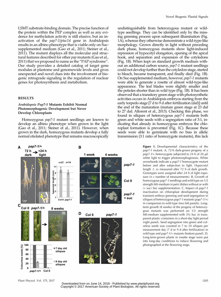
undistinguishable from heterozygous mutant or wild-type seedlings. They can be identified only by the miss-ing greening process upon subsequent illumination (Fig.1A), whereas they otherwise demonstrate awild-type-likemorphology. Grown directly in light without precedingdark phase, homozygous mutants show light-inducedrepression of hypocotyl elongation, opening of the apicalhook, and separation and expansion of the cotyledons(Fig. 1B). When kept on standard growth medium with-out an additional carbon source, pap7-1 mutant seedlingscouldnot develop further than the cotyledon stage, startedto bleach, became transparent, and finally died (Fig. 1B).On Suc-supplementedmedium, however, pap7-1mutantswere able to generate a rosette of almost wild-type-likeappearance. The leaf blades were slightly smaller andthe petioles shorter than in wild type (Fig. 1B). It has beenobserved that a transitory green stagewith photosyntheticactivities occurs in Arabidopsis embryos starting from theearly torpedo stage [7 d to 9 d after fertilization (daf)] untilthe end of the maturation (mature green stage at 23 dafto 27 daf; Allorent et al., 2013). Checking this phase, wefound in siliques of heterozygous pap7-1 mutants bothgreen and white seeds with a segregation ratio of 3:1, in-dicating that already in homozygous embryos the chlo-roplast formation is prevented (Fig. 1C). Because theseseeds were able to germinate with no bias in allelictransmission (3:1 ratio of homozygote mutants), this lack
Figure 1. Developmental characteristics of thepap7-1 mutant. A, 72-h dark-grown progeny of apap7-1/+ heterozygote subjected to 24 h of 20 mEwhite light to trigger photomorphogenesis. Whitearrowheads indicate a pap7-1 homozygote mutantbefore and after subjection to light. Hypocotyllength 6 SD measured after 72 h of dark growth.Genotypes were assigned after 24 h of light expo-sure (n = number of measurements). B, Growth ofhomozygous pap7-1 seedlings andwild type on 1/2strength MS-medium in petri dishes without or with(+ suc) Suc supplementation. C, Impact of pap7-1inactivation on chloroplast development duringtransient embryo greening and seed segregation insiliques of heterozygous pap7-1mutants (pap7-1/+)in comparison to wild type (two left panels). Long-term growth (8 weeks) of the progeny of heterozy-gous mutants was performed on 1/2 strengthMS-medium supplemented with 3% Suc in trans-parent plastic containers in a short-day light period(right panel). Seed segregation into green and col-orless seeds was counted in 7 to 10 siliques permeasurement day (7 d or 9 d after fertilization) inwild type and pap7-1/+ mutants (bottom panel). D,Long-term-grown plants in rosette stage were putinto long-day conditions to induce flowering andphotographed at the flowering stage.
Plant Physiol. Vol. 175, 2017 1205
Novel Biogenic Plastid Signals
www.plantphysiol.orgon June 14, 2018 - Published by Downloaded from Copyright © 2017 American Society of Plant Biologists. All rights reserved.

of chloroplast biogenesis appears nonessential for properseed development and maturation. When growing theprogeny of such heterozygous pap7-1 mutants on sugar-supplemented medium in short-day conditions undervery dim light (approximately 8 to 12 mE WL), homozy-gous mutant plants developed a reasonable rosette thatwas even able to initiate the flowering program aftershifting the plant container to long-day-conditions (Fig. 1,C and D). Architecture and size of the resulting inflores-cence did not exhibit major differences in comparison togreen plants, as indicated by control plants grown in thesame containers (Fig. 1D). In sum, these observations in-dicate that the major photomorphogenic programs (andhence the action of the corresponding PRs) are functionalin the mutant.
Transcript Accumulation in Albino Pap7-1 Plastids
All papmutants analyzed so far display largely reducedaccumulation of PEP-dependent transcripts,whereasNEP-dependent gene transcripts do overaccumulate (Börneret al., 2015; Pfannschmidt et al., 2015). In all published re-ports, however, only a few representative genes per geneclasswere investigated.Here,weperformeda comparativeplastomewide analysis in which we determined the accu-mulation of all plastidial mRNAs in light-grown pap7-1mutant plants and in wild-type plants grown in parallel(Fig. 2). To this end we used a custom-made macroarraythat, in addition to sense RNAs, also allowed the detectionof all corresponding antisenseRNAs (Demarsy et al., 2012).Plastidial antisense RNAs were found, in Arabidopsis, toaccumulate specifically in early phases of germination andradicle out-growth—two developmental steps precedingchloroplast biogenesis (Demarsy et al., 2012).
We observed reduced transcript accumulation formany PEP-dependent class-1 genes. However, mainlypsb genes (encoding PSII components) were affected inthe mutant (Fig. 2, light-green boxes), whereas, for psagenes (encoding PSI components), the reduction wasfound to be less pronounced or even nonexistent. Genesfor the cytochrome b6 f complex (pet genes) displayedmixed responses ranging from almost none to strongreduction of transcript accumulation. In contrast, allclass-2 genes (genes with PEP and NEP promoters) dis-played a stable or even enhanced transcript accumula-tion in the mutant. This class includes the ndh genes(encoding components of the NADH dehydrogenasecomplex), the genes encoding the proteins of the small(rps) and large (rpl) ribosomal subunits, and genes forcomponents of the ATP synthase (atp). Enhanced accu-mulation could be also observed for class-3 genes (geneswith NEP promoters only) rpoA and rpoB/C1/C2 (en-coding the PEP core subunits; Fig. 2, yellow boxes), butnot for the class-3 genes ycf1 and accD. A general down-regulation of PEP-dependent genes and a correspondingup-regulation of NEP-dependent genes, as suggested,thus cannot be confirmed by our macroarray analysis.
These observations were independently supportedby the microarray analysis that includes the full setof plastome-located genes. In addition, the microarray
also includes all tRNA genes that are not covered by themacroarray (Supplemental Table S1). These displayedall strongly reduced accumulation in the pap7-1mutantline, suggesting that they are transcribed by PEP. This isin agreement with results obtained in a recent study onpap mutants of maize (Williams-Carrier et al., 2014).
Antisense RNA accumulation for most genes dis-played no major differences between wild-type andpap7-1 mutant plants. However, we observed specificoveraccumulation of antisense transcripts for the psbB/psbT/psbH/petB/petD operon, the rpl33 and rps18 genes,and the two genes ycf1 and accD (encoding importmachinery subunit TIC214 and the b-subunit of theacetyl-CoA carboxylase complex, respectively; Fig. 2).This differential accumulation suggests that antisenseproduction is not just a concomitant by-product ofread-through transcription, but that a distinct unknownmechanism is at its origin.
In summary, our macroarray experiment uncoveredthat the disturbance in PEP activity does not causegene-class-specific transcription changes but rathermany gene-specific effects, suggesting a much morecomplex transcription regulation in arrested albinoplastids than current models anticipate.
Separation of Light- and Plastid-Dependent GeneRegulation during Photomorphogenesis by TrilateralDifferential Gene Expression Profiling
Chloroplast biogenesis is embedded into the generalphotomorphogenic program of seedling developmentthat strongly impedes a clear distinction of light-,development-, and plastid-dependent signaling (López-Juez et al., 1998, 2007). However, because illuminatedpap7-1mutants develop normally on Suc-supplementedmedia (Fig. 1; Gao et al., 2011; Steiner et al., 2011) indi-cating that chloroplast biogenesis can be separated fromphotomorphogenesis, we used it as a tool to separate thegene groups regulated by either light or plastid devel-opmental stage. To this end, we performed genomewidedifferential gene expression profiling in Arabidopsisby microarray hybridization. To unravel truly light-and plastid-dependent gene expression changes, wedid a trilateral comparative profiling including light-grown pap7-1 mutant seedlings (pap7-1 light, contain-ing white plastids), light-grown wild-type seedlings(wild-type light, containing chloroplasts), and dark-grown wild-type seedlings (wild-type dark, containingetioplasts, ETs), all at the two-cotyledon-stage (Fig. 3A).We performed a supervised analysis of the expressiondata using the MapMan tool (Usadel et al., 2005) and anunsupervised analysis by performing a weighted geneexpression network analysis (WGCNA) using gene on-tology (GO) groups to combine the advantages of bothgene categorization tools (Klie and Nikoloski, 2012).
In our supervised strategy, we assumed that a wild-type-light to wild-type-dark comparison reveals ex-pression changes controlled by the photomorphogenicprogram (PM; Fig. 3A). This should identify all genesactivated or inactivated by light and plastid signals. A
1206 Plant Physiol. Vol. 175, 2017
Grübler et al.
www.plantphysiol.orgon June 14, 2018 - Published by Downloaded from Copyright © 2017 American Society of Plant Biologists. All rights reserved.

parallel comparison of pap7-1 light- and wild-type-darkshould identify genes being regulated only by light,whereas the developmental status of the plastid is negli-gible (light signaling, LS; Fig. 3A) because both samples donot develop chloroplasts. Subsequent comparison of thesignificantly regulated gene groups identified in PM andLS then should identify genes specifically regulated bylight, by plastid stage (PS), or by both (Fig. 3A).Significantly regulated genes in the data sets were
identified (Supplemental Table S1) andwere imported intotheMapMan visualization tool (Fig. 3B; Supplemental Fig.S1). As expected, the PM data set revealed large genome-wide changes known to be characteristic for the photo-morphogenic program (Ma et al., 2001). This included amassive up-regulation of genes for photosynthesis, energymetabolism, and tetrapyrrole biosynthesis as well as sul-fate reduction and a great number of other biosynthetic
pathways (Supplemental Fig. S1A). In the LS data set, weobserved an impact on mostly the same gene groups andwith similar strength, indicating that the light-regulation inthe pap7-1 mutant works in a comparable manner as inwild type (Supplemental Fig. S1B). Direct comparison ofpap7-1 light versuswild-type-light gene expression profilesdemonstrated only limited differences (Supplemental Fig.S1C), indicating that the gene regulation in the light-grownpap7-1 mutant resembles much more that of light-grownthan dark-grownwild type. This indicates that the lackof functional chloroplasts in the pap7-1 mutant exertsonly a minor impact on light-regulated gene expres-sion networks.
The quality of the expression profiles obtained inthe microarray approaches was tested by quantitativeRT-PCR of selected genes being representative for dif-ferent expression classes found. To this end, RNA was
Figure 2. Macroarray analysis comparing plastid transcript accumulation in light-grown wild-type and pap7-1 Arabidopsisseedlings. Given is the transcript accumulation in wild-type (top panel) and mutant (bottom panel) seedlings both for sense (bluebars) and antisense (red bars) transcripts. Hybridization signals were normalized to the total signal intensity of the membrane andare given as arbitrary units in the left margin. Genes are labeled at the bottomof each panel according to accepted nomenclatures.Sequence of genes corresponds to their organization on the plastome separated between inner strand (left parts) and outer strandof the plastome (right parts). High transcript accumulation of PEP-dependent transcripts in wild type is highlighted by green boxes.High transcript accumulation of NEP-dependent transcripts in the pap7-1mutant is highlighted by yellow boxes. WT, wild type.
Plant Physiol. Vol. 175, 2017 1207
Novel Biogenic Plastid Signals
www.plantphysiol.orgon June 14, 2018 - Published by Downloaded from Copyright © 2017 American Society of Plant Biologists. All rights reserved.

Figure 3. Identification of gene modules responsive to light and/or biogenic plastid signals. A, Strategy for differential expressionprofiling of 4-d-old to 5-d-old Arabidopsis seedlings in wild type and pap7-1 mutants (pap7-1). Left panel displays photographs ofseedlings with representative phenotypic appearance. The right panel indicates the corresponding developmental transition of theplastids in eachof these seedlings. Smallwhite ovals represent undifferentiatedpro/eoplasts from seeds.Upon illumination, theydevelopinto green CPs in wild type (wild-type light) or arrested albino plastids (AAP) in pap7mutants (pap7-1 light). Growth in the dark leads todevelopment of yellow ETs in wild type (wild-type dark). Analysis of differences between expression profiles in these plant samples(indicated by brackets) identify genes for PM, for plastid-independent LS, and for PS (for details, see text). B, Flow diagram of bio-informatic analysis done with primary expression data from the microarray analysis. C, Detailed comparison of differentially expressedgenes (indicated by numbers) within the PM and LS expressionmodules according to their direction of expression change (indicated byarrows). Only genes exceeding a threshold of log2 fc$ 2were taken into analysis. Genes found in bothmoduleswere further separatedaccording to their direction and degree of gene expression change. Arrows of different size but same direction indicate genes that displaythe same direction of expression change but with a difference of at least log2 fc$ 1. Green arrows are expression change in wild type.White arrows are expression change inpap7-1mutants. Formore details, see legendbox.D, Proportional distribution of geneswithin thedifferent gene groups defined in Fig. 3C according to their functional association within MapMan bins. WT, wild type.
1208 Plant Physiol. Vol. 175, 2017
Grübler et al.
www.plantphysiol.orgon June 14, 2018 - Published by Downloaded from Copyright © 2017 American Society of Plant Biologists. All rights reserved.

isolated from identically, but independently grownplant samples from wild type and pap7-1mutants. As afurther independent control for the arrested plastiddevelopment another pap mutant, pap6-1, was used(Gilkerson et al., 2012). All genes tested in the quanti-tative reverse transcription PCR (qRT-PCR) displayedhighly reproducible expression data (correlation factorsabove 0.9) when compared to the corresponding dataobtained from the microarrays (Fig. 4; Supplemental Fig.S2). Furthermore, the expression data in the two differentpapmutants displayed a remarkable lowvariation (Fig. 4),indicating that the observed expression profiles in themutant background are robust and likely representativenot only for pap7-1 but also for other pap mutants.
Identification of Gene Groups Responding to Either Lightor Biogenic Plastid Signals
Next, we performed a more detailed analysis of thevarious regulated gene groups. To restrict the analysisto strongly regulated genes, we introduced a thresholdof log2-fold change (fc) $ 2 for definition of signifi-cantly regulated genes (881 genes in total). Identitiesand expression changes of these genes are given inSupplemental Table S2. Seven-hundred-and-twentygenes in the PM data set and 568 genes in the LS dataset met this criterion (Fig. 3C). Four-hundred-and-seven genes were identified in both data sets with amajority being regulated in the same direction in bothconditions (159 up-regulated and 207 down-regulatedgenes; Fig. 3C). These 366 equally regulated genes werenot influenced by the developmental state of the plas-tid, but by illumination only. Thus, they representplastid-independent, light-regulated genes. Only 41 ofthe 407 genes displayed either opposite (one gene) ordifferent strength in their regulation, where we regar-ded only differences of at least log2 fc$ 1 as significant.These genes appear to be partly affected by both plastidstage and light. The largest group (23 genes) displayedup-regulation in wild-type light versus wild-type dark,but significantly less up-regulation in pap7-1- lightversus wild-type dark. This expression pattern corre-sponds to the regulationmode attributed to the classicaldefinition of a plastid signal that causes a lower accu-mulation of light-induced nuclear gene transcriptswhen chloroplast development is inhibited by chemicalor genetic means (Pfannschmidt 2010).In addition,we identified 313genes thatwere exclusively
up- (162) or down- (151) regulated only when chloroplastbiogenesis (CP) occurred (CP-dependent genes, Fig. 3).These genes may either cause chloroplast biogenesis or arerelated to specific functions exerted by this fully devel-oped plastid type such as retrograde redox regulationfrom photosynthesis. Interestingly, we identified 161genes that displayed exclusive up- (83) or down- (78)regulation only in the presence of the arrested albinoplastid (AAP-dependent genes, Fig. 3). These genes arelikely regulated because of the missing chloroplastbiogenesis or function (i.e. photosynthesis or connectedbiosynthesis pathways). Both developmental plastid
stages apparently send distinct, light-independent sig-nals to the nucleus that either repress or enhance sep-arate sets of genes. Because of their light-independency,these signals are not identical with the classical plastidsignal and imply the existence of plastid-type specificpositive and negative signals not yet defined by currentmodels of retrograde signaling.
In total, 41.5% of the 881 strongly regulated genesappear to be light-regulated, 4.6% are regulated bycombined light and plastid signals, and 53.8% (corre-sponding to 474 genes) are regulated by plastid signalsonly (CP and AAP). Decreasing the threshold for sig-nificant regulation to log2 fc $ 1 identified 3658 regu-lated genes, which is roughly four timesmore thanwithlog2 fc$ 2 as threshold (Supplemental Table S3). In thislarger group, 46.4% of the genes appear to be light-regulated, 3.9% are regulated by light and plastidsignals, and 49.5% are regulated by plastid signalsonly (Supplemental Fig. S3). Thus, enlargement of thenumber of investigated genes did not result in majorrelative changes between the three regulation modes,indicating that the arbitrarily chosen thresholds didnot produce a bias for a specific regulation pattern. Theinclusion of dark-grown plants as an additional ref-erence point instead of doing a direct comparison ofwild-type light and pap7-1 light provides a much moreprecise separation of plastid- and light-regulated genes.Both factors control very distinct sets of genes and theoverlap between the two signaling pathways/expressionnetworks appears to be very limited.
Among Photosynthesis-Associated Nuclear Genes, OnlyLHCB Genes Are Affected by Plastid Signals in Pap7-1
The CP-, light-, and AAP-dependent geneswere finallysorted according to their functional categories (MapManbin; Fig. 3D). The majority of functional categories wereidentical for all three groups comprising the MapManbins “protein”, “RNA”, “stress”, “transport”, “signaling”,“development”, and “hormone metabolism”. Interest-ingly, the group of CP-dependent genes did not include“photosynthesis genes”, whereas the group of AAP-dependent genes did not include the bin “secondarymetabolism”, but instead the bin “cell wall”. The largeoverlap in functional categories, however, is not reflectedat the level of individual gene identities (SupplementalTable S1), and our analysis clearly demonstrates that eachgene group represents a distinct set of regulated genes(see also results from WGCNA).
The lack of photosynthesis genes in the CP-dependentgroup was surprising with respect to the notion thatphotosynthesis-associated nuclear genes (PhANGs) areconsidered to be a prime target for plastid signals duringbiogenic control. We, therefore, analyzed these genes sep-arately and compared their relative expression changes inthe PMand LS data sets (Fig. 5A). Inwild-type light versuswild-type dark, 76 genes exhibited a light-induced ex-pression increase of log2 fc $ 1. Ninety-three photosyn-thesis genes (including all plastome-localized genes)displayed expression variations that remained below
Plant Physiol. Vol. 175, 2017 1209
Novel Biogenic Plastid Signals
www.plantphysiol.orgon June 14, 2018 - Published by Downloaded from Copyright © 2017 American Society of Plant Biologists. All rights reserved.

this threshold, suggesting that they are not or just mildlyaffected by light. Only 15 genes exhibited a strong lightinduction of log2 fc $ 2. The top five of these genes allencode proteins of the light-harvesting complex of PSII(LHCB; framed in Fig. 5A). In the pap7-1 light versus wild-type dark comparison, we observed a similar expressionprofile for photosynthesis genes. Here, 62 genes exhibitedlight-induced expression of log2 fc $ 1 and 11 genes dis-played expression changes of log2 fc $ 2. Ten of thesegeneswere also found as top-regulated genes inwild type.However, we observed that, specifically, LHCB genesdisplayed significant less accumulation in pap7-1 com-pared to wild type, whereas all other genes remainedfairly constant. Comparable results were obtained in theindependent qRT-PCR controls (Fig. 4). This reduced ex-pression can be attributed to the impact of biogenic signalsfrom the arrested albino plastid. In addition, we also
observed nine significantly down-regulated genes in thiscomparison, all of them being plastome-localized (Fig.5A). This confirms the observation using the MapManvisualization (Supplemental Fig. S1) inwhich inhibition ofchloroplast development had only limited impact on theoverall nuclear gene expression profile. We conclude thatinhibition of chloroplast development in the pap7-1 mu-tant represses specifically plastome-localized photosyn-thesis genes and a small set of nuclear LHCB genes,whereas all other PhANGs are either not affected orare just mildly attenuated.
Another interesting question was whether other nu-clear genes for components of the plastid transcriptionmachinery were affected in their expression by the re-pressed PEP activity in pap7-1 plastids. We, therefore,analyzed the expression data of genes for all s-factors,PAPs, PTAC components, and for NEP. The majority of
Figure 4. Expression profiles of selected genes from the microarray analysis tested by qRT-PCR. A, Expression changes of genesrepresentative for distinct expression classes in wild-type light, pap7-1, and pap6-1 mutants compared to wild-type dark. Genes forlight-harvesting complex II protein1.4, light harvesting complex II protein1.2, and “overexpression leads to pseudo-etiolation in light”represent genes displaying a reduced light induction in pap7-1when compared to wild type. Genes encoding protein state transitionkinase7, PGR5-like protein 1a, and s-factor5 represent genes displaying no effect of pap7-1 on light induction, whereas the gene forprotochlorophyllide oxidoreductase B represents an example for strong light repression both in wild type and the pap7-1mutant. Thegenes for glutathione peroxidase 7 and (6 to 4) DNA photolyase were used as examples displaying light induction in wild type andfurther promotion in pap7-1. Genes for SEN1 and HFR1 senescence-associated protein DIN1 and the transcription factor LongHypocotyl after far-red1 represent genes displaying repression inwild type but promotion in themutant. As additional genetic control,a defect in the gene for a phospho-fructokinase like1 protein (pap6-1/fln1) was used to detect potential mutation-specific responses.Expression of both pap geneswas tested in all RNA samples as further control. B, Difference in expression change between light-grownwild-type and the pap7-1 and pap6-1mutants. Negative values indicate lower expression, positive values higher expression than inwild type. Data given represent means of three independent experiments. Primers used are given in Supplemental Table S4. LHCB1.2,light-harvesting complex II protein 1.2; LHCB1.4, light-harvesting complex II protein 1.4; STN7, state transition kinase7; PGRL1a,PGR5-like protein1a; PORB, protochlorophyllide oxidoreductase B; SIG5, s-factor 5; ATGPX7, glutathione peroxidase7; oePEL,“overexpression leads to pseudo-etiolation in light”; UVR3, (6 to 4) DNA photolyase; SEN1, senescence-associated protein DIN1;HFR1, transcription factor Long Hypocotyl after far-red1; WT, wild type.
1210 Plant Physiol. Vol. 175, 2017
Grübler et al.
www.plantphysiol.orgon June 14, 2018 - Published by Downloaded from Copyright © 2017 American Society of Plant Biologists. All rights reserved.

these components displayed light induction in wild typewhen compared to the dark control (Fig. 5B). This is inaccordance with earlier bioinformatic analyses. The ob-served regulation patterns were largely maintained in themutant with only minor deviations from the wild type,indicating that the developmental state of the plastid hasalso no significant impact on the expression of these geneswhereas light appears to be a dominant regulator even inthemutant. The functional PEP deficiency does not exert aretrograde repressive control of other nuclear-encodedPEP or NEP components.
WGCNA
The supervised analysis (Fig. 3) used presettingsbased on assumptions drawn from literature and our
own experiences. To avoid any unwanted bias, we per-formed an additional unsupervised analysis of the geneexpression data sets employing aWGCNA(SupplementalFig. S4). As in our supervised analysis, the cluster analysisof the expression data revealed a much closer correlationbetween pap7-1 light and wild-type light samples thanbetween pap7-1 light and wild-type dark samples (Fig. 6).Nevertheless, the gene expression profiles of pap7-1 lightand wild-type light samples displayed a number of spe-cific differences. In total, six significant gene expressionmodules could be identified by WGCNA within the dataset (Supplemental Fig. S4). These modules describe char-acteristic similarities and differences between the threeplant samples and largely correspond to the groupsdefined in the supervised differential analysis. Becausethe unsupervised analysis did not include expression
Figure 5. Light-induced expression changes of genes for photosynthesis and plastid transcription. A, Graphs display the expression valuesof significantly light-affected photosynthesis genes (log2$ 1) detected by comparison of expression profiles for wild-type light versuswild-type dark (top graph) and pap7-1 light versus wild-type dark (bottom graph). From the 169 photosynthesis genes present in the corre-sponding MapMan bin, 72 and 62 genes, respectively, exhibited an expression change of at least log2 $ 1. In the wild-type light versuswild-type dark comparison, all plastid genes remained below this threshold. Genes exceeding an expression change of log2 $ 2 wereboxed in the graph and listed in a panel aside. Nuclear-encoded genes are given in black, plastid-encoded genes are given in green. Thedifference in the expression change of a particular gene that occurs between the top and the bottom panel reflects the plastid influence onits expression. Strongly down-regulated genes in pap7-1 light versus wild-type dark (log2 $ 21) are given in a left box. B, Expressionchanges for all 31 nuclear genes encoding components of the plastid gene transcriptionmachinery inwild type andpap7-1. The respectivecomparisons are indicated in the top panel, gene identities in the left, and protein identities in the right panels. Given valuesrepresent log2-fold changes; a corresponding color-code is depicted at the bottom level. WT, wild type.
Plant Physiol. Vol. 175, 2017 1211
Novel Biogenic Plastid Signals
www.plantphysiol.orgon June 14, 2018 - Published by Downloaded from Copyright © 2017 American Society of Plant Biologists. All rights reserved.
thresholds, it covered much larger gene numbers. Theidentified expression modules divide into two groups(Supplemental Fig. S4B). Group 1 comprises threemodules (modules blue, yellow, and green) in whichtwo samples are highly similar, whereas the third oneis opposing. Group 2 comprises three modules (mod-ules turquoise, brown, and red) in which two samplesare opposing, whereas the third one is in an interme-diate state between the two others.
Genes in module “blue” (in total 1838 genes) displayhighly similar expression in pap7-1 light and wild-typelight samples and an opposing expression in wild-typedark. The dominant regulating factor in this module isthe light, whereas the plastid state appears to play norole or only a very minor one. Module “blue”, therefore,covers the light-regulated genes.Genes inmodule “yellow”(in total 769 genes) display highly similar expression inpap7-1 light and wild-type-dark samples and an opposingexpression in wild-type-light. The dominant regulatingfactor here is theplastid state,whereas light appears tohavenomajor impact. Module “yellow”, therefore, covers genesunder aplastidial regulation that is exerted fromanormallydeveloped chloroplast. Genes in module “green” (in total671 genes) display a highly similar expression in wild-type-light and wild-type-dark samples and an opposingexpression in pap7-1-light. Light appears to be of no im-portance for the regulation of these genes, but the arrestedplastid development and the genetic background of themutant. Module “green”, therefore, covers genes that aremisregulated because of the disturbance in plastid devel-opment and the pap7-1 protein deficiency. The expressionprofiles of the genes covered by these three modules cor-respond to those in the three gene groups defined in oursupervised analysis (Fig. 3C), and provide independentanalytical confirmation for the correctness of our selectioncriteria. Understanding and interpretation of the modulesin group 2 appeared to be much more difficult as the in-termediate expression profiles in each module prevent anidentification of the dominant regulating factor. Genegroups defined by these modules are likely under multi-factorial control andwere, therefore, excluded from furtheranalysis.
We analyzed the three genemodules for enriched GOgroups (Supplemental Figs. S5 and S7) and indicatedmost important GO groups with respect to the mutantphenotype below. Light-regulated genes in module“blue” included the GO groups for “Photosynthesis”,“Isoprenoid biosynthesis”, “Tetrapyrrole biosynthesis”,and for “Anatomical structure andmorphogenesis” (Fig.7A). All these gene groups are apparently activated bylight without displaying a major impact of the devel-opmental state of the plastid. Like in the supervisedanalysis, PhANGs occur under the light-regulated genegroups supporting the notion that the impact of theplastid stage on the expression of this gene group isvery limited. Chloroplast-regulated genes in module“yellow” (Fig. 7B) contained GO groups for “Cellularmetabolic processes”, “Photoperiodism and flower-ing”, “Fatty acid beta oxidation”, “Protein localizationto peroxisome”, “Glucosinolate biosynthetic process”,
and “Indoleacetic acid metabolic processes”. Althoughthe first four groups appear to be repressed, the lattertwo are mainly activated by the presence of functionalchloroplasts. Finally, in the pap7-1-regulated module“green” (Fig. 7C), we found GO groups for “Nitrogencompoundmetabolism processes”, “Circadian rhythm”,and “Reproductive system development”. These genegroups were either activated or stayed active upon thegenetic arrest of chloroplast biogenesis. Our triplicateanalyses describe distinct gene sets that are specificallytargeted by the three factors of light, chloroplasts, andarrested albino plastids, in both a positive and a negativemanner.
DISCUSSION
Our plastome- and genomewide gene expressionanalysis in the pap7-1 mutant provides the first detailedview of the disturbances occurring at the transcript levelof this mutant. This revealed interesting and unexpectedfacts that help us to better understand the causes for thealbinism in this mutant. In addition, the obtained dataprovide important novel clues for our understanding ofother pap mutants as well as retrograde biogenic signalsand their target genes.
Impact of Pap7-1 Deficiency on Plastid Gene Expression
Elucidation of the plastid transcriptome in the pap7-1mutant uncovered that many PEP-dependent class Itranscripts displayed only low accumulation whereasNEP-dependent rpo transcripts exhibited enhanced ac-cumulation (Fig. 2). This is in coincidence with earlierreports. However, we observed significant differencesbetween psa and psb gene groups, suggesting differen-tial effects on class-I transcription. In addition, NEP-dependent ycf1 and accD gene transcripts did notoveraccumulate as rpo transcripts, but exhibited clearlyreduced transcript accumulation (Fig. 2). Furthermore,class-II transcripts revealed largely wild-type-like ac-cumulation in the mutant (Fig. 2). These effects cannotbe reconciled with this model of plastid transcription inalbino plastids of papmutants, which assumes a generalinactivation of PEP activity, whereas the NEP activitiesare up-regulated. Instead, these data imply differentialand gene-specific effects on transcription activities inthe albino plastids. Because all class-I transcripts didaccumulate to a certain degree in pap7-1 plastids, it islikely that these arise from a basal activity of the PEPcore-complex. Such a basal activity was identified inETs of mustard, and was shown to be able in faithfulpromoter recognition using s-factors, but in a differentmanner than the corresponding activity from chloro-plasts (Eisermann et al., 1990; Tiller and Link, 1993;Pfannschmidt and Link, 1994). This scenario couldprovide a realistic explanation for the observed tran-scriptome in the pap7-1 mutant plastids, and suggestthat the gene expression mechanisms in the albinoplastids are likely arrested in a stage similar to ETs.Alternatively, the residual class-I transcripts may arise
1212 Plant Physiol. Vol. 175, 2017
Grübler et al.
www.plantphysiol.orgon June 14, 2018 - Published by Downloaded from Copyright © 2017 American Society of Plant Biologists. All rights reserved.

from read-through transcription performed by NEP en-zyme activities; however, such a model would lack anexplanation for differential gene group transcription.Regardless of their origin, the residual class-I transcriptsapparently are not sufficient to elicit the formation of aphotosynthetic apparatus, suggesting the involvement ofadditional constraints for the chloroplast formation.One such constraint could be the enhanced antisense
transcript accumulation in the mutant that was ob-served notably for the psbB/T/H/petB/D operon, therpl33/rps18 operon, and the ycf1 and accD genes (Fig. 2).As this effect appears to be rather gene-specific, it likelydoes not represent just an arbitrary accumulation ofread-through transcription of the respective oppositestrand of the plastome. Mechanistically, one could ex-pect that enhanced antisense transcript accumulationinterferes with translation efficiency of the correspondingsense transcript due to duplex formation, as it was sug-gested for the plastid psbT gene (Zghidi-Abouzid et al.,2011). This would provide a gene-group-specific mecha-nism that could prevent the formation of functionalphotosystem II and Cytb6f complexes. Whether this an-tisense production is based on a specific transcriptionevent will be an interesting field of future research.Our microarray analysis of pap7-1 RNA samples
indicated a strongly reduced accumulation of most
plastidial tRNAs (Supplemental Fig. S1). This is inagreement with a recent study performed in severalmaize pap mutants, proposing that PEP activity has amajor role in the expression of plastidial tRNAs (Williams-Carrier et al., 2014). Reduction in plastid tRNA accumu-lation restricts plastid translation, as the tRNA moleculestransfer the amino acids. Furthermore, because the tRNAE
is the precursor of amino-levulinic acid, tetrapyrrole bio-synthesis and the generation of chlorophylls might beseverely affected.
In sum, the differential accumulation of gene-specifictranscripts of all classes implies the existence of specific,yet unknown, transcription events in the arrested albinoplastids, suggesting a more defined and diversifieddivision of labor between the PEP and NEP enzymesduring early steps of chloroplast biogenesis than thecurrent models propose.
Retrograde Control of Nuclear Gene Expression byBiogenic Plastidial Signals
A major improvement of our study when comparedto earlier work in this field arose from our experimentalsetup, which included dark-grown wild-type plants asan additional reference point. Inclusion of homozygouspap7-1mutants grown in the dark as further controlwas
Figure 6. Cluster analysis of genes differentiallyregulated in the three growth setups as defined byANOVA. Genes displaying an FDR of , 0.05were included. The right margin identified thedata sets from the microarray analysis; the leftmargin indicates a cladogram defining the corre-lation in gene expression profiles between them.On top, clustering of gene groups according totheir expression is indicated. Diagram in top-leftcorner gives the color key and numbers of geneswith corresponding gene expression values. Redindicates up-regulation; green indicates down-regulation. WT, wild type.
Plant Physiol. Vol. 175, 2017 1213
Novel Biogenic Plastid Signals
www.plantphysiol.orgon June 14, 2018 - Published by Downloaded from Copyright © 2017 American Society of Plant Biologists. All rights reserved.

Figure 7. GOgroups of differentially expressed gene setswithin the genemodules identified byWGCNA. A, Genemodule “blue”with fourmajor genegroups regulated by light. B, Genemodule “yellow”with six major gene groups regulated by the chloroplast. C, Genemodule “green” with threemajorgene groups specifically regulated by the arrested plastid. Genes displaying an FDR, 0.05 were included. On top of each heat map, the selected GOgroup is given; underneath, a clustering of the genes in this group, according to their expression, is indicated. The right margin identifies the data setsfrom the microarray analysis. Red indicates up-regulation; green indicates down-regulation. For further gene groups, see Supplemental Figs. S8 to S10.WT, wild type.
1214 Plant Physiol. Vol. 175, 2017
Grübler et al.
www.plantphysiol.orgon June 14, 2018 - Published by Downloaded from Copyright © 2017 American Society of Plant Biologists. All rights reserved.

not feasible, as homozygous mutant seedlings in thisstage remain macroscopically indistinguishable fromwild-type or heterozygous mutants (Fig. 1A). Dis-crimination between wild-type and mutant genotypesat this stage would be technically possible only at themolecular level of individual seedlings, exacerbatinglargely the harvest of sufficient material for RNA prep-aration. The indistinguishability of dark-grown homo-zygous mutants is in accordance with our workinghypothesis that the mutation becomes effective onlyunder illumination, although it does not affect theskotomorphogenic program. Consequently, segrega-tion of the progeny of heterozygous pap7-1mutant plantsbecomes macroscopically visible only in the light whenthe transition toward chloroplasts is arrested. Neverthe-less, the trilateral expression profiling allowed us to defineunambiguously distinct gene sets responding specificallyto 1) light, 2) chloroplast signals, and 3) signals fromarrested albino plastids. This distinction is impossible by asimple bilateral mutant-wild-type comparison in the lightas this provides only the relative differences between thetwo conditions. The impact of light in each of them re-quires the additional comparison with the dark controlthat then reveals whether light has a promoting, inhibit-ing, or no effect and allows finally for the distinction be-tween retrograde and light control.The three gene modules identified by our approach
contain both activated and repressed genes, implyingthe existence of retrograde biogenic signals that are ofeither a positive or a negative nature. Because the genesets regulated by chloroplasts and arrested plastids aredifferent, it is likely that the controlling signals are trans-mitted via separate pathways. Whether these are fully
independent from each other or mutually exclude eachother remains to be investigated. The question of whethera weaker nuclear gene expression indicates the action of anegative signal or the missing action of a positive signalhas been debated in the past (Pfannschmidt, 2010; Terryand Smith, 2013; Hills et al., 2015). A recent study (Pageet al., 2017a, 2017b) and our data presented here indicatethat the action of both positive and negative signals needsto be considered in current models.
The phenotypic analysis (Fig. 1) revealed that despitechloroplast deficiency, the general photomorphogenicprogram in the pap7-1mutant appears to be operationalas long as an external carbon source is available. Oldermutant plants generate a complete albino rosette that iscomparable to those of dark-grown mutants with consti-tutively active phytochromes (Su and Lagarias, 2007),confirming that general plant development and chloro-plast biogenesis represent largely separate processes. Eventhe initiation of the flowering transition was found to befunctional, which corresponds to observations reportedfor rpo deletion mutants of tobacco (De Santis-MacIosseket al., 1999). We assume that illumination activates the PRsystems in both wild-type and pap7-1 mutant seed-lings in the same manner, suggesting that synthesisand action of the PRs (including their chromophores)are fully functional in the albino mutant.
This explains the similarity of the expression profiles oflight-grownwild-type and pap7-1mutants (SupplementalFig. S1), including the expression of the correspondinglight-regulated gene module (module “blue”; Fig. 8). Themodule contains the GO groups for isoprenoid and tet-rapyrrole biosynthesis (Fig. 7) that produce importantprecursors for primary products such as carotenoids,chlorophylls, haem, phytochromobilin, plastoquinones,or different plant hormones (Pulido et al., 2012). Both bio-synthesis pathways provide essential metabolites forplant metabolism and development and, therefore, arelikely active in the albino plastids with the apparent ex-ception of Chl biosynthesis. For the same reasons, the GOgroup for morphogenesis and anatomical structure (Fig.7)may appear in thismodule because it is part of the light-initiation of photomorphogenesis. Unexpectedly, how-ever, was the identification of PhANGs that, in contrast togeneral assumptions, did not exhibit anymajor repressionby retrograde signals from the arrested plastid develop-ment. Only a few Lhcb genes appeared to be selectivelytargeted by plastid signals (Fig. 5) and these, in addition,seem to interact with light (Figs. 3 and 5). This may ex-plain the manifold connections identified between lightand plastid signaling using promoters of these genes ingenetic screens and corresponding mutants (Larkin,2014). Interestingly, the structural and functional defectin the PEP-PAP complex in pap7-1mutants does not affectthe transcript accumulation of other nuclear-encodedcomponents of the plastid transcription machinery (Fig.5B), implying that these genes are not under retrogradecontrol.
It remains for us to understand why chloroplastbiogenesis does not work in the mutant. A major de-terminant of this developmental block certainly is the
Figure 8. Model of anterograde and retrograde signaling during earlysteps of chloroplast biogenesis in wild-type and pap7-1 mutants. Lightsignals (flash) are perceived by PRs (orange circle). Small white ovalsrepresent undifferentiated proteoplasts present in seeds. Large ovals ingreen and white represent the different plastid types in wild-type (CP)and pap7-1 (AAP) seedlings. Gray-shaded arrows indicate the devel-opmental process leading to these plastid types. Boxes indicate andname the genes or gene modules regulated during these processes. Thinblack arrows represent positive regulation; lines with a blocking barrepresent negative regulation. WT, wild type.
Plant Physiol. Vol. 175, 2017 1215
Novel Biogenic Plastid Signals
www.plantphysiol.orgon June 14, 2018 - Published by Downloaded from Copyright © 2017 American Society of Plant Biologists. All rights reserved.

reduced expression of plastid photosystem II genes,but the finding of residual transcripts suggests thatother molecular reasons also likely play a role, such asthe obvious inhibition of Chl biosynthesis (Gao et al.,2012) and further defects that are unknown to date.Weconclude that in the pap7-1 mutant, plastidial photo-system II genes are especially strongly repressed,whereas retrograde signals from the arrested albinoplastid neither modulate nor antagonistically coun-teract light regulation of PhANGs. This is differentfrom conclusions obtained in recent studies on retro-grade signaling using lincomycin, an inhibitor ofplastid translation (Ruckle et al., 2007; Martín et al.,2016). Because pap7-1 mutants developed rather nor-mally in the first few days even without sugar, weregard it as likely that either the genetic block in pap7-1mutants results in milder effects than a lincomycintreatment, or the observed differences are due to technicaldifferences in the respective setups, e.g. light intensity.Both scenarios suggest that there exists a threshold for theeffectiveness of retrograde signals. Elucidating the mo-lecular nature for thiswill be an interesting topic for futureresearch. The retrograde-controlled gene groups iden-tified in this study provide a useful first tool for suchinvestigations.
An interesting result of our expression profiling wasthe identification of separate biogenic signals fromchloroplasts and albino plastids. Chloroplast signal-dependent GO groups were mostly related to metabo-lism, likely because chloroplast biogenesis initiates theconversion from a heterotrophic to autotrophic lifestyle. This includes the down-regulation of b-oxidationof fatty acids that takes place in glyoxysomes. Oppo-sitely, GO groups for glucosinolate biosynthesis andindoleacetic acid biosynthesis were activated, both be-ing highly important for pathogen defense and growthof green plants (Fig. 7B). GO groups regulated by sig-nals from the arrested albino plastid relate mostly tostarvation processes and the mobilization of storageenergies (nitrogen compound metabolic processes, re-productive system development) being indicative ofthe nonautotrophic metabolism in the albino plantletsthat requires the mobilization of all internal resources.Surprisingly, a significant impact on the GO group ofcircadian rhythms was observed that suggests an in-fluence of the plastid developmental stage on circadianclock genes. A circadian control from the nucleus influ-encing chloroplast transcription has been recently reported(Noordally et al., 2013). Furthermore, iron metabolismand plastid developmental stage were demonstrated tohave a significant influence on the period of the circadianclock (Salomé et al., 2013). These observations suggestthat our results reflect a mutual influence between plas-tids and the circadian clock of larger significance, pro-viding an interesting target for future research (Fig. 7C).
In sum, the arrest of chloroplast development in thepap7-1 mutant can be best explained by a specific dis-turbance in the light-induced buildup of the PEP com-plex during the pro/eoplast-to-chloroplast transition.This leads to concomitant defects in the expression of
PSII components and tRNAs, which in turn limits Chlbiosynthesis and (likely) translation. The albinism of themutant likely is the result of a multifactorial syndromethat prevents chloroplast biogenesis without destroyingthe plastid. Therefore, the functional and metabolic stateof the arrested albino plastid resembles that of an ETdespite it perceiving light. This likely allows the mutantto develop normally when the lack of photosynthesis iscomplemented by an external carbon source. Correspond-ingly, the overall gene expressionprofiles of pap7-1mutantsdo exhibit a chimerical character, with metabolic genesregulated like in dark-grown seedlings and photoregulatedgenes like in light-grown plants. In sum, the pap7-1mutant,but likely also other pap mutants, provide an interestingtool for dissecting further the molecular processes duringthe early steps in chloroplast biogenesis.
MATERIALS AND METHODS
Plant Material
We used Arabidopsis (Arabidopsis thaliana) Columbia (Col) 0 as wild typethroughout the study. As null alleles for the genes pap7-1/ptac14 and pap6/fln1we used the Arabidopsis T-DNA inactivation lines SAIL_566_F06 andGK-443A08, respectively. These lines (named pap7-1 and pap6-1 in this study)were characterized earlier in detail for singularity of T-DNA insertion and forcausal connection between the albino phenotype and the corresponding genedefect (Arsova et al., 2010; Gao et al., 2011; Steiner et al., 2011; Gilkerson et al.,2012). Seeds of wild-type and heterozygous mutants were surface-sterilizedand spread on half-strength standard Murashige & Skoog (MS) medium sup-plemented with indicated amounts of Suc in petri dishes, stratified for 3 d, andgrown to the two-cotyledon stage at 21°C for further analyses. Light-grownplants used for array analyses were grown under permanent white light of120 mE to 150 mE photon flux density. In long-term growth experiments, theillumination intensity of the white light source was reduced to 8 mE to 12 mE.
Plastid Macroarray Analysis
Plant material was grown 6 d on MS medium supplemented with 0.5% Suc.Approximately 500 mg each of green wild-type and albino mutant cotyledonswere harvested separately and shock-frozen in liquid nitrogen. Total RNA wasisolated following published procedures (Demarsy et al., 2006, 2012). PotentialDNA contaminations were removed by DNase treatment and its absence wasproven by PCR. For preparation of the hybridization probe, 4 mg of each RNApreparation was reverse-transcribed using a gene-specific primer mix anneal-ing to 80 protein-coding genes and their corresponding antisense sequences.Subsequently, a reverse transcription reactionwas performed using SuperscriptII Reverse Transcriptase (Invitrogen) in the presence of all four nucleotides and100 mCi [a-32P] dATP (Demarsy et al., 2012). Unincorporated nucleotides werefinally removed from the labeled cDNAsbygel-filtration througha SephadexG50column. For evaluation of relative differences in plastid transcription between thebiological backgrounds of the two samples,wemeasured the total radioactivity ofeach cDNA sample after synthesis and verified the cDNAprofile on a sequencinggel. In the experiment described here, radioactively labeled cDNAs of wild typeand pap7-1 differed by just 3% (wild type . pap7-1) in incorporation, indicatingthat total plastid gene expression is not significantly different in wild-type andpap7-1 plantlets. Subsequent hybridization of the radiolabeled probes with themacroarray, washing conditions, subsequent signal detection in a phosphor-imager (FLA-8000; Fujifilm), and final data analysis were done essentially asdescribed in Demarsy et al. (2012). Construction of the plastome macroarray,including description of the gene-specific sense and antisense probes and theirspotting pattern, was published elsewhere (Lerbs-Mache, 2011).
Genomewide Microarray Analysis
Plant material was grown on medium supplemented with 1% (w/v) sugar toharmonize thegermination. Illuminatedwild-type andhomozygouspap7-1mutantswere grown for 5 d for full expansion of the cotyledons, whereas dark-grown
1216 Plant Physiol. Vol. 175, 2017
Grübler et al.
www.plantphysiol.orgon June 14, 2018 - Published by Downloaded from Copyright © 2017 American Society of Plant Biologists. All rights reserved.

wild-type seedlings were grown for 4 d to avoid mechanical stress imposedby physical contact with the lid of the petri dishes. Plant materials wereharvested at 10:00 a.m. and shock-frozen in liquid N. Dark-grown materialwas harvested and shock-frozen at the same time point of the day, but under agreen safe-light to exclude any light effects in the profiles. Total RNA fromthese materials was then basically prepared as described in Logemann et al. (1987).In brief, 250 mg of frozen plant material was ground in a mortar and purified withthe RNeasy purification kit (Qiagen). Concentration and purity of RNA sampleswere determined spectroscopically and intactnesswas proven by ethidiumbromidestaining after separation on 1.2% (w/v) agarose gels. Purified samples from threebiological replicates each were sent on dry ice to a commercial service (Kompe-tenzzentrum für Fluoreszente Bioanalytik) where a second quality check, cDNAsynthesis, and labelingwasperformedaccording to theGeneChip 39 IVTExpressKitprotocol (Affymetrix). Hybridization and reading of signals was performed usingthe Arabidopsis Genome Array ATH1 (Affymetrix) and according to standardprotocols of the service.
qRT-PCR
Reverse transcription was performed with 1 mg total RNA isolated from threeindependent biological replicates of wild-type, pap7-1, and pap6 mutants grownidentically to those for the microarray analyses. cDNA was synthesized usingoligo(dT) primers and the SuperScript II Reverse Transcriptase (Invitrogen) followingthe manufacturers’ recommendations. The qRT-PCR analysis was performed usingthe GoTaq qPCR Master Mix (Promega) and the Rotor-Gene 3000 equipment(Qiagen). Primer sequences for genes of interest were designed using the softwareApE-A Plasmid Editor (v2.0.47) and NCBI/Primer-BLASTwith preference to intronspanning amplicons. Each primer pair was tested for amplification efficiency usingthe synthesized cDNA. Only primer pairs with amplification efficiencies between90% and 110% were used for further analysis. For used primer sequences and geneidentities, see Supplemental Table S4. The relative quantification was calculatedaccording to describedmethods (Pfaffl, 2001). The Arabidopsis genes for actin 7 andubiquitin 5 were used as internal control and reference genes for quantification.
Bioinformatics
In the supervised analysis, the hybridization signal data from themicroarrayanalysis performed by a commercial service (KFB Regensburg) were analyzedwith the ROBINA (http://www.mapman.gabipd.org/web/guest) and MapMan(http://www.mapman.gabipd.org/web/guest) programs (Usadel et al., 2005,2009). Statistical analysis by t test and subsequent calculation of the false-discoveryrate (FDR) were performed according to the ROBINA program. Gene expressionchanges with an FDR of P # 0.05 were regarded as statistically significant. Themicroarray data given in the Supplemental File S1 are based on three biologicalreplicates each. The data discussed in this publication have been deposited inNCBI’sGene Expression Omnibus (Edgar et al., 2002; Barrett et al., 2013) and are accessiblethrough GEO Series accession no. GSE88988 (https://www.ncbi.nlm.nih.gov/geo/query/acc.cgi?acc=GSE88988). Visualization of the cellular pathways and functionalcategories of the expression data were carried out using the MapMan and PegManpackages according toAth_AFFY_ATH1_TAIR8_Jan 2010 (http://mapman.gabipd.org; Usadel et al., 2005). The visualization tool of MapMan was used to identifysimilarities and differences in the different metabolic pathways. A Wilcoxon ranksum testwasused to visualize significantly expressedgenes inPegMan (Usadel et al.,2009). Venn diagrams were calculated using the expression log values of theMapMan package. In the unsupervised WGCNA, the original CEL filessupplied by the commercial service were imported into R with Bioconductor(package oligo; Carvalho and Irizarry, 2010) followed by normalization andbackground correction of the raw data using RMA. After selecting 27,296annotated genes, a scaling and centering followed by a cluster analysis wasperformed. Differentially expressed genes were selected with ANOVA andan FDR threshold of 0.05, followed by a WGCNA (Langfelder and Horvath,2008) and a GO enrichment analysis using the R package topGO (http://www.mpi-sb.mpg.de/;alexa).
Supplemental Data
The following supplemental materials are available.
Supplemental Figure S1. Relative gene expression profiles of wild-typeand pap7-1 mutants visualized using the MapMan tool.
Supplemental Figure S2. Correlation of expression data from selectedgenes obtained from microarrays and qRT-PCR.
Supplemental Figure S3. Identification of gene modules responsive tolight and/or biogenic plastid signals with a significance threshold of log2
fc $ 1.
Supplemental Figure S4. Weighted gene coexpression network clusteranalysis.
Supplemental Figure S5. Gene ontology groups within module “blue”.
Supplemental Figure S6. Gene ontology groups within module “yellow”.
Supplemental Figure S7. Gene ontology groups within module “green”.
Supplemental Table S1. Gene expression changes of genes sorted accord-ing to the encoding genomic compartment.
Supplemental Table S2. Gene sets with an expression change larger thanthe threshold 2 [log2].
Supplemental Table S3. Gene sets with an expression change larger thanthe threshold 1 [log2].
Supplemental Table S4.Nucleotide sequences of primers used for qRT-PCR.
Supplemental Dataset 1.
Supplemental Dataset 2.
Supplemental Dataset 3.
Received July 27, 2017; accepted September 20, 2017; published September 21,2017.
LITERATURE CITED
Allorent G, Courtois F, Chevalier F, Lerbs-Mache S (2013) Plastid geneexpression during chloroplast differentiation and dedifferentiation intonon-photosynthetic plastids during seed formation. Plant Mol Biol 82:59–70
Arsova B, Hoja U, Wimmelbacher M, Greiner E, Ustün S, Melzer M,Petersen K, Lein W, Börnke F (2010) Plastidial thioredoxin z interactswith two fructokinase-like proteins in a thiol-dependent manner: evi-dence for an essential role in chloroplast development in Arabidopsisand Nicotiana benthamiana. Plant Cell 22: 1498–1515
Arsovski AA, Galstyan A, Guseman JM, Nemhauser JL (2012) Photo-morphogenesis. Arabidopsis Book 10: e0147
Barrett T, Wilhite SE, Ledoux P, Evangelista C, Kim IF, Tomashevsky M,Marshall KA, Phillippy KH, Sherman PM, Holko M, Yefanov A, LeeH, et al (2013) NCBI GEO: archive for functional genomics data sets—update. Nucleic Acids Res 41: D991–D995
Börner T, Aleynikova AY, Zubo YO, Kusnetsov VV (2015) ChloroplastRNA polymerases: role in chloroplast biogenesis. Biochim Biophys Acta1847: 761–769
Brown NJ, Sullivan JA, Gray JC (2005) Light and plastid signals regulatethe expression of the pea plastocyanin gene through a common region atthe 59 end of the coding region. Plant J 43: 541–552
Carvalho BS, Irizarry RA (2010) A framework for oligonucleotide micro-array preprocessing. Bioinformatics 26: 2363–2367
Chan KX, Phua SY, Crisp P, McQuinn R, Pogson BJ (2016) Learning thelanguages of the chloroplast: retrograde signaling and beyond. AnnuRev Plant Biol 67: 25–53
Chi W, Sun X, Zhang L (2013) Intracellular signaling from plastid to nu-cleus. Annu Rev Plant Biol 64: 559–582
De Santis-MacIossek G, Kofer W, Bock A, Schoch S, Maier RM, WannerG, Rüdiger W, Koop HU, Herrmann RG (1999) Targeted disruption ofthe plastid RNA polymerase genes rpoA, B and C1: molecular biology,biochemistry and ultrastructure. Plant J 18: 477–489
de Souza A, Wang JZ, Dehesh K (2016) Retrograde signals: integrators ofinterorganellar communication and orchestrators of plant development.Annu Rev Plant Biol 68: 85–108
Demarsy E, Buhr F, Lambert E, Lerbs-Mache S (2012) Characterization ofthe plastid-specific germination and seedling establishment transcrip-tional programme. J Exp Bot 63: 925–939
Demarsy E, Courtois F, Azevedo J, Buhot L, Lerbs-Mache S (2006)Building up of the plastid transcriptional machinery during germinationand early plant development. Plant Physiol 142: 993–1003
Plant Physiol. Vol. 175, 2017 1217
Novel Biogenic Plastid Signals
www.plantphysiol.orgon June 14, 2018 - Published by Downloaded from Copyright © 2017 American Society of Plant Biologists. All rights reserved.

Edgar R, Domrachev M, Lash AE (2002) Gene expression omnibus: NCBIgene expression and hybridization array data repository. Nucleic AcidsRes 30: 207–210
Eisermann A, Tiller K, Link G (1990) In vitro transcription and DNAbinding characteristics of chloroplast and etioplast extracts from mustard(Sinapis alba) indicate differential usage of the psbA promoter. EMBO J 9:3981–3987
Estavillo GM, Crisp PA, Pornsiriwong W, Wirtz M, Collinge D, Carrie C,Giraud E, Whelan J, David P, Javot H, Brearley C, Hell R, et al (2011)Evidence for a SAL1-PAP chloroplast retrograde pathway that functionsin drought and high light signaling in Arabidopsis. Plant Cell 23: 3992–4012
Galvez-Valdivieso G, Fryer MJ, Lawson T, Slattery K, TrumanW, Smirnoff N,Asami T, DaviesWJ, Jones AM, Baker NR, Mullineaux PM (2009) The highlight response in Arabidopsis involves ABA signaling between vascular andbundle sheath cells. Plant Cell 21: 2143–2162
Gao ZP, Chen GX, Yang ZN (2012) Regulatory role of Arabidopsis pTAC14in chloroplast development and plastid gene expression. Plant SignalBehav 7: 1354–1356
Gao ZP, Yu QB, Zhao TT, Ma Q, Chen GX, Yang ZN (2011) A functionalcomponent of the transcriptionally active chromosome complex, Ara-bidopsis pTAC14, interacts with pTAC12/HEMERA and regulatesplastid gene expression. Plant Physiol 157: 1733–1745
Gilkerson J, Perez-Ruiz JM, Chory J, Callis J (2012) The plastid-localizedpfkB-type carbohydrate kinases FRUCTOKINASE-LIKE 1 and 2 are es-sential for growth and development of Arabidopsis thaliana. BMC PlantBiol 12: 102
Hajdukiewicz PT, Allison LA, Maliga P (1997) The two RNA polymerasesencoded by the nuclear and the plastid compartments transcribe distinctgroups of genes in tobacco plastids. EMBO J 16: 4041–4048
Hills AC, Khan S, López-Juez E (2015) Chloroplast biogenesis-associatednuclear genes: control by plastid signals evolved prior to their regula-tion as part of photomorphogenesis. Front Plant Sci 6: 1078
Isemer R, Mulisch M, Schäfer A, Kirchner S, Koop HU, Krupinska K(2012) Recombinant Whirly1 translocates from transplastomic chloro-plasts to the nucleus. FEBS Lett 586: 85–88
Kleine T, Leister D (2016) Retrograde signaling: organelles go networking.Biochim Biophys Acta 1857: 1313–1325
Klie S, Nikoloski Z (2012) The choice between MapMan and gene ontology forautomated gene function prediction in plant science. Front Genet 3: 115
Kusnetsov V, Bolle C, Lübberstedt T, Sopory S, Herrmann RG, OelmüllerR (1996) Evidence that the plastid signal and light operate via the samecis-acting elements in the promoters of nuclear genes for plastid proteins.Mol Gen Genet 252: 631–639
Lagrange T, Hakimi MA, Pontier D, Courtois F, Alcaraz JP, Grunwald D,Lam E, Lerbs-Mache S (2003) Transcription factor IIB (TFIIB)-relatedprotein (pBrp), a plant-specific member of the TFIIB-related proteinfamily. Mol Cell Biol 23: 3274–3286
Langfelder P, Horvath S (2008) WGCNA: an R package for weightedcorrelation network analysis. BMC Bioinformatics 9: 559
Larkin RM (2014) Influence of plastids on light signalling and develop-ment. Philos Trans R Soc Lond B Biol Sci 369: 20130232
Lee KP, Kim C, Landgraf F, Apel K (2007) EXECUTER1- and EXECUTER2-dependent transfer of stress-related signals from the plastid to the nu-cleus of Arabidopsis thaliana. Proc Natl Acad Sci USA 104: 10270–10275
Legen J, Kemp S, Krause K, Profanter B, Herrmann RG, Maier RM (2002)Comparative analysis of plastid transcription profiles of entire plastidchromosomes from tobacco attributed to wild-type and PEP-deficienttranscription machineries. Plant J 31: 171–188
Lerbs-Mache S (2011) Function of plastid sigma factors in higher plants:regulation of gene expression or just preservation of constitutive tran-scription? Plant Mol Biol 76: 235–249
Liebers M, Grübler B, Chevalier F, Lerbs-Mache S, Merendino L, BlanvillainR, Pfannschmidt T (2017) Regulatory shifts in plastid transcription play akey role in morphological conversions of plastids during plant development.Front Plant Sci 8: 23
Liere K, Weihe A, Börner T (2011) The transcription machineries of plantmitochondria and chloroplasts: composition, function, and regulation. JPlant Physiol 168: 1345–1360
Logemann J, Schell J, Willmitzer L (1987) Improved method for the iso-lation of RNA from plant tissues. Anal Biochem 163: 16–20
López-Juez E (2007) Plastid biogenesis, between light and shadows. J ExpBot 58: 11–26
López-Juez E, Jarvis RP, Takeuchi A, Page AM, Chory J (1998) NewArabidopsis cue mutants suggest a close connection between plastid-and phytochrome regulation of nuclear gene expression. Plant Physiol118: 803–815
Ma L, Li J, Qu L, Hager J, Chen Z, Zhao H, Deng XW (2001) Light controlof Arabidopsis development entails coordinated regulation of genomeexpression and cellular pathways. Plant Cell 13: 2589–2607
Martín G, Leivar P, Ludevid D, Tepperman JM, Quail PH, Monte E (2016)Phytochrome and retrograde signalling pathways converge to antago-nistically regulate a light-induced transcriptional network. Nat Com-mun 7: 11431
Noordally ZB, Ishii K, Atkins KA, Wetherill SJ, Kusakina J, Walton EJ,Kato M, Azuma M, Tanaka K, Hanaoka M, Dodd AN (2013) Circadiancontrol of chloroplast transcription by a nuclear-encoded timing signal.Science 339: 1316–1319
Page MT, Kacprzak SM, Mochizuki N, Okamoto H, Smith AG, Terry MJ(2017a) Seedlings lacking the PTM protein do not show a genomes un-coupled (gun) mutant phenotype. Plant Physiol 174: 21–26
Page MT, McCormac AC, Smith AG, Terry MJ (2017b) Singlet oxygeninitiates a plastid signal controlling photosynthetic gene expression.New Phytol 213: 1168–1180
Park SW, Li W, Viehhauser A, He B, Kim S, Nilsson AK, Andersson MX,Kittle JD, Ambavaram MM, Luan S, Esker AR, Tholl D, et al (2013)Cyclophilin 20-3 relays a 12-oxo-phytodienoic acid signal during stressresponsive regulation of cellular redox homeostasis. Proc Natl Acad SciUSA 110: 9559–9564
Pfaffl MW (2001) A new mathematical model for relative quantification inreal-time RT-PCR. Nucleic Acids Res 29: e45
Pfalz J, Liere K, Kandlbinder A, Dietz KJ, Oelmüller R (2006) pTAC2, -6, and-12 are components of the transcriptionally active plastid chromosome thatare required for plastid gene expression. Plant Cell 18: 176–197
Pfalz J, Pfannschmidt T (2013) Essential nucleoid proteins in early chlo-roplast development. Trends Plant Sci 18: 186–194
Pfannschmidt T (2010) Plastidial retrograde signalling—a true “plastidfactor” or just metabolite signatures? Trends Plant Sci 15: 427–435
Pfannschmidt T, Blanvillain R, Merendino L, Courtois F, Chevalier F, LiebersM, Grübler B, Hommel E, Lerbs-Mache S (2015) Plastid RNA polymerases:orchestration of enzymes with different evolutionary origins controls chlo-roplast biogenesis during the plant life cycle. J Exp Bot 66: 6957–6973
Pfannschmidt T, Link G (1994) Separation of two classes of plastid DNA-dependent RNA polymerases that are differentially expressed in mus-tard (Sinapis alba L.) seedlings. Plant Mol Biol 25: 69–81
Pfannschmidt T, Munne-Bosch S (2013) Plastidial signaling during the plant lifecycle. In B Biswal, K Krupinska, eds, Plastid Development in Leaves duringGrowth and Senescence. Springer, Dordrecht, the Netherlands
Pogson BJ, Ganguly D, Albrecht-Borth V (2015) Insights into chloroplastbiogenesis and development. Biochim Biophys Acta 1847: 1017–1024
Pogson BJ, Woo NS, Förster B, Small ID (2008) Plastid signalling to thenucleus and beyond. Trends Plant Sci 13: 602–609
Pulido P, Perello C, Rodriguez-Concepcion M (2012) New insights intoplant isoprenoid metabolism. Mol Plant 5: 964–967
Ramel F, Birtic S, Ginies C, Soubigou-Taconnat L, Triantaphylidès C,Havaux M (2012) Carotenoid oxidation products are stress signals thatmediate gene responses to singlet oxygen in plants. Proc Natl Acad SciUSA 109: 5535–5540
Ruckle ME, Burgoon LD, Lawrence LA, Sinkler CA, Larkin RM (2012)Plastids are major regulators of light signaling in Arabidopsis. PlantPhysiol 159: 366–390
Ruckle ME, DeMarco SM, Larkin RM (2007) Plastid signals remodel lightsignaling networks and are essential for efficient chloroplast biogenesisin Arabidopsis. Plant Cell 19: 3944–3960
Salomé PA, Oliva M, Weigel D, Krämer U (2013) Circadian clock adjust-ment to plant iron status depends on chloroplast and phytochromefunction. EMBO J 32: 511–523
Steiner S, Schröter Y, Pfalz J, Pfannschmidt T (2011) Identification of essentialsubunits in the plastid-encoded RNA polymerase complex reveals buildingblocks for proper plastid development. Plant Physiol 157: 1043–1055
Su YS, Lagarias JC (2007) Light-independent phytochrome signaling me-diated by dominant GAF domain tyrosine mutants of Arabidopsisphytochromes in transgenic plants. Plant Cell 19: 2124–2139
Sullivan JA, Gray JC (2002) Multiple plastid signals regulate the expressionof the pea plastocyanin gene in pea and transgenic tobacco plants. PlantJ 32: 763–774
1218 Plant Physiol. Vol. 175, 2017
Grübler et al.
www.plantphysiol.orgon June 14, 2018 - Published by Downloaded from Copyright © 2017 American Society of Plant Biologists. All rights reserved.

Sun X, Feng P, Xu X, Guo H, Ma J, Chi W, Lin R, Lu C, Zhang L (2011) Achloroplast envelope-bound PHD transcription factor mediates chloro-plast signals to the nucleus. Nat Commun 2: 477
Terry MJ, Smith AG (2013) A model for tetrapyrrole synthesis as the pri-mary mechanism for plastid-to-nucleus signaling during chloroplastbiogenesis. Front Plant Sci 4: 14
Tiller K, Link G (1993) Sigma-like transcription factors from mustard (Sinapisalba L.) etioplast are similar in size to, but functionally distinct from, theirchloroplast counterparts. Plant Mol Biol 21: 503–513
Usadel B, Nagel A, Thimm O, Redestig H, Blaesing OE, Palacios-Rojas N,Selbig J, Hannemann J, Piques MC, Steinhauser D, Scheible WR,Gibon Y, et al (2005) Extension of the visualization tool MapMan toallow statistical analysis of arrays, display of corresponding genes, andcomparison with known responses. Plant Physiol 138: 1195–1204
Usadel B, Poree F, Nagel A, Lohse M, Czedik-Eysenberg A, Stitt M (2009)A guide to using MapMan to visualize and compare omics data in
plants: a case study in the crop species, maize. Plant Cell Environ 32:1211–1229
Waters MT, Langdale JA (2009) The making of a chloroplast. EMBO J 28:2861–2873
Williams-Carrier R, Zoschke R, Belcher S, Pfalz J, Barkan A (2014) Amajor role for the plastid-encoded RNA polymerase complex in theexpression of plastid transfer RNAs. Plant Physiol 164: 239–248
Woodson JD, Perez-Ruiz JM, Chory J (2011) Heme synthesis by plastid ferro-chelatase I regulates nuclear gene expression in plants. Curr Biol 21: 897–903
Xiao Y, Savchenko T, Baidoo EE, Chehab WE, Hayden DM, Tolstikov V,Corwin JA, Kliebenstein DJ, Keasling JD, Dehesh K (2012) Retrogradesignaling by the plastidial metabolite MEcPP regulates expression ofnuclear stress-response genes. Cell 149: 1525–1535
Zghidi-Abouzid O, Merendino L, Buhr F, Malik Ghulam M, Lerbs-MacheS (2011) Characterization of plastid PsbT sense and antisense RNAs.Nucleic Acids Res 39: 5379–5387
Plant Physiol. Vol. 175, 2017 1219
Novel Biogenic Plastid Signals
www.plantphysiol.orgon June 14, 2018 - Published by Downloaded from Copyright © 2017 American Society of Plant Biologists. All rights reserved.