light microscopic immunocytochemical localization of ... · pdf filelight microscopic...
TRANSCRIPT
Light microscopic immunocytochemical localization of hepatic and intestinal types of fatty acid-binding proteins in rat small intestine'
Helen M. Shields,* Margaret L. Bates,t Nathan M. Bass,** Cynthia J. Best,* David H. Alpers,t and Robert K. Ockner** Charles A. Dana Research Institute and Harvard-Thorndike Laboratory, Division of Gastroenterology, Department of Medicine, Beth Israel Hospital and Harvard Medical School, Boston, MA 02215;* Department of Medicine, Washington University School of Medicine, St. Louis, MO 63110; t and Department of Medicine, University of California, San Francisco, CA 94143**
Abstract Monospecific antisera to purified hepatic fatty acid- binding protein (hFABP) and gut fatty acid-binding protein (gFABP) have been used to localize these two proteins in the small intestine of fed rats at the light microscopic level. Pieces of duodenum, jejunum, and ileum were removed from 4-, lo-, 20-, 22-, and 60-day-old Sprague-Dawley rats. Both cryostat and paraffin sections were studied for the presence of hFABP or gFABP by the avidin-biotin immunoperoxidase method. Slides were graded blind for the intensity of staining. Despite the struc- tural and immunological differences between these two proteins, we showed no major differences between their staining patterns or their staining intensity throughout the intestine during post- natal development. The staining for both fatty acid-binding pro- teins was cytoplasmic. No brush border staining was found. Staining was more intense in the proximal rather than distal in- testine, in the villus rather than crypt cells, and in the apex rather than the base of intestinal cells. Shifts in staining pat- terns, and staining intensity occurring during development may be related to variations in dietary fat intake, rates of cell prolifera- tion, intestinal anatomy, and mechanisms for fat absorption. - Shields, H. M., M. L. Bates, N. M. Bass, C. J. Best, D. H. Alpers, and R. I(. Ocher. Light microscopic immunocyto- chemical localization of hepatic and intestinal types of fatty acid- binding proteins in rat small intestine. J. Lipid Res. 1986. 27: 549-557.
Supplementary key w o r d s chemistry avidin-biotin immunoperoxidase method
fatty acid-binding protein immunocyto-
Small molecular weight cytosolic proteins that bind long chain fatty acids have been found in a variety of tissues, including liver, small intestine, kidney, adipose tissue, cardiac muscle, and skeletal muscle and have been called fatty acid-binding proteins (FABP) (1). Hepatic fatty acid-binding protein (hFABP) and gut FABP (gFABP) (2) have been purified (3-5), and monospecific antisera have been raised to each form (3, 4). The two proteins are structurally and immunologically distinct (3-6). hFABP is probably identical to Z protein and one of the sterol car- rier proteins isolated from liver (7-9). hFABP is present
in both liver and intestine (10) while gFABP is present largely in the intestine (3). The exact function(s) of these proteins in the small intestine is not known, but it has been suggested that they may act as intracellular trans- port proteins for fatty acids andlor be involved in intra- cellular fat metabolism (1, 3, 4, 11, 12). Knowledge of the anatomic location of these proteins is very slight. A single study localized Z protein in the liver at the light and elec- tron microscope level by immunocytochemical techniques (13). By light microscopy, staining was limited to the hepatic cytoplasm. By electron microscopy, staining was found in the cytoplasm adjacent to smooth endoplasmic reticulum and mitochondria (13). In the absence of prior morphological data on the intestinal FABPs, we have used monospecific antisera to hFABP and gFABP for their im- munocytochemical localization in the rat small intestine throughout development as an initial step towards a better understanding of their functional roles in the enterocyte.
METHODS
hFABP and gFABP are small molecular weight proteins found in the 105,000 g supernatant of liver and small in- testine (3, 4). Each was purified as previously described by either sucrose density gradient (gFABP) (3) or Sepha- dex thin-layer, isoelectric focusing (hFABP) (4). hFABP or gFABP were both single protein bands as determined by SDS polyacrylamide gel electrophoresis. Antisera to puri-
Abbreviations: FABP, fatty acid-binding protein; gFABP, intestinal type fatty acid-binding protein; hFABP, hepatic type fatty acid-binding protein; TBS, Tris-buffered saline; NSS, normal sheep serum.
'Part of this paper was presented at the Annual Meeting of the Ameri- can Gastroenterological Association in Washington, DC, May 1983.
Journal of Lipid Research Volume 27, 1986 549
by guest, on April 26, 2018
ww
w.jlr.org
Dow
nloaded from
fied hFABP and gFABP were raised in rabbits as previ- ously described (3, 4). Antisera to each type were identi- fied as being monospecific by the Ouchterlony double immunodiffusion method (3, 4), and by the fact that each antiserum immunoprecipitated a unique polypeptide from cell-free translation mixtures (5, 6).
Full thickness pieces of duodenum (1 cm distal to the pylorus), jejunum (middle of the intestine), and distal ileum (1 cm proximal to the cecum) were removed from
suckling (4-day-old and 10-day-old), weanling (20-, 21-, and 22-day-old), and adult (60-day-old, equal numbers of male and female) Sprague-Dawley rats under ether anes- thesia. Tissue from the 20-, 21-, and 22-day-old rats will be considered together under the label weanling through- out the subsequent experiments. Sprague-Dawley rats were used, since both FARPs had previously been purified from this strain (2, 3). Rats were permitted to feed up until the time they were killed.
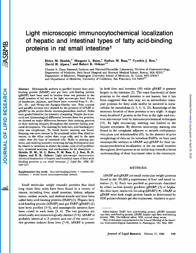
Fig. 1. Low power light micrograph of a paraffin section of a 10-day-old rat duodenum stained for gFARP showing irnmunoperoxidase staining in the villus tip and mid-villus cells. Vacuoles are scattered throughout the apical areas of the villus tip cells. Apical staining is heavier than basal staining. No staining is noted in the crypt areas. X750.
550 Journal of Lipid Research Volnme 27, 1986
by guest, on April 26, 2018
ww
w.jlr.org
Dow
nloaded from
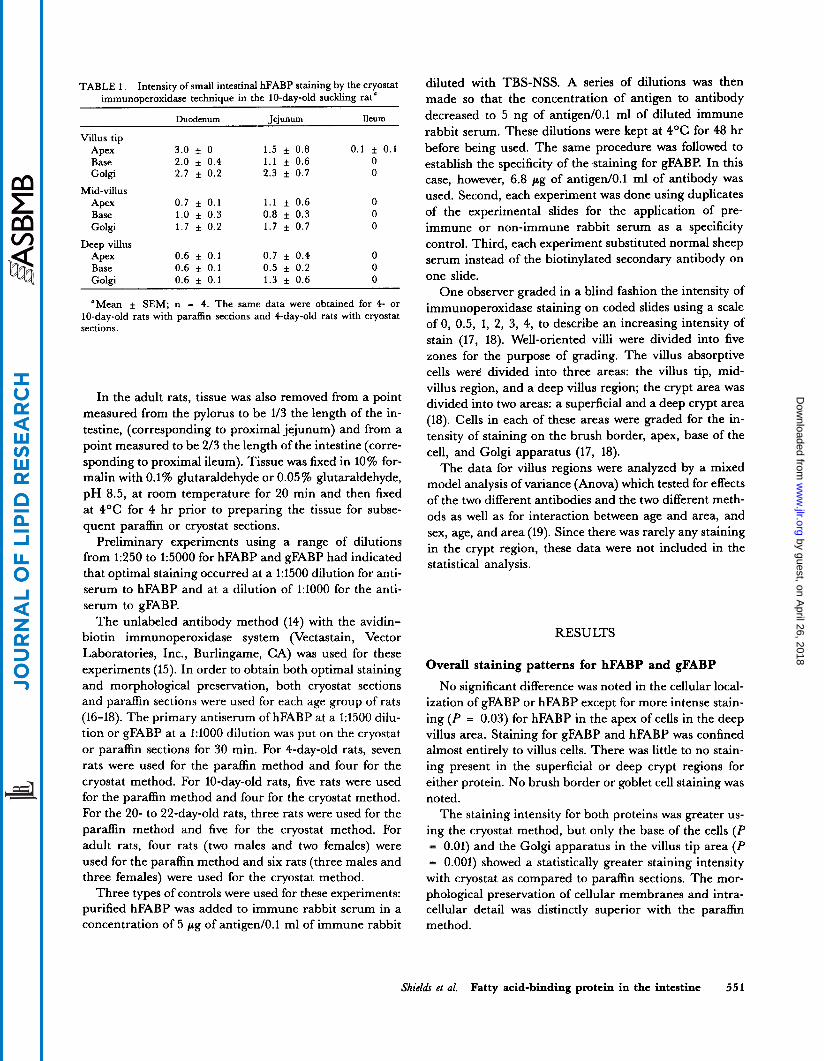
TABLE 1. Intensity of small intestinal hFABP staining by the cryostat immunoperoxidase technique in the 10-day-old suckling rat‘
Duodenum leiunum Ileum _____~
Villus tip
Base Golgi
Mid-villus Apex Base Golgi
Deep villus Apex Base Golgi
Apex 3.0 f 0 2.0 f 0.4 2.7 f 0.2
0.7 f 0.1 1.0 f 0.3 1 . 7 f 0.2
0.6 f 0.1 0.6 + 0.1 0.6 f 0.1
1.5 f 0.8 1.1 f 0.6 2.3 f 0.7
1 . 1 f 0.6 0.8 f 0.3 1 . 7 f 0.7
0.7 f 0.4 0.5 f 0.2 1.3 f 0.6
0.1 f 0.1 0 0
0 0 0
0 0 0
“Mean f SEM; n = 4. The same data were obtained for 4- or 10-day-old rats with paraffin sections and 4-day-old rats with cryostat sections.
In the adult rats, tissue was also removed from a point measured from the pylorus to be 1/3 the length of the in- testine, (corresponding to proximal jejunum) and from a point measured to be 2/3 the length of the intestine (corre- sponding to proximal ileum). Tissue was fixed in 10% for- malin with 0.1% glutaraldehyde or 0.05 % glutaraldehyde, pH 8.5, at room temperature for 20 min and then fixed at 4OC for 4 hr prior to preparing the tissue for subse- quent paraffin or cryostat sections.
Preliminary experiments using a range of dilutions from 1:250 to 1:5000 for hFABP and gFABP had indicated that optimal staining occurred at a 1:1500 dilution for anti- serum to hFABP and at a dilution of 1:lOOO for the anti- serum to gFABP.
The unlabeled antibody method (14) with the avidin- biotin immunoperoxidase system (Vectastain, Vector Laboratories, Inc., Burlingame, CA) was used for these experiments (15). In order to obtain both optimal staining and morphological preservation, both cryostat sections and paraffin sections were used for each age group of rats (16-18). The primary antiserum of hFABP at a 1:1500 dilu- tion or gFABP at a 1:lOOO dilution was put on the cryostat or paraffin sections for 30 min. For 4-day-old rats, seven rats were used for the paraffin method and four for the cryostat method. For 10-day-old rats, five rats were used for the paraffin method and four for the cryostat method. For the 20- to 22-day-old rats, three rats were used for the paraffin method and five for the cryostat method. For adult rats, four rats (two males and two females) were used for the paraffin method and six rats (three males and three females) were used for the cryostat method.
Three types of controls were used for these experiments: purified hFABP was added to immune rabbit serum in a concentration of 5 pg of antiged0.1 ml of immune rabbit
diluted with TBS-NSS. A series of dilutions was then made so that the concentration of antigen to antibody decreased to 5 ng of antiged0.l ml of diluted immune rabbit serum. These dilutions were kept at 4OC for 48 hr before being used. The same procedure was followed to establish the specificity of the staining for gFABP. In this case, however, 6.8 pg of antiged0.1 ml of antibody was used. Second, each experiment was done using duplicates of the experimental slides for the application of pre- immune or non-immune rabbit serum as a specificity control. Third, each experiment substituted normal sheep serum instead of the biotinylated secondary antibody on one slide.
One observer graded in a blind fashion the intensity of immunoperoxidase staining on coded slides using a scale of 0, 0.5, 1, 2, 3, 4, to describe an increasing intensity of stain (17, 18). Well-oriented villi were divided into five zones for the purpose of grading. The villus absorptive cells were divided into three areas: the villus tip, mid- villus region, and a deep villus region; the crypt area was divided into two areas: a superficial and a deep crypt area (18). Cells in each of these areas were graded for the in- tensity of staining on the brush border, apex, base of the cell, and Golgi apparatus (17, 18).
The data for villus regions were analyzed by a mixed model analysis of variance (Anova) which tested for effects of the two different antibodies and the two different meth- ods as well as for interaction between age and area, and sex, age, and area (19). Since there was rarely any staining in the crypt region, these data were not included in the statistical analysis.
RESULTS
Overall staining patterns for hFABP and gFABP
No significant difference was noted in the cellular local- ization of gFABP or hFABP except for more intense stain- ing (P = 0.03) for hFABP in the apex of cells in the deep villus area. Staining for gFABP and hFABP was confined almost entirely to villus cells. There was little to no stain- ing present in the superficial or deep crypt regions for either protein. No brush border or goblet cell staining was noted.
The staining intensity for both proteins was greater us- ing the cryostat method, but only the base of the cells (P = 0.01) and the Golgi apparatus in the villus tip area (P = 0.001) showed a statistically greater staining intensity with cryostat as compared to paraffin sections. The mor- phological preservation of cellular membranes and intra- cellular detail was distinctly superior with the paraffin method.
Shields et al. Fatty acid-binding protein in the intestine 551
by guest, on April 26, 2018
ww
w.jlr.org
Dow
nloaded from
Fig. 2. High power light micrograph of a paraffin section of a IO-day-old rat jejunum stained for hFABP showing very prominent globular staining in the Golgi apparatus (arrow). No brush border or nuclear staining is noted. There is no difference between apical and basal staining. X7,500.
Specific patterns of staining throughout development for hFABP and gFABP
Since no major differences were noted between staining patterns for gFABP and hFABP, the two staining patterns will be described together.
During the suckling period there were two sharply defined gradients of staining noted. The first one was the decreasing gradient of staining going from top of the villus to the crypt with a fairly abrupt cut-off of staining at the level of the crypt (Fig. 1). The second gradient was that going from the duodenum to the ileum showing a
marked diminution in staining going from intense stain- ing in the proximal intestine to essentially no staining in the distal intestine (Table 1). Staining in the villus cells was found diffusely throughout the cytoplasm with apical staining being greater than basal staining. Staining in the duodenal cytoplasm often outlined droplets in the apical portion of the cell (data not shown). No staining was noted in the large apical vacuoles of the ileum. The apical portion of the duodenal villus tip cells had significantly ( P = 0.01) more staining for FABP during the suckling period (4- and/or 10-day-old rats) compared to the wean- ling or adult period. In addition, the Golgi apparatus
552 Journal of Lipid Research Volume 27, 1986
by guest, on April 26, 2018
ww
w.jlr.org
Dow
nloaded from
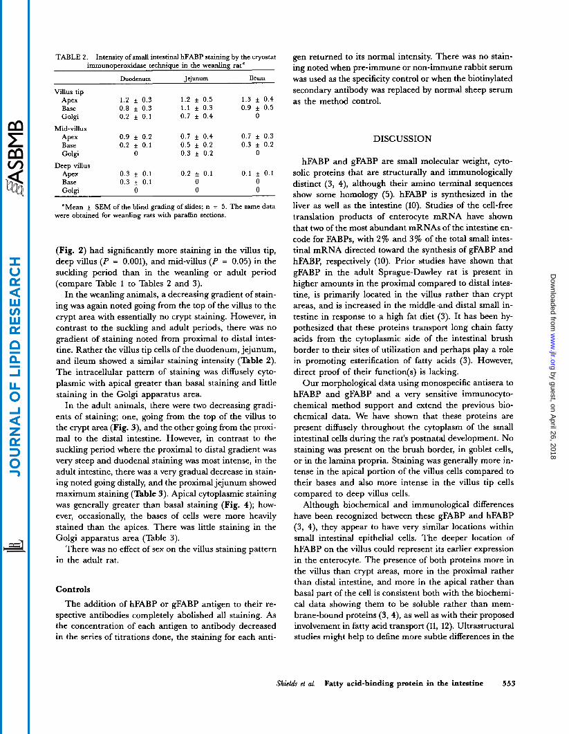
TABLE 2. Intensity of small intestinal hFABP staining by the cryostat immunoDeroxidase techniaue in the weanlinP rat’
Duodenum Jejunum Ileum
Villus tip Apex 1 . 2 f 0.3 1.2 f 0.5 1.3 f 0.4
1 . 1 f 0.3 0.9 f 0.5 Base 0.8 f 0.3 Golgi 0.2 f 0.1 0.7 f 0.4 0
Mid-villus Apex 0.9 f 0.2 0.7 f 0.4 0.7 f 0.3 Base 0.2 f 0.1 0.5 f 0.2 0.3 f 0.2 Golgi 0 0.3 f 0.2 0
Apex 0.3 f 0.1 0.2 f 0.1 0.1 f 0.1 Base 0 .3 f 0.1 0 0 Golgi 0 0 0
Deep villus
“Mean f SEM of the blind grading of slides; n = 5. The same data were obtained for weanling rats with paraffin sections.
(Fig. 2) had significantly more staining in the villus tip, deep villus ( P = O.OOl), and mid-villus ( P = 0.05) in the suckling period than in the weanling or adult period (compare Table 1 to Tables 2 and 3).
In the weanling animals, a decreasing gradient of stain- ing was again noted going from the top of the villus to the crypt area with essentially no crypt staining. However, in contrast to the suckling and adult periods, there was no gradient of staining noted from proximal to distal intes- tine. Rather the villus tip cells of the duodenum, jejunum, and ileum showed a similar staining intensity (Table 2). The intracellular pattern of staining was diffusely cyto- plasmic with apical greater than basal staining and little staining in the Golgi apparatus area.
In the adult animals, there were two decreasing gradi- ents of staining; one, going from the top of the villus to the crypt area (Fig. 3), and the other going from the proxi- mal to the distal intestine. However, in contrast to the suckling period where the proximal to distal gradient was very steep and duodenal staining was most intense, in the adult intestine, there was a very gradual decrease in stain- ing noted going distally, and the proximal jejunum showed maximum staining (Table 3). Apical cytoplasmic staining was generally greater than basal staining (Fig. 4); how- ever, occasionally, the bases of cells were more heavily stained than the apices. There was little staining in the Golgi apparatus area (Table 3).
There was no effect of sex on the villus staining pattern in the adult rat.
Controls
The addition of hFABP or gFABP antigen to their re- spective antibodies completely abolished all staining. As the concentration of each antigen to antibody decreased in the series of titrations done, the staining for each anti-
gen returned to its normal intensity. There was no stain- ing noted when pre-immune or non-immune rabbit serum was used as the specificity control or when the biotinylated secondary antibody was replaced by normal sheep serum as the method control.
DISCUSSION
hFABP and gFABP are small molecular weight, cyto- solic proteins that are structurally and immunologically distinct (3, 4), although their amino terminal sequences show some homology (5). hFABP is synthesized in the liver as well as the intestine (10). Studies of the cell-free translation products of enterocyte mRNA have shown that two of the most abundant mRNAs of the intestine en- code for FABPs, with 2% and 3% of the total small intes- tinal mRNA directed toward the synthesis of gFABP and hFABP, respectively (10). Prior studies have shown that gFABP in the adult Sprague-Dawley rat is present in higher amounts in the proximal compared to distal intes- tine, is primarily located in the villus rather than crypt areas, and is increased in the middle .and distal small in- testine in response to a high fat diet (3). It has been hy- pothesized that these proteins transport long chain fatty acids from the cytoplasmic side of the intestinal brush border to their sites of utilization and perhaps play a role in promoting esterification of fatty acids (3). However, direct proof of their function(s) is lacking.
Our morphological data using monospecific antisera to hFABP and gFABP and a very sensitive immunocyto- chemical method support and extend the previous bio- chemical data. We have shown that these proteins are present diffusely throughout the cytoplasm of the small intestinal cells during the rat’s postnatal development. No staining was present on the brush border, in goblet cells, or in the lamina propria. Staining was generally more in- tense in the apical portion of the villus cells compared to their bases and also more intense in the villus tip cells compared to deep villus cells.
Although biochemical and immunological differences have been recognized between these gFABP and hFABP (3, 4), they appear to have very similar locations within small intestinal epithelial cells. The deeper location of hFABP on the villus could represent its earlier expression in the enterocyte. The presence of both proteins more in the villus than crypt areas, more in the proximal rather than distal intestine, and more in the apical rather than basal part of the cell is consistent both with the biochemi- cal data showing them to be soluble rather than mem- brane-bound proteins (3, 4), as well as with their proposed involvement in fatty acid transport (11, 12). Ultrastructural studies might help to define more subtle differences in the
Shiela!r et al. Fatty acid-binding protein in the intestine 553
by guest, on April 26, 2018
ww
w.jlr.org
Dow
nloaded from
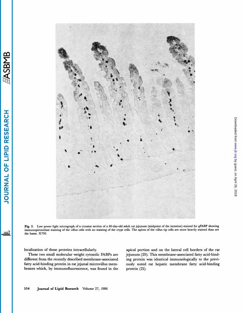
Fig. 9. Low power light micrograph of a cryostat section of a 60-day-old adult rat jejunum (midpoint of the intestine) stained for gFABP showing
the bases. X750. immunoperoxidase staining of the villus cells with no staining of the crypt cells. The apices of the villus tip cells are more heavily stained than are
localization of these proteins intracellularly. apical portion and on the lateral cell borders of the rat These two small molecular weight cytosolic FABPs are jejunum (20). This membrane-associated fatty acid-bind-
different from the recently described membrane-associated ing protein was identical immunologically to the pmvi- fatty acid-binding protein in rat jejunal microvillus mem- ously noted rat hepatic membrane fatty acid-binding branes which, by immunofluorescence, was found in the protein (21).
554 Journal of Lipid Research Volume 27, 1986
by guest, on April 26, 2018
ww
w.jlr.org
Dow
nloaded from
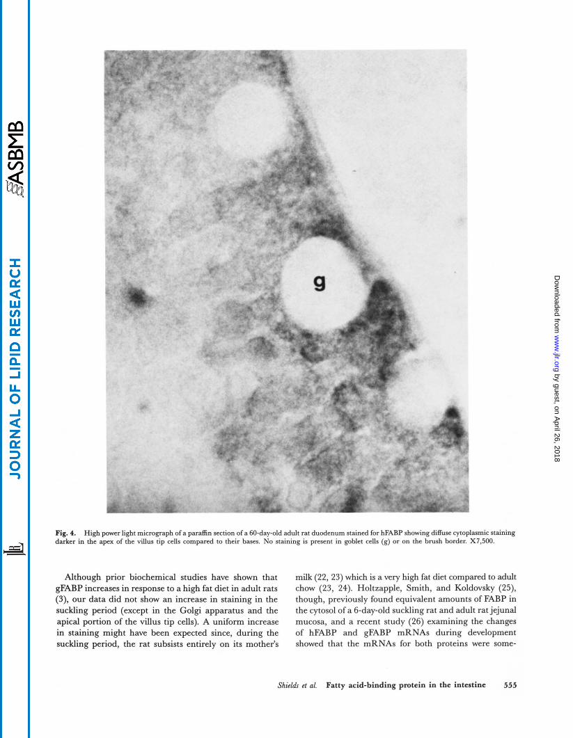
Fig. 4. High power light micrograph of a paraffin section of a 60-day-old adult rat duodenum stained for hFABP showing diffuse cytoplasmic staining darker in the apex of the villus tip cells compared to their bases. No staining is present in goblet cells (g) or on the brush border. X7.500.
Although prior biochemical studies have shown that milk (22,23) which is a very high fat diet compared to adult gFABP increases in response to a high fat diet in adult rats chow (23, 24). Holtzapple, Smith, and Koldovsky (25), (3), our data did not show an increase in staining in the though, previously found equivalent amounts of FABP in suckling period (except in the Golgi apparatus and the the cytosol of a 6-day-old suckling rat and adult rat jejunal apical portion of the villus tip cells). A uniform increase mucosa, and a recent study (26) examining the changes in staining might have been expected since, during the of hFABP and gFABP mRNAs during development suckling period, the rat subsists entirely on its mother’s showed that the mRNAs for both proteins were some-
Shields et 01. Fatty acid-binding protein in the intestine 555
by guest, on April 26, 2018
ww
w.jlr.org
Dow
nloaded from
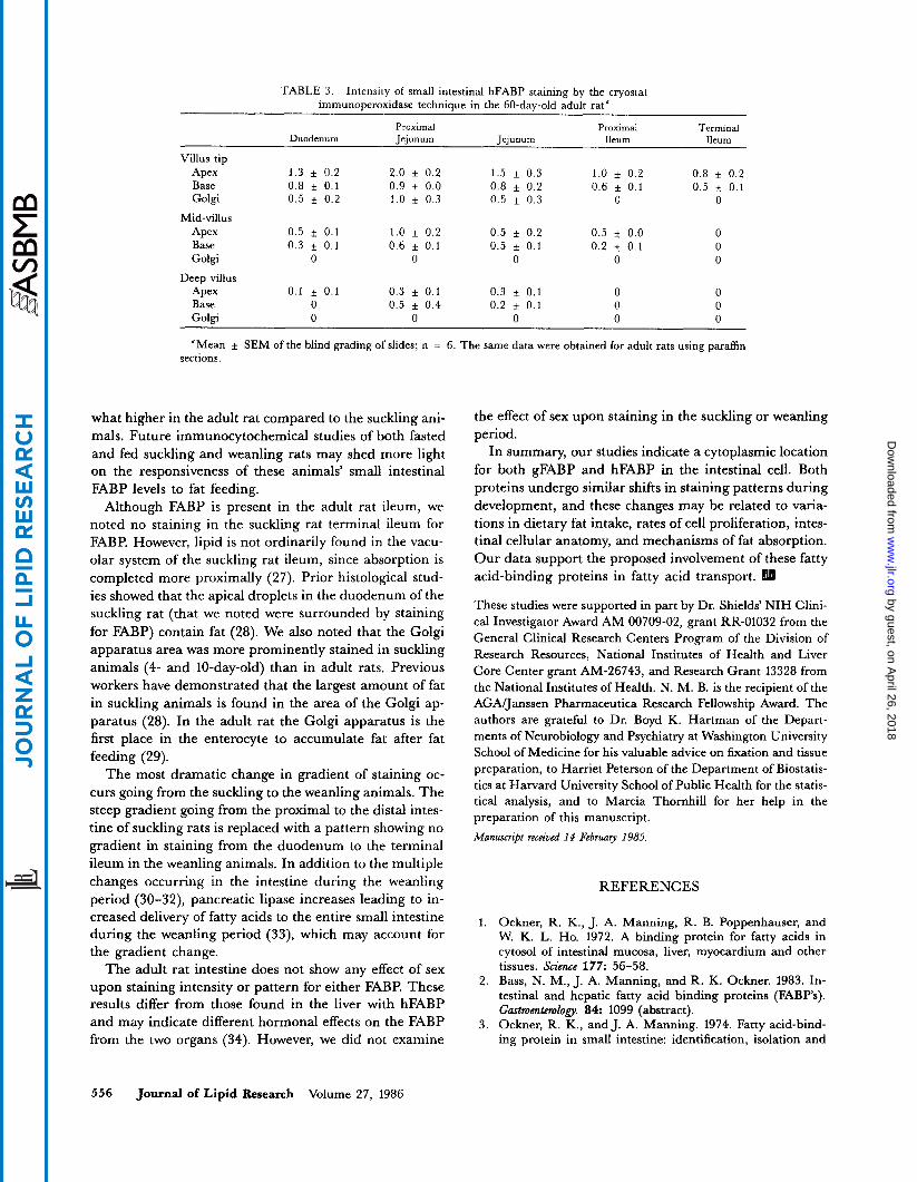
TABLE 3. Intensity of small intestinal hFABP staining by the cryostat immunoperoxidase technique in the 60-day-old adult rat a
~ ~~~~
Terminal Proximal Proximal Duodenum Jejunum Jejunum Ileum Ileum
Villus tip Apex 1 . 3 f 0.2 2.0 f 0.2 1.5 f 0.3 1.0 f 0.2 0.8 + 0.2 Base 0.8 0.1 0.9 f 0.0 0.8 f 0.2 0.6 + 0.1 0.5 f 0.1 Golgi 0.5 + 0.2 1.0 f 0.3 0.5 f 0.3 0 0
Apex 0.5 f 0.1 1.0 f 0.2 0.5 f 0.2 0.5 f 0.0 0 Base 0.3 + 0.1 0.6 f 0.1 0.5 f. 0.1 0.2 f 0.1 0 Golgi 0 0 0 0 0
Apex 0.1 f 0.1 0.3 f 0.1 0.3 f 0.1 0 0 Base 0 0.5 + 0.4 0.2 f 0.1 0 0 Golgi 0 0 0 0 0
Mid-villus
Deep villus
"Mean f SEM of the blind grading of slides; n = 6. The same data were obtained for adult rats using paraffin sections.
what higher in the adult rat compared to the suckling ani- mals. Future immunocytochemical studies of both fasted and fed suckling and weanling rats may shed more light on the responsiveness of these animals' small intestinal FABP levels to fat feeding.
Although FABP is present in the adult rat ileum, we noted no staining in the suckling rat terminal ileum for FABP. However, lipid is not ordinarily found in the vacu- olar system of the suckling rat ileum, since absorption is completed more proximally (27). Prior histological stud- ies showed that the apical droplets in the duodenum of the suckling rat (that we noted were surrounded by staining for FABP) contain fat (28). We also noted that the Golgi apparatus area was more prominently stained in suckling animals (4- and 10-day-old) than in adult rats. Previous workers have demonstrated that the largest amount of fat in suckling animals is found in the area of the Golgi ap- paratus (28). In the adult rat the Golgi apparatus is the first place in the enterocyte to accumulate fat after fat feeding (29).
The most dramatic change in gradient of staining oc- curs going from the suckling to the weanling animals. The steep gradient going from the proximal to the distal intes- tine of suckling rats is replaced with a pattern showing no gradient in staining from the duodenum to the terminal ileum in the weanling animals. In addition to the multiple changes occurring in the intestine during the weanling period (30-32), pancreatic lipase increases leading to in- creased delivery of fatty acids to the entire small intestine during the weanling period (33), which may account for the gradient change.
The adult rat intestine does not show any effect of sex upon staining intensity or pattern for either FABP. These results differ from those found in the liver with hFABP and may indicate different hormonal effects on the FABP from the two organs (34). However, we did not examine
the effect of sex upon staining in the suckling or weanling period.
In summary, our studies indicate a cytoplasmic location for both gFABP and hFABP in the intestinal cell. Both proteins undergo similar shifts in staining patterns during development, and these changes may be related to varia- tions in dietary fat intake, rates of cell proliferation, intes- tinal cellular anatomy, and mechanisms of fat absorption. Our data support the proposed involvement of these fatty acid-binding proteins in fatty acid transport.
These studies were supported in part by Dr. Shields' NIH Clini- cal Investigator Award AM 00709-02, grant RR-01032 from the General Clinical Research Centers Program of the Division of Research Resources, National Institutes of Health and Liver Core Center grant AM-26743, and Research Grant 13328 from the National Institutes of Health. N. M. B. is the recipient of the AGADanssen Pharmaceutica Research Fellowship Award. The authors are grateful to Dr. Boyd K. Hartman of the Depart- ments of Neurobiology and Psychiatry at Washington University School of Medicine for his valuable advice on fixation and tissue preparation, to Harriet Peterson of the Department of Biostatis- tics at Harvard University School of Public Health for the statis- tical analysis, and to Marcia Thomhill for her help in the preparation of this manuscript. Manuscript received 14 February 1985.
REFERENCES
1. Ockner, R. K., J. A. Manning, R. B. Poppenhauser, and W. K. L. Ho. 1972. A binding protein for fatty acids in cytosol of intestinal mucosa, liver, myocardium and other tissues. Science 177: 56-58.
2. Bass, N. M., J. A. Manning, and R. K. Ockner. 1983. In- testinal and hepatic fatty acid binding proteins (FABPs). Gmtmmtemlofi. 84: 1099 (abstract).
3. Ockner, R. K., and J. A. Manning. 1974. Fatty acid-bind- ing protein in small intestine: identification, isolation and
556 Journal of Lipid Research Volume 27, 1986
by guest, on April 26, 2018
ww
w.jlr.org
Dow
nloaded from
evidence for its role in cellular fatty acid transport. J. Clin. Invest. 54: 326-338.
4. Ockner, R. K., J. A. Manning, and J. P. Kane. 1982. Fatty acid-binding protein: isolation from rat liver, characteriza- tion and immunochemical quantification. J. Biol. Chem.
5. Alpen, D. H., A. W. Strauss, R. K. Ockner, N. M. Bass, and J. I. Gordon. 1984. Cloning of a cDNA encoding rat intestinal fatty acid-binding protein. Pmc. Natl. Acad. Sci.
6. Gordon, J. I., D. H. Alpers, R. K. Ockner, and A. W. Strauss. 1983. The nucleotide sequence of rat liver fatty acid binding protein mRNA.J. Biol. Chem. 258: 3356-3363.
7. Mishkin, S., L. Stein, Z. Gatmaitan, and I. M. Arias. 1972. The binding of fatty acids to cytoplasmic proteins: binding to Z protein in liver and other tissues of the rat. Biochem. Biokbs. Res. Commun. 47: 997-1003.
8. Billheimer, J. T., and J. L. Gaylor. 1980. Cytosolic modula- tors of activities of microsomal enzymes of cholesterol bio- synthesis: role of a cytosolic protein with properties similar to Z protein (fatty acid-binding protein).J. Biol. Chem. 255:
9. Dempsey, M. E., K. E. McCoy, H. N. Baker, A. Dimitri- adou-Vafiadou, T. Lorsbach, and J. B. Howard. 1981. Large scale purification and structural characterization of squalene and sterol carrier protein. J. Biol. Chem. 256: 1867-1873.
10. Gordon, J. I., D. P. Smith, D. H. Alpers, and A. W. Strauss. 1982. Cloning of a complementary deoxyribonucleic acid encoding a portion of rat intestinal preapolipoprotein A-IV messenger ribonucleic acid. Biochemistry. 21: 5424-5431. Ockner, R. K., and J. A. Manning. 1976. Fatty acid-bind- ing protein. Role in esterification of absorbed long chain fatty acid in rat intestine. J. Clin. Invest. 58: 632-641.
12. Gangl, A., and R. K. Ockner. 1975. Intestinal metabolism of plasma free fatty acids. Intracellular compartmentation and mechanisms of control. J. Clin. Invest. 55: 803-813.
13. Capron, F., B. Coltoff-Schiller, A. B. Johnson, G. M. Fleischner, and S. Goldfischer. 1979. Immunocytochemical localization of hepatic ligandin and Z protein utilizing frozen sections for light and electron microscopy. J. Histo-
14. Sternberger, L. A. 1979. Immunocytochemistry. 2nd ed. John Wiley and Sons, New York. 122-127.
15. Hsu, S. M., L. Raine, and H. Fanger. 1981. Use of avidin- biotin-peroxidase complex (ABC) in immunoperoxidase techniques: a comparison between ABC and unlabeled antibody (PAP) procedures. J. Histochem. Cytochem. 29:
16. Lev, R., and W. C. Griffiths. 1982. Colonic and small intes- tinal alkaline phosphatase: a histochemical and biochemical study. Gastmentedogy. 82: 1427-1435.
17. Shields, H. M., F. A. Bair, M. L. Bates, S. T. Yedlin, and D. H. Alpers. 1982. Localization of immunoreactive alka- line phosphatase in the rat small intestine at the light micro- scopic level by immunocytochemistry. Gastmentemlogy. 82: 39-45.
18. Shields, H. M., M. L. Bates, S. T. Yedlin, and C. J. Best. 1984. The distribution of immunoreactive alkaline phospha- tase in the adult rat ileum by immunoperoxidase staining
257: 7872-7878.
USA. 81: 313-317.
8128-8135.
11.
chem. Cytochem. 27: 961-966.
577-580.
19.
20.
21.
22.
23.
24.
25.
26.
27.
28.
29.
30.
31.
32.
33.
34.
at the light microscope level. Gastmentmlopy. 87: 827-835. Barr, A. J., J. H. Goodnight, and J. P. Sall. 1976. Users Guide to SAS 76. SAS, Inc., Raleigh, NC. Stremmel, W., S. Kochwa, and P. D. Berk. 1983. Studies of oleate binding to rat liver plasma membranes. Biochem. Bio- phys. Res. Commun. 112: 88-95. Stremmel, W., G. Lotz, G. Strohmeyer, and P. D. Berk. 1985. Identification, isolation, and partial characterization of a fatty acid-binding protein from rat jejunal microvillus membranes. J. Clin. Invest. 75: 1068-1076. Fernando-Warnakulasuriya, G. J. P., J. E. Staggers, S. C. Frost, and M. A. Wells. 1981. Studies on fat digestion, ab- sorption, and transport in the suckling rat. I. Fatty acid composition and concentrations of major lipid components. J. Lipid Res. 22: 668-674. Staggers, J. E., G. J. P. Fernando-Warnakulasuriya, and M. A. Wells. 1981. Studies on fat digestion, absorption, and transport in the suckling rat. 11. Triacylglycerols: molecular species, stereospecific analysis, and specificity of hydrolysis by lingual lipase. J. Lipid Res. 22: 675-679. Aw, T. Y., and M. R. Grigor. 1980. Digestion and absorp- tion of milk triacylglycerols in 14-day-old suckling rats. J.
Holtzapple, P. G., G. Smith, and 0. Koldovsky. 1975. Up- take, activation, and esterification of fatty acids in the small intestine of the suckling rat. F'edktr. Res. 9: 786-791. Gordon, J. I., N. Elshourbagy, J. B. Lowe, W. S. Liao, D. H. Alpers, and J. M. Taylor. 1985. Tissue specific ex- pression and developmental regulation of two genes coding for rat fatty acid binding proteins. J. Biol. Chem. 260:
Cornell, R., and H. A. Padykula. 1969. A cytological study of intestinal absorption in the suckling rat. Am. J. Anat.
Hahn, P., and 0. Koldovsky. 1966. Utilization of Nutrients during Postnatal Development. Pergamon Press, New York.
Marenus, K. D., and E S. Sjostrand. 1982. Sequence of structural changes in columnar epithelium of small intestine during early stages of fat absorption. J. Ultrastruct. Res. 79:
Herbst, J. J., and P. Sunshine. 1969. Postnatal development of the small intestine of the rat: changes in mucosal mor- phology at weaning. Pediatr. Res. 3: 27-33. Lee, P. C., and E. Lebenthal. 1983. Early weanling and pre- cocious development of small intestine in rats: genetic, di- etary or hormonal control. Pediatr. Res. 17: 645-650. Koldovsky, O., P. Sunshine, and N. Kretchmer. 1966. Cellu- lar migration of intestinal epithelia in suckling and weaned rats. Nature. 212: 1389-1390. Rokos, J., P. Hahn, 0. Koldovsky, and P. Prochazka. 1963. The postnatal development of lipolytic activity in the pan- creas and small intestine of the rat. Physiol. Bohemoslov. 12:
Bass, N. M., J. A. Manning, R. K. Ockner, J. I. Gordon, S. Seetharam, and D. H. Alpers. 1985. Regulation of the biosynthesis of two distinct fatty acid-binding proteins in rat liver and intestine: influences of sex difference and of clofibrate. J. Biol. Chem. 260: 1432-1436.
Nutr. 110: 2133-2140.
1995-1998.
125: 291-316.
84-96.
92-109.
213-219.
Shields et al. Fatty acid-binding protein in the intestine 557
by guest, on April 26, 2018
ww
w.jlr.org
Dow
nloaded from