linear and nonlinear rheology of living cells -...
TRANSCRIPT

MR41CH04-Fabry ARI 3 June 2011 7:57
Linear and Nonlinear Rheologyof Living CellsPhilip Kollmannsberger1,2 and Ben Fabry1
1Department of Physics, University of Erlangen-Nuremberg, 91052 Erlangen, Germany;email: [email protected] of Biomaterials, Max Planck Institute of Colloids and Interfaces, 14424 Potsdam,Germany; email: [email protected]
Annu. Rev. Mater. Res. 2011. 41:75–97
First published online as a Review in Advance onMarch 8, 2011
The Annual Review of Materials Research is online atmatsci.annualreviews.org
This article’s doi:10.1146/annurev-matsci-062910-100351
Copyright c© 2011 by Annual Reviews.All rights reserved
1531-7331/11/0804-0075$20.00
Keywords
cell mechanics, cytoskeleton, microrheology, soft glassy rheology,contractile prestress, stress stiffening
Abstract
Living cells are an active soft material with fascinating mechanical prop-erties. Under mechanical loading, cells exhibit creep and stress relaxationbehavior that follows a power-law response rather than a classical exponen-tial response. Such a response puts cells in the context of soft colloidal glassesand other disordered metastable materials that share the same properties. Incells, however, both the power-law exponent and stiffness are related to thecontractile prestress in the cytoskeleton. In addition, cells are made of ahighly nonlinear material that stiffens and fluidizes under mechanical stress.They show active and adaptive mechanical behavior such as contraction andremodeling that sets them apart from any other nonliving material. Strik-ingly, all these observations can be linked by simple relationships with thepower-law exponent as the only organizing parameter. Current theoreticalmodels capture specific facets of cell mechanical behavior, but a comprehen-sive understanding is still emerging.
75
Ann
u. R
ev. M
ater
. Res
. 201
1.41
:75-
97. D
ownl
oade
d fr
om w
ww
.ann
ualr
evie
ws.
org
Acc
ess
prov
ided
by
WIB
6276
- U
nive
rsita
tsbi
blio
thek
Erl
ange
n-N
uern
berg
on
04/1
4/15
. For
per
sona
l use
onl
y.

MR41CH04-Fabry ARI 3 June 2011 7:57
Cytoskeleton: adynamic network ofprotein filamentsspanning the entirecell body; it maintainsthe cell’s shape andmechanical function
Prestress: permanentmechanical stress,either internallygenerated byactomyosincontraction orexternally applied, infilament networks suchas the cytoskeleton
1. THE CELL AS A MATERIAL
1.1. Structure and Dynamics of the Cell
Eukaryotic cells are highly complex structures consisting of a large number of different proteins andother constituents. Mechanically, they are stabilized by the cytoskeleton, a contractile filamentousnetwork that spans the entire cell body. In humans, several thousand different proteins formand regulate the cytoskeleton (1). Together, these proteins assemble into a structure of stunningcomplexity in a process that is far from thermodynamic equilibrium. To connect two proteins toeach other, the cell spends approximately one molecule of high-energy phosphate such as ATP orGTP. It is therefore even more remarkable that this intricate cytoskeletal structure, which coststhe cell a large part of its metabolic energy, is constantly destroyed and then reassembled over andover again. This ongoing remodeling makes the cytoskeleton highly versatile and allows the cell tochange shape, to move, to contract, to divide, to merge, and to engulf other objects. It is thereforenot so much the structural architecture but rather the dynamic behavior of the cytoskeleton thatmakes the cell fundamentally different from any other nonliving material. But is it then possibleor even sensible to treat living cells just like an ordinary inert material and to characterize themin terms of stresses, strains, and deformations? And what can we learn from such a mechanicalcharacterization?
1.2. Cytoskeleton: A Primer
The most abundant proteins in cells of all tissue origin are those that form the cytoskeleton, suchas actin and myosin. The cytoskeleton of nucleated (eukaryotic) cells consists of three distinctcomponents: the actin-myosin network, the microtubule network, and the intermediate-filamentnetwork (2).
The actin-myosin network is generally located at the cortex and outer edges close to the cellmembrane. It is mechanically linked to the adhesive contacts that anchor the cell to the extracellularsubstrate. These contacts counterbalance the contractile forces built up by the active myosinmotors in the network, leading to a constant prestress in the cytoskeleton. In highly contractilecells, the actin and myosin filaments can align and form stress fibers that closely resemble musclefibrils. These stress fibers are not confined to the cortex but can span the entire cell body. Apartfrom myosin, there are a large number of other, passive actin cross-linkers with widely varyingproperties. Such cross-linkers turn the actin-myosin network into a highly versatile structure thatcan fulfill a variety of functions, such as forming different types of protrusions.
Microtubules are rather stiff filaments that extend from a common origin (centrosome) locatednear the cell nucleus. They serve as transport pathways for intracellular cargo and play an importantrole in defining cell polarity and in orienting the chromosomes during mitosis. In most adherentcells, however, they play only a minor mechanical role in stabilizing the cytoskeleton and balancingstress fluctuations (3).
Intermediate filaments consist of a large, tissue-specific group of proteins. Examples includekeratin in epithelial cells, vimentin in mesenchymal cells, internexin in neuronal cells, and desminin muscle cells. They start to significantly contribute to the overall mechanical response of the cellduring large cell deformation, when intermediate filaments become fully extended and stretched(4–6).
All these three components of the cytoskeleton are highly connected to each other, to the nu-cleus, and to the cell membrane (7–10). Their individual contribution to cell mechanical behaviorcan therefore not easily be separated. This needs to be taken into account when comparing cellmechanical studies with experiments on reconstituted cytoskeletal networks.
76 Kollmannsberger · Fabry
Ann
u. R
ev. M
ater
. Res
. 201
1.41
:75-
97. D
ownl
oade
d fr
om w
ww
.ann
ualr
evie
ws.
org
Acc
ess
prov
ided
by
WIB
6276
- U
nive
rsita
tsbi
blio
thek
Erl
ange
n-N
uern
berg
on
04/1
4/15
. For
per
sona
l use
onl
y.

MR41CH04-Fabry ARI 3 June 2011 7:57
Microrheology:measures themicroscopicmechanical propertiesof soft materials bytracking the movementof embeddedmicrometer-sizedparticles in response tothermal or nonthermalforces
2. MECHANICAL BEHAVIOR OF CELLS
2.1. Methods for Measuring Cell Mechanics
Rheometers operate by applying a defined mechanical stress to the material and by measuring theresulting deformations or, vice versa, by measuring the mechanical stress necessary to achieve adesired deformation. Because of the exceedingly low stiffness and small size of cells, the mechanicalforces and deformations are very low, in the range of pico-Newtons and nanometers, respectively.Precise quantitative mechanical measurements on single, living cells became possible only withthe development of modern microrheological techniques in recent decades such as magnetictweezers, laser tweezers, atomic force microscopy, cell poking, microplates, and cell stretchers.Nonetheless, in 1922 Heilbronn (11), using an early version of magnetic tweezers, reported thefirst microrheological studies of the cytoplasm. Francis Crick (of DNA fame) & Arthur Hughes(12) improved on this method in the late 1940s. Despite experimental limitations, they observed
1012
1011
1010
109
108
107
106
105
104
103
102
Diamond
Steel
Window glass
BoneWood (oak)
Actin
Polyethylene
Soft rubberFoamed polymers
Cells
Youn
g's
mod
ulus
(Pa)
Volumefraction(0.1–1%)
Filament bending;stretching of cross-linkers
Dynamics
Figure 1(Left) Young’s modulus of some common materials. Cells are exceedingly soft, with a Young’s modulus manyorders of magnitude lower than that of most common materials. Starting with the high stiffness of actin, themain protein constituent of cells, several mechanisms work together to lower the stiffness down to a levelcomparable to that of soft pastes, slurries, or weak gels. (Right) Actin cytoskeleton of an NIH-3T3 fibroblastcell.
www.annualreviews.org • Linear and Nonlinear Rheology of Living Cells 77
Ann
u. R
ev. M
ater
. Res
. 201
1.41
:75-
97. D
ownl
oade
d fr
om w
ww
.ann
ualr
evie
ws.
org
Acc
ess
prov
ided
by
WIB
6276
- U
nive
rsita
tsbi
blio
thek
Erl
ange
n-N
uern
berg
on
04/1
4/15
. For
per
sona
l use
onl
y.

MR41CH04-Fabry ARI 3 June 2011 7:57
the full range of the irritatingly rich and complex viscoelastic behavior of cells, including plasticityand shear thinning, and reported stiffness values of the cell interior in the range of 2 to 50 Pa.
Why are cells so soft? The filamentous network of the cytoskeleton, when viewed in isolation,is actually made of rather strong materials. Intermediate filaments, for example, keratin, which isthe main component of wool, have a stiffness (1–5 GPa) and tensile strength (0.2 GPa) that makethem a useful material for many technical applications (2). The cytoskeletal protein actin, the mostabundant intracellular protein, has a Young’s modulus in the range of 1–2 GPa (2). The cell as amaterial, however, is approximately six orders of magnitude softer (Figure 1) for the same reasonas why a wool jumper is soft to the touch: The volume fraction of the keratin fibers is low, and theindividual fibers undergo bending rather than stretching deformations. In cells, the metastability,or dynamics, of the fiber network reduces the overall stiffness further still.
From the measured value of cell stiffness, however, it is impossible to estimate the relative con-tributions of volume fraction, filament bending, cross-linker stretching, and dynamic remodelingmechanisms. The concept of cell stiffness as a single number neglects, or rather averages over,any inhomogeneity, granularity, anisotropy, nonlinearity, and time fluctuations. Moreover, theparticularities of this averaging depend on the probe, e.g., its shape, size, surface functionalization,contact time with the cell, or position relative to the cell body. The averaging further depends onthe applied force or deformation magnitude and on the time or frequency range over which themeasurement is performed. Different measurement techniques therefore measure different facetsof cell rheology, and each has particular characteristics, advantages, and limitations (Table 1).
Large range of frequencies (0.01–1,000 Hz)
Parallel measurements of ~100 cells possible
High time resolution of stiffness changes (~1 s)
Large range of forces (up to ~100 nN)
Good measurement throughput (30 cells h–1)
High time resolution of stiffness changes(~1 s)
Good measurement throughput (30 cells h–1)
Large range of forces (up to ~1 °N)
Control of cellular prestress
Quantitative measurements of shear moduluspossible
High frequency (up to 100 kHz)
Maximum specific torque<140 Pa
Only for unidirectionalforces
No frequency modulation(lock-in) possible
Low throughput
No subcellular resolution
Only for soft materials(G << 100 Pa)
44, 55, 64
25, 102
14, 103
35, 63, 65
48, 104
Magnetic twistingcytometry withoptical detectionof bead movements
Magnetic tweezers
Optical tweezers
Microplaterheometer
Particle trackingmicrorheology
High spatial resolution (10 nm, depending ontip geometry)
Quantitative measurements of shear moduluspossible
High time resolution of stiffness changes (~1 s)
Large range of forces (up to ~100 nN)
•
•
•
•
•
•
•
•
•
•
•
•
•
•
•
Low scanning speed
Low throughput
•
•
•
Maximum forces <500 pN
Heating caused by lasertraps
•
•
•
•
•
•
•
51, 100, 101Atomic forcemicroscopy
AdvantagesMethod Limitations References
Table 1 Comparison of common methods to measure cell rheology
78 Kollmannsberger · Fabry
Ann
u. R
ev. M
ater
. Res
. 201
1.41
:75-
97. D
ownl
oade
d fr
om w
ww
.ann
ualr
evie
ws.
org
Acc
ess
prov
ided
by
WIB
6276
- U
nive
rsita
tsbi
blio
thek
Erl
ange
n-N
uern
berg
on
04/1
4/15
. For
per
sona
l use
onl
y.

MR41CH04-Fabry ARI 3 June 2011 7:57
Moreover, whether by ligation and/or mechanotransduction, the interaction of a probe (such asan AFM tip, ligand-coated bead, or microplate) with the cell unavoidably induces local remodelingevents that alter the structure being probed (13–20). This remodeling is a principal feature of allapproaches in which an external probe is used. Other limitations of bead-based methods, except forthe cases in which a bead is glued onto an AFM tip, are that the position at which the bead settlesonto the cell surface cannot be controlled (although it can be measured) (21) and that the geometryof the bead-cell interaction is also not controlled (although, again, it can be measured) (22).
As with all the available methods for measuring cell mechanics, with the exception of two-pointparticle tracking microrheology (23, 24), a length scale must be invoked to convert raw displace-ment data into cellular strain and subsequently into a proper elastic modulus. The intracellularstrain distribution can be analytically derived (24, 25), computed using a finite element model ofcell deformation (26), or estimated from simple dimensional arguments (27), all under the assump-tion that the cell is a homogeneous, isotropic, and mostly linear elastic material. Even under thesesimplifying assumptions, the strain and stress field within the cell close to a bead or AFM tip ishighly complicated (26, 28) but is predicted to decay rapidly with larger distances from the probe.In practice, however, one finds in violation with continuum mechanics predictions that locallyapplied stresses can be focused and transmitted at great lengths through the entire cell body viastress fibers (28).
From all these considerations, one might conclude that quantitative cell mechanical measure-ments are a pointless endeavor; indeed, reported values of elastic moduli measured by differenttechniques vary by more than one order of magnitude, and this scatter in the available data onlyincreases when different cell types are compared (29, 30). The current situation, however, looksmuch more positive. Several recent experimental investigations have revealed a set of cell me-chanical properties that can be reproduced consistently and compared between various probingtechniques and even across different cell types (29, 31, 32). Such universal aspects of cell mechanicsare the basis for understanding the complex cell rheology from a physical point of view (6, 29, 32)and are discussed below.
2.2. Linear Viscoelasticity: Springs and Dashpots
Depending on the experimental approach and the timescales probed, cells show both solid-likeelastic and fluid-like viscous properties. This behavior is termed viscoelastic. In engineering,viscoelastic materials are traditionally described by mechanical equivalent circuits of connectedHookean elastic springs and Newtonian viscous dashpots. Any arbitrary linear viscoelastic behaviorcan be modeled with networks of springs and dashpots arranged in series or in parallel.
The aim of the spring-dashpot approach is that the different elements of the mechanical equiv-alent circuit can be ascribed to different structural elements. In the case of cells, different elasticand viscous elements were thought to reflect the membrane, actin cortex, deep cytoskeleton, andso on (25). Due to the limited resolution of early experimental results on viscoelastic creep andstress relaxation in cells, simple spring-dashpot models with only one time constant were sufficientto fit the data (33–35). With the development of more sophisticated experimental techniques, theaccessible ranges of time and frequency and the resolution of the obtained data were increased,which made it necessary to introduce more complex models of cell mechanics (25, 36).
The problem with this approach is that larger numbers of model parameters make it ambiguousor outright impossible to equate the spring-dashpot elements with real cell components. Moreover,as we see below, basic underlying assumptions such as linearity or the decoupling of elastic andviscous properties are not correct. Additional components such as nonlinear plastic elements wereintroduced to explain more complex behavior such as plasticity (36), but the increasing number of
www.annualreviews.org • Linear and Nonlinear Rheology of Living Cells 79
Ann
u. R
ev. M
ater
. Res
. 201
1.41
:75-
97. D
ownl
oade
d fr
om w
ww
.ann
ualr
evie
ws.
org
Acc
ess
prov
ided
by
WIB
6276
- U
nive
rsita
tsbi
blio
thek
Erl
ange
n-N
uern
berg
on
04/1
4/15
. For
per
sona
l use
onl
y.

MR41CH04-Fabry ARI 3 June 2011 7:57
Time (s)
Stre
ss (n
orm
aliz
ed)
100 101 102 10310–110–210–3
100
10–1
10–2
10–3
τ = 0.01 s τ = 0.1 s τ = 1 s
τ = 10 s
τ = 100 s
Figure 2Stress relaxation. A Maxwell body (the combination of an elastic spring with elasticity k in series with aviscous dissipative dashpot element with viscosity ν, as shown at left) shows an exponential relaxation ofmechanical stress after a step increase in mechanical strain (red curves). The ratio ν/k defines a characteristictimescale τ after which a dominant fraction of the initial stress has decayed. A power-law decay of stressescan be reasonably well fitted with a combination of Maxwell bodies (blue curve) when, for each decade intime, an additional spring-dashpot element is added in parallel to the other elements. The fitted values of τ ,the spring elasticities, and dashpot viscosities, however, are determined solely by the timescale of theexperiment and therefore have no meaning and no relationship with the state of the cell (37).
model fit parameters necessarily decreases the confidence of the actual fit. As explained in the nextsection, traditional viscoelastic models of the cell, except for very special cases, are not mechanisticand have no physical meaning.
2.3. Power-Law Rheology
As the time and frequency resolution of microrheology became better, researchers set out to recorda broader viscoelastic spectrum and to find precise time constants describing cell mechanics.Surprisingly, the viscoelastic spectrum of living cells lacks any distinct timescales that can beidentified with discrete structural elements or processes. The number of spring-dashpot elementsneeded to fit the data, and their specific values, depends only on the scale of the measurement timeor frequency and for that reason cannot have any physical meaning (Figure 2) (37). Instead, theviscoelastic behavior of cells can be described by a power law with a single exponent over manyorders of magnitude of time or frequency.
Power-law stress relaxation of biological samples was described in the first part of the nineteenthcentury (38–40). With the development of linear viscoelasticity, however, the description of stressrelaxation by power laws (41) was discarded in favor of mechanistically more intuitive spring-dashpot equivalent circuits with exponential relaxation behavior (42). More than 100 years later,Hildebrandt (43) finally discovered the simplicity and accuracy of power laws for describing tissuebiomechanics.
Power-law behavior can best be explained by a creep experiment in which the material ismechanically loaded at time t = 0 with a constant force F, and the material deformation d versustime t is recorded. The ratio d(t)/F defines the creep function J(t):
J(t) = d (t)/F = j0 · (t/τ0)β . 1.
80 Kollmannsberger · Fabry
Ann
u. R
ev. M
ater
. Res
. 201
1.41
:75-
97. D
ownl
oade
d fr
om w
ww
.ann
ualr
evie
ws.
org
Acc
ess
prov
ided
by
WIB
6276
- U
nive
rsita
tsbi
blio
thek
Erl
ange
n-N
uern
berg
on
04/1
4/15
. For
per
sona
l use
onl
y.
MR41CH04-Fabry ARI 3 June 2011 7:57
Elastic
Viscous
0 5 10 15 20
0
0.2
0.4
0.6
0.8
1
1.2
Time (s)
d(t)
/dm
ax
PAA (β = 0)
PDMS (β = 1)Cell (β ≈ 0.3)
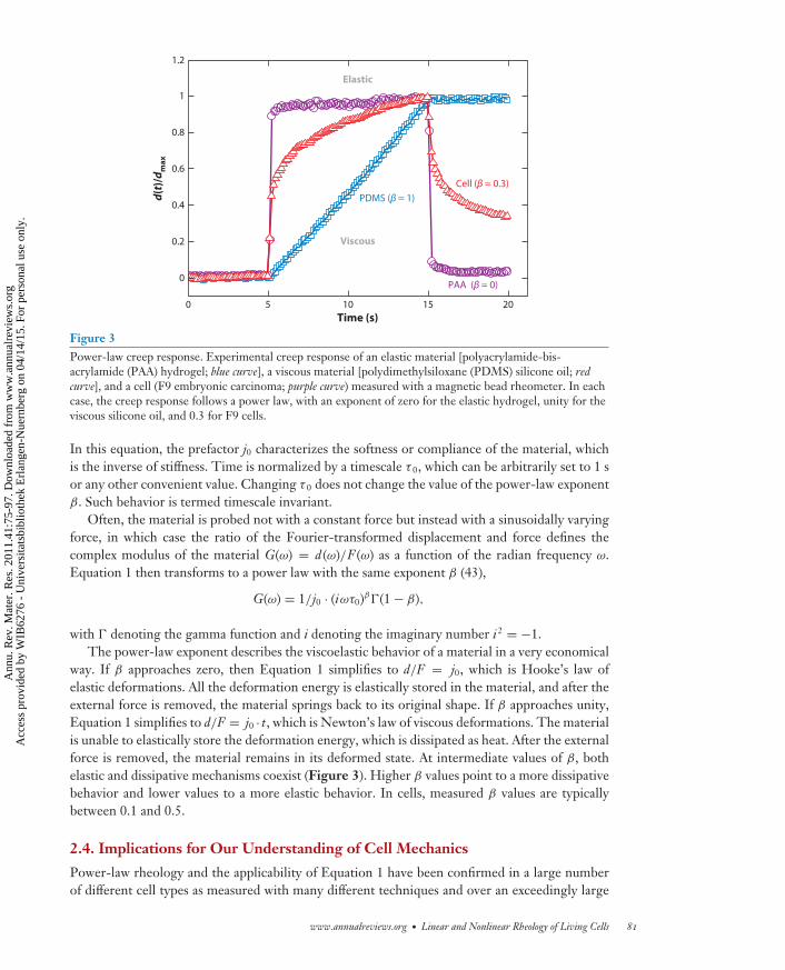
Figure 3Power-law creep response. Experimental creep response of an elastic material [polyacrylamide-bis-acrylamide (PAA) hydrogel; blue curve], a viscous material [polydimethylsiloxane (PDMS) silicone oil; redcurve], and a cell (F9 embryonic carcinoma; purple curve) measured with a magnetic bead rheometer. In eachcase, the creep response follows a power law, with an exponent of zero for the elastic hydrogel, unity for theviscous silicone oil, and 0.3 for F9 cells.
In this equation, the prefactor j0 characterizes the softness or compliance of the material, whichis the inverse of stiffness. Time is normalized by a timescale τ 0, which can be arbitrarily set to 1 sor any other convenient value. Changing τ 0 does not change the value of the power-law exponentβ. Such behavior is termed timescale invariant.
Often, the material is probed not with a constant force but instead with a sinusoidally varyingforce, in which case the ratio of the Fourier-transformed displacement and force defines thecomplex modulus of the material G(ω) = d (ω)/F (ω) as a function of the radian frequency ω.Equation 1 then transforms to a power law with the same exponent β (43),
G(ω) = 1/j0 · (iωτ0)β�(1 − β),
with � denoting the gamma function and i denoting the imaginary number i2 = −1.The power-law exponent describes the viscoelastic behavior of a material in a very economical
way. If β approaches zero, then Equation 1 simplifies to d/F = j0, which is Hooke’s law ofelastic deformations. All the deformation energy is elastically stored in the material, and after theexternal force is removed, the material springs back to its original shape. If β approaches unity,Equation 1 simplifies to d/F = j0 · t, which is Newton’s law of viscous deformations. The materialis unable to elastically store the deformation energy, which is dissipated as heat. After the externalforce is removed, the material remains in its deformed state. At intermediate values of β, bothelastic and dissipative mechanisms coexist (Figure 3). Higher β values point to a more dissipativebehavior and lower values to a more elastic behavior. In cells, measured β values are typicallybetween 0.1 and 0.5.
2.4. Implications for Our Understanding of Cell Mechanics
Power-law rheology and the applicability of Equation 1 have been confirmed in a large numberof different cell types as measured with many different techniques and over an exceedingly large
www.annualreviews.org • Linear and Nonlinear Rheology of Living Cells 81
Ann
u. R
ev. M
ater
. Res
. 201
1.41
:75-
97. D
ownl
oade
d fr
om w
ww
.ann
ualr
evie
ws.
org
Acc
ess
prov
ided
by
WIB
6276
- U
nive
rsita
tsbi
blio
thek
Erl
ange
n-N
uern
berg
on
04/1
4/15
. For
per
sona
l use
onl
y.

MR41CH04-Fabry ARI 3 June 2011 7:57
range of timescales or frequencies (Figure 4) (31, 44). Scale-free power-law rheology appears to bea universal property of adherent cells and holds even when the cells are treated with an exhaustiverange of cytoskeletally active drugs (45). Considering this universality, Equation 1 is a remarkablysimple empirical relationship. It captures the essence of the data with only a single parameter: thepower-law exponent β. (We show below that the parameters j0 and τ 0 are not free-fit parametersbut are constants for a given system such as a cell line.)
This behavior may at first glance appear somewhat disappointing because if no characteristictimescale is evident, it is not possible to identify a dominating relaxation process. If all imaginablepharmacological interventions or differences between different cell types can be characterizedby changes in the power-law exponent β alone, then obviously β cannot explain a molecularmechanism. But what does it explain?
A possible answer comes—surprisingly—from a theory of soft glassy materials (SGMs) (46).SGMs are a diverse group of substances that includes foams, pastes, colloids, emulsions, andslurries. These substances are very soft (in the range of pascals to kilopascals) and are scale freeaccording to Equation 1, with power-law exponents on the order of 0.1. Because these materialsare so diverse, it has been reasoned that the common rheological features must be not so much areflection of specific molecular mechanisms as a reflection of generic systems properties at the level
Scal
ed e
last
ic m
odul
us (K
'/Ks)
ω (rad s–1)101 10310–110–3 10510–5
Cells analyzed by OMTC (Reference 44)
Cells analyzed by OMTC (Reference 31)
Cells analyzed by MT (Reference 110)
Cells analyzed by AFM (Reference 101)
Cells analyzed by AFM (Reference 100)
Cells analyzed by MPR (Reference 63)
Cells analyzed by OT (Reference 103)
Cells analyzed by PTM (Reference 48)
Cells analyzed by PTM (Reference 31)
Renal cortex (Reference 111)
Hepatic tissue (Reference 111)
Cell monolayer (Reference 29)
Adipose tissue (Reference 112)
Actin-FLNa-myosin (Reference 69)
Actin-biotin-myosin (Reference 68)
Actin-FLNa (Reference 62)
Figure 4Universality of power-law responses in biological materials. The frequency response of cells (blue) follows a weak power law overseveral decades of frequency, regardless of cell type or experimental technique. Cytoskeletal networks under active or passive stress, cellensembles, and soft tissues also display a power-law frequency. The absolute values of the differential elastic modulus K ′ measured at afixed prestress vary according to specimen type and experimental technique and are difficult to compare between different studies. Forclarity, the curves have been shifted in the y direction to emphasize the similarity of the frequency dependency. In some cases, thereported time-dependent responses were Laplace transformed into the frequency domain. Abbreviations: AFM, atomic forcemicroscopy; FLNa, filamin A; MPR, microplate rheometer; MT, magnetic tweezers; OMTC, optical magnetic twisting cytometry;OT, optical tweezers; PTM, particle tracking microrheology.
82 Kollmannsberger · Fabry
Ann
u. R
ev. M
ater
. Res
. 201
1.41
:75-
97. D
ownl
oade
d fr
om w
ww
.ann
ualr
evie
ws.
org
Acc
ess
prov
ided
by
WIB
6276
- U
nive
rsita
tsbi
blio
thek
Erl
ange
n-N
uern
berg
on
04/1
4/15
. For
per
sona
l use
onl
y.

MR41CH04-Fabry ARI 3 June 2011 7:57
Soft glassy rheology(SGR): a concept toexplain glassy behaviorand power-lawrheology in softdisordered materialssuch as foams andcolloids
of structural organization (46). All SGMs are composed of elements that are discrete, numerous,and aggregated with one another via weak interactions. In addition, these materials exist far awayfrom thermodynamic equilibrium and are arrayed in a microstructural geometry that is inherentlydisordered and metastable. The data presented above establish that living cells satisfy all thesecriteria and features, and it has been proposed that they be added to the list of SGMs (44).
Soft glassy rheology (SGR) theory considers that each individual element of the matrix existswithin an energy landscape containing many wells, or traps, of differing depth E. In the case ofliving cells, those traps might plausibly be thought of as being formed by binding energies betweenneighboring cytoskeletal proteins through hydrophilic interactions, charge effects, or simple stericconstraints. Each energy well is regarded as being so deep that the elements are unlikely to escapethe well by thermal fluctuations alone. Instead, elements are imagined to be agitated by someother energy source (46). In cells, ATP-dependent mechanisms are thought to be the cause ofsuch nonthermal agitation (47). The magnitude of this agitation is represented by an effectivetemperature, or noise level, x.
For a soft glass to deform elastically, its elements must remain in energy wells; to flow, theelements must hop out of these wells. Once an element has escaped its energy well, all its elasticallystored deformation energy is dissipated as heat. Dissipation is therefore intrinsically linked to anelastic stress, not to a viscous stress, and the locus of both friction and elasticity lies within thesame elements and their binding energies and cannot be attributed to distinct components (32).
With increasing effective temperature x, the elements more frequently manage to hop out oftheir well only to fall into another. The hop can be thought of as the fundamental molecularremodeling event and the origin of fluid-like behavior. An increase in x causes the system to melt,and a decrease in x causes the system to freeze. If x tends toward unity, the elements become trappedin deeper and deeper wells; all hopping events abate, and the system approaches a glass transition.
In the special case of an exponential distribution of energy well depths, the creep responseof such a system follows Equation 1. The parameters in this equation are defined as follows.The power-law exponent β can be identified as the noise temperature x minus unity (x − 1); theprefactor j0 is the lowest possible compliance (i.e., the highest stiffness) of the system, which occursat the glass transition temperature (x = 1); and the timescale τ 0 is the minimum residence timeof an element in a well before it can hop out. The parameters j0 and τ 0 are thus system-specificstructural constants, whereas the power-law exponent β (or the noise temperature x − 1) reflectsthe system’s dynamics.
If j0 and τ 0 are constants, the consequences for cell behavior are striking. The cell cannotchange its elastic and dissipative properties independently but can change such properties onlyalong a special trajectory and only through changes in the exponent β. To increase its stiffness, thecell must become more solid like (i.e., have a smaller exponent β), and vice versa, to decrease itsstiffness, it must become more fluid like (i.e., have a larger exponent β). Measurement of the creepresponse of cells with different stiffnesses experimentally demonstrates this behavior. The creepcurves may differ dramatically, but when extrapolated, they intersect at the point j0 at time τ 0 (or atfrequency 2π/τ0) (Figure 5). This striking behavior has been seen in a wide range of different celllines treated with a plethora of drugs and has been measured using a range of different methods(45, 48–51). This behavior is still utterly mystifying, but it implies that the power-law exponentβ plays a central organizing role leading to the collapse of all data onto universal master curves(Figure 6) (32, 49).
The extrapolation of the creep response J(t) to very short timescales, or equivalently the ex-trapolation of the complex modulus G(ω) to very high frequencies, should not be confused witha prediction by SGR theory. Equation 1 is valid only in the limit of long timescales or lowfrequencies. At higher frequencies, a viscous dissipation of the thermally driven bending
www.annualreviews.org • Linear and Nonlinear Rheology of Living Cells 83
Ann
u. R
ev. M
ater
. Res
. 201
1.41
:75-
97. D
ownl
oade
d fr
om w
ww
.ann
ualr
evie
ws.
org
Acc
ess
prov
ided
by
WIB
6276
- U
nive
rsita
tsbi
blio
thek
Erl
ange
n-N
uern
berg
on
04/1
4/15
. For
per
sona
l use
onl
y.

MR41CH04-Fabry ARI 3 June 2011 7:57
Soft
er
Fluid like (β = 1)
Solid like (β = 1)
Time (s)10–6 10–4 10–2 10010–810–1010–12
10–1
10–3
10–5
10–7
J(t)
(Pa–1
)
τ0
j0
Figure 5Intersection of creep responses. Data between individual cells varied, but when the cells were arranged ingroups with different stiffnesses, a relationship emerged. Cells in the stiffest group displayed the lowestpower-law exponent β, whereas cells in the softest group displayed the highest power-law exponent β.Remarkably, the data defined a family of curves that, when extrapolated, appeared to intersect at a singlevalue ( j0) at a very small timescale τ 0.
fluctuations of the cytoskeletal filaments is thought to dominate the system’s frictional response.Theoretical considerations of the relaxation spectrum (or creep behavior) in this high-frequencyregime also predict a power law, with exponents of 3/4 for freely fluctuating filaments (52) andexponents of 1/2 for tensed filaments (53). Microrheological measurements on cross-linked actinfilament networks and living cells are in agreement with these predictions (54, 55).
0
0.1
0.2
0.3
0.4
0.5
Scaled elastic modulus (K'/Ks)
Expo
nent
β Cells (Reference 49)
Cells (Reference 50)
Cells (Reference 70)
Actin-FLNa (Reference 62)
Prediction by Equation 1
Figure 6Scaling the data. The elastic modulus and the power-law exponent in cells are not independent but arerelated to each other by Equation 1. When data from different cell types as well as flexibly cross-linked actinnetworks are scaled by the prefactor j0 and the timescale τ 0, such data collapse into a single masterrelationship. FLNa denotes filamin A. Note the logarithmic scaling of the x axis. The differential elasticmodulus K ′ is the inverse of the creep compliance J(t) measured at time t = 1 s or the elastic modulus G′(ω)measured at a radian frequency ω = 1 s−1 for a fixed prestress.
84 Kollmannsberger · Fabry
Ann
u. R
ev. M
ater
. Res
. 201
1.41
:75-
97. D
ownl
oade
d fr
om w
ww
.ann
ualr
evie
ws.
org
Acc
ess
prov
ided
by
WIB
6276
- U
nive
rsita
tsbi
blio
thek
Erl
ange
n-N
uern
berg
on
04/1
4/15
. For
per
sona
l use
onl
y.

MR41CH04-Fabry ARI 3 June 2011 7:57
2.5. The Role of Cytoskeletal Prestress
A second law of cell mechanics states that the stiffness of adherent cells increases with contractiletension (56). This can be demonstrated in a simple experiment by pressing the biceps musclewhile lifting a heavy weight: The stronger the muscle cells contract, the stiffer they become.Such behavior can also be seen in contractile cells other than skeletal muscle. Moreover, therelationship between the differential elastic modulus K ′ (measured at a fixed time or frequency)and contractile tone is linear (with a geometry-dependent prefactor a) except for a small offsetK ′
0 (Figure 7) (21, 56–58):
K ′ = K ′0 + aσp. 2.
The contractile tone is here expressed as the internal stress σ p in the cell body (56, 57). Thisinternal stress exists even when no external stress is applied to the cell, and it is therefore referredto as prestress. K ′ is the response to small forces or deformations at a fixed value of prestress.
The theory of SGR does not explain force generation, prestress, or contractile stiffening. Unlikecolloids and slurries, cells are not an inert material. Rather, cells are active and can generate largecontractile stresses through the interaction between actin filaments and myosin motors. Thisinteraction occurs at the molecular level through the formation of actomyosin bridges. When abridge is formed and chemical energy in the form of ATP is available, parts of the myosin proteinundergo conformational changes, termed a power stroke, that lead to a contractile force generationbetween the actin filament and the myosin filament. The myosin protein can then detach fromits actin-binding site and reattach at a different location. The cyclic attachment, contraction, anddetachment of multiple actomyosin bridges lead to the sliding of myosin and actin filaments againsteach other in opposite directions.
Scaled prestress (σp/σs)
Scal
ed e
last
ic m
odul
us (K
'/K' s)
Cells (Reference 70)
Cells (Reference 57)
Cells (Reference 113)
Prediction from Equation 2
Figure 7Contractile stiffening. The elastic modulus of cells is proportional to the active cytoskeletal prestress exceptfor a small residual stiffness at zero prestress. Different data points correspond to different cell types. Datafrom different experiments were scaled (shifted in the x-y direction) to account for differences inexperimental conditions or cell types.
www.annualreviews.org • Linear and Nonlinear Rheology of Living Cells 85
Ann
u. R
ev. M
ater
. Res
. 201
1.41
:75-
97. D
ownl
oade
d fr
om w
ww
.ann
ualr
evie
ws.
org
Acc
ess
prov
ided
by
WIB
6276
- U
nive
rsita
tsbi
blio
thek
Erl
ange
n-N
uern
berg
on
04/1
4/15
. For
per
sona
l use
onl
y.

MR41CH04-Fabry ARI 3 June 2011 7:57
l
Detachment/hop
Agitation
Attachmentat equilibrium
position
Attachmentwith forcegeneration
E =
½kl
2
F = k Δl
Δl
Figure 8Potential well picture of the trap dynamics in a soft glassy rheology model with active force generation.Elements hop between quadratic potential wells of different depths or yielding forces. If attachment takesplace at a position outside the trap minimum, a force is generated and leads to sliding as the elementeventually moves toward the equilibrium position (60).
Tensegrity(tensional integrity):a concept adaptedfrom architecture;stability in aprestressed structurearises from a balancebetween tension andcompression
The sliding-filament theory by A.F. Huxley (59) captures this behavior. In its mathematicalstructure, Huxley’s theory closely resembles Sollich’s SGR model (46, 59). The SGR elements canbe identified with myosin motors, and the energy wells can be identified with the binding energiesbetween myosin and actin. The important difference is that in Huxley’s model, the free elementsthat fall into a well do not fall in at the equilibrium position. Hence, these elements exert a force.The same idea of a so-called Brownian ratchet can be applied to Sollich’s model and turns an inertsoft glass into an active force-generating material (Figure 8) (60). Huxley’s model as well as theactive soft glassy model give a plausible explanation for a strictly linear relationship between forceand stiffness because the number of elements that fall into energy wells sets both the stiffness andthe force of the system.
An alternative explanation for contractile stiffening considers the spatial arrangements of thecontractile actin and myosin network. When prestressed network fibers are laterally deformed,they resist with an apparent stiffness that is strictly proportional to the prestress (60), a fact well-known by musicians tuning their string instruments or by campers tightening the ropes of theirtent. The tensegrity model of cell rheology expresses the same idea (3, 56, 57).
2.6. Connection Between Power-Law Response, Prestress, and Stiffness
We introduce above two universal laws of cell mechanics, one stating that stiffness is a function ofthe power-law exponent and the other stating that stiffness is a function of contractile prestress.For both laws to hold at the same time, the following relationship between prestress, stiffness, andthe power-law exponent must also hold (61):
β = ln[
j0(K ′0 + aσp)
]
ln τ0. 3.
Indeed, the creep exponent obtained from the data in Figure 5, when plotted against the prestress,closely follows Equation 3 without any further adjustment of parameters (Figure 9).
Equation 3 can also be written with the exponent β as the independent parameter. Inthe framework of SGR theory, the exponent β reflects the noise temperature that determinesthe cytoskeletal prestress according to Equation 3, which in turn links the universal master
86 Kollmannsberger · Fabry
Ann
u. R
ev. M
ater
. Res
. 201
1.41
:75-
97. D
ownl
oade
d fr
om w
ww
.ann
ualr
evie
ws.
org
Acc
ess
prov
ided
by
WIB
6276
- U
nive
rsita
tsbi
blio
thek
Erl
ange
n-N
uern
berg
on
04/1
4/15
. For
per
sona
l use
onl
y.

MR41CH04-Fabry ARI 3 June 2011 7:57
Cells (Reference 61)
Cells (Reference 70)
Actin-FLNa-myosin (Reference 69)
Prediction from Equation 3
0
0.1
0.2
0.3
0.4
Expo
nent
β
Scaled prestress (σp/σs)
Figure 9Power-law response and prestress. The power-law exponent decreases with increasing prestress according toEquation 3 (black line). Prestress data from different cell types and experimental conditions were scaled(shifted along the x axis) to account for differences in contractility. Also shown are data from actin–filamin A(FLNa) networks with active (myosin) cross-linkers.
relationships expressed in Equations 1 and 2. For cells and actin gels cross-linked with myosinmotors, ATP-consuming processes set the contractile prestress, and they may also be at the rootof the agitation energy and noise temperature x (47).
Surprisingly, Equation 3 also holds for inert cross-linked actin gels in the absence of activemyosin motors. An externally applied stress can replace the contractile prestress in these sys-tems. Under increasing external stress, cross-linked actin polymer solutions show a decrease inthe power-law exponent and exhibit a more solid-like behavior (62). This implies that the totalmechanical stress in the filament network is important, not so much whether the stress is internallygenerated by active motors or externally applied. As we see in the next section, this equivalence ofinternal and external stress has important consequences for the nonlinear behavior of cells underlarge external stresses.
3. NONLINEAR MECHANICAL PROPERTIES
3.1. Passive Stress Stiffening
As long as the applied external stress or strain is small, the viscoelastic response of the cell is linear,that is, independent of the magnitude of the external stress or strain (50, 63). This considerablysimplifies the analysis of cell mechanical measurements and viscoelastic modeling. The studiesdiscussed above were carried out mostly in the linear regime. In some cases, deviations from linearbehavior were noted, but such deviations were not important enough to sacrifice linear responsetheory (25). Only a few studies explicitly reported a dependency of stiffness on external stress(34, 64), but the stiffening observed in these experiments was later attributed to geometricaleffects of bead-based microrheology (26).
www.annualreviews.org • Linear and Nonlinear Rheology of Living Cells 87
Ann
u. R
ev. M
ater
. Res
. 201
1.41
:75-
97. D
ownl
oade
d fr
om w
ww
.ann
ualr
evie
ws.
org
Acc
ess
prov
ided
by
WIB
6276
- U
nive
rsita
tsbi
blio
thek
Erl
ange
n-N
uern
berg
on
04/1
4/15
. For
per
sona
l use
onl
y.
MR41CH04-Fabry ARI 3 June 2011 7:57
Stress stiffening:increase in a material’sstiffness in response toincreasing mechanicalstress (externallyapplied or internallygenerated)
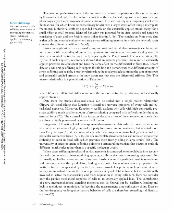
The first comprehensive study of the nonlinear viscoelastic properties of cells was carried outby Fernandez et al. (65), exploring for the first time the mechanical response of cells over a large,physiologically relevant range of cytoskeletal stresses. This was done by superimposing small stressfluctuations (for which linear response theory holds) over a larger stress offset using a microplaterheometer. Differential stiffness depended linearly on the externally applied stress, except for asmall offset at small stresses. Identical behavior was reported for in vitro cytoskeletal networksconsisting of actin and the flexible cross-linker filamin A (66). The conclusion from these datais that cells and cytoskeletal polymers are a stress-stiffening material in which the network stresscontrols the differential stiffness (66, 67).
Instead of application of an external stress, reconstituted cytoskeletal networks can be turnedinto a contractile material by adding active myosin motor proteins as cross-linkers and by control-ling the amount of contractile prestress by adjusting the ATP and cross-linker concentration (68).By use of such a system, researchers showed that an actively generated stress and an externallyapplied prestress are equivalent and have the same effect on the differential stiffness (69). Recentdata on a wide range of living cells support this finding and demonstrate that different amounts ofstress stiffening can be fit by a master relationship; the total cytoskeletal stress (the sum of prestressand externally applied stress) is the only parameter that sets the differential stiffness (70). Thismaster relationship is a generalization of Equation 2:
K ′(σ ) = δσ
δγ= K ′
0 + aσ, 4.
where K ′ is the differential stiffness and σ is the sum of contractile prestress σ p and externallyapplied stress σ e.
Data from the studies discussed above can be scaled into a single master relationship(Figure 10), establishing that Equation 4 describes a universal property of living cells and cy-toskeletal networks. Moreover, Equation 4 readily explains why cells with high contractile pre-stress exhibit a much smaller amount of stress stiffening compared with soft cells under the sameexternal force (70): The external force increases the total stress of the cytoskeleton in cells thatare already highly prestressed by only a small fraction.
Integration of Equation 4 yields an exponential stress-strain relationship. Exponential stiffeningat large strain values is a highly unusual property for most common materials, but as noted morethan 150 years ago (71), it is a universal, characteristic property of many biological materials, inparticular connective tissue (72, 73). Use of a microplate rheometer has also revealed exponentialstiffening to occur in fixed cells (which prevents them from yielding at large strains) (65). Theuniversality of stress or strain stiffening points to a structural mechanism that occurs at multipledifferent length scales rather than to a specific molecular origin.
When stress stiffening in cells and in vitro networks is compared, one should take into accountthat cells, in contrast to inert nonliving systems, exhibit active mechanosensing responses (74).Externally applied force is sensed and transduced into biochemical signals that result in remodelingand reinforcement of the cytoskeleton, leading to a drastic change of mechanical properties. Thematter is further complicated by the fact that some cross-linker proteins such as filamin A seemto play an important role for the passive properties in cytoskeletal networks but are additionallyinvolved in active mechanosensing and force regulation in living cells (27). Here we consideronly the passive mechanical response of cells to an externally applied load. The contributionof active mechanochemical signaling responses can be filtered out by oscillatory loading andlock-in techniques or minimized by keeping the measurement time sufficiently short. Data onthe low-frequency or long-time passive behavior of cells are therefore exceedingly difficult toanalyze (75).
88 Kollmannsberger · Fabry
Ann
u. R
ev. M
ater
. Res
. 201
1.41
:75-
97. D
ownl
oade
d fr
om w
ww
.ann
ualr
evie
ws.
org
Acc
ess
prov
ided
by
WIB
6276
- U
nive
rsita
tsbi
blio
thek
Erl
ange
n-N
uern
berg
on
04/1
4/15
. For
per
sona
l use
onl
y.
MR41CH04-Fabry ARI 3 June 2011 7:57
Scaled stress (σ/σs)10–2 10–1 100 101 10210–3
102
101
100
10–1
Scal
ed e
last
ic m
odul
us (K
'/Ks)
Cells (Reference 70)
Cells (Reference 65)
Cells (Reference 57)
Cells (Reference 113)
Actin-FLNa (Reference 69)
Prediction from Equation 4
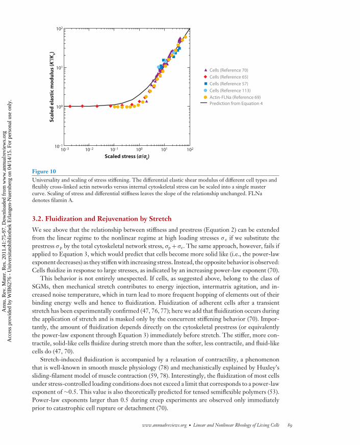
Figure 10Universality and scaling of stress stiffening. The differential elastic shear modulus of different cell types andflexibly cross-linked actin networks versus internal cytoskeletal stress can be scaled into a single mastercurve. Scaling of stress and differential stiffness leaves the slope of the relationship unchanged. FLNadenotes filamin A.
3.2. Fluidization and Rejuvenation by Stretch
We see above that the relationship between stiffness and prestress (Equation 2) can be extendedfrom the linear regime to the nonlinear regime at high loading stresses σ e if we substitute theprestress σ p by the total cytoskeletal network stress, σp + σe. The same approach, however, fails ifapplied to Equation 3, which would predict that cells become more solid like (i.e., the power-lawexponent decreases) as they stiffen with increasing stress. Instead, the opposite behavior is observed:Cells fluidize in response to large stresses, as indicated by an increasing power-law exponent (70).
This behavior is not entirely unexpected. If cells, as suggested above, belong to the class ofSGMs, then mechanical stretch contributes to energy injection, intermatrix agitation, and in-creased noise temperature, which in turn lead to more frequent hopping of elements out of theirbinding energy wells and hence to fluidization. Fluidization of adherent cells after a transientstretch has been experimentally confirmed (47, 76, 77); here we add that fluidization occurs duringthe application of stretch and is masked only by the concurrent stiffening behavior (70). Impor-tantly, the amount of fluidization depends directly on the cytoskeletal prestress (or equivalentlythe power-law exponent through Equation 3) immediately before stretch. The stiffer, more con-tractile, solid-like cells fluidize during stretch more than the softer, less contractile, and fluid-likecells do (47, 70).
Stretch-induced fluidization is accompanied by a relaxation of contractility, a phenomenonthat is well-known in smooth muscle physiology (78) and mechanistically explained by Huxley’ssliding-filament model of muscle contraction (59, 78). Interestingly, the fluidization of most cellsunder stress-controlled loading conditions does not exceed a limit that corresponds to a power-lawexponent of ∼0.5. This value is also theoretically predicted for tensed semiflexible polymers (53).Power-law exponents larger than 0.5 during creep experiments are observed only immediatelyprior to catastrophic cell rupture or detachment (70).
www.annualreviews.org • Linear and Nonlinear Rheology of Living Cells 89
Ann
u. R
ev. M
ater
. Res
. 201
1.41
:75-
97. D
ownl
oade
d fr
om w
ww
.ann
ualr
evie
ws.
org
Acc
ess
prov
ided
by
WIB
6276
- U
nive
rsita
tsbi
blio
thek
Erl
ange
n-N
uern
berg
on
04/1
4/15
. For
per
sona
l use
onl
y.

MR41CH04-Fabry ARI 3 June 2011 7:57
4. THEORETICAL DESCRIPTION
4.1. Theoretical Models of Cell Mechanics
The constitutive relationships between elastic modulus, power-law exponent, and prestress de-scribed by Equations 1–4 are purely phenomenological. The small number of empirical parametersgreatly simplifies the description of cell mechanical behavior but does not provide a mechanisticunderstanding. Researchers have developed a number of theoretical models that are able to explainand to predict certain facets of cell mechanics (Table 2), but currently no single model capturesthe full phenomenology of cell mechanical behavior.
Bottom up
Linearviscoelasticity
Tensegrity
Soft glassyrheology
Worm-likechain
Entropic filamentelasticity, viscousbackground, prestress
25, 33, 35,36, 103
44, 47, 109
52, 54, 81, 83
105–108
Equivalent circuit ofelastic springs andviscous dashpots
Prestressed structure madeof stiff rods and tensed cables
Elastic elements hoppingbetween energy traps,effective noisetemperature
Crossover fromelastic plateauto G' ~ ω0.75
PrestressbroadenscrossoverregimeG' ~ ω0.5
Top down
Cross-linkunfolding
Glassyworm-likechain
Prestressedsemiflexiblechain
Stiff filamentswith flexiblelinkers
Slidingfilament
Nonaffinenetworkdeformation
Geometrical effects,bending-to-stretchingtransition of filamentnetworks
Network of stiffpolymers with seriallyunfolding cross-links
Filament relaxationslowed down due tosticky interactions
Propagation of stress,thermal fluctuations
Randomly orientedcross-links and effectivemedium
Stiff filaments linked byactive crossbridges
85, 86
93
91
59, 96
87–90
94, 95
No
No (softening at highstress)
Requires a largenumber ofelements
No
No
No
No
No
NoNo
No
Broadly distributedrelaxation spectrum
Model Concept Stress stiffening? Power-lawrheology?
Stress-dependentexponent?
References
Model Concept Stress stiffening? Power-lawrheology?
Stress-dependentexponent?
References
Stiffness proportionalto prestress + externalstress
Yes
Yes
Fluidizationwith stress
Yes
Yes
Yes
Yes
Yes
Yes
Yes (for prestress)
No (for external stress)
Power-lawregime
Broad distributionof unfolding forces
Fluidizationwith stress
Prestressdecreasesapparentexponent
Prestressdecreasesexponent
aRed text denotes that the model does not predict the behavior in question. Blue text denotes that the model does predict the behavior in question.
Table 2 Comparison of the predictions of different theoretical models of cell mechanicsa
90 Kollmannsberger · Fabry
Ann
u. R
ev. M
ater
. Res
. 201
1.41
:75-
97. D
ownl
oade
d fr
om w
ww
.ann
ualr
evie
ws.
org
Acc
ess
prov
ided
by
WIB
6276
- U
nive
rsita
tsbi
blio
thek
Erl
ange
n-N
uern
berg
on
04/1
4/15
. For
per
sona
l use
onl
y.

MR41CH04-Fabry ARI 3 June 2011 7:57
Worm-like chain(WLC): a minimalphysical model thatdescribes the entropicelasticity of individualsemiflexible polymerssuch as cytoskeletalfilaments
4.2. Cytoskeletal Filaments and Networks
Although the springs and dashpots of linear viscoelasticity (Section 2.2) as well as the elements ofSGR (Section 2.4) and the cables and rods in tensegrity structures (Section 2.5) can be identifiedwith distinct structural components of the cytoskeleton, these models rest on generic principlesthat are valid independently of the details of the microscopic structure. All three approaches cantherefore be considered phenomenological, or top-down. The reductionist method of physics, incontrast to the phenomenological approach, explains the properties of a system from the propertiesof its constituents. In the case of cell mechanics, this bottom-up strategy starts with quantitativein vitro studies of the constituents of the cytoskeleton. Such in vitro reconstituted systems can beprepared in a reproducible way with well-defined structural properties such as filament lengthsor cross-linker densities (79). Once these systems are understood theoretically, it is expected thatthe functional modules can then be linked together into higher-order structures with emergingbiological complexity (80).
The cytoskeleton of eukaryotic cells is a cross-linked network of semiflexible filaments (witha persistence length on the order of the distance between cross-links). A minimal model for thebehavior of individual semiflexible filaments is the worm-like chain (WLC) model. The elasticityof a semiflexible polymer is entropic, with the individual filaments more likely to be curved thanto be straight. Upon stretching, the number of available conformations and therefore the entropyare reduced, which generates an opposing force (81). The dynamic frequency-dependent prop-erties can be obtained by considering friction of the thermally fluctuating filaments in a viscousbackground fluid (52). This model accurately describes the elastic as well as dynamic propertiesof reconstituted actin and other semiflexible polymer networks (82, 83).
The elastic and dissipative properties, however, are determined not only by the filaments andthe viscous background but also by the properties of the cross-linkers. For instance, unbindingof transient cross-links introduces additional dissipation and alters the frequency response (84).Simulations of filament networks with serially unfolding cross-links showed that under prestress,many links are at the threshold of domain unfolding, which can give rise to collective unfolding,a broad distribution of lifetimes, and power-law rheology (85, 86).
When filaments and cross-linkers are combined into networks, additional geometric effectscome into play. At large strains, the deformation is nonaffine (87, 88), meaning that individualfilament strain and network strain differ. As more and more fibers are pulled straight, filamentstretching begins to dominate over filament bending (89, 90). Broedersz et al. (91) use a self-consistent effective medium approach to examine the role of flexible cross-linkers in stiff filamentnetworks. Here, the onset of stiffening occurs when the cross-links are stretched toward their fullextension. These investigators show that the stiffening response crucially depends on cross-linkerand filament length, in good agreement with experiments (92).
These models provide several mechanisms to account for the stress-stiffness response of cy-toskeletal networks, but they are less successful in modeling the weak power-law dependency ofelastic moduli on frequency. Two recently developed theoretical descriptions deal with this issue.Rosenblatt et al. (93) relate the propagation of force along a prestressed nonlinear semiflexiblechain to the creep response of cells. The model is a combination of tensegrity and the WLC de-scription, and it correctly predicts a decrease in the power-law exponent with increasing prestress.The other approach, termed the glassy worm-like chain (GWLC) model, combines the WLCmodel with concepts from SGR (94). It describes the retardation of filament relaxation due tosticky interfilament interactions, leading to an exponential stretching of the ω3/4 relaxation spec-trum of individual filaments. The idea behind this concept is generic and applicable to cells andother heterogeneous biological materials. The model can explain power-law rheology and stress
www.annualreviews.org • Linear and Nonlinear Rheology of Living Cells 91
Ann
u. R
ev. M
ater
. Res
. 201
1.41
:75-
97. D
ownl
oade
d fr
om w
ww
.ann
ualr
evie
ws.
org
Acc
ess
prov
ided
by
WIB
6276
- U
nive
rsita
tsbi
blio
thek
Erl
ange
n-N
uern
berg
on
04/1
4/15
. For
per
sona
l use
onl
y.

MR41CH04-Fabry ARI 3 June 2011 7:57
stiffening as well as fluidization and plasticity (94, 95). As with all microscopic filament–baseddescriptions, however, a quantitative prediction of the shear modulus for cells is difficult becausestructural parameters of the cytoskeleton such as mesh size or filament length are highly variable.
None of the above models can account for the active contractility of the cytoskeleton. On amicroscopic level, the mechanism of contractile force generation is the interaction of actin andmyosin filaments, which is well captured by Huxley’s sliding-filament model of skeletal muscle(59, 96). Highly contractile cells such as fibroblasts and myoblasts exhibit a remarkably muscle-likemechanical phenotype, and Huxley’s model captures the dynamics of force generation in thesecells against an elastic load (97, 98). Moreover, the model can explain the linear dependency ofstiffness on prestress but in its simple form does not predict a power-law frequency dependency.The Huxley model resembles the SGR model in that it contains multiple binding interactionenergies with the addition of a force-generating mechanism based on a Brownian ratchet. Fromthis perspective, a combination of the SGR model and muscle contraction models appears to bea promising approach for describing cell mechanics (60).
4.3. Top Down Versus Bottom Up: Lessons from Muscle
The history of muscle modeling provides an interesting example of how phenomenological andmechanistic models can complement each other. When A.V. Hill formulated his active-state modelof muscle contraction (99), which is a viscoelastic equivalent circuit with springs and dashpots, heknew next to nothing about the microscopic structure of the muscle and the mechanisms of forcegeneration. Huxley’s sliding-filament model, on the contrary, follows a reductionist approach toquantitatively explain overall muscle tension from the molecular interactions between actin andmyosin. Both models can generate valid macroscopic predictions, and neither of them is corrector incorrect; they differ only in their complexity. The Hill model is still used today to fit datawhenever the molecular details are not considered relevant or are unknown.
The quantitative match between bottom-up and top-down predictions in the case of musclecontraction was possible because muscle has a well-defined and regular structure. In the caseof cells, however, scaling up molecular-level models to the whole-cell level is difficult due tothe structural complexity of the cytoskeleton. Although the mechanical properties of variousmolecular constituents such as cytoskeletal filaments and cross-linkers are well characterized,deriving macroscopic mechanical predictions from this knowledge in any but the most simplemodel systems is not possible. Moreover, theoretical models based on a full microscopic descriptionare not likely to yield much more additional insight. The structural complexity of the cell requiresa different approach, discarding the reductionist search for a microscopic mechanistic model towhich all macroscopic observations can be traced back. A successful comprehensive model ofcell mechanics will likely be both generic and mechanistic. It will incorporate mechanisms suchas filament fluctuations, protein unfolding, bond dynamics, actin-myosin cycling, and geometricnetwork mechanisms on different hierarchical levels to relate microscopic interactions in thecytoskeleton to the macroscopic mechanical behavior of cells.
SUMMARY POINTS
1. Measurement of mechanical properties of living cells has become a standard tool incell biology. A wide range of microrheology techniques are available, each with specificadvantages and limitations.
92 Kollmannsberger · Fabry
Ann
u. R
ev. M
ater
. Res
. 201
1.41
:75-
97. D
ownl
oade
d fr
om w
ww
.ann
ualr
evie
ws.
org
Acc
ess
prov
ided
by
WIB
6276
- U
nive
rsita
tsbi
blio
thek
Erl
ange
n-N
uern
berg
on
04/1
4/15
. For
per
sona
l use
onl
y.

MR41CH04-Fabry ARI 3 June 2011 7:57
2. The linear and nonlinear viscoelastic properties of cells conform to a limited set ofuniversal scaling laws. Creep and stress relaxation behavior follows a power law overseveral orders of magnitude of time or frequency. The power-law exponent characterizesthe state of the cell between a solid and a fluid and is related to the contractile prestressof the cytoskeleton as well as to stiffening and fluidization behavior in response to largestresses or strains.
3. This universal behavior can be reproduced consistently with different microrheologytechniques and across different cell types treated with different pharmacological inter-ventions. It can also be reproduced in simplified model networks of cytoskeletal filaments.
4. The biological relevance of such universal scaling behavior is wide ranging. By a singlemechanism that is present in all eukaryotic cell types, namely, by modulating the activityof actomyosin contraction and hence the cytoskeletal prestress, cells can stiffen or fluidizeand thus adapt to different mechanical loading conditions. Moreover, by modulating theircontractile prestress, cells also change the mechanical state of their environment, whichis again sensed by neighboring cells. This mechanical feedback loop may be essential forthe control and robustness of many developmental and growth processes.
5. Investigators have proposed numerous different theoretical models that capture differentaspects of cell mechanical behavior at different functional and structural levels, rang-ing from considerations of the cytoskeletal network architecture down to single-proteinbinding kinetics. A comprehensive understanding of the physical mechanisms that governthese universal scaling laws, however, is still emerging.
DISCLOSURE STATEMENT
The authors are not aware of any affiliations, memberships, funding, or financial holdings thatmight be perceived as affecting the objectivity of this review.
ACKNOWLEDGMENTS
We thank Stefan Munster for valuable discussions and proofreading.
LITERATURE CITED
1. Venter JC, Adams MD, Myers EW, Li PW, Mural RJ, et al. 2001. The sequence of the human genome.Science 291:1304–51
2. Howard J. 2001. Mechanics of Motor Proteins and the Cytoskeleton. Sunderland, MA: Sinauer3. Stamenovic D, Mijailovich SM, Tolic-Norrelykke IM, Chen J, Wang N. 2002. Cell prestress. II. Con-
tribution of microtubules. Am. J. Physiol. Cell Physiol. 282:617–244. Janmey PA, Euteneuer U, Traub P, Schliwa M. 1991. Viscoelastic properties of vimentin compared with
other filamentous biopolymer networks. J. Cell Biol. 113:155–605. Wang N, Stamenovic D. 2000. Contribution of intermediate filaments to cell stiffness, stiffening, and
growth. Am. J. Physiol. Cell Physiol. 279:188–946. Stamenovic D. 2008. Rheological behavior of mammalian cells. Cell Mol. Life Sci. 65:3592–6057. Eckes B, Dogic D, Colucci-Guyon E, Wang N, Maniotis A, et al. 1998. Impaired mechanical stability,
migration and contractile capacity in vimentin-deficient fibroblasts. J. Cell Sci. 111(Pt. 13):1897–9078. Chancellor TJ, Lee J, Thodeti CK, Lele T. 2010. Actomyosin tension exerted on the nucleus through
nesprin-1 connections influences endothelial cell adhesion, migration, and cyclic strain-induced reori-entation. Biophys. J. 99:115–23
www.annualreviews.org • Linear and Nonlinear Rheology of Living Cells 93
Ann
u. R
ev. M
ater
. Res
. 201
1.41
:75-
97. D
ownl
oade
d fr
om w
ww
.ann
ualr
evie
ws.
org
Acc
ess
prov
ided
by
WIB
6276
- U
nive
rsita
tsbi
blio
thek
Erl
ange
n-N
uern
berg
on
04/1
4/15
. For
per
sona
l use
onl
y.

MR41CH04-Fabry ARI 3 June 2011 7:57
9. Maniotis AJ, Chen CS, Ingber DE. 1997. Demonstration of mechanical connections between integrins,cytoskeletal filaments, and nucleoplasm that stabilize nuclear structure. Proc. Natl. Acad. Sci. USA 94:849–54
10. Rodriguez OC, Schaefer AW, Mandato CA, Forscher P, Bement WM, Waterman-Storer CM. 2003.Conserved microtubule-actin interactions in cell movement and morphogenesis. Nat. Cell Biol. 5:599–609
11. Heilbronn A. 1922. Eine neue Methode zur Bestimmung der Viskositat lebender Protoplasten. Jahrb.Wiss. Bot. 61:284–338
12. Crick FHC, Hughes AFW. 1950. The physical properties of cytoplasm. Exp. Cell Res. 1:37–8013. Plopper G, Ingber DE. 1993. Rapid induction and isolation of focal adhesion complexes. Biochem. Biophys.
Res. Commun. 193:571–7814. Schmidt CE, Horwitz AF, Lauffenburger DA, Sheetz MP. 1993. Integrin-cytoskeletal interactions in
migrating fibroblasts are dynamic, asymmetric, and regulated. J. Cell Biol. 123:977–9115. Felsenfeld DP, Choquet D, Sheetz MP. 1996. Ligand binding regulates the directed movement of β1
integrins on fibroblasts. Nature 383:438–4016. Choquet D, Felsenfeld DP, Sheetz MP. 1997. Extracellular matrix rigidity causes strengthening of
integrin-cytoskeleton linkages. Cell 88:39–4817. Schmidt C, Pommerenke H, Durr F, Nebe B, Rychly J. 1998. Mechanical stressing of integrin receptors
induces enhanced tyrosine phosphorylation of cytoskeletally anchored proteins. J. Biol. Chem. 273:5081–85
18. Deng L, Fairbank NJ, Fabry B, Smith PG, Maksym GN. 2004. Localized mechanical stress inducestime-dependent actin cytoskeletal remodeling and stiffening in cultured airway smooth muscle cells.Am. J. Physiol. Cell Physiol. 287:440–48
19. Puig-de-Morales M, Millet E, Fabry B, Navajas D, Wang N, et al. 2004. Cytoskeletal mechanics inadherent human airway smooth muscle cells: probe specificity and scaling of protein-protein dynamics.Am. J. Physiol. Cell Physiol. 287:643–54
20. Metzner C, Raupach C, Mierke CT, Fabry B. 2010. Fluctuations of cytoskeleton-bound microbeads:the effect of bead-receptor binding dynamics. J. Phys. Condens. Matter 22:194105
21. Park CY, Tambe D, Alencar AM, Trepat X, Zhou EH, et al. 2010. Mapping the cytoskeletal prestress.Am. J. Physiol. Cell Physiol. 298:1245–52
22. Fabry B, Maksym GN, Hubmayr RD, Butler JP, Fredberg JJ. 1999. Implications of heterogeneous beadbehavior on cell mechanical properties measured with magnetic twisting cytometry. J. Magn. Magn.Mater. 194:120–25
23. Lau AW, Hoffman BD, Davies A, Crocker JC, Lubensky TC. 2003. Microrheology, stress fluctuations,and active behavior of living cells. Phys. Rev. Lett. 91:198101
24. Levine AJ, Lubensky TC. 2000. One- and two-particle microrheology. Phys. Rev. Lett. 85:1774–7725. Bausch AR, Ziemann F, Boulbitch AA, Jacobson K, Sackmann E. 1998. Local measurements of viscoelas-
tic parameters of adherent cell surfaces by magnetic bead microrheometry. Biophys. J. 75:2038–4926. Mijailovich SM, Kojic M, Zivkovic M, Fabry B, Fredberg JJ. 2002. A finite element model of cell
deformation during magnetic bead twisting. J. Appl. Physiol. 93:1429–3627. Kasza KE, Nakamura F, Hu S, Kollmannsberger PB, Bonakdar N, et al. 2009. Filamin A is essential for
active cell stiffening but not passive stiffening under external force. Biophys. J. 96:4326–3528. Hu S, Chen J, Fabry B, Numaguchi Y, Gouldstone A, et al. 2003. Intracellular stress tomography
reveals stress focusing and structural anisotropy in cytoskeleton of living cells. Am. J. Physiol. Cell Physiol.285:1082–90
29. Pullarkat PA, Fernandez PA, Ott A. 2007. Rheological properties of the eukaryotic cell cytoskeleton.Phys. Rep. 449:29–53
30. Janmey PA, McCulloch CA. 2007. Cell mechanics: integrating cell responses to mechanical stimuli.Annu. Rev. Biomed. Eng. 9:1–34
31. Found power-lawrheology in differentcell types using fourdifferent techniques.
31. Hoffman BD, Massiera G, Van Citters KM, Crocker JC. 2006. The consensus mechanics ofcultured mammalian cells. Proc. Natl. Acad. Sci. USA 103:10259–64
32. Trepat X, Lenormand G, Fredberg JJ. 2008. Universality in cell mechanics. Soft Matter 4:1750–5933. Schmid-Schonbein GW, Sung KL, Tozeren H, Skalak R, Chien S. 1981. Passive mechanical properties
of human leukocytes. Biophys. J. 36:243–56
94 Kollmannsberger · Fabry
Ann
u. R
ev. M
ater
. Res
. 201
1.41
:75-
97. D
ownl
oade
d fr
om w
ww
.ann
ualr
evie
ws.
org
Acc
ess
prov
ided
by
WIB
6276
- U
nive
rsita
tsbi
blio
thek
Erl
ange
n-N
uern
berg
on
04/1
4/15
. For
per
sona
l use
onl
y.

MR41CH04-Fabry ARI 3 June 2011 7:57
34. Wang N, Ingber DE. 1994. Control of cytoskeletal mechanics by extracellular matrix, cell shape, andmechanical tension. Biophys. J. 66:2181–89
35. Thoumine O, Ott A. 1997. Time scale dependent viscoelastic and contractile regimes in fibroblastsprobed by microplate manipulation. J. Cell Sci. 110:2109–16
36. Laurent VM, Fodil R, Canadas P, Fereol S, Louis B, et al. 2003. Partitioning of cortical and deepcytoskeleton responses from transient magnetic bead twisting. Ann. Biomed. Eng. 31:1263–78
37. Wilson TA. 1994. Time constants may be meaningless in exponentials fit to pressure relaxation data.J. Appl. Physiol. 77:1570–71
38. Weber W. 1841. Ueber die Elasticitaet fester Koerper. Ann. Phys. Chem. 54:1–1839. Weber W. 1835. Ueber die Elasticitaet der Seidenfaeden. Ann. Phys. Chem. 34:247–5740. Kohlrausch R. 1847. Nachtrag ueber die elastische Nachwirkung beim Cocon- und Glasfaden, und die
hygroskopische Eigenschaft des ersteren. Ann. Phys. Chem. 72:393–9841. Kohlrausch F. 1866. Beitrage zur Kenntniss der elastischen Nachwirkung. Ann. Phys. Chem. 128:1–20,
207–27, 399–41942. Maxwell JC. 1867. On the dynamical theory of gases. Philos. Trans. R. Soc. Lond. 157:49–8843. Hildebrandt J. 1969. Comparison of mathematical models for cat lung and viscoelastic balloon derived
by Laplace transform methods from pressure-volume data. Bull. Math. Biophys. 31:651–6744. Discovered thepower-law frequencydependency and scalingrelationship for theelastic modulus of cells.
44. Fabry B, Maksym GN, Butler JP, Glogauer M, Navajas D, Fredberg JJ. 2001. Scaling the mi-crorheology of living cells. Phys. Rev. Lett. 87:148102
45. Laudadio RE, Millet EJ, Fabry B, An SS, Butler JP, Fredberg JJ. 2005. Rat airway smooth muscle cellduring actin modulation: rheology and glassy dynamics. Am. J. Physiol. Cell Physiol. 289:1388–95
46. Sollich P. 1998. Rheological constitutive equation for a model of soft glassy materials. Phys. Rev. E58:738–59
47. Bursac P, Lenormand G, Fabry B, Oliver M, Weitz DA, et al. 2005. Cytoskeletal remodelling and slowdynamics in the living cell. Nat. Mater. 4:557–61
48. Yamada S, Wirtz D, Kuo SC. 2000. Mechanics of living cells measured by laser tracking microrheology.Biophys. J. 78:1736–47
49. Fabry B, Maksym GN, Butler JP, Glogauer M, Navajas D, et al. 2003. Time scale and other invariantsof integrative mechanical behavior in living cells. Phys. Rev. E 68:041914
50. Lenormand G, Millet E, Fabry B, Butler JP, Fredberg JJ. 2004. Linearity and time-scale invariance ofthe creep function in living cells. J. R. Soc. Interface 1:91–97
51. Smith BA, Tolloczko B, Martin JG, Grutter P. 2005. Probing the viscoelastic behavior of cultured airwaysmooth muscle cells with atomic force microscopy: stiffening induced by contractile agonist. Biophys. J.88:2994–3007
52. Gittes F, MacKintosh FC. 1998. Dynamic shear modulus of a semiflexible polymer network. Phys. Rev.E 58:R1241–44
53. Granek R. 1997. From semi-flexible polymers to membranes: anomalous diffusion and reptation. J. Phys.II 7:1761–88
54. Gardel ML, Shin JH, MacKintosh FC, Mahadevan L, Matsudaira PA, Weitz DA. 2004. Scaling ofF-actin network rheology to probe single filament elasticity and dynamics. Phys. Rev. Lett. 93:188102
55. Deng L, Trepat X, Butler JP, Millet E, Morgan KG, et al. 2006. Fast and slow dynamics of the cytoskele-ton. Nat. Mater. 5:636–40 56. Reported the linear
relationship betweenstiffness andcytoskeletal prestress incells.
56. Wang N, Naruse K, Stamenovic D, Fredberg JJ, Mijailovich SM, et al. 2001. Mechanical behaviorin living cells consistent with the tensegrity model. Proc. Natl. Acad. Sci. USA 98:7765–70
57. Wang N, Tolic-Norrelykke IM, Chen J, Mijailovich SM, Butler JP, et al. 2002. Cell prestress. I. Stiffnessand prestress are closely associated in adherent contractile cells. Am. J. Physiol. Cell Physiol. 282:606–16
58. Krishnan R, Park CY, Lin YC, Mead J, Jaspers RT, et al. 2009. Reinforcement versus fluidization incytoskeletal mechanoresponsiveness. PLoS ONE 4:e5486
59. Huxley AF. 1957. Muscle structure and theories of contraction. Prog. Biophys. Biophys. Chem. 7:255–31860. Kollmannsberger P, Fabry B. 2009. Active soft glassy rheology of adherent cells. Soft Matter 5:1771–
74
61. Showed therelationship betweencontractile prestress andpower-law exponent ofthe complex modulus.61. Stamenovic D, Suki B, Fabry B, Wang N, Fredberg JJ. 2004. Rheology of airway smooth muscle
cells is associated with cytoskeletal contractile stress. J. Appl. Physiol. 96:1600–5
www.annualreviews.org • Linear and Nonlinear Rheology of Living Cells 95
Ann
u. R
ev. M
ater
. Res
. 201
1.41
:75-
97. D
ownl
oade
d fr
om w
ww
.ann
ualr
evie
ws.
org
Acc
ess
prov
ided
by
WIB
6276
- U
nive
rsita
tsbi
blio
thek
Erl
ange
n-N
uern
berg
on
04/1
4/15
. For
per
sona
l use
onl
y.
MR41CH04-Fabry ARI 3 June 2011 7:57
62. Gardel ML, Nakamura F, Hartwig J, Crocker JC, Stossel TP, Weitz DA. 2006. Stress-dependent elas-ticity of composite actin networks as a model for cell behavior. Phys. Rev. Lett. 96:088102
63. Desprat N, Richert A, Simeon J, Asnacios A. 2005. Creep function of a single living cell. Biophys. J.88:2224–33
64. Wang N, Butler JP, Ingber DE. 1993. Mechanotransduction across the cell surface and through thecytoskeleton. Science 260:1124–27
65. The firstcomprehensive study ofnonlinear cellmechanics; reportsuniversal stressstiffening in cells. 65. Fernandez P, Pullarkat PA, Ott A. 2006. A master relation defines the nonlinear viscoelasticity
of single fibroblasts. Biophys. J. 90:3796–80566. Showed thatprestressed actinnetworks cross-linkedwith flexible filamin Alinkers displaymechanical behaviorsimilar to that of cells.
66. Gardel ML, Nakamura F, Hartwig JH, Crocker JC, Stossel TP, Weitz DA. 2006. PrestressedF-actin networks cross-linked by hinged filamins replicate mechanical properties of cells. Proc.
Natl. Acad. Sci. USA 103:1762–6767. Kasza KE, Rowat AC, Liu J, Angelini TE, Brangwynne CP, et al. 2007. The cell as a material. Curr.
Opin. Cell Biol. 19:101–768. Mizuno D, Tardin C, Schmidt CF, MacKintosh FC. 2007. Nonequilibrium mechanics of active cy-
toskeletal networks. Science 315:370–73
69. Showed that activeinternal prestress andpassive externalprestress are equivalentin controlling therheology of cytoskeletalnetworks.
69. Koenderink GH, Dogic Z, Nakamura F, Bendix PM, MacKintosh FC, et al. 2009. An activebiopolymer network controlled by molecular motors. Proc. Natl. Acad. Sci. USA 106:15192–97
70. Kollmannsberger P, Mierke CT, Fabry B. 2011. Nonlinear viscoelasticity of adherent cells is controlledby cytoskeletal tension. Soft Matter 7:3127–32
71. Wertheim MG. 1847. Memoire sur l’elasticite et la cohesion des principaux tissues du corps humain.Ann. Chim. Phys. (21):385–414
72. Fung YC. 1967. Elasticity of soft tissues in simple elongation. Am. J. Physiol. 213:1532–4473. Fung YC. 1993. Mechanical Properties of Living Tissues. New York: Springer-Verlag74. Riveline D, Zamir E, Balaban NQ, Schwarz US, Ishizaki T, et al. 2001. Focal contacts as mechanosensors:
externally applied local mechanical force induces growth of focal contacts by an mDia1-dependent andROCK-independent mechanism. J. Cell Biol. 153:1175–86
75. Stamenovic D, Rosenblatt N, Montoya-Zavala M, Matthews BD, Hu S, et al. 2007. Rheological behaviorof living cells is timescale-dependent. Biophys. J. 93:L39–41
76. Bursac P, Fabry B, Trepat X, Lenormand G, Butler JP, et al. 2007. Cytoskeleton dynamics: fluctuationswithin the network. Biochem. Biophys. Res. Commun. 355:324–30
77. Trepat X, Deng L, An SS, Navajas D, Tschumperlin DJ, et al. 2007. Universal physical responses tostretch in the living cell. Nature 447:592–95
78. Fredberg JJ, Inouye D, Miller B, Nathan M, Jafari S, et al. 1997. Airway smooth muscle, tidal stretches,and dynamically determined contractile states. Am. J. Respir. Crit. Care Med. 156:1752–59
79. Janmey PA, Hvidt S, Kas J, Lerche D, Maggs A, et al. 1994. The mechanical properties of actin gels.Elastic modulus and filament motions. J. Biol. Chem. 269:32503–13
80. Bausch AR, Kroy K. 2006. A bottom-up approach to cell mechanics. Nat. Phys. 2:231–3881. MacKintosh FC, Kas J, Janmey PA. 1995. Elasticity of semiflexible biopolymer networks. Phys. Rev. Lett.
75:4425–2882. Gardel ML, Shin JH, MacKintosh FC, Mahadevan L, Matsudaira P, Weitz DA. 2004. Elastic behavior
of cross-linked and bundled actin networks. Science 304:1301–583. Storm C, Pastore JJ, MacKintosh FC, Lubensky TC, Janmey PA. 2005. Nonlinear elasticity in biological
gels. Nature 435:191–9484. Lieleg O, Claessens MM, Luan Y, Bausch AR. 2008. Transient binding and dissipation in cross-linked
actin networks. Phys. Rev. Lett. 101:10810185. DiDonna BA, Levine AJ. 2006. Filamin cross-linked semiflexible networks: fragility under strain. Phys.
Rev. Lett. 97:06810486. Hoffman BD, Massiera G, Crocker JC. 2007. Fragility and mechanosensing in a thermalized cytoskeleton
model with forced protein unfolding. Phys. Rev. E 76:05190687. Head DA, Levine AJ, MacKintosh FC. 2003. Deformation of cross-linked semiflexible polymer networks.
Phys. Rev. Lett. 91:10810288. Heussinger C, Frey E. 2006. Stiff polymers, foams, and fiber networks. Phys. Rev. Lett. 96:017802
96 Kollmannsberger · Fabry
Ann
u. R
ev. M
ater
. Res
. 201
1.41
:75-
97. D
ownl
oade
d fr
om w
ww
.ann
ualr
evie
ws.
org
Acc
ess
prov
ided
by
WIB
6276
- U
nive
rsita
tsbi
blio
thek
Erl
ange
n-N
uern
berg
on
04/1
4/15
. For
per
sona
l use
onl
y.

MR41CH04-Fabry ARI 3 June 2011 7:57
89. Huisman EM, van Dillen T, Onck PR, Van der Giessen E. 2007. Three-dimensional cross-linked F-actinnetworks: relation between network architecture and mechanical behavior. Phys. Rev. Lett. 99:208103
90. Onck PR, Koeman T, van Dillen T, Van der Giessen E. 2005. Alternative explanation of stiffening incross-linked semiflexible networks. Phys. Rev. Lett. 95:178102
91. Broedersz CP, Storm C, MacKintosh FC. 2008. Nonlinear elasticity of composite networks of stiffbiopolymers with flexible linkers. Phys. Rev. Lett. 101:118103
92. Kasza KE, Broedersz CP, Koenderink GH, Lin YC, Messner W, et al. 2010. Actin filament length tuneselasticity of flexibly cross-linked actin networks. Biophys. J. 99:1091–100
93. Rosenblatt N, Alencar AM, Majumdar A, Suki B, Stamenovic D. 2006. Dynamics of prestressed semi-flexible polymer chains as a model of cell rheology. Phys. Rev. Lett. 97:168101
94. Combined themechanistic WLCdescription with thephenomenological SGRconcept.
94. Semmrich C, Storz T, Glaser J, Merkel R, Bausch AR, Kroy K. 2007. Glass transition andrheological redundancy in F-actin solutions. Proc. Natl. Acad. Sci. USA 104:20199–203
95. Wolff L, Fernandez P, Kroy K. 2010. Inelastic mechanics of sticky biopolymer networks. N. J. Phys.12:1–18
96. Huxley AF, Simmons RM. 1971. Proposed mechanism of force generation in striated muscle. Nature233:533–38
97. Fernandez P, Ott A. 2008. Single cell mechanics: stress stiffening and kinematic hardening. Phys. Rev.Lett. 100:238102
98. Mitrossilis D, Fouchard J, Guiroy A, Desprat N, Rodriguez N, et al. 2009. Single-cell response to stiffnessexhibits muscle-like behavior. Proc. Natl. Acad. Sci. USA 106:18243–48
99. McMahon TA. 1984. Muscles, Reflexes, and Locomotion. Princeton, NJ: Princeton Univ. Press100. Alcaraz J, Buscemi L, Grabulosa M, Trepat X, Fabry B, et al. 2003. Microrheology of human lung
epithelial cells measured by atomic force microscopy. Biophys. J. 84:2071–79101. Hiratsuka S, Mizutani Y, Tsuchiya M, Kawahara K, Tokumoto H, Okajima T. 2009. The number dis-
tribution of complex shear modulus of single cells measured by atomic force microscopy. Ultramicroscopy109:937–41
102. Kollmannsberger P, Fabry B. 2007. High-force magnetic tweezers with force feedback for biologicalapplications. Rev. Sci. Instrum. 78:114301
103. Balland M, Desprat N, Icard D, Fereol S, Asnacios A, et al. 2006. Power laws in microrheology experi-ments on living cells: comparative analysis and modeling. Phys. Rev. E 74:021911
104. Crocker JC, Valentine MT, Weeks ER, Gisler T, Kaplan PD, et al. 2000. Two-point microrheology ofinhomogeneous soft materials. Phys. Rev. Lett. 85:888–91
105. Ingber DE. 1997. Tensegrity: the architectural basis of cellular mechanotransduction. Annu. Rev. Physiol.59:575–99
106. Stamenovic D, Coughlin MF. 1999. The role of prestress and architecture of the cytoskeleton anddeformability of cytoskeletal filaments in mechanics of adherent cells: a quantitative analysis. J. Theor.Biol. 201:63–74
107. Sultan C, Stamenovic D, Ingber DE. 2004. A computational tensegrity model predicts dynamic rheo-logical behaviors in living cells. Ann. Biomed. Eng. 32:520–30
108. Canadas P, Wendling-Mansuy S, Isabey D. 2006. Frequency response of a viscoelastic tensegrity model:structural rearrangement contribution to cell dynamics. J. Biomech. Eng. 128:487–95
109. Sollich P, Lequeux F, Hebraud P, Cates ME. 1997. Rheology of soft glassy materials. Phys. Rev. Lett.78:2020–23
110. Mierke CT, Kollmannsberger P, Paranhos-Zitterbart D, Smith J, Fabry B, Goldmann WH. 2008.Mechano-coupling and regulation of contractility by the vinculin tail domain. Biophys. J. 94:661–70
111. Nicolle S, Vezin P, Palierne JF. 2010. A strain-hardening bi-power law for the nonlinear behaviour ofbiological soft tissues. J. Biomech. 43:927–32
112. Geerligs M, Peters GWM, Ackermans PAJ, Oomens CWJ, Baaijens FPT. 2008. Linear viscoelasticbehavior of subcutaneous adipose tissue. Biorheology 45:677–88
113. Engler AJ, Sen S, Sweeney HL, Discher DE. 2006. Matrix elasticity directs stem cell lineage specification.Cell 126:677–89
www.annualreviews.org • Linear and Nonlinear Rheology of Living Cells 97
Ann
u. R
ev. M
ater
. Res
. 201
1.41
:75-
97. D
ownl
oade
d fr
om w
ww
.ann
ualr
evie
ws.
org
Acc
ess
prov
ided
by
WIB
6276
- U
nive
rsita
tsbi
blio
thek
Erl
ange
n-N
uern
berg
on
04/1
4/15
. For
per
sona
l use
onl
y.

MR41-FrontMatter ARI 4 June 2011 12:1
Annual Review ofMaterials Research
Volume 41, 2011Contents
Materials Science of Biological Systems
Advances in Drug DeliveryBrian P. Timko, Kathryn Whitehead, Weiwei Gao, Daniel S. Kohane,
Omid Farokhzad, Daniel Anderson, and Robert Langer � � � � � � � � � � � � � � � � � � � � � � � � � � � � � � � � � � 1
Crystallization Pathways in BiomineralizationSteve Weiner and Lia Addadi � � � � � � � � � � � � � � � � � � � � � � � � � � � � � � � � � � � � � � � � � � � � � � � � � � � � � � � � � � � � � � � �21
Deformation and Fracture Mechanisms of Bone and NacreRizhi Wang and Himadri S. Gupta � � � � � � � � � � � � � � � � � � � � � � � � � � � � � � � � � � � � � � � � � � � � � � � � � � � � � � � � �41
Linear and Nonlinear Rheology of Living CellsPhilip Kollmannsberger and Ben Fabry � � � � � � � � � � � � � � � � � � � � � � � � � � � � � � � � � � � � � � � � � � � � � � � � � � � � �75
Mussel-Inspired Adhesives and CoatingsBruce P. Lee, P.B. Messersmith, J.N. Israelachvili, and J.H. Waite � � � � � � � � � � � � � � � � � � � � � �99
Nanomechanics of the Cartilage Extracellular MatrixLin Han, Alan J. Grodzinsky, and Christine Ortiz � � � � � � � � � � � � � � � � � � � � � � � � � � � � � � � � � � � � � � 133
Plant Stems: Functional Design and MechanicsThomas Speck and Ingo Burgert � � � � � � � � � � � � � � � � � � � � � � � � � � � � � � � � � � � � � � � � � � � � � � � � � � � � � � � � � � � 169
Current Interest
Elastic and Mechanical Properties of the MAX PhasesMichel W. Barsoum and Miladin Radovic � � � � � � � � � � � � � � � � � � � � � � � � � � � � � � � � � � � � � � � � � � � � � � � � 195
Electrocaloric MaterialsJ.F. Scott � � � � � � � � � � � � � � � � � � � � � � � � � � � � � � � � � � � � � � � � � � � � � � � � � � � � � � � � � � � � � � � � � � � � � � � � � � � � � � � � � � � � 229
Electrochromic MaterialsRoger J. Mortimer � � � � � � � � � � � � � � � � � � � � � � � � � � � � � � � � � � � � � � � � � � � � � � � � � � � � � � � � � � � � � � � � � � � � � � � � � � 241
Nanowire Solar CellsErik C. Garnett, Mark L. Brongersma, Yi Cui, and Michael D. McGehee � � � � � � � � � � � � 269
Nonconventional (Non-Silicon-Based) Photovoltaic MaterialsT. Unold and H.W. Schock � � � � � � � � � � � � � � � � � � � � � � � � � � � � � � � � � � � � � � � � � � � � � � � � � � � � � � � � � � � � � � � � 297
v
Ann
u. R
ev. M
ater
. Res
. 201
1.41
:75-
97. D
ownl
oade
d fr
om w
ww
.ann
ualr
evie
ws.
org
Acc
ess
prov
ided
by
WIB
6276
- U
nive
rsita
tsbi
blio
thek
Erl
ange
n-N
uern
berg
on
04/1
4/15
. For
per
sona
l use
onl
y.

MR41-FrontMatter ARI 4 June 2011 12:1
On the Future Availability of the Energy MetalsT.E. Graedel � � � � � � � � � � � � � � � � � � � � � � � � � � � � � � � � � � � � � � � � � � � � � � � � � � � � � � � � � � � � � � � � � � � � � � � � � � � � � � � � 323
Oxide Electronics Utilizing Ultrafast Metal-Insulator TransitionsZheng Yang, Changhyun Ko, and Shriram Ramanathan � � � � � � � � � � � � � � � � � � � � � � � � � � � � � � � � 337
Point Defects in Oxides: Tailoring Materials ThroughDefect EngineeringHarry L. Tuller and Sean R. Bishop � � � � � � � � � � � � � � � � � � � � � � � � � � � � � � � � � � � � � � � � � � � � � � � � � � � � � � 369
Recent Developments in Semiconductor Thermoelectric Physicsand MaterialsAli Shakouri � � � � � � � � � � � � � � � � � � � � � � � � � � � � � � � � � � � � � � � � � � � � � � � � � � � � � � � � � � � � � � � � � � � � � � � � � � � � � � � � 399
Thermoelectric Phenomena, Materials, and ApplicationsTerry M. Tritt � � � � � � � � � � � � � � � � � � � � � � � � � � � � � � � � � � � � � � � � � � � � � � � � � � � � � � � � � � � � � � � � � � � � � � � � � � � � � � 433
Index
Cumulative Index of Contributing Authors, Volumes 37–41 � � � � � � � � � � � � � � � � � � � � � � � � � � � 449
Errata
An online log of corrections to Annual Review of Materials Research articles may befound at http://matsci.annualreviews.org/errata.shtml
vi Contents
Ann
u. R
ev. M
ater
. Res
. 201
1.41
:75-
97. D
ownl
oade
d fr
om w
ww
.ann
ualr
evie
ws.
org
Acc
ess
prov
ided
by
WIB
6276
- U
nive
rsita
tsbi
blio
thek
Erl
ange
n-N
uern
berg
on
04/1
4/15
. For
per
sona
l use
onl
y.

AnnuAl Reviewsit’s about time. Your time. it’s time well spent.
AnnuAl Reviews | Connect with Our expertsTel: 800.523.8635 (us/can) | Tel: 650.493.4400 | Fax: 650.424.0910 | Email: [email protected]
New From Annual Reviews:
Annual Review of Statistics and Its ApplicationVolume 1 • Online January 2014 • http://statistics.annualreviews.org
Editor: Stephen E. Fienberg, Carnegie Mellon UniversityAssociate Editors: Nancy Reid, University of Toronto
Stephen M. Stigler, University of ChicagoThe Annual Review of Statistics and Its Application aims to inform statisticians and quantitative methodologists, as well as all scientists and users of statistics about major methodological advances and the computational tools that allow for their implementation. It will include developments in the field of statistics, including theoretical statistical underpinnings of new methodology, as well as developments in specific application domains such as biostatistics and bioinformatics, economics, machine learning, psychology, sociology, and aspects of the physical sciences.
Complimentary online access to the first volume will be available until January 2015. table of contents:•What Is Statistics? Stephen E. Fienberg•A Systematic Statistical Approach to Evaluating Evidence
from Observational Studies, David Madigan, Paul E. Stang, Jesse A. Berlin, Martijn Schuemie, J. Marc Overhage, Marc A. Suchard, Bill Dumouchel, Abraham G. Hartzema, Patrick B. Ryan
•The Role of Statistics in the Discovery of a Higgs Boson, David A. van Dyk
•Brain Imaging Analysis, F. DuBois Bowman•Statistics and Climate, Peter Guttorp•Climate Simulators and Climate Projections,
Jonathan Rougier, Michael Goldstein•Probabilistic Forecasting, Tilmann Gneiting,
Matthias Katzfuss•Bayesian Computational Tools, Christian P. Robert•Bayesian Computation Via Markov Chain Monte Carlo,
Radu V. Craiu, Jeffrey S. Rosenthal•Build, Compute, Critique, Repeat: Data Analysis with Latent
Variable Models, David M. Blei•Structured Regularizers for High-Dimensional Problems:
Statistical and Computational Issues, Martin J. Wainwright
•High-Dimensional Statistics with a View Toward Applications in Biology, Peter Bühlmann, Markus Kalisch, Lukas Meier
•Next-Generation Statistical Genetics: Modeling, Penalization, and Optimization in High-Dimensional Data, Kenneth Lange, Jeanette C. Papp, Janet S. Sinsheimer, Eric M. Sobel
•Breaking Bad: Two Decades of Life-Course Data Analysis in Criminology, Developmental Psychology, and Beyond, Elena A. Erosheva, Ross L. Matsueda, Donatello Telesca
•Event History Analysis, Niels Keiding•StatisticalEvaluationofForensicDNAProfileEvidence,
Christopher D. Steele, David J. Balding•Using League Table Rankings in Public Policy Formation:
Statistical Issues, Harvey Goldstein•Statistical Ecology, Ruth King•Estimating the Number of Species in Microbial Diversity
Studies, John Bunge, Amy Willis, Fiona Walsh•Dynamic Treatment Regimes, Bibhas Chakraborty,
Susan A. Murphy•Statistics and Related Topics in Single-Molecule Biophysics,
Hong Qian, S.C. Kou•Statistics and Quantitative Risk Management for Banking
and Insurance, Paul Embrechts, Marius Hofert
Access this and all other Annual Reviews journals via your institution at www.annualreviews.org.
Ann
u. R
ev. M
ater
. Res
. 201
1.41
:75-
97. D
ownl
oade
d fr
om w
ww
.ann
ualr
evie
ws.
org
Acc
ess
prov
ided
by
WIB
6276
- U
nive
rsita
tsbi
blio
thek
Erl
ange
n-N
uern
berg
on
04/1
4/15
. For
per
sona
l use
onl
y.