lines is required for normal operation of wingless...
TRANSCRIPT

1211RESEARCH ARTICLE
INTRODUCTIONDevelopment of a multicellular organism requires coordination inspace and time of cellular specification and cellular proliferation.This coordination relies in part on the activity of several signalingpathways that contribute to gene regulation by the activation ofspecific transcription factors. Some of these conserved signalingpathways are Notch (N), Wnt, Hedgehog (Hh), TGFβ/BMP,EGFR/Ras and JAK/STAT pathways. Knowing the mechanism thatintegrates these pathways is fundamental to understanding thedevelopment of multicellular tissues.
The Drosophila wing is a discrete organ that has been used tostudy the coordination of signaling pathways during development.The developing wing disc is a sac-like structure composed of thecolumnar epithelium or disc proper cells (DP), the cuboidal marginalcells (MC) and the overlying squamous cells (SC); MC and SCconstitute the peripodial epithelium (PE) (Fig. 1A-D). During larvaldevelopment, imaginal cells proliferate extensively and arepatterned. After metamorphosis, the DP cells differentiate into thecuticle that forms the adult wing and notum, whereas PE cellscontribute little to these structures (Milner et al., 1984).
Here, we summarize key signaling events that take place in theDP during development. At very early stages, localized Wingless(Wg) signaling restricts the activity of the EGFR pathway to theproximal region to subdivide the wing imaginal disc into wing andbody wall (notum) precursors, where it appears to be requiredcontinuously to allocate notal cell fates from neighboring wing fates(Wang et al., 2000; Zecca and Struhl, 2002b). This subdivision is theprimary manifestation of the proximodistal (PD) patterning.
The growth and patterning of the wing disc depends on theestablishment of two organizing/signaling centers. One isestablished at the boundary between anterior (A) and posterior (P)cells through the activity of Hh, produced in the P compartment.Hh induces expression of the secreted signaling moleculeDecapentaplegic (Dpp; a member of the TGF-beta family) in athin stripe of A cells that acts as a long-range morphogen tocoordinate patterning and growth along the AP boundary(reviewed by Blair, 2007). A second organizer is establishedduring the second larval instar to subdivide the wing disc intodorsal (D) and ventral (V) compartments (Diaz-Benjumea andCohen, 1995), resulting in a stripe of cells with elevated Nactivation at the interface of DV cells (de Celis et al., 1996; Diaz-Benjumea and Cohen, 1995; Doherty et al., 1996). N in turnactivates the expression of Wg in cells along the DV boundary(Diaz-Benjumea and Cohen, 1995; Rulifson et al., 1996), andfurther refinement involves a series of positive- and negative-feedback loops between both pathways. In addition to theseprimary signaling events, several other pathways are alsoimportant for coordination of cell proliferation in developingtissues, including the JAK/STAT pathway (Mukherjee et al.,2005).
One key question is how can different pathways be integrated andcoordinated during the wing development? In the nucleus, severalof these pathways use to share common regulators that actsimultaneously on their transcriptional control. One of thesecomponents is the co-repressor Groucho/TLE (Gro). Gro is recruitedto target promoters by association with DNA-binding proteinsthrough conserved eh1 or WRPW domains (Buscarlet and Stifani,2007). Transcriptional regulators of the N, Wnt, Dpp, EGFR and Hhpathways all interact with Gro (Hasson et al., 2005). A secondpossible integrator is the regulatory cassette formed by Lines (Lin)and the Odd-skipped gene family of zinc finger proteins [bowl,drumstick (drm), odd-skipped (odd) and sister of odd and bowl(sob)] (Bras-Pereira et al., 2006; de Celis Ibeas and Bray, 2003; Haoet al., 2003; Hatini et al., 2005; Iwaki et al., 2001). The cassette
Lines is required for normal operation of Wingless,Hedgehog and Notch pathways during wing developmentElvira Benítez1, Sarah J. Bray2, Isabel Rodriguez1 and Isabel Guerrero1,*
The regulatory Lines/Drumstick/Bowl gene network is implicated in the integration of patterning information at several stagesduring development. Here, we show that during Drosophila wing development, Lines prevents Bowl accumulation in the wingprimordium, confining its expression to the peripodial epithelium. In cells that lack lines or over-expressing Drumstick, Bowlstabilization is responsible for alterations such as dramatic overgrowths and cell identity changes in the proximodistal patterningowing to aberrant responses to signaling pathways. The complex phenotypes are explained by Bowl repressing the Winglesspathway, the earliest effect seen. In addition, Bowl sequesters the general co-repressor Groucho from repressor complexesfunctioning in the Notch pathway and in Hedgehog expression, leading to ectopic activity of their targets. Supporting this model,elimination of the Groucho interaction domain in Bowl prevents the activation of the Notch and Hedgehog pathways, although notthe repression of the Wingless pathway. Similarly, the effects of ectopic Bowl are partially rescued by co-expression of either Hairlessor Master of thickveins, co-repressors that act with Groucho in the Notch and Hedgehog pathways, respectively. We conclude thatby preventing Bowl accumulation in the wing, primordial Lines permits the correct balance of nuclear co-repressors that control theactivity of the Wingless, Notch and Hedgehog pathways.
KEY WORDS: Lines, Drumstick, Bowl, Wingless, Hedgehog, Notch, Groucho, Hairless, Drosophila wing development
Development 136, 1211-1221 (2009) doi:10.1242/dev.021428
1Centro de Biología Molecular ‘Severo Ochoa’ (C.S.I.C.-UAM), UniversidadAutónoma de Madrid, Cantoblanco, E-28049 Madrid, Spain. 2Department ofPhysiology, Development and Neuroscience, Anatomy Building, University ofCambridge, Downing Street, Cambridge CB2 3DY, UK.
*Author for correspondence (e-mail: [email protected])
Accepted 3 February 2009 DEVELO
PMENT

1212
Drm/Lin/Bowl controls the morphogenesis at several stages: in theembryo, they coordinate epidermal cell differentiation throughregulating Hh and Wg signaling inputs (Bokor and DiNardo, 1996;Hatini et al., 2000; Hatini et al., 2005); and, in the gut, they regulatemorphogenesis by controlling the JAK-STAT proliferative pathway(Green et al., 2002; Iwaki et al., 2001). During imaginal discdevelopment, they are regulated by the N signaling pathway in theleg disc (de Celis Ibeas and Bray, 2003; Hao et al., 2003); in the eyedisc, the Odd-skipped family regulates the activation of Hh duringretinogenesis (Bras-Pereira et al., 2006).
In this work, we investigated whether Lin/Drm/Bowl contributesto signal integration during wing development. As in the embryo,we find that Lin prevents Bowl accumulation in imaginal cells (DP),except where Drm is expressed (PE). Mutations in lin oroverexpression of Drm cause tissue hyper-proliferation. Thisphenotype appears to be a consequence of a deregulation of Wg, Hhand N pathways, which has long-term effects on JAK-STAT, EGFRand Dpp pathways. Most of the effects can be partially reverted byeither Bowl mutations that affect its interaction with the co-repressorGro or by the co-expression of the Hh and N pathway repressorsMaster of thickveins (Mtv) and Hairless (H). This suggests thatLin/Drm/Bowl are mediators of signal integration. We conclude thatwing development requires Lin in the DP cells to restrict Bowlexpression to the PE and permit the function of the Wg, N and Hhpathways at the appropriate levels.
MATERIALS AND METHODSDrosophila stocksWe used the following fly stocks: linG1 (Bokor and DiNardo, 1996), bowl2
(Wang and Coulter, 1996), gro1 (Preiss et al., 1988), STAT92E-lacZ(http://flystocks.bio.indiana.edu/), Hh-lacZ (Lee et al., 1992), brk-lacZ(Minami et al., 1999), kekkon-lacZ (Musacchio and Perrimon, 1996) andpuckered-lacZ (pucE69) (Martin-Blanco et al., 1998). The following Gal4drivers were used: ptc-Gal4 (Hinz et al., 1994), ubx-Gal4 (Pallavi andShashidhara, 2003), pnr-Gal4, ap-Gal4 (Calleja et al., 1996) and sd-Gal4(Mullor et al., 1997).
Transgenic fly lines previously described are: UAS-bowl (de Celis Ibeasand Bray, 2003), UAS-drm (gift from Judith Lengyel), UAS-armS10 (Pai etal., 1997), UAS-dTcf (van de Wetering et al., 1997), UAS-H (Klein et al.,2000), UAS-gro (Apidianakis et al., 2001), UAS-mtv (Funakoshi et al.,2001), UAS-dicer (Dietzl et al., 2007) and UAS-linRNAi, UAS-bowlRNAi(VDRC, http://stockcenter.vdrc.at). To generate the Bowleh1– construct, afragment of the bowl cDNA lacking the last 13 codons, which encodes theeh-1 domain (RTGFFSIEDI), was amplified. To generate the Bowleh1–VP16construct, this bowl cDNA was ligated in frame with herpes virus proteinVP16 (pHK3NVP16).
Overexpression experiments and generation of clonesMitotic recombination clonesRandom clones were generated by FLP-mediated recombination. Flies ofthe genotype FRT42D linG1/CyO were crossed to flies FLP; FRT42D arm-lacZ/CyO or FLP; FRT42D Ubi-GFP/CyO, and mosaic clones were inducedby incubating larvae at 37°C for 30 minutes at 24-48, 48-72 and 72-96 hoursafter egg laying (AEL).
Flip-out clonesThe transgene abx/ubx<FRT, stop, f+, FRT< Gal4-UAS-lacZ (de Celis et al.,1998) and the transgene Act>CD2>Gal4 (Pignoni and Zipursky, 1997) wereused to generate ectopic expression clones by incubating larvae at 37°C for15 minutes at 48-72 hours AEL.
MARCM clonesTo generate linG1 clones that ectopically express ArmS10 or Gro males UAS-armS10; FRT42DlinG1/CyO or FRT42DlinG1/CyO; UAS-Gro were crossed tofemales: y,w,FLP,Tub Gal4,UAS-GFP; FRT42D Gal80/CyO. To generatebowl2 clones marked by UAS-GFP, males bowl2 FRT40A were crossed tofemales y,w,FLP,Tub Gal4,UAS-GFP; Gal80 FRT40A/CyO. In all cases,larvae were incubated at 37°C for 30 minutes at 48-72 hours AEL.
Transient expression of transgenesTransient expression of UAS transgenes was induced using different Gal4drivers and maintaining crosses at 18°C and inactivating the Gal80ts for 7 to36 hours at the restrictive temperature (29°C).
In situ hybridizationDioxigenin (Roche) probes were used to detect lin, bowl and drm mRNA inimaginal discs. To prepare the antisense riboprobes, fragments from lin(clone LD 43682), drm (clone LD 26791) and bowl (clone LD 15350)cDNAs were cloned into pGEMT, pOT2 or pBS SK vectors.
Generation of the anti-Lin antibodyFor generating the anti-Lin antibody, a region of 1.6 kb of the lin cDNA wasamplified and subcloned in the BamHI/KpnI site of the expression vectorpT7-7. The induced Lin protein was purified by electrophoresis inacrylamide-SDS gels and extracted and injected in guinea pigs.
Antibodies and immunohistochemistryImmunostaining was performed according to standard protocols. Antibodieswere used at the following dilutions: rabbit anti-β-gal 1/1000 (JacksonLaboratories); rabbit anti-β-gal 1/100 (Promega); anti-Dl 1/5, anti-Wg 1/50and anti-Ubx antibody 1/10 from the Hybridoma Bank; rabbit anti-Bowl1/500 (de Celis Ibeas and Bray, 2003); rabbit anti-Hth 1/200 (Aldaz et al.,2005); rat anti-Iro 1/200 (Diez del Corral et al., 1999); mouse anti-MAPK-P 1/1000 (Sigma); mouse anti-Nub 1/50 (Yeo et al., 1995); guinea-pig anti-Sens 1/1000 (Nolo et al., 2000); rat anti-STAT92E 1/20 (gift from AurelBetz); rabbit anti-STAT-p 1/1000 (Cell Signaling Technology); rabbit anti-Tsh 1/1000 (Gallet et al., 1998); rabbit anti-Zfh2 1/250 (Whitworth and
RESEARCH ARTICLE Development 136 (7)
Fig. 1. Lin/Drm/Bowl regulatory interaction inthe wing imaginal disc. (A-D) Schematicrepresentation of the epithelial layers of the wingimaginal disc: peripodial epithelium (containing thesquamous cells and the marginal cells) (A),longitudinal section (B), disc proper (C) and cross-section (D), showing SC in red, MC in green and DPin yellow. (E-G) In situ hybridization in a wild-typedisc showing lin (E), drm (F) and bowl (G)transcripts. (H-K) Lin protein is in the nucleus andcytoplasm in DP cells (H) but is cytoplasmic in MC(arrowheads in H,I) and SC cells (K). (L) Bowl proteinis stabilized in MC cells but is not present in DP cells.(M) Bowl expression in the PE.
DEVELO
PMENT

Russell, 2003); rabbit anti-Caspase3 1/50 (Hybridoma bank); mouse anti-Ptc 1/50 (Capdevila and Guerrero, 1994); rabbit anti Hh antibody 1/800(Takei et al., 2004); and rabbit anti-phospho-Histone-3 1/400 (Cell SignalingTechnology).
RESULTSLin function involves deployment of theLin/Drm/Bowl regulatory cassetteDuring embryonic development, Lin expression is ubiquitous and islargely cytoplasmic, except in those epidermic cells where linfunction is required (Green et al., 2002; Hatini et al., 2000; Hatini etal., 2005). To investigate whether similar mechanisms apply in thewing imaginal disc, we monitored lin mRNA and protein distribution.Both, the lin transcript and Lin protein, were ubiquitously expressed(Fig. 1E,H); however, the subcellular localization of the protein wasclearly modulated. Whereas Lin was detected in both nucleus andcytoplasm in most of DP cells (Fig. 1H,J), it was restricted to thecytoplasm in the cubiodal MC, a narrow stripe of cells along theanterior border of the notum and pleura (Fig. 1I), and in SC (Fig. 1K).If the Lin/Drm/Bowl cassette functions as in the embryo, thecytoplasmic localization should indicate where Lin is inactive.Conversely, the presence of nuclear Lin in most of the wing imaginalcells would be suggestive of a functional role.
To analyze the role of lin during wing development, we inducedmitotic recombinant clones of the null allele linG1 (lin–) (Bokor andDiNardo, 1996). Clones induced mid-way through larval development(48-72 hours AEL) gave rise to dramatic overgrowths that segregatedfrom wild-type tissue, forming smooth borders. The increased indivision rate in lin– clones is monitored by higher expression ofphospho-Histone 3 (PH3), a marker of cell division. Only few of suchclones persisted into adult structures, most probably as a consequenceof cell death observed using the apoptotic markers puckered-lacZ, andactivated caspase 3. Notably, clones induced earlier (24-48 hoursAEL) fail to survive even to larval stages, as only wild-type twinclones were detected under these conditions. Strikingly, some lateinduced clones (72 hours AEL) were able to regenerate a completewing or notum when they were located near the wing hinge region(see Fig. S1 in the supplementary material).
In many developmental contexts, Lin is regulated by Drm, whichprevents the interaction between Lin and Bowl, allowing nuclearaccumulation of Bowl (Bras-Pereira et al., 2006; de Celis Ibeas andBray, 2003; Hao et al., 2003; Hatini et al., 2005; Iwaki et al., 2001).In the wing imaginal disc, drm transcript was detected in both theMC along the anterior notum border and in the SC, where Lin iscytoplasmic (Fig. 1F). However, although bowl transcript waspresent uniformly throughout the disc (Fig. 1G), Bowl protein wasfound only in the nucleus of drm-expressing cells (Fig. 1L,M). Aspredicted, in lin– clones Bowl protein accumulated dramatically,independently of the position of the clone in the DP cells (Fig. 2A).Therefore, we conclude that in the wing disc Lin regulates Bowlprotein stability, probably through similar mechanisms as duringembryonic development (Hatini et al., 2005).
Furthermore, ectopic expression of Drm in the DP cells results ina relocalization of Lin to the cytoplasm (Fig. 2B) and acorresponding stabilization of Bowl (Fig. 2C) supporting our model.Therefore, Drm overexpression in DP cells should cause the samephenotypes as lin– clones. Indeed, ectopic Drm expression gaveovergrowths and cell identity changes similar to lin– clones (seebelow) (see Fig. S2 in the supplementary material). Therefore, insubsequent experiments, we use both Drm gain-of-function (GOF)and lin– clones for analyzing the lin requirement in the wingimaginal disc.
To determine whether the phenotypes observed in lin– (or DrmGOF) clones were a consequence of Bowl stabilization, we knockedout both lin and bowl functions by co-expressing RNAi against bothgenes. We first tested the functionality of the corresponding RNAi.Expression of Lin-RNAi in D cells of the wing disc severelycompromised Lin protein expression and led to Bowl proteinstabilization (Fig. 2D). Expression of Bowl-RNAi in P cells in theleg disc, where Bowl has a characteristic rings pattern correspondingto the joint primordia (de Celis Ibeas and Bray, 2003; Hao et al.,2003; Hatini et al., 2005), is able to ablate Bowl expression in thesecells, demonstrating efficient knock down (Fig. 2E).
To confirm that the effects of removing Lin depend on Bowl, weexamined the expression of either Lin-RNAi or Bowl-RNAi, orboth, in DP cells of the wing pouch. Expression of Bowl-RNAi did
1213RESEARCH ARTICLELines/Bowl function during wing development
Fig. 2. Functional interaction between Lin, Drm and Bowl in wingdevelopment. (A,A�) Bowl protein expression (green) in a wing disccontaining a linG1 clone. (B,B�) Subcellular localization of Lin protein inDrm GOF clones (red in B). In a higher magnification of B, Lin protein(green in B,B�) is relocalized to the cytoplasm. (C,C�) Bowl proteinstabilization (green in C,C�) in Drm GOF clones (red in C). (D-D�) ap-Gal4>UAS-linRNAi wing disc shows the absence of Lin protein (blue inD, grey in D�) and the stabilization of Bowl (red in D, grey in D�).(E-E�) en-Gal4>UAS-bowlRNAi leg disc (labeled with GFP, green in E,E�)shows absence of Bowl protein in the P compartment (red in E,E�). (F) Asd-Gal4>UAS-bowlRNAi wing with Bowl expression knocked down inthe wing pouch. (G) The sd-Gal4>UAS-linRNAi wing shows a severephenotype in terms of reduction of wing size. (H) This lin– phenotype issuppressed by co-expression of UAS-linRNAi and UAS-bowlRNAi usingsd-Gal4. Scale bars: 500 μm. D
EVELO
PMENT

1214
not cause any wing alteration (Fig. 2F). However, Lin-RNAiexpression resulted in a drastic reduction of wing size, indicatingthat the development was severely compromised (Fig. 2G). Thisphenotype was completely suppressed when Lin-RNAi and Bowl-RNAi were co-expressed (Fig. 2H). Hence, we can conclude thatBowl is the primary effector of the changes observed in lin– (andDrm GOF) clones in the wing disc.
Role of Lin/Drm/Bowl on the regulation of Wgresponses.As mentioned above, early induced lin– or Drm GOF clones (48-60hours AEL) over-proliferate in the wing pouch. When lin– clonessurvived to the adulthood, they look segregated from the wild-typetissue and produced ectopic structures, such as macrochaetae,suggestive of cell identity transformation into proximal identity(notum, pleura or hinge transformation) (Fig. 3C). In agreement,these clones do not express specific genes for wing pouch. Forexample, the nubbin (nub) gene, a distal marker (Cifuentes andGarcia-Bellido, 1997; Ng et al., 1995; Whitworth and Russell, 2003)was absent in lin– clones in the wing pouch (Fig. 3A). Conversely,genes normally expressed in the hinge and notal regions of the disc,such as the zinc finger homeodomain-2 (zfh-2), teashirt (tsh) andhomothorax (hth) (Fig. 3F,H,J) (Azpiazu and Morata, 2000; Casaresand Mann, 2000; Terriente et al., 2008; Whitworth and Russell,2003; Wu and Cohen, 2002; Zirin and Mann, 2007), were nowexpressed at high levels in lin– cells, indicating a change in their PDidentity (Fig. 3G-I).
As the PD specification involves an antagonistic interactionbetween Wg and EGFR (Zecca and Struhl, 2002a; Zecca andStruhl, 2002b), the phenotypes might be caused by a requirementof lin for the activation of Wg target genes. To test whether thiswas the case, we generated lin– clones that simultaneously
expressed a constitutively active form of Arm (ArmS10) or dTcf(Pangolin), transcriptional effectors of the Wg pathway (reviewedby Tolwinski and Wieschaus, 2004). Neither rescue of over-proliferation nor aberrant gene expression was detected in eithercombination (Fig. 3D,E), suggesting that Lin acts in parallel to ordownstream of Arm and dTcf/Pangolin to regulate Wg pathway,as it was observed in the embryo (Hatini et al., 2000; Hatini et al.,2005).
Lin/Bowl regulate Wg signaling at the D/Vcompartment border of the wing discWg is required at multiple stages during wing development. Toinvestigate whether Lin could regulate the later Wg function at theDV compartment boundary, we analyzed Senseless (Sens)expression, a specific Wg target, and found that its expression wasabsent in both lin– and Drm GOF clones (Fig. 4B). These results areconsistent with the role of Lin regulating Wg signaling, as at earlierstages. Moreover, the initial DV border specification involves anantagonistic interaction between N and Wg pathway; thus, analternate possibility could be that N signaling is ectopically activatedin the lin– cells. In support of this hypothesis, we observed that lin-
clones induced close to the DV boundary ectopically expressed twoN targets, as Wg and Cut (Ct) (Fig. 4D,F). This result suggests thatthe effect on Sens expression, a Wg target gene, could be an indirectconsequence of N pathway activation.
To distinguish whether the effects on the Wg activity is causeddirectly or through N activation, we used the Gal4/Gal80ts techniqueto induce Drm expression (representing lin– function) at differentdevelopmental times. Using this approach, we examined the effectof Drm on targets of both Wg and N pathways after the DVboundary is established. The earliest effect caused by ectopic Drmexpression was Bowl stabilization, 7 hours after induction (Fig. 4G).
RESEARCH ARTICLE Development 136 (7)
Fig. 3. Repression of Wg responses; P/D transformation of linG1 clones in the wing disc. (A-A�) Nub repression (green in A,A�) in linG1clonesinduced in a wing disc (lack of red, arrowheads in A-A�). Inset in A shows Nub expression in the wild-type wing disc. (B) Proximal part of an adultwild-type wing. (C) Proximal part of a wing containing linG1 clones labeled by the cuticle marker forked. Arrowheads indicate ectopic macrocheatae.(D,D�) Hth ectopic expression (red) in MARCM linG1 clones overexpressing ArmS10 (labeled with GFP, green). (E,E�) Nub repression (green) in randomclones co-expressing Drm and dTcf (red in E). (F,H,J) Wild-type expression of Zfh-2 (F), Tsh (H) and Hth (J) in the wing disc. (G,G�,I,I�,K,K�) Ectopicactivation of Zfh-2 (red in G,G�), Tsh (green in I,I�) and Hth (red in K,K�) in linG1 clones (labeled with GFP in G and K, and lack of red in I). All cloneswere induced between 48 and 72 hours AEL. D
EVELO
PMENT

Next, we detected total repression of Sens before 18 hours (Fig. 4H);at that time, the ectopic expression of N targets Wg and Ct was alsoobserved in and adjacent to the Drm-expressing clones (Fig. 4I,J).Thus, upregulation of N and Wg targets appear at similar times,suggesting that effects on these pathways are independent. We notethat the strongest induction of Ct occurs at the boundary of the Drm-expressing clone, suggesting that it might be augmented due to theinduction of N ligand expression within these clones (see Fig. S2Bin the supplementary material), a characteristic of ectopic Notchpathway activity in the late stages of wing margin development (deCelis and Bray, 1997).
Vestigial (Vg), is another gene regulated independently by bothN (at the DV boundary) and Wg (elsewhere in the wing pouch)(Zecca and Struhl, 2007). We therefore analyzed Vg expression inDrm-expressing clones induced for 25 hours. We observed Vgexpression in clones at the DV border, but in the clones located at adistance from the DV border Vg is repressed. These spatially distinctphenotypes indicate that Drm-expressing cells can both upregulateN pathway activity (to maintain Vg at DV) and downregulate Wgpathway activity (to repress Vg at distant positions; see Fig. S2C inthe supplementary material).
As Bowl appears to be the primary effector in lin– or Drm GOFclones in the wing disc, we examined N and Wg targets in cloneswhere Bowl and Lin were simultaneously eliminated by co-
expression of UASLin-RNAi and UASBowl-RNAi. Under theseconditions, neither the repression of Sens nor the activation of the Ntargets was detected (Fig. 4K). These data suggest that the inhibitionof Bowl by Lin is essential for normal Wg and N functions.
Lin/Drm/Bowl regulates Hh expressionIn the dorsal embryonic epidermis lin plays an essential roleregulating the antagonistic interaction between the Wg and Hhpathways (Hatini et al., 2005). We therefore analyzed whether theHh pathway was also affected in lin– or Drm GOF cells in thewing disc. We transiently overexpressed Drm in a stripe in the Acompartment and monitored the expression of Hh and its target,Patched (Ptc). Both were ectopically expressed in the Acompartment cells (Fig. 5B, compare with Fig. 5A). Using thehh-lacZ reporter, we confirmed that this regulation occurs attranscriptional level. The Hh derepression was more pronouncedin V cells, as we also observed for N pathway targets (Fig. 5C,see also Fig. 4I,J), although the reason for this is unclear. Next,inducing UAS-linRNAi clones randomly we observed that hh wasonly activated in the A compartment clones close to the APborder (Fig. 5D,D�). However, as discussed earlier, repression ofthe Wg target Sens occurred in all ectopic UAS-linRNAi clonesthat touch the DV border (Fig. 5D,D�). The spatially restricted hhinduction (in A clones close to the AP compartment border) is
1215RESEARCH ARTICLELines/Bowl function during wing development
Fig. 4. Deregulation of Wg and Notch responses at the DV border of the wing disc. (A,C,E) Expression of Sens (A), Wg (C) and Ct (E) in awild-type wing disc. (B,B�) Sens repression (red) in a wing disc containing linG1 clones (lack of GFP and outlined). (D,D�) Wg is ectopically activated(green) in linG1 clones (red) induced in the wing pouch. (F,F�) Ct activation (green) in some linG1 clones in the wing pouch (red). Very large clones donot activate Ct probably because they were induced before the onset of the DV border. (G-J�) Wing discs containing transient ectopic UAS-drmclones (red) produced by the tubGal80ts technique. After 7 hours at the restrictive temperature, Bowl protein (green) (G,G�) is stabilized. After 18hours, Sens (H,H�) is autonomously repressed (green) and Wg (see brackets in I,I�) and Ct (J,J�) are activated. (K-K�) Ectopic clones co-expressing LinRNAi and Bowl RNAi (green). The effects of knocking down lin by ectopic Lin RNAi are suppressed by co-expression of Bowl RNAi.
DEVELO
PMENT

1216
similar to that seen in gro (Apidianakis et al., 2001) and mtv(Apidianakis et al., 2001; Bejarano et al., 2007) mutant clones.Mtv is a target of Hh at the AP compartment border, which,together with Gro, helps to maintain hh repression in theresponding cells. Taken together, these results suggest thatLin/Bowl plays a similar role in the wing pouch to that observedin the dorsal embryonic epidermis, regulating the antagonisticinteraction between the Wg and Hh pathways in both contexts(Hatini et al., 2000; Hatini et al., 2005).
Gro is involved in the deregulation of N and Hh byLin/BowlThe co-repressor Gro is a component shared by the repressorcomplexes regulating Hh, Wg and N pathways (Apidianakis et al.,2001; Barolo et al., 2002; Bejarano et al., 2007; Cavallo et al., 1998;
de Celis and Ruiz-Gomez, 1995; Lawrence et al., 2000; Morel et al.,2001; Nagel et al., 2005). As Bowl contains an eh-1 motif thatrecruits Gro, one way that Lin could exert its effects is by regulatingBowl/Gro interactions (Goldstein et al., 2005). Thus, nuclear Bowlin lin– or Drm GOF cells could interact with Gro and sequester itfrom the N and Hh repressor complex.
To investigate whether the effects of Lin/Bowl could be mediatedthrough sequestration of Gro, we tested whether the phenotypescaused by ectopic Bowl could be suppressed by co-expressing Gro.On its own, ectopic Bowl induces expression of Ct (or Wg) and Hh(Fig. 6A,E; and data not shown) in a similar manner to lin– or DrmGOF clones (albeit to a much weaker extent because Lin is stillcompetent to destabilize the ectopically expressed Bowl protein). Co-expression of Gro and Bowl was sufficient to prevent the activationof these targets (Fig. 6B). However, when the eh-1 motif waseliminated in Bowleh1– (Fig. 6C,F) or substituted by the VP16activation domain in Bowleh1–VP16 (Fig. 6D), Bowl is unable toactivate ectopic expression of Ct, Wg or Hh. These results indicatethat Bowl needs to interact with Gro to activate N targets and Hhexpression. Therefore, we propose that Lin prevents the Bowl/Grointeraction. As Gro is required for repression in the N pathway and Hhexpression, in lin– or Drm GOF cells, Bowl sequesters Gro from therepressor complexes, triggering ectopic expression of the target genes.
Conversely, the effect on Sens expression suggests that Bowlmight act through a different mechanism to regulate the Wgpathway. First, co-expression of Bowl and Gro yields the sameeffects on Wg targets (Fig. 6H) as when either Gro (see Fig. S4A inthe supplementary material) or Bowl (Fig. 6G) are expressed alone,arguing against the sequestration model. Second, expression ofBowleh1–, still repressed Sens (Fig. 6I), indicating that Bowl mayacts as a transcriptional repressor independently of its interactionwith Gro. Third, expression of Bowleh1–VP16 can activate Sensexpression (Fig. 6J), although activation was variable and primarilydetected in clones close to the endogenous source of Wg.Nevertheless these results suggest that the repression of Sens byBowl can be reversed by the presence of an activation domain(VP16) and it is independent of its interaction with Gro. Therefore,Bowl represses Wg targets via a Gro-independent mechanism.
Our model implies a functional relationship for lin/bowl and gro,which might be detected by genetic interactions between alleles oflin and gro genes. gro1 individuals display tufts of bristles in thedorsal head and in the scutelum. Removing one dose of lin in thisbackground (linG1/+; gro1/+) results in a high incidence of lethalityand the few escapers showed enhanced phenotypes, such as ectopiceyes, leg truncations and duplications, loss of proboscis, duplicationof antenna segments and nicks in the wing margin (see Fig. S3A-Hin the supplementary material). The interaction between lin and growas also evident from the rescue of ectopic Wg expression whenGro was overexpressed in lin– clones (see Fig. S3I,I� in thesupplementary material).
Bowl recruits Gro from the N and Hh repressorcomplexesOur results suggest that the effects of lin on the N and Hh pathwaysare a consequence of the ability of Bowl to bind Gro, a crucialcomponent for repression of both pathways. Thus, the sequestrationof Gro by Bowl can explain both the ectopic activation of N targetsand the Hh expression.
Repression of N target genes by Gro is mediated by Suppressorof Hairless [Su(H)], and Hairless (H), the adaptor that binds directlyto Gro (Barolo et al., 2002; Furriols and Bray, 2000; Morel et al.,2001). If the activation of N targets in lin– or Drm GOF clones is
RESEARCH ARTICLE Development 136 (7)
Fig. 5. Drm/Bowl induces the ectopic activation of Hh in the wingdisc. (A-A�) Hh (red in A and grey in A�) and Ptc (blue in A and grey inA�) expression in a wild-type wing disc. (B-B�) Ptc and Hh expressions ina ptc-Gal4> tubGal80ts; UAS-drm/UAS-GFP wing disc (after 29 hours ofDrm induction). Note the ectopic expression of Hh (red in B, grey in B�)and Ptc (blue in B, grey in B�) in the A compartment (arrowheads).(C-C�) Expression of hh-lacZ (red in C and grey in C�) and Wg (green in Cand grey in C�) in a ptcGal>tubGal80ts; UAS-drm/hh-lacZ wing disc (after29 hours of Drm induction). Hh and Wg are activated within the Ptcdomain (arrowheads). (D-D�) UAS-linRNAi clones (lack of green andmarked with broken lines) induced using Act>CD2>Gal4; tubGal80ts
system (after 29 hours of linRNAi induction). hh-lacZ (red in D and greyin D�) is activated only in the clones touching the AP border(arrowheads), and the repression of Sens (blue in D and grey in D�) ispresent in all clones touching the DV border (asterisks).
DEVELO
PMENT
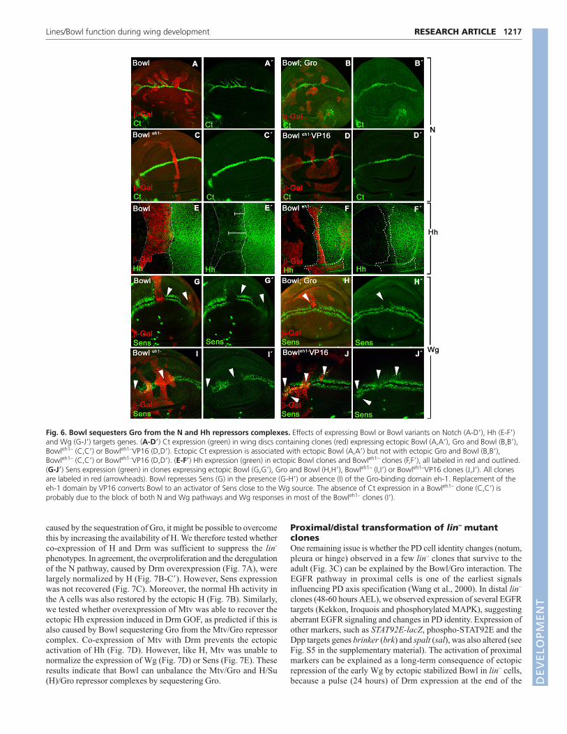
caused by the sequestration of Gro, it might be possible to overcomethis by increasing the availability of H. We therefore tested whetherco-expression of H and Drm was sufficient to suppress the lin-
phenotypes. In agreement, the overproliferation and the deregulationof the N pathway, caused by Drm overexpression (Fig. 7A), werelargely normalized by H (Fig. 7B-C�). However, Sens expressionwas not recovered (Fig. 7C). Moreover, the normal Hh activity inthe A cells was also restored by the ectopic H (Fig. 7B). Similarly,we tested whether overexpression of Mtv was able to recover theectopic Hh expression induced in Drm GOF, as predicted if this isalso caused by Bowl sequestering Gro from the Mtv/Gro repressorcomplex. Co-expression of Mtv with Drm prevents the ectopicactivation of Hh (Fig. 7D). However, like H, Mtv was unable tonormalize the expression of Wg (Fig. 7D) or Sens (Fig. 7E). Theseresults indicate that Bowl can unbalance the Mtv/Gro and H/Su(H)/Gro repressor complexes by sequestering Gro.
Proximal/distal transformation of lin– mutantclonesOne remaining issue is whether the PD cell identity changes (notum,pleura or hinge) observed in a few lin– clones that survive to theadult (Fig. 3C) can be explained by the Bowl/Gro interaction. TheEGFR pathway in proximal cells is one of the earliest signalsinfluencing PD axis specification (Wang et al., 2000). In distal lin–
clones (48-60 hours AEL), we observed expression of several EGFRtargets (Kekkon, Iroquois and phosphorylated MAPK), suggestingaberrant EGFR signaling and changes in PD identity. Expression ofother markers, such as STAT92E-lacZ, phospho-STAT92E and theDpp targets genes brinker (brk) and spalt (sal), was also altered (seeFig. S5 in the supplementary material). The activation of proximalmarkers can be explained as a long-term consequence of ectopicrepression of the early Wg by ectopic stabilized Bowl in lin– cells,because a pulse (24 hours) of Drm expression at the end of the
1217RESEARCH ARTICLELines/Bowl function during wing development
Fig. 6. Bowl sequesters Gro from the N and Hh repressors complexes. Effects of expressing Bowl or Bowl variants on Notch (A-D�), Hh (E-F�)and Wg (G-J�) targets genes. (A-D�) Ct expression (green) in wing discs containing clones (red) expressing ectopic Bowl (A,A�), Gro and Bowl (B,B�),Bowleh1– (C,C�) or Bowleh1–VP16 (D,D�). Ectopic Ct expression is associated with ectopic Bowl (A,A�) but not with ectopic Gro and Bowl (B,B�),Bowleh1– (C,C�) or Bowleh1–VP16 (D,D�). (E-F�) Hh expression (green) in ectopic Bowl clones and Bowleh1– clones (F,F�), all labeled in red and outlined.(G-J�) Sens expression (green) in clones expressing ectopic Bowl (G,G�), Gro and Bowl (H,H�), Bowleh1– (I,I�) or Bowleh1–VP16 clones (J,J�). All clonesare labeled in red (arrowheads). Bowl represses Sens (G) in the presence (G-H�) or absence (I) of the Gro-binding domain eh-1. Replacement of theeh-1 domain by VP16 converts Bowl to an activator of Sens close to the Wg source. The absence of Ct expression in a Bowleh1– clone (C,C�) isprobably due to the block of both N and Wg pathways and Wg responses in most of the Bowleh1– clones (I�).
DEVELO
PMENT

1218
second instar was sufficient to induce changes in the PD identity,even though Drm was not present continuously (see Fig. S2D in thesupplementary material).
As several of the upregulated genes in lin– clones, such asSTAT92E, brk, hth and tsh, are also normally expressed in the PEcells, it is possible that lin– cells are transformed to PE identity(rather than proximal DP identity). However, as lin– (48-60 AEL)clones also upregulated genes that are not present in the SC of thePE cells, such as dachsous (ds) (data not shown), iro, zfh2 orkekkon, this cannot be the whole explanation. Second DP lin– cellscould be transformed to MC fate. Supporting this possibility, all thegenes upregulated in lin– cells are normally expressed in the MC ofthe disc, including Bowl. Moreover, lin– clones differentiatetricomes and macrochaetes, compatible with a transformation toproximal structures such as pleura/hinge/notum (Fig. 3C).However, as the early-induced lin– clones died and only twin cloneswere recovered, we cannot exclude the possibility that Lin mighthave an early function in preventing the transformation to both MCand SC of the PE, as has been recently proposed (Nusinow et al.,2008).
Bowl function in the peripodial epitheliumThe regulatory interaction between Lin, Drm and Bowl restrictsBowl protein to the SC and MC within the PE. To determine thefunction of Bowl in these domains, we induced early bowl–
clones, marked by the expression of GFP (MARCM clones).Although the frequency of recovered clones in PE (Fig. 8A) isusually lower than in the DP (Fig. 8B), we could visualize largeclones in the PE and observed that they still expressed theperipodial markers Ubx (Fig. 8A,A�) and Hth (Fig. 8A,A�). Wealso expressed Bowl-RNAi in the PE and in the MC using ubx-Gal4 (Pallavi and Shashidhara, 2003). We found that some Ubx-Gal>UAS-bowlRNAi wing discs were smaller than wild-typediscs (Fig. 8C) and showed altered expression of Ubx (Fig.8D,D�) and Hth (Fig. 8D,D�). ubx-Gal4>UAS-bowlRNAi adultwings display (30%) reduction of the proximal wing andoccasionally the whole wing was missing (Fig. 8F,F�). Likewise,expressing UAS-bowlRNAi with pnr-Gal4 (expressed in thenotum primordium, including the MC expressing Bowl) results ina cleft in the thorax and absence of dorso/central bristles (Fig. 8I).
These results suggest that Bowl is required for normal wing andnotum development, possibly differentiating the signalingresponse between the SC/MC and DP cells.
DISCUSSIONThe Lin/Drm/Bowl cassette is emerging as an important molecularmechanism with which to coordinate various pathways in differentdevelopmental contexts (de Celis Ibeas and Bray, 2003; Hao et al.,2003; Hatini et al., 2005; Nusinow et al., 2008). In all cases, thesteady-state accumulation of Bowl is regulated by the relative levelsof Drm and Lin proteins. High levels of Drm impede binding of Linto Bowl and, thus, this transcriptional repressor becomes stabilizedin the nucleus. Here, we have found that regulatory interactionLin/Drm/Bowl also functions during wing development. In lin– orDrm GOF cause ectopic expression of Bowl and dramaticovergrowths within the wing disc. These overgrowths frequentlyshowed altered cell identity, resembling more proximal disc margincells. Some of the effects can be explained by the ability of Bowl tointeract with Gro co-repressor through the eh-1 motif, forming acomplex that sequesters Gro from other repressors complexes suchas Su(H)/H/Gro and Mtv/Gro.
Lin/Drm/Bowl regulative interactionAlthough Bowl is ubiquitously transcribed in the wing disc, Bowlprotein is present only in the SC and MC, being normally absentfrom the DP cells. The spatial distribution of nuclear Bowl isdependent on Drm, which causes Lin to relocalize to the cytoplasm.Drm is absent from most of the DP cells and, therefore, Lin turnsdown the steady-state accumulation of Bowl protein in these cells.In the absence of Lin, Bowl accumulates in the DP cell nuclei andelicits the dramatic alterations observed in lin– mutant cells.Therefore, the main function of Lin is to prevent Bowl accumulationin the DP cells, restricting Bowl protein to MC and SC of the PE.
The main alterations in lin–, Drm GOF or Bowl GOF clones canbe classified according to the signaling pathways temporallyaffected. The earliest defect observed is the repression ofWg pathway responses and the evidence suggests that Bowlfunctions as a repressor of the Wg pathway. However, activatedforms of nuclear Wg pathway components, such as ArmS10
or dTcf, cannot restore the expression of the proximal-distal
RESEARCH ARTICLE Development 136 (7)
Fig. 7. H or Mtv partially suppress the effects of ectopicDrm in the wing. (A-A�) ptc-Gal4> UAS-drm/hh-lacZ wingdisc showing a huge overgrowth and the induction of hh(red in A, grey in A�) and Wg (green in A, grey in A�) in theAP compartment region where Drm is induced. (B,B�) ptc-Gal4>UAS-H;UAS-drm/hh-lacZ wing disc. The co-expressionof H and Drm largely normalizes the overgrowth and theexpression of hh-lacZ (red in B and grey in B�) and partiallyrescues at the DV border by the ectopic Wg (green in B)caused by ectopic Drm. (C,C�) ptc-Gal4>UAS-H;UAS-drm.Note that Sens is still repressed (red in C, grey in C�).(D,D�) ptc-Gal4/UAS-mtv;UAS-drm/hh-lacZ wing disc.Overgrowth and activation of hh-lacZ are largely normalized(red in D and grey in D�) but the ectopic Wg expression(green in D) is not. (E,E�) ptc-Gal4>UAS-mtv;UAS-drm/UAS-GFP wing disc (GFP labels the Ptc expression domain in E).Sens repression is not normalized (red in E, grey in E�). ThePtc expression domain is marked with a broken line in allpanels.
DEVELO
PMENT

markers owing to repression of the Wg targets in lin–, indicatingthat Bowl must act in parallel to or downstream of Arm and dTcf,as was previously suggested (Green et al., 2002; Hatini et al.,2005).
Gro acts in the crosstalk of different signalingpathwaysBowl is a zinc-finger protein that can interact with the co-repressorGro directly through the eh-1 motif (Goldstein et al., 2005). Ourresults indicate that this mechanism is also important underconditions where Bowl accumulates in the wing disc. Most of thealterations observed in lin– or Drm GOF clones can be explained byBowl sequestering Gro from other repression complexes (causingactivation of N targets and Hh). Several results support this model.First, the strong genetic interaction between lin and gro alleles,where trans-heterozygous combinations between lin and gro allelesresult in dramatic phenotypes, argue that Gro is a limiting factor.Second, removal of eh-1 motif that recruits Gro, eliminates theeffects of Bowl on the Hh and N pathways. Third, ectopic expressionof Gro, H or Mtv partially suppress the phenotypes of ectopic Drmor Bowl. These observations imply a ‘tug of war’ between Bowl, Hand Mtv for Gro. Increased H or Mtv would shift the balance backin favor of N target repression and Hh repression.
By contrast, the repression of Wg pathway observed in lin– cellsappears to involve a different mechanism. Although the effect isBowl dependent, repression of Wg targets also occurs with Bowleh1–,indicating that Gro sequestration is not required. Similarly, co-expression of Bowl with H or Mtv cannot re-establish the repressionof the Wg targets. These results show that Bowl is able to repressWg targets independently of Gro and the observation that Bowleh1–
VP16 can cause some ectopic expression of Sens suggests that thismay involve a direct effect of Bowl on Wg targets.
Wnt/Wg, N and Hh signaling represent major conserved signalingchannels to control cell identity and behavior during development.An antagonistic interaction between the Wg and Hh has also beendescribed in the embryo (Hatini et al., 2005) and at the intersectionof the D/V and A/P compartment borders of the wing disc (Glise etal., 2002). Similarly, Wnt/Wg and N activities are closely entangledin many different systems. Mutual dependent interactions betweenN and Wnt signaling have been observed in vertebrate skinprecursors (Estrach et al., 2006), in rhombomere patterning (Chenget al., 2004) and in somitogenesis (Aulehla et al., 2003; Dale et al.,2003; Hofmann et al., 2004). It has also been reported thatorthologues of the Odd-skipped family, Osr1 and Osr2, function astranscriptional repressors during kidney formation (Tena et al.,2007). It is possible therefore that Lin/Bowl/Gro interaction isevolutionary conserved and it will be interesting to discover whetherlin is an important regulatory factor in other systems.
Bowl function in wing developmentBy analyzing lin– clones in the wing primordium, we haveuncovered the consequences of stabilizing Bowl in the DP cells.There are, however, two regions where Bowl accumulates normally,in the MC and SC within the PE. Removal of Bowl in the PE mightlead to ectopic Wg protein and thus to ectopic activity of the Wgsignaling to transform PE from squamous to columnar cells (Baena-Lopez et al., 2003). In this context, recently, it has shown that Bowlinhibition by ectopic expression of Lin results in the replacement ofthe PE by a mirror image duplication of the DP cells (Nusinow et al.,2008). However, we did not observe much alteration in cellmorphology nor in the expression of markers such as Ubx or Hthwhen Bowl was depleted in PE cells (bowl– clones and UAS-BowlRNAi). It could be that the recovered bowl– clones were notinduced early enough or that the levels of Bowl-RNAi were notsufficient to completely eliminate the Bowl function in these cells.Nevertheless, our manipulations revealed that bowl– phenotypes inthe proximal wing and notum were consistent with a functional role
1219RESEARCH ARTICLELines/Bowl function during wing development
Fig. 8. Bowl requirement in the peripodial cells of the wingdisc. (A-B�) Wing discs containing MARCM bowl2 clones (green) inthe PE (A) and in the DP (B). Note that Ubx (red in A,B and grey inA�,B�) and Hth (blue in A,B and grey in A�,B�) expression is notmodified in bowl2 clones in either the PE (A) or in the DP (B).(C-C�) PE cells of a wild-type disc showing the expression of Ubx (redin C and grey in C�) and Hth (green in C and grey in C�). (D-D�) ubx-Gal4>UAS-bowlRNAi wing discs showing the expression of Ubx (redin D and grey in D�), Hth (green in D and grey in D�) and Nub (blue inD). Both the size of the PE and the Ubx and Hth expression domainsare reduced. The outlined area corresponds to the DP cells expressingNub (blue in D). (E) Wild-type adult wing. (F,F�) ubx-Gal4>UAS-bowlRNAi wings. (G) Wild-type pnr expression domain (GFP in green)in the notum region. (H) Wild-type adult notum. (I) pnr-Gal4>UAS-bowl RNAi notum. Note the cleft in the notum and thedisorganization of micro and macrocheatae.
DEVELO
PMENT
1220
in MC. Therefore, we conclude that Lin has an important role inrestricting Bowl to the MC (and PE), delimiting a Bowl-freeterritory that forms the DP cells and enables their responsiveness tokey signaling pathways such as Wg.
We thank Aphrodite Bilioni and Fernando Díaz-Benjumea for critical reading ofthe manuscript, and members of the Bray and Guerrero laboratories fordiscussions and advice, especially Maki Daniels and Ainhoa Callejo. We arevery grateful to Carmen Ibáñez, Emma Harrison and Marie Quick for excellenttechnical assistance; to A. Betz, N. Azpiazu, H. Bellen, C. Delidakis, F. J. Diaz-Benjumea, S. Kerridge, J. Modolell, T. Tabata and the Developmental StudiesHybridoma Bank for providing antibodies; and to V. Hatini, M. Calleja, S.DiNardo, C. Delidakis, J. Lengyel, C. Rauskolb, S. Shashidara, T. Tabata, A.Martinez-Arias, Vienna Drosophila RNAi Center and the Bloomington StockCenter for Drosophila stocks. This work has been supported by grantsBFU2005-04183 and CSD2007-00008 from the Spanish MEC by aninstitutional grant from Fundación Areces given to the Centro de BiologíaMolecular ‘Severo Ochoa’ to I.G. and MEC fellowship (BMC2002-03839) toE.B. Work in the Bray laboratory was supported by a Programme Grant fromthe Medical Research Council (MRC) to S.J.B., with additional funding fromEMBO Short Term Fellowship (ASTF 143-2006) to E.B. and Royal SocietyTravelling Fellowship to I.G. Deposited in PMC for release after 6 months.
Supplementary materialSupplementary material for this article is available athttp://dev.biologists.org/cgi/content/full/136/7/1211/DC1
ReferencesAldaz, S., Morata, G. and Azpiazu, N. (2005). Patterning function of
homothorax/extradenticle in the thorax of Drosophila. Development 132, 439-446.
Apidianakis, Y., Grbavec, D., Stifani, S. and Delidakis, C. (2001). Grouchomediates a Ci-independent mechanism of hedgehog repression in the anteriorwing pouch. Development 128, 4361-4370.
Aulehla, A., Wehrle, C., Brand-Saberi, B., Kemler, R., Gossler, A., Kanzler, B.and Herrmann, B. G. (2003). Wnt3a plays a major role in the segmentationclock controlling somitogenesis. Dev. Cell 4, 395-406.
Azpiazu, N. and Morata, G. (2000). Function and regulation of homothorax inthe wing imaginal disc of Drosophila. Development 127, 2685-2693.
Baena-Lopez, L. A., Pastor-Pareja, J. C. and Resino, J. (2003). Wg and Egfrsignalling antagonise the development of the peripodial epithelium inDrosophila wing discs. Development 130, 6497-6506.
Barolo, S., Stone, T., Bang, A. G. and Posakony, J. W. (2002). Defaultrepression and Notch signaling: hairless acts as an adaptor to recruit thecorepressors Groucho and dCtBP to Suppressor of Hairless. Genes Dev. 16,1964-1976.
Bejarano, F., Perez, L., Apidianakis, Y., Delidakis, C. and Milan, M. (2007).Hedgehog restricts its expression domain in the Drosophila wing. EMBO Rep. 8,778-783.
Blair, S. S. (2007). Wing vein patterning in Drosophila and the analysis ofintercellular signaling. Annu. Rev. Cell Dev. Biol. 23, 293-319.
Bokor, P. and DiNardo, S. (1996). The roles of hedgehog, wingless and lines inpatterning the dorsal epidermis in Drosophila. Development 122, 1083-1092.
Bras-Pereira, C., Bessa, J. and Casares, F. (2006). Odd-skipped genes specify thesignaling center that triggers retinogenesis in Drosophila. Development 133,4145-4149.
Buscarlet, M. and Stifani, S. (2007). The ‘Marx’ of Groucho on development anddisease. Trends Cell Biol. 17, 353-361.
Calleja, M., Moreno, E., Pelaz, S. and Morata, G. (1996) Visualization of geneexpression in living adult Drosophila. Science 274, 252-255.
Capdevila, J. and Guerrero, I. (1994). Targeted expression of the signalingmolecule decapentaplegic induces pattern duplications and growth alterations inDrosophila wings. EMBO J. 13, 4459-4468.
Casares, F. and Mann, R. S. (2000). A dual role for homothorax in inhibiting wingblade development and specifying proximal wing identities in Drosophila.Development 127, 1499-1508.
Cavallo, R. A., Cox, R. T., Moline, M. M., Roose, J., Polevoy, G. A., Clevers,H., Peifer, M. and Bejsovec, A. (1998). Drosophila Tcf and Groucho interact torepress Wingless signalling activity. Nature 395, 604-608.
Cifuentes, F. J. and Garcia-Bellido, A. (1997). Proximo-distal specification in thewing disc of Drosophila by the nubbin gene. Proc. Natl. Acad. Sci. USA 94,11405-11410.
Cheng, Y. C., Amoyel, M., Qiu, X., Jiang, Y. J., Xu, Q. and Wilkinson, D. G.(2004). Notch activation regulates the segregation and differentiation ofrhombomere boundary cells in the zebrafish hindbrain. Dev. Cell 6, 539-550.
Dale, J. K., Maroto, M., Dequeant, M. L., Malapert, P., McGrew, M. andPourquie, O. (2003). Periodic notch inhibition by lunatic fringe underlies the
chick segmentation clock. Nature 421, 275-278.de Celis, J. F. and Ruiz-Gomez, M. (1995). groucho and hedgehog regulate
engrailed expression in the anterior compartment of the Drosophila wing.Development 121, 3467-3476.
de Celis, J. F. and Bray, S. (1997). Feed-back mechanisms affecting Notchactivation at the dorsoventral boundary in the Drosophila wing. Development124, 3241-3251.
de Celis, J. F., Garcia-Bellido, A. and Bray, S. J. (1996). Activation and functionof Notch at the dorsal-ventral boundary of the wing imaginal disc. Development122, 359-369.
de Celis, J. F., Tyler, D. M., de Celis, J. and Bray, S. J. (1998). Notch signallingmediates segmentation of the Drosophila leg. Development 125, 4617-4626.
de Celis Ibeas, J. M. and Bray, S. J. (2003). Bowl is required downstream ofNotch for elaboration of distal limb patterning. Development 130, 5943-5952.
Diaz-Benjumea, F. J. and Cohen, S. M. (1995). Serrate signals through Notch toestablish a Wingless-dependent organizer at the dorsal/ventral compartmentboundary of the Drosophila wing. Development 121, 4215-4225.
Dietzl, G., Chen, D., Schnorrer, F., Su, K. C., Barinova, Y., Fellner, M., Gasser,B., Kinsey, K., Oppel, S., Scheiblauer, S. et al. (2007). A genome-widetransgenic RNAi library for conditional gene inactivation in Drosophila. Nature448, 151-156.
Diez del Corral, R., Aroca, P., Gómez-Skarmeta, J. L., Cavodeassi, F. andModolell, J. (1999). The Iroquois homeodomain proteins are required to specifybody wall identity in Drosophila. Genes Dev. 13, 1754-1761.
Doherty, D., Feger, G., Younger-Shepherd, S., Jan, L. Y. and Jan, Y. N. (1996).Delta is a ventral to dorsal signal complementary to Serrate, another Notchligand, in Drosophila wing formation. Genes Dev. 10, 421-434.
Estrach, S., Ambler, C. A., Lo Celso, C., Hozumi, K. and Watt, F. M. (2006).Jagged 1 is a beta-catenin target gene required for ectopic hair follicle formationin adult epidermis. Development 133, 4427-4438.
Funakoshi, Y., Minami, M. and Tabata, T. (2001). mtv shapes the activitygradient of the Dpp morphogen through regulation of thickveins. Development128, 67-74.
Furriols, M. and Bray, S. (2000). Dissecting the mechanisms of suppressor ofhairless function. Dev. Biol. 227, 520-532.
Gallet, A., Erkner, A., Charroux, B., Fasano, L. and Kerridge, S. (1998). Trunk-specific modulation of wingless signalling in Drosophila by teashirt binding toarmadillo. Curr. Biol. 8, 893-902.
Glise, B., Jones, D. L. and Ingham, P. W. (2002). Notch and Wingless modulatethe response of cells to Hedgehog signalling in the Drosophila wing. Dev. Biol.248, 93-106.
Goldstein, R. E., Cook, O., Dinur, T., Pisante, A., Karandikar, U. C., Bidwai, A.and Paroush, Z. (2005). An eh1-like motif in odd-skipped mediates recruitmentof Groucho and repression in vivo. Mol. Cell. Biol. 25, 10711-10720.
Green, R. B., Hatini, V., Johansen, K. A., Liu, X. J. and Lengyel, J. A.(2002). Drumstick is a zinc finger protein that antagonizes Lines to controlpatterning and morphogenesis of the Drosophila hindgut. Development 129,3645-3656.
Hao, I., Green, R. B., Dunaevsky, O., Lengyel, J. A. and Rauskolb, C. (2003).The odd-skipped family of zinc finger genes promotes Drosophila legsegmentation. Dev. Biol. 263, 282-295.
Hasson, P., Egoz, N., Winkler, C., Volohonsky, G., Jia, S., Dinur, T., Volk, T.,Courey, A. J. and Paroush, Z. (2005). EGFR signaling attenuates Groucho-dependent repression to antagonize Notch transcriptional output. Nat. Genet.37, 101-105.
Hatini, V., Bokor, P., Goto-Mandeville, R. and DiNardo, S. (2000). Tissue- andstage-specific modulation of Wingless signaling by the segment polarity genelines. Genes Dev. 14, 1364-1376.
Hatini, V., Green, R. B., Lengyel, J. A., Bray, S. J. and Dinardo, S. (2005). TheDrumstick/Lines/Bowl regulatory pathway links antagonistic Hedgehog andWingless signaling inputs to epidermal cell differentiation. Genes Dev. 19, 709-718.
Hinz, U., Giebel, B. and Campos-Ortega, J. A. (1994). The basic-helix-loop-helixdomain of Drosophila lethal of scute protein is sufficient for proneural functionand activates neurogenic genes. Cell 76, 77-87.
Hofmann, M., Schuster-Gossler, K., Watabe-Rudolph, M., Aulehla, A.,Herrmann, B. G. and Gossler, A. (2004). WNT signaling, in synergy withT/TBX6, controls Notch signaling by regulating Dll1 expression in the presomiticmesoderm of mouse embryos. Genes Dev. 18, 2712-2717.
Iwaki, D. D., Johansen, K. A., Singer, J. B. and Lengyel, J. A. (2001).drumstick, bowl, and lines are required for patterning and cell rearrangement inthe Drosophila embryonic hindgut. Dev. Biol. 240, 611-626.
Klein, T., Seugnet, L., Haenlin, M. and Martinez Arias, A. (2000). Two differentactivities of Suppressor of Hairless during wing development in Drosophila.Development 127, 3553-3566.
Lawrence, N., Dearden, P., Hartley, D., Roose, J., Clevers, H. and Arias, A. M.(2000). dTcf antagonises Wingless signalling during the development andpatterning of the wing in Drosophila. Int. J. Dev. Biol. 44, 749-756.
RESEARCH ARTICLE Development 136 (7)
DEVELO
PMENT

Lee, J. J., von Kessler, D. P., Parks, S. and Beachy, P. A. (1992). Secretion andlocalized transcription suggest a role in positional signaling for products of thesegmentation gene hedgehog. Cell 71, 33-50.
Martin-Blanco, E., Gampel, A., Ring, J., Virdee, K., Kirov, N., Tolkovsky, A. M.and Martinez-Arias, A. (1998). puckered encodes a phosphatase thatmediates a feedback loop regulating JNK activity during dorsal closure inDrosophila. Genes Dev. 12, 557-570.
Milner, M., Bleasby, A. and Kelly, S. (1984). The role of the peripodialmembrane of leg and wing imaginal discs of Drosophila melanogaster duringevagination and differentiation in vitro. Wilhelm Roux’s Arch. Dev. Biol. 193,180-186.
Minami, M., Kinoshita, N., Kamoshida, Y., Tanimoto, H. and Tabata, T.(1999). brinker is a target of Dpp in Drosophila that negatively regulates Dpp-dependent genes. Nature 398, 242-246.
Morel, V., Lecourtois, M., Massiani, O., Maier, D., Preiss, A. andSchweisguth, F. (2001). Transcriptional repression by suppressor of hairlessinvolves the binding of a hairless-dCtBP complex in Drosophila. Curr. Biol. 11,789-792.
Mukherjee, T., Hombria, J. C. and Zeidler, M. P. (2005). Opposing roles forDrosophila JAK/STAT signalling during cellular proliferation. Oncogene 24, 2503-2511.
Mullor, J. L., Calleja, M., Capdevila, J. and Guerrero, I. (1997). Hedgehogactivity, independent of decapentaplegic, participates in wing disc patterning.Development 124, 1227-1237.
Musacchio, M. and Perrimon, N. (1996). The Drosophila kekkon genes: novelmembers of both the leucine-rich repeat and immunoglobulin superfamiliesexpressed in the CNS. Dev. Biol. 178, 63-76.
Nagel, A. C., Krejci, A., Tenin, G., Bravo-Patino, A., Bray, S., Maier, D. andPreiss, A. (2005). Hairless-mediated repression of notch target genes requiresthe combined activity of Groucho and CtBP corepressors. Mol. Cell. Biol. 25,10433-10441.
Ng, M., Diaz-Benjumea, F. J. and Cohen, S. M. (1995). Nubbin encodes a POU-domain protein required for proximal-distal patterning in the Drosophila wing.Development 121, 589-599.
Nolo, R., Abbott, L. A. and Bellen, H. J. (2000). Senseless, a Zn fingertranscription factor, is necessary and sufficient for sensory organ development inDrosophila. Cell 102, 349-362.
Nusinow, D., Greenberg, L. and Hatini, V. (2008). Reciprocal roles for bowl andlines in specifying the peripodial epithelium and the disc proper of theDrosophila wing primordium. Development 135, 3031-3041.
Pai, L. M., Orsulic, S., Bejsovec, A. and Peifer, M. (1997). Negative regulation ofArmadillo, a Wingless effector in Drosophila. Development 124, 2255-2266.
Pallavi, S. K. and Shashidhara, L. S. (2003). Egfr/Ras pathway mediatesinteractions between peripodial and disc proper cells in Drosophila wing discs.Development 130, 4931-4941.
Pignoni, F. and Zipursky, S. L. (1997). Induction of Drosophila eye developmentby decapentaplegic. Development 124, 271-278.
Preiss, A., Hartley, D. A. and Artavanis-Tsakonas, S. (1988). The moleculargenetics of Enhancer of split, a gene required for embryonic neural developmentin Drosophila. EMBO J. 7, 3917-3927.
Rulifson, E. J., Micchelli, C. A., Axelrod, J. D., Perrimon, N. and Blair, S. S.(1996). wingless refines its own expression domain on the Drosophila wingmargin. Nature 384, 72-74.
Takei, Y., Ozawa, Y., Sato, M., Watanabe, A. and Tabata, T. (2004). ThreeDrosophila EXT genes shape morphogen gradients through synthesis of heparansulfate proteoglycans. Development 131, 73-82.
Tena, J. J., Neto, A., de la Calle-Mustienes, E., Bras-Pereira, C., Casares, F.and Gomez-Skarmeta, J. L. (2007). Odd-skipped genes encode repressors thatcontrol kidney development. Dev. Biol. 301, 518-531.
Terriente, J., Perea, D., Suzanne, M. and Diaz-Benjumea, F. J. (2008). TheDrosophila gene zfh2 is required to establish proximal-distal domains in the wingdisc. Dev. Biol. 320, 102-112.
Tolwinski, N. S. and Wieschaus, E. (2004). A nuclear escort for beta-catenin.Nat. Cell Biol. 6, 579-580.
van de Wetering, M., Cavallo, R., Dooijes, D., van Beest, M., van Es, J.,Loureiro, J., Ypma, A., Hursh, D., Jones, T., Bejsovec, A. et al. (1997).Armadillo coactivates transcription driven by the product of the Drosophilasegment polarity gene dTCF. Cell 88, 789-799.
Wang, L. and Coulter, D. E. (1996). bowel, an odd-skipped homolog, functionsin the terminal pathway during Drosophila embryogenesis. EMBO J. 15, 3182-3196.
Wang, S. H., Simcox, A. and Campbell, G. (2000). Dual role for Drosophilaepidermal growth factor receptor signaling in early wing disc development.Genes Dev. 14, 2271-2276.
Whitworth, A. J. and Russell, S. (2003). Temporally dynamic response toWingless directs the sequential elaboration of the proximodistal axis of theDrosophila wing. Dev. Biol. 254, 277-288.
Wu, J. and Cohen, S. M. (2002). Repression of Teashirt marks the initiation ofwing development. Development 129, 2411-2418.
Yeo, S. L., Lloyd, A., Kozak, K., Dinh, A., Dick, T., Yang, X., Sakonju, S. andChia, W. (1995). On the functional overlap between two Drosophila POUhomeo domain genes and the cell fate specification of a CNS neural precursor.Genes Dev. 9, 1223-1236.
Zecca, M. and Struhl, G. (2002a). Control of growth and patterning of theDrosophila wing imaginal disc by EGFR-mediated signaling. Development 129,1369-1361.
Zecca, M. and Struhl, G. (2002b). Subdivision of the Drosophila wing imaginaldisc by EGFR-mediated signaling. Development 129, 1357-1368.
Zecca, M. and Struhl, G. (2007). Recruitment of cells into the Drosophila wingprimordium by a feed-forward circuit of vestigial autoregulation. Development134, 3001-3010.
Zirin, J. D. and Mann, R. S. (2007). Nubbin and Teashirt mark barriers to clonalgrowth along the proximal-distal axis of the Drosophila wing. Dev. Biol. 304,745-758.
1221RESEARCH ARTICLELines/Bowl function during wing development
DEVELO
PMENT