lipid-induced epigenomic changes in human macrophages
TRANSCRIPT

University of KentuckyUKnowledge
Gill Heart & Vascular Institute Faculty Publications Heart & Vascular
4-2-2015
Lipid-Induced Epigenomic Changes in HumanMacrophages Identify a Coronary Artery Disease-Associated Variant that Regulates PPAP2BExpression through Altered C/EBP-Beta BindingMichael E. ReschenUniversity of Oxford, UK
Kyle J. GaultonUniversity of Oxford, UK
Da LinUniversity of Oxford, UK
Elizabeth J. SoilleuxUniversity of Oxford, UK
Andrew J. MorrisUniversity of Kentucky, [email protected]
See next page for additional authors
Right click to open a feedback form in a new tab to let us know how this document benefits you.Follow this and additional works at: https://uknowledge.uky.edu/heart_facpub
Part of the Cardiology Commons
This Article is brought to you for free and open access by the Heart & Vascular at UKnowledge. It has been accepted for inclusion in Gill Heart &Vascular Institute Faculty Publications by an authorized administrator of UKnowledge. For more information, please [email protected].
Repository CitationReschen, Michael E.; Gaulton, Kyle J.; Lin, Da; Soilleux, Elizabeth J.; Morris, Andrew J.; Smyth, Susan S.; and O'Callaghan,Christopher A., "Lipid-Induced Epigenomic Changes in Human Macrophages Identify a Coronary Artery Disease-Associated Variantthat Regulates PPAP2B Expression through Altered C/EBP-Beta Binding" (2015). Gill Heart & Vascular Institute Faculty Publications.6.https://uknowledge.uky.edu/heart_facpub/6

AuthorsMichael E. Reschen, Kyle J. Gaulton, Da Lin, Elizabeth J. Soilleux, Andrew J. Morris, Susan S. Smyth, andChristopher A. O'Callaghan
Lipid-Induced Epigenomic Changes in Human Macrophages Identify a Coronary Artery Disease-AssociatedVariant that Regulates PPAP2B Expression through Altered C/EBP-Beta Binding
Notes/Citation InformationPublished in PLOS Genetics, v. 11, no. 4, article e1005061, p. 1-33.
© 2015 Reschen et al.
This is an open access article distributed under the terms of the Creative Commons Attribution License,which permits unrestricted use, distribution, and reproduction in any medium, provided the original authorand source are credited
Digital Object Identifier (DOI)http://dx.doi.org/10.1371/journal.pgen.1005061
This article is available at UKnowledge: https://uknowledge.uky.edu/heart_facpub/6

RESEARCH ARTICLE
Lipid-Induced Epigenomic Changes in HumanMacrophages Identify a Coronary ArteryDisease-Associated Variant that RegulatesPPAP2B Expression through Altered C/EBP-Beta BindingMichael E. Reschen1☯, Kyle J. Gaulton1☯¤, Da Lin1, Elizabeth J. Soilleux2, AndrewJ. Morris3,4, Susan S. Smyth3,4, Christopher A. O'Callaghan1*
1 Wellcome Trust Centre for Human Genetics, University of Oxford, Oxford, United Kingdom, 2 NuffieldDepartment of Clinical Laboratory Sciences, University of Oxford and Department of Cellular Pathology, JohnRadcliffe Hospital, Oxford, United Kingdom, 3 Division of Cardiovascular Medicine, The Gill Heart Institute,University of Kentucky, Lexington, Kentucky, United States of America, 4 Department of Veterans AffairsMedical Center, Lexington, Kentucky, United States of America
☯ These authors contributed equally to this work.¤ Current address: Department of Genetics, Stanford University, Stanford California United States of America* [email protected]
AbstractGenome-wide association studies (GWAS) have identified over 40 loci that affect risk of cor-
onary artery disease (CAD) and the causal mechanisms at the majority of loci are unknown.
Recent studies have suggested that many causal GWAS variants influence disease
through altered transcriptional regulation in disease-relevant cell types. We explored
changes in transcriptional regulation during a key pathophysiological event in CAD, the en-
vironmental lipid-induced transformation of macrophages to lipid-laden foam cells. We used
a combination of open chromatin mapping with formaldehyde-assisted isolation of regulato-
ry elements (FAIRE-seq) and enhancer and transcription factor mapping using chromatin
immuno-precipitation (ChIP-seq) in primary human macrophages before and after exposure
to atherogenic oxidized low-density lipoprotein (oxLDL), with resultant foam cell formation.
OxLDL-induced foam cell formation was associated with changes in a subset of open chro-
matin and active enhancer sites that strongly correlated with expression changes of nearby
genes. OxLDL-regulated enhancers were enriched for several transcription factors includ-
ing C/EBP-beta, which has no previously documented role in foam cell formation. OxLDL
exposure up-regulated C/EBP-beta expression and increased genomic binding events,
most prominently around genes involved in inflammatory response pathways. Variants at
CAD-associated loci were significantly and specifically enriched in the subset of chromatin
sites altered by oxLDL exposure, including rs72664324 in an oxLDL-induced enhancer at
the PPAP2B locus. OxLDL increased C/EBP beta binding to this site and C/EBP beta bind-
ing and enhancer activity were stronger with the protective A allele of rs72664324. In addi-
tion, expression of the PPAP2B protein product LPP3 was present in foam cells in human
PLOSGenetics | DOI:10.1371/journal.pgen.1005061 April 2, 2015 1 / 33
a11111
OPEN ACCESS
Citation: Reschen ME, Gaulton KJ, Lin D, SoilleuxEJ, Morris AJ, Smyth SS, et al. (2015) Lipid-InducedEpigenomic Changes in Human MacrophagesIdentify a Coronary Artery Disease-AssociatedVariant that Regulates PPAP2B Expression throughAltered C/EBP-Beta Binding. PLoS Genet 11(4):e1005061. doi:10.1371/journal.pgen.1005061
Editor: Gioacchino Natoli, European Institute ofOncology, ITALY
Received: June 17, 2014
Accepted: February 9, 2015
Published: April 2, 2015
Copyright: © 2015 Reschen et al. This is an openaccess article distributed under the terms of theCreative Commons Attribution License, which permitsunrestricted use, distribution, and reproduction in anymedium, provided the original author and source arecredited.
Data Availability Statement: The data discussed inthis publication have been deposited in NCBI's GeneExpression Omnibus and are accessible throughGEO Super Series accession number GSE54975.
Funding: This work was supported by the WellcomeTrust (097089/Z/11/Z), the Medical Research Council(G116/165) and the National Institute for HealthResearch Oxford Comprehensive BiomedicalResearch Centre Program. We thank the HighThroughput Genomics Group at the Wellcome TrustCentre for Human Genetics (Wellcome Trust grant

atherosclerotic plaques and oxLDL exposure up-regulated LPP3 in macrophages resulting
in increased degradation of pro-inflammatory mediators. Our results demonstrate a genetic
mechanism contributing to CAD risk at the PPAP2B locus and highlight the value of study-
ing epigenetic changes in disease processes involving pathogenic environmental stimuli.
Author Summary
Coronary artery disease is a complex disease where over 40 genomic loci contributing togenetic risk have been identified. However, identifying the precise variants, genomic ele-ments and genes that mediate this risk at each locus has proved challenging. We hypothe-sized that some genetic risk variants may influence a key step in development of coronaryartery disease, which occurs when macrophages encounter environmentally-derived lipid.These cells take up lipid and accumulate in atherosclerotic plaques in the walls of bloodvessels where they contribute to the inflammatory atherosclerotic disease process. There-fore, we studied the effects of this lipid exposure on the genomic activity of these cells. En-vironmental lipid exposure triggered changes in transcriptional regulation and geneexpression. Variants at coronary artery disease risk loci were enriched for genomic regionsaltered by lipid exposure. We studied one such risk variant rs72664324 in detail and foundthat it altered binding of the C/EBP-beta transcription factor and altered expression of thePPAP2B gene. PPAP2B encodes an enzyme that degrades pro-inflammatory substances.Our study demonstrates a hitherto unknown genetic mechanism underlying atherosclerot-ic heart disease and demonstrates the value of studying changes in transcriptional regula-tion in key disease processes involving environmental influences.
IntroductionCoronary artery disease (CAD) is the leading cause of death worldwide [1]. Most cases arecaused by atherosclerosis, a form of chronic inflammation in arterial walls that involves the ac-cumulation of lipid-containing plaques [2,3]. Blood levels of low-density lipoproteins (LDL)are a major environmentally-influenced risk factor for CAD and one of the more successfulpreventative treatments, statin therapy, lowers LDL levels [4–6]. A greater understanding ofthe molecular events by which lipoproteins cause atherosclerosis is a prerequisite for rationaldevelopment of new therapies targeting this aspect of atherosclerosis.
CAD is a complex disease with a strong heritable component [7]. A major development inthe study of the heritable component of complex diseases has been the application of genome-wide association studies (GWAS) to identify regions of the genome that contain genetic vari-ants that mediate this heritable risk. These studies have identified over 40 genomic loci harbor-ing genetic variants that influence CAD risk [8]. Although the contribution of each locus to theoverall risk is typically small, the gene pathways mediating risk at each individual locus containuseful biological information and may involve potential therapeutic targets [8]. The majority ofCAD risk variants do not alter the sequence of protein coding genes; thus the mechanism bywhich a risk locus operates is not typically identified by GWAS studies themselves. Further-more, the particular SNP identified by a GWAS study may only be in linkage disequilibrium(LD) with the causative SNP. Several studies have identified CAD-associated variants that altertranscription factor binding or miRNA binding, suggesting that many causal variants underly-ing CAD risk influence gene regulatory processes [9–11]. However, for most CAD loci the
Lipid-Induced Macrophage Epigenomic Changes Identify CAD Risk Variant
PLOS Genetics | DOI:10.1371/journal.pgen.1005061 April 2, 2015 2 / 33
reference 090532/Z/09/Z and MRC Hub grantG0900747 91070) for the generation of thesequencing data. This study was supported by grantsfrom the NIH, American Heart Association andDepartment of Veterans Affairs and conducted in partusing resources provided by the Lexington VeteransAffairs Medical Center. The recruitment of participantsfrom the Oxford Biobank was supported by the NIHROxford Biomedical Research Centre and the NationalNIHR Bioresource. The funders had no role in studydesign, data collection and analysis, decision topublish, or preparation of the manuscript.
Competing Interests: The authors have declaredthat no competing interests exist.

mechanism by which CAD-associated variants affect the disease is not known. Indeed for mostCAD loci, it is not clear in which cell type the risk variants exert their effects.
In atherosclerosis, LDL cholesterol is deposited in the arterial wall and undergoes modifica-tions, such as oxidation, that in turn promote pro-inflammatory processes [2,12]. Monocytesare recruited to these sites and differentiate into macrophages, which have a variety of scaven-ger surface receptors for oxidized LDL (oxLDL) [2,12]. The uptake of oxLDL by macrophagesoccurs via scavenger receptors, phagocytosis and macro-pinocytosis and is fundamental to thedevelopment of atherosclerotic lesions [12]. An imbalance in the uptake and degradative me-tabolism of oxLDL leads to the accumulation of lipid-laden vesicles in macrophages giving riseto a foam cell phenotype [2,12]. A central role for macrophages in atherosclerosis is evidencedby the inhibition of atherosclerosis in mice with severe macrophage deficiency due to knockoutof macrophage colony-stimulating factor [13].
Foam cells contribute to the pathogenesis of atherosclerosis by releasing pro-inflammatorymediators that recruit additional cells and matrix metallo-proteinases that can destabilize pla-ques [2,12]. In addition, foam cell apoptosis causes release of toxic lipids into the necrotic coreof the lesion [14,15]. Plaque rupture can expose this thrombogenic mixture to luminal bloodleading to thrombosis and blood vessel occlusion [2]. Several studies have documented changesin lipid response and oxidative stress response pathways during foam cell formation, but un-derstanding of how oxLDL influences transcriptional regulation in macrophages is incomplete[16,17]. Nevertheless, reprogramming of the macrophage response to lipid is a plausibletherapeutic strategy.
Development and maintenance of cell type identity depends on the binding of hundredsof different transcription factors to thousands of cis-regulatory elements [18]. Active cis-regulatory regions are typically characterized by nucleosome depletion, a necessary conditionfor DNA binding by many transcription factors [19–21]. A variety of techniques have emergedwhich allow mapping of this accessible open chromatin, such as DNAseI hypersensitivity orformaldehyde-assisted isolation of regulatory elements (FAIRE-seq) [22,23]. In parallel, chro-matin immunoprecipitation (ChIP-seq) approaches have been developed to identify genomicsites where histones in nucleosomes that flank open chromatin have undergone post-transla-tion modifications that indicate the function, such as enhancer function, of the open chromatin[18,24]. The identification of cis-regulatory elements is useful in the study of functional geneticrisk variants. Overlap of a risk variant with a functional regulatory element in a particular celltype indicates that the variant might exert its effect in that cell type by altering the activity ofthe regulatory element [23,25,26]. Several large-scale studies have demonstrated that transcrip-tional regulatory sites identified via high-throughput assay in specific human cell types are en-riched for disease-associated GWAS variants, highlighting the importance of pinpointing celltype regulatory elements [27,28].
In this study we employ FAIRE-seq and ChIP-seq assays to, first, show that exposure of pri-mary human macrophages to oxLDL causes changes in chromatin accessibility and histonemodification at a subset of regulatory sites. We then demonstrate that these changes are corre-lated with local expression of genes involved in foam cell and atherosclerotic processes and alsobinding of key transcription factors such as C/EBP-beta. We also show that these sites prefer-entially harbor variants influencing CAD risk and identify a regulatory variant rs72664324 thataffects PPAP2B expression through altered C/EBP-beta binding and enhancer activity. Finally,we demonstrate that the protein product of PPAP2B (Lipid Phosphate Phosphohydrolase3LPP3) is upregulated in foam cells and that this upregulation is associated with increasedLPP3 enzymatic activity for degrading pro-inflammatory mediators. Our approach of studyingchromatin profiles in the context of a major environmental disease stimulus as a means toidentify the causal mechanisms of GWAS loci is widely applicable to other diseases.
Lipid-Induced Macrophage Epigenomic Changes Identify CAD Risk Variant
PLOS Genetics | DOI:10.1371/journal.pgen.1005061 April 2, 2015 3 / 33

Results
Oxidized LDL triggers changes in expression of genes near CAD riskvariantsExposure of primary human macrophages to oxLDL resulted in marked lipid uptake and trans-formation to a lipid-laden foam cell phenotype, as demonstrated by oil red O lipid staining (S1Fig). We measured gene expression levels before and after exposure to oxLDL and identified1,283 and 1,376 genes significantly up- and down-regulated respectively (S1A/B Table,Fig. 1A). Among the most differentially expressed genes were several with previously knowninvolvement in foam cell formation, including the low-density lipoprotein receptor (LDLR)and perilipin 2 (PLIN2) [2,29] (Table 1). We observed enrichment among all differentially ex-pressed genes for gene ontology terms pertaining to lipid and sterol handling (Fig. 1B). Path-way analyses on the differentially regulated genes further highlighted pathways involved ininflammation (Fig. 1C).
We next explored the relationship between gene expression changes during foam cell for-mation and CAD risk variants. For this we identified genomic intervals containing all SNPs inhigh linkage disequilibrium (LD) (r2 > 0.8) with known CAD-associated variants and thenidentified all genes within 50kb of these CAD risk locus intervals (S2 Table). These 132 genesat CAD risk loci were enriched for processes relevant to atherosclerosis, including lipid han-dling and foam cell formation (S3, S4 Tables). Exposure of macrophages to oxLDL altered theexpression of 19 of these 132 genes, a significant excess compared to expectation across all ex-pressed genes (Binomial p = 0.025). This suggests that CAD variants may act on nearby genesto influence the macrophage response to oxLDL, a key stage in the pathogenesisof atherosclerosis.
OxLDL induces major changes in macrophage chromatin accessibilityTo understand the transcriptional pathways mediating the effect of oxLDL on gene expression,we identified candidate regulatory DNA elements that underwent oxLDL-induced changes inchromatin structure. We began by generating genome-wide maps of open chromatin in prima-ry human macrophages before and after oxLDL exposure, with consequent foam cell forma-tion. Formaldehyde-assisted isolation of regulatory elements with high-throughput sequencing(FAIRE-seq) identified 130,491 and 123,400 nucleosome-depleted open chromatin sites inmacrophages and foam cells respectively (S5, S6 Tables) [19,22]. Sites at promoters, definedas� 1kb upstream of the Refseq transcriptional start site (TSS), accounted for 6.1% of openchromatin sites in macrophages and 7.1% in foam cells (S2 Fig). The promoters of genes with alow level of expression had low FAIRE signal consistent with a relatively nucleosome-boundchromatin profile. By contrast, the promoters of maximally expressed genes had a high FAIREsignal consistent with nucleosome-depletion (Fig. 2A, 2B) [19]. Genes whose expression wasaltered by oxLDL exposure had an intermediate chromatin profile at their promoters. OxLDLsignificantly increased nucleosome depletion at the promoters of up-regulated genes, (p = 1.08x 10⁻²⁷) and reduced it at the promoters of down-regulated genes (p = 5.42 x 10⁻²¹) indicatingits capacity to trigger chromatin remodelling (Fig. 2A, 2B).
All genomic sites with significant oxLDL-induced changes in chromatin profile were identi-fied and termed dynamic chromatin sites (Fig. 3A). OxLDL changed the chromatin profile ataround 10% of all open chromatin sites (13,516 sites of which 12,754 (94%) were non-promot-er sites (defined as 1kb from a known RefSeq TSS)(S7 Table). 7,276 (54%) of these 13,516 siteshad a more nucleosome-depleted profile and 6,240 had a more nucleosome-bound profile infoam cells (Fig. 3B). The size distribution of the dynamic chromatin sites demonstrates that
Lipid-Induced Macrophage Epigenomic Changes Identify CAD Risk Variant
PLOS Genetics | DOI:10.1371/journal.pgen.1005061 April 2, 2015 4 / 33

changes at most non-promoter sites involve displacement of a single nucleosome, whereasthose at promoter sites may involve displacement of one or two nucleosomes (S3 Fig). Overall,only a fraction of macrophage open chromatin sites, the majority distal to known promoter re-gions, are changed in response to oxLDL.
Fig 1. Effect of oxLDL-induced foam cell formation on gene expression in humanmacrophages. (A)Heatmap showing genes differentially expressed between macrophages and foam cells (subset withadjusted p< 0.0001). Each column in each half of the main panel represents one of the 5 donors. Each row inthe figure represents one gene. Most changes are common to all donors. Dendrograms indicate clustering ofgenes by expression profile (left axis) and by sample (top axis). Dashed vertical lines indicate the medianexpression signal and solid vertical lines indicate expression for that gene and sample relative to the median.The inset indicates the expression distribution for the data and the color key for the heatmap. (B and C)Enrichment of differentially expressed genes for gene sets related to biological processes (B) and biologicalpathways (C).
doi:10.1371/journal.pgen.1005061.g001
Lipid-Induced Macrophage Epigenomic Changes Identify CAD Risk Variant
PLOS Genetics | DOI:10.1371/journal.pgen.1005061 April 2, 2015 5 / 33

Table 1. Top 10 most up-regulated and down-regulated genes during oxLDL-induced foam cell formation.
Up-regulated genes Down-regulated genes
Rank Genesymbol
Gene Fold change(log2)
Gene symbol Gene Fold change(log2)
1 PLIN2 Perilipin 2 2.70 LDLR Low Density Lipoprotein Receptor -3.09
2 AKR1C4 Aldo-Keto Reductase Family 1,Member C4
2.50 F13A1 Coagulation Factor XIII, A1Polypeptide
-2.81
3 AKR1C3 Aldo-Keto Reductase Family 1,Member C3
2.45 SC4MOL(MSMO1)
Methylsterol Monooxygenase 1 -2.57
4 ABCG1 ATP-Binding Cassette, Sub-Family G(WHITE), Member 1
2.44 SQLE Squalene Epoxidase -2.46
5 PDK4 Pyruvate Dehydrogenase Kinase,Isozyme 4
1.95 INSIG1 Insulin Induced Gene 1 -2.15
6 PPAP2B Phosphatidic Acid Phosphatase Type2B
1.65 FCGBP Fc Fragment Of IgG Binding Protein -2.01
7 LOC644496 Pseudo gene similar to cathepsin L1preproprotein
1.54 CD93 CD93 Molecule -2.00
8 TMEM158 Transmembrane Protein 158 1.51 ALOX5AP Arachidonate 5-Lipoxygenase-Activating Protein
-1.91
9 FABP4 Fatty Acid Binding Protein 4, Adipocyte 1.50 DHCR7 7-Dehydrocholesterol Reductase -1.83
10 SLC7A11 Solute Carrier Family 7 1.45 FADS1 Fatty acid desaturase 1 -1.81
doi:10.1371/journal.pgen.1005061.t001
Fig 2. OxLDL-induced changes in chromatin structure determined by FAIRE-seq. (A) Normalized FAIRE-seq signal in 2000bp windows centered onthe TSS for gene sets grouped by their expression characteristics. OxLDL-induced changes in open chromatin were associated with concordant changes ingene expression (higher FAIRE signal indicates more open chromatin, *** p< 0.0005). (B) Profile view of FAIRE-seq signal around TSSs showing sharplydelineated promoter open chromatin profiles and their relationship with expression.
doi:10.1371/journal.pgen.1005061.g002
Lipid-Induced Macrophage Epigenomic Changes Identify CAD Risk Variant
PLOS Genetics | DOI:10.1371/journal.pgen.1005061 April 2, 2015 6 / 33

We next determined whether dynamic chromatin sites had characteristics representative oftrue regulatory elements. First, we tested the extent to which these sites overlapped regulatoryelements identified in the ENCODE project, compared to control DNA sequences randomlychosen from within 10kb of each site (see Methods). Dynamic sites were most prominently en-riched for overlap with enhancer and TFBS elements (p< 0.01) (Fig. 3C). Second, we deter-mined whether the dynamic chromatin sites were evenly distributed across the genome. Weobserved significant clustering (p< 0.05) of dynamic sites suggesting a non-random distribu-tion of these sites. Gene set enrichment analysis of the clustered sites identified processes
Fig 3. Characterization of the dynamic chromatin landscape resulting from oxLDL exposure. (A) Representative chromatin profile at theCD36 locusshowing a dynamic chromatin cluster comprising three dynamic chromatin sites; the lower panel close-up view shows the relevant FAIRE-seq signals formacrophages, foam cells and the normalized dynamic signal (foam cell minus macrophage signal). Dynamic sites are highlighted in red boxes and areknown DNAse hypersensitivity sites in other cell types in the ENCODE database; one site shows conservation in vertebrates. (B) Heatmap of openchromatin profile at all dynamic chromatin sites, split into panels showing sites with increased or decreased FAIRE-seq signal. Each line represents a 5kbwindow centered on an individual site. Sites are clustered into groups with similar signal patterns. (C) Dynamic chromatin sites are enriched for non-codingregulatory elements found in 9 other cell types (* p< 0.05, ** p< 0.005, TFBS—transcription factor binding site). (D) Dynamic chromatin clusters areenriched for gene sets involved in foam cell formation and lipid handling.
doi:10.1371/journal.pgen.1005061.g003
Lipid-Induced Macrophage Epigenomic Changes Identify CAD Risk Variant
PLOS Genetics | DOI:10.1371/journal.pgen.1005061 April 2, 2015 7 / 33

relevant to foam cell formation and cellular lipid handling (Fig. 3D). Further, we performedmotif analysis using the genomic sequence within dynamic chromatin sites. We identified sev-eral motifs enriched in these sequences, including JunD/AP1, NFE2L2 and EGR1 (S8 and S9Tables). These results suggest dynamic chromatin sites are enriched for regulatory potential.
Macrophage enhancers undergo major oxLDL-induced changesAs the majority of dynamic chromatin sites were distal to known promoters, we hypothesizedthat many of these sites represented enhancer elements involved in the macrophage responseto oxLDL and thus in foam cell identity. We, therefore, mapped the enhancer landscape inmacrophages and foam cells using ChIP-seq for the histone modification H3K27ac, which isknown to mark active enhancers [24,30]. We observed significant oxLDL-induced changes inH3K27ac signal at 39,194 sites, termed ‘dynamic enhancer sites’, 95% of which were distal toknown promoter regions (Fig. 4A, S10 Table). We found a strong positive correlation betweenthe change in enhancer signal and the change in expression of the nearest differentially express-ed gene (r2 = 0.45; p< 2.2x10⁻¹⁶) (Fig. 4B, 4C). We identified ‘super-enhancer’ elements,which are spatially clustered enhancers that, in other contexts, are relevant to the expression ofcell-type specific genes [31]. We identified 1,081 and 1,098 super enhancers in macrophagesand foam cells, respectively (Fig. 4D, S11 and S12 Tables). Genes overlapping or adjacent tosuper enhancers had significantly higher expression than those associated with other enhancers(Fig. 4D inset). Of the 1098 foam cell super-enhancer clusters, 213 were evident as super-enhancer clusters only in foam cells and genes nearest to or overlapping them had significantlyhigher expression in foam cells compared to macrophages (p = 4.61x10⁻¹⁰). The analysis of en-hancers indicates that oxLDL-induced changes in gene expression are mediated in large partthrough changes in local enhancer activity.
We next identified sites that had significant oxLDL-induced changes in both chromatin sig-nal and enhancer signal. 1743 dynamic chromatin sites overlapped one or more of 2004 dy-namic enhancer sites. Of these 2004 enhancer sites there was concordance in the direction ofchange in enhancer and open chromatin signals in 1817 sites (90.6%). These 1743 dynamicchromatin sites were, in turn, significantly associated with a directionally concordant change inexpression of the nearest genes and chromatin signal (oxLDL-induced chromatin sitesp = 6.9x10⁻²⁷, oxLDL-suppressed chromatin sites p = 0.0004, Fig. 5A). We also observed en-richment among genes nearest to these 1,743 dynamic sites for gene sets related to atheroscle-rotic vascular disease (Fig. 5B). These results demonstrate that a discrete number of sites withchanges in both chromatin accessibility and enhancer signal are associated with disease-rele-vant changes in gene expression.
Dynamic enhancer sites identify C/EBP-beta as a novel oxLDL-regulated transcription factor in macrophagesWe used the dynamic chromatin and enhancer sites to identify potential upstream transcrip-tional regulators driving oxLDL-induced changes in macrophage chromatin and gene expres-sion. We performed motif enrichment analysis using the genomic sequence of the 1,743 siteswith changes in both chromatin and enhancer signal (Fig. 5C, S13 Table). We observed enrich-ment for AP1-related factors, such as JunD, which bind enhancer elements, and NFE2L2,which is involved in the anti-oxidant response [17,21]. We also observed enrichment for fac-tors regulated directly by cellular lipids, including LXRA and PPARG [32]. Notably, we ob-served enrichment for several predicted C/EBP motifs including C/EBP-beta, which is a knownmyeloid transcription factor, but has no known role in foam cell formation [33]. These results
Lipid-Induced Macrophage Epigenomic Changes Identify CAD Risk Variant
PLOS Genetics | DOI:10.1371/journal.pgen.1005061 April 2, 2015 8 / 33

Fig 4. OxLDL-regulated dynamic enhancer signals correlate with expression changes in local genes. (A) Open chromatin (FAIRE) and enhancer(H3K27ac) profile at the IL6R locus showing a dynamic chromatin cluster that overlaps a large, intronic dynamic enhancer site with increased signal inoxLDL-treated cells. (B) Correlation between fold change in gene expression for differentially expressed genes and mean fold change in enhancer signal fordynamic sites annotated to their nearest gene (r2 = 0.45, Pearson r = 0.67). (C) The proportion of the genes in each quartile that are associated with adynamic enhancer site is plotted. Differentially expressed genes were split into oxLDL up and down regulated genes and further split into quartiles based onfold change in expression (-/+ and—-/++++ indicate least and most differential quartiles, respectively). The proportion of differentially expressed genes ineach quartile that are associated with a directionally concordant change in a dynamic enhancer site is correlated with the magnitude of gene expressionchange. (D) Enhancers in macrophages or foam cells were aggregated if within 12.5kb and plotted according to H3k27ac signal rank. The insets show geneexpression (log2, normalized) for genes overlapping either enhancers within super enhancer domains or other enhancers.
doi:10.1371/journal.pgen.1005061.g004
Lipid-Induced Macrophage Epigenomic Changes Identify CAD Risk Variant
PLOS Genetics | DOI:10.1371/journal.pgen.1005061 April 2, 2015 9 / 33

suggest hypotheses concerning the role of specific regulatory proteins in the macrophage re-sponse to oxLDL, including proteins such as C/EBP-beta with no prior links to this process.
Given enrichment for C/EBP-beta motifs in dynamic enhancer sites, in addition to up-regu-lation of the CEBPB gene itself after oxLDL exposure (S1B Table, Fig. 5C), we hypothesized
Fig 5. OxLDL induces changes in both open chromatin and enhancer status at a subset of sites withimportant regulatory function. (A) The oxLDL regulated change in open chromatin, at sites which alsohave changes in enhancer signal, is correlated with expression of the nearest gene (*** p< 0.0005). (B) Therefined subset of sites with dynamic open chromatin and dynamic enhancer status shows enrichment forproximity to disease-associated gene sets linked with atherosclerosis. (C) The subset of sites with dynamicopen chromatin and enhancer signal was analyzed for transcription factor motif enrichment and the top 25motifs are shown with the p values for enrichment. Motifs were identified using SeqPos by comparison withknownmotifs from the Cistrome database and by identification of de novomotifs. SeqPos clusters similarmotifs and the transcription factors listed are representative of each cluster. The position weight matrices forthe de novomotifs are provided in S13 Table.
doi:10.1371/journal.pgen.1005061.g005
Lipid-Induced Macrophage Epigenomic Changes Identify CAD Risk Variant
PLOS Genetics | DOI:10.1371/journal.pgen.1005061 April 2, 2015 10 / 33

that C/EBP-beta regulatory activity in macrophages would be stimulated by oxLDL exposure.We, therefore, performed ChIP-seq for C/EBP-beta binding in macrophages before and afteroxLDL exposure and consequent foam cell formation. We observed 10,212 genomic sites withsignificantly different C/EBP-beta binding signal between cell types, almost all of which (97%,9,907 sites) had increased signal in foam cells (S14 and S15 Tables, Fig. 6A). Dynamic C/EBP-beta sites were enriched for proximity to genes relating to innate immune functions (Fig. 6B),suggesting that C/EBP-beta may be involved in inflammatory processes triggered by oxLDLduring foam cell development.
Fig 6. C/EBP-beta binding is responsive to oxLDL. (A) Track view of a dynamic open chromatin andenhancer site which also overlaps a dynamic C/EBP-beta binding site with increased C/EBP-beta binding infoam cells (horizontal black bars indicate dynamic sites). (B) Enrichment of gene sets associated withdifferent biological processes for dynamic C/EBP-beta binding sites.
doi:10.1371/journal.pgen.1005061.g006
Lipid-Induced Macrophage Epigenomic Changes Identify CAD Risk Variant
PLOS Genetics | DOI:10.1371/journal.pgen.1005061 April 2, 2015 11 / 33

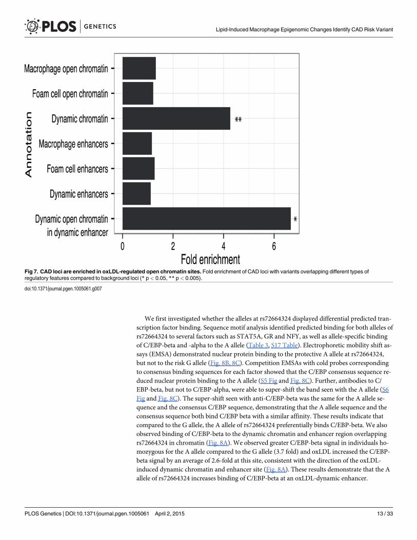
CAD-associated genetic variants are enriched in regulatory DNA sitesaltered by oxLDLDisease-associated genetic variants may alter transcriptional regulation by affecting transcrip-tion factor binding to regulatory DNA elements [9]. Therefore, we sought to profile the rela-tionship between the oxLDL-induced changes we had identified in macrophages and geneticvariants associated with CAD risk. We identified 45 independent CAD-associated genomic lociand catalogued the reported index SNP and all SNPs in high LD (r2 > 0.8) [34]. We then inter-sected macrophage and foam cell epigenomic data with the resulting set of variants at CAD-associated loci (Table 2, S2 and S16 Tables). At 22 of the 45 CAD loci, one or more variants laywithin an open chromatin or enhancer site in macrophages and/or foam cells. We tested forenrichment of variants at CAD-associated loci in dynamic sites compared to the expected over-lap derived from a background set of matched non-CAD GWAS loci (see Methods). We ob-served significant enrichment of CAD-associated loci in dynamic chromatin sites (fold = 4.26;binomial p = 0.0027) (Fig. 7), an effect that was stronger when considering sites with both dy-namic chromatin and enhancer signal (fold = 6.66; p = 0.036). Conversely, we observed no sig-nificant enrichment when considering chromatin sites in macrophages and foam cells alone(p = 0.19, p = 0.31). These results suggest that variants at CAD-associated loci are specificallyenriched in the subset of macrophage regulatory sites altered by oxLDL exposure.
CAD-associated variants at the IL6R and PPAP2B loci were within overlapping dynamicchromatin and enhancer sites. At both loci the CAD-associated variants (rs7549338 andrs7553796 at IL6R; rs72664324 at PPAP2B) overlap single dynamic sites that had increased sig-nals after oxLDL exposure; further, both IL6R and PPAP2B had significantly up-regulated ex-pression in response to oxLDL (S4 Fig). IL6R encodes the receptor for the pro-inflammatorycytokine interleukin-6 and circulating levels of a soluble form of IL6R have been associated withcoronary artery disease [35,36]. PPAP2B encodes lipid phosphate phosphohydrolase 3 (LPP3),an enzyme that metabolizes and so deactivates pro-inflammatory mediators [37]. The mecha-nism through which the PPAP2B locus influences CAD risk is unknown and the expression pat-tern and regulation of PPAP2B by oxLDL has not been studied previously. These resultsdemonstrate that dynamic sites can highlight candidate causal variants and genes at CAD-associated loci.
CAD-associated variant rs72664324 demonstrates allelic differences inoxLDL-induced enhancer activityWe sought to identify and characterize candidate causal CAD-associated variants at thePPAP2B locus. A single CAD-associated variant rs72664324 overlapped a dynamic chromatinand enhancer site, which in turn lies in an oxLDL-induced ‘super-enhancer’ cluster (Fig. 8A).
Table 2. Number of CAD loci and individual SNPs overlapping regulatory elements in macrophages and foam cells.
Feature type Number of CAD loci Number of SNPs
Open chromatin Macrophage 12 26
Foam cell 12 26
Enhancer Macrophage 18 109
Foam cell 19 114
Dynamic chromatin 6 9
Dynamic enhancer 9 38
doi:10.1371/journal.pgen.1005061.t002
Lipid-Induced Macrophage Epigenomic Changes Identify CAD Risk Variant
PLOS Genetics | DOI:10.1371/journal.pgen.1005061 April 2, 2015 12 / 33
We first investigated whether the alleles at rs72664324 displayed differential predicted tran-scription factor binding. Sequence motif analysis identified predicted binding for both alleles ofrs72664324 to several factors such as STAT5A, GR and NFY, as well as allele-specific bindingof C/EBP-beta and -alpha to the A allele (Table 3, S17 Table). Electrophoretic mobility shift as-says (EMSA) demonstrated nuclear protein binding to the protective A allele at rs72664324,but not to the risk G allele (Fig. 8B, 8C). Competition EMSAs with cold probes correspondingto consensus binding sequences for each factor showed that the C/EBP consensus sequence re-duced nuclear protein binding to the A allele (S5 Fig and Fig. 8C). Further, antibodies to C/EBP-beta, but not to C/EBP-alpha, were able to super-shift the band seen with the A allele (S6Fig and Fig. 8C). The super-shift seen with anti-C/EBP-beta was the same for the A allele se-quence and the consensus C/EBP sequence, demonstrating that the A allele sequence and theconsensus sequence both bind C/EBP beta with a similar affinity. These results indicate thatcompared to the G allele, the A allele of rs72664324 preferentially binds C/EBP-beta. We alsoobserved binding of C/EBP-beta to the dynamic chromatin and enhancer region overlappingrs72664324 in chromatin (Fig. 8A). We observed greater C/EBP-beta signal in individuals ho-mozygous for the A allele compared to the G allele (3.7 fold) and oxLDL increased the C/EBP-beta signal by an average of 2.6-fold at this site, consistent with the direction of the oxLDL-induced dynamic chromatin and enhancer site (Fig. 8A). These results demonstrate that the Aallele of rs72664324 increases binding of C/EBP-beta at an oxLDL-dynamic enhancer.
Fig 7. CAD loci are enriched in oxLDL-regulated open chromatin sites. Fold enrichment of CAD loci with variants overlapping different types ofregulatory features compared to background loci (* p< 0.05, ** p< 0.005).
doi:10.1371/journal.pgen.1005061.g007
Lipid-Induced Macrophage Epigenomic Changes Identify CAD Risk Variant
PLOS Genetics | DOI:10.1371/journal.pgen.1005061 April 2, 2015 13 / 33

We next tested the effects of rs72664324 on oxLDL-induced enhancer activity using lucifer-ase reporter assays in primary human macrophages and foam cells. We observed increased en-hancer activity with the A allele compared to the G allele in both macrophages (1.73 fold,p = 0.0005) and foam cells (2.25 fold, p = 0.025) (Fig. 8D). Furthermore, we observed a greateroxLDL-induced increase in enhancer activity at the rs72664324 site with the A allele (1.43 fold,p = 0.03) compared to the G allele (1.098, p = 0.78) (Fig. 8D). Overexpression of C/EBP-beta
Fig 8. An intronic SNP at the PPAP2B locus regulates enhancer activity and oxLDL-induced expression of PPAP2B. (A) Chromatin profile at thePPAP2B locus where rs72664324 is in a dynamic chromatin, enhancer and C/EBP beta site (red box). The region is part of an oxLDL-induced superenhancer. (B) Comparison of the human rs72664324 alleles and the corresponding mouse sequence with the CEBPmotif aligned above. (C) EMSAdemonstrating that only the A allele at rs72664324 binds nuclear protein, which is shown to be C/EBP beta by a super-shift in the presence of anti-C/EBPbeta antibody. Also, only a cold probe with the A allele competes off binding to a C/EBP consensus probe. (D) Luciferase reporter assays in primary humanmacrophages and foam cells with a reporter element containing rs72664324 with either allele (* p< 0.05, ** p< 0.005). (E) Effect of C/EBP betaoverexpression on luciferase reporter activity with either the A or G allele relative to empty vector (* p< 0.05). (F) Induction of PPAP2B expression by oxLDLin primary humanmacrophages from individuals with allele G or at least one copy of the A allele (long lines indicate mean, short lines indicate median, Mann-Whitney U test, p = 0.0013, GG n = 10, GA n = 2, AA n = 6).
doi:10.1371/journal.pgen.1005061.g008
Lipid-Induced Macrophage Epigenomic Changes Identify CAD Risk Variant
PLOS Genetics | DOI:10.1371/journal.pgen.1005061 April 2, 2015 14 / 33

increased transcriptional enhancer activity of the A allele significantly more than that of the Gallele (p = 0.012) (Fig. 8E). These results suggest that, compared to the G allele, the A allelepreferentially increases enhancer activity upon oxLDL induction through increased C/EBP-beta binding.
We obtained primary macrophages from healthy individuals with different rs72664324 ge-notypes. We observed significantly higher oxLDL-induced PPAP2B expression in macro-phages from individuals with at least one copy of the A allele compared to those with only theG allele (p = 0.0013) (Fig. 8F). The A allele of rs72664324 is in LD with the protective allele ofthe reported index SNP at this locus, so higher PPAP2B expression is associated with reducedCAD risk.
OxLDL up-regulates PPAP2B expression and influences pro-inflammatory mediatorsThe role of PPAP2B in the response of primary human macrophages to oxLDL has not beenstudied previously. Given the influence of the CAD risk variant rs72664324 on PPAP2B ex-pression in the macrophage response to oxLDL and our observation that PPAP2B was the sev-enth most up-regulated gene in response to oxLDL exposure (Table 1), we studied the effects ofoxLDL on the activity of the resulting protein product LPP3, and the expression pattern ofLPP3 in atherosclerotic lesions. LPP3 is an enzyme that dephosphorylates and thus deactivatespro-inflammatory mediators including lysophosphatidic acid and sphingosine 1-phosphate[37–39]. Protein expression of LPP3 in primary human macrophages was strongly increasedby oxLDL treatment, as demonstrated using western blotting and mass spectrometry (Fig. 9A).Immunohistochemistry for LPP3 in human arterial atherosclerotic plaques confirmed thatfoam cells express LPP3 in vivo and that these cells constitute the major source of LPP3 withinthe plaque (Fig. 9B). Enzymatic activity of LPP3 against both LPA and S1P was strongly in-duced in primary macrophages by oxLDL exposure (Fig. 9C, 9D). Mass spectrometry-basedanalysis also identified significant oxLDL-induced changes in substrates and products of LPP3(Fig. 9E). These results indicate that the CAD protective A allele of rs72664324 at the PPAP2Blocus increases the transcriptional enhancer response to oxLDL in macrophages, resulting inaltered LPP3 activity, which in turn promotes increased metabolism of pro-inflammatory me-diators within atherosclerosis lesions (Fig. 10).
Table 3. Differential transcription factor motif affinity at the rs72664324 site for the risk G allele and the protective A allele.
Rank Difference log(p) for two sequences A allele p value G allele p value Matrix ID (Transfac) Transcription factor
1 1.71 0.0137 0.693 M00190 CEBP
2 1.48 0.0263 0.794 M00116 CEBPA
3 0.978 0.0452 0.429 M00621 CEBPDELTA
4 0.874 0.0102 0.0762 M01147 DMRT2
5 0.828 0.104 0.698 M01334 NKX11
6 0.822 0.0471 0.312 M00309 ACAAT
7 0.821 0.0498 0.33 M00775 NFY
8 0.816 0.0902 0.591 M00109 CEBPB
9 -0.814 0.936 0.144 M00975 RFX
10 -0.802 0.199 0.0314 M00792 SMAD
doi:10.1371/journal.pgen.1005061.t003
Lipid-Induced Macrophage Epigenomic Changes Identify CAD Risk Variant
PLOS Genetics | DOI:10.1371/journal.pgen.1005061 April 2, 2015 15 / 33

Fig 9. OxLDL exposure induces PPAP2B-encoded LPP3 expression and activity in foam cells. (A)Western blotting demonstrates up-regulation of glycosylated and non-glycosylated LPP3 protein (asindicated) in macrophage-derived foam cells (data from 3 donors shown). (B) Human atherosclerotic plaquecontains an abundance of LPP3-expressing foam cells (brown stain, nuclei counterstained blue withhaematoxylin, scale bar indicates 50 um, inset shows close-up of foam cell—arrowed). All 5 casesimmunostained showed similar LPP3 expression in foam cells. (C and D) OxLDL exposure inducedincreased specific activity of LPP3, measured by the degradation of receptor active species lysophosphatidicacid (LPA) to mono-acylglycerol (MG) (C) and sphingosine-1-phosphate (S1P) to sphingosine (D) (n = 3donors, * p< 0.05). (E) OxLDL induced changes in the levels of LPP3 substrates and products (n = 3donors).
doi:10.1371/journal.pgen.1005061.g009
Lipid-Induced Macrophage Epigenomic Changes Identify CAD Risk Variant
PLOS Genetics | DOI:10.1371/journal.pgen.1005061 April 2, 2015 16 / 33

DiscussionBoth environmental and genetic factors influence an individual’s risk of developing coronaryartery disease. We have used epigenetic techniques for mapping chromatin structure at cis-regulatory elements to study the interacting effects of an environmental stimulus and risk-asso-ciated genetic variants.
Overwhelming evidence indicates that LDL is the major environmentally influenced con-tributor to the development of atherosclerosis. In humans circulating levels of LDL are influ-enced by diet and are tightly correlated with CAD risk; lowering LDL lowers risk [4–6]. Invarious animal models, diets that elevate LDL accelerate atherosclerosis [40]. The most strikingcellular effect of lipid in atherosclerosis results from the interaction of oxLDL with macro-phages. OxLDL is formed by modification of protein and lipids within LDL particles that havebecome trapped in the arterial vessel wall [2,41]. Macrophages within the arterial wall take upthis oxLDL in an uncontrolled manner, predominantly via scavenger receptors [2,42]. Progres-sive oxLDL uptake results in the formation of lipid-laden foam cells [2,42]. Foam cells contrib-ute to the inflammatory nature of the atherosclerotic lesions in various ways, as they havereduced motility, release inflammatory cytokines, chemokines, degradative enzymes, reactiveoxygen species and ultimately undergo apoptosis with release of a cocktail of further pro-inflammatory cellular contents [2,15,42].
Fig 10. Model showing the interplay between oxLDL, C/EBP beta and rs72664324 on expression of PPAP2B.OxLDL-induced C/EBP beta binding isgreater to the A allele and leads to increased induction of PPAP2B in response to oxLDL compared to the G allele. PPAP2B encodes LPP3 whose enzymaticactivity reduces pro-inflammatory signalling mediated by LPA.
doi:10.1371/journal.pgen.1005061.g010
Lipid-Induced Macrophage Epigenomic Changes Identify CAD Risk Variant
PLOS Genetics | DOI:10.1371/journal.pgen.1005061 April 2, 2015 17 / 33

In this study we have mapped the effects of oxLDL on chromatin remodeling and gene ex-pression in macrophages and integrated these data with the results of GWAS in CAD to identi-fy candidate functional variants. Further, these results provide a detailed map of the epigeneticchanges arising from exposure of macrophages to oxLDL and so provide a rational basis forthe study of approaches to ameliorate foam cell formation.
Open chromatin mapping with FAIRE-seq identifies the locations of all types of cis-regula-tory elements and can be used to study changes in chromatin structure in cells subjected to avariety of stimuli or undergoing ontogenetic change [23,43–46]. However, as FAIRE-seq doesnot directly provide information about the function of DNA elements, we simultaneously un-dertook ChIP-seq for H3K27ac which marks active enhancers [24,47]. Identifying the subset ofopen chromatin sites with active enhancer status is important because they play a central rolein gene regulation, particularly the control of cell-type specific gene expression [48]. H3K27acalso marks promoter sites, which are easily distinguishable by their genomic context.
FAIRE-seq derived open chromatin sites that vary between macrophages and foam cellscontain regulatory DNA sequences that play a role in oxLDL-induced foam cell formation. Wefound that the number of open chromatin sites that were modified by oxLDL was substantiallygreater than the number of differentially expressed genes. This is likely to reflect the combina-torial effects of sets of these dynamic chromatin sites and clusters of spatially linked sites wereenriched around genes whose expression was altered by oxLDL. Although we detected a signifi-cant change in open chromatin structure at oxLDL-regulated promoters, the fold change in sig-nal was smaller than that seen at non-promoter sites. This is consistent with data indicatingthat promoter chromatin structure is largely set at an early stage during cell ontogeny as 70%of promoter sites remained constant during adipogenesis compared to only 25–40% of non-promoter sites [46].
OxLDL also induced widespread changes in the H3K27ac enhancer signal and a close corre-lation was demonstrated between differential gene expression and the mean change in enhanc-er signal across multiple dynamic H3K27ac sites annotated to their nearest gene. Other studieshave found that not all sites of H3K27ac signal are associated with an open chromatin site [49].We found that at a subset of 1743 non-promoter sites there were oxLDL-induced changes inboth the open chromatin site and the surrounding enhancer marks and that the changes weredirectionally concordant at over 90% of these sites. The value of combining H3K27ac andFAIRE-seq data in this way is evident from the increased strength of the relationship of thesedually identified sites with nearby gene expression [50]. Similarly, we found a robust relation-ship between the oxLDL-induced change in FAIRE signal at these sites and expression of thenearest gene. The importance of this set of sites was underlined by their proximity to gene-setsstrongly enriched for genes known to be involved in coronary artery disease. As a subset of en-hancers for further study, these sites represent a valuable prioritized set, since the dynamicopen chromatin sites pinpoint the precise locations where altered transcription factor bindingmust occur.
Given the strong association of these dynamic enhancers with gene expression, identifyingthe transcription factors that bind them would very valuable in defining the upstream regulato-ry pathways influenced by oxLDL. Within the 1,743 sites we identified a distinct signature oftranscription factor binding using motif analysis. As validation of our approach, we found en-richment for binding sites for NRF2 (NFE2L2) and PPARG, which are both known to be asso-ciated with foam cell formation. NRF2 mediates the oxidative stress response to oxLDL andPPARG is a nuclear receptor for constituents of oxLDL [51,52]. The list of enriched transcrip-tion factors included C/EBP-beta and AP1, which are both known to act as pioneer factorsand so can bind relatively closed chromatin, causing chromatin remodeling and thus allowingfurther transcription factors to bind [21]. Using ChIP-seq we confirmed oxLDL increased C/
Lipid-Induced Macrophage Epigenomic Changes Identify CAD Risk Variant
PLOS Genetics | DOI:10.1371/journal.pgen.1005061 April 2, 2015 18 / 33

EBP-beta binding at multiple genomic sites. In keeping with these results of an effect of oxLDLon C/EBP-beta, the saturated fatty acid palmitate has been shown to induce inflammatorychanges in murine macrophages by a C/EBP-beta-dependent mechanism [53]. In adipocytesC/EBP beta causes chromatin remodeling which opens up sites for PPARG binding [49]; ourdata raise the possibility of a similar process in foam cell formation with oxLDL exposure re-sulting in C/EBP-beta opening up chromatin for other transcription factors [49].
Profiling the co-localization of regulatory sites with genetic risk variants identified byGWAS has been useful in the identification of cell types in which disease risk variants operate[28,54]. Given the established role of macrophages and foam cells in atherosclerosis it was ini-tially surprising that we found no enrichment for CAD risk loci in either context alone. Howev-er, we found that those sites that underwent oxLDL-induced changes in chromatin accessibilitywere enriched for CAD-associated loci. This demonstrates that the response to oxLDL, withconcomitant foam cell formation, induces chromatin changes at the sites of a subset of CADvariants and provides a cellular context for risk variants at these sites to operate in altering dis-ease risk. Our data show that the regulatory capacity of key SNPs in dynamic sites is actuatedin macrophages by an environmentally influenced stimulus, oxLDL, so demonstrating themechanism for a direct interplay between environmental and genetic risk factors. To ourknowledge this is the first time enrichment for CAD loci has been demonstrated in open chro-matin in an adult cell type.
The PPAP2B CAD locus was of particular interest because oxLDL induced more open chro-matin, greater enhancer activity and up-regulated PPAP2B expression. Our finding thatPPAP2B and it protein product, LPP3 were up-regulated by oxLDL fits with a growing body ofevidence implicating dysregulation of its substrates, LPA and S1P in atherosclerosis. LPP3 hy-drolyzes LPA and S1P to their non-receptor active forms [37,55]. LPP3 is known to have a rolein vascular development and endothelial integrity, but its role in macrophages and foam cellsremains unknown [56,57]. We found that PPAP2B was one of the most up-regulated genes infoam cells, was abundant in human plaque foam cells and its specific enzymatic activity to-wards LPA and S1P in macrophages was increased by oxLDL.
LPA is an obligate intermediary of triglyceride and glycerophospholipid synthesis and anextracellular ligand for 6 different LPA receptors [58,59]. There is an accumulation of LPA inplaques in both human atherosclerotic disease and murine models of atherosclerosis [60–63].LPA has pro-atherogenic effects on most of the cells involved in atherosclerosis, promotingvascular smooth muscle proliferation, endothelial cell adhesion molecule expression, and stim-ulating oxLDL uptake by macrophages [64]. In addition, LPA is a highly thrombogenic media-tor and its release upon atherosclerotic plaque rupture can contribute to thrombotic occlusionof the artery [61]. Strategies that modulate LPA levels in plaques could, among other beneficialeffects, reduce thrombogenicity.
S1P is a receptor active sphingolipid which signals via 5 G-protein linked cell surface recep-tors resulting in diverse effects including immune cell trafficking and angiogenesis [65]. Recentstudies have sought to characterize the precise role of S1P in atherosclerosis using murine mod-els in which S1P receptors have been knocked out. Mice deficient for S1P2R or S1P3R andApoE have reduced numbers of macrophages and foam cells in plaques [66]. Bone marrow chi-meras indicate that this effect in S1P2R deficient mice is dependent on hemopoeitic cells andS1P2R deficiency reduced macrophage inflammatory response. Nevertheless, the precise roleof S1P in different aspects of atherosclerosis remains to be determined
Our immunohistochemical studies of human atherosclerotic plaque indicated that the pre-dominant source of LPP3 was in foam cells. In keeping with an accumulation of LPA in athero-sclerotic plaque, we found that foam cells contained significantly more LPA than macrophages.Taken together our findings suggest that by degrading LPA, LPP3 could have a role as a
Lipid-Induced Macrophage Epigenomic Changes Identify CAD Risk Variant
PLOS Genetics | DOI:10.1371/journal.pgen.1005061 April 2, 2015 19 / 33

negative regulator of pro-inflammatory LPA signaling during the macrophage response tooxLDL and in foam cells.
The PPAP2B locus is well validated as a CAD risk locus and our data demonstrate that therisk allele reduced the induction of PPAP2B by oxLDL in macrophages [8,67,68]. This suggestsa plausible model whereby the risk allele lowers transcription of PPAP2B in response to oxLDLexposure in macrophages (Fig. 10). This in turn would lower levels of LPP3 in foam cells, re-sulting in higher levels of LPA in plaque and consequently increased pro-inflammatory signal-ing, vascular smooth muscle proliferation, retention of foam cells in lesions andthrombogenicity. Our data demonstrate that an effect on PPAP2B transcription may in part bemediated through rs72664324, a SNP that is in high linkage disequilibrium with the reportedGWAS SNP (rs17114036). The risk allele at rs72664324 reduces C/EBP-beta binding at thissite and so reduces the enhancer activity of the site. The overall mechanism we propose is inkeeping with that shown for two other CAD loci where rs12740374 affected SORT1 transcrip-tion and SNPs at the 9p21 locus altered STAT1 binding and interferon gamma signaling[9,10].
Further evidence suggests that rs72664324 is a causal SNP. None of the other 20 SNPs inhigh linkage disequilibrium were in the coding sequence or even the 3’UTR, where alteredmiRNA binding is an additional mechanism by which CAD SNPs might act [11]. The chroma-tin structure at the reported SNP, rs17114036, remained closed before and after oxLDL expo-sure. Moreover, no other SNP in high linkage disequilibrium at the locus was within an openchromatin site before or after oxLDL treatment. Interestingly, we found that the DNA sequenceat the dynamic chromatin site containing rs72664324 has been highly conserved across verte-brate species. Active enhancers have been associated with the production of small RNA tran-scripts and using CAGE-seq to measure enhancer transcripts across the genome in hundredsof predominantly ‘healthy’ primary cells and tissues, the only enhancer RNA that overlappedany of the 21 SNPs in high linkage disequilibrium with the reported risk SNP was that from thers72664324 site [69]. This enhancer was only detected in monocytes, the precursors of macro-phages [69]. Finally, we show that the presence of the protective A allele at the rs72664324 siteenhances the upregulation of PPAP2B expression that is triggered by oxLDL in primary mac-rophages. Nevertheless we cannot exclude that other linked SNPs may be important, especiallysince our primary open chromatin data and enhancer mapping was performed in individualshomozygous for the major allele of the CAD reported SNP.
Our data indicate that a CAD risk variant operates to alter the response of macrophages tooxLDL exposure by altering binding of C/EBP beta to an enhancer site regulating PPAP2B ex-pression. This will influence inflammatory and other aspects of the atherosclerotic disease pro-cess. Targeting the activity of PPAP2B-encoded LPP3 in macrophages and foam cells is aplausible therapeutic strategy. Future studies could directly address the role of PPAP2B in theresponse to oxLDL using murine models in which PPAP2B is selectively knocked out from my-eloid cells. Overall, our study establishes a link between CAD genetic susceptibility, the macro-phage response to atherogenic lipid and receptor active lipid signaling. This studydemonstrates the utility of chromatin and enhancer mapping in primary human cells beforeand after a pathogenic environmental stimulus and this approach may have applications in thestudy of other diseases.
Materials and Methods
Cell cultureEthical approval for the study was obtained from the NHS Research Ethics Committee (SouthCentral-Hampshire B, reference 13/SC/0392) and all participants provided informed consent.
Lipid-Induced Macrophage Epigenomic Changes Identify CAD Risk Variant
PLOS Genetics | DOI:10.1371/journal.pgen.1005061 April 2, 2015 20 / 33

CD14+ monocytes were isolated from healthy human volunteers by centrifugation of pe-ripheral blood over Ficoll-Paque PLUS (GE Healthcare LifeSciences, Piscataway, NJ) followedby extraction with magnetic beads conjugated to anti-CD14 antibody (Miltenyi Biotec, Ber-gisch Gladbach, Germany). Monocyte purity was assessed by flow cytometry using anti-CD14antibody (AbD Serotec, Raleigh, NC) and was� 95%. Cells were maintained in RPMI 1640medium with 10% fetal calf serum, 4 mM L-glutamine, 50 units/ml penicillin and 50 μg /mlstreptomycin (Sigma, St Louis, MO), supplemented with 50 ng/ml macrophage colony stimu-lating factor (eBioscience, San Diego, CA). After 7 days these macrophages were treated witheither control buffer (1 mM ethylenediaminetetraacetic acid (EDTA), 25 μmCuCl2, phos-phate-buffered saline (PBS)) or 50 μg/ml oxLDL for 48hrs. Oil red O staining of intracellularlipids and measurement of cellular cholesterol and cholesterol ester content by mass spectrom-etry confirmed foam cell formation in oxLDL-treated cells. Viability of cells was confirmed tobe> 98% using the Invitrogen LIVE/DEAD fluorescent microscopy kit (Life Technologies,Carlsbad, CA). THP1 cells (ATCC) were maintained in RPMI 1640 with 10% fetal calf serum,4 mM L-glutamine, 50 units/ml penicillin and 50 μg/ml streptomycin. THP1 cells were treatedwith 50 ng/ml phorbol myristate acetate (PMA, Sigma) for 48 hours to obtain adherentmacrophage cells.
Preparation of oxLDLLDL (d 1.019–1.063 g/ml) was freshly isolated from human plasma by ultracentrifugationusing a discontinuous potassium bromide gradient [70]. Precautions were taken to prevent en-dotoxin contamination and maintain sterility. LDL was extensively dialyzed against PBS insterile gamma-irradiated cartridges (Pierce, Rockford, Il) and protein concentration was mea-sured with the BCA method (Pierce, Rockford, Il). LDL was oxidized by incubation with 25 μmCuCl2 at 37° C for 18 hours. Oxidation was confirmed by thiobarbituric reactive substancesassay (Caymen Chemical, Ann Arbor, MI). Oxidation was terminated by addition of 1 mMEDTA and storage at 4#x00B0;C. OxLDL was used within 2 weeks of production. For endotox-in testing, samples were first heated to 75°C for 15 minutes to remove the plasma inhibitor andthen assayed using the gel clot method according to the manufacturer’s instructions (Associatesof Cape Cod, East Falmouth, MA). Levels were< 0.1 EU/ml.
FAIREFAIRE was performed as described with modifications [22]. Five million primary human mac-rophages or foam cells were cross-linked, lyzed and sonicated (30 pulses of 15 seconds at maxi-mum intensity using a BioRuptor (Diagenode, Denville, NJ)). Nucleosome-depleted DNA wasextracted using 4 phenol-chloroform extractions and purified by ethanol precipitation. Threeindependent biological replicates were produced using cells from three healthy donors. Donorswere homozygous for the reference allele of rs17114036/rs72664324.
ChIPChIP was performed using the Invitrogen Magnify kit (Invitrogen) according to the manufac-turer’s instructions using 200,000 primary human macrophages or foam cells. Cells were cross-linked with formaldehyde for 10 minutes on ice, lyzed and sonicated (32 pulses of 15 seconds,maximum intensity, Diagenode Bioruptor). H3K27ac-enriched DNA was immunoprecipitatedusing rabbit polyclonal anti-histone H3 (acetyl K27) antibody (ab4729, Abcam, Cambridge,MA). ChIP was performed in technical duplicates (pooled—total 400,000 cells) and two inde-pendent biological replicates were produced from two donors. Donors were homozygous forthe reference allele of rs17114036/rs72664324. C/EBP-beta enriched DNA was
Lipid-Induced Macrophage Epigenomic Changes Identify CAD Risk Variant
PLOS Genetics | DOI:10.1371/journal.pgen.1005061 April 2, 2015 21 / 33

immunoprecipitated using rabbit polyclonal anti-C/EBP-beta antibody (SC-150X, Santa Cruz,Santa Cruz, CA). ChIP was performed in technical duplicates (pooled total 400,000) cells and 4biological replicates were produced from 4 donors (2 homozygotes for the reference allele and2 for the non-reference allele of rs17114036/rs72664324).
Library preparation and sequencingLibraries were generated from gel-purified ~200 bp DNA fragments. After adapter ligation andPCR-based amplification, samples were sequenced on the Illumina HiSeq 2000 or 2500 plat-form (Illumina, San Diego, CA). 50 bp paired-end reads were mapped against the UCSC hg19reference genome using STAMPY v1.021 [71]. For FAIRE and H3K27ac ChIP samples, readswere filtered (MAPQminimum 15) in SAMTOOLS yielding at least 54 and 49 million reads re-spectively per sample [72]. For C/EBP-beta ChIP, paired end reads were mapped against thehs37d5 genome and filtered for a minimumMAPQ score of 4 and further filtered to removeduplicates (using the markduplicates tool in PICARD). For all subsequent analysis one read ofeach proper pair was retained along with all unpaired reads passing the requisite MAPQthreshold. Reads mapping to chrM, random contigs, unplaced contigs and ENCODE black-listed regions were removed from subsequent analysis. To confirm reproducibility of FAIREdata, peaks were called on individual FAIRE samples using Fseq V1.84 (default parameters)and PeakDeck [73,74]. FAIRE peaks between replicates were intersected and shown to exceedENCODE FAIRE-seq guideline standards (ENCODE and modENCODE Guidelines for Exper-iments Generating CHIP, DNase, FAIRE, and DNAMethylation GenomeWide Location DataVersion 2.0[18]) before pooling for further analysis. ChIP samples peaks were called usingMACS1.4.2 for H3k27ac and MACS2 for C/EBP-beta (versus input control DNA) and con-firmed to exceed ENCODE guideline standards[75].
FAIRE and ChIP-seq analysisFor FAIRE, dynamic chromatin sites between macrophages and foam cells were identifiedfrom pooled biological replicates using Diffreps V1.55 (windows 200 bp, step 20 bp, G-test,FDR< 2.5%) [76]. Clusters of dynamic chromatin were identified using the Diffreps HotSpotalgorithm (p< 0.05). FAIRE-seq peaks were also determined for macrophages and foam cellsindividually using Fseq on pooled replicates (default settings, with threshold 8.5 for macro-phages and 8 for foam cells). Peaks were merged if within 140 bp and filtered out if< 50 bpor> 5 kb in width. For ChIP dynamic H3K27ac enriched sites were identified with Diffrepsusing separate biological replicates (windows 500 bp, step 50 bp, FDR< 2.5%). ChIP-seq peakswere determined for each cell type individually using MACS1.4.2 on pooled biological repli-cates versus input control DNA with default parameters. Super enhancer sites were determinedusing the ROSE algorithm according to the originally established method[31]. For C/EBP betaChIP-seq dynamic sites were identified from pooled biological replicates using Diffreps V1.55(windows 200 bp, step 20 bp, G-test, FDR<2.5%, filtered on minimum 50 reads in dynamicsite). Quantile normalized read counts in dynamic C/EBP-beta binding sites were determinedfor each genotype in order to compare signal at the rs72664324 dynamic site.
Annotation of FAIRE and CHIP-seq sitesGenomic features were annotated using HOMER (V4.1), Diffreps and CEAS [76–78]. For aver-age signal profiling at transcription start sites using CEAS, wig files of pooled biological repli-cates were produced using the Java genomics toolkit (https://github.com/timpalpant/java-genomics-toolkit) and subsets of ~200 genes were interrogated (for differentially expressedgenes the top two quartiles of upregulated/downregulated genes based on fold change were
Lipid-Induced Macrophage Epigenomic Changes Identify CAD Risk Variant
PLOS Genetics | DOI:10.1371/journal.pgen.1005061 April 2, 2015 22 / 33

used). To compare the FAIRE-seq signal between macrophages and foam cell promoters in dif-ferent subsets of genes, the FAIRE-seq signal at all promoters was first quantified usingHOMER (+ /- 1 kb transcriptional start site) and then quantile normalized. Signal at the pro-moters of all differentially expressed genes was then compared (paired Student’s t-test, signifi-cance p< 0.05) and displayed as boxplots. For correlating dynamic chromatin/dynamicenhancer sites with gene expression, the dynamic sites were annotated with the nearest geneand expression compared using a paired Student’s t-test, significance p< 0.05 and simple line-ar regression. For assigning dynamic enhancer status to dynamic chromatin sites, the dynamicchromatin sites were expanded by 300 bp in each direction to identify colocalization with his-tone marks of enhancer status that are on adjacent nucleosomes; finally the two datasets wereintersected using bedtools. For genes with more than one Illumina microarray probe the differ-ential expression data were extracted from the differential probe set with the most significantfold change between the two conditions. Gene set enrichment analysis of various dynamicchromatin/enhancer sites was performed using GREAT V2.0.2 and default parameters [79].
Display in genome browserFor display of FAIRE-seq, H3K27ac or C/EBP-beta signal in the UCSC genome browser wigfiles were generated using ngs.BaseAlignCounts function in the Java genomics toolkit. Wig fileswere then normalized using the wigmath.scale function (default parameters) and macrophagesignal was subtracted from foam cell signal to generate a track of dynamic signal.
Transcription factor analysisTranscription factor motif enrichment analysis was performed using SeqPos in the CistromeGalaxy environment with the curated Cistrome motif database and de novomotif generator[80]. De novomotif position weight matrices are given in Tables S9 and S13.
SNP enrichment analysisWe identified SNPs with genome-wide association (p< 5x10⁻⁸) to any trait in European indi-viduals present in the GWAS catalogue. Index SNPs were pruned (r2 > 0.1 in CEU samples) sothat each ‘locus’ was only represented by one index SNP to avoid counting redundant loci.Each index SNP was then used to identify variants in 1000 Genomes Project (1KG) pilot 1 datain high LD (r2 > 0.8) in CEU samples using HaploReg [81]. Thus, an associated ‘locus’ consistsof an index SNP and the set of 1KG SNPs in high CEU LD. We then created a background setcontaining all qualifying loci, binned based on the number of total variants (index + high LDSNPs) in the locus.
We then identified published reports of CAD-associated variants with p< 5x10⁻⁸ in Euro-pean samples. Where studies had reported different variants at the same locus we used the vari-ant with the most significant p value [8,67,82–86]. For each CAD-associated variant we createdloci containing 1KG variants in high LD (r2 > 0.8) in CEU samples [81].
Using the set of CAD-associated loci, we calculated the number of loci containing a variantoverlapping a given annotation. We performed 100,000 permutations of the set of loci drawingfrom matching bins in the background set and recalculated the number of loci with a variantoverlapping an annotation.
We then compared the observed number of loci, the total number of loci and the expectednumber obtained via permutation using a binomial test.
Lipid-Induced Macrophage Epigenomic Changes Identify CAD Risk Variant
PLOS Genetics | DOI:10.1371/journal.pgen.1005061 April 2, 2015 23 / 33

Non-coding element enrichmentWe obtained non-coding element data for ChromHMM chromatin state, TFBS, and multi-species conservation from the UCSC genome browser. For chromatin state data, we pooled‘Enhancers’, ‘Promoters’ and ‘Insulators’ identified as such in any cell type.
We calculated the overlap of dynamic chromatin sites with each regulatory class, and thenperformed peak-shifting of the chromatin sites a random distance within a 10 kb window. Wethen re-calculated the overlap with the shifted sites, and derived a background distribution ofexpected overlap over 100 permutations. Fold-enrichment values were calculated relative tothe background mean and p values for each overlap were obtained directly via permutation.
Gene expressionPrimary human macrophages and foam cells were lyzed and total RNA extracted using theTRIZOL RNA PLUS extraction kit (Life Technologies) with on-column Purelink DNAse treat-ment (Life Technologies). The integrity of the total RNA was analyzed on an Agilent Bioanaly-zer 2100 (Agilent Technologies, Santa Clara, CA). Total RNA was reverse transcribed,amplified and biotinylated using the Illumina TotalPrep-96 RNA Amplification Kit (Ambion,Austin, TX). Biotinylated cRNA was hybridized to a single Human HT-12 V4 BeadCHIP (Illu-mina) at 58°C for 18 hours. The BeadCHIP was scanned using the Illumina Iscanner and datapre-processed using the Illumina Bead Studio to correct for local background effects, removeoutlier beads, to compute average bead signal and SD for each probe and gene and to calculatep values. Hierarchical clustering showed that macrophages samples and foam cells samplesclustered together respectively. The LUMI pipeline was used for data analysis. Data were trans-formed (variance-stabilizing transformation or log2 where indicated) and normalized (robustspline normalization). Differentially expressed genes were determined using both a pairedmacrophage-foam cell design and unpaired design (probe-centric) after removal of probeswith absent expression. For differential expression an FDR-corrected p value threshold of 0.05was deemed significant [87]. Enrichment p values for differentially expressed genes for candi-date CAD genes were calculated using the binomial distribution. Heatmaps of differentially ex-pressed genes were generated using the heatmap.2 function in R based on the subset of thegenes with adjusted p value< 0.0001, and for genes with more than one microarray probe, themost significant probe was used (using the unpaired design). Gene set enrichment analysis wasperformed using GREAT.
For real time quantitative PCR (RTqPCR) 1 μg of DNaseI-treated total RNA was reversetranscribed using Bioscript (Bioline, London, UK) with random hexamers. RTqPCR was per-formed with SYBR Green reagents using a Step One Plus machine (Applied Biosystems, FosterCity, CA) using technical triplicates and biological triplicates. Primers used: PPAP2B CO5547TTCTGGCAGGATTTGCTCAA, CO5548 AGGGAGAGCGTCGTCTTAGTCTT; IL6R CO4830GCATTGCCATTGTTCTGAGGTT, CO4831 ACCAGCTGCCCCAAAGAGT, GAPDH CO3744TTGCCATCAATGACCCCTTCA, CO3745 CGCCCCACTTGATTTTGGA. For PPAP2B expres-sion in genotyped healthy individuals; rs72664324-G n = 10, rs72664324-G/A n = 2,rs72664324-A n = 6.
GenotypingGenomic DNA was extracted from blood using Quick-gDNA minipreps (Zymo Research, Ir-vine, CA). Genotyping of the rs72664324 SNP was performed using a custom Taqman assay(Applied Biosystems, Foster City, CA). Forward primer AGGTGACCAGATATGCAAGTTGTC,reverse primer ACAGGGACTAGGACGAAGGAA. Allele specific MGB probes: A- AGGAAAT-GAACCAATGTCT, G- AGGAAATGAACCGATGTCT. Genotyping of rs17114036 was
Lipid-Induced Macrophage Epigenomic Changes Identify CAD Risk Variant
PLOS Genetics | DOI:10.1371/journal.pgen.1005061 April 2, 2015 24 / 33

performed using an inventoried Taqman assay (C__33268873_10, Applied Biosystems). For al-lelic expression analysis, genotyped individuals were recruited from a cohort of healthy partici-pants in the Oxford Biobank. Homozygous individuals at rs72664324 and rs17114046 all hadalleles consistent with the known high LD such that rs72664324-AA was always associatedwith rs17114036-GG.
Electrophoretic mobility shift assays (EMSA)EMSA was performed using nuclear extracts from primary human macrophages treated with32P gamma-ATP end-labeled double-stranded DNA probes (PerkinElmer, Waltham, MA) aspreviously described[88]. The forward strand probe sequences were: rs72664324-A aggaaat-gaaccAatgtctgttcct, rs72664324-G aggaaatgaaccGatgtctgttcct, CEBP consensus TGCA-GATTGCGCAATCTGCA, NFYA consensus CGTCTCCACCAATGGGAGGGCTGGGC, STAT5Aconsensus AGATTTCTAGGAATTCAATCC, GR consensus AGAGGATCTGTACAGGATGTTC-TAGAT. For standard EMSA, 5 μg of nuclear extract was incubated with 100 fmol labeledprobe in a 10 ul binding reaction containing 1 μg poly(dI-dC). For competition assays unla-beled probe at 100-fold excess was added to the binding reaction before addition of labeledprobe. For super-shift assays the nuclear extract was pre-incubated with 1 μg antibody for30 minutes on ice before probe was added. The following antibodies were used: rabbit poly-clonal anti-C/EBP beta (SC-150, Santa Cruz), rabbit polyclonal anti-C/EBP alpha (SC-61,Santa Cruz).
Luciferase reporter assaysTo test enhancer activity at the rs72664324 locus in response to oxLDL, primary human mac-rophages were transfected with PGL4.23 reporter plasmids using nucleofection in biologicaltriplicates as per the manufacturer’s protocol (Amaxa, Lonza, Portsmouth, NH). After 24hours cells were then treated with oxLDL 50 μg/ml or buffer for a further 24 hours. To test en-hancer activity in response to C/EBP beta over-expression, THP1 cells were cultured in 6 welldishes and transfected in at least triplicates using Lipofectamine LTX PLUS (Life Technologies)with 2.5 μg of vector for 36 hours. To control for transfection efficiency in each replicate, fireflyluciferase plasmids were co-transfected with pRL-SV40 encoding Renilla luciferase (1/20 DNAamount compared to Firefly). Cells were lyzed for luciferase assay using the Dual Luciferase Re-porter Assay System (Promega, Fitchburg, WI). The luciferase activity of each sample was nor-malized to Renilla luciferase activity and shown as relative light units (RLU). For eachconstruct two individual clones were tested independently and representative data are shown.Data were analyzed using Student’s t-test on at least triplicates. rs72664324 reporter plasmidswere generated in pGL4.23 for each allele, by cloning in four tandem repeats of GGAAAT-GAACCAATGTCT or GGAAATGAACCGATGTCT (Plasmid IDs; allele A = pOC1250, alleleG = pOC1251). Human C/EBP beta LAP transcript was cloned into pCDNA3.1 (PlasmidID = pOC1252). As control empty pCDNA3.1 was co-transfected with reporter plasmids. Forco-transfections 1.25 μg of firefly plasmid and 1.25 μg of C/EBP beta LAP plasmid or controlplasmid were transfected. All plasmids were verified by Sanger sequencing using BigDye (LifeTechnologies).
Western blottingLPP3 was detected using an extensively characterized and validated rabbit polyclonal anti-human LPP3 antibody and fluorescently multiplexed with beta-actin staining. Images shownare monochrome images of multiplex western blots [37,89–91].
Lipid-Induced Macrophage Epigenomic Changes Identify CAD Risk Variant
PLOS Genetics | DOI:10.1371/journal.pgen.1005061 April 2, 2015 25 / 33

Measurement of LPP3 phosphatase activityLPP3 was immunopreciptated from Triton X-100 extracted macrophage/foam cell proteinsand phosphatase activity determined using heptadecanoyl lysophosphatidic acid and S1P assubstrate and measuring heptadecanoyl monoacylglycerol and sphingosine produced usingHPLC electrospray ionization tandem mass spectrometry. The reagents and methods em-ployed have been described in detail elsewhere [37,39].
Measurement of LPP3 substrates and productsCholesterol, cholesterol ester and a series of LPP3 substrates and their cognate dephosphoryla-tion products were quantitated in macrophage and foam cell lipid extracts by electrospray ioni-zation tandem mass spectrometry using methods that have been described previously [92,93].
Proteomic quantitation of LPP3LPP3-derived tryptic peptides were quantitated using capillary flow reverse phase HPLC andelectrospray ionization tandem mass spectrometry using an AB Sciex 5600 Q-TOF mass spec-trometer (AB SCIEX, Framingham, MA) operated in high-resolution selected ion monitoringmode. We quantitated the following peptides STIQNPYVAALYK, NGGSPALNNNPR,EILSPVDIIDR and used a mass-labelled derivative of the latter of these (New England Pep-tides, Gardner, MA) as an internal standard [94].
ImmunohistochemistryImmunohistochemical staining for LPP3 was undertaken using mouse monoclonal antibodyclone 7H7D3 (Biosensis, Temecula, CA), which has previously been validated, including forimmunohistochemistry [95]. Formalin-fixed paraffin-embedded tissue samples of non-athero-sclerotic aorta (n = 5), and atherosclerotic aorta (n = 5) obtained with full ethical approvalfrom the National Research and Ethics Service (Oxfordshire Research and Ethics CommitteeA: reference 04/Q1604/21), were immunostained using the Bond Max™ fully automated immu-nohistochemistry system (Leica Microsystems, Wetzlar, Germany). Heat induced antigen re-trieval was performed using the EDTA-based Novocastra Bond Epitope Retrieval Solution 2(pH 9.0) for 30 minutes and the NovolinkTM max polymer detection system (Leica Microsys-tems) was used, as per the manufacturer’s instructions. Detection reagents only were used as anegative control. Slides were mounted in Aquatex mounting medium (Merck, Darmstadt, Ger-many). Stained sections were photographed with a Nikon DS-FI1 camera with a Nikon DS-L2control unit (Nikon, Kingston-upon-Thames, UK) and an Olympus BX40 microscope (Olym-pus, Watford, UK). All immunohistochemical staining was analyzed by and performed underthe supervision of an experienced board certified consultant pathologist (ES).
Supporting InformationS1 Fig. Foam cell formation in human primary macrophages. Light microscopy of primaryhuman macrophages (A, C) and oxLDL treated macrophages (B, D) stained with oil red O andvisualized with (C, D) and without (A, B) phase contrast demonstrates extensive red-coloredintracellular lipid in oxLDL-treated cells which confirms foam cell formation (scale bar indi-cates 100 um).(EPS)
S2 Fig. Genomic distribution of open chromatin sites in macrophages and foam cells. Theimages were annotated using CEAS and the background genome distribution derived from
Lipid-Induced Macrophage Epigenomic Changes Identify CAD Risk Variant
PLOS Genetics | DOI:10.1371/journal.pgen.1005061 April 2, 2015 26 / 33

percentage of genome covered by RefSeq annotations.(EPS)
S3 Fig. Width of dynamic FAIRE sites. The frequency distributions of widths of dynamicchromatin sites suggest most are not more than one nucleosome in width with a relative in-crease in wider sites at promoters.(EPS)
S4 Fig. Up-regulation of IL6R and PPAP2B in foam cells compared to macrophages as as-sessed by quantitative RT-PCR. (A) OxLDL induces expression of IL6R at the mRNA level(n = 3). (B) OxLDL induces expression of PPAP2B at the mRNA level (n = 3, homozygousmajor genotype rs72664324, rs17114036).(EPS)
S5 Fig. EMSA with cold probe competition assays.Only the C/EBP consensus sequence com-petes off binding to the A allele. Cold probes for NFY, STAT5A and GR did not compete offbinding. No protein binding to the G allele was detectable.(EPS)
S6 Fig. EMSA with probes for the rs72664324 alleles. The A allele binds C/EBP beta as dem-onstrated by a super-shift with anti-C/EBP beta antibody. The anti-C/EBP alpha antibody pro-duces no super-shift.(EPS)
S1 Table. (A) List of differentially expressed genes (probe centric) between macrophagesand foam cells using paired data (FDR adjusted p< 0.05). For some genes several differentprobes are reported. (B) List of differentially expressed genes (probe centric) without usingpairing information (FDR adjusted p< 0.05). For some genes several different probes arereported.(XLSX)
S2 Table. CAD loci and SNPs in high linkage disequilibrium.(XLSX)
S3 Table. Genes within 50kb of CAD SNPs.(XLSX)
S4 Table. Gene set enrichment analysis for genes within 50kb of CAD SNPs.(XLSX)
S5 Table. Macrophage open chromatin sites identified by FAIRE-seq.(XLSX)
S6 Table. Foam cell open chromatin sites identified by FAIRE-seq.(XLSX)
S7 Table. Dynamic FAIRE-seq sites.(XLSX)
S8 Table. Top 10 enriched transcription factor binding site motifs in dynamic open chro-matin sites (top 3000 open dynamic chromatin sites, see also S9 Table).(PDF)
S9 Table. Complete output of transcription factor motif enrichment analysis in dynamicchromatin sites (top 3000 open chromatin sites, see also S8 Table).(XLSX)
Lipid-Induced Macrophage Epigenomic Changes Identify CAD Risk Variant
PLOS Genetics | DOI:10.1371/journal.pgen.1005061 April 2, 2015 27 / 33

S10 Table. Dynamic H3K27ac sites.(XLSX)
S11 Table. Macrophage super enhancer clusters.(TXT)
S12 Table. Foam cell super enhancer clusters.(TXT)
S13 Table. Complete output of transcription factor motif enrichment analysis in the subsetof 1743 dynamic chromatin sites with dynamic enhancer status.(XLSX)
S14 Table. Dynamic C/EBP beta binding sites.(TXT)
S15 Table. Transcription factor motif enrichment in dynamic C/EBP beta sites to confirmC/EBP beta motif enrichment (on top 3000 sites).(XLSX)
S16 Table. Count of overlaps of CAD SNPs with chromatin and enhancer maps.(XLSX)
S17 Table. Transcription factor motif affinities for rs72664324 alleles.(PDF)
AcknowledgmentsWe thank the High-Throughput Genomics Group at the Wellcome Trust Centre for HumanGenetics for the generation of the sequencing data and the participants and staff of the OxfordBioBank for support with obtaining blood samples.
Author ContributionsConceived and designed the experiments: MER CAO. Performed the experiments: MER AJMEJS. Analyzed the data: MR KJG DL CAO AJM SSS EJS. Wrote the paper: MER KJG DL EJSAJM SSS CAO.
References1. World Health Organization. World health statistics. Geneva, Switzerland: World Health Organization.
2. Glass CK, Witztum JL (2001) Atherosclerosis. the road ahead. Cell 104: 503–516. PMID: 11239408
3. Hansson GK, Hermansson A (2011) The immune system in atherosclerosis. Nature immunology 12:204–212. doi: 10.1038/ni.2001 PMID: 21321594
4. Stamler J, Wentworth D, Neaton JD (1986) Is relationship between serum cholesterol and risk of pre-mature death from coronary heart disease continuous and graded? Findings in 356,222 primaryscreenees of the Multiple Risk Factor Intervention Trial (MRFIT). JAMA 256: 2823–2828. PMID:3773199
5. Pekkanen J, Linn S, Heiss G, Suchindran CM, Leon A, et al. (1990) Ten-year mortality from cardiovas-cular disease in relation to cholesterol level among men with and without preexisting cardiovascular dis-ease. N Engl J Med 322: 1700–1707. PMID: 2342536
6. Scandinavian Simvastatin Survival Study G (1994) Randomised trial of cholesterol lowering in 4444 pa-tients with coronary heart disease: the Scandinavian Simvastatin Survival Study (4S). The Lancet 344:1383–1389. PMID: 7968073
7. Sayols-Baixeras S, Lluis-Ganella C, Lucas G, Elosua R (2014) Pathogenesis of coronary artery dis-ease: focus on genetic risk factors and identification of genetic variants. Appl Clin Genet 7: 15–32. doi:10.2147/TACG.S35301 PMID: 24520200
Lipid-Induced Macrophage Epigenomic Changes Identify CAD Risk Variant
PLOS Genetics | DOI:10.1371/journal.pgen.1005061 April 2, 2015 28 / 33

8. Consortium CAD, Deloukas P, Kanoni S, Willenborg C, Farrall M, et al. (2013) Large-scale associationanalysis identifies new risk loci for coronary artery disease. Nat Genet 45: 25–33. doi: 10.1038/ng.2480 PMID: 23202125
9. Musunuru K, Strong A, Frank-Kamenetsky M, Lee NE, Ahfeldt T, et al. (2010) From noncoding variantto phenotype via SORT1 at the 1p13 cholesterol locus. Nature 466: 714–719. doi: 10.1038/nature09266 PMID: 20686566
10. Harismendy O, Notani D, Song X, Rahim NG, Tanasa B, et al. (2011) 9p21 DNA variants associatedwith coronary artery disease impair interferon-gamma signalling response. Nature 470: 264–268. doi:10.1038/nature09753 PMID: 21307941
11. Miller CL, Haas U, Diaz R, Leeper NJ, Kundu RK, et al. (2014) Coronary heart disease-associated vari-ation in TCF21 disrupts a miR-224 binding site and miRNA-mediated regulation. PLoS Genet 10:e1004263. doi: 10.1371/journal.pgen.1004263 PMID: 24676100
12. Moore KJ, Sheedy FJ, Fisher EA (2013) Macrophages in atherosclerosis: a dynamic balance. Nat RevImmunol 13: 709–721. doi: 10.1038/nri3520 PMID: 23995626
13. Smith JD, Trogan E, Ginsberg M, Grigaux C, Tian J, et al. (1995) Decreased atherosclerosis in mice de-ficient in both macrophage colony-stimulating factor (op) and apolipoprotein E. Proc Natl Acad Sci U SA 92: 8264–8268. PMID: 7667279
14. Ball RY, Stowers EC, Burton JH, Cary NR, Skepper JN, et al. (1995) Evidence that the death of macro-phage foam cells contributes to the lipid core of atheroma. Atherosclerosis 114: 45–54. PMID:7605375
15. Silvestre-Roig C, deWinther MP, Weber C, DaemenMJ, Lutgens E, et al. (2014) Atherosclerotic pla-que destabilization: mechanisms, models, and therapeutic strategies. Circ Res 114: 214–226. doi: 10.1161/CIRCRESAHA.114.302355 PMID: 24385514
16. Hopkins PN (2013) Molecular biology of atherosclerosis. Physiol Rev 93: 1317–1542. doi: 10.1152/physrev.00004.2012 PMID: 23899566
17. Kobayashi E, Suzuki T, Yamamoto M (2013) Roles nrf2 plays in myeloid cells and related disorders.Oxid Med Cell Longev 2013: 529219. doi: 10.1155/2013/529219 PMID: 23819012
18. Consortium EP, Bernstein BE, Birney E, Dunham I, Green ED, et al. (2012) An integrated encyclopediaof DNA elements in the human genome. Nature 489: 57–74. doi: 10.1038/nature11247 PMID:22955616
19. Song L, Zhang Z, Grasfeder LL, Boyle AP, Giresi PG, et al. (2011) Open chromatin defined by DNaseIand FAIRE identifies regulatory elements that shape cell-type identity. Genome Res 21: 1757–1767.doi: 10.1101/gr.121541.111 PMID: 21750106
20. John S, Sabo PJ, Thurman RE, Sung MH, Biddie SC, et al. (2011) Chromatin accessibility pre-determines glucocorticoid receptor binding patterns. Nat Genet 43: 264–268. doi: 10.1038/ng.759PMID: 21258342
21. Biddie SC, John S, Sabo PJ, Thurman RE, Johnson TA, et al. (2011) Transcription factor AP1 potenti-ates chromatin accessibility and glucocorticoid receptor binding. Mol Cell 43: 145–155. doi: 10.1016/j.molcel.2011.06.016 PMID: 21726817
22. Simon JM, Giresi PG, Davis IJ, Lieb JD (2012) Using formaldehyde-assisted isolation of regulatory ele-ments (FAIRE) to isolate active regulatory DNA. Nat Protoc 7: 256–267. doi: 10.1038/nprot.2011.444PMID: 22262007
23. Gaulton KJ, Nammo T, Pasquali L, Simon JM, Giresi PG, et al. (2010) A map of open chromatin inhuman pancreatic islets. Nat Genet 42: 255–259. doi: 10.1038/ng.530 PMID: 20118932
24. Creyghton MP, Cheng AW,Welstead GG, Kooistra T, Carey BW, et al. (2010) Histone H3K27ac sepa-rates active from poised enhancers and predicts developmental state. Proc Natl Acad Sci U S A 107:21931–21936. doi: 10.1073/pnas.1016071107 PMID: 21106759
25. Pasquali L, Gaulton KJ, Rodriguez-Segui SA, Mularoni L, Miguel-Escalada I, et al. (2014) Pancreaticislet enhancer clusters enriched in type 2 diabetes risk-associated variants. Nat Genet.
26. Ward LD, Kellis M (2012) Interpreting noncoding genetic variation in complex traits and human disease.Nat Biotechnol 30: 1095–1106. doi: 10.1038/nbt.2422 PMID: 23138309
27. Trynka G, Sandor C, Han B, Xu H, Stranger BE, et al. (2013) Chromatin marks identify critical cell typesfor fine mapping complex trait variants. Nat Genet 45: 124–130. doi: 10.1038/ng.2504 PMID:23263488
28. Maurano MT, Humbert R, Rynes E, Thurman RE, Haugen E, et al. (2012) Systematic Localization ofCommon Disease-Associated Variation in Regulatory DNA. Science 337: 1190–1195. doi: 10.1126/science.1222794 PMID: 22955828
29. Buechler C, Ritter M, Duong CQ, Orso E, Kapinsky M, et al. (2001) Adipophilin is a sensitive marker forlipid loading in human blood monocytes. Biochim Biophys Acta 1532: 97–104. PMID: 11420178
Lipid-Induced Macrophage Epigenomic Changes Identify CAD Risk Variant
PLOS Genetics | DOI:10.1371/journal.pgen.1005061 April 2, 2015 29 / 33

30. Nord AS, Blow MJ, Attanasio C, Akiyama JA, Holt A, et al. (2013) Rapid and pervasive changes in ge-nome-wide enhancer usage during mammalian development. Cell 155: 1521–1531. doi: 10.1016/j.cell.2013.11.033 PMID: 24360275
31. Hnisz D, Abraham BJ, Lee TI, Lau A, Saint-Andre V, et al. (2013) Super-enhancers in the control of cellidentity and disease. Cell 155: 934–947. doi: 10.1016/j.cell.2013.09.053 PMID: 24119843
32. Nagy ZS, Czimmerer Z, Nagy L (2013) Nuclear receptor mediated mechanisms of macrophage choles-terol metabolism. Mol Cell Endocrinol 368: 85–98. doi: 10.1016/j.mce.2012.04.003 PMID: 22546548
33. Huber R, Pietsch D, Panterodt T, Brand K (2012) Regulation of C/EBPbeta and resulting functions incells of the monocytic lineage. Cell Signal 24: 1287–1296. doi: 10.1016/j.cellsig.2012.02.007 PMID:22374303
34. Genomes Project C, Abecasis GR, Auton A, Brooks LD, DePristo MA, et al. (2012) An integrated mapof genetic variation from 1,092 human genomes. Nature 491: 56–65. doi: 10.1038/nature11632 PMID:23128226
35. Collaboration IRGCERF, Sarwar N, Butterworth AS, Freitag DF, Gregson J, et al. (2012) Interleukin-6receptor pathways in coronary heart disease: a collaborative meta-analysis of 82 studies. Lancet 379:1205–1213. doi: 10.1016/S0140-6736(11)61931-4 PMID: 22421339
36. Ferreira RC, Freitag DF, Cutler AJ, Howson JM, Rainbow DB, et al. (2013) Functional IL6R 358Ala al-lele impairs classical IL-6 receptor signaling and influences risk of diverse inflammatory diseases.PLoS Genet 9: e1003444. doi: 10.1371/journal.pgen.1003444 PMID: 23593036
37. Sciorra VA, Morris AJ (1999) Sequential actions of phospholipase D and phosphatidic acid phosphohy-drolase 2b generate diglyceride in mammalian cells. Mol Biol Cell 10: 3863–3876. PMID: 10564277
38. Long JS, Pyne NJ, Pyne S (2008) Lipid phosphate phosphatases form homo- and hetero-oligomers:catalytic competency, subcellular distribution and function. The Biochemical journal 411: 371–377. doi:10.1042/BJ20071607 PMID: 18215144
39. PanchatcharamM, Miriyala S, Salous A, Wheeler J, Dong A, et al. (2013) Lipid phosphate phosphatase3 negatively regulates smooth muscle cell phenotypic modulation to limit intimal hyperplasia. Arterios-cler Thromb Vasc Biol 33: 52–59. doi: 10.1161/ATVBAHA.112.300527 PMID: 23104851
40. Getz GS, Reardon CA (2012) Animal models of atherosclerosis. Arterioscler Thromb Vasc Biol 32:1104–1115. doi: 10.1161/ATVBAHA.111.237693 PMID: 22383700
41. Steinberg D, Parthasarathy S, Carew TE, Khoo JC, Witztum JL (1989) Beyond cholesterol. Modifica-tions of low-density lipoprotein that increase its atherogenicity. N Engl J Med 320: 915–924. PMID:2648148
42. Moore KJ, Tabas I (2011) Macrophages in the pathogenesis of atherosclerosis. Cell 145: 341–355.doi: 10.1016/j.cell.2011.04.005 PMID: 21529710
43. Magnani L, Ballantyne EB, Zhang X, Lupien M (2011) PBX1 genomic pioneer function drives ERalphasignaling underlying progression in breast cancer. PLoS Genet 7: e1002368. doi: 10.1371/journal.pgen.1002368 PMID: 22125492
44. Nammo T, Rodriguez-Segui SA, Ferrer J (2011) Mapping open chromatin with formaldehyde-assistedisolation of regulatory elements. Methods in molecular biology 791: 287–296. doi: 10.1007/978-1-61779-316-5_21 PMID: 21913087
45. Paul DS, Albers CA, Rendon A, Voss K, Stephens J, et al. (2013) Maps of open chromatin highlight celltype-restricted patterns of regulatory sequence variation at hematological trait loci. Genome Res 23:1130–1141. doi: 10.1101/gr.155127.113 PMID: 23570689
46. Waki H, Nakamura M, Yamauchi T, Wakabayashi K, Yu J, et al. (2011) Global mapping of cell type-specific open chromatin by FAIRE-seq reveals the regulatory role of the NFI family in adipocyte differ-entiation. PLoS Genet 7: e1002311. doi: 10.1371/journal.pgen.1002311 PMID: 22028663
47. Zhang B, Day DS, Ho JW, Song L, Cao J, et al. (2013) A dynamic H3K27ac signature identifiesVEGFA-stimulated endothelial enhancers and requires EP300 activity. Genome Res 23: 917–927. doi:10.1101/gr.149674.112 PMID: 23547170
48. Shlyueva D, Stampfel G, Stark A (2014) Transcriptional enhancers: from properties to genome-widepredictions. Nat Rev Genet 15: 272–286. doi: 10.1038/nrg3682 PMID: 24614317
49. Siersbaek R, Nielsen R, John S, Sung MH, Baek S, et al. (2011) Extensive chromatin remodelling andestablishment of transcription factor 'hotspots' during early adipogenesis. EMBO J 30: 1459–1472. doi:10.1038/emboj.2011.65 PMID: 21427703
50. Rhie SK, Hazelett DJ, Coetzee SG, Yan C, Noushmehr H, et al. (2014) Nucleosome positioning andhistone modifications define relationships between regulatory elements and nearby gene expression inbreast epithelial cells. BMCGenomics 15: 331. doi: 10.1186/1471-2164-15-331 PMID: 24885402
Lipid-Induced Macrophage Epigenomic Changes Identify CAD Risk Variant
PLOS Genetics | DOI:10.1371/journal.pgen.1005061 April 2, 2015 30 / 33

51. Nagy L, Szanto A, Szatmari I, Szeles L (2012) Nuclear hormone receptors enable macrophages anddendritic cells to sense their lipid environment and shape their immune response. Physiol Rev 92:739–789. doi: 10.1152/physrev.00004.2011 PMID: 22535896
52. Gupte AA, Lyon CJ, HsuehWA (2013) Nuclear factor (erythroid-derived 2)-like-2 factor (Nrf2), a keyregulator of the antioxidant response to protect against atherosclerosis and nonalcoholic steatohepati-tis. Curr Diab Rep 13: 362–371. doi: 10.1007/s11892-013-0372-1 PMID: 23475581
53. Rahman SM, Janssen RC, Choudhury M, Baquero KC, Aikens RM, et al. (2012) CCAAT/enhancer-binding protein beta (C/EBPbeta) expression regulates dietary-induced inflammation in macrophagesand adipose tissue in mice. J Biol Chem 287: 34349–34360. doi: 10.1074/jbc.M112.410613 PMID:22902781
54. Trynka G, Sandor C, Han B, Xu H, Stranger BE, et al. (2012) Chromatin marks identify critical cell typesfor fine mapping complex trait variants. Nat Genet.
55. Kai M, Wada I, Imai S, Sakane F, Kanoh H (1997) Cloning and characterization of two human isozymesof Mg2+-independent phosphatidic acid phosphatase. The Journal of biological chemistry 272:24572–24578. PMID: 9305923
56. Ren H, PanchatcharamM, Mueller P, Escalante-Alcalde D, Morris AJ, et al. (2013) Lipid phosphatephosphatase (LPP3) and vascular development. Biochim Biophys Acta 1831: 126–132. doi: 10.1016/j.bbalip.2012.07.012 PMID: 22835522
57. Escalante-Alcalde D, Sanchez-Sanchez R, Stewart CL (2007) Generation of a conditional Ppap2b/Lpp3 null allele. Genesis 45: 465–469. PMID: 17610274
58. Knowlden S, Georas SN (2014) The autotaxin-LPA axis emerges as a novel regulator of lymphocytehoming and inflammation. J Immunol 192: 851–857. doi: 10.4049/jimmunol.1302831 PMID: 24443508
59. Yung YC, Stoddard NC, Chun J (2014) LPA Receptor Signaling: Pharmacology, Physiology, andPathophysiology. J Lipid Res.
60. Bot M, Bot I, Lopez-Vales R, van de Lest CH, Saulnier-Blache JS, et al. (2010) Atherosclerotic lesionprogression changes lysophosphatidic acid homeostasis to favor its accumulation. Am J Pathol 176:3073–3084. doi: 10.2353/ajpath.2010.090009 PMID: 20431029
61. SiessW, Zangl KJ, Essler M, Bauer M, Brandl R, et al. (1999) Lysophosphatidic acid mediates therapid activation of platelets and endothelial cells by mildly oxidized low density lipoprotein and accumu-lates in human atherosclerotic lesions. Proc Natl Acad Sci U S A 96: 6931–6936. PMID: 10359816
62. Smyth SS, Mueller P, Yang F, Brandon JA, Morris AJ (2014) Arguing the case for the autotaxin-lysophosphatidic acid-lipid phosphate phosphatase 3-signaling nexus in the development and compli-cations of atherosclerosis. Arterioscler Thromb Vasc Biol 34: 479–486. doi: 10.1161/ATVBAHA.113.302737 PMID: 24482375
63. Stegemann C, Drozdov I, Shalhoub J, Humphries J, Ladroue C, et al. (2011) Comparative lipidomicsprofiling of human atherosclerotic plaques. Circ Cardiovasc Genet 4: 232–242. doi: 10.1161/CIRCGENETICS.110.959098 PMID: 21511877
64. Schober A, Siess W (2012) Lysophosphatidic acid in atherosclerotic diseases. Br J Pharmacol 167:465–482. doi: 10.1111/j.1476-5381.2012.02021.x PMID: 22568609
65. Mendelson K, Evans T, Hla T (2014) Sphingosine 1-phosphate signalling. Development 141: 5–9. doi:10.1242/dev.094805 PMID: 24346695
66. Skoura A, Michaud J, Im DS, Thangada S, Xiong Y, et al. (2011) Sphingosine-1-phosphate receptor-2function in myeloid cells regulates vascular inflammation and atherosclerosis. Arterioscler ThrombVasc Biol 31: 81–85. doi: 10.1161/ATVBAHA.110.213496 PMID: 20947824
67. Schunkert H, Konig IR, Kathiresan S, Reilly MP, Assimes TL, et al. (2011) Large-scale associationanalysis identifies 13 new susceptibility loci for coronary artery disease. Nature genetics 43: 333–338.doi: 10.1038/ng.784 PMID: 21378990
68. Coronary Artery Disease Genetics C (2011) A genome-wide association study in Europeans and SouthAsians identifies five new loci for coronary artery disease. Nat Genet 43: 339–344. doi: 10.1038/ng.782PMID: 21378988
69. Andersson R, Gebhard C, Miguel-Escalada I, Hoof I, Bornholdt J, et al. (2014) An atlas of active en-hancers across human cell types and tissues. Nature 507: 455–461. doi: 10.1038/nature12787 PMID:24670763
70. Havel RJ, Eder HA, Bragdon JH (1955) The distribution and chemical composition of ultracentrifugallyseparated lipoproteins in human serum. The Journal of clinical investigation 34: 1345–1353. PMID:13252080
71. Lunter G, Goodson M (2011) Stampy: a statistical algorithm for sensitive and fast mapping of Illuminasequence reads. Genome Res 21: 936–939. doi: 10.1101/gr.111120.110 PMID: 20980556
Lipid-Induced Macrophage Epigenomic Changes Identify CAD Risk Variant
PLOS Genetics | DOI:10.1371/journal.pgen.1005061 April 2, 2015 31 / 33

72. Li H, Handsaker B, Wysoker A, Fennell T, Ruan J, et al. (2009) The Sequence Alignment/Map formatand SAMtools. Bioinformatics 25: 2078–2079. doi: 10.1093/bioinformatics/btp352 PMID: 19505943
73. Boyle AP, Guinney J, Crawford GE, Furey TS (2008) F-Seq: a feature density estimator for high-throughput sequence tags. Bioinformatics 24: 2537–2538. doi: 10.1093/bioinformatics/btn480 PMID:18784119
74. McCarthy MT, O'Callaghan CA (2014) PeaKDEck: a kernel density estimator-based peak calling pro-gram for DNaseI-seq data. Bioinformatics 30: 1302–1304. doi: 10.1093/bioinformatics/btt774 PMID:24407222
75. Zhang Y, Liu T, Meyer CA, Eeckhoute J, Johnson DS, et al. (2008) Model-based analysis of ChIP-Seq(MACS). Genome Biol 9: R137. doi: 10.1186/gb-2008-9-9-r137 PMID: 18798982
76. Shen L, Shao NY, Liu X, Maze I, Feng J, et al. (2013) diffReps: detecting differential chromatin modifi-cation sites from ChIP-seq data with biological replicates. PLoS One 8: e65598. doi: 10.1371/journal.pone.0065598 PMID: 23762400
77. Pham T-H, Benner C, Lichtinger M, Schwarzfischer L, Hu Y, et al. (2012) Dynamic epigenetic enhancersignatures reveal key transcription factors associated with monocytic differentiation states. Blood 119:e161–e171. doi: 10.1182/blood-2012-01-402453 PMID: 22550342
78. Shin H, Liu T, Manrai AK, Liu XS (2009) CEAS: cis-regulatory element annotation system. Bioinformat-ics 25: 2605–2606. doi: 10.1093/bioinformatics/btp479 PMID: 19689956
79. McLean CY, Bristor D, Hiller M, Clarke SL, Schaar BT, et al. (2010) GREAT improves functional inter-pretation of cis-regulatory regions. Nat Biotechnol 28: 495–501. doi: 10.1038/nbt.1630 PMID:20436461
80. Liu T, Ortiz JA, Taing L, Meyer CA, Lee B, et al. (2011) Cistrome: an integrative platform for transcrip-tional regulation studies. Genome Biol 12: R83. doi: 10.1186/gb-2011-12-8-r83 PMID: 21859476
81. Ward LD, Kellis M (2012) HaploReg: a resource for exploring chromatin states, conservation, and regu-latory motif alterations within sets of genetically linked variants. Nucleic Acids Res 40: D930–934. doi:10.1093/nar/gkr917 PMID: 22064851
82. Samani NJ, Erdmann J, Hall AS, Hengstenberg C, Mangino M, et al. (2007) Genomewide associationanalysis of coronary artery disease. N Engl J Med 357: 443–453. PMID: 17634449
83. Myocardial Infarction Genetics C, Kathiresan S, Voight BF, Purcell S, Musunuru K, et al. (2009) Ge-nome-wide association of early-onset myocardial infarction with single nucleotide polymorphisms andcopy number variants. Nat Genet 41: 334–341. doi: 10.1038/ng.327 PMID: 19198609
84. Erdmann J, Willenborg C, Nahrstaedt J, Preuss M, Konig IR, et al. (2011) Genome-wide associationstudy identifies a new locus for coronary artery disease on chromosome 10p11.23. Eur Heart J 32:158–168. doi: 10.1093/eurheartj/ehq405 PMID: 21088011
85. Wild PS, Zeller T, Schillert A, Szymczak S, Sinning CR, et al. (2011) A genome-wide association studyidentifies LIPA as a susceptibility gene for coronary artery disease. Circ Cardiovasc Genet 4: 403–412.doi: 10.1161/CIRCGENETICS.110.958728 PMID: 21606135
86. O'Donnell CJ, Kavousi M, Smith AV, Kardia SL, Feitosa MF, et al. (2011) Genome-Wide AssociationStudy for Coronary Artery Calcification With Follow-Up in Myocardial Infarction. Circulation 124:2855–2864. doi: 10.1161/CIRCULATIONAHA.110.974899 PMID: 22144573
87. Du P, KibbeWA, Lin SM (2008) lumi: a pipeline for processing Illumina microarray. Bioinformatics 24:1547–1548. doi: 10.1093/bioinformatics/btn224 PMID: 18467348
88. Lin D, Lavender H, Soilleux EJ, O'Callaghan CA (2012) NF-kappaB regulates MICA gene transcriptionin endothelial cell through a genetically inhibitable control site. J Biol Chem 287: 4299–4310. doi: 10.1074/jbc.M111.282152 PMID: 22170063
89. Escalante-Alcalde D, Hernandez L, Le Stunff H, Maeda R, Lee HS, et al. (2003) The lipid phosphataseLPP3 regulates extra-embryonic vasculogenesis and axis patterning. Development 130: 4623–4637.PMID: 12925589
90. Lopez-Juarez A, Morales-Lazaro S, Sanchez-Sanchez R, Sunkara M, Lomeli H, et al. (2011) Expres-sion of LPP3 in Bergmann glia is required for proper cerebellar sphingosine-1-phosphate metabolism/signaling and development. Glia 59: 577–589. doi: 10.1002/glia.21126 PMID: 21319224
91. Breart B, Ramos-Perez WD, Mendoza A, Salous AK, Gobert M, et al. (2011) Lipid phosphate phospha-tase 3 enables efficient thymic egress. The Journal of experimental medicine 208: 1267–1278. doi: 10.1084/jem.20102551 PMID: 21576386
92. Salous AK, PanchatcharamM, Sunkara M, Mueller P, Dong A, et al. (2013) Mechanism of rapid elimi-nation of lysophosphatidic acid and related lipids from the circulation of mice. J Lipid Res 54:2775–2784. doi: 10.1194/jlr.M039685 PMID: 23948545
Lipid-Induced Macrophage Epigenomic Changes Identify CAD Risk Variant
PLOS Genetics | DOI:10.1371/journal.pgen.1005061 April 2, 2015 32 / 33

93. Federico L, Ren H, Mueller PA, Wu T, Liu S, et al. (2012) Autotaxin and its product lysophosphatidicacid suppress brown adipose differentiation and promote diet-induced obesity in mice. Mol Endocrinol26: 786–797. doi: 10.1210/me.2011-1229 PMID: 22474126
94. Dillen L, Cools W, Vereyken L, LorreyneW, Huybrechts T, et al. (2012) Comparison of triple quadrupoleand high-resolution TOF-MS for quantification of peptides. Bioanalysis 4: 565–579. doi: 10.4155/bio.12.3 PMID: 22409554
95. Humtsoe JO, Liu M, Malik AB, Wary KK (2010) Lipid phosphate phosphatase 3 stabilization of beta-catenin induces endothelial cell migration and formation of branching point structures. Molecular andcellular biology 30: 1593–1606. doi: 10.1128/MCB.00038-09 PMID: 20123964
Lipid-Induced Macrophage Epigenomic Changes Identify CAD Risk Variant
PLOS Genetics | DOI:10.1371/journal.pgen.1005061 April 2, 2015 33 / 33