liver mrna probes disclose two cytochrome p-450 genes
TRANSCRIPT

Proc. Nad. Acad. Sci. USAVol. 82, pp. 4453-4457, July 1985Genetics
Liver mRNA probes disclose two cytochrome P-450 genesduplicated in tandem with the complement C4 loci of themouse H-2S region
(steroid 21-hydroxylase/gene family/DNA sequence)
MOUNiRA AMOR*, MARIO TosI*, CHRISTIANE DUPONCHEL*, MICHAEL STEINMETZt, AND TOMMASO MEO*
*Unitd d'Immunogdndtique and Institut National de la Santd et de la Recherche MWdicale U.276, Institut Pasteur, 75724 Paris Cddex 15, France; and tBaselInstitute for Immunology, CH-4005 Basel, Switzerland
Communicated by R. R. Porter, March 4, 1985
ABSTRACT A search for uncharacterized genes of the Sregion of the murineH-2 major histocompatibility complex wasundertaken; a series of cosmid clones previously aligned byoverlap hybridizations were used as radiolabeled probes. Se-quences hybridizing with liver poly(A)+ RNA were foundwithin a cosmid covering a region 3' to the C4-Slp gene (the geneencoding the hemolytically inactive isoform of the fourthcomponent of serum complement). Radiolabeled, short cDNAcomplementary to liver poly(A)+ RNA was used to establish thetranscriptional polarity of the newly detected gene and to definefragments containing its 3' end. DNA sequence analyses andcomparisons with porcine peptides established that the geneencodes the enzyme steroid 21-hydroxylase (EC 1.14.99.10), acytochrome P-450 often referred to as P-450(C21), whosemajor site of expression is the adrenal gland. Two copies of theP-450(C21) gene, very similar yet distinguishable by restrictionendonuclease analysis, were found individually associated withC4 and C4-Slp, genes that encode isoforms of mouse fourthcomponent of complement. One of the P450(C21) genes iscoamplified with C4-Slp in H-2 7, a haplotype carrying a rareelongation of the S region. Comparisons with other members ofthe P450 gene family show that the P-450(C21) genes encodepeptides of extraordinary evolutionary conservation. The de-tection of a liver transcript of P-450(C21) raises the issue of thespecific metabolic role of this enzyme in this organ and mayhave implications for the interpretation of human congenitaladrenal hyperplasia.
The S region of the murine H-2 major histocompatibilitycomplex is commonly viewed as composed of loci related tothe complement system, or class III genes. Although itrepresents the largest genetic segment (0.4 centimorgan) ofthe H-2 complex, surprisingly few marker genes have beendefinitely mapped in this region.To extend the functional characterization of this H-2
segment, we sought coding DNA sequences in clonedgenomic fragments by using mRNA hybridization and over-lapping cosmid clones. We now report the finding of DNAsequences that hybridize with a 20S liver mRNA and that lieimmediately adjacent to the gene (C4) encoding the fourthcomponent of complement (C4) and to the gene (C4-Slp)encoding the nonhemolytic isoform of C4, sex-limited protein(Slp). By sequencing genomic DNA, we have established thatthese sequences correspond to two copies of the geneencoding steroid 21-hydroxylase [21-OHase; steroid 21-monooxygenase; steroid,hydrogen-donor:oxygen oxidore-ductase (21-hydroxylating), EC 1.14.99.10], a member of thecytochrome P-450 isozyme superfamily (1).
MATERIALS AND METHODS
Preparation of Liver DNA and RNA. Genomic DNA wasisolated (2) from liver nuclei prepared by the citric acidmethod (3). RNA was isolated essentially as described (4) andprecipitated in 2.7 M lithium chloride. The polyadenylylatedRNA fraction was bound to poly(U)-Sephadex (BethesdaResearch Laboratories) according to the instructions of themanufacturer but was eluted at 500C in 50% formamide/10mM Na Hepes, pH 7.0/1 mM EDTA/0.4% NaDodSO4. Forsome experiments, poly(A)+ was size-fractionated by de-naturation with 15mM methylmercury hydroxide followed bysedimentation in 10-33% sucrose exponential gradients pre-pared in 25 mM NaCl/5 mM EDTA/10 mM Na Hepes, pH7.0.
Nucleic Acid Blotting and Hybridizations. Total genomicDNA or cloned DNA was digested with the restrictionendonuclease specified in the figure legends, and the restric-tion fragments were separated by agarose gel electrophore-sis, denatured, and blotted onto a GeneScreen membrane(New England Nuclear) in 1Ox NaCl/Cit (lx is 150 mMNaCl/15 mM sodium citrate, pH 7) (5). RNA was denaturedfor 15 min at room temperature with 10 mM methylmercu-ry hydroxide in borate buffer (25 mM boric acid/2.5 mMsodium tetraborate/4 mM sodium sulfate/0.2 mM EDTA)and then adjusted to 3% (wt/vol) formaldehyde. RNAs wereseparated in 1.2% agarose gels in borate buffer containing 3%formaldehyde (running buffer) (2). After electrophoresis, thegels were washed with distilled water for 1 hr and with 0.2 Mammonium acetate for 30 min and then stained with ethidiumbromide in 0.2 M ammonium acetate. Before blotting to aGeneScreen membrane, the gel was reequilibrated for 1 hr inthe running buffer.
After a 2-hr baking in a vacuum oven at 80°C, DNA orRNAfilters were prehybridized for 2-6 hr at 43°C in 50%formamide/5 x Denhardt's solution (lx is 0.02% FicollW0.02% polyvinylpyrrolidone/0.02% bovine serum albumin)/5 x NaCl/Cit/50 mM sodium phosphate buffer, pH 6.5/0.2%NaDodSO4/yeast RNA (250 ,ug/ml)/depurinated and de-natured salmon sperm DNA (100 ,g/ml). Hybridization wasfor 40 hr at 43°C in the same solution but supplemented withpoly(A) (50 ,ug/ml), poly(C) (25 ,ug/ml), and probe (_106dpm/ml). Filters were washed extensively at room tempera-ture and twice for 30 min at 67°C in lx NaClI/Cit/0.2%NaDodSO4/0.05% sodium pyrophosphate/15 mM sodiumphosphate buffer, pH 7.3, and then twice for 30 min at 67°Cin 0.1x NaClI/Cit/0.2% NaDodSO4. Dried filters were ex-
Abbreviations: C4 and C4-Slp, hemolytically active and inactiveisoforms, respectively, of the fourth component of murine comple-ment; H-2, murine major histocompatibility complex; HLA, humanmajor histocompatibility complex; 21-OHase, steroid 21-hydroxyl-ase [also referred to as P-450(C21)]; kb, kilobase(s); bp, base pair(s).
4453
The publication costs of this article were defrayed in part by page chargepayment. This article must therefore be hereby marked "advertisement"in accordance with 18 U.S.C. §1734 solely to indicate this fact.

Proc. Natl. Acad. Sci. USA 82 (1985)
posed to Kodak XAR5 films for 1-6 days at -70'C with aCronex Lightning Plus intensifying screen (DuPont).
Identification of Coding Sequences on Genomic Clones. Theconstruction and screening ofthe AKR mouse cosmid libraryand the alignment of cosmid clones are described elsewhere(6). DNA from cosmid clones was 32P-labeled by nick-transla-tion (7) and hybridized to blots of electrophoretically sepa-rated liver poly(A)+ RNA. To localize the hybridizing se-quences, restriction endonuclease fragments of cosmidclones were separated on agarose gels, blotted as describedabove, and hybridized with oligo(dT)-primed, 32P-labeledcDNA (2) synthesized against a sucrose-gradient-enrichedfraction of liver poly(A)+ RNA. In parallel hybridizations,repetitive DNA sequences were detected by use of 32p_labeled total mouse DNA. [The average length of thesecDNA probes was varied as indicated in the figure legends byusing final concentrations of either 10 jLM or 3 gM limiting32P-labeled deoxynucleoside triphosphates and a constantconcentration (200 ,uM) of the remaining deoxynucleosidetriphosphates.] These hybridizations were for 48 hr at 60TC,with constant agitatiop, in the hybridization solution de-scribed above, except that formamide was omitted.DNA Sequence Analysis. DNA fragments were made blunt-
ended with the Klenow fragment of DNA polymerase I andinserted into the Sma I site of bacteriophage vector M13mp8.Complementary strands were sequenced by the chain-termination method of Sanger et al. (8). The sequence wasconfirmed by subcloning and sequencing internal Hae IIIfragments.
A 3,7kbC4-Slp -
II
Ba b c d e
0-
2.9-
1.8-
1.5-
RESULTS
Detection of a Previously Unknown H-2S Region Gene. Wehave previously described two cosmid clones, isolated froman AKR/J (H-2k) mouse library and containing the genesencoding the two isoforms C4 and C4-Slp of the fourthcomponent of murine complement (6, 9, 10). Starting fromthese clones, we isolated and linked by overlap-hybridizationmethods a number of S-region cosmids. Several new genescould be expected on these cosmids, primarily those antici-pated from analogy with the major histocompatibility com-plex of other mammals (11). However, the structural genesfor the complement components C2 and factor B (see ref. 12for their molecular map in the human genome) were strongercandidates also because their location in the H-2S region hadbeen inferred directly by formal genetic studies in the mouse(13, 14). As these genes are primarily expressed in the liver,we chose hepatic poly(A)+ RNA for detecting coding DNAsequences possibly carried by the newly isolated cosmids.Whereas a 150-kilobase (kb) cluster of clones contiguous tothe 3' end side of the I-region gene E' failed to hybridize toblots of electrophoretically fractionated mouse liverpoly(A)+ RNA, clone 3.3, which overlaps a cosmid contain-ing the C4-Slp gene (Fig. 1) yielded a strong hybridizationsignal.The sequences responsible for these hybridizations were
localized by use of radiolabeled cDNA. A positive 2.7-kbEcoRI fragment ofcosmid 9.2, surprisingly close to the 3' endof the C4-Slp gene, was selected for subcloning on thegrounds of its lack of repetitive DNA sequences. Fig. 1Bshows the detection of a 20S liver RNA species with this2.7-kb EcoRI fragment as probe. The size of this mRNA (2.1kb) and the location of the probe with respect to C4-Slp ruledout a possible identification of this mRNA as a transcript ofthe gene for complement component C2 or factor B. Thephysical location of the latter gene has meanwhile beenestablished 5' to the C4-Slp locus (15). These data, therefore,strongly suggest the presence, within 7 kb from the 3' end ofC4-Slp, of a hitherto unknown gene. The direction of tran-scription of this putative gene was deduced from differential
FIG. 1. Location within cosmid clones of sequences hybridizingwith liver mRNA. (A) Cleavage map of overlapping AKR/J (H-2k)cosmid clones. Lines indicate the entire insertion of clone 3.3 andpart of the insertion of clone 9.2, for which the 3' portion of theC4-Slp gene is boxed. An arrow marks the 3' end ofa21-OHase gene.The 10.6-kb Hpa I fragment (expanded) contains a 2.7-kb EcoRIfragment (thick line) which is devoid of repetitive sequences andhybridizes with 32P-labeled cDNA synthesized against total poly(A)+liver RNA. The left boundary of the enlarged segment is dotted toindicate that no otherEcoRI (R) site is present in clone 9.2. This madeit convenient to isolate the 2.7-kb EcoRI fragment from clone 9.2instead of clone 3.3 for subcloning in the vector pBR322 (2). (B)Hybridization of mouse liver RNA with the 2.7-kb EcoRI fragment.Lane a: 10 ,ug of poly(A)- RNA. Lane b: 4 ,ug of total poly(A)+RNA. Lanes c-e: 0.8 ,g of contiguous sucrose gradient fractions ofpoly(A)+ RNA that sedimented around the 18S ribosomal RNAmarker. The side markers indicate the origin of migration (o) and thesize in kb of Escherichia coli and mouse rRNAs used as standards.Autoradiographic exposure was for 6 days.
hybridization of short and longer oligo(dT)-primed cDNAprobes corresponding to sequences within the 2.7-kb EcoRIfragment (Fig. 2). The orientation of this fragment withrespect to the cosmid map of Fig. 1 then was determined byhybridizing appropriate restriction fragments of cosmid 9.2either with the 547-bp Msp I-Taq I probe or with the 281-bpTaq I-Msp I probe (data not shown). The resulting orienta-tion of this EcoRI fragment demonstrates that the gene hasthe same transcriptional polarity as C4-Slp.
Demonstration that the Gene Encodes 21-Ollase. The nucle-otide sequence of the 828-bp Msp I fragment, which hybrid-izes with short liver cDNA and thus covers the 3' end of theputative gene, was then determined. The sequence (Fig. 3A)contains, within the stretch corresponding to the 281-bp Taq
9.23.3
I Kpn I
Xho
Sac II
Hpa I
em..4 4
4454 Genetics: Amor et al.
.1
RI ,.
Proc. Natl. Acad. Sci. USA 82 (1985) 4455
-
I i .2 It I II I~~~~~~~~~~~~~~
1> 547 bp 1281 bpi
o 0.5 1.0 1.5 2.0 2.5 kb
Msp
--828 bp
a b c d
_T
Taq
'-_-1900 bpArmbh-1200bp
e f g h
AMspI (T-37argproleuleuglyglnthrserleualaleuh is leuleuprothraspArgPheLLu
b
CGGCCTCTGCTGGGACAAACTTCTCTAGCTCTCCATCTACTACCCACAGATCGCTTCCTG 60
Ala Ala SerAlaLeuAla )(Tsa-23GluProglULysAsnPro rgThrPePheGlCYsGlYAlaAraliXsteuGlYGAACCTGGAAGAATCCCAGAACACCATCCTTTGGCTGTGGGGCACGCGTG1GCCTG6ebA 1 20
ValGIn
jluProLeuAlaAr LeuGluLeuPheValValLeuAl aArgLeuLeuGl le
GAGCCTCTGGCACGGCTGGAGCTCTTTGTGGTCTGCTCGTCTGCTGCAGGCCTTCACT 180
---ThrGlu Ala Val X LeuLeuLeu!ro~roProAspjalh eurohrLeuGI nEU roETyrAlaaGI1 v Il1eCTGCTG-C-CTCCCAG GWATACCCTCTTCCTGCAGCCCCAGCCTTATGCTGGCATC 240
AsnLeuProlleProProPheGlnValArgLeuGlnProArgAsnLeuAlaProGlnAspAATCTCCCGATTCCTCCTTTCCAGGTGCGGCTGCAGCCCAGAAACCTGGCGCCCCAAGAC 300
G lnGLyGl uArgProCAGGGTGAGCGTCCTE7CAGGATAGGACGAGTCTCTTTAAAGTTTCTCCTTTATTGCTC 360
AGTTCCCCCCCCCCCCCCCCCGTAAACATGGTGCTGTGAGATTGTGGCAGAGAAGGCTTC 420
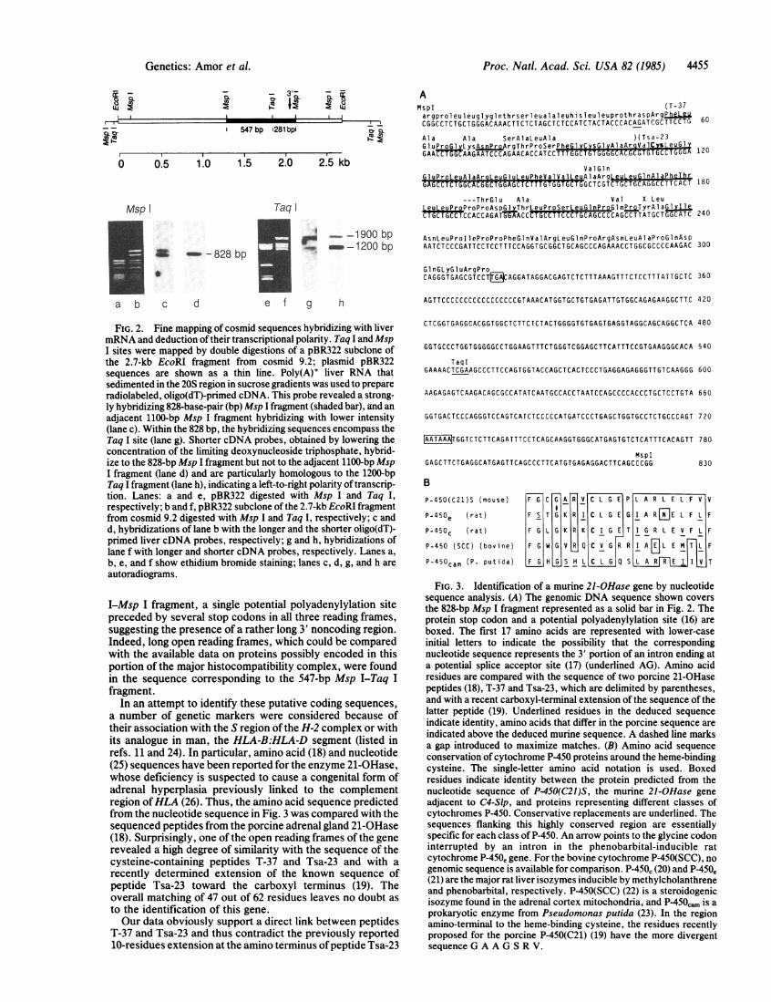
FIG. 2. Fine mapping of cosmid sequences hybridizing with livermRNA and deduction of their transcriptional polarity. Taq I and MspI sites were mapped by double digestions of a pBR322 subclone ofthe 2.7-kb EcoRI fragment from cosmid 9.2; plasmid pBR322sequences are shown as a thin line. Poly(A)+ liver RNA thatsedimented in the 20S region in sucrose gradients was used to prepareradiolabeled, oligo(dT)-primed cDNA. This probe revealed a strong-ly hybridizing 828-base-pair (bp) Msp I fragment (shaded bar), and anadjacent 1100-bp Msp I fragment hybridizing with lower intensity(lane c). Within the 828 bp, the hybridizing sequences encompass theTaq I site (lane g). Shorter cDNA probes, obtained by lowering theconcentration of the limiting deoxynucleoside triphosphate, hybrid-ize to the 828-bp Msp I fragment but not to the adjacent 1100-bp MspI fragment (lane d) and are particularly homologous to the 1200-bpTaq I fragment (lane h), indicating a left-to-right polarity of transcrip-tion. Lanes: a and e, pBR322 digested with Msp I and Taq I,respectively; b and f, pBR322 subclone of the 2.7-kb EcoRI fragmentfrom cosmid 9.2 digested with Msp I and Taq I, respectively; c andd, hybridizations of lane b with the longer and the shorter oligo(dT)-primed liver cDNA probes, respectively; g and h, hybridizations oflane f with longer and shorter cDNA probes, respectively. Lanes a,b, e, and f show ethidium bromide staining; lanes c, d, g, and h are
autoradiograms.
I-Msp I fragment, a single potential polyadenylylation sitepreceded by several stop codons in all three reading frames,suggesting the presence of a rather long 3' noncoding region.Indeed, long open reading frames, which could be comparedwith the available data on proteins possibly encoded in thisportion of the major histocompatibility complex, were foundin the sequence corresponding to the 547-bp Msp I-Taq Ifragment.
In an attempt to identify these putative coding sequences,a number of genetic markers were considered because oftheir association with the S region of the H-2 complex or withits analogue in man, the HLA-B:HLA-D segment (listed inrefs. 11 and 24). In particular, amino acid (18) and nucleotide(25) sequences have been reported for the enzyme 21-OHase,whose deficiency is suspected to cause a congenital form ofadrenal hyperplasia previously linked to the complementregion ofHLA (26). Thus, the amino acid sequence predictedfrom the nucleotide sequence in Fig. 3 was compared with thesequenced peptides from the porcine adrenal gland 21-OHase(18). Surprisingly, one of the open reading frames of the generevealed a high degree of similarity with the sequence of thecysteine-containing peptides T-37 and Tsa-23 and with arecently determined extension of the known sequence ofpeptide Tsa-23 toward the carboxyl terminus (19). Theoverall matching of 47 out of 62 residues leaves no doubt asto the identification of this gene.Our data obviously support a direct link between peptides
T-37 and Tsa-23 and thus contradict the previously reported10-residues extension at the amino terminus ofpeptide Tsa-23
CTCGGTGAGGCACGGTGGCTCTTCTCTACTGGGGTGTGAGTGAGGTAGGCAGCAGGCTCA 480
GGTGCCCTGGTGGGGGCCTGGAAGTTTCTGGGTCGGAGCTTCATTTCCGTGAAGGGCACA 540
TaqIGAAAACTCGAAGCCCTTCCAGTGGTACCAGCTCACTCCCTGAGGAGAGGGTTGTCAAGGG 600
AAGAGAGTCAAGACAGCGCCATATCAATGCCACCTAATCCAGCCCCACCCTGCTCCTGTA 660
GGTGACTCCCAGGGTCCAGTCATCTCCCCCATGATCCCTGAGCTGGTGCCTCTGCCCAGT 720
|AATAAAGGTCTCTTCAGATTTCCTCAGCAAGGTGGGCATGAGTGTCTCATTTCACAGTT 780
MspIGAGCTTCTGAGGCATGAGTTCAGCCCTTCATGTGAGAGGACTTCAGCCCGG 830
B
P-450(C21)S (mouse) F G C G A R V C L G E P L A R L E L F V V
P-450e (rat ) F S T G K R I C L G E G I A RFEJE L F L F
P-450C (rat) F G L G K R K C I G E T I G R L E V F L F
P-450 (SCC) (bovine) F G W G V R Q C V G R R I ALIL E MTL F
P-450cam (P- putida) F G H G S H L C L G Q S LLAR lRE I I T
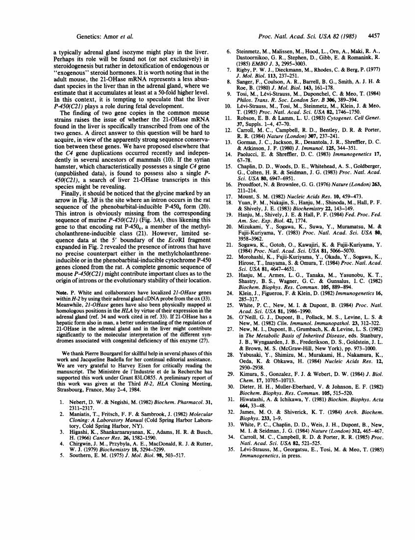
FIG. 3. Identification of a murine 21-OHase gene by nucleotidesequence analysis. (A) The genomic DNA sequence shown coversthe 828-bp Msp I fragment represented as a solid bar in Fig. 2. Theprotein stop codon and a potential polyadenylylation site (16) areboxed. The first 17 amino acids are represented with lower-caseinitial letters to indicate the possibility -that the correspondingnucleotide sequence represents the 3' portion of an intron ending ata potential splice acceptor site (17) (underlined AG). Amino acidresidues are compared with the sequence of two porcine 21-OHasepeptides (18), T-37 and Tsa-23, which are delimited by parentheses,and with a recent carboxyl-terminal extension of the sequence of thelatter peptide (19). Underlined residues in the deduced sequenceindicate identity, amino acids that differ in the porcine sequence areindicated above the deduced murine sequence. A dashed line marksa gap introduced to maximize matches. (B) Amino acid sequenceconservation of cytochrome P-450 proteins around the heme-bindingcysteine. The single-letter amino acid notation is used. Boxedresidues indicate identity between the protein predicted from thenucleotide sequence of P450(C21)S, the murine 21-OHase gene
adjacent to C4-Slp, and proteins representing different classes ofcytochromes P-450. Conservative replacements are underlined. Thesequences flanking this highly conserved region are essentiallyspecific for each class of P-450. An arrow points to the glycine codoninterrupted by an intron in the phenobarbital-inducible ratcytochrome P-450, gene. For the bovine cytochrome P-450(SCC), no
genomic sequence is available for comparison. P-450C (20) and P-450e(21) are the major rat liver isozymes inducible by methylcholanthreneand phenobarbital, respectively. P-450(SCC) (22) is a steroidogenicisozyme found in the adrenal cortex mitochondria, and P-450',,, is aprokaryotic enzyme from Pseudomonas putida (23). In the regionamino-terminal to the heme-binding cysteine, the residues recentlyproposed for the porcine P-450(C21) (19) have the more divergentsequence G A A G S R V.
_Lcx co
Genetics: Amor et al.

Proc. Natl. Acad. Sci. USA 82 (1985)
(19). Formally, we cannot exclude that this discrepancyreflects a species difference. Moreover, the cDNA sequencerecently reported for the bovine adrenal gland 21-OHase (25)is uninformative in this context because it does not cover thiscarboxyl-terminal region of the enzyme. Peptide Tsa-23 lieswithin the most conserved sequence of all cytochromesP-450, as shown by previous comparisons around thecysteine (bold print in Fig. 3) believed to bind the heme ironatom in these isozymes (22). The new murine sequenceallows extension of such comparisons to a region of 21-OHase amino-terminal to this heme-binding cysteine (Fig.3B). Interestingly, these protein alignments reveal that, atvariance with the aforementioned porcine sequence (19), thesequence deduced from the murine gene more faithfullycomplies with the canonical matches traced for this regionfrom all other cytochromes P-450, including the prokaryoticcamphor hydroxylase.Tandem Duplications of C4 and P-450(C21) Genes. A direct
implication of our findings is that a gene encoding 21-OHase,typically expressed in the adrenal gland, is also transcribed inthe liver. This observation raises the question of whetherthere are multiple copies ofgenes encoding 21-OHase, whichcould be expressed in a tissue-specific fashion. Most restric-tion endonuclease analyses were uninformative on this point.Essentially a single DNA fragment was detected (data notshown) in each case, when Southern blots of genomic DNAfrom various mouse strains were probed with the 2.7-kbEcoRI fragment subcloned from cosmid 9.2, after digestion ofthe DNA with BamHI (4.0 kb; strains BALB/c, B10.W7R,and B10.HTT), HindIII (16.5 kb; strains BALB/c, AKR/J,DBA/1, and C57BL/6), Pvu II (2 kb; strains BALB/c,DBA/1, and C57BL/6), or EcoRI (2.7 kb; strains BALB/c,B10.W7R, and B10.HTT). Although single hybridizationbands could hide multiple conserved 21-OHase gene copies,a DNA blot analysis of hamster-BALB/c somatic cellhybrids showed that all hybridizing sequences reside onmouse chromosome 17 (Fig. 4). However, the endonucleaseTaq I permitted the detection of two nonallelic gene copiesin the BALB.K (H-2k) strain (lane e) as well as in all strainsmentioned above. As one would have suspected from thevery close proximity of a 21-OHase-encoding gene to theC4-Slp gene on cosmid 9.2 and from the existence of twononallelic C4 genes in the mouse (10, 15), the other copy islocated on the cosmid containing the second C4 gene (refs. 6and 10; see Fig. 4). To distinguish the 21-OHase geneassociated with the C4 gene from that associated with theC4-Slp gene, we here refer to the two gene copies asP-450(C2l)C and P-4S0(C21)S, respectively, in line with thedesignation more commonly used for this gene family. Theclose association of P-450(C21) and C4 genes is furtherunderscored by a genetic peculiarity of strain B10.W7R,which carries an amplification of the H-2S region correlatingwith a testosterone-independent form of expression of theC4-Slp protein (10). As seen in lane f of Fig. 4, B10.W7Rcarries one P-450(C2l)C but multiple (probably four) copiesof the P-450(C2l)S gene, in line with our finding that only theC4-Slp isotype is amplified in this strain (35).
DISCUSSIONA most surprising outcome ofour search for new H-2S regiongenes that might be expressed in the liver was the detectionofP-450(C21) sequences tandemly duplicated with the genesencoding isoforms (C4 and Slp) of the fourth component ofcomplement. The enzyme 21-OHase is typically produced inthe adrenal gland, and a deficiency of this form of the enzymehas long been correlated with an HLA-linked disorder ofsteroidogenesis (27).Our data clearly establish that at least one P-450(C21) gene
copy is transcribed in the liver. In fact, it could be argued that
a b c d e f g h
kb
4.3-3.8- w
_k __-_ _ _nd
FIG. 4. Duplications of the P.450(C21) gene within H-2 ofdifferent strains. Uncloned genomic DNA from cell lines (lanes b-d;30 pg), mouse liver (a, e, f, and g; 15 ,ug) or cloned genomic DNA(lanes h and i; 1 ng) was digested with Taq I, electrophoresed in a0.8% agarose gel, blotted, and hybridized with the M13mp8 subcloneof the 547-bp Msp I-Taq I fragment (Fig. 2) labeled in its vectorportion by use of a M13 probe primer. All P-450(C21) sequences lieon chromosome 17, as shown by the comparison of the genomicDNAs of BALB/c mouse liver (lane a), of a hamster-BALB/chybrid cell line carrying the mouse chromosome 17 (lane b), and ofits sister line, which lacks mouse chromosome 17 but carries at leastone copy of each of the other mouse chromosomes (10) (lane c). Thehamster P-450(C21) sequences are not detectable under these condi-tions in the parental hamster cell line (lane d). In the H-2k haplotype(BALB.K; lane e) the probe reveals two Taq I fragments of 3.8 kband 4.3 kb, which correspond to the P-450(C21)S gene (cosmid 9.2containing C4-Slp; lane h) and to the P-450(C21)C gene (cosmid 10.8,containing C4; lane i), respectively. Strains B1O.W7R (lane f) andC3H.W7 (lane g), possess multiple copies of the P-450(C21)S gene.Note that a Taq I polymorphism distinguishes the P-450(C21)C geneof BALB/c mouse H-2d; lane a) from the corresponding gene of theH-2k and of the H-2w7 haplotypes, because the hybridization band of4.3 kb is replaced in BALB/c by a 0.9-kb Taq I fragment (see arrows).
the mRNA detected in this tissue does not encode 21-OHasebut another isozyme species whose mRNA expression in theliver is sufficiently high to offset a weak sequence homologywith P-450(C21). Two observations contradict this hypoth-esis. First, no other member of the P-4S0 gene family wasfound to hybridize with the short P-450(C21) probe used inthe Southern blot shown in Fig. 4, even after longer autora-diographic exposure. This probe includes the nucleotidesencoding the conserved region around the heme-bindingcysteine, which is essentially shared by all P-450 proteins(Fig. 3B). Second, radiolabeled liver cDNA hybridizes witha 1.2-kb Taq I fragment (Fig. 2B) containing exclusively the3' noncoding sequences ofmouse P-450(C2l)S. Significantly,the 3' noncoding regions of the known major P-450 mRNAsshow a high degree of length and sequence heterogeneityamong (20, 21, 28) or even within (29) subfamilies. Accord-ingly, probes that include the 3' noncoding region of P-450(C2l)S, such as the subcloned 2.7-kb EcoRI fragment(Fig. 2), also failed to reveal crosshybridizing genes.A 21-OHase activity in organs other than the adrenal gland
has been previously reported in a wide variety of species,including rabbit (30), ox (31), and even spiny lobster (32).However, due to the biochemical complexity of cytochromesP450, this activity was explained as cross-metabolization bynonsteroidogenic isozymes (18, 31). Thus, our detection of amRNA homologous in sequence to the gene encoding 21-OHase directly raises the issue ofthe metabolic role that such
4456 Genetics: Amor et al.
Proc. Natl. Acad. Sci. USA 82 (1985) 4457
a typically adrenal gland isozyme might play in the liver.Perhaps its role will be found not (or not exclusively) insteroidogenesis but rather in detoxification of endogenous or"exogenous" steroid hormones. It is worth noting that in theadult mouse, the 21-OHase mRNA represents a less abun-dant species in the liver than in the adrenal gland, where weestimate that it accumulates at least at a 50-fold higher level.In this context, it is tempting to speculate that the liverP-450(C21) plays a role during fetal development.The finding of two gene copies in the common mouse
strains raises the issue of whether the 21-OHase mRNAfound in the liver is specifically transcribed from one of thetwo genes. A direct answer to this question will be hard toacquire, in view of the apparently strong sequence conserva-tion between these genes. We have proposed elsewhere thatthe C4 gene duplications occurred recently and indepen-dently in several ancestors of mammals (10). If the syrianhamster, which characteristically possesses a single C4 gene(unpublished data), is found to possess also a single P-450(C21), a search of liver 21-OHase transcripts in thisspecies might be revealing.
Finally, it should be noticed that the glycine marked by anarrow in Fig. 3B is the site where an intron occurs in the ratsequence of the phenobarbital-inducible P450e form (20).This intron is obviously missing from the correspondingsequence of murine P-450(C21) (Fig. 3A), thus likening thisgene to that encoding rat P450e, a member of the methyl-cholanthrene-inducible class (21). However, limited se-quence data at the 5' boundary of the EcoRI fragmentexpanded in Fig. 2 revealed the presence of introns that haveno precise counterpart either in the methylcholanthrene-inducible or in the phenobarbital-inducible cytochrome P-450genes cloned from the rat. A complete genomic sequence ofmouse P-450(C21) might contribute important clues as to theorigin of introns or the evolutionary stability oftheir location.
Note. P. White and collaborators have localized 21-OHase geneswithin H-2 by using their adrenal gland cDNA probe from the ox (33).Meanwhile, 21-OHase genes have also been physically mapped athomologous positions in the HLA by virtue of their expression in theadrenal gland (ref. 34 and work cited in ref. 33). If 21-OHase has ahepatic form also in man, a better understanding of the regulation of21-OHase in the adrenal gland and in the liver might contributesignificantly to the molecular interpretation of the different syn-dromes associated with congenital deficiency of this enzyme (27).
We thank Pierre Bourgarel for skillful help in several phases of thiswork and Jacqueline Badella for her continual editorial assistance.We are very grateful to Harvey Eisen for critically reading themanuscript. The Ministere de l'Industrie et de la Recherche hassupported this work under Grant 83LO855. A preliminary report ofthis work was given at the Third H-2, HLA Cloning Meeting,Strasbourg, France, May 2-4, 1984.
1. Nebert, D. W. & Negishi, M. (1982) Biochem. Pharmacol. 31,2311-2317.
2. Maniatis, T., Fritsch, F. F. & Sambrook, J. (1982) MolecularCloning: A Laboratory Manual (Cold Spring Harbor Labora-tory, Cold Spring Harbor, NY).
3. Higashi, K., Shankarnarayanan, K., Adams, H. R. & Busch,H. (1966) Cancer Res. 26, 1582-1590.
4. Chirgwin, J. M., Przybyla, A. E., MacDonald, R. J. & Rutter,W. J. (1979) Biochemistry 18, 5294-5299.
5. Southern, E. M. (1975) J. Mol. Biol. 98, 503-517.
6. Steinmetz, M., Malissen, M., Hood, L., Om, A., Maki, R. A.,Dastoorikoo, G. R., Stephen, D., Gibb, E. & Romanink, R.(1985) EMBO J. 3, 2995-3003.
7. Rigby, P. W. J., Dieckmann, M., Rhodes, C. & Berg, P. (1977)J. Mol. Biol. 113, 237-251.
8. Sanger, F., Coulson, A. R., Barrell, B. G., Smith, A. J. H. &Roe, B. (1980) J. Mol. Biol. 143, 161-178.
9. Tosi, M., Levi-Strauss, M., Duponchel, C. & Meo, T. (1984)Philos. Trans. R. Soc. London Ser. B 306, 389-394.
10. Levi-Strauss, M., Tosi, M., Steinmetz, M., Klein, J. & Meo,T. (1985) Proc. Natl. Acad. Sci. USA 82, 1746-1750.
11. Robson, E. B. & Lamm, L. U. (1983) Cytogenet. Cell Genet.37, Suppls. 1-4, 47-70.
12. Carroll, M. C., Campbell, R. D., Bentley, D. R. & Porter,R. R. (1984) Nature (London) 307, 237-241.
13. Gorman, J. C., Jackson, R., Desantola, J. R., Shreffler, D. C.& Atkinson, J. P. (1980) J. Immunol. 125, 344-351.
14. Paolucci, E. & Shreffler, D. C. (1983) Immunogenetics 17,67-78.
15. Chaplin, D. D., Woods, D. E., Whitehead, A. S., Goldberger,G., Colten, H. R. & Seidman, J. G. (1983) Proc. Natl. Acad.Sci. USA 80, 6947-6951.
16. Proudfoot, N. & Brownlee, G. G. (1976) Nature (London) 263,211-214.
17. Mount, S. M. (1982) Nucleic Acids Res. 10, 459-473.18. Yuan, P. M., Nakajin, S., Hanju, M., Shinoda, M., Hall, P. F.
& Shively, J. E. (1983) Biochemistry 22, 143-149.19. Hanju, M., Shively, J. E. & Hall, P. F. (1984) Fed. Proc. Fed.
Am. Soc. Exp. Biol. 42, 1774.20. Mizukami, Y., Sogawa, K., Suwa, Y., Muramatsu, M. &
Fujii-Kuriyama, Y. (1983) Proc. Natl. Acad. Sci. USA 80,3958-3962.
21. Sogawa, K., Gotoh, O., Kawajiri, K. & Fujii-Kuriyama, Y.(1984) Proc. Natl. Acad. Sci. USA 81, 5066-5070.
22. Morohashi, K., Fujii-Kuriyama, Y., Okada, Y., Sogawa, K.,Hirose, T., Inayama, S. & Omura, T. (1984) Proc. Natl. Acad.Sci. USA 81, 4647-4651.
23. Hanju, M., Armes, L. G., Tanaka, M., Yasunobu, K. T.,Shastry, B. S., Wagner, G. C. & Gunsalus, I. C. (1982)Biochem. Biophys. Res. Commun. 105, 889-894.
24. Klein, J., Figueroa, F. & Klein, D. (1982) Immunogenetics 16,285-317.
25. White, P. C., New, M. I. & Dupont, B. (1984) Proc. Natl.Acad. Sci. USA 81, 1986-1990.
26. O'Neill, G. J., Dupont, B., Pollack, M. S., Levine, L. S. &New, M. (1982) Clin. Immunol. Immunopathol. 23, 312-322.
27. New, M. I., Dupont, B., Grumbach, K. & Levine, L. S. (1982)in The Metabolic Basis of Inherited Disease, eds. Stanbury,J. B., Wyngaarden, J. B., Frederikson, D. S., Goldstein, J. L.& Brown, M. S. (McGraw-Hill, New York), pp. 973-1000.
28. Yabusaki, Y., Shimizu, M., Murakami, H., Nakamura, K.,Oeda, K. & Ohkawa, H. (1984) Nucleic Acids Res. 12,2930-2938.
29. Kimura, S., Gonzalez, F. J. & Webert, D. W. (1984) J. Biol.Chem. 17, 10705-10713.
30. Dieter, H. H., Muller-Eberhard, V. & Johnson, E. F. (1982)Biochem. Biophys. Res. Commun. 105, 515-520.
31. Hiwatashi, A. & Ichikawa, Y. (1981) Biochim. Biophys. Acta664, 33-48.
32. James, M. 0. & Shiverick, K. T. (1984) Arch. Biochem.Biophys. 233, 1-9.
33. White, P. C., Chaplin, D. D., Weis, J. H., Dupont, B., New,M. I. & Seidman, J. G. (1984) Nature (London) 312, 465-467.
34. Carroll, M. C., Campbell, R. D. & Porter, R. R. (1985) Proc.Natl. Acad. Sci. USA 82, 521-525.
35. Levi-Strauss, M., Georgatsu, E., Tosi, M. & Meo, T. (1985)Immunogenetics, in press.
Genetics: Amor et al.