liver parenchymal cell injury viii. lesions of …jcb.rupress.org/content/jcb/41/3/736.full.pdf ·...
TRANSCRIPT

LIVER PARENCHYMAL CELL INJURY
VIII. Lesions of Membranous Cellular
Components following Iodoform
DAVID A . SELL and EDWARD S . REYNOLDS
From the Department of Pathology, Peter Bent Brigham Hospital and Harvard Medical School,Boston, Massachusetts 02115 . Dr . Sell's present address is Base Hospital, Keesler Air Force Base,Biloxi, Mississippi .
ABSTRACT
Iodoform, a relatively water-insoluble yellow solid, chemically reactive in free-radicalreactions, produces early hepatocellular injury qualitatively similar to that of carbontetrachloride . 2 hr after administration of radioactively labeled iodoform, nonvolatile 11Cis preferentially recovered in microsomal lipid and protein . By 30 min microsomal propertiesare profoundly affected : oxidative demethylation decreases abruptly ; increased lipoperoxidedecomposition products are detected ; and amino acid incorporation into liver protein isdepressed. By 1 hr glucose-6-phosphatase is suppressed centrolobularly and increasedstainable calcium is present in the midzone. Increased cell sap RNA contents are observedby 2 hr . Morphologically, the biochemical and histochemical changes are associated withprogressive dispersion, vacuolation, and degranulation of the granular endoplasmic re-ticulum . Calcium-associated masses accumulate within the mitochondrial matrix, andmitochondria become progressively pleomorphic . Golgi components dilate and disperse .Membranous components of the cytoplasm of parenchymal cells conglomerate into laby-rinthine tubular aggregates . Lipid accumulates in cytoplasmic droplets . Ultimately, centro-lobular necrosis ensues . The close cytochemical and morphological similarities between thecellular injury produced in the liver by iodoform and that produced by carbon tetrachloridesuggest common pathogenetic mechanisms associated with damage to membranes .
INTRODUCTION
The liver lesion of carbon tetrachloride poisoningis one of the most extensively analyzed modelsystems of cellular injury (2, 29, 33, 35, 36, 45) .The chlorocarbon appears and is concentrated inthe liver within minutes following its administra-tion (32, 39) . Liver lipids, proteins, and nucleicacids become labeled with the products of itsmetabolism (38, 39) . Increased products of lipo-peroxidation accumulate in isolated componentsof the endoplasmic reticulum (29, 31) . Enzymaticfunctions of the endoplasmic reticulum, includingglucose-6-phosphatase, oxidative demethylation,
736
and the ability to synthesize protein, are affectedby the end of the 1st hr following poisoning(24, 33, 35, 45, 47) . Concomitantly, increasedcalcium enters midzonal parenchymal cells andthen spontaneously disappears by 2 hr (35) . Withtime, lipid accumulates and centrolobular necrosisensues (25, 29, 33, 36) . Although other hepato-toxins, specifically dimethylnitrosamine and thio-acetamide, may interfere with protein synthesisearly in the course of their lesions, centrolobularnecrosis ensues without prior alterations ofglucose-6-phosphatase, drug metabolism, or the
on May 8, 2018jcb.rupress.org Downloaded from http://doi.org/10.1083/jcb.41.3.736Published Online: 1 June, 1969 | Supp Info:

permeability of the plasma membrane to
(5, 14, 19, 21, 40) .
halomethanes,Among
calcium
iodoform, a virtuallywater-insoluble yellow powder (119 ° C) sparinglysoluble in lipids, is at least as reactive as carbontetrachloride in homolytic cleavage (free radical)reactions (6) . This study examines in detail theearly morphologic and cytochemical componentsof the liver lesion of iodoform poisoning . Thestriking similarities in the toxic effects of the twohalomethanes on liver parenchymal cells support
a common pathogenetic mechanism and providea basis for its further elucidation .
MATERIALS AND METHODS
Healthy young male rats (Charles River BreedingLaboratories, Inc., North Wilmington, Mass .),weighing between 150-300 g, were maintained on adiet of Purina Chow and water ad libitum . lodoform-mineral oil suspensions containing 0 .68 g iodoformper ml were prepared by high-speed homogenization .
For morphologic studies, replicate rats were sacri-ficed at 1Z, 1, 2, 4, and 8 hr following the oral adminis-tration by polyethylene stomach tube of a single doseof 2,600 mmole iodoform per 100 g of animal. Controlanimals were fed 1 .5 ml mineral oil per 100 g of ani-mal and sacrificed at 2 hr . Animals were sacrificed bydecapitation and exsanguination following a 16 hrfast. The left lateral lobe of the liver was removedimmediately and sliced . Cubes of liver were fixed ins-collidine-buffered osmium tetroxide (0-4 °C) (3),postfixed in saturated aqueous uranyl acetate (18),and embedded in Epon 812 . Serial sections (1 µ) werestained with toluidine blue O for light microscopy .Sections for electron microscopy, stained sequentiallywith uranyl acetate and lead citrate, were examinedin an RCA EMU 3G electron microscope.
For histochemical studies, cryostat sections of liversof control and treated animals sacrificed at 1 and 8 hrfollowing feeding of the standard dose of iodoform(2,600 mmole/100 g animal) were stained for glucose-6-phosphatase activity by the method of Wachsteinand Meisel (50) . Calcium was stained with alizarinred S by the method of McGee-Russell (22) . Succinicand glutamic dehydrogenase activities were deter-mined by incubation of cryostat sections with nitro-tetrazolium blue in appropriate media (23) .
Patterns of incorporation of 14C and 125 1 from iodo-form were determined in livers of animals fasted for16 hr and then fed the appropriately labeled iodoform2 hr prior to sacrifice . 14C HI3 (New England NuclearCorporation, Boston, Mass .) and CH12513 (RadioChemical Centre, Amersham, England) were storedin the dark at -16 °C until used . All preparationswere carried out at 0-4 °C. Livers of replicate ratswere removed, homogenized in 0 .25 M sucrose, and
the homogenates were fractionated by differentialcentrifugation into residue, mitochondria, micro-somes, and cell sap (17) . Liver homogenate and sub-cellular fractions were chemically fractionated intoacid-soluble, lipid, nucleic acid, and protein constit-uents by a modified Schmidt-Thannhauser procedure(37) . Total nucleic acids, DNA, RNA, and proteincontents of each fraction were determined (37) . Non-volatile 14C in liver homogenates, subcellular frac-tions, and their chemical constituents was measuredin aliquots, and dried for 1 hr at 80 °C. Dried sampleswere counted at infinite thickness in a thin-windowgas-flow counter. 125I contents were determined in awell-type scintillation counter . 14C and 1251 contentsof the samples were compared with 14C and 1251
standards counted concurrently . Excess potassiumor perchlorate present which might interfere withthe determination of 14C was removed by precipita-tion as potassium perchlorate (38) .
Functional and chemical properties of microsomesisolated from livers of rats, at times up to 4 hr follow-ing intragastric administration of the standard dose ofiodoform, were determined . Oxidative demethylationwas assayed according to the method of Orreniusand his associates (26) . Nicotinamide adenine dinu-cleotide phosphate (NADPH) -neotetrazolium reduc-tase was determined according to the method ofDallner (7) . The glucose-6-phosphatase assay systemcontained 15 µm glucose-6-phosphate, 30 µm Trismaleate buffer, pH 6.7, 20 µm EDTA, pH 6 .7, 250 µmsucrose, and 1 mg microsomes (wet weight) in a finalvolume of 1 .5 ml . After incubation at 37 °C for 20 minthe reaction was stopped by the addition of an equalvolume of 10 0%o TCA, and aliquots of the super-natant were analyzed for inorganic phosphorusaccording to the method of Fiske and SubbaRow(12) .Conjugated diene contents of microsomal lipids
were determined by a modification of the method ofRecknagel (30) . 1 j ml aliquots of microsomal sus-pensions in 0.25 M sucrose containing 300-400 mgmicrosomes (wet weight) per ml were extracted with19 volumes 2 :1 chloroform : methanol . The super-natant was separated into two phases by the additionof 2 .0 ml water according to Folch et al . (13), and theupper phase was discarded. 0.2 ml methanol wasadded to the lower phase, and the optical density wasdetermined between 210-300 mµ by using freshlyprepared lower phase as blank (25 ml water to 95 ml2 :1 chloroform: methanol) . Peak absorption for dieneconjugation products of lipid peroxidation (4) isolatedunder these conditions shifts from 233 to 240 mµ, inthe presence of chloroform. Absorption at the latterwavelength, expressed as optical density units pergram microsomes (wet weight), was used as an indi-cator of the extent of lipoperoxidation .
14 C-glycine incorporation into protein was deter-
DAVID A. SELL AND EDWARD S. REXN96DS Jodoform Poisoning
7 37

mined following the intraperitoneal injection of 25µc uniformly-labeled 14C-glycine (New EnglandNuclear Corporation) 30 min prior to sacrifice .Ribosomal, cisternal, and membranous subfractionsof microsomes were obtained by first treating micro-somes with a final concentration of 0.010 M EDTA(pH 7 .4) to remove ribosomes (27 and footnote 1)followed by hypotonic stress of low ionic strength toextract cisternal protein (51) . Proteins isolated frontthe homogenate, microsomes, and microsomal sub-fractions were dried to constant weight at 104 °C, dis-solved in a small amount of 0.3 M KOH, and thendiluted for counting .
RESULTS
Recovery Patterns of lodoform Derived14C and 1251
Nonvolatile 14C and 125I derived from isotopicallylabeled iodoform are recovered from chemicalcomponents of liver homogenates and subcellularfractions 2 hr following poisoning (Table I) .14C from iodoform preferentially labels proteinsand lipids, particularly the protein and lipids ofthe residue and microsomal fractions (Fig . 1) .In contrast, most of the 1251 is recovered from theacid-soluble fraction of the cell sap and, to amuch lesser extent, from lipids of all fractions(Fig. 1, Table I) . Under the conditions used inthis study, the bulk of 1251 in the acid-solublefraction is considered to be inorganic iodide (38) .The distribution of I'll in chemical constituentsof subcellular components following feeding ofiodoform differs greatly from that resulting fromthe addition of the labeled compound to liverhomogenates in vitro . In the latter instance,between 80 and 90% of the 1251 is recovered inlipid fractions .
TABLE I
Nonvolatile Radioactivity Recovered in Liver 2 Hr after Oral Feeding of Isotopic lodoform
* Numbers in parentheses refer to number of animals.
738
THE JOURNAL OF CELL BIOLOGY • VOLUME 41, 1969
Light Microscopy
Lipid accumulation, diffuse non-lipid vacuola-tion, and increased mitochondrial irregularityfollowing iodoform poisoning, first observed insome centrolobular cells by 1 hr, extend to involvecells in the midzonal and periportal parenchymabetween 4 and 8 hr (Fig . 2) . Concomitantly,detectable suppression of glucose-6-phosphataseoccurs centrolobularly by 1 hr (Fig. 3 b) and abroad band of midzonal liver parenchymal cellsstain forincreased calcium (Fig . 3 e) . Centrolobularglucose-6-phosphatase activity is virtually abol-ished by 8 hr (Fig. 3 c) while the diffuse in-crease in midzonal calcium disappears leaving onlyscattered, intensely stained cells (Fig . 3f) . Histo-chemical patterns of succinic and glutamic dehy-drogenase activities at 8 hr are indistinguishablefrom controls .
Electron Microscopy
Earliest discernible changes following iodoforminvolve the endoplasmic reticulum . By 1 hr theergastoplasm is dispersed, the membranes of thegranular endoplasmic reticulum are partially de-granulated, and increased numbers of free mon-omer ribosomes appear in the cytoplasmic matrix(Figs . 4-6, Table II) . Cisternae of the granularendoplasmic reticulum may be dilated . Thesechanges are progressive with time, and by 8 hr, inthose cells with the most severe degranulation ofthe endoplasmic reticulum, well-granulated strandsare observed only in close approximation tomitochondria (Figs . 7-9) .
The extent of degranulation was semiquantita-tively estimated in representative electron micro-graphs . Ribosomes within less than one ribosomal
Distribution of isotope recovered
Subcellular fractions Chemical constituents
Isotope' Dose/100 g rat
Nonvolatileisotope
recovered Residue Mito Micro LipidNucleicacids ProteiCell sap Acid-sol .
pc am/IM/100 gliver (mm) mean % total isotope bound
19CH1 3 (3) 33 17 0 .8 41 8 14 26
25 28 1 36
CH1 251 3 (3) 250 140 3 .9 25 1 4 60
101 6 0 0

RESIDUE MITO- MICRO- CELLCHONDRIA SOMES SAP
c
central veinCa
intramitochondrial calcium-associatedelectron-opaque masses
•
space of DisseLTA labyrinthine tubular aggregates•
lipidMb microbodies
M
mitochondrion•
portal triadRp
polysoines•
ribosomes, freeRm ribosomes, membrane boundSer
agranular endoplasmic reticulum•
vacuoleScale line in all electron micrographs is one micron in length .
FIGURE 1 Patterns of incorporation of 14CH1 3 and CH12513 into protein, nucleic acid, lipid, and acid-soluble constituents of residue, mitochondria, microsomes, and cell sap of livers of adult rats at 2 hrsHeight of each bar represents that fraction of total nonvolatile radioactivity of the homogenate recoveredin the respective chemical component of the subcellular fraction . Bar heights are average of values fromthree animals .
diameter of a membrane were considered mem-brane-bound . Non-membrane-bound ribosomeswere considered polysomal if they were within oneribosomal diameter of another ribosome . Freeribosomes were separated from other ribosomesand membranes in two dimensions by a distancegreater than 200 A . In the most severely affectedcells decreased numbers of membrane-boundribosomes and increased numbers of free monomerribosomes are present in the cytoplasm by 1 hr
(Table II) . As time following poisoning lengthens,increasing numbers of ribosomes appear free inthe cytoplasmic matrix both in the least and in themost severely affected cells (Table II) . As ageneral rule the most severely affected cells aremidzonal and centrolobular .Membranous components of the endoplasmic
reticulum decrease in many cells by 8 hr, andlarge conglomerations of closely packed, inter-lacing, smooth-surfaced tubules confluent with
DAVID A. SELL AND EDWAun S. REYNOLDS lodoform Poisoning 739
20 4CHIO
t0TOTALCPM 0
QRO &'\H~1C\.E\C\-\Q\OQ .C\O S ∎L~ ∎
60CH
125
50
0 40TOTALCPM 30
20
10 , '
0 ∎
,
QRO~E\H `H~1CEE\C A. ~'∎
_ __-
∎ ∎`AC\O SOE .

FIGURE 2 Light microscopic illustration of alterations in periportal (Figs . 2 a, 2 b) and centrolobular(Figs . 2 c, 2 d) cells following poisoning with iodofortn . Osmium tetroxide fixation ; Epon embedded ; 1 gsections, toluidine blue O . X 1400 . 2 a . Periportal region in control. 2 b . 8 hr following poisoning with iodo-form . Some cells (upper left) are diffusely vacuolated . Other cells show increased numbers of lipid vacuoles .Areas devoid of mitochondria are prominent in the centers of cells (arrow) . Mitochondria appear morepleomorphic than in controls . 2 c . Centrolobular region in control . 2 d. 8 hr following poisoning with iodo-form. Prominent, diffuse vacuolation is present in some cells, and lipid vacuoles are seen in other cells .The central areas of many cells are again devoid of mitochondria (arrows) . Mitochondria appear morepleomorphic than in controls .
recognizable remnants of both smooth and roughendoplasmic reticulum appear (Figs . 7-9) .These conglomerations or labyrinthine tubularaggregates (LTA) differ from smooth endoplasmicreticulum in that their tubular components aresmaller, more intricately interwoven, and asso-ciated with amorphous electron-opaque deposits(Figs . 7-9) .
740
THE JOURNAL OF CELL BIOLOGY • VOLUME 41, 1969
Striking mitochondrial changes are evident at1 hr following poisoning . In midzonal parenchy-mal cells rosette and ring-like calcium-associatedelectron-opaque granules appear in the mito-chondrial matrix immediately adjacent to innermembranes (Figs . 4, 5) . Normal matrix granulesappear decreased in these mitochondria .
Outlines of some mitochondria become markedly

FIGURE 3 Time course of glucose-6-phosphatase suppression (Figs . 3 a-3 c) and calcium influx (Figs .3 d-3 f) following iodoform . Fresh-frozen cryostat sections, 6 µ thick. X 60 . 3 a. Glucose-6-phosphataseactivity in control liver is uniform throughout . 3 b. At 1 hr following iodoform the centrolobular activityis moderately suppressed . 3 c . At 8 hr the activity is markedly suppressed except in periportal areas . 3 d.Alizarin red-S staining of control liver . Focal areas of increased stainable calcium are not present . 3 e . At1 hr a faint midzonal band of increased staining is prominent . 3 f. At 8 hr the mid-zonal band has disap-peared and scattered cells in midzonal and centrolobular areas stain intensely for calcium .

FIGURE 4 Parenchymal cell 1 hr after poisoning with iodoform . Small granular calcium-associated,electron-opaque deposits of varying size are present within the mitochondrial matrix . These deposits areclose to the internal mitochondrial membranes . An enlarged mitochondrion (MI) contains a pale flocculentmatrix . Ribosomes are predominantly membrane associated although, focally, ribosomes appear to liefree within the cytoplasmic matrix. Nucleus and nucleolus appear as in controls . X 30,000.

FIGURE 5 Portions of liver cells 1 hr after poisoning with iodoform . Increased numbers of free ribosomesare noted in the cytoplasmic matrix. X :27,000 . 5 a . Calcium-associated, electron-opaque deposits areevident close to inner mitochondrial membranes . A similar deposit (arrow) is present in an enlarged mito-chondrion (MI) with a pale matrix . Inset : Calcium-associated electron-opaque deposit within mitochon-drion of parenchymal cell 4 hr following administration of iodoform shows concentric rings . X 91,000 .5 b . Occasional mitochondria with apparently normal matrix density have markedly scalloped boundaries(M2) . Note absence of matrix granules .
irregular, and their boundaries scalloped (Fig .5). Other mitochondria may enlarge slightly andcontain a less dense matrix, although normalnumbers of cristae mitochondriales persist (Fig .4, 9). Electron-opaque, intramitochondrial, cal-cium-associated masses may occasionally be seenwithin such mitochondria (Fig . 5) . The density ofthe mitochondrial matrix may increase in damagedcells and approach that of microbodies and nearbyerythrocytes . In such mitochondria the matrixconsists of homogeneously distributed, electron-lucent granules of relatively uniform size (Fig . 9) .Mitochondria with less dense, more flocculentmatrices of uneven granularity may be seenwithin the same cell (Fig . 9), and in these cellsribosome-like granules and small dark lipid-like
droplets appear (Fig. 9) . In cells with markedcisternal dilatation of the endoplasmic reticulumat 8 hr, concentric membranous inclusionsappear both within the outer mitochondrialspace and within the adjacent cytoplasmic matrix(Fig . 6) .
Cell boundaries are well preserved at 1 hr .Occasional focal separations between adjacentcell membranes occur by 8 hr. Microvilli in theperisinusoidal spaces of Dissé do not show markedvariations, although clubbing and areas devoidof microvilli are occasionally seen (Figs . 6, 7) .Vascular changes are not apparent except forthe presence of rare fibrin thrombi within sinusoids .
The Golgi apparatus balloon progressivelywith time and, by 8 hr, the few that are recognized
DAVID A. SELL AND EDWARD S. REYNOLDS Iodoforln Poisoning
743

FIGURE 6 Cytoplasm of centrolobular liver cell 8 hr after poisoning with iodoform . There is markeddilation of cisternae of endoplasmic reticulum . Intracytoplasmic lipid droplets are abundant. The ma-jority of ribosomes are free in the cytoplasmic matrix as both monomers and polysomes . Mitochondria, allhave a relatively dense matrix . Concentric membranous bodies (arrows) apparently lie free within thecytoplasmic matrix and also within outer mitochondrial space . Microvilli lining the space of Disse arefocally absent . X 27,000 . Inset : Membranous inclusion evaginates the outer mitochondrial membrane .X 81,000 .

FIGURE 7 Cytoplasm of two midzonal cells 8 hr after poisoning with iodoform . Intracytoplasmic lipiddroplets are present in both cells. Tubules consisting of smooth membranes in the upper cell representsmooth endoplasmic reticulum . Well-developed labyrinthine tubular aggregates are present in the lowercell in association with characteristic foci of amorphous increases in density (arrows) . Note the pale cyto-plasm, obvious free ribosomes, and dilated cisternae of endoplasmic reticulum in the lower cell as com-pared to the upper cell . Differences in density of mitochondrial matrix exist between the two cells . Micro-villi lining the cell-cell interface are clubbed . X 20,000 .

TABLE IIEstimation of Ribosomal Populations in Cytoplasm
of Liver Cells after Poisoning with Iodoform
Ribosomes counted
Ribosomal distribution
consist of dispersed clusters of dilated vacuolescontaining few of the 600-800 A electron-opaquegranules normally present.
Microbodies and lysosomes, present in paren-chyma) cells of livers of both control and experi-mental animals, do not increase in number orappear to be morphologically altered during thisinitial period . Definite changes in nucleoli at-tributable to iodoform were not recognized .
Biochemical Functions and Properties of theEndoplasmic Reticulum
The striking changes in the morphology of theendoplasmic reticulum within 1 hr followingiodoform are concomitant with alterations inenzymatic and chemical properties of its micro-somal counterpart . Within /'z hr following poison-ing the ability of microsomes to oxidatively
FIGURE 8 Labyrinthine tubular aggregate in centrolobular cell 8 hr following iodoform .Strands of preserved perimitochondrial granular endoplasmic reticulum are continuouswith membranes at the periphery of the labyrinthine tubular aggregate . Amorphous areasof increased electron opacity are apparent (arrows) . Increased numbers of free ribosomesare present in the adjacent cytoplasmic matrix . X 60,000 . Inset : Occasional botryoidalclusters of 60-80 A dense granules (arrow) are present within a labyrinthine tubular ag-gregate. X 91,000 .
746
THE JOURNAL OF CELL BIOLOGY . VOLUME 41, 1969
demethylate antipyrine is decreased by nearlyone-third, and a significant increase in dieneconjugation products of lipoperoxidation occurs(lipid E 240 , Table III) . 14 C-glycine incorporationinto protein is also decreased to approximatelyone-half the control values at this time . Decreasesin oxidative demethylase activity and 14C-glycineincorporation are progressive with time, respec-tively falling to 13 and ' s their control levels by2 hr (Table III) . Although similar amounts ofribosomal, cisternal, and membrane protein arerecovered from control and iodoform-poisonedanimals, reduction of glycine incorporation isgreatest in protein isolated from ribosomes andmembranes at 2 hr and is least in cisternal pro-teins . Lipid peroxidation decreases slightly follow-ing its initial l,z hr elevation, while increasedcell sap RNA contents, a biochemical indicatorof degranulation of the rough endoplasmic re-ticulum, is first noted at 2 hr . In contrast, NADPH-NT-reductase and microsomal protein contentsare only slightly altered from normal throughoutthis early time period (Table III) . Glucose-6-phosphatase activity, profoundly affected in thehistochemical assay procedure (Fig . 3), is notsignificantly decreased biochemically until 4 hrfollowing poisoning .
DISCUSSION
Comparison of cellular injury following iodoformwith that following other hepatotoxins offers abasis for further elucidation of some basic patho-genetic mechanisms. Recovery of products ofiodoform metabolism within liver cell componentsearly in the course of poisoning indicates that thishepatotoxin might be most meaningfully com-pared with other hepatotoxins which are metabo-lized (21, 34, 38, 40) . The major early morpho-logical and biochemical alterations produced byiodoform reveal a striking similarity to carbon
Mem-branebound
Cytoplasmicmatrix
Poly- Mon-somal orner
Total qo TotalLeast affected cells
Controls 226 81 14 5?Z hr. 221 80 15 51 hr. 216 71 20 78 hr. 98 57 6 37
Most affected cellsControl 237 68 29 3
12 hr. 258 62 33 51 hr. 102 38 21 418 hr. 71 32 18 50

DAVID A. SELL AND EDWARD S. REYNOLDS Iodoform Poisoning
747
tetrachloride hepatotoxicity in particular (2, 24,25, 29-33, 35, 36, 38, 39, 45, 47) .
Disaggregation of the ergastoplasm and disper-sion of the granular endoplasmic reticulumthroughout the cytoplasm is the earliest recog-nizable morphologic change following iodoform .Concomitantly, the membranes are degranulated,free ribosomes appear within the cytoplasmicmatrix and, in time, cisternae of the granularendoplasmic reticulum dilate . However, there isstriking preservation of the flattened granulatedcisternae about mitochondria. Such early dispersalof ergastoplasm and degranulation of endoplasmicreticulum is also a major morphologic feature ofpoisoning with dimethylnitrosamine (8) andethionine (1) as well as with carbon tetrachloride(25, 35, 47) . This change which results from thefeeding of these apparently dissimilar poisonsmay be but a secondary response to differingprimary molecular events. Dimethylnitrosaminemethylates nucleic acids and proteins (20, 21) .Ethionine produces ATP starvation and, ergo,suppression of synthesis of messenger RNA (11) .Cleavage products from the metabolism of carbontetrachloride may primarily attack the membranesof the cell (40) . Iodoform, which is at least asreactive in free-radical reactions as carbon tetra-chloride (6), may have a similar mechanism ofaction.
The early iodoform-induced changes in theendoplasmic reticulum are followed by the forma-tion of labyrinthine tubular aggregates, i .e .,conglomerations of closely packed interconnectingsmooth-surfaced tubules . Identical structuresform following carbon tetrachloride (37) . "Neben-kern" observed in livers of rats following di-methylnitrosamine (9) and thioacetamide (42)may represent similar structures . The apparentmorphologic differences between labyrinthine
748
tubular aggregates and smooth endoplasmicreticulum suggest that the former may be theresult of degenerative changes in the membranesof the endoplasmic reticulum. Indeed, labyrinthinetubular aggregates persist in hepatocytes recover-ing from carbon tetrachloride injury in whichnewly synthesized endoplasmic reticulum is alsopresent .'
The early and transient influx of calcium intothe midzone 1 hr following iodoform, precedingby hours the terminal reentry of calcium into theliver lobule, had previously been obser~ ed to beunique for carbon tetrachloride poisoning (40) .Such an early influx suggests the occurrence of atransient alteration in plasma membrane permea-bility secondary to direct chemical attack by thetoxin. The appearance of calcium-associatedelectron-opaque intramitochondrial masses atthese times following both iodoform and carbontetrachloride, is interpreted as the result of thephysiologic response of mitochondria to increasedcalcium entry into the cell (10, 28, 35, 37, 48),although primary injury to the membranes of themitochondria as a participating event cannot beruled out .
Mitochondrial changes following iodoform alsoinclude progressive decrease and increase inmitochondrial matrix density which at times occurswithin the same cell . These changes, which arenot apparently related to calcium accumulation,may be a reflection of differing respiratory statesof the mitochondria (15) . Small mitochondriawith dense matrices previously designated "pyk-notic mitochondria" (49) may be the result of thesame processes .The striking morphologic changes involving
the endoplasmic reticulum in the liver lesions of
'E. S . Reynolds . Unpublished data.
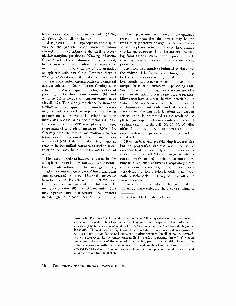
FIGURE 9 Portion of centrolobular liver cell 8 hr following iodoform . The difference inmitochondrial matrix densities and state of aggregation is apparent . The darker mito-chondria (M3) have occasional small (200-300 A) granules (arrows) within a finely granu-lar matrix. The matrix of the light mitochondrion (MI) is more flocculent in appearancewith an uneven granularity and occasional darker granules (small arrow) of approxi-mately 140-180 A. An intramitochondrial lipid inclusion is present (arrow) . The outermitochondrial space is of the same width in both types of mitochondria . Labyrinthinetubular aggregates with their characteristic amorphous densities are present as are in-creased free ribosomes . Preserved strands of granular endoplasmic reticulum are presentabout mitochondria. X 40,000 .
THE JOURNAL OF CELL BIOLOGY • VOLUME 41, 1969

DAVID A. SELL AND EDWARD S. REYNOLDS lodoform Poisoning
749

g microsomes (wet wt .) .$ Numbers in parentheses refer to number of animals .§ P = <0 .05 .11 P = <0.005 .
both iodoform and carbon tetrachloride poisoningare accompanied by marked alterations in thecytochemical properties of the endoplasmic reticu-lum. The recovery patterns of non-volatile 14C and,1251 from iodoform in chemical components ofsubcellular fractions are qualitatively similar tothose of isotopic carbon and chlorine followingadministration of carbon tetrachloride (38, 39) .Both agents suppress glucose-6-phosphatase earlyin the course of poisoning (35, 40) . Within 30 minfollowing iodoform, lipoperoxides are increased,oxidative demethylation decreases, and proteinsynthesis is affected . Similar changes occur follow-ing carbon tetrachloride (31, 46, 47) . 1 Oddly,following iodoform, increases in cell sap RNA, abiochemical indicator of degranulation of granularendoplasmic reticulum (25, 35, 41, 45, 47), donot occur until after 1 hr . The temporal rela-tionships whereby amino acid incorporation intomicrosomal protein decreases prior to degranula-tion of the rough endoplasmic reticulum suggestthat protein synthesis fails while the ribosomes arestill membrane attached . In sharp contrast tothe changes which occur in the endoplasmicreticulum, histochemical analyses of mitochondrialsuccinic and glutamic dehydrogenase activityshow no detectable changes during the early periodstudied .
Recovery of increased products of lipoperoxida-tion from microsomes during the time of concen-tration of the poison by the liver strongly suggeststhat lipoperoxidation is an important process inthe initial cytopathologic lesion (31) . Smallamounts of diene conjugation products of lipo-
7 5 0 THE JOURNAL OF CELL BIOLOGY ' VOLUME 41, 1969
peroxidation are recoverable from lipids of"microsomes" isolated from livers of normal rats(30, 31, and footnote 1) . Increased amounts mayresult from increased rates of auto-oxidation or offree-radical reactions (4, 52) . Iodoform andcarbon tetrachloride are readily reactive in free-radical reactions and may produce increasedlipoperoxidation either by increased reactivity offree-radicals formed or by increased rates ofproduction of free radicals in ongoing free-radicalreactions postulated to occur in the membranesof the endoplasmic reticulum of normal liver cells(16, 26, 44) .
Increased free-radical generation is an attrac-tive hypothesis to explain the subsequent changesin the membranes of the endoplasmic reticulum .Free-radicals react in chain reactions (4, 52), andsuch reactions in a lipid phase of a membranecould result in partial polymerization of its lipid,protein, and nucleic acid components (52) . De-struction of highly unsaturated lipids would alsooccur (30, 52) . Decreased contents of arachidonicand linolenic acid in microsomes following carbontetrachloride have been postulated to be theresult of such reactions (30) . Thus, such changes asdegranulation of the granular endoplasmic re-ticulum and the formation of labyrinthine tubularaggregates may be but morphologic manifesta-tions of these underlying chemical events .The dissociation between carbon and iodine
labeling in this study is not incompatible with sucha postulated free-radical mechanism. The libera-tion of iodide ion into the cell sap can result eitherfrom the sequestering of an electron by nascent
TABLE III
Functions and Properties of the Endoplasmic Reticulum after Poisoning with CHI ;;
Microsomalproteincontent
Oxidativedemethylation
NADPH-NTreductase
G-6-phosphatas Lipid E 0 „
uC-glycine incor-poration Cell sap RN A
mg/g *mmle
CII0O/,g* minmmole
NTH/g* nainmmole h
m oo/gm* rpm/mg pral . nannte/g lac r
Control (9) $ 74 ± 1 0 .36 ± 0.02 1 .8 ± 0 .1 21 ± 1 4 .4 ± 0 .4 1240 ± 170 3.2 ± 0.2
?z hr (5) 68 ± 3 0 .26 ± 0 .03§ 1 .9 ± 0 .2 22 ± I 7 .6 ± 0 .7)1 590 ± 250 3 .6 ± 0.4
2hr(7) 66±3§ 0 .12 ± 0 .02) 1 .6 ± 0 .1 17 ± 1 5 .8 ± 0 .4§ 200 ± 50) 5 .1 ± 0 .3)1

iodine, or from the subsequent heterolytic cleavage
of iodomethyl free radicals.Concentric membranous inclusions within the
outer mitochondrial space of mitochondria with
dense matrices in severely injured cells mayresult from similar degenerative changes in
mitochondrial membranes. Mitochondrial en-largement, the formation of cup-shaped mito-
chondria, and the occurrence of mitochondriawith markedly irregular contours seen within 2
hr following carbon tetrachloride (35) as well as
following iodoform may be early reflections ofthese degenerative changes .
The close cytochemical and morphological
similarities between the hepatic lesions of carbontetrachloride and iodoform described in this study
are considered to support the hypothesis that bothagents have a common pathogenetic mechanism
of hepatocellular injury. Consideration of such anhypothesis in view of the known physical and
chemical characteristics of both halocarbons sug-
REFERENCES
1 . BAGLIO, C . M., and E . FARBER . 1965 . J. Mol.Biol . 12 :466 .
2 . BASSI, M . 1960 . Exp. Cell Res . 20 :313 .3 . BENNETT, R. S ., and J . H . LUFT. 1959 . J. Biophys.
Biochem. Cytol. 6 :133.4. BOLAND, J . L., and H . P. KocH . 1945 . J. Chem .
Soc. (London) 445 .5 . BROUWERS, J . A., and P . EMMELOT. 1960 . Exp .
Cell Res . 19:467 .6 . DAILEY, B. P . 1960 . J. Chem . Phys . 33 :1641 .7. DALLNER, G. 1963 . Acta Pathol . Microbiol . Scand.
Supp . 166.8. EMMELOT, P ., and E . L. BENEDETTI. 1960. J. Bio-
phys. Biochem. Cytol. 7 :393 .9 . EMMELOT, P ., and E . L. BENEDETTI . 1961 . In PrG-
tein Biosynthesis . R. J. C. Harris, editor.Academic Press Inc., New York . 99 .
10. ENGSTROM, G. W., and H. DELUCA. 1964. Bio-chemistry 3 :379 .
11 . FARBER, E ., K. H. SHULL, S . VILLA-TREVINO, B .LOMBARDI, and M. THOMAS. 1964. Nature(London) 203 :34 .
12 . FISKE, C. H., and Y. SUBBAROW. 1925 . J. Biol.Chem . 66:375 .
13. FOLCH, J ., M. LEES, and G. H. SLOANE-STANLEY .1956 . J. Biol . Chem . 226 :497 .
14. GUPTA, D. M. 1956. J. Pathol. Bacteriol . 72 :183 .15. HACKENBROCK, C . R. 1968. J. Cell Biol . 37 :345 .16 . HOCHSTEIN, P., and L . ERNSTER . 1964 . In Cellular
Injury. A. V. S. DeReuck, and J . Knight,
gests that the pathogenetic mechanisms may in-
volve free-radical reactions . Consideration of free-radical induced lipoperoxidation mechanisms inview of changes in membranes common to bothtoxins suggests that the cellular membranes,specifically those of the endoplasmic reticulumand related organelles, may be primary sites ofsuch an action. Consideration of cellular mem-branes as the possible primary site of action raisesthe possibility of conformational change in mem-brane structure as the initial event which producesthe subsequent morphologic and biochemical
changes .
This work was supported by Grants GM-14323, 5-T1-HE-5274, and HE-1771 from the National Institutesof Health, U . S . Public Health Service . (Preliminaryreports of this work have been presented at the 52ndAnnual Meeting of the Federation of AmericanSocieties for Experimental Biology, Atlantic City,New Jersey, 19 April 1968 [43]) .
Received for publication 26 August 1968, and in revised form29 January 1969 .
editors. Little, Brown & Co ., Boston, Mass .,123 .
17. HOGEBOOM, G. H., W . C . SCHNEIDER, and G. E .PALADE. 1948 . J. Biol. Chem. 172 :619 .
18. KELLENBERGER, E., A. RYTER, and J. SÉCHAUD .1958 . J. Biophys. Biochem . Cytol . 4 :671 .
19. MAGEE, P . N. 1958 . Biochem. J. 70:606.20. MAGEE, P . N. 1964. In Cellular Injury, A . V . S .
DeReuck and J . Knight, editors . Little, Brown& Co ., Boston, Mass .
21 . MAGEE, P. N., and E. FARBER . 1962 . Biochem . J .83:114 .
22. MAGEE-RUSSELL, S. M. 1958 . J. Histochem . Cyto-chem . 6:22 .
23 . NACHLAS, N. M., K. TSAU, E. DESOUZA, C .CHENG, and A. M. SELIGMAN . 1957 . J. Histo-chem . Cytochem. 5 :420 .
24 . NEUBEAT, D., and D . MAIBAUER . 1959 . Arch. exp .Pathol. Pharmakol . (Naunyn-Schmiede -Bergfis) 235 :291 .
25. OBERLING, C ., and C . ROUILLER . 1956 . Ann. Anat .Pathol. (Paris) 1 :401 .
26. ORRENIUS, S . 1965 . J. Cell. Biol . 26 :713 .27 . PALADE, G. E., and P . SIEKEVITZ . 1958 . J. Bio-
phys. Biochem . Cytol. 4 :557.28. PEACHEY, L. D. 1964 . J. Cell Biol. 20 :95 .29 . RECKNAGEL, R. O. 1967 . Pharmacol . Rev. 19:145.30 . RECKNAGEL, R. O., and A . K. GHOSHAL . 1966.
Lab. Invest . 15 :132 .
DAVID A . SELL AND EDWARD S . REYNOLDS Iodoform Poisoning 751

31 . RECKNAGEL, R. O ., and A . K. GHOSHAL. 1966 .Nature (London) 210 :1162 .
32 . RECKNAGEL, R. O., and M . LITTERIA . 1960 .Amer. J. Pathol. 36 :521 .
33 . RECKNAGEL, R. O., and B . LOMBARDI. 1961 . J.Biol. Chem. 236:564 .
34. REES, K. R ., G . F. ROWLAND, and J . S. VARCOE .1966. Int. J . Cancer (Switzerland) 1 :197 .
35. REYNOLDS, E . S . 1963 . J. Cell Biol . 19 :139 .36. REYNOLDS, E . S . 1964 . Lab . Invest. 13 :1457 .37. REYNOLDS, E . S . 1965 . J . Cell Biol. 25 :53 .38. REYNOLDS, E. S. 1967 . J. Pharmacol. Exp. Ther.
155 :117 .39. REYNOLDS, E . S ., and A . G. YEE . 1967 . Lab . In-
vest. 16 :591 .40. REYNOLDS, E . S ., and A. G. YEE . 1968 . Lab . In-
vest . 19 :273 .41. RICHTER, G. 1962 . Biochim . Biophys. Acta 61 :144 .
7 52
THE JOURNAL OF CELL BIOLOGY . VOLUME 41, 1969
42 . SALOMON, J . C. 1962 . J. Ultrastruct . Res . 7 :293 .43. SELL, D. A ., and E . S . REYNOLDS . 1968 . Fed. Proc.
27 :606 .44. SLATER, T . F . 1966 . Nature (London) 209:36.45. SMUCKLER, E. A., and E. P . BENDITT . 1965 .
Biochem . 4 :671 .46. SMUCKLER, E. A ., E . ARRHENIUS, and T. HULTIN .
1967 . Biochem. J. 103 :55 .47. SMUCKLER, E. A ., O . A . 1SERI, and E . P. BENDITT .
1962 . J. Exp . Med. 116 :55 .48. VASSINGTON, F . D., and J . V . MURPHY. 1962 .
J. Biol . Chem . 237 :2670 .49 . WACHSTEIN, M., and M . BESEN . 1964. Amer. J.
Pathol. 44 :383 .50 . WACHSTEIN, M., and E . MEISEL. 1956 . J. Histo-
chem . Cytochem . 4 :592 .51 . WALLACH, D. F. H., and V . B . KAMAT. 1966 .
Methods Enzymol. 8: 164.52. WALLING, C. 1963 . Radiat . Res . Suppl . 3 :3 .