liver - ptcog
TRANSCRIPT

Educational Workshop
Liver
Toshiyuki Okumura M.D.
Proton Medical Research Center
University of Tsukuba
2013 PTCOG EW, Essen

Liver : Topics for today
• Anatomy
• Tolerance to RT
• Epidemiology
• Risk factors of liver cancer
• Pathology
• Treatment options
• Particle therapy

Liver : Topics for today
• Anatomy
• Tolerance to RT
• Epidemiology
• Risk factors of liver cancer
• Pathology
• Treatment options
• Particle therapy

Anatomy
• The largest organ in
the body (1200 –
1500g)
• Lies in the right upper
quadrant
• Pyramid like shape
• Has double blood
supply: portal vein
and hepatic artery

The structure of normal human liver
Lymphatic duct
Connective tissue
Central vein
sinusoid
hepatocytes
Portal vein
Hepatic
artery
Intrahepatic
Bile duct
From: Sheila Sherlock, Diseases of the Liver and Biliary System
Hepatic lobule
consisted of a central tributary of the hepatic vein and
at the periphery a portal tract containing bile duct,
portal vein radicle and hepatic artery branch.

Function of the liver
• Bile production
• Metabolism of ingested nutrient
• Elimination of waste products
• Glycogen storage
• Protein synthesis
Pan CC,et.al: IJROBP 2010

Liver : Topics for today
• Anatomy
• Tolerance to RT
• Epidemiology
• Risk factors of liver cancer
• Pathology
• Treatment options
• Particle therapy
Radiation-induced liver
disease
(RILD)
What can happen when the liver is irradiated?

RILD
• Classic
– anicteric hepatomegaly and ascites, ALP↑
– occuring between 2 W – 3 M after RT
– retrograde congenstion followed by liver
failure.
• Nonclassic
– AST↑, ALT↑, worsening of Child-Pugh score
– occuring between 1 W – 3 M after RT
Pan CC, et al. IJROBP 76, 2010
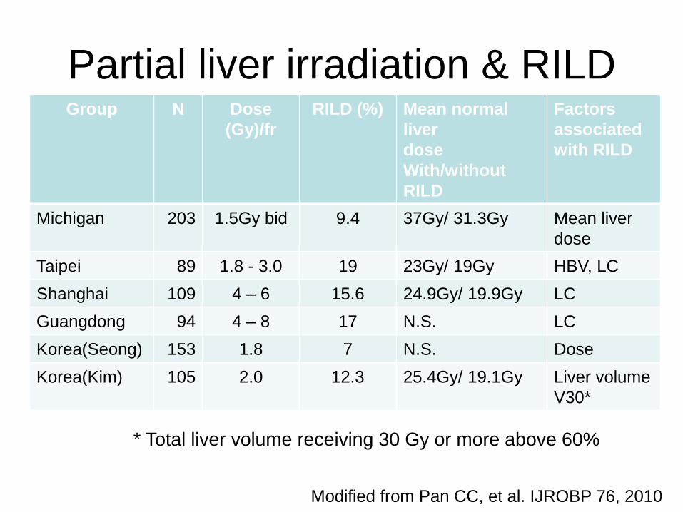
Partial liver irradiation & RILD Group N Dose
(Gy)/fr
RILD (%) Mean normal
liver
dose
With/without
RILD
Factors
associated
with RILD
Michigan 203 1.5Gy bid 9.4 37Gy/ 31.3Gy Mean liver
dose
Taipei 89 1.8 - 3.0 19 23Gy/ 19Gy HBV, LC
Shanghai 109 4 – 6 15.6 24.9Gy/ 19.9Gy LC
Guangdong 94 4 – 8 17 N.S. LC
Korea(Seong) 153 1.8 7 N.S. Dose
Korea(Kim) 105 2.0 12.3 25.4Gy/ 19.1Gy Liver volume
V30*
* Total liver volume receiving 30 Gy or more above 60%
Modified from Pan CC, et al. IJROBP 76, 2010
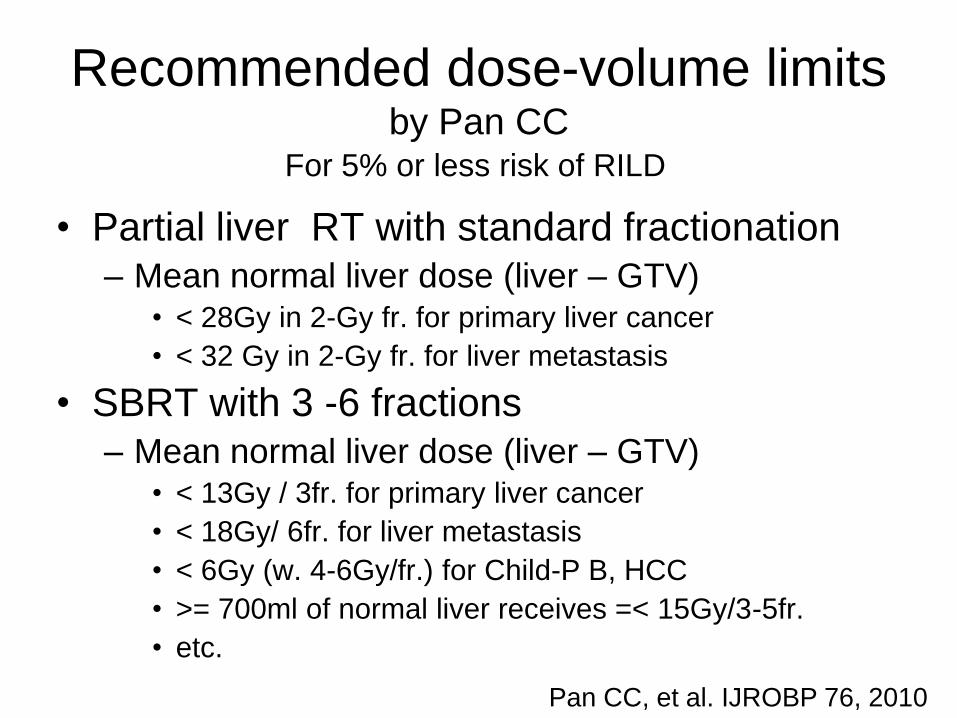
Recommended dose-volume limits by Pan CC
• Partial liver RT with standard fractionation
– Mean normal liver dose (liver – GTV) • < 28Gy in 2-Gy fr. for primary liver cancer
• < 32 Gy in 2-Gy fr. for liver metastasis
• SBRT with 3 -6 fractions
– Mean normal liver dose (liver – GTV) • < 13Gy / 3fr. for primary liver cancer
• < 18Gy/ 6fr. for liver metastasis
• < 6Gy (w. 4-6Gy/fr.) for Child-P B, HCC
• >= 700ml of normal liver receives =< 15Gy/3-5fr.
• etc.
Pan CC, et al. IJROBP 76, 2010
For 5% or less risk of RILD
• How about particle therapy?

Dose-volume histogram analysis of
proton beam therapy for
unresectable hapatocellular carcinoma Kawashima M., et al. IJROBP 79, 2011
• Single nodular or single CTV encompassing multiple lesions
• ICG R15: < 20/ 20-50/ > 50 = 20/ 32/ 8
• Tumor size: median 45mm (20 – 90)
• PBT: 76CGE/20fr., 65CGE/26fr., 60CGE/ 10fr.
• Proton-induced hepatic insufficiency (PHI) – hepatic insufficiency presented with anicteric
ascites and/or asterixis within 6 M. after completion of PRT in the absence of disease progression

Dose-volume histogram analysis of
proton beam therapy for
unresectable hapatocellular carcinoma Kawashima M., et al. IJROBP 79, 2011
• ICG R 15 < 20%
– No PHI
• ICG R 15 20 – 50%
– should minimize the irradiated volume
– V30 < 25% in the noncancerous portion of the
liver
• ICG R 15 > 50%
– Indication of PBT is limited

Evaluation of liver function after proton
beam therapy for hepatocellular carcinoma. Mizumoto M. et al, IJROBP 2012
• 259 patients, 2001 -2007
• CTV encompassing all active lesions
• Child A/ B/ C = 198/ 58/ 3
• Tumor size : median 34 mm (6 – 130)
• PBT: 77GyE/ 35fr., 72.6Gy/ 22fr., 66GyE/
10fr.
• Adverse event: increase of >=1 in Child-
Pugh score

Evaluation of liver function after proton
beam therapy for hepatocellular carcinoma. Mizumoto M. et al, IJROBP 2012
• 91/ 259 : no disease progression for 12 Mo. – 66/ 91 : no increase of Child-Pugh score
– 15/ 91 : 1 point increase of CP score
– 10/ 91 : >=2 point increase of CP score
• Optimal cut-off – V0/ V10/ V20/ V30 = 30%/ 20%/ 26%/ 18%
– (Kawashima’s report: V30 < 25%)
• Liver function after PBT is significantly related to the percentage volume of normal liver that is not irradiated.
Liver : Topics for today
• Anatomy
• Tolerance to RT
• Epidemiology
• Risk factors of liver cancer
• Pathology
• Treatment options
• Particle therapy

Neoplasms in the liver Classification of Liver Cancer
Liver Cancer Study Group of Japan
• Primary liver cancer
– Hepatocellular Carcinoma (HCC)
– Intrahepatic Cholangiocarcinoma (CCC)
– Combined HCC & CCC
– Cystadenocarcinoma
– Hepatoblastoma
• Metastatic Liver cancer
– Colon, Rectum, Breast etc.

• Primary liver cancer is the fifth most
common cancer worldwide and the third
most common cause of cancer mortality.
• Hepatocellular carcinoma (HCC) accounts
for between 85% and 90% of primary liver
cancers.
Today’s talk will be focused on HCC

Epidemiology of HCC
• Variations among geographic regions,
racial and ethnic groups
• Men : Women = 2 : 1 – 4 : 1
• Environmental potentially preventable risk
factors
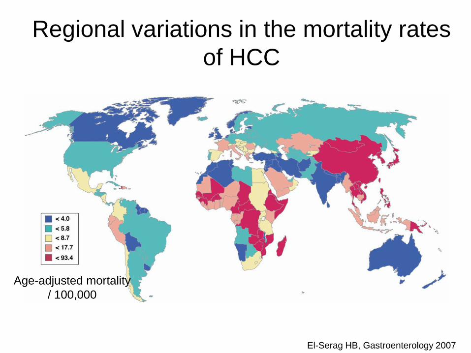
Regional variations in the mortality rates
of HCC
El-Serag HB, Gastroenterology 2007
Age-adjusted mortality
/ 100,000

Liver : Topics for today
• Anatomy
• Tolerance to RT
• Epidemiology
• Risk factors of liver cancer
• Pathology
• Treatment options
• Particle therapy

Risk factors of HCC
• Hepatitis virus infection HBV, HCV
• Alcohol
• Toxic exposure
– Afratoxin, Vinyl chloride
• Nonalcoholic fatty liver disease (NASH)
• Obesity
• Diabetes Mellitus

Proportion of patients with HCC
related to HCV viral hepatitis
Hassan MM, J Clin Gastroenterol 2002

Liver : Topics for today
• Anatomy
• Tolerance to RT
• Epidemiology
• Risk factors of liver cancer
• Pathology
• Treatment options
• Particle therapy
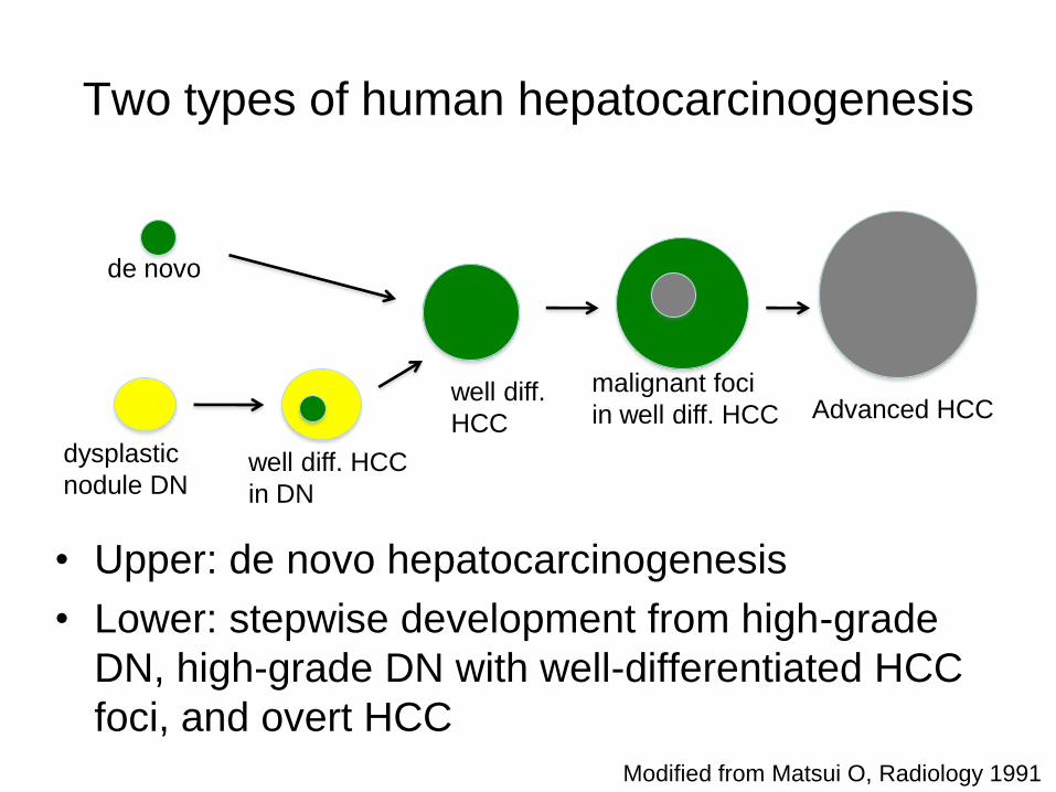
Two types of human hepatocarcinogenesis
• Upper: de novo hepatocarcinogenesis
• Lower: stepwise development from high-grade
DN, high-grade DN with well-differentiated HCC
foci, and overt HCC
de novo
dysplastic
nodule DN well diff. HCC
in DN
well diff.
HCC
malignant foci
in well diff. HCC Advanced HCC
Modified from Matsui O, Radiology 1991
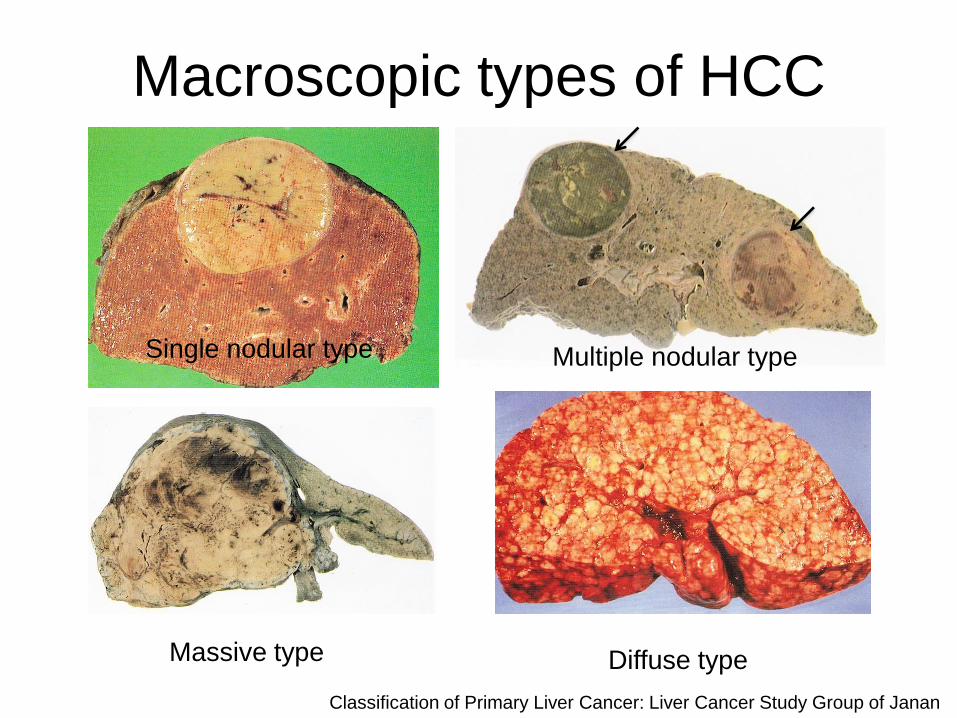
Macroscopic types of HCC
Single nodular type Multiple nodular type
Massive type Diffuse type
Classification of Primary Liver Cancer: Liver Cancer Study Group of Janan

Liver : Topics for today
• Anatomy
• Tolerance to RT
• Epidemiology
• Risk factors of liver cancer
• Pathology
• Treatment options
• Particle therapy

Treatment options for HCC
• Surgical resection
– The mainstay Tx but majority are not eligible
• Liver transplantation
– Solitary, < 5cm or < 3 nodules, < 3cm
• Percutaneous ablation: RFA, PEI
– < 3 nodules, < 3 cm
• Transcatheter Arterial Chemoembolization
– Suitable for multiple, unresectable HCC
• Radiotherapy: particles, SBRT, Y90-IRT

Cohort study conducted by
Liver Cancer Study Group Japan
n 1 year 3 year 5 year
surgery radical 19845
(25066)
91.9 74.6 58.9
RFA Solitary
tumor
6474
(9643)
95.7 80.0 61.7
TACE Solitary
tumor
7942
(31600)
83.6 54.6 32.4
OS by various standard treatment for HCC
(2009)
Data are from most favorable group of patients
Liver : Topics for today
• Anatomy
• Tolerance to RT
• Epidemiology
• Risk factors of liver cancer
• Pathology
• Treatment options
• Particle therapy

PBT for Hepatocellular
Carcinoma

Primary liver cancer 33.6%
Prostate 11.5%
Lung 11.4%
Metastatic tumor 9.8%
H & N 6.1%
Pediatric 4.8%
Esophagus 5.5%
Brain 4.2% Bladder 2.9%
3381
Cases at PMRC, Tsukuba
(1983 – 2013.3)

Standard procedure
for treating liver tumors @ PMRC
• Fiducial marker implant under
ultrasonographic guidance
• Real-time tumor localization using
fluoroscope
• Respiratory gated irradiation

In-situ fiducial marker
platinum

A fiducial marker is used to adjust the daily positioning
under the respiratory gating.
digital radiography for
tumor localization
laser gauge fiducial markers

Internal Target Volume (ITV) of the liver
• >= 2 cm displacement during regular breathing (mostly C-C direction)
• Management for RT
– Abdominal compression
– Shallow breathing
– Breath holding
– Deformation modeling
– Gated treatment
– Real-time tumor tracking
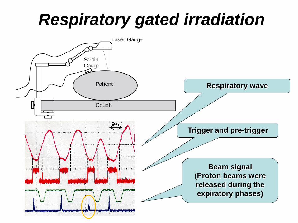
Respiratory gated irradiation Laser Gauge
Patient
Couch
StrainGauge
Trigger and pre-trigger
Beam signal
(Proton beams were
released during the
expiratory phases)
Respiratory wave

Dose, fractionation according to
tumor location
Tumor location dose/ fraction EQD2 α/β: 10
Ex Standard @
PMRC_KEK for
peripheral type
79.2 GyE/ 16fr 98.7 Gy
A Peripheral type 66 GyE/ 10fr 91.3 Gy
B Central type 72.6 GyE/ 22fr 80.5 Gy
C Close to GI 77 (74)GyE/ 35 (37) fr 78.3 (74)Gy
(*): since 2008

Dose, fractionation according to
tumor location
Tumor location dose/ fraction EQD2 α/β: 10
Ex Standard @
PMRC_KEK for
peripheral type
79.2 GyE/ 16fr 98.7 Gy
A Peripheral type 66 GyE/ 10fr 91.3 Gy
B Central type 72.6 GyE/ 22fr 80.5 Gy
C Close to GI 77 (74)GyE/ 35 (37) fr 78.3 (74)Gy
(*): since 2008

Dose distribution for a peripheral type HCC
A B
C D
右
腹
頭
腹
頭
右
背
white
GTV
red
100% dose
blue
10% dose

Hepatocellular carcinoma
peripheral type: 66GyE/ 10fr./ 15D
pre PBT 2 months
after PBT 4 months 46 months

Dose, fractionation according to
tumor location
Tumor location dose/ fraction EQD2 α/β: 10
Ex Standard @
PMRC_KEK for
peripheral type
79.2 GyE/ 16fr 98.7 Gy
A Peripheral type 66 GyE/ 10fr 91.3 Gy
B Central type 72.6 GyE/ 22fr 80.5 Gy
C Close to GI 77 (74)GyE/ 35 (37) fr 78.3 (74)Gy
(*): since 2008

Central type HCC
72.6GyE/ 22fr.
Pre PBT
post PBT 8
Mo.

Dose, fractionation according to
tumor location
Tumor location dose/ fraction EQD2 α/β: 10
Ex Standard @
PMRC_KEK for
peripheral type
79.2 GyE/ 16fr 98.7 Gy
A Peripheral type 66 GyE/ 10fr 91.3 Gy
B Central type 72.6 GyE/ 22fr 80.5 Gy
C Close to GI 77 (74)GyE/ 35 (37) fr 78.3 (74)Gy
(*): since 2008

Tumor close to the GI tract
Gastroesophageal junction
pre PBT
1 Y after PBT
74GyE/ 37fr.

Clinical results

Proton Beam Therapy for Hepatocellular Carcinoma:
A Comparison of Three Treatment Protocols
• Period: 2001 Jan. – 2007 Dec.
• Eligibility criteria • No active tumor outside the target volume
• PS <=2
• Child-Pugh score <=10
• No extrahepatic metastasis
• WBC => 1000/mm3, Hgb => 6.5 g/dl, Plt =>
25000/mm3
Mizumoto M, et al: IJROBP 2011;
81: 1039-1045

Proton Beam Therapy for Hepatocellular Carcinoma:
A Comparison of Three Treatment Protocols
• N= 266 (66GyE: 104, 72.6GyE: 95, 77GyE: 60)
• OS @ 1/ 3/ 5 yr. : 87%/ 61%/ 48%
• Local control @ 1/ 3/ 5 yr. : 98%/ 87%/ 81%
• No significant difference between the protocols
• Predictive factors for OS: liver function, small CTV, no prior treatment
Mizumoto M, et al: IJROBP 2011;
81: 1039-1045

toxicity
Acute dermatitis Grade 1 127
Grade 2 12
Grade 3 2
Late Rib fracture 3
dermatitis Grade 1 2
Grade 3 1
Gastro intestinal* Grade 2 3
Grade 3 3
*: all were “ close to GI “ type
Mizumoto IJROBP 81, 2011
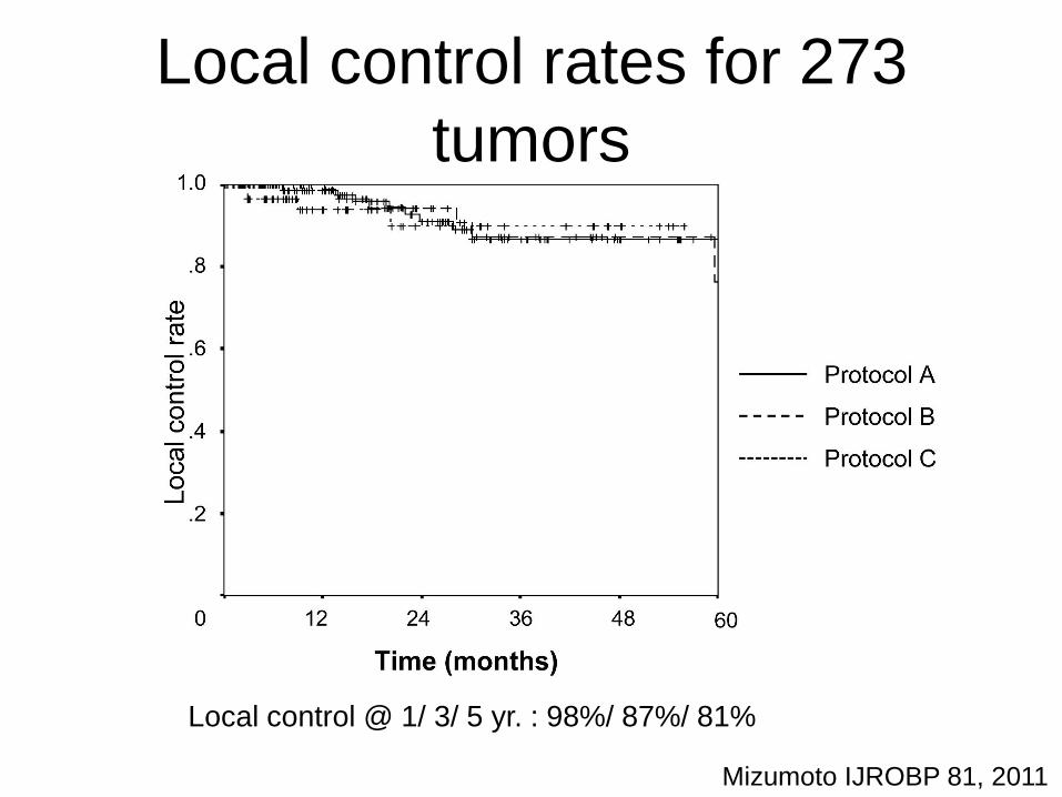
Local control rates for 273
tumors
Mizumoto IJROBP 81, 2011
Local control @ 1/ 3/ 5 yr. : 98%/ 87%/ 81%

Overall survival rates for 259
patients
n
104
95
60
Mizumoto IJROBP 81, 2011
OS @ 1/ 3/ 5 yr. : 87%/ 61%/ 48%

Cohort study conducted by
Liver Cancer Study Group Japan (2009)
n 1 year 3 year 5 year
surgery radical 19845
(25066)
91.9 74.6 58.9
RFA Solitary
tumor
6474
(9643)
95.7 80.0 61.7
TACE Solitary
tumor
7942
(31600)
83.6 54.6 32.4
proton 259 87 61 48

Clinical results of particle therapy for HCC
Series particle n dose
(GyE)/Fx
ED in
2Gy/Fx
Preceding
Tx
Local
control
OS
Kawashima
2005 P 30 76/ 20 87.4 none 96%
@2yr
66%
@2yr
Bush 2004
P 34 63/ 15 74.6 none 75%
@2yr
55%
@2yr
Fukumitsu 2009
P 51 66/ 10 91.3 65% 94.5%
@3yr
49.2%
@3yr
Mizumoto 2008
P 53 72.6/ 22 80.5 72% 86%
@3yr
45.1%
@3yr
Kim PTCOG
2012 P 12 72 / 24 78 none 82.5%
@ 3yr
70.7%
@
3yr
Kato 2004
C 24 49.5-79.5/
15
none 81%
@3yr
50%
@3yr

from our experience
• The choice of fractionation schedule
based on the proximity of the dose limiting
structures is useful.
• Careful treatment planning should be
made for the tumor close to GI tract to
avoid late morbidity.
Mizumoto IJROBP 81, 2011

Proton-Beam Therapy for
Hepatocellular Carcinoma associated
with Portal Vein Tumor Thrombosis
S Sugahara, H Nakayama, K Fukuda,
M Mizumoto, M Tokita, M Abei, J Shoda,
Y Matsuzaki, E Thono, K Tsuboi, K Tokuuye
Strahlenther Onkol 2009

Central type HCC 81 yr., LC (C), Pugh score: 6, Vp4
pre PBT
axilal coronal
72.6GyE/ 22回/ 37日

Central type HCC 81 yr., LC (C), Pugh score: 6, Vp4
6 Mo. after
pre PBT

Radiation (X ray) Therapy for PVTT
Author No.Case PVTT treatment method RR* MST(Mo.)
Tazawa J 24 Vp3,4 TACE+RT50Gy 50 CR,PR;9.7 2001
NC,PD;3.8
Yamada K 8 Vp3 TACE+RT60Gy+TACE 38 5.7(+2) 2001
Ishikura S 20 Vp3 TACE+RT 50 5.3 2002
Nakagawa K 52 Vp2,3,4 3DCRT57Gy (39-60) 50 (25.3%;2YSR) 2005
Kim DY 59 Vp3,4 3DCRT30-54Gy 45.8 CR,PR;10.7 2005
NC,PD;5.3
Lin CS 43 Vp3,4 RT45Gy/15fr:22 75 6.0 2006
3DCRT45Gy/25fr:21 83 6.7
*RR: response rate

Patients and Method
• Period: February 1991 – September 2005
• Tumor thrombus in the main trunk and/or
major branches of the portal vein
• No extrahepatic metastases
• Not diffusely infiltrating tumor
• Child – Pugh score: A or B
• PS: 0-1
Sugahara S, Strahlenther Onkol 185, 2009

Overall survival for all 35 cases
MST 22 months
OS @ 2Y: 48%
Sugahara S, Strahlenther Onkol 185, 2009
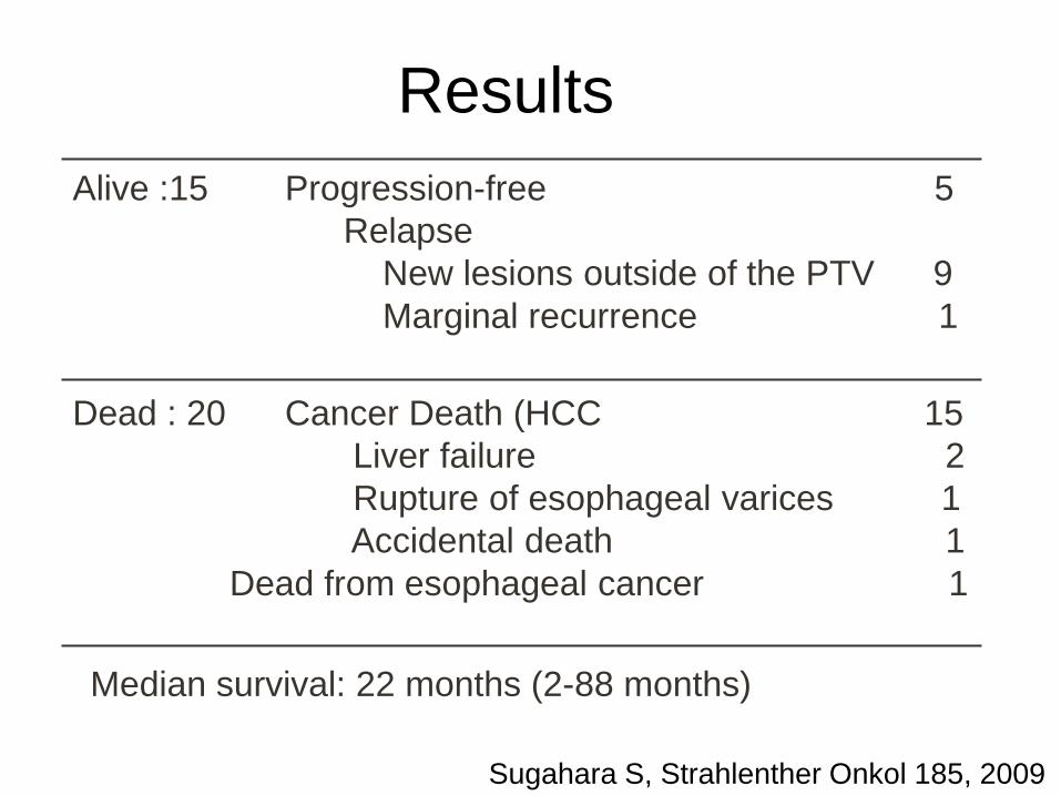
Alive :15 Progression-free 5
Relapse
New lesions outside of the PTV 9
Marginal recurrence 1
Results
Dead : 20 Cancer Death (HCC 15
Liver failure 2
Rupture of esophageal varices 1
Accidental death 1
Dead from esophageal cancer 1
Median survival: 22 months (2-88 months)
Sugahara S, Strahlenther Onkol 185, 2009

Overall survival in
patients treated for PVTT and other active HCC foci
vs.
those for whom PBT covered PVTT only
Sugahara S, Strahlenther Onkol 185, 2009
n = 30
MST 26 months
OS @ 2Y 57%
n = 5
MST 8 months
OS @ 2Y 0% p = 0.019

Radiation Therapy for PVTT
Author No.Case PVTT treatment method RR* MST(Mo.)
Tazawa J 24 Vp3,4 TACE+RT50Gy 50 CR,PR;9.7 2001
NC,PD;3.8
Yamada K 8 Vp3 TACE+RT60Gy+TACE 38 5.7(+2) 2001
Ishikura S 20 Vp3 TACE+RT 50 5.3 2002
Nakagawa K 52 Vp2,3,4 3DCRT57Gy (39-60) 50 (25.3%;2YSR) 2005
Kim DY 59 Vp3,4 3DCRT30-54Gy 45.8 CR,PR;10.7 2005
NC,PD;5.3
Lin CS 43 Vp3,4 RT45Gy/15fr:22 75 6.0 2006
3DCRT45Gy/25fr:21 83 6.7
Tsukuba 35 Vp3,4 PBT 72.6GyE (55-77) 91 22 2009
*RR: response rate
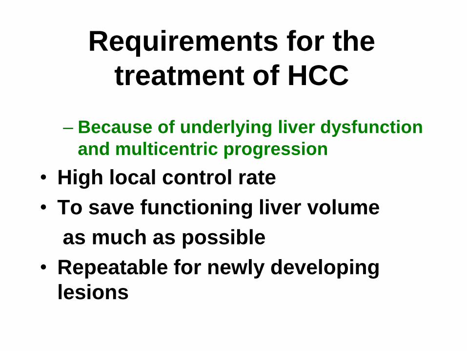
Requirements for the
treatment of HCC
– Because of underlying liver dysfunction
and multicentric progression
• High local control rate
• To save functioning liver volume
as much as possible
• Repeatable for newly developing
lesions

Case: a 47 year old woman with chronic
hepatitis (B) and aplastic anemia
1st: 63 Gy (1992) 2nd: 72 Gy (1994) 3rd: 72 Gy (1995)
4th: 70 Gy (1998) 5th: 72 Gy (1999) 6th: 60 Gy (2002)
Proton beam therapy can be repeatable.
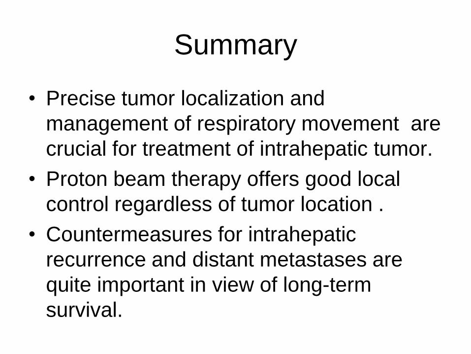
Summary
• Precise tumor localization and
management of respiratory movement are
crucial for treatment of intrahepatic tumor.
• Proton beam therapy offers good local
control regardless of tumor location .
• Countermeasures for intrahepatic
recurrence and distant metastases are
quite important in view of long-term
survival.

Acknowledgment
@ KEK: Shigeki SUWA, Toshio KITAGAWA,
Sadayoshi FUKUMOTO, Tetsuo INADA,
Hirohiko TSUJII,
Masayoshi AKISADA, Yuji ITAI, Akira MARUHASHI,
Yoshihisa TAKADA, Yoshinori HAYAKAWA,
Junichiro TADA, Kiyoshi OHARA, Yutaka HIROKAWA,
Takuro ARIMOTO, Shigeyuki MURAYAMA,
Hiroshi TSUJI, Toshiya CHIBA, Kenji HASEZAWA
@ Univ.Camp.: Yasuyuki AKINE, Koji TSUBOI,
Hideyuki SAKURAI, Takeji SAKAE, Koichi TOKUUYE,
Kiyoshi YASUOKA, Shinji SUGAHARA,
Toshiyuki TERUNUMA, Yoshiyuki SHIOYAMA,
Kenji KAGEI, Hiroshi IGAKI, Masaharu HATA,
Nobuyoshi FUKUMITSU, Hidetsugu NAKAYAMA,
Takayuki HASHIMOTO,
Masashi MIZUMOTO, Yoshiko OSHIRO