liver: update on staging of fibrosis and cirrhosis
TRANSCRIPT
5/24/2013
1
Linda Ferrell, MDDistinguished ProfessorVice ChairDirector of Surgical PathologyDept of Pathology
LIVERUpdate on Staging of Fibrosis
and Cirrhosis
Staging and Liver FibrosisTwo important concepts for consideration: - Stage is more than histologic fibrosisAn integrated clinical/pathophysiologic approach is needed to accurately stage the disease- Cirrhosis is not the “end” of the story: Histologic scoring may need to evolve to identify regression or remodeling of cirrhosis, and evaluate for very advanced nonreversible, or “end-stage” cirrhosis, based on degree of fibrosis
Stage is more than liver fibrosisClinical Modalities to Stage Chronic Liver Disease Measurements of liver function and patho-physiology include the following among others:• Transient elastography (Fibroscan )• Clinical scores including Child-Pughs and MELD
scores • Serum markers and panels, such as Fibrotest ,
Hepascore, FibroSpect , ELF score, AAR, APRI, etc.
• Hepatic venous pressure gradient (HVPG)

5/24/2013
2
Going “Beyond Cirrhosis”Proposal from the International Liver Pathology Study GroupConcept: Cirrhosis has historically implied end-stage disease with the imminent death of patient as there was no cure and no treatmentBut now, many patients remain compensated, and function improves with therapy, particularly notable in chronic viral hepatitis
Going “Beyond Cirrhosis”Proposal: It may be time to put aside the “one-term-fits-all” approach, and stage liver disease as related to etiology and pathophysiology
Should we drop the term cirrhosis or at least recognize different “degree’s of cirrhosis” for a better method of describing advanced liver injury based on etiology and patterns of injury??
Assessment of Advanced Chronic Liver DiseaseAdapted from Figs 1, Beyond Cirrhosis (AJCP 2012)
and Exploring Beyond Cirrhosis (Hepatol 2012, 56:779)Patient with chronic liver disease
Clinical workup• Assessment or reassessment of
etiology, comorbidities or cofactors
• Assessment of severity – Laboratory tests– Transient elastography– HVPG, etc
Clinicopathologic correlation
Liver biopsy with advanced stage of chronic disease-Activity of disease-Features of regression-Presence of other diseases-Risk factors for malignancy
Final diagnosis as the sum of: Etiology Stage
-Advanced stage with no complications-Advanced stage with complications-Advanced stage with regression-End stage
Disease activity Risk for HCC
Staging and Liver FibrosisLiver biopsy is still considered an important component of staging
Questions:• How do we use the liver biopsy in the best
way?• What are the histological aspects we need to
consider?
5/24/2013
3
Staging and Liver Fibrosis
An important starting point is the adequate biopsy!!
Adequacy of Biopsy for Grading/Staging
Short summary (more details in syllabus)Current acceptable recommendations• 5 portal areas minimum, probably >11 for
optimal value• And/or approx 2 cm core of reasonable
width (17 gauge or larger)
Staging and Liver Fibrosis:Other considerations
• Etiology of the injury• Pattern and degree of histological injury• Treatment effects resulting in
remodeling, or regression, of fibrosis-What changes are reversible?
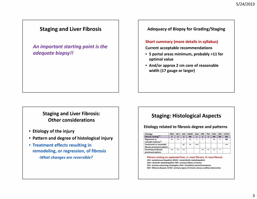
Staging: Histological AspectsEtiology related to fibrosis degree and patterns
Etiology HBV HCV AIH NASH ASH PBC PSC HHC WD CVOO Fibrosis ranking*13 3 3 3 NA 1 2 2 NA NA NA Regression or remodel evidence15
++ ++ + ++ + + ++ + NA
Centrizonal or sinusoidal fibrosis prominent pattern
+/- ++ +++ +++
Portal-based fibrosis prominent pattern
++ ++ ++ ++ ++ ++ +
Fibrosis ranking on explanted liver, 1= most fibrosis, 3= least fibrosisAIH= autoimmune hepatitis; NASH= nonalcoholic steatohepatitis; ASH= alcoholic steatohepatitis; PBC= primary biliary cirrhosis; PSC= primary sclerosing cholangitis; HHC= hereditary hemochromatosis; WD= Wilsons disease; CVOO= primary types of chronic venous outflow obstruction
5/24/2013
4
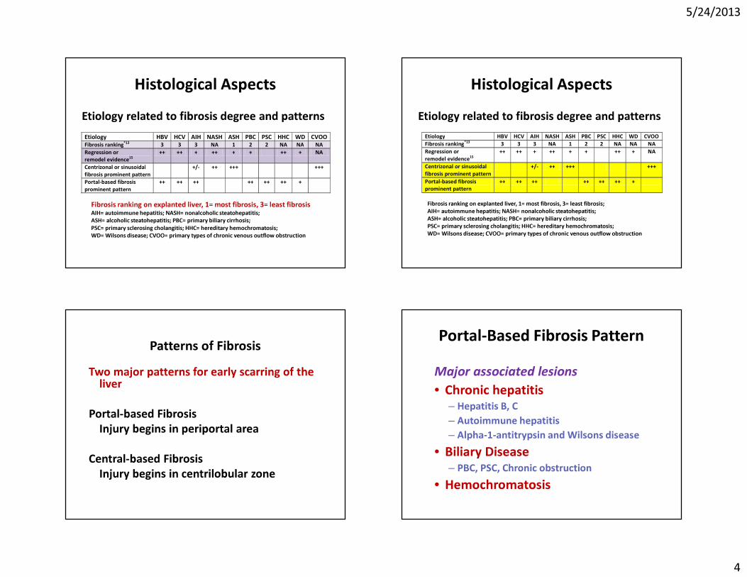
Histological AspectsEtiology related to fibrosis degree and patternsEtiology HBV HCV AIH NASH ASH PBC PSC HHC WD CVOO Fibrosis ranking*13 3 3 3 NA 1 2 2 NA NA NA Regression or remodel evidence15
++ ++ + ++ + + ++ + NA
Centrizonal or sinusoidal fibrosis prominent pattern
+/- ++ +++ +++
Portal-based fibrosis prominent pattern
++ ++ ++ ++ ++ ++ +
Fibrosis ranking on explanted liver, 1= most fibrosis, 3= least fibrosis AIH= autoimmune hepatitis; NASH= nonalcoholic steatohepatitis; ASH= alcoholic steatohepatitis; PBC= primary biliary cirrhosis; PSC= primary sclerosing cholangitis; HHC= hereditary hemochromatosis; WD= Wilsons disease; CVOO= primary types of chronic venous outflow obstruction
Histological AspectsEtiology related to fibrosis degree and patterns
Etiology HBV HCV AIH NASH ASH PBC PSC HHC WD CVOO Fibrosis ranking*13 3 3 3 NA 1 2 2 NA NA NA Regression or remodel evidence15
++ ++ + ++ + + ++ + NA
Centrizonal or sinusoidal fibrosis prominent pattern
+/- ++ +++ +++
Portal-based fibrosis prominent pattern
++ ++ ++ ++ ++ ++ +
Fibrosis ranking on explanted liver, 1= most fibrosis, 3= least fibrosis; AIH= autoimmune hepatitis; NASH= nonalcoholic steatohepatitis; ASH= alcoholic steatohepatitis; PBC= primary biliary cirrhosis; PSC= primary sclerosing cholangitis; HHC= hereditary hemochromatosis; WD= Wilsons disease; CVOO= primary types of chronic venous outflow obstruction
Patterns of FibrosisTwo major patterns for early scarring of the
liver
Portal-based FibrosisInjury begins in periportal area
Central-based FibrosisInjury begins in centrilobular zone
Portal-Based Fibrosis PatternMajor associated lesions• Chronic hepatitis
– Hepatitis B, C– Autoimmune hepatitis– Alpha-1-antitrypsin and Wilsons disease
• Biliary Disease– PBC, PSC, Chronic obstruction
• Hemochromatosis
5/24/2013
5
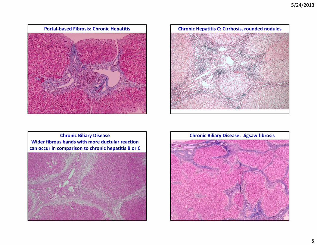
Portal-based Fibrosis: Chronic Hepatitis Chronic Hepatitis C: Cirrhosis, rounded nodules
Chronic Biliary Disease Wider fibrous bands with more ductular reaction
can occur in comparison to chronic hepatitis B or C
Chronic Biliary Disease: Jigsaw fibrosis

5/24/2013
6
Fibrosis Scoring of Chronic Hepatitis
Practical tips and common problems • First step: Use a system that is
– Simple– Reproducible– Useful in clinical setting
Commonly used Grading/Staging systems• Scheuer/Batts-Ludwig/Tsui:
– Grade and Stage on scale 0-4– Simple, reproducible, validated clinically
• METAVIR: – Grade 0-3, Fibrosis 0-4– Simple, reproducible, validated clinically
• Ishak, et al:– Grades four categories of activity/necrosis, 0-4 or 0-6
• Generally considered too complex, not necessary– Staging 0-6
• Preferred in many clinical trials• Still reproducible and validated clinically
Scheuer / Batts-Ludwig / TsuiGrading and Staging
• Simple, reproducible, validated• Essentially same methodology so
interchangeable for the most part• Most commonly used day-to-day in USA and
validated for studies as well• #1 Recommended for typical usage for
grading
Scheuer/Batts,Ludwig/TsuiFibrosis scoring for Chronic Hepatitis
Stage Description 0 No fibrosis, normal amount of connective tissue 1 Portal/periportal fibrosis 2 Septal fibrosis 3 Bridging fibrosis with architectural distortion. 4 Cirrhosis, probable cirrhosis
5/24/2013
7
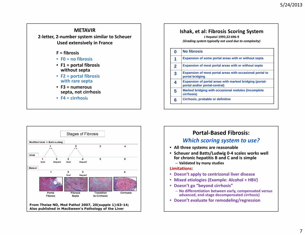
METAVIR2-letter, 2-number system similar to Scheuer
Used extensively in FranceF = fibrosis• F0 = no fibrosis• F1 = portal fibrosis
without septa• F2 = portal fibrosis
with rare septa• F3 = numerous
septa, not cirrhosis• F4 = cirrhosis
Ishak, et al: Fibrosis Scoring SystemJ Hepatol 1995;22:696-9
(Grading system typically not used due to complexity)
0 No fibrosis
1 Expansion of some portal areas with or without septa
2 Expansion of most portal areas with or without septa
3 Expansion of most portal areas with occasional portal to portal bridging
4 Expansion of portal areas with marked bridging (portal-portal and/or portal-central)
5 Marked bridging with occasional nodules (incomplete cirrhosis)
6 Cirrhosis, probable or definitive
From Theise ND, Mod Pathol 2007, 20(supple 1):S3-14;Also published in MacSween’s Pathology of the Liver
Portal-Based Fibrosis: Which scoring system to use?
• All three systems are reasonable• Scheuer and Batts/Ludwig 0-4 scales works well
for chronic hepatitis B and C and is simple– Validated by many studies
Limitations:• Doesn’t apply to centrizonal liver disease• Mixed etiologies (Example: Alcohol + HBV)• Doesn’t go “beyond cirrhosis”
– No differentiation between early, compensated versus advanced, end-stage decompensated cirrhosis)
• Doesn’t evaluate for remodeling/regression
5/24/2013
8
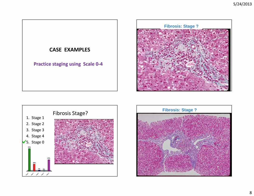
CASE EXAMPLES
Practice staging using Scale 0-4
Fibrosis: Stage ?
Fibrosis Stage?
S t ag e
1
S t ag e
2
S t ag e
3
S t ag e
4
S t ag e
0
55%
16%
27%
0%1%
1. Stage 12. Stage 23. Stage 34. Stage 45. Stage 0
Fibrosis: Stage ?

5/24/2013
9
Fibrosis stage?
S t ag e
1
S t ag e
2
S t ag e
3
S t ag e
4
S t ag e
0
23%
55%
0%0%
22%
1. Stage 12. Stage 23. Stage 34. Stage 45. Stage 0
Fibrosis: Stage ?
Fibrosis stage?
S t ag e
1
S t ag e
2
S t ag e
3
S t ag e
4
S t ag e
0
52%
0%
48%
0%0%
1. Stage 12. Stage 23. Stage 34. Stage 45. Stage 0
Fibrosis: Stage ?

5/24/2013
10
Fibrosis stage ?
S t ag e
1
S t ag e
2
S t ag e
3
S t ag e
4
S t ag e
0
0%9%
0%8%
84%
1. Stage 12. Stage 23. Stage 34. Stage 45. Stage 0
Fibrosis: Stage ?
Fibrosis score?
S t ag e
1
S t ag e
2
S t ag e
3
S t ag e
4
S t ag e
0
22%
61%
0%
8%9%
1. Stage 12. Stage 23. Stage 34. Stage 45. Stage 0
Fibrosis: Stage ?
5/24/2013
11
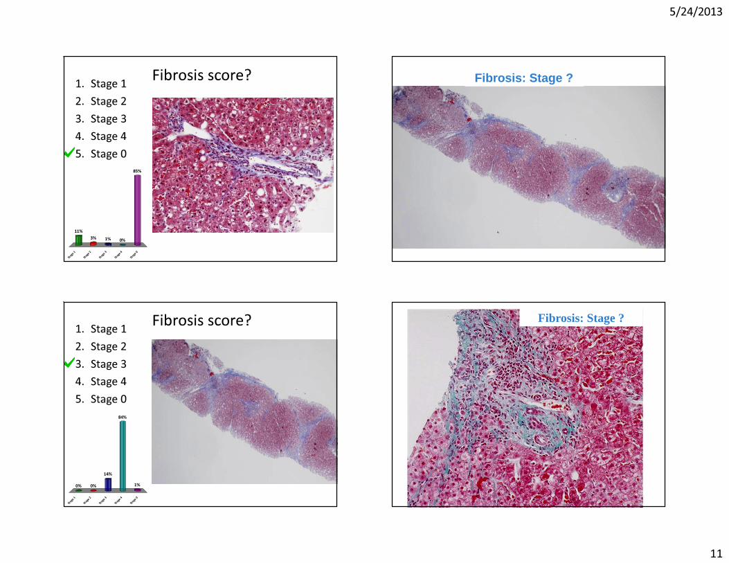
Fibrosis score?
S t ag e
1
S t ag e
2
S t ag e
3
S t ag e
4
S t ag e
0
11%3%
85%
0%1%
1. Stage 12. Stage 23. Stage 34. Stage 45. Stage 0
Fibrosis: Stage ?
Fibrosis score?
S t ag e
1
S t ag e
2
S t ag e
3
S t ag e
4
S t ag e
0
0% 0% 1%
84%
14%
1. Stage 12. Stage 23. Stage 34. Stage 45. Stage 0
Fibrosis: Stage ?
5/24/2013
12
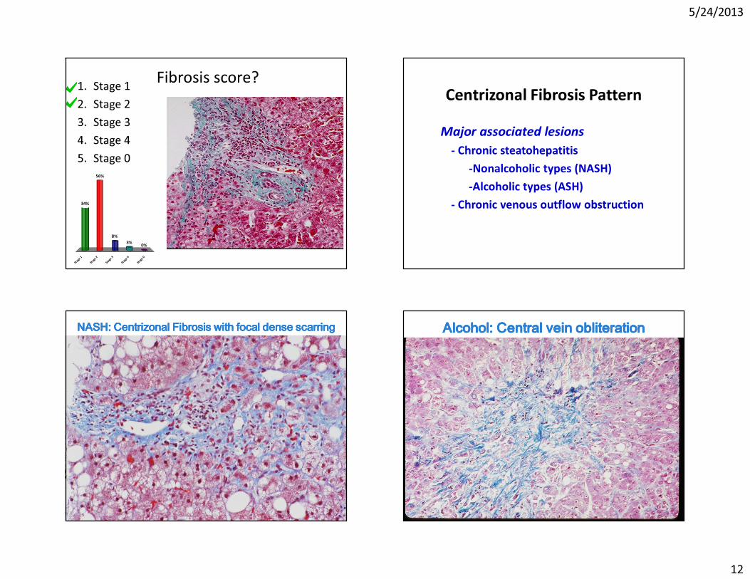
Fibrosis score?
S t ag e
1
S t ag e
2
S t ag e
3
S t ag e
4
S t ag e
0
34%
56%
0%3%8%
1. Stage 12. Stage 23. Stage 34. Stage 45. Stage 0
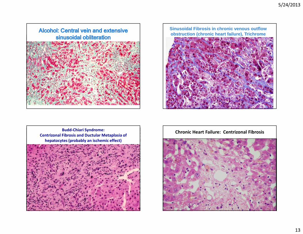
Centrizonal Fibrosis PatternMajor associated lesions
- Chronic steatohepatitis-Nonalcoholic types (NASH)-Alcoholic types (ASH)
- Chronic venous outflow obstruction
NASHNASHNASHNASH: : : : CentrizonalCentrizonalCentrizonalCentrizonal Fibrosis with focal dense scarringFibrosis with focal dense scarringFibrosis with focal dense scarringFibrosis with focal dense scarring Alcohol: Central vein obliterationAlcohol: Central vein obliterationAlcohol: Central vein obliterationAlcohol: Central vein obliteration
5/24/2013
13
Alcohol: Central vein and extensive Alcohol: Central vein and extensive Alcohol: Central vein and extensive Alcohol: Central vein and extensive sinusoidal sinusoidal sinusoidal sinusoidal obliterationobliterationobliterationobliteration
Sinusoidal Fibrosis in chronic venous outflow obstruction (chronic heart failure), Trichrome
Budd-Chiari Syndrome: Centrizonal Fibrosis and Ductular Metaplasia of
hepatocytes (probably an ischemic effect)Chronic Heart Failure: Centrizonal Fibrosis
5/24/2013
14
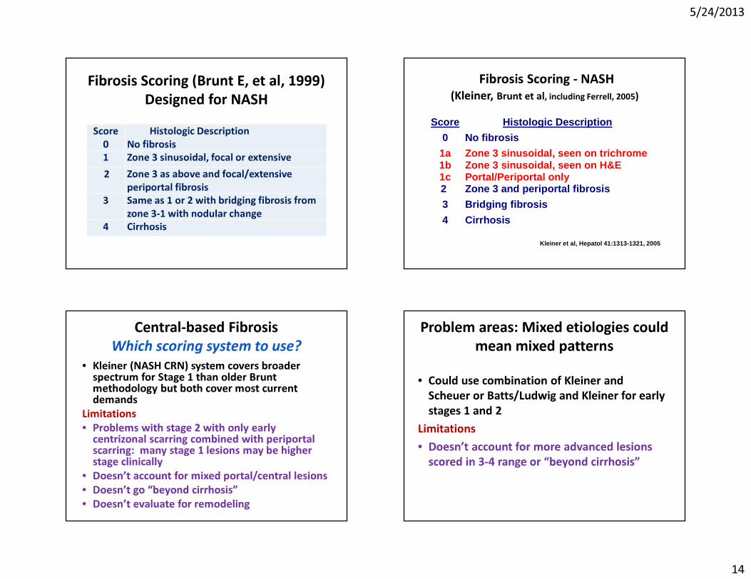
Fibrosis Scoring (Brunt E, et al, 1999)Designed for NASH
Score0
Histologic DescriptionNo fibrosis
1 Zone 3 sinusoidal, focal or extensive2 Zone 3 as above and focal/extensive
periportal fibrosis3 Same as 1 or 2 with bridging fibrosis from
zone 3-1 with nodular change 4 Cirrhosis
Fibrosis Scoring - NASH(Kleiner, Brunt et al, including Ferrell, 2005)
Score Histologic Description
0 No fibrosis
1a 1b 1c
Zone 3 sinusoidal, seen on trichrome Zone 3 sinusoidal, seen on H&E Portal/Periportal only
2 Zone 3 and periportal fibrosis
3 Bridging fibrosis
4 Cirrhosis
Kleiner et al, Hepatol 41:1313-1321, 2005
Central-based FibrosisWhich scoring system to use?
• Kleiner (NASH CRN) system covers broader spectrum for Stage 1 than older Brunt methodology but both cover most current demands
Limitations• Problems with stage 2 with only early
centrizonal scarring combined with periportalscarring: many stage 1 lesions may be higher stage clinically
• Doesn’t account for mixed portal/central lesions• Doesn’t go “beyond cirrhosis”• Doesn’t evaluate for remodeling
Problem areas: Mixed etiologies could mean mixed patterns
• Could use combination of Kleiner and Scheuer or Batts/Ludwig and Kleiner for early stages 1 and 2
Limitations• Doesn’t account for more advanced lesions
scored in 3-4 range or “beyond cirrhosis”

5/24/2013
15
NASH + HCV or HBVNOTE Pattern of disease locations PORTAL: favors chronic hepatitis• Portal-based chronic inflammation, fibrosis, and
interface hepatitis• HBV or HCV markersCENTRAL: favors steatohepatitis• Centrizonal fat, fibrosis, ballooned cells,
inflammation associated with fat• Risk factors for NASH/ASH
NASH and HCVCentrizonal and Periportal fibrosis
NASH and HCVCentrizonal and Periportal fibrosis
How to stage?
NASH + HCV or HBV STAGINGStage separately for earlier stages if possible• NASH: Brunt or Kleiner stage
Case example – if all fibrosis due to NASH, Stage 2 NASH– If periportal likely due to HCV, then Stage 1 NASH
• Viral hepatitis: Do not include central fibrosis– Scheuer or Batts/Ludwig stage 1 or 2 Note prominent pattern or combination of patterns as centrizonal or portal if possible
5/24/2013
16
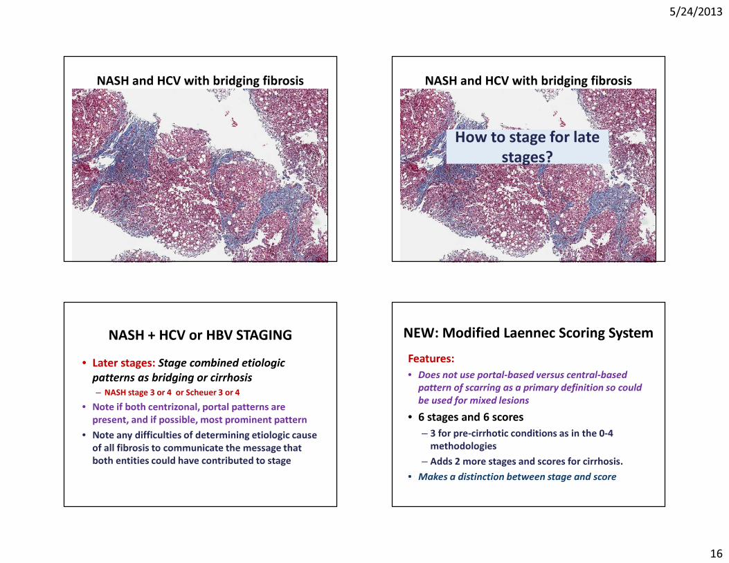
NASH and HCV with bridging fibrosis NASH and HCV with bridging fibrosis
How to stage for late stages?
NASH + HCV or HBV STAGING• Later stages: Stage combined etiologic
patterns as bridging or cirrhosis – NASH stage 3 or 4 or Scheuer 3 or 4
• Note if both centrizonal, portal patterns are present, and if possible, most prominent pattern
• Note any difficulties of determining etiologic cause of all fibrosis to communicate the message that both entities could have contributed to stage
NEW: Modified Laennec Scoring SystemFeatures: • Does not use portal-based versus central-based
pattern of scarring as a primary definition so could be used for mixed lesions
• 6 stages and 6 scores – 3 for pre-cirrhotic conditions as in the 0-4
methodologies– Adds 2 more stages and scores for cirrhosis.
• Makes a distinction between stage and score
5/24/2013
17
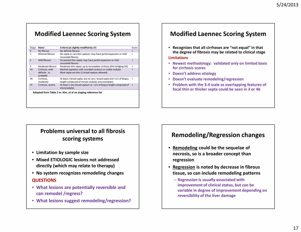
Modified Laennec Scoring SystemStage Name Criteria (as slightly modified by LF) Score 0 No fibrosis No definite fibrosis 0 1 Minimal fibrosis No septa or rare thin septum; may have portal expansion or mild
sinusoidal fibrosis 1
2 Mild fibrosis Occasional thin septa; may have portal expansion or mild sinusoidal fibrosis
2
3 Moderate fibrosis Moderate thin septa; up to incomplete cirrhosis (thin bridging OK) 3 4A Cirrhosis, mild
definite, or probable
Marked septation with rounded contours or visible nodules Most septa are thin (1 broad septum allowed)
4
4B Cirrhosis, moderate
At least 2 broad septa, but no very broad septa and <1/2 of biopsy length composed of minute nodules (micronodules)
5
4C Cirrhosis, severe At least 1 very broad septum or >1/2 of biopsy length composed of micronodules
6
Adapted from Table 2 in: Kim, et al on staging reference list
Modified Laennec Scoring System• Recognizes that all cirrhoses are “not equal” in that
the degree of fibrosis may be related to clinical stageLimitations• Newest methodology: validated only on limited basis
for cirrhosis scores• Doesn’t address etiology • Doesn’t evaluate remodeling/regression• Problem with the 3-4 scale as overlapping features of
focal thin or thicker septa could be seen in 3 or 4b
Problems universal to all fibrosis scoring systems
• Limitation by sample size• Mixed ETIOLOGIC lesions not addressed
directly (which may relate to therapy)• No system recognizes remodeling changesQUESTIONS• What lesions are potentially reversible and
can remodel /regress?• What lesions suggest remodeling/regression?
Remodeling/Regression changes• Remodeling could be the sequelae of
necrosis, so is a broader concept than regression
• Regression is noted by decrease in fibrous tissue, so can include remodeling patterns – Regression is usually associated with
improvement of clinical status, but can be variable in degree of improvement depending on reversibility of the liver damage
5/24/2013
18
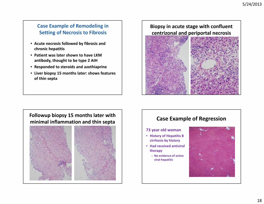
Case Example of Remodeling in Setting of Necrosis to Fibrosis
• Acute necrosis followed by fibrosis and chronic hepatitis
• Patient was later shown to have LKM antibody, thought to be type 2 AIH
• Responded to steroids and azothiaprine• Liver biopsy 15 months later: shows features
of thin septa
Biopsy in acute stage with confluent centrizonal and periportal necrosis
Followup biopsy 15 months later with minimal inflammation and thin septa Case Example of Regression
73 year old woman• History of Hepatitis B
cirrhosis by history• Had received antiviral
therapy– No evidence of active
viral hepatitis
5/24/2013
19
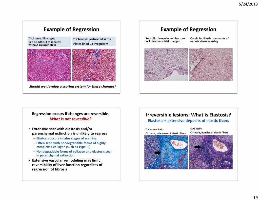
Example of RegressionTrichrome: Thin septaCan be difficult to identify without collagen stain
Trichrome: Perforated septaPlates lined up irregularly
Should we develop a scoring system for these changes?
Example of RegressionReticulin: irregular architecture includes sinusoidal changes
Orcein for Elastic: remnants of remote dense scarring
Regression occurs if changes are reversible.What is not reversible?
• Extensive scar with elastosis and/or parenchymal extinction is unlikely to regress– Elastosis occurs in later stages of scarring – Often seen with nondegradable forms of highly-
complexed collagen (such as Type III)– Nondegradable forms of collagen and elastosis seen
in parenchymal extinction• Extensive vascular remodeling may limit
reversibility of liver function regardless of regression of fibrosis
Irreversible lesions: What is Elastosis?Elastosis = extensive deposits of elastic fibers
Trichrome Stain:Cirrhosis, pale areas of elastic fibers
EVG Stain: Cirrhosis, bundles of elastic fibers

5/24/2013
20
What is Parenchymal Extinction?Parenchymal Extinction = Extensive scar
What is Parenchymal Extinction?Parenchymal Extinction = Extensive scar• Dark, dense fibers predominate = highly
complexed collagen• Indicates a late stage in the fibrotic process
as in Laennec stage 4c• Much of the extensive scarring probably
related to either venous outflow or arterial inflow alterations and chronic ischemic effects in advanced “end-stage” cirrhosis
Vascular Alterations in Cirrhosis
Vascular collaterals/modifications develop in fibrosis and cirrhosis. Fibrosis leads to intraparenchymal vascular resistanceMicro- and Macrocirculatory changes occur in conjunction with alterations in hepatic flow dynamics
Rappaport et al. The scarring of liver acini (cirrhosis). Tridimensional and microcirculatory considerations. Virchows Arch of Anat Path, 1983
Microcirculatory RemodelingExample: Arterialization of Centrizonal ScarsGill R…Ferrell L: AJSP, 35, 1400-04, 2011. • Increased arteries and
microvessels in centrizonalscars
• Increased CD34 staining of sinusoidal endothelial cells as effect of loss of fenestrations (“capillarization”)
• Occurs prior to cirrhosis, but most prevalent in fibrosis score 4-6 by ISHAK
Arrows point to Arteries
5/24/2013
21
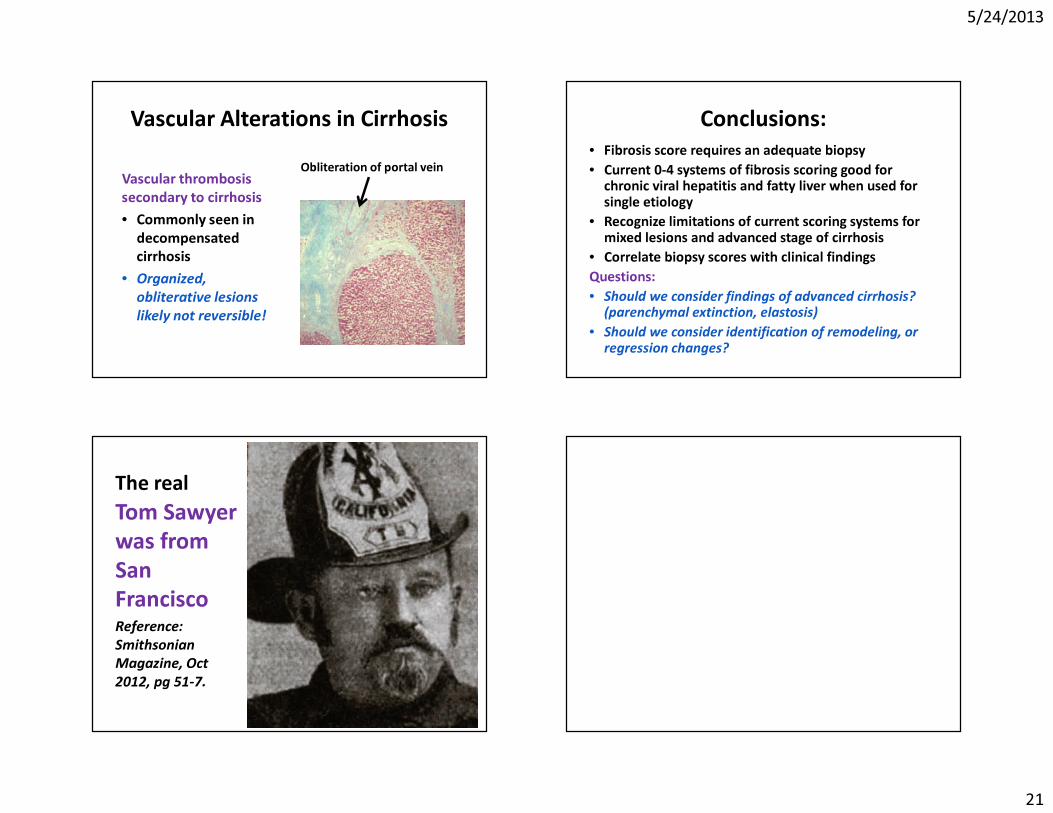
Vascular Alterations in Cirrhosis
Vascular thrombosis secondary to cirrhosis• Commonly seen in
decompensated cirrhosis
• Organized, obliterative lesions likely not reversible!
Obliteration of portal vein
Conclusions: • Fibrosis score requires an adequate biopsy • Current 0-4 systems of fibrosis scoring good for
chronic viral hepatitis and fatty liver when used for single etiology
• Recognize limitations of current scoring systems for mixed lesions and advanced stage of cirrhosis
• Correlate biopsy scores with clinical findingsQuestions:• Should we consider findings of advanced cirrhosis?
(parenchymal extinction, elastosis)• Should we consider identification of remodeling, or
regression changes?
The realTom Sawyer was from San FranciscoReference: Smithsonian Magazine, Oct 2012, pg 51-7.
5/24/2013
22
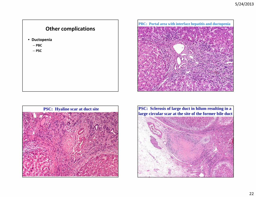
Other complications• Ductopenia
– PBC– PSC
PBC: Portal area with interface hepatitis and ductopenia
PSC: Hyaline scar at duct site PSC: Sclerosis of large duct in hilum resulting in a large circular scar at the site of the former bile duct