living at the extremes: extremophiles and the limits of
TRANSCRIPT

Limits of Life in Planetary Context
Living at the Extremes: Extremophiles and the Limits ofLife in a Planetary Context
PREPRINT
Nancy Merino1,2, Heidi S. Aronson3, Diana Bojanova1, Jayme Feyhl-Buska1, Michael L.Wong4,5, Shu Zhang6, Donato Giovannelli2,7,8,9*1
1Department of Earth Sciences, University of Southern California, Los Angeles, CA, USA2Earth-Life Science Institute, Tokyo Institute of Technology, Tokyo, Japan3Department of Biology, University of Southern California, Los Angeles, CA, USA4Department of Astronomy & Astrobiology Program, University of Washington, Seattle, WA, USA5NASA Astrobiology Institute’s Virtual Planetary Laboratory, University of Washington, Seattle, WA, USA6Section of Infection and Immunity, Herman Ostrow School of Dentistry of USC, University of Southern California, Los Angeles, CA, USA7Department of Biology, University of Naples “Federico II”, Naples, Italy8Department of Marine and Coastal Science, Rutgers University, New Brunswick, NJ, USA9Institute for Biological Resources and Marine Biotechnology, National Research Council of Italy, CNR-IRBIM, Ancona, Italy
Abstract
Prokaryotic life has dominated most of the evolutionary history of our planet, evolving to occupy virtually all available environmental niches. Extremophiles, especially those thriving under multiple extremes, represent a key area of research for multiple disciplines, spanning from the study of adaptations to harsh conditions, to the biogeochemical cycling of elements. Extremophile research also has implications for origin of life studies and the search for life on other planetary and celestial bodies. In this article, we will review the current state of knowledge for the biospace in which life operates on Earth and will discuss it in a planetary context, highlighting knowledge gaps and areas of opportunity.
Keywords: Polyextremophiles, Limits of life, Astrobiology, Habitability and astrobiology, extremophiles/extremophily, Search for life
1. (Poly)extremophiles help us predict the boundaries of life
Since the first non-spore-formingextremophile, Thermus aquaticus, was isolated 50years ago in 1969 (Brock and Freeze, 1969), theboundary conditions under which life can thrivehave been pushed in every possible direction,encompassing broader swaths of temperature, pH,pressure, radiation, salinity, energy, and nutrientlimitation. Microorganisms do not only thrive
under such a broad spectrum of parameters onEarth, but can also survive the harsh conditions ofspace, an environment with extreme radiation,vacuum pressure, extremely variable temperature,and microgravity (Horneck et al., 2010;Yamagishi et al., 2018). The definition of“extreme conditions” has strong anthropocentriccriteria, rather than microbial criteria, and can bethe cause of confusion (Rothschild andMancinelli, 2001). When consideringextremophilic (as opposed to extremotolerant)
1 Correspondence: Donato Giovannelli – [email protected]
1
Limits of Life in Planetary Context
organisms, it is important to keep in mind thatthese are highly adapted organisms for theconditions considered and that the “extreme”condition constitutes the norm under which theorganism is able to metabolically andbiochemically operate. Moreover, t There aremyriad environments on our planet’s surface –and especially subsurface – that exhibit extremesin one or more physical or chemical condition.Therefore, extremophiles and, in particular,polyextremophiles (Capece et al., 2013) might bethe most abundant lifeforms on our planet. Inaddition, if we consider that the current planetarysurface conditions on Earth (such as meantemperature, redox state and oxygenicatmosphere) have only occurred for a short periodof time compared to the existence of life (Knoll,2015), we might conclude that the extremophilicway of life has actually dominated theevolutionary history of life on our planet.
Over the past several decades, theisolation of culturable (poly)extremophiles andthe identification of extreme microbialcommunities through various culture-independentapproaches have provided key insights into theboundaries of life. Research on(poly)extremophiles has led to numerousadvances in molecular biology and medicine(Babu et al., 2015; Coker, 2016; Durvasula andRao, 2018), while simultaneously reshaping ourunderstanding of the origins and evolution of life(Bertrand et al., 2015) and the potential for life onother planetary bodies (Schulze-Makuch, 2013).Several reviews have defined extremophiles(Table 1) (e.g., Capece et al., 2013; Fang et al.,2010; Rothschild and Mancinelli, 2001; Seckbachet al., 2013) and discussed the physiology andgenetics of (poly)extremophiles in detail (e.g.,chapters within Polyextremophiles: Life UnderMultiple Forms of Stress, edited by Seckbach andcollaborators (2013)). To build upon thesediscussions, this paper will review the parametersthat limit life, providing ranges under which lifehas been detected. In addition, we will map thecurrently known boundary conditions of life onEarth to the theoretical space that life couldoccupy on Earth (defined here as the parameter
space of possible conditions present on theplanet) and explore the prospect of using thisinformation for the search of life on otherplanetary bodies.
2. Parameters that limit life
Our knowledge of life is based on theobservable and measurable phenomena that occuron Earth, and is therefore limited to this instanceof life. However, the laws of chemistry andphysics have universal principles which enable usto extrapolate to the conditions under which lifecould survive elsewhere. These principles suggestthat life requires a liquid solvent, an energysource, and building blocks (Schwieterman et al.,2018).
While the bulk abundance of (inorganic)building blocks appears not to be a factorlimiting the distribution of life on Earth (withsubsurface environments as a possible exception,e.g., Hoehler and Jørgensen, 2013) and,potentially, other planetary bodies, theavailability of a solvent is considered to be a keyfactor. While the potential for other liquidsolvents to sustain extraterrestrial life is discussedin detail elsewhere (Schwieterman et al., 2018and references therein), water is considered themost likely liquid solvent because of its cosmicabundance and physicochemical properties(Michiels et al., 2008; Schwieterman et al., 2018).Water, especially the availability of liquid water,appears to be the main factor controlling thedimensions of the biospace for life on Earth (i.e.,the parameter space occupied by life). Liquidwater acts both as a solvent and areactant/product in biochemical reactions, and itsnumerous unique physicochemical propertieshave profoundly shaped the emergence andevolution of life on our planet. As discussed inthis review below, water activity appears to be thesingle key parameter controlling the biospace ofEarth’s life, and numerous other parameterslimiting life (e.g., temperature and salinity) are, infact, acting on the availability of water. At theecosystem level, water can indirectly influencethe variation of key physicochemical conditions,which in turn controls microbial community
2

Limits of Life in Planetary Context
composition and diversity, profoundlyinfluencing geobiochemical cycling (sensu Shockand Boyd, 2015).
Life also needs a source of energy topower chemical reactions, and redox chemistryappears to be universal (Jelen et al., 2016).Physicochemical gradients create non-equilibriumredox conditions that have played an importantrole in the origins, evolution, and diversity of life.Redox and proton gradients were likely the twomain mechanisms involved in the origins of life,initiating the necessary energy flux to drivemetabolism and growth (Lane et al., 2010; Laneand Martin, 2012). Therefore, the current searchfor life’s limits have been extended beyondtemperature, pH, pressure, salinity, and radiationgradients (each parameter discussed in theirrespective sections) and also includes the possibleenergetic and nutrient limits of life (discussed inHoehler and Jørgensen, 2013, Jones et al., 2018,and LaRowe and Amend, 2015).
The parameters discussed herein(temperature, pH, pressure, and salinity, andradiation) correlate with each other and caninfluence the availability of nutrients and energysources. Depending on the environment, certainparameters can more strongly influence microbialdiversity over others, such as temperature ingeothermal waters (Sharp et al., 2014), pH in soilcommunities (Rousk et al., 2010), salinity insaline lakes (Yang et al., 2016), and water contentin dry climates (Dose et al., 2001). On the nano-and micro-scale level, the two most importantfactors are likely water activity and pH, whichinfluence the chemiosmotic, energy-generatinggradient at the cell level (Lane et al., 2010; Laneand Martin, 2012). In contrast, parameters thatinfluence the macro-scale level vary with theecosystem. For example, temperature plays asignificant role in geothermal environments andinfluences such processes as water-rockinteractions and degassing (Cole et al., 2013;Fouke, 2011; Nordstrom et al., 2005; Price andGiovannelli, 2017). Water-rock interactions canthen impact microorganisms by limiting the
availability of trace elements and electron donors/acceptors.
Microorganisms have been detected in avariety of extreme environments (Figure 1),virtually in any location where liquid water isavailable for life to use. This demonstrates thatlife can adapt to a wide range of parameters(Figure 2). It is therefore imperative to determinethe minima and maxima for each parameter(temperature, pH, pressure, and salinity, andradiation), and even more importantly, tounderstand their combined effects, in order toevaluate the limits of Earth’s life and advance ourunderstanding of the potential for life elsewhere.
2a. Acidity and alkalinity
Extremely low and high pH environmentshave been observed for different ecosystems onEarth (Table 2). Extreme pH values wereobserved for ecosystems contaminated by miningwaste, with current extremes reported from IronMountain (Shasta County, CA, USA) (pH -3.6)(Nordstrom et al., 2000) and Gorka Lake(Chrzanow region, Poland) (pH 13.3; Czop et al.,2011). While there has yet to be any microbialcommunity studies or isolation attempts forGorka Lake, to the best of our knowledge,microbial communities have been explored atIron Mountain (Baker and Banfield, 2003), withseveral microorganisms isolated (e.g.,Thermoplasmales (Edwards et al., 2000),Acidithiobacillus ferrooxidans (Kelly and Wood,2000; Schrenk et al., 1998), and Leptospirillumferrooxidans (Schrenk et al., 1998)). Despite this,there are currently no cultured or isolatedmicroorganisms which can be grown at either ofthe listed extremes. Currently, the most extremeacidophile and alkaliphile can survive at pH 0 andpH 12.5, respectively (pHopt 0.7 and 11) (Table3). The lowest pHmin -0.06 was observed for twohyperacidophilic Archaea known as Picrophilusoshimae and P. torridus (pHopt 0.7), isolated froma solfataric hot spring in Noboribetsu (Hokkaido,Japan) (Schleper et al., 1996). Theseheterotrophic and aerobic polyextremophiles canalso withstand temperatures of up to 65ºC (Topt =60ºC, Tmin = 47ºC), potentially through increased
3

Limits of Life in Planetary Context
cyclization of their tetraether membrane lipids asa generalized response to pH, temperature, andnutrient stress (Feyhl-Buska et al., 2016). Incomparison to extreme acidophily, the highestpHmax of 12.5 was observed for an alkaliphilic,aerobic, mesophilic bacterium known asSerpentinomonas sp. B1 (pHopt 11), isolated froma terrestrial serpentinizing system, The Cedars(CA, USA) (Suzuki et al., 2014). Although thereis a report of the highest pHmax 13 held byPlectonema nostocorum (Kingsbury, 1954) thishas not been further confirmed. The largest pHrange, as compared to other isolatedmicroorganisms, was observed for Halomonascampisalis (pHrange 6–12), a haloalkaliphilicbacterium isolated from a soda lake (Soap Lake,WA, USA) (Aston and Peyton, 2007; Mormile etal., 1999) (Table 4).
The pH has a significant effect onmicroorganisms and microbial consortia, rangingfrom the nano- to macro-scale level. Allmicroorganisms must maintain a near neutralcytoplasmic pH to enable cellular functions forsurvival and metabolism (Jin and Kirk, 2018;Krulwich et al., 2011). The cytoplasmic pH ofacidophilic bacteria is ~6.0 while alkaliphilicbacteria have a cytoplasmic pH around 7.2–8.7(Krulwich et al., 2011). For more information onthe molecular mechanisms behind pHhomeostasis, Krulwich and colleagues provide adetailed review (Krulwich et al., 2011). Thehomeostasis of protons (and other ions) throughvarious transporters, including the ion-utilizingATP synthase, was likely one of the firstfunctions to develop within the earliest cells(Lane and Martin, 2012). Indeed, chemiosmosisis a property of both archaeal and bacterial cells(Lane et al., 2010). In addition to intracellular pH,microorganisms can excrete organic metabolites,such as lactic acid or acetic acid, therebychanging the immediate, surrounding pH (Zhanget al., 2016). Many acidophiles also have organicacid degradation pathways to prevent protonuncoupling by organic acids (Baker-Austin andDopson, 2007). It has been demonstrated both innatural settings and laboratory cultures thatmicroorganisms can significantly alter their
environmental pH as a result of metabolicreactions. For example, sulfide thiosulfate, andelemental sulfur oxidizers secrete sulfate andprotons as by-products, significantly acidifyingtheir environment. This ability is used industriallyfor the bio-leaching of sulfide ore deposit (Olsonet al., 2003; Rohwerder et al., 2003) and it islargely responsible for the low pH of acid minedrainage fluids and other acidic environments.Recent work by Colman et al. (2018) suggeststhat thermoacidophilic archaea and the acidity oftheir habitats co-evolved after the evolution ofoxygenic photosynthesis (since oxygen is used asprimary electron acceptor in the metabolisms),showing a significant example of nicheengineering and geosphere-biospherecoevolution. All together, these findings suggestthat pH can be metabolically controlled either atthe intracellular or local level, as compared totemperature, radiation, salinity, and pressure.
On the macro-scale level, pH candominate as the main parameter affectingmicrobial community composition andabundances. Several studies demonstrate that pHaffects microbial community diversity more thanany other parameter tested (e.g., Kuang et al.,2013; Lauber et al., 2009; Rousk et al., 2010;Xiong et al., 2012; Zhalnina et al., 2014) Forexample, distinct microbial communities wereobserved with changes in pH (pHrange 1.9–4.1), inwhich the genus Ferrovum dominated at higherpH while the phyla Alphaproteobacteria,Gammaproteobacteria, Nitrospirae, andEuryarchaeota were present at lower pHs(Kuang et al., 2013) Similarly, bacterialcommunity composition changed with increasingpH in alkaline sediments of a Tibetan plateau(pHrange 6.88–10.37) (Xiong et al., 2012). Changesin community composition are likely derivedfrom the range in which microorganisms cansurvive (Fernández-Calviño and Bååth, 2010).Most cultured microbes live within a narrow pHrange of 3–4 units (Rosso et al., 1995), althoughsome exceptions occur (e.g., fungal isolates cangrow over 5–9 pH units (Nevarez et al., 2009;Wheeler et al., 1991)). Moreover, it has beensuggested that archaeal (Kuang et al., 2013) and
4

Limits of Life in Planetary Context
fungal communities (Rousk et al., 2010) may beless affected by changes in pH compared tobacteria.
2b. Salinity and water activity
Salinity has a significant impact onmicrobial community composition (Lozuponeand Knight, 2007; Swan et al., 2010). Salineenvironments comprise a large portion of theEarth and range from the marine environment(~3–4% salinity), hot springs (up to 10.5%salinity), and to soda lakes (up to 37.1% salinity),and even salt inclusions (up to 49.7% salinity(Scambelluri et al., 1997)) (Figure 2, Table 2).Salinity can also vary significantly on smallerscales, for example, in tidal pools (Morris andTaylor, 1983), or on salt mineral grains due towater deliquescence (Davila et al., 2008). A widerange of different ions, including Na+, Cl-, SO4
2-,Ca2+, and Mg2+ (Oren, 2013) can contribute tototal salinity in the environment. The ioniccomposition can significantly influence wateractivities, especially in the presence of highconcentrations chaotrophic salts, like in theathalassic deep-sea hypersaline anoxic basins ofthe Mediterranean Sea (Yakimov et al., 2015). Inaddition, water availability in terrestrial salineenvironments is further influenced byprecipitation rates relative to evaporation,resulting in increasing concentration of salts(Finlayson et al., 2018).
The salinity range and optimum forcultivable and isolated microorganisms isbetween 0–35%. The current highest salinityrecord holder is Halarsenatibacter silvermaniistrain SLAS-1T, isolated from the alkalinehypersaline Searles Lake (California, USA)(salinityopt 35% NaCl) (Blum et al., 2009).Halophiles are found in all three domains of life(DasSarma and DasSarma, 2017). Currenthyperhalophiles in culture include bacteria andarchaea which can grow over a salinity of ~15%NaCl. There are also polyextremophiles, forexample, the bacterium Halomonas campisalis(Table 4), isolated from a soda lake (Soap Lake,USA) is a moderate halophile and alkaliphile(salinityopt = 8.8%, pHopt = 9.5) and can tolerate
extreme pH up to 12 and salinities up to 26.3%(Aston and Peyton, 2007; Mormile et al., 1999).
Halophiles achieve the necessary osmoticbalance by one of two strategies: (1)accumulating K+ in the cytoplasm as a ‘salt-in’strategy or (2) excluding salts by synthesizingcompatible organic solutes, such as polyols,amino acids, sugars, and betaines. The ‘salt-in’strategy has been identified only in a fewhalophiles (e.g., Salinibacter andHalanaerobiales) which require KCl to havefunctional proteins. In contrast, manymicroorganisms that utilize the salt exclusionstrategy can tolerate a wider range of saltconcentrations due to the production of organicsolutes to counter the concentration of salts(Oren, 2011). The necessary energy needed tomaintain osmosis, and the thermodynamics ofsurviving under saline conditions has beenthoroughly discussed by Oren (2011).
Many microorganisms in salineenvironments must also adapt to low wateractivity (the mole fraction of water) and increasedradiation (discussed in section “2e. Radiation”).Although salts can lower the freezing point ofwater, saturated salt solutions have low wateractivity. Water activity is the only otherparameter, aside from pH and salinity, that somemicroorganisms can regulate through theproduction of metabolites capable of storing orattracting water (e.g., proteins andpolysaccharides from EPS) (Frösler et al., 2017).The theoretical water activity minima forhalophilic archaea and bacteria is 0.611 aw whileit is 0.632 aw for fungi (Stevenson et al., 2015). Incomparison, the water activity of NaCl saturatedsolutions is estimated to be 0.755 aw while purewater is 1 aw (Hallsworth et al., 2007; Stevensonet al., 2015).
The theoretical water activity limit has beensurpassed by microbial life. When there are highconcentrations of the chaotropic MgCl2 or CaCl2,the water activity is lowered even more (e.g., 0.3aw for a saturated MgCl2 solution). For example,environmental surveys reported microbialcommunities in the brines of two athalassic deep-
5

Limits of Life in Planetary Context
sea hypersaline anoxic basin (DHAB), Discovery(MgCl2 ≥ 5 M, T = 14.5ºC) (Van Der Wielen etal., 2005) and Kryos Basin (saturated MgCl2, ~0.4aw, T = 16.5ºC) (Alcaide et al., 2015; Steinle etal., 2018), both located in the Mediterranean Sea.The Kryos Basin microbial community, located inthe brine, consisted of active sulfate-reducers,with sulfate reduction reaching up to 460µmol/kg-day (Steinle et al., 2018). In contrast tothe DHABs, microbial life has yet to be shown toexist in a CaCl2-dominated brine with up to 474g/L total dissolved salts (Don Juan Pond,Antarctica) (Oren, 2013). This is likely due toboth extreme temperature and salinity conditions,as Don Juan Pond is an unfrozen lake (pH 4.6)with an average depth of 11 cm and temperaturesreaching below -36ºC (Tmax ~ 20ºC) (Dickson etal., 2013; Samarkin et al., 2010; Torii et al.,1981). The estimated water activity in Don JuanPond is likely below 0.45 aw (Oren, 2013) butcould be between 0.28 aw (25ºC) to 0.61 aw (–50ºC), as estimated for a CaCl2-dominated brinewith antarcticite (CaCl2·6H2O) precipitation(Toner et al., 2017).
2c. Temperature
The temperature on Earth’s surfaceranges from -98.6–495ºC (ultra-cold locations inEast Antarctica (Scambos et al., 2018) andextremely hot deep-sea hydrothermal vents(McDermott et al., 2018)), with much highertemperatures possible in magma influencedsubsurface environments (Table 2). Fluidtemperatures above 100°C are possible wheneverthe combination of hydrothermal or magmaticactivity is present together with high pressure, forexample, in the deep subsurface near volcanoesor at deep-sea hydrothermal vents. In the absenceof geothermal influence, the highest surfacetemperature reported on Earth is ~71°C, in theLut Desert (Iran) (Mildrexler et al., 2011). Thecurrent temperature extreme that microbial lifecan survive extends from -25ºC (Tmin,Deinococcus geothermalis DSM 11300) (Frösleret al., 2017) to 130ºC (Tmax, “Geogemmabarossii” 121) (Kashefi and Lovley, 2003)(Table 4). Around -26ºC to -10ºC, microbial cells
will likely become vitrified (without intracellularfreezing), enabling cells to survive lowtemperatures (Clarke et al., 2013). Thetemperature range in which microorganisms arereported to be metabolically active is currentlybetween -20ºC (an enrichment culture from theSiberian permafrost soil) (Rivkina et al., 2000)and 122ºC (Methanopyrus kandleri 116; Takai etal., 2008). In comparison, the lowest temperaturein which a pure culture isolate is capable ofgrowing is -15ºC with 18% salinity (Planococcushalocryophilus Or1; (Mykytczuk et al., 2012,2013).
The upper temperature of life has been raisedseveral times in the past 50 years of research(Brock and Freeze, 1969; Ferrera andReysenbach, 2007), and current environmentaland theoretical studies suggest that the upper limitof life might lay near ~150°C, due primarily tothe instability of macromolecules above thistemperature. Similarly, thermodynamicconsiderations suggest that life might beimpossible below -40°C (Price and Sowers,2004), thus the current theoretical boundaries forlife are -40°C to 150°C. It is still possiblehowever that the boundary conditions of lifemight extend past these limits, and the surpassingof previous historical theoretical limits suggestthat future studies might unveil unexpectedadaptation strategies.
Extreme temperature adaptations bypsychrophiles and thermophiles generally involveeither high saline or pressure conditions. Highsaline, cold environments enable the growth ofhalopsychrophiles (Deming, 2007). Liquidinclusions in sea ice are due to the highconcentrations of salts, which lower the freezingpoint of water, and this liquid fraction can still beobserved at -40ºC (theoretical seawater eutectictemperature is -55ºC) (Deming, 2007). Microbialconsortia are likely to inhabit subzero brine veins,especially those surrounding soil particles, wheresalts and organic materials (e.g., the microbially-produced extracellular polymeric substances orEPS) are concentrated. Indeed, the majority ofactive bacteria and archaea observed in Arctic
6
Limits of Life in Planetary Context
wintertime sea-ice cores at -20ºC were allparticle-associated (Junge et al., 2004). Incontrast to halopsychrophiles, there are very fewhalothermophiles, with a combined temperaturerange of 17–70ºC (Topt = 50–65ºC) and salinityrange 2.9–29.2% (salinityopt = 11.7–26.3% NaCl)(Mesbah and Wiegel, 2005). Severalhyperthermophiles (growth at >80ºC) must growat high pressure conditions because high pressureallows water to remain liquid at highertemperatures, with an upper theoretical limit of407°C at 29.8 MPa pressure (Koschinsky et al.,2008; McDermott et al., 2018).Hyperthermopiezophilic microorganisms, such asMethanopyrus kandleri strain 116 (Takai et al.,2008) and “Geogemma barossii” strain 121(Kashefi and Lovley, 2003)(Table 4), are able tomaintain cell structural integrity due to thecontrasting effects of high temperature and highpressure.
Macro-scale temperature gradientsdemonstrate the influence of temperature onmicrobial community composition within anecosystem (Cole et al., 2013; Everroad et al.,2012; Miller et al., 2009; Purcell et al., 2007;Sharp et al., 2014). In this regard, the effect ofincreasing temperature gradients, especially ingeothermal-influenced environments, have beenstudied to greater extent compared to decreasingtemperature gradients. In general, the communitycomplexity decreases with increasingtemperatures on the scale of centimeters tometers. For example, the soil microbialcommunity of Tengchong Geothermal Field(China) shifted towards lower diversity withincreasing temperatures (50–90.2ºC and 32–36MPa) and became dominated by Archaea (Li etal., 2015). Similar patterns have been alsoreported for deep-sea and shallow-waterhydrothermal vents (Flores et al., 2012;Giovannelli et al., 2013). Temperature gradientslikely have more influence on the microbialcommunity of geothermal environments (Sharp etal., 2014), as compared to other environments(e.g., soil), where pH and salinity have beenshown to be the dominant factor (see sections
“2a. Acidity and Alkalinity” and “2b. Salinityand Water Activity”).
2d. Pressure
As mentioned above, pressure influencesmicrobial growth, especially under extremetemperatures. On Earth’s surface, pressure rangesfrom 0.1–112 MPa (Table 2), with higherpressures observed at subduction zones (e.g., 900MPa at the top of a subducting plate, MarianaForearc; Mottl et al., 2004) and subsurfaceenvironments (e.g., Miettinen et al., 2015). It isestimated that microbial life could be supported atsubduction zone forearcs with pressures ~340MPa (Plümper et al., 2017). Several piezophilesand piezotolerant microorganisns have beenisolated from deep-sea locations (Table 3), andthe current record holder is Thermococcuspiezophilus, a thermophilic archaeon able tosurvive up to 125 MPa (Popt = 50 MPa, Pgrowth range =0.1–125 MPa) (Dalmasso et al., 2016).Piezophiles have lower generation times at higherpressure than at atmospheric pressure (Bartlett etal., 2007), and considering the average depth ofthe ocean is 3,800 m (average pressure 38 MPa),with bottom temperatures between 0–3°C, thereis likely a vast number of uncultured piezophilesacross a range of temperatures, including a vastmajority of psychropiezophiles (Alazard et al.,2003; Fang et al., 2010). Despite the smallnumber of strict piezophiles currently in culture,environmental studies suggest that life can easilyaccommodate high pressures, and studies onpiezotolerant strains have demonstrated that lifecan survive brief exposures up to 2,000 MPa(Sharma et al., 2002; Vanlint et al., 2011). Underthese extreme conditions, cells have been shownto be metabolically active in fluid inclusionsfound in ice-VI crystals within diamond anvilcells (Sharma et al., 2002).
(Hyper)piezophiles have adapted toextreme pressures through various strategies. Inparticular, the cell membrane is packed with moreunsaturated fatty acids to increase membranefluidity at high pressures. Other adaptations couldinclude upregulation chaperone-encoding genes,modification of the respiratory chain, expression
7

Limits of Life in Planetary Context
of different porins, and production of osmolytes(Jebbar et al., 2015; Oger and Jebbar, 2010).Several detailed reviews on piezophile adaptationstrategies are available, including Fang andcolleagues (2010), Picard and Daniel, (2013),Jebbar and colleages (2015), and Oger and Jebbar(2010).
In contrast to high pressureenvironments, the low pressure found at highaltitude in mountain formations (0.0033 MPa atthe summit of Mount Everest) is unlikely to affectmicrobial survival per se, and the lowest pressureis found in space vacuum or low Earth orbit (10-13
to 10-10 MPa) (Horneck et al., 2010). Despite this,several prokaryotes, fungi, and lichen can surviveexposure for several months to years under spaceconditions (De Vera et al., 2012; Horneck et al.,2010; Onofri et al., 2018; Yamagishi et al., 2018),due to sporulation or formation of biofilms(Frösler et al., 2017). It is possible that the toplayer of a biofilm protects the lower layers,enabling the survival of microorganisms underspace conditions. For example, Deinococcusaetherius ST survived a one-year exposure tospace conditions only when ≥ 500 µm cell layerwas utilized (Yamagishi et al., 2018). However,longer exposure to space vacuum can causedetrimental effects, such as dehydration and DNAdenaturation, and likely requires pre-driedmicrobial spores or biofilm within a protectivesubstance (e.g., sugars or buffer salts). For moreinformation, Horneck and colleagues havewritten a detailed review on space conditioneffects on microorganisms (Horneck et al., 2010).
The effects of pressure on microbialcommunity composition can be observed mostobviously in deep-sea environments. However, itis likely that other parameters dominate as themajor contributors to community compositionand abundances, such as salinity, temperature,oxygen concentrations, and UV radiation (Amendand Shock, 2001; Phoenix et al., 2006; Walsh etal., 2016), rather than pressure. In contrast todeep-sea environments, there have been fewstudies examining the microbial communitydiversity with increasing elevation, where surface
air pressure decreases with altitude. However, itis still likely that other parameters affectmicroorganisms, as suggested by the change inbacterial diversity with elevation at Mount Fuji(Japan) (Singh et al., 2012). The highest bacterialdiversity was observed at 2,500 m, along the treeline, and declined towards ~3,700 m (near thesummit), where extreme temperatures, UVradiation, and a lack of nutrients likely affectedthe microbial community more significantly thanpressure changes. In addition, the Earth’satmosphere is a unique ecosystem that enables thedistribution of microorganisms (~102–105
cells/mL in cloud or fog) through aerosolization(DasSarma and DasSarma, 2018; Delort et al.,2010). In the atmosphere, microorganisms haveto contend with multiple hazards, including UV-Cand cosmic radiation, low temperatures,desiccation, and oxidants (DasSarma andDasSarma, 2018), and it is unlikely thatdecreasing pressure plays the most significantrole in microbial community diversity (Amato etal., 2007). Under these conditions, sporulation,resting stages, and biofilm formation arestrategies used to withstand the multiple extremes(Delort et al., 2010). It is possible that the toplayer of a biofilm protects the lower layers,enabling the survival of microorganisms underspace conditions. For example, Deinococcusaetherius ST survived a one-year exposure tospace conditions only when ≥ 500 µm cell layerwas utilized (Yamagishi et al., 2018). However,longer exposure to space vacuum can causedetrimental effects, such as dehydration and DNAdenaturation, and likely requires pre-driedmicrobial spores or biofilm within a protectivesubstance (e.g., sugars or buffer salts). For moreinformation on space condition effects onmicroorganisms, see Horneck and co-workers fora detailed review (Horneck et al., 2010).
2d. Radiation
Radiation sources include UV radiation, X-rays,gamma rays and more generally, cosmic rays.These different types of ionizing radiation, inparticular UV and gamma rays, can impactmicrobial cells via direct and indirect (e.g., the
8

Limits of Life in Planetary Context
formation of reactive oxygen species)mechanisms. The reactive oxygen species canthen damage DNA, proteins, lipids, and RNA, inaddition to initiating Fenton-type reactions withinthe cell due to the release of Fe2+ from Fe-Sclusters (Webb and DiRuggiero, 2013).Radiation-resistant microorganisms have beenshown to resist up to 30 kGy of γ-radiation, in thecase of a thermophilic bacterium Thermococcusgammatolerans EJ3 (Jolivet et al., 2003) and amesophilic bacterium Deinococcus hohokamensis(Rainey et al., 2005)) aand 100–1000 J/m2 ofUV254, in a xerotolerant bacteriumPsychrobacter pacificensis L0S3S-03b (La Ducet al., 2007). Additionally, these microorganismsare often polyextremophiles (Table 4;Fredrickson et al., 2008; Webb and DiRuggiero,2013).
Many ecosystems on Earth are affected by sometype of radiation, with the most extreme radiationemanating from man-made radioactive-contaminated sites. These range from 0.5 Bq/kgat the Great Lakes, USA (Trapeznikov, 1983) to109 Bq/kg at Hanford Site in Richland,Washington, USA (Fredrickson et al., 2004).Radiation can additionally be found in subsurfaceenvironments, due to the radioactive decay ofradiogenic isotopes (e.g. 238U, 232Th and 40K),which could also be responsible for radiolytichydrogen production (Dzaugis et al., 2016)potentially supporting in situ microbialproductivity. Indeed, a hyperthermophilic andradiation-tolerant Archaeon was isolated(Thermococcus gammatolerans EJ3) from a deep-sea hydrothermal environment located at the EastPacific Rise, where natural radioactivity occurs(210Pb, 210Po, 222Rn) (Jolivet et al., 2003).
There are several isolated microorganismswhich can survive exposure to extreme radiation(kGy), including exposure to space conditions forhundreds of days (De Vera et al., 2012). UVradiation likely influenced the evolution of life,especially during the Archean, when the ozonelayer had yet to develop in the upper atmospheredue to a lack of atmospheric O2. During this time,there were also intervals in which a
photochemically-produced organic haze wouldform, creating a UV shield (Arney et al., 2016).As such, the earliest life would have to contendwith periods of intense UV radiation until enoughO2 was produced by oxygenic phototrophs untilafter the Great Oxidation Event (ca. 2.8–2.4 Ga).Through photochemical reactions at short UVradiation wavelengths (<242 nm), a protectiveozone layer could be established, thus preventinga significant amount of short wavelength (<290nm) radiation from penetrating to the surface(Caldwell et al., 1989; Phoenix et al., 2006). It islikely that microorganisms had to develop thenecessary resistance to both UV and ionizingradiation. Indeed, model simulations demonstratethat the 200–300 nm wavelength range wereseveral orders of magnitude higher about 4–3.5Ga compared to current levels (Cnossen et al.,2007; Cockell and Raven, 2007). Moreover, ahyperthermophilic and radiation-tolerantArchaeon was isolated (Thermococcusgammatolerans EJ3) from a deep-seahydrothermal environment located at the EastPacific Rise, where natural radioactivity occurs(210Pb, 210Po, 222Rn) (Jolivet et al., 2003).Microbial adaptation to radiation include moregenome copies for genome redundancy (Anitori,2012, chapter 2), changes in DNA repairfunctions (Byrne et al., 2014), a condensednucleoid (Anitori, 2012, chapter 2), utilization ofsmaller amino acids (Sghaier et al., 2013),accumulation of Mn(II) (Daly et al., 2004),production of pigments (Mojib et al., 2013), andmore, as described elsewhere (Anitori, 2012;Confalonieri and Sommer, 2011; Krisko andRadman, 2013).
3. Potential expanded ranges for life
Earth’s ecosystems often have wider ranges foreach of the environmental parameters consideredin this review compared to the current knownlimits for life (Figure 2). As described in theprevious sections, the physical and chemicalconditions of Earth’s environments exhibit a widerange, much of which, but not all, has beenshown to be exploited by microbial life. Since thefirst extremophile discoveries in the 1969, each
9

Limits of Life in Planetary Context
decade of exploration has broadened our view ofthe boundaries of microbial environmentalhabitability. Therefore, it is likely that the truelimits of life have yet to be found. For example,observed limits for temperature are -20–130°C,the theoretical temperature limit is considered tobe between -40–150ºC due to decreasingmetabolic rates at -40ºC (~100 million years toturn over all of the cellular carbon; Price andSowers, 2004) and the denaturation of cellularcomponents at 150ºC (Schulze-Makuch et al.,2017 and references therein). The ability of life toadapt and thrive under extreme conditions can befurther supported by the analysis of thecommunities adapted to pH changes caused byhuman activity, including the dumping of minedrainage and steel slag. Earth’s naturalecosystems have a pH range of 0.02–12.5, butcontaminated sites extend the range to pH -3.6–13.3 and have observable microbial communities(Mendez-Garcia et al., 2015) (Table 2). Similarto pH, the current pressure range of microbial life(Prange 0.1–125 MPa) extends beyond that ofEarth’s surface ecosystems (Prange 0.1–112 MPa),demonstrating life can resist more extreme valuesof both low and high pressure (see “section 2d.Pressure”). Similarly, microorganisms living inextreme salinity (salinitylife = 0–35%, salinityEarth =0–50%) also need to contend with water activity.As mentioned previously, the lowest aw for life iscurrently estimated ~0.611 aw (Stevenson et al.,2015), but microbial life surpassed this wateractivity limit in DHABs (~0.4 aw) (see section“2b. Salinity and Water Activity”).
Although there are many(poly)extremophiles currently in culture (seeTable 4 for some examples of notablepolyextremophiles), data concerning the ability towithstand multiple stressors are extremely limited(Harrison et al., 2013). Moreover, the number ofcultured microorganisms is tiny if compared tothe diversity of uncultured clades (Hug et al.,2016). The number of uncultured microorganismsat the genus level has been recently estimated tobe on average 7.3x1029, with ~81% of microbialcells in environments such as the terrestrialsubsurface, hypersaline environments, marine
sediment, hot springs, and hydrothermal vents(Lloyd et al., 2018). These uncultivatedmicroorganisms are very likely to include(poly)extremophiles and will aid in expandingour understanding of the boundary conditions oflife.
4. Can life originate, evolve, or survive on other planetary bodies?
Different classification schemes havebeen published to describe planetary bodies basedon their ‘habitability’ (e.g., Lammer et al., 2009;Noack et al., 2016; Schulze-Makuch et al., 2017).Several studies have also demonstrated thegrowth of microorganisms under lab-simulatedplanetary conditions, including Mars-like(Fajardo-Cavazos et al., 2018; Nicholson et al.,2013; Schuerger and Nicholson, 2016) andEnceladus-like (Taubner et al., 2018) conditions.In this context, defining the boundary limits oflife on Earth is a crucial step in identifying theconditions likely to originate or support life onother planetary bodies. Therefore, studies on thelimits of life are important to understand fourareas: (1) the potential for panspermia, (2)forward contamination due to human explorationventures, (3) planetary colonization by humans,and (4) the exploration of extinct and extant life.In this review, we outline the physical andchemical boundary conditions of Earth’senvironments and those of life on Earth andcompare them to the conditions observed on otherplanetary bodies in order to discuss whether lifecould originate, evolve, or survive elsewhere inour solar system and beyond.
Similar to Earth, other planetary bodiesmight have different environments with varyingranges for each parameter. Since our knowledgeof individual niches or habitats is extremelylimited for other planetary bodies, we consideredthe range of each parameter (temperature,salinity, pH, and pressure) across three planetarylayers: 1) atmosphere, 2) surface, and 3)subsurface (Table 5). Many planetary bodiesstudied thus far have the potential for extinct orextant life, based on our knowledge of life onEarth. Depending on the planetary body, different
10

Limits of Life in Planetary Context
(poly)extremophiles could persist. For example,halopsychrophiles might be able to persist onTitan, Ceres, and Europa, which likely havesaline subsurface oceans (Grindrod et al., 2008;Neveu and Desch, 2015; Zolotov and Kargel,2009), and also on Mars which could have Cl-richsubsurface brines (Clifford et al., 2010; Jones etal., 2011). These lifeforms would also need towithstand high pressures. For example, thehydrostatic pressure of the subsurface ocean atTitan ranges from 140–800 MPa (Sohl et al.,2014). While such pressures are beyond the rangeof the most extreme cultured piezophile on Earth(Thermococcus piezophilus, Pmax = 125 MPa)(Dalmasso et al., 2016), microorganisms havesuccessfully been exposed to pressures up to2,000 MPa and found to be metabolically activein fluid inclusions within type-IV ice (Vanlint etal., 2011). Based on these observations it ispossible that other planetary bodies may bewithin reach for Earth-based life (Table 5),including Enceladus (Pmax = 50 MPa; Hsu et al.,2015) and Europa (Pmax = 30 MPa; Muñoz-Iglesias et al., 2013).
The atmospheres of some planetarybodies could potentially harbor life as well. Inparticular, the upper-to-middle cloud layers ofVenus (0–60ºC; pH~0) might be suitable forthermo- or psychro-acidophilic microorganisms(Table 4). Titan also has a dense atmosphere, butit is extremely cold (-183 – -78ºC) and life onEarth can only metabolize at temperatures greaterthan -20ºC (Rivkina et al., 2000). Other planetarybodies presented in Table 5 have transient ortenuous atmospheres that have extremely lowpressures and likely cannot support life. Incomparison, on Earth, microorganisms have beenobserved and cultured from the upperatmosphere, although stresses such as UV-Cradiation, low temperatures, and oxidants make itdifficult to survive (DasSarma and DasSarma,2018). Microorganisms, in particularpsychrophiles, with the capability of biofilmformation, clumping, and repair systems are morelikely to tolerate Earth’s atmospheric conditions(DasSarma and DasSarma, 2018). Similar
strategies may be needed on other planetarybodies.
The surface of other planetary bodies, such asCeres, Europa, and Mars, experience high levelsof radiation, and thus, may be unsuitable tosupport life. UV radiation is damaging for Earth-based life, and several studies have shown thatthere is a 99% loss in viability formicroorganisms placed under Mars-like surfaceconditions, with UV-C as the most harmfulsource (Schuerger et al., 2003). However,shielding from UV-C radiation increases thechance of survival and includes shielding byatmospheric dust or burial (Barbier et al., 1998;Cockell et al., 2002, 2005; Hansen et al., 2009;Johnson et al., 2011; Mancinelli and Klovstad,2000; Schuerger et al., 2003). Shielding is alsonecessary against charged particle radiation andcan be achieved by burial at only centimeterdepths below the surface. Indeed, the harshradiation exposed to Europa’s surface inside theJovian magnetosphere is predicted to onlypenetrate about 1–20 cm below the surface ofEuropa, as modeled by Nordheim and colleagues(2018).
This suggests the subsurface is one of themost important locations in the search for extinctand extant extraterrestrial life (Jones et al., 2018).On Earth alone, the subsurface is estimated tohouse 50 to 87% of the Earth’s microorganisms(Kallmeyer et al., 2012; Magnabosco et al.,2018). The subsurface of other planetary bodies ispotentially warmer than the surface andatmosphere (Table 5), influenced by geothermalprocesses (e.g., on Mars (Jones et al., 2011),thermal convection (e.g., on Enceladus and Titan(Mitri and Showman, 2008)) and radiolysis (e.g.,on Mars (Dzaugis et al., 2018)). Several planetarybodies (Enceladus, Titan, Ceres, and Europa)likely have subsurface oceans, and Mars couldpotentially have a limited supply of groundwater(Clifford et al., 2010). Potential communities inthese extraterrestrial subsurface environments areunlikely to be supported by surface exports oforganic carbon like on our planet (Kallmeyer etal., 2012), but rather by in situ production fueled
11

Limits of Life in Planetary Context
by H2 and abiotic CH4. The abiotic production ofH2 can occur through a variety of mechanisms,including the radiolysis of water (Dzaugis et al.,2018; Lin et al., 2005) and serpentinization atboth high and low temperatures (McCollom,2016; Neubeck et al., 2011).
Serpentinization consists of water-rockinteractions involving the hydration of Fe2+-richminerals (primarily olivines), resulting in alkalinepH, production of H2 and potentially low-molecular weight organic carbon (e.g., formate,methane and a wide variety of other organiccompounds) (Schrenk et al., 2013). Thus,serpentinization may have played a role in theorigins of life on Earth (Russell et al., 2010) andperhaps on icy worlds as well (Russell et al.,2014, 2017). Several planetary bodies could haveongoing serpentinization in a subsurface ocean,including Enceladus, Titan, Ceres, and Europa(Table 5), and serpentinization reactions could bewidespread in the cosmos (Holm et al., 2015).Mars might also have serpentinization occurringin the subsurface or had serpentinizationoccurring millions of years ago, as indicated bythe observation of hydrated minerals, such asserpentine phases, on the surface of Mars(Ehlmann et al., 2010). Serpentinite-hosted siteson planetary bodies could likely supportchemoautotrophic life, such as methanogens(McCollom, 1999). For example, thepiezotolerant thermophile Methanothermococcusokinawensis was capable of growing underEnceladus-like conditions up to 5 MPa (Taubneret al., 2018), and the thermophilic methanogen,Methanothermobacter wolfeii, could survivesubsurface Mars-like conditions across pH 5–9,pressure 0.1–122 MPa, and temperature at 55ºC(Sinha et al., 2017). The 55ºC temperaturecorresponds to a Martian depth of 1–30 km and10–304 MPa (Sinha et al., 2017).
In contrast to serpentinization, radiolysisconsists of radionuclides decay, such as uranium,thorium, and radioactive potassium, decomposingwater molecules into oxidizing radicals that thenreact with oxidizable substrates, such as pyrite,generating the necessary chemical energy for life
to survive. For example, the sulfate-reducingbacterium Candidatus Desulforudis audaxviatoris the only species observed in fracture fluids atdepths >1.5 km (Mponeng mine, Johannesburg,South Africa) and is likely influenced by theradiolytic production of such chemical species asH2 and sulfate (Chivian et al., 2008). It is possiblethat radiolysis could support such life on otherplanetary bodies, including the Europan ocean(Altair et al., 2018) and the martian subsurface(Michalski et al., 2018).
It is important to note that the presence ofliquid water (or other liquid solvent) is the mainindicator to consider the possibility of extinct orextant life on a planetary body. Planetary bodieswith low water activity (aw <0.6, see section 2b)may not have the capability to harbor life. Inplaces with low water activity, desiccation-tolerance could become an important factor indetermining the survivability of organisms,coupled with the transient availability of waterover time (either by precipitation, moisture, fog,or atmospheric humidity). For example,desiccation tolerant organisms may be able tosurvive under Mars-like surface conditions(Johnson et al., 2011).
While it is possible to use our knowledgeof the boundary conditions of Earth’s life to mappossible habitable environments on otherplanetary bodies, the discussion regarding thepotential for life to originate elsewhere remainsmore elusive. Given the limited understanding ofthe processes that have led to life on our planet,discussions regarding the conditions under whichlife might originate on other planets remainsrather speculative (McKay, 2014). As suggestedpreviously (McKay, 2014), we might assumeonly planets possessing boundary conditionsencompassing Earth’s biospace (Figure 2) and/orhaving all fundamental life requirements (e.g.,energy source, solvent and building blocks) mightbe generative for life. An additional point to keepin mind while discussing the origin—and long-term persistence—of life on a planetary body isthe necessity of elemental cycling on planetaryscales (Jelen et al., 2016), a role often
12

Limits of Life in Planetary Context
accomplished on our planet by a combination ofgeological and biological processes on our planetlinked by a complex set of feedback processesover time (Chopra and Lineweaver, 2016; Mooreet al., 2017).
5. Future directions and outlook
Extremophiles have pushed ourunderstanding of the boundaries of life in alldirections since they were first discovered. Asalready highlighted by Harrison et al. (2013) andby the data presented in Table 3 and 4, ananalysis of cultured extremophiles highlights thatthe majority of organisms in culture are in factpolyextremophiles. Despite this, there is afundamental lack of studies addressing thetolerance of microorganisms to multiple extremes(Harrison et al., 2013; Rothschild and Mancinelli,2001), potentially hindering our understanding ofthe limits of life. In the past 50 years ofextremophile research it has become apparent thatthe limit of life varies when organisms face co-occurring multiple extremes. For example, theupper limit of life has been raised beyond 100°Cwhen high pressure was also present (Stetter,1982). Future research will need to focus more onthe interaction factor between multipleparameters.
While considering the basic requirementsof life discussed in the introduction (namely,energy, solvent, and building blocks), it ispossible that the true limits of life are actuallycontrolled by practical implications of theserequirements. For example, the current theoreticallimits of life regarding temperature, pressure, andsalinity are directly linked to the water activity orthe stability of biological molecules under suchconditions (Price and Sowers, 2004). In thesearch of life’s true limits, it is thereforeimportant to consider the effect (and combinedeffects) of any parameters directly controlling theavailability of water, both at the community andsubcellular level, and the stability ofmacromolecules.
The comparative and historical analysisof the limits of Earth’s life provides insight intothe epistemology of life’s boundary research.
Despite ongoing scientific investigations of ourplanet for most of recorded human history, westill find life in unexpected places, and given thenumber of Earth ecosystems that still need to beexplored in detail, we expect the currentboundary of life to be pushed even further.Comparing Earth’s parameter space with thebiospace of Earth’s life (Figure 2), one canhypothesize that life might, indeed, have adaptedto occupy nearly all available planetary niches,even transiently. Taken together, theseobservations suggest that the true shape of theterrestrial biosphere remains undefined.Moreover, the astonishing diversity of planetarybodies and exoplanets (Seager, 2013) will mostlikely expand the combinatorial space ofenvironmental conditions, allowing us tospeculate wildly about possible extraterrestriallifeforms.
While considering the possibility for lifeto originate and exist on other planetary bodies, itis important to consider the variability of Earthlocal conditions when compared to the planetarymean (Table 2 and 5). The majority ofparameters considered in this review are unlikelyto be extreme over an entire planet, and local ortransient conditions might still support life. Anoutstanding example are communities present inmicrobialites in the Atacama Desert, whereseasonal water deliquescence on salt grains wassufficient to sustain a productive and diversecommunity (Davila et al., 2008). Similarly,Recurring Slope Lineae on the surface of Mars(McEwen et al., 2011) are an extraterrestrialexample of a transient condition in which thepresence of hydrated salts (Ojha et al., 2015) andseasonality suggests a role for water, albeitlimited (Dundas et al., 2017). Therefore, it isunlikely that time-limited, coarse-grainedobservation of any extraterrestrial environmentwill be enough to definitely rule out the existenceof life or conditions within the boundary space ofEarth life, at least transiently.
Whether or not other planetary bodiessuch as Mars, Enceladus, or Europa could or didsupport life, the search for Earth’s life true limits
13

Limits of Life in Planetary Context
will inform our exploration of space and couldprovide insight into processes that have led to theorigin of life on our planet.
Acknowledgements
We thank Karla Abuyen for insightful discussions onthe limitations of life. We thank Patricia BarcalaDominguez for assistance with figure illustration. Theauthors acknowledge the support of the Deep CarbonObservatory and C-DEBI (Center for Dark EnergyBiosphere Investigations).
Funding
NM was supported by NASA Grant NNA13AA92Aand by Air Force Office of Scientific Research GrantFA9550-14-1-0114. This work was in part supportedby NSF grant MCB 15-17567 and by the “BiologyMeets Subduction” grant from the Alfred P. SloanFoundation and the Deep Carbon Observatory to DG.DG and NM were also partially supported by an ELSIOrigins Network (EON) research fellowship, which issupported by a grant from the John TempletonFoundation. The opinions expressed in this publicationare those of the authors and do not necessarily reflectthe views of the John Templeton Foundation. DG wasalso partially supported a Deep Life Modeling andVisualization Fellowship, which is supported by theDeep Carbon Observatory. HSA and JFB weresupported by NSF Graduate Research Fellowships.HSA, DB, and JFB were supported by the Center forDark Energy Biosphere Investigations NSF Award#0939564 and the NASA Astrobiology InstituteAward #NNA13AA92A. This is C-DEBI Contribution### and NAI Contribution ###
Author Contributions
NM conducted literature search, created figures, andwrote the paper. HSA, DB, JFB, SZ, and MWconducted literature search and wrote the paper. DGdevised the topic, supervised paper structure and datacollection, conducted literature search, created figuresand wrote the paper.
Conflict of Interest StatementThe authors declare no competing interests in relation to this work.
References
Airey, M. W., Mather, T. A., Pyle, D. M., and Ghail, R. C. (2017). The distribution of volcanism in the Beta-Atla-Themis region of Venus: Its relationship to rifting and implications for globaltectonic regimes. J. Geophys. Res. Planets 122, 1626–1649. doi:10.1002/2016JE005205.
Aislabie, J. M., Chhour, K. L., Saul, D. J., Miyauchi, S., Ayton, J., Paetzold, R. F., et al. (2006). Dominant bacteria in soils of Marble Point and Wright Valley, Victoria Land, Antarctica. Soil Biol. Biochem. 38, 3041–3056. doi:10.1016/j.soilbio.2006.02.018.
Alazard, D., Dukan, S., Urios, A., Verhé, F., Bouabida, N., Morel, F., et al. (2003). Desulfovibrio hydrothermalis sp. nov., a novel sulfate-reducing bacterium isolated from hydrothermal vents. Int. J. Syst. Evol. Microbiol.53, 173–178. doi:10.1099/ijs.0.02323-0.
Alcaide, M., Stogios, P. J., Lafraya, Á., Tchigvintsev, A., Flick, R., Bargiela, R., et al. (2015). Pressureadaptation is linked to thermal adaptation in salt-saturated marine habitats. Environ. Microbiol. 17, 332–345. doi:10.1111/1462-2920.12660.
Altair, T., De Avellar, M. G. B., Rodrigues, F., and Galante, D. (2018). Microbial habitability of Europa sustained by radioactive sources. Sci. Rep. 8, 1–8. doi:10.1038/s41598-017-18470-z.
Amato, P., Parazols, M., Sancelme, M., Laj, P., Mailhot, G., and Delort, A. M. (2007). Microorganisms isolated from the water phase oftropospheric clouds at the Puy de Dôme: Major groups and growth abilities at low temperatures. in FEMS Microbiology Ecology (Oxford University Press), 242–254. doi:10.1111/j.1574-6941.2006.00199.x.
Amend, J. P., and Shock, E. L. (2001). Energetics of overall metabolic reaction of thermophilic and hyperthermophylic Archaea and Bacteria. FEMSMicrob. Revs. 25, 175–243.
Anitori, R. P. (2012). Extremophiles : microbiology and biotechnology. Caister Academic Press Available at: https://www.caister.com/extremophiles [Accessed January 6, 2019].
Arney, G., Domagal-Goldman, S. D., Meadows, V. S.,Wolf, E. T., Schwieterman, E., Charnay, B., et al. (2016). The Pale Orange Dot: The Spectrum and Habitability of Hazy Archean Earth. Astrobiology 16, 873–899. doi:10.1089/ast.2015.1422.
Aston, J. E., and Peyton, B. M. (2007). Response of
14

Limits of Life in Planetary Context
Halomonas campisalis to saline stress: changes in growth kinetics, compatible solute production and membrane phospholipid fatty acid composition. FEMS Microbiol. Lett. 274, 196–203. doi:10.1111/j.1574-6968.2007.00851.x.
Babu, P., Chandel, A. K., and Singh, O. V. (2015). Extremophiles and Their Applications in Medical Processes. doi:10.1007/978-3-319-12808-5.
Baker-Austin, C., and Dopson, M. (2007). Life in acid:pH homeostasis in acidophiles. Trends Microbiol. 15, 165–171. doi:10.1016/j.tim.2007.02.005.
Baker, B. J., and Banfield, J. F. (2003). Microbial communities in acid mine drainage. FEMS Microbiol. Ecol. 44, 139–152.
Baland, R. M., Tobie, G., Lefèvre, A., and Van Hoolst,T. (2014). Titan’s internal structure inferred from its gravity field, shape, and rotation state. Icarus 237, 29–41. doi:10.1016/j.icarus.2014.04.007.
Barbier, B., Chabin, A., Chaput, D., and Brack, A. (1998). Photochemical processing of amino acids in Earth orbit. Planet. Space Sci. 46, 391–398. doi:10.1016/S0032-0633(97)00150-5.
Bartlett, D. H., Eloe, E. A., and Lauro, F. M. (2007). “Microbial Adaptation to High Pressure,” in Physiology and Biochemistry of Extremophiles (American Society of Microbiology), 333–348. doi:10.1128/9781555815813.ch25.
Basilevsky, A. T., and Head, J. W. (2003). The surfaceof Venus. Reports Prog. Phys. 66, 1699–1734. doi:10.1088/0034-4885/66/10/R04.
Becker, K., Langseth, M. G., and Hyndman, R. D. (1984). “5. Temperature measurements in Hole 395A, Leg 78B,” in Initial Reports of the Deep Sea Drilling Project, 689–698. doi:doi:10.2973/dsdp.proc.78b.105.1984.
Bertaux, J.-L., Vandaele, A.-C., Korablev, O., Villard, E., Fedorova, A., Fussen, D., et al. (2007). A warm layer in Venus’ cryosphere and high-altitude measurements of HF, HCl, H2O and HDO. Nature 450, 646–649. doi:10.1038/nature05974.
Bertrand, J. C., Brochier-Armanet, C., Gouy, M., and Westall, F. (2015). “For three billion years, microorganisms were the only inhabitants of the earth,” in Environmental Microbiology: Fundamentals and Applications (Dordrecht: Springer Netherlands), 25–71. doi:10.1007/978-94-017-9118-2_4.
Blum, J. S., Han, S., Lanoil, B., Saltikov, C., Witte, B.,Tabita, F. R., et al. (2009). Ecophysiology of “Halarsenatibacter silvermanii” strain SLAS-1 T, gen. nov., sp. nov., a facultative chemoautotrophic arsenate respirer from salt-saturated Searles Lake, California. Appl. Environ. Microbiol. 75, 1950–1960. doi:10.1128/AEM.02614-08.
Brassé, C., Buch, A., Coll, P., and Raulin, F. (2017). Low-Temperature Alkaline pH Hydrolysis of Oxygen-Free Titan Tholins: Carbonates’ Impact.Astrobiology 17, 8–26. doi:10.1089/ast.2016.1524.
Brock, T. D., and Freeze, H. (1969). Thermus aquaticus gen. nov. and sp. nov., a non-sporulating extreme thermophile. J. Bacteriol. 98, 289–97. Available at: http://jb.asm.org/content/98/1/289.short [Accessed November 9, 2018].
Byrne, R. T., Klingele, A. J., Cabot, E. L., Schackwitz,W. S., Martin, J. A., Martin, J., et al. (2014). Evolution of extreme resistance to ionizing radiation via genetic adaptation of DNA repair. Elife 2014, 1322. doi:10.7554/eLife.01322.
Caldwell, M. M., Teramura, A. H., and Tevini, M. (1989). The changing solar ultraviolet climate and the ecological consequences for higher plants. Trends Ecol. Evol. 4, 363–367. doi:10.1016/0169-5347(89)90100-6.
Capece, M. C., Clark, E., Saleh, J. K., Halford, D., Heinl, N., Hoskins, S., et al. (2013). “Polyextremophiles and the Constraints for Terrestrial Habitability,” in, 3–59. doi:10.1007/978-94-007-6488-0_1.
Cassidy, T. A., Paranicas, C. P., Shirley, J. H., Dalton, J. B., Teolis, B. D., Johnson, R. E., et al. (2013). Magnetospheric ion sputtering and water ice grain size at Europa. Planet. Space Sci. 77, 64–73. doi:10.1016/j.pss.2012.07.008.
Castillo-Rogez, J., Neveu, M., McSween, H. Y., Fu, R.R., Toplis, M. J., and Prettyman, T. (2018). Insights into Ceres’s evolution from surface composition. Meteorit. Planet. Sci. 53, 1820–1843. doi:10.1111/maps.13181.
Cavalazzi, B., and Westall, F. (2018). Biosignatures for Astrobiology. Springer, Cham doi:https://doi.org/10.1007/978-3-319-96175-0.
Chan, C. S., Chan, K. G., Ee, R., Hong, K. W., Urbieta, M. S., Donati, E. R., et al. (2017). Effects of physiochemical factors on prokaryoticBiodiversity in Malaysian circumneutral hot
15

Limits of Life in Planetary Context
springs. Front. Microbiol. 8, 1252. doi:10.3389/fmicb.2017.01252.
Chivian, D., Brodie, E. L., Alm, E. J., Culley, D. E., Dehal, P. S., DeSantis, T. Z., et al. (2008). Environmental genomics reveals a single-speciesecosystem deep within earth. Science . 322, 275–278. doi:10.1126/science.1155495.
Chopra, A., and Lineweaver, C. H. (2016). The Case for a Gaian Bottleneck: The Biology of Habitability. Astrobiology 16, 7–22. doi:10.1089/ast.2015.1387.
Chyba, C. F., and Phillips, C. B. (2001). Possible ecosystems and the search for life on Europa. Proc. Natl. Acad. Sci. 98, 801–804. doi:10.1073/pnas.98.3.801.
Clarke, A., Morris, G. J., Fonseca, F., Murray, B. J., Acton, E., and Price, H. C. (2013). A Low Temperature Limit for Life on Earth. PLoS One 8, e66207. doi:10.1371/journal.pone.0066207.
Clifford, S. M., Lasue, J., Heggy, E., Boisson, J., McGovern, P., and Max, M. D. (2010). Depth ofthe Martian cryosphere: Revised estimates and implications for the existence and detection of subpermafrost groundwater. J. Geophys. Res. 115, E07001. doi:10.1029/2009JE003462.
Cnossen, I., Sanz-Forcada, J., Favata, F., Witasse, O., Zegers, T., and Arnold, N. F. (2007). Habitat of early life: Solar X-ray and UV radiation at Earth’s surface 4–3.5 billion years ago. J. Geophys. Res. 112, E02008. doi:10.1029/2006JE002784.
Cockell, C. S. (1999). Life on Venus. Planet. Space Sci. 47, 1487–1501. doi:10.1016/S0032-0633(99)00036-7.
Cockell, C. S., Lee, P., Osinski, G., Horneck, G., and Broady, P. (2002). Impact-induced microbial endolithic habitats. Meteorit. Planet. Sci. 37, 1287–1298. doi:10.1111/j.1945-5100.2002.tb01029.x.
Cockell, C. S., and Raven, J. A. (2007). Ozone and lifeon the Archaean Earth. Philos. Trans. A. Math. Phys. Eng. Sci. 365, 1889–901. doi:10.1098/rsta.2007.2049.
Cockell, C. S., Schuerger, A. C., Billi, D., Friedmann, E. I., and Panitz, C. (2005). Effects of a Simulated Martian UV Flux on the Cyanobacterium, Chroococcidiopsis. Astrobiology 5, 127–140. doi:doi:10.1089/ast.2005.5.127.
Coker, J. A. (2016). Extremophiles and biotechnology:current uses and prospects. F1000Research 5,
396. doi:10.12688/f1000research.7432.1.Cole, J. K., Peacock, J. P., Dodsworth, J. A., Williams,
A. J., Thompson, D. B., Dong, H., et al. (2013). Sediment microbial communities in Great Boiling Spring are controlled by temperature anddistinct from water communities. ISME J. 7, 718–729. doi:10.1038/ismej.2012.157.
Colman, D. R., Poudel, S., Hamilton, T. L., Havig, J. R., Selensky, M. J., Shock, E. L., et al. (2018). Geobiological feedbacks and the evolution of thermoacidophiles. ISME J. 12, 225–236. doi:10.1038/ismej.2017.162.
Confalonieri, F., and Sommer, S. (2011). Bacterial andarchaeal resistance to ionizing radiation. in Journal of Physics: Conference Series (IOP Publishing), 012005. doi:10.1088/1742-6596/261/1/012005.
Cordier, D., Garciá-Sánchez, F., Justo-Garciá, D. N., and Liger-Belair, G. (2017). Bubble streams in Titan’s seas as a product of liquid N2 + CH4 + C2H6 cryogenic mixture. Nat. Astron. 1, 0102. doi:10.1038/s41550-017-0102.
Czop, M., Motyka, J., Sracek, O., and Szuwarzyński, M. (2011). Geochemistry of the hyperalkaline Gorka pit lake (pH > 13) in the Chrzanow region, southern Poland. Water. Air. Soil Pollut. 214, 423–434. doi:10.1007/s11270-010-0433-x.
Dalmasso, C., Oger, P., Selva, G., Courtine, D., L’Haridon, S., Garlaschelli, A., et al. (2016). Thermococcus piezophilus sp. nov., a novel hyperthermophilic and piezophilic archaeon witha broad pressure range for growth, isolated from a deepest hydrothermal vent at the Mid-Cayman Rise. Syst. Appl. Microbiol. 39, 440–444. doi:10.1016/j.syapm.2016.08.003.
Daly, M. J., Gaidamakova, E. K., Matrosova, V. Y., Vasilenko, A., Zhai, M., Venkateswaran, A., et al. (2004). Accumulation of Mn(II) in Deinicoccus radiodurans facilitates gamma-radiation resistance. Science . 306, 1025–1028. doi:10.1126/science.1103185.
Danovaro, R., Company, J. B., Corinaldesi, C., D’Onghia, G., Galil, B., Gambi, C., et al. (2010).Deep-Sea Biodiversity in the Mediterranean Sea:The Known, the Unknown, and the Unknowable.PLoS One 5, e11832. doi:10.1371/journal.pone.0011832.
DasSarma, P., and DasSarma, S. (2018). Survival of microbes in Earth’s stratosphere. Curr. Opin. Microbiol. 43, 24–30. doi:10.1016/j.mib.2017.11.002.
16

Limits of Life in Planetary Context
DasSarma, S., and DasSarma, P. (2017). “Halophiles,”in eLS (Chichester, UK: John Wiley & Sons, Ltd), 1–13. doi:10.1002/9780470015902.a0000394.pub4.
Davila, A. F., Gómez-Silva, B., de los Rios, A., Ascaso, C., Olivares, H., McKay, C. P., et al. (2008). Facilitation of endolithic microbial survival in the hyperarid core of the Atacam Desert by mineral deliquescence. J. Geophys. Res. Biogeosciences 113, n/a-n/a. doi:10.1029/2007JG000561.
de Kok, R., Irwin, P. G. J., Teanby, N. A., Lellouch, E., Bézard, B., Vinatier, S., et al. (2007). Oxygen compounds in Titan’s stratosphere as observed by Cassini CIRS. Icarus 186, 354–363.doi:10.1016/J.ICARUS.2006.09.016.
De Vera, J. P., Boettger, U., Noetzel, R. de la T., Sánchez, F. J., Grunow, D., Schmitz, N., et al. (2012). Supporting Mars exploration: BIOMEX in Low Earth Orbit and further astrobiological studies on the Moon using Raman and PanCam technology. in Planetary and Space Science (Pergamon), 103–110. doi:10.1016/j.pss.2012.06.010.
Delmelle, P., and Bernard, A. (1994). Geochemistry, mineralogy, and chemical modeling of the acid crater lake of Kawah Ijen Volcano, Indonesia. Geochim. Cosmochim. Acta 58, 2445–2460. doi:10.1016/0016-7037(94)90023-X.
Delort, A. M., Vaïtilingom, M., Amato, P., Sancelme, M., Parazols, M., Mailhot, G., et al. (2010). A short overview of the microbial population in clouds: Potential roles in atmospheric chemistry and nucleation processes. Atmos. Res. 98, 249–260. doi:10.1016/j.atmosres.2010.07.004.
Deming, J. W. (2007). “Life in Ice Formations at VeryCold Temperatures,” in Physiology and Biochemistry of Extremophiles (American Society of Microbiology), 133–144. doi:10.1128/9781555815813.ch10.
Dickson, J. L., Head, J. W., Levy, J. S., and Marchant, D. R. (2013). Don Juan Pond, Antarctica: Near-surface CaCl2-brine feeding Earth’s most saline lake and implications for Mars. Sci. Rep. 3, 1166. doi:10.1038/srep01166.
Dion, P., Nautiyal, C. S., and Dion, P. (2008). Microbiology of Extreme Soils. Springer doi:10.1007/978-3-540-74231-9.
Dose, K., Bieger-Dose, A., Ernst, B., Feister, U., Gómez-Silva, B., Klein, A., et al. (2001). Survival of microorganisms under the extreme
conditions of the Atacama desert. Orig. Life Evol. Biosph. 31, 287–303. doi:10.1023/A:1010788829265.
Dundas, C. M., McEwen, A. S., Chojnacki, M., Milazzo, M. P., Byrne, S., McElwaine, J. N., et al. (2017). Granular flows at recurring slope lineae on Mars indicate a limited role for liquid water. Nat. Geosci. 10, 903–907. doi:10.1038/s41561-017-0012-5.
Durvasula, R., and Rao, D. V. S. (2018). “Extremophiles: from Biology to Biotechnology,” in Extremophiles (Boca Raton : Taylor & Francis, a CRC title, part of the Taylor & Francis imprint, a member of the Taylor & Francis Group, the academic division of T&F Informa plc, 2018.: CRC Press), 1–18. doi:10.1201/9781315154695-1.
Dzaugis, M. E., Spivack, A. J., Dunlea, A. G., Murray,R. W., and D’Hondt, S. (2016). Radiolytic hydrogen production in the subseafloor basaltic aquifer. Front. Microbiol. 7, 76. doi:10.3389/fmicb.2016.00076.
Dzaugis, M., Spivack, A. J., and D’Hondt, S. (2018). Radiolytic H2 Production in Martian Environments. Astrobiology 18, 1137–1146. doi:10.1089/ast.2017.1654.
Edwards, K. J., Bond, P. L., Gihring, T. M., and Banfield, J. F. (2000). An Archaeal iron-oxidizing extreme acidophile important in acid mine drainage. Science . 287, 1796–1799. doi:10.1126/science.287.5459.1796.
Ehlmann, B. L., Mustard, J. F., and Murchie, S. L. (2010). Geologic setting of serpentine deposits on Mars. Geophys. Res. Lett. 37, 1–5. doi:10.1029/2010GL042596.
El-Demerdash, M. A., Hegazy, A. K., and Zilay, A. M.(1995). Vegetation-soil relationships in Tihamahcoastal plains of Jazan region, Saudi Arabia. J. Arid Environ. 30, 161–174. doi:10.1016/S0140-1963(05)80067-9.
Emeis, K. C., Robertson, A. H. F., and Richter, D. (1996). Reports of the Ocean Drilling Program.
Everroad, R. C., Otaki, H., Matsuura, K., and Haruta, S. (2012). Diversification of Bacterial Community Composition along a Temperature Gradient at a Thermal Spring. Microbes Environ. 27, 374–381. doi:10.1264/jsme2.ME11350.
Fairén, A. G., Davila, A. F., Gago-Duport, L., Amils, R., and McKay, C. P. (2009). Stability against freezing of aqueous solutions on early Mars.
17

Limits of Life in Planetary Context
Nature 459, 401–404. doi:10.1038/nature07978.Fairén, A. G., Fernández-Remolar, D., Dohm, J. M.,
Baker, V. R., and Amils, R. (2004). Inhibition ofcarbonate synthesis in acidic oceans on early Mars. Nature 431, 423–426. doi:10.1038/nature02911.
Fajardo-Cavazos, P., Morrison, M. D., Miller, K. M., Schuerger, A. C., and Nicholson, W. L. (2018). Transcriptomic responses of Serratia liquefaciens cells grown under simulated Martian conditions of low temperature, low pressure, and CO2-enriched anoxic atmosphere. Sci. Rep. 8, 14938. doi:10.1038/s41598-018-33140-4.
Fanale, F. P., and Salvail, J. R. (1989). The water regime of asteroid (1) Ceres. Icarus 82, 97–110. doi:10.1016/0019-1035(89)90026-2.
Fang, J., Zhang, L., and Bazylinski, D. A. (2010). Deep-sea piezosphere and piezophiles: geomicrobiology and biogeochemistry. Trends Microbiol. 18, 413–422. doi:10.1016/j.tim.2010.06.006.
Fernández-Calviño, D., and Bååth, E. (2010). Growth response of the bacterial community to pH in soils differing in pH. FEMS Microbiol. Ecol. 73,149–156. doi:10.1111/j.1574-6941.2010.00873.x.
Ferrera, I., and Reysenbach, A.-L. (2007). “Thermophiles,” in Encyclopedia of Life Sciences (Chichester, UK: John Wiley & Sons, Ltd). doi:10.1002/9780470015902.a0000406.
Feyhl-Buska, J., Chen, Y., Jia, C., Wang, J. X., Zhang,C. L., and Boyd, E. S. (2016). Influence of growth phase, pH, and temperature on the abundance and composition of tetraether lipids in the thermoacidophile Picrophilus torridus. Front. Microbiol. 7, 1323. doi:10.3389/fmicb.2016.01323.
Finlayson, C. M., Milton, R., Prentice, C., and Davidson, N. C. (2018). The Wetland Book II, Distribution, Description, and Conservation. , eds. C. M. Finlayson, R. Milton, C. Prentice, andN. C. Davidson Springer.
Flores, G. E., Shakya, M., Meneghin, J., Yang, Z. K., Seewald, J. S., Geoff Wheat, C., et al. (2012). Inter-field variability in the microbial communities of hydrothermal vent deposits froma back-arc basin. Geobiology 10, 333–346. doi:10.1111/j.1472-4669.2012.00325.x.
Fouke, B. W. (2011). Hot-spring Systems Geobiology:Abiotic and biotic influences on travertine
formation at Mammoth Hot Springs, Yellowstone National Park, USA. Sedimentology 58, 170–219. doi:10.1111/j.1365-3091.2010.01209.x.
Frank, Y. A., Kadnikov, V. V., Gavrilov, S. N., Banks,D., Gerasimchuk, A. L., Podosokorskaya, O. A., et al. (2016). Stable and variable parts of microbial community in Siberian deep subsurface thermal aquifer system revealed in a long-term monitoring study. Front. Microbiol. 7,2101. doi:10.3389/fmicb.2016.02101.
Fredrickson, J. K., Li, S. M. W., Gaidamakova, E. K., Matrosova, V. Y., Zhai, M., Sulloway, H. M., et al. (2008). Protein oxidation: Key to bacterial desiccation resistance? ISME J. 2, 393–403. doi:10.1038/ismej.2007.116.
Fredrickson, J. K., Zachara, J. M., Balkwill, D. L., Kennedy, D., Li, S. M. W., Kostandarithes, H. M., et al. (2004). Geomicrobiology of high-level nuclear waste-contaminated vadose sediments at the Hanford Site, Washington State. Appl. Environ. Microbiol. 70, 4230–4241. doi:10.1128/AEM.70.7.4230-4241.2004.
Frösler, J., Panitz, C., Wingender, J., Flemming, H.-C.,and Rettberg, P. (2017). Survival of Deinococcus geothermalis in Biofilms under Desiccation and Simulated Space and Martian Conditions. Astrobiology 17, 431–447. doi:10.1089/ast.2015.1431.
Fulchignoni, M., Ferri, F., Angrilli, F., Ball, A. J., Bar-Nun, A., Barucci, M. A., et al. (2005). In situ measurements of the physical characteristics of Titan’s environment. Nature 438, 785–791. doi:10.1038/nature04314.
Gioia, G., Chakraborty, P., Marshak, S., and Kieffer, S. W. (2007). Unified model of tectonics and heat transport in a frigid Enceladus. Proc. Natl. Acad. Sci. 104, 13578–13581. doi:10.1073/pnas.0706018104.
Giovannelli, D., D’Errico, G., Manini, E., Yakimov, M., and Vetriani, C. (2013). Diversity and phylogenetic analyses of bacteria from a shallow-water hydrothermal vent in Milos island(Greece). Front. Microbiol. 4, 184. doi:10.3389/fmicb.2013.00184.
Glein, C. R., Baross, J. A., and Waite, J. H. (2015). The pH of Enceladus’ ocean. Geochim. Cosmochim. Acta 162, 202–219. doi:10.1016/j.gca.2015.04.017.
Grindrod, P. M., Fortes, A. D., Nimmo, F., Feltham, D. L., Brodholt, J. P., and Vočadlo, L. (2008).
18

Limits of Life in Planetary Context
The long-term stability of a possible aqueous ammonium sulfate ocean inside Titan. Icarus 197, 137–151. doi:10.1016/j.icarus.2008.04.006.
Hallsworth, J. E., Yakimov, M. M., Golyshin, P. N., Gillion, J. L. M., D’Auria, G., De Lima Alves, F., et al. (2007). Limits of life in MgCl2-containing environments: Chaotropicity defines the window. Environ. Microbiol. 9, 801–813. doi:10.1111/j.1462-2920.2006.01212.x.
Hand, K. P., and Carlson, R. W. (2015). Europa’s surface color suggests an ocean rich with sodiumchloride. Geophys. Res. Lett. 42, 3174–3178. doi:10.1002/2015GL063559.
Hans Wedepohl, K. (1995). The composition of the continental crust. Geochim. Cosmochim. Acta 59, 1217–1232. doi:10.1016/0016-7037(95)00038-2.
Hansen, A. A., Jensen, L. L., Kristoffersen, T., Mikkelsen, K., Merrison, J., Finster, K. W., et al.(2009). Effects of Long-Term Simulated MartianConditions on a Freeze-Dried and Homogenized Bacterial Permafrost Community. Astrobiology 9, 229–240. doi:10.1089/ast.2008.0244.
Harrison, J. P., Gheeraert, N., Tsigelnitskiy, D., and Cockell, C. S. (2013). The limits for life under multiple extremes. Trends Microbiol. 21, 204–212. doi:10.1016/j.tim.2013.01.006.
Hayne, P. O., and Aharonson, O. (2015). Thermal stability of ice on Ceres with rough topography. J. Geophys. Res. E Planets 120, 1567–1584. doi:10.1002/2015JE004887.
Hecht, M. H., Kounaves, S. P., Quinn, R. C., West, S. J., Young, S. M. M., Ming, D. W., et al. (2009). Detection of perchlorate and the soluble chemistry of martian soil at the phoenix lander site. Science . 325, 64–67. doi:10.1126/science.1172466.
Hendrix, A. R., Vilas, F., and Li, J. Y. (2016). Ceres: Sulfur deposits and graphitized carbon. Geophys. Res. Lett. 43, 8920–8927. doi:10.1002/2016GL070240.
Hoehler, T. M., and Jørgensen, B. B. (2013). Microbial life under extreme energy limitation. Nat. Rev. Microbiol. 11, 83–94. doi:10.1038/nrmicro2939.
Holm, D. A., Owen, L., and Ochsenwald, W. L. (2017). Arabian Desert. Encycl. Br. Available at:https://www.britannica.com/place/Arabian-Desert [Accessed January 7, 2019].
Holm, N. G., Oze, C., Mousis, O., Waite, J. H., and Guilbert-Lepoutre, A. (2015). Serpentinization
and the Formation of H2 and CH4 on Celestial Bodies (Planets, Moons, Comets). Astrobiology 15, 587–600. doi:10.1089/ast.2014.1188.
Horneck, G., Klaus, D. M., and Mancinelli, R. L. (2010). Space Microbiology. Microbiol. Mol. Biol. Rev. 74, 121–156. doi:10.1128/MMBR.00016-09.
Hsu, H. W., Postberg, F., Sekine, Y., Shibuya, T., Kempf, S., Horányi, M., et al. (2015). Ongoing hydrothermal activities within Enceladus. Nature 519, 207–210. doi:10.1038/nature14262.
Hug, L. A., Baker, B. J., Anantharaman, K., Brown, C.T., Probst, A. J., Castelle, C. J., et al. (2016). A new view of the tree of life. Nat. Microbiol. 1, 1–6. doi:10.1038/nmicrobiol.2016.48.
Jaakkola, S. T., Ravantti, J. J., Oksanen, H. M., and Bamford, D. H. (2016). Buried Alive: Microbes from Ancient Halite. Trends Microbiol. 24, 148–160. doi:10.1016/j.tim.2015.12.002.
James, J. J., Tiller, R. L., and Richards, J. H. (2005). Multiple resources limit plant growth and function in a saline-alkaline desert community. J. Ecol. 93, 113–126. doi:10.1111/j.0022-0477.2004.00948.x.
Javor, B. (1984). Growth potential of halophilic bacteria isolated from solar salt environments: carbon sources and salt requirements. Appl. Environ. Microbiol. 48, 352–60. Available at: http://www.ncbi.nlm.nih.gov/pubmed/16346609 [Accessed December 19, 2018].
Jebbar, M., Franzetti, B., Girard, E., and Oger, P. (2015). Microbial diversity and adaptation to high hydrostatic pressure in deep-sea hydrothermal vents prokaryotes. Extremophiles 19, 721–740. doi:10.1007/s00792-015-0760-3.
Jelen, B. I., Giovannelli, D., and Falkowski, P. G. (2016). The Role of Microbial Electron Transfer in the Coevolution of the Biosphere and Geosphere. Annu. Rev. Microbiol. 70, 45–62. doi:10.1146/annurev-micro-102215-095521.
Jennings, D. E., Cottini, V., Nixon, C. A., Achterberg, R. K., Flasar, F. M., Kunde, V. G., et al. (2016). Surface temperatures on Titan during northern winter and spring. Astrophys. J. 816, L17. doi:10.3847/2041-8205/816/1/L17.
Jin, Q., and Kirk, M. F. (2018). pH as a Primary Control in Environmental Microbiology: 1. Thermodynamic Perspective. Front. Environ. Sci. 6, 21. doi:10.3389/fenvs.2018.00021.
Johnson, A. P., Pratt, L. M., Vishnivetskaya, T., Pfiffner, S., Bryan, R. A., Dadachova, E., et al.
19

Limits of Life in Planetary Context
(2011). Extended survival of several organisms and amino acids under simulated martian surfaceconditions. Icarus 211, 1162–1178. doi:10.1016/j.icarus.2010.11.011.
Jolivet, E., L’Haridon, S., Corre, E., Forterre, P., and Prieur, D. (2003). Thermococcus gammatoleranssp. nov., a hyperthermophilic archeon from a deep-sea hydrothermal vent that resists ionizing radiation. Int. J. Syst. Evol. Microbiol. 53, 847–851. doi:10.1099/ijs.0.02503-0.
Jones, B. F., Eugster, H. P., and Rettig, S. L. (1977). Hydrochemistry of the Lake Magadi basin, Kenya. Geochim. Cosmochim. Acta 41, 53–72. doi:10.1016/0016-7037(77)90186-7.
Jones, E. G., Lineweaver, C. H., and Clarke, J. D. (2011). An Extensive Phase Space for the Potential Martian Biosphere. Astrobiology 11, 1017–1033. doi:10.1089/ast.2011.0660.
Jones, R. M., Goordial, J. M., and Orcutt, B. N. (2018). Low Energy Subsurface Environments as Extraterrestrial Analogs. Front. Microbiol. 9, 1–18. doi:10.3389/fmicb.2018.01605.
Junge, K., Eicken, H., and Deming, J. W. (2004). Bacterial Activity at -2 to -20°C in Arctic Wintertime Sea Ice. Appl. Environ. Microbiol. 70, 550–557. doi:10.1128/AEM.70.1.550-557.2004.
Kallmeyer, J., Pockalny, R., Adhikari, R. R., Smith, D.C., and D’Hondt, S. (2012). Global distribution of microbial abundance and biomass in subseafloor sediment. Proc. Natl. Acad. Sci. 109,16213–16216. doi:10.1073/pnas.1203849109.
Karbe, L. (1986). “Hot Brines and the Deep Sea Environment,” in Red Sea, eds. A. J. Edwards and S. M. Head (Pergamon Press).
Kargel, J. S., Kaye, J. Z., Head, J. W., Marion, G. M., Sassen, R., Crowley, J. K., et al. (2000). Europa’s Crust and Ocean: Origin, Composition,and the Prospects for Life. Icarus 148, 226–265. doi:10.1006/ICAR.2000.6471.
Kashefi, K., Holmes, D. E., Reysenbach, A. L., and Lovley, D. R. (2002). Use of Fe(III) as an electron acceptor to recover previously uncultured hyperthermophiles: Isolation and characterization of Geothermobacterium ferrireducens gen. nov., sp. nov. Appl. Environ. Microbiol. 68, 1735–1742. doi:10.1128/AEM.68.4.1735-1742.2002.
Kashefi, K., and Lovley, D. R. (2003). Extending the upper temperature limit for life. Science . 301, 934. doi:10.1126/science.1086823.
Kattenhorn, S. A., and Prockter, L. M. (2014). Evidence for subduction in the ice shell of Europa. Nat. Geosci. 7, 762–767. doi:10.1038/NGEO2245.
Kavak, M. T., and Karadogan, S. (2012). Investigationof sea surface temperature variation of Lake Vanusing AVHRR. Available at: https://www.researchgate.net/publication/256512293_INVESTIGATION_SEA_SURFACE_TEMPERATURE_VARIATION_OF_LAKE_VAN_USING_AVHRR [Accessed January 7, 2019].
Kelly, D. P., and Wood, A. P. (2000). Reclassification of some species of Thiobacillus Acidithiobacillus gen . nov ., Halothiobacillus. Int. J. Syst. Evol. Microbiol. 50, 511–516. doi:10.1099/00207713-50-2-511.
Kimura, J., and Kitadai, N. (2015). Polymerization of Building Blocks of Life on Europa and Other IcyMoons. Astrobiology 15, 430–441. doi:10.1089/ast.2015.1306.
Kingsbury, J. M. (1954). On the isolation, physiology, and development of a minute, hardy, bluegreen alga. Dr. Diss. Harvard Univ.
Knoll, A. H. (2015). Life on a Young Planet: The FirstThree Billion Years of Evolution on Earth. Princeton University Press doi:10.1515/9781400866045.
Konn, C., Charlou, J. L., Donval, J. P., Holm, N. G., Dehairs, F., and Bouillon, S. (2009). Hydrocarbons and oxidized organic compounds in hydrothermal fluids from Rainbow and Lost City ultramafic-hosted vents. Chem. Geol. 258, 299–314. doi:10.1016/J.CHEMGEO.2008.10.034.
Koschinsky, A., Garbe-Schönberg, D., Sander, S., Schmidt, K., Gennerich, H. H., and Strauss, H. (2008). Hydrothermal venting at pressure-temperature conditions above the critical point of seawater, 5°S on the Mid-Atlantic Ridge. Geology 36, 615–618. doi:10.1130/G24726A.1.
Kottemann, M., Kish, A., Iloanusi, C., Bjork, S., and DiRuggiero, J. (2005). Physiological responses of the halophilic archaeon Halobacterium sp. strain NRC1 to desiccation and gamma irradiation. Extremophiles 9, 219–227. doi:10.1007/s00792-005-0437-4.
Krisko, A., and Radman, M. (2013). Biology of extreme radiation resistance: The way of Deinococcus radiodurans. Cold Spring Harb. Perspect. Biol. 5, a012765–a012765.
20

Limits of Life in Planetary Context
doi:10.1101/cshperspect.a012765.Krulwich, T. A., Sachs, G., and Padan, E. (2011).
Molecular aspects of bacterial pH sensing and homeostasis. Nat. Rev. Microbiol. 9, 330–343. doi:10.1038/nrmicro2549.
Kuang, J.-L., Huang, L.-N., Chen, L.-X., Hua, Z.-S., Li, S.-J., Hu, M., et al. (2013). Contemporary environmental variation determines microbial diversity patterns in acid mine drainage. ISME J.7, 1038–1050. doi:10.1038/ismej.2012.139.
Küppers, M., O’Rourke, L., Bockelée-Morvan, D., Zakharov, V., Lee, S., Von Allmen, P., et al. (2014). Localized sources of water vapour on thedwarf planet (1) Ceres. Nature 505, 525–527. doi:10.1038/nature12918.
La Duc, M. T., Benardini, J. N., Kempf, M. J., Newcombe, D. A., Lubarsky, M., and Venkateswaran, K. (2007). Microbial Diversity of Indian Ocean Hydrothermal Vent Plumes: Microbes Tolerant of Desiccation, Peroxide Exposure, and Ultraviolet and γ -Irradiation. Astrobiology 7, 416–431. doi:10.1089/ast.2006.0060.
Lammer, H., Bredehöft, J. H., Coustenis, A., Khodachenko, M. L., Kaltenegger, L., Grasset, O., et al. (2009). What makes a planet habitable?Astron. Astrophys. Rev. 17, 181–249. doi:10.1007/s00159-009-0019-z.
Lane, N., Allen, J. F., and Martin, W. (2010). How didLUCA make a living? Chemiosmosis in the origin of life - Lane - 2010 - BioEssays - Wiley Online Library. BioEssays. Available at: https://onlinelibrary.wiley.com/doi/abs/10.1002/bies.200900131 [Accessed October 12, 2018].
Lane, N., and Martin, W. F. (2012). The origin of membrane bioenergetics. Cell 151, 1406–1416. doi:10.1016/j.cell.2012.11.050.
Lang, N. P., and Hansen, V. L. (2006). Venusian channel formation as a subsurface process. J. Geophys. Res. E Planets 111, E04001. doi:10.1029/2005JE002629.
Langmuir, D. (1971). The geochemistry of some carbonate ground waters in central Pennsylvania.Geochim. Cosmochim. Acta 35, 1023–1045. doi:10.1016/0016-7037(71)90019-6.
LaRowe, D. E., and Amend, J. P. (2015). Power limits for microbial life. Front. Microbiol. 6, 718. doi:10.3389/fmicb.2015.00718.
Lauber, C. L., Hamady, M., Knight, R., and Fierer, N. (2009). Pyrosequencing-based assessment of soilpH as a predictor of soil bacterial community
structure at the continental scale. Appl. Environ. Microbiol. 75, 5111–5120. doi:10.1128/AEM.00335-09.
Lechmann, S. M., Schmalholz, S. M., Hetényi, G., May, D. A., and Kaus, B. J. P. (2014). Quantifying the impact of mechanical layering and underthrusting on the dynamics of the modern India-Asia collisional system with 3-D numerical models. J. Geophys. Res. Solid Earth 119, 616–644. doi:10.1002/2012JB009748.
Li, H., Yang, Q., Li, J., Gao, H., Li, P., and Zhou, H. (2015). The impact of temperature on microbial diversity and AOA activity in the Tengchong Geothermal Field, China. Sci. Rep. 5. doi:10.1038/srep17056.
Lin, L. H., Hall, J., Lippmann-Pipke, J., Ward, J. A., Lollar, B. S., DeFlaun, M., et al. (2005). Radiolytic H2 in continental crust: Nuclear power for deep subsurface microbial communities. Geochemistry, Geophys. Geosystems 6, n/a-n/a. doi:10.1029/2004GC000907.
Lloyd, K. G., Steen, A. D., Ladau, J., Yin, J., and Crosby, L. (2018). Phylogenetically Novel Uncultured Microbial Cells Dominate Earth Microbiomes. mSystems 3. doi:10.1128/mSystems.00055-18.
Longstaff, A. (2014). Astrobiology : an introduction.Lozupone, C. A., and Knight, R. (2007). Global
patterns in bacterial diversity. Proc. Natl. Acad. Sci. 104, 11436–11440. doi:10.1073/pnas.0611525104.
Lu, J., Nogi, Y., and Takami, H. (2001). Oceanobacillus iheyensis gen. nov., sp. nov., a deep-sea extremely halotolerant and alkaliphilic species isolated from a depth of 1050 m on the Iheya Ridge. FEMS Microbiol. Lett. 205, 291–297. doi:10.1016/S0378-1097(01)00493-1.
Magnabosco, C., Lin, L. H., Dong, H., Bomberg, M., Ghiorse, W., Stan-Lotter, H., et al. (2018). The biomass and biodiversity of the continental subsurface. Nat. Geosci. 11, 707–717. doi:10.1038/s41561-018-0221-6.
Mancinelli, R. L., and Klovstad, M. (2000). Martian soil and UV radiation: Microbial viability assessment on spacecraft surfaces. Planet. SpaceSci. 48, 1093–1097. doi:10.1016/S0032-0633(00)00083-0.
Mantyla, A. W., and Reid, J. L. (1983). Abyssal characteristics of the World Ocean waters. DeepSea Res. Part A, Oceanogr. Res. Pap. 30, 805–
21

Limits of Life in Planetary Context
833. doi:10.1016/0198-0149(83)90002-X.Mapelli, F., Barozzi, A., Michoud, G., Merlino, G.,
Crotti, E., Borin, S., et al. (2017). An updated view of the microbial diversity in deep hypersaline anoxic basins. Adapt. Microb. Life to Environ. Extrem. Nov. Res. Results Appl. Second Ed., 23–40. doi:10.1007/978-3-319-48327-6_2.
Marion, G. M., Kargel, J. S., Catling, D. C., and Jakubowski, S. D. (2005). Effects of pressure on aqueous chemical equilibria at subzero temperatures with applications to Europa. Geochim. Cosmochim. Acta 69, 259–274. doi:10.1016/j.gca.2004.06.024.
Martin, A., and McMinn, A. (2018). Sea ice, extremophiles and life on extra-terrestrial ocean worlds. Int. J. Astrobiol. 17, 1–16. doi:10.1017/S1473550416000483.
Mastrogiuseppe, M., Poggiali, V., Hayes, A., Lorenz, R., Lunine, J., Picardi, G., et al. (2014). The bathymetry of a Titan sea. Geophys. Res. Lett. 41, 1432–1437. doi:10.1002/2013GL058618.
McCollom, T. M. (1999). Methanogenesis as a potential source of chemical energy for primary biomass production by autotrophic organisms in hydrothermal systems on Europa. J. Geophys. Res. Planets 104, 30729–30742. doi:10.1029/1999JE001126.
McCollom, T. M. (2016). Abiotic methane formation during experimental serpentinization of olivine. Proc. Natl. Acad. Sci. 113, 13965–13970. doi:10.1073/pnas.1611843113.
McCord, T. B., and Castillo-Rogez, J. C. (2018). Ceres’s internal evolution: The view after Dawn.Meteorit. Planet. Sci. 53, 1778–1792. doi:10.1111/maps.13135.
McCord, T. B., and Sotin, C. (2005). Ceres: Evolution and current state. J. Geophys. Res. E Planets 110, 1–14. doi:10.1029/2004JE002244.
McCord, T. B., and Zambon, F. (2019). The surface composition of Ceres from the Dawn mission. Icarus 318, 2–13. doi:10.1016/j.icarus.2018.03.004.
McDermott, J. M., Sylva, S. P., Ono, S., German, C. R., and Seewald, J. S. (2018). Geochemistry of fluids from Earth’s deepest ridge-crest hot-springs: Piccard hydrothermal field, Mid-Cayman Rise. Geochim. Cosmochim. Acta 228, 95–118. doi:10.1016/j.gca.2018.01.021.
McDonough, W. F., and Sun, S. s. (1995). The composition of the Earth. Chem. Geol. 120, 223–
253. doi:10.1016/0009-2541(94)00140-4.McEwen, A. S., Ojha, L., Dundas, C. M., Mattson, S.
S., Byrne, S., Wray, J. J., et al. (2011). Seasonal flows on warm Martian slopes. Science . 333, 740–743. doi:10.1126/science.1204816.
McGrath, M. A., Hansen, C. J., and Hendrix, A. R. (2009). “Observations of Europa’s Tenuous Atmosphere,” in Europa, 485–506. Available at:http://www.igpp.ucla.edu/public/mkivelso/refs/PUBLICATIONS/McGrath et al submitted.pdf [Accessed December 21, 2018].
McKay, C. P. (2014). Requirements and limits for life in the context of exoplanets. Proc. Natl. Acad. Sci. 111, 12628–12633. doi:10.1073/pnas.1304212111.
McKay, C. P. (2016). Titan as the Abode of Life. Life 6, 8. doi:10.3390/life6010008.
Meklat, A., Bouras, N., Zitouni, A., Mathieu, F., Lebrihi, A., Schumann, P., et al. (2013). Actinopolyspora mzabensis sp. nov., a halophilicactinomycete isolated from an Algerian Saharan soil. Int. J. Syst. Evol. Microbiol. 63, 3787–3792.doi:10.1099/ijs.0.046649-0.
Mendez-Garcia, C., Pelaez, A. I., Mesa, V., Sanchez, J., Golyshina, O. V., and Ferrer, M. (2015). Microbial diversity and metabolic networks in acid mine drainage habitats. Front. Microbiol. 6,475. doi:10.3389/fmicb.2015.00475.
Merlino, G., Barozzi, A., Michoud, G., Ngugi, D. K., and Daffonchio, D. (2018). Microbial ecology ofdeep-sea hypersaline anoxic basins. FEMS Microbiol. Ecol. 94. doi:10.1093/femsec/fiy085.
Mesbah, N., and Wiegel, J. (2005). Halophilic thermophiles: a novel group of extremophiles. Microb. Divers. Curr. Perspect. … 2605, 1–38. Available at: https://books.google.com/books?hl=en&lr=&id=4MoZ2vM1d0MC&oi=fnd&pg=PA91&dq=halothermophiles&ots=eFKpWdv0Wq&sig=ukXMyqURctb6QzT34LFL37hL2hQ#v=onepage&q=halothermophiles&f=false [Accessed January 5, 2019].
Michalski, J. R., Cuadros, J., Niles, P. B., Parnell, J., Deanne Rogers, A., and Wright, S. P. (2013). Groundwater activity on Mars and implications for a deep biosphere. Nat. Geosci. 6, 133–138. doi:10.1038/ngeo1706.
Michalski, J. R., Onstott, T. C., Mojzsis, S. J., Mustard, J., Chan, Q. H. S., Niles, P. B., et al. (2018). The Martian subsurface as a potential window into the origin of life. Nat. Geosci. 11, 21–26. doi:10.1038/s41561-017-0015-2.
22

Limits of Life in Planetary Context
Michiels, C., Bartlett, D. H., and Aersten, A. (2008). High-Pressure Microbiology. , eds. Michiels, Bartlett, and Aersten American Society of Microbiology doi:10.1128/9781555815646.
Miettinen, H., Kietäväinen, R., Sohlberg, E., Numminen, M., Ahonen, L., and Itävaara, M. (2015). Microbiome composition and geochemical characteristics of deep subsurface high-pressure environment, Pyhäsalmi mine Finland. Front. Microbiol. 6, 1203. doi:10.3389/fmicb.2015.01203.
Mildrexler, D. J., Zhao, M., and Running, S. W. (2011). Satellite finds highest land skin temperatures on Earth. Bull. Am. Meteorol. Soc. 92, 855–860. doi:10.1175/2011BAMS3067.1.
Miller, S. R., Strong, A. L., Jones, K. L., and Ungerer, M. C. (2009). Bar-coded pyrosequencing revealsshared bacterial community properties along the temperature gradients of two alkaline hot springsin Yellowstone National Park. Appl. Environ. Microbiol. 75, 4565–4572. doi:10.1128/AEM.02792-08.
Millero, F. J., and Rabindra, N. R. (1997). A chemical equilibrium model for the carbonate system in natural waters. Croat. Chem. Acta 70, 1–38. Available at: https://hrcak.srce.hr/134777 [Accessed January 7, 2019].
Mitchell, J. L., and Lora, J. M. (2016). The Climate of Titan. Annu. Rev. Earth Planet. Sci. 44, 353–380. doi:10.1146/annurev-earth-060115-012428.
Mitri, G., Meriggiola, R., Hayes, A., Lefevre, A., Tobie, G., Genova, A., et al. (2014). Shape, topography, gravity anomalies and tidal deformation of Titan. Icarus 236, 169–177. doi:10.1016/j.icarus.2014.03.018.
Mitri, G., and Showman, A. P. (2008). Thermal convection in ice-I shells of Titan and Enceladus. Icarus 193, 387–396. doi:10.1016/j.icarus.2007.07.016.
Mojib, N., Farhoomand, A., Andersen, D. T., and Bej, A. K. (2013). UV and cold tolerance of a pigment-producing Antarctic Janthinobacterium sp. Ant5-2. Extremophiles 17, 367–378. doi:10.1007/s00792-013-0525-9.
Moore, E. K., Jelen, B. I., Giovannelli, D., Raanan, H.,and Falkowski, P. G. (2017). Metal availability and the expanding network of microbial metabolisms in the Archaean eon. Nat. Geosci. 10, 629–636. doi:10.1038/ngeo3006.
Mormile, M. R., Romine, M. F., Garcia, M. T., Ventosa, A., Bailey, T. J., and Peyton, B. M.
(1999). Halomonas campisalis sp, nov., a denitrifying, moderately haloalkaliphilic bacterium. Syst. Appl. Microbiol. 22, 551–558. doi:10.1016/S0723-2020(99)80008-3.
Morris, S., and Taylor, A. C. (1983). Diurnal and seasonal variation in physico-chemical conditions within intertidal rock pools. Estuar. Coast. Shelf Sci. 17, 339–355. doi:10.1016/0272-7714(83)90026-4.
Mottl, M. J., Komor, S. C., Fryer, P., and Moyer, C. L.(2003). Deep-slab fluids fuel extremophilic Archaea on a Mariana forearc serpentinite mud volcano: Ocean drilling program leg 195. Geochemistry, Geophys. Geosystems 4, 9009. doi:10.1029/2003GC000588.
Mottl, M. J., Wheat, C. G., Fryer, P., Gharib, J., and Martin, J. B. (2004). Chemistry of springs acrossthe Mariana forearc shows progressive devolatilization of the subducting plate. Geochim. Cosmochim. Acta 68, 4915–4933. doi:10.1016/j.gca.2004.05.037.
Muñoz-Iglesias, V., Bonales, L. J., and Prieto-Ballesteros, O. (2013). pH and Salinity Evolution of Europa’s Brines: Raman Spectroscopy Study of Fractional Precipitation at1 and 300 Bar. Astrobiology 13, 693–702. doi:10.1089/ast.2012.0900.
Mykytczuk, N. C. S., Foote, S. J., Omelon, C. R., Southam, G., Greer, C. W., and Whyte, L. G. (2013). Bacterial growth at -15 °C; molecular insights from the permafrost bacterium Planococcus halocryophilus Or1. ISME J. 7, 1211–1226. doi:10.1038/ismej.2013.8.
Mykytczuk, N. C. S., Wilhelm, R. C., and Whyte, L. G. (2012). Planococcus halocryophilus sp. nov., an extreme sub-zero species from high arctic permafrost. Int. J. Syst. Evol. Microbiol. 62, 1937–1944. doi:10.1099/ijs.0.035782-0.
Namsaraev, Z. B., Gorlenko, V. M., Namsaraev, B. B.,Buryukhaev, S. P., and Yurkov, V. V. (2003). The structure and biogeochemical activity of the phototrophic communities from the bol’sherechenskii alkaline hot spring. Microbiology 72, 193–202. doi:10.1023/A:1023272131859.
NASA Mars Fact Sheet. Available at: https://nssdc.gsfc.nasa.gov/planetary/factsheet/marsfact.html [Accessed December 21, 2018].
Neubeck, A., Duc, N. T., Bastviken, D., Crill, P., and Holm, N. G. (2011). Formation of H2 and CH4 by weathering of olivine at temperatures
23

Limits of Life in Planetary Context
between 30 and 70°C. Geochem. Trans. 12, 6. doi:10.1186/1467-4866-12-6.
Nevarez, L., Vasseur, V., Le Madec, A., Le Bras, M. A., Coroller, L., Leguérinel, I., et al. (2009). Physiological traits of Penicillium glabrum strainLCP 08.5568, a filamentous fungus isolated from bottled aromatised mineral water. Int. J. Food Microbiol. 130, 166–171. doi:10.1016/j.ijfoodmicro.2009.01.013.
Neveu, M., and Desch, S. J. (2015). Geochemistry, thermal evolution, and cryovolcanism on Ceres with a muddy ice mantle. Geophys. Res. Lett. 42,10197–10206. doi:10.1002/2015GL066375.
Nicholson, W. L., Krivushin, K., Gilichinsky, D., and Schuerger, A. C. (2013). Growth of Carnobacterium spp. from permafrost under low pressure, temperature, and anoxic atmosphere has implications for Earth microbes on Mars. Proc. Natl. Acad. Sci. 110, 666–671. doi:10.1073/pnas.1209793110.
Nicholson, W. L., and Schuerger, A. C. (2005). Bacillus subtilis Spore Survival and Expression of Germination-Induced Bioluminescence After Prolonged Incubation Under Simulated Mars Atmospheric Pressure and Composition: Implications for Planetary Protection and Lithopanspermia. Astrobiology 5, 536–544. doi:10.1089/ast.2005.5.536.
Noack, L., Höning, D., Rivoldini, A., Heistracher, C., Zimov, N., Journaux, B., et al. (2016). Water-rich planets: How habitable is a water layer deeper than on Earth? Icarus 277, 215–236. doi:10.1016/j.icarus.2016.05.009.
Noell, A. C., Ely, T., Bolser, D. K., Darrach, H., Hodyss, R., Johnson, P. V., et al. (2015). Spectroscopy and Viability of Bacillus subtilis Spores after Ultraviolet Irradiation: Implications for the Detection of Potential Bacterial Life on Europa. Astrobiology 15, 20–31. doi:10.1089/ast.2014.1169.
Nogi, Y., Hosoya, S., Kato, C., and Horikoshi, K. (2004). Colwellia piezophila sp. nov., a novel piezophilic species from deep-sea sediments of the Japan Trench. Int. J. Syst. Evol. Microbiol. 54, 1627–1631. doi:10.1099/ijs.0.03049-0.
Nordheim, T. A., Hand, K. P., and Paranicas, C. (2018). Preservation of potential biosignatures inthe shallow subsurface of Europa. Nat. Astron. 2,673–679. doi:10.1038/s41550-018-0499-8.
Nordstrom, D. K., Alpers, C. N., Ptacek, C. J., and Blowes, D. W. (2000). Negative pH and
extremely acidic mine waters from Iron Mountain, California. Environ. Sci. Technol. 34, 254–258. doi:10.1021/es990646v.
Nordstrom, D. K., Ball, J. W., and Mccleskey, R. B. (2005). Ground Water to Surface Water : Chemistry of Thermal Outflows in Yellowstone National Park. Chem. Therm. Outflows, 73–94.
Norman, L. H. (2011). Is there life on … Titan? Astron. Geophys. 52, 1.39-1.42. doi:10.1111/j.1468-4004.2011.52139.x.
Oger, P. M., and Jebbar, M. (2010). The many ways ofcoping with pressure. Res. Microbiol. 161, 799–809. doi:10.1016/j.resmic.2010.09.017.
Ojha, L., Wilhelm, M. B., Murchie, S. L., McEwen, A.S., Wray, J. J., Hanley, J., et al. (2015). Spectral evidence for hydrated salts in recurring slope lineae on Mars. Nat. Geosci. 8, 829–832. doi:10.1038/ngeo2546.
Olson, G. J., Brierley, J. A., and Brierley, C. L. (2003).Bioleaching review part B: Progress in bioleaching: Applications of microbial processesby the minerals industries. Appl. Microbiol. Biotechnol. 63, 249–257. doi:10.1007/s00253-003-1404-6.
Onofri, S., Selbmann, L., Pacelli, C., de Vera, J., Horneck, G., Hallsworth, J., et al. (2018). Integrity of the DNA and Cellular Ultrastructure of Cryptoendolithic Fungi in Space or Mars Conditions: A 1.5-Year Study at the International Space Station. Life 8, 23. doi:10.3390/life8020023.
Oremland, R., Kulp, T., Blum, J., Hoeft, S., Baesman, S., Miller, L., et al. (2005). A microbial arsenic cycle in a salt-saturated, extreme environment. Science . 308, 1305–1308.
Oren, A. (2011). Thermodynamic limits to microbial life at high salt concentrations. Environ. Microbiol. 13, 1908–1923. doi:10.1111/j.1462-2920.2010.02365.x.
Oren, A. (2013). “Life In Magnesium- And Calcium-Rich Hypersaline Environments: Salt Stress By Chaotropic Ions,” in Polyextremophiles. Cellular Origin, Life in Extreme Habitats and Astrobiology (Springer, Dordrecht), 215–232. doi:10.1007/978-94-007-6488-0.
Pandit, A. S., Joshi, M. N., Bhargava, P., Shaikh, I., Ayachit, G. N., Raj, S. R., et al. (2015). A snapshot of microbial communities from the Kutch: one of the largest salt deserts in the World. Extremophiles 19, 973–987. doi:10.1007/s00792-015-0772-z.
24

Limits of Life in Planetary Context
Pankova, E. I., and Konyushkova, M. V. (2013). Climate and soil salinity in the deserts of CentralAsia. Eurasian Soil Sci. 46, 721–727. doi:10.1134/S1064229313070065.
Pasek, M. A., and Greenberg, R. (2012). Acidification of Europa’s Subsurface Ocean as a Consequenceof Oxidant Delivery. Astrobiology 12, 151–159. doi:10.1089/ast.2011.0666.
Pavlov, A., Cheptsov, V., Tsurkov, D., Lomasov, V., Frolov, D., Vasiliev, G., et al. (2018). Survival of Radioresistant Bacteria on Europa’s Surface after Pulse Ejection of Subsurface Ocean Water. Geosciences 9, 9. doi:10.3390/geosciences9010009.
Phoenix, V. R., Bennett, P. C., Engel, A. S., Tyler, S. W., and Ferris, F. G. (2006). Chilean high-altitude hot-spring sinters: A model system for UV screening mechanisms by early Precambriancyanobacteria. Geobiology 4, 15–28. doi:10.1111/j.1472-4669.2006.00063.x.
Picard, A., and Daniel, I. (2013). Pressure as an environmental parameter for microbial life - A review. Biophys. Chem. 183, 30–41. doi:10.1016/j.bpc.2013.06.019.
Pikuta, E., Lysenko, A., Chuvilskaya, N., Mendrock, U., Hippe, H., Suzina, N., et al. (2000). Anoxybacillus pushchinensis gen. nov., sp. nov.,a novel anaerobic, alkaliphilic, moderately thermophilic bacterium from manure, and description of Anoxybacillus flavithermus comb.nov. Int. J. Syst. Evol. Microbiol. 50, 2109–2117. doi:10.1099/00207713-50-6-2109.
Plümper, O., King, H. E., Geisler, T., Liu, Y., Pabst, S., Savov, I. P., et al. (2017). Subduction zone forearc serpentinites as incubators for deep microbial life. Proc. Natl. Acad. Sci. 114, 4324–4329. doi:10.1073/pnas.1612147114.
Pontefract, A., Zhu, T. F., Walker, V. K., Hepburn, H.,Lui, C., Zuber, M. T., et al. (2017). Microbial diversity in a hypersaline sulfate lake: A terrestrial analog of ancient mars. Front. Microbiol. 8, 1819. doi:10.3389/fmicb.2017.01819.
Postberg, F., Kempf, S., Schmidt, J., Brilliantov, N., Beinsen, A., Abel, B., et al. (2009). Sodium saltsin E-ring ice grains from an ocean below the surface of Enceladus. Nature 459, 1098–1101. doi:10.1038/nature08046.
Postberg, F., Khawaja, N., Abel, B., Choblet, G., Glein, C. R., Gudipati, M. S., et al. (2018). Macromolecular organic compounds from the
depths of Enceladus. Nature 558, 564–568. doi:10.1038/s41586-018-0246-4.
Price, P. B., and Sowers, T. (2004). Temperature dependence of metabolic rates for microbial growth, maintenance, and survival. Proc. Natl. Acad. Sci. 101, 4631–4636. doi:10.1073/pnas.0400522101.
Price, R. E., and Giovannelli, D. (2017). A Review of the Geochemistry and Microbiology of Marine Shallow-Water Hydrothermal Vents. Ref. Modul. Earth Syst. Environ. Sci. doi:10.1016/B978-0-12-409548-9.09523-3.
Prieto, G. A., Froment, B., Yu, C., Poli, P., and Abercrombie, R. (2017). Earthquake rupture below the brittle-ductile transition in continental lithospheric mantle. Sci. Adv. 3, e1602642. doi:10.1126/sciadv.1602642.
Purcell, D., Sompong, U., Yim, L. C., Barraclough, T. G., Peerapornpisal, Y., and Pointing, S. B. (2007). The effects of temperature, pH and sulphide on the community structure of hyperthermophilic streamers in hot springs of northern Thailand. FEMS Microbiol. Ecol. 60, 456–466. doi:10.1111/j.1574-6941.2007.00302.x.
Qi, J., Xu, M., An, C., Wu, M., Zhang, Y., Li, X., et al.(2017). Characterizations of geothermal springs along the Moxi deep fault in the western Sichuanplateau, China. Phys. Earth Planet. Inter. 263, 12–22. doi:10.1016/j.pepi.2017.01.001.
Rainey, F. A., Ray, K., Ferreira, M., Gatz, B. Z., Nobre, M. F., Bagaley, D., et al. (2005). Extensive diversity of ionizing-radiation-resistant bacteria recovered from Sonoran Desertsoil and description of nine new species of the genus Deinococcus obtained from a single soil sample. Appl. Environ. Microbiol. 71, 5225–5235. doi:10.1128/AEM.71.9.5225-5235.2005.
Resing, J. A., Baker, E. T., Lupton, J. E., Embley, R. W., Massoth, G. J., Chadwick, J. W., et al. (2007). Venting of acid-sulfate fluids in a high-sulfidation setting at NW Rota-1 submarine volcano on the Mariana Arc. Econ. Geol. 102, 1047–1061. doi:10.2113/gsecongeo.102.6.1047.
Rivkina, E. M., Friedmann, E. I., McKay, C. P., and Gilichinsky, D. A. (2000). Metabolic activity of Permafrost Bacteria below the freezing point. Appl. Environ. Microbiol. 66, 3230–3233. doi:10.1128/AEM.66.8.3230-3233.2000.
Roadcap, G. S., Sanford, R. A., Jin, Q., Pardinas, J. R.,and Bethke, C. M. (2006). Extremely alkaline
25

Limits of Life in Planetary Context
(pH > 12) ground water hosts diverse microbial community. Ground Water 44, 511–517. doi:10.1111/j.1745-6584.2006.00199.x.
Rohwerder, T., Gehrke, T., Kinzler, K., and Sand, W. (2003). Bioleaching review part A: Progress in bioleaching: fundamentals and mechanisms of bacterial metal sulfide oxidation. Appl. Microbiol. Biotechnol. 63, 239–248. doi:10.1007/s00253-003-1448-7.
Rosso, L., Lobry, J. R., Bajard, S., and Flandrois, J. P. (1995). Convenient Model To Describe the Combined Effects of Temperature and pH on Microbial Growth. Appl. Environ. Microbiol. 61,610–6. Available at: http://www.ncbi.nlm.nih.gov/pubmed/16534932 [Accessed October 12, 2018].
Rothschild, L. J., and Mancinelli, R. L. (2001). Life in extreme environments. Nature 409, 1092–1101. doi:10.1038/35059215.
Rousk, J., Bååth, E., Brookes, P. C., Lauber, C. L., Lozupone, C., Caporaso, J. G., et al. (2010). Soilbacterial and fungal communities across a pH gradient in an arable soil. ISME J. 4, 1340–1351.doi:10.1038/ismej.2010.58.
Rubin, A. E., and Swindle, T. D. (2011). Flattened chondrules in the LAP 04581 LL5 chondrite: Evidence for an oblique impact into LL3 material and subsequent collisional heating. Meteorit. Planet. Sci. 46, 587–600. doi:10.1111/j.1945-5100.2011.01176.x.
Russell, M. J., Barge, L. M., Bhartia, R., Bocanegra, D., Bracher, P. J., Branscomb, E., et al. (2014). The Drive to Life on Wet and Icy Worlds. Astrobiology 14, 308–343. doi:10.1089/ast.2013.1110.
Russell, M. J., Hall, A. J., and Martin, W. (2010). Serpentinization as a source of energy at the origin of life. Geobiology 8, 355–371. doi:10.1111/j.1472-4669.2010.00249.x.
Russell, M. J., Murray, A. E., and Hand, K. P. (2017). The Possible Emergence of Life and Differentiation of a Shallow Biosphere on Irradiated Icy Worlds: The Example of Europa. Astrobiology 17, ast.2016.1600. doi:10.1089/ast.2016.1600.
Samarkin, V. A., Madigan, M. T., Bowles, M. W., Casciotti, K. L., Priscu, J. C., McKay, C. P., et al. (2010). Abiotic nitrous oxide emission from the hypersaline Don Juan Pond in Antarctica. Nat. Geosci. 3, 341–344. doi:10.1038/ngeo847.
Scambelluri, M., Piccardo, G., Philippot, P., Robbiano,
A., and Negretti, L. (1997). High salinity fluid inclusions formed from recycled seawater in deeply subducted alpine serpentinite. Earth Planet. Sci. Lett. 148, 485–499. doi:10.1016/S0012-821X(97)00043-5.
Scambos, T. A., Campbell, G. G., Pope, A., Haran, T., Muto, A., Lazzara, M., et al. (2018). Ultralow Surface Temperatures in East Antarctica From Satellite Thermal Infrared Mapping: The ColdestPlaces on Earth. Geophys. Res. Lett. 45, 6124–6133. doi:10.1029/2018GL078133.
Schleper, C., Puehler, G., Holz, I., Gambacorta, A., Janekovic, D., Santarius, U., et al. (1995). Picrophilus gen. nov., fam. nov.: A novel aerobic, heterotrophic, thermoacidophilic genus and family comprising archaea capable of growth around pH 0. J. Bacteriol. 177, 7050–7059. doi:10.1128/jb.177.24.7050-7059.1995.
Schleper, C., Puhler, G., Klenk, H.-P., and Zillig, W. (1996). Picrophilus oshimae and Picrophilus torridus fam. nov., gen. nov., sp. nov., two species of hyperacidophilic, thermophilic, heterotrophic, aerobic archaea. Int. J. 46, 814–816. doi:10.1099/00207713-46-3-814.
Schrenk, M. O., Brazelton, W. J., and Lang, S. Q. (2013). Serpentinization, Carbon, and Deep Life.Rev. Mineral. Geochemistry 75, 575–606. doi:10.2138/rmg.2013.75.18.
Schrenk, M. O., Edwards, K. J., Goodman, R. M., Hamers, R. J., and Banfield, J. F. (1998). Distribution of Thiobacillus ferrooxidans and Leptospirillum ferrooxidans: Implications for generation of acid mine drainage. Science . 279, 1519–1522. doi:10.1126/science.279.5356.1519.
Schuerger, A. C., Mancinelli, R. L., Kern, R. G., Rothschild, L. J., and McKay, C. P. (2003). Survival of endospores of Bacillus subtilis on spacecraft surfaces under simulated martian environments: Implications for the forward contamination of Mars. Icarus 165, 253–276. doi:10.1016/S0019-1035(03)00200-8.
Schuerger, A. C., and Nicholson, W. L. (2016). Twenty Species of Hypobarophilic Bacteria Recovered from Diverse Soils Exhibit Growth under Simulated Martian Conditions at 0.7 kPa. Astrobiology 16, 964–976. doi:10.1089/ast.2016.1587.
Schulze-Makuch, D. (2013). “Extremophiles on Alien Worlds: What Types of Organismic Adaptations are Feasible on Other Planetary Bodies,” in Habitability of Other Planets and Satellites, eds.
26

Limits of Life in Planetary Context
J.-P. de Vera and J. Seckbach, 253–265. doi:10.1007/978-94-007-6546-7_14.
Schulze-Makuch, D., Airo, A., and Schirmack, J. (2017). The adaptability of life on earth and the diversity of planetary habitats. Front. Microbiol. 8, 2011. doi:10.3389/fmicb.2017.02011.
Schulze-Makuch, D., Grinspoon, D. H., Abbas, O., Irwin, L. N., and Bullock, M. A. (2004). A Sulfur-Based Survival Strategy for Putative Phototrophic Life in the Venusian Atmosphere. Astrobiology 4, 11–18. doi:10.1089/153110704773600203.
Schwieterman, E. W., Kiang, N. Y., Parenteau, M. N., Harman, C. E., DasSarma, S., Fisher, T. M., et al. (2018). Exoplanet Biosignatures: A Review of Remotely Detectable Signs of Life. Astrobiology 18, 663–708. doi:10.1089/ast.2017.1729.
Seager, S. (2013). Exoplanet Habitability. Science . 340, 577–581. doi:10.1126/science.1232226.
Seckbach, J., Oren, A., and Stan-Lotter, H. (2013). Polyextremophiles. , eds. J. Seckbach, A. Oren, and H. Stan-Lotter Dordrecht: Springer Netherlands doi:10.1007/978-94-007-6488-0.
Segerer, A., Neuner, A., Kristjansson, J. K., and Stetter, K. O. (1986). Acidianus infernus gen. nov., sp. nov., and Acidianus brierleyi Comb. nov.: Facultatively Aerobic, Extremely Acidophilic Thermophilic Sulfur-Metabolizing Archaebacteria. Int. J. Syst. Bacteriol. 36, 559–564. doi:10.1099/00207713-36-4-559.
Sghaier, H., Thorvaldsen, S., and Saied, N. M. (2013). There are more small amino acids and fewer aromatic rings in proteins of ionizing radiation-resistant bacteria. Ann. Microbiol. 63, 1483–1491. doi:10.1007/s13213-013-0612-2.
Sharma, A., Scott, J. H., Cody, G. D., Fogel, M. L., Hazen, R. M., Hemley, R. J., et al. (2002). Microbial activity at gigapascal pressures. Science . 295, 1514–1516. doi:10.1126/science.1068018.
Sharp, C. E., Brady, A. L., Sharp, G. H., Grasby, S. E.,Stott, M. B., and Dunfield, P. F. (2014). Humboldt’s spa: Microbial diversity is controlled by temperature in geothermal environments. ISME J. 8, 1166–1174. doi:10.1038/ismej.2013.237.
Shock, E. L., and Boyd, E. S. (2015). Principles of Geobiochemistry. Elements 11, 395–401. doi:10.2113/gselements.11.6.395.
Siegert, M. J., Ellis-Evans, J. C., Tranter, M., Mayer,
C., Petit, J. R., Salamatin, A., et al. (2001). Physical, chemical and biological processes in Lake Vostok and other Antarctic subglacial lakes. Nature 414, 603–609. doi:10.1038/414603a.
Singh, D., Takahashi, K., Kim, M., Chun, J., and Adams, J. M. (2012). A Hump-Backed Trend in Bacterial Diversity with Elevation on Mount Fuji, Japan. Microb. Ecol. 63, 429–437. doi:10.1007/s00248-011-9900-1.
Sinha, N., Nepal, S., Kral, T., and Kumar, P. (2017). Survivability and growth kinetics of methanogenic archaea at various pHs and pressures: Implications for deep subsurface life on Mars. Planet. Space Sci. 136, 15–24. doi:10.1016/j.pss.2016.11.012.
Smith, D. J., Schuerger, A. C., Davidson, M. M., Pacala, S. W., Bakermans, C., and Onstott, T. C. (2009). Survivability of Psychrobacter cryohalolentis K5 Under Simulated Martian Surface Conditions. Astrobiology 9, 221–228. doi:10.1089/ast.2007.0231.
Soderlund, K. M., Schmidt, B. E., Wicht, J., and Blankenship, D. D. (2014). Ocean-driven heating of Europa’s icy shell at low latitudes. Nat. Geosci. 7, 16–19. doi:10.1038/ngeo2021.
Sohl, F., Solomonidou, A., Wagner, F. W., Coustenis, A., Hussmann, H., and Schulze-Makuch, D. (2014). Structural and tidal models of Titan and inferences on cryovolcanism. J. Geophys. Res. Planets 119, 1013–1036. doi:10.1002/2013JE004512.
Spencer, J. R., Tamppari, L. K., Martin, T. Z., and Travis, L. D. (1999). Temperatures on Europa from Galileo Photopolarimeter-Radiometer: Nighttime Thermal Anomalies. Science . 284, 1514–1516. doi:10.1126/science.284.5419.1514.
Steinle, L., Knittel, K., Felber, N., Casalino, C., De Lange, G., Tessarolo, C., et al. (2018). Life on the edge: Active microbial communities in the Kryos MgCl2-brine basin at very low water activity. ISME J. 12, 1414–1426. doi:10.1038/s41396-018-0107-z.
Stetter, K. O. (1982). Ultrathin mycelia-forming organisms from submarine volcanic areas havingan optimum growth temperature of 105 °C. Nature 300, 258–260. doi:10.1038/300258a0.
Stevenson, A., Cray, J. A., Williams, J. P., Santos, R., Sahay, R., Neuenkirchen, N., et al. (2015). Is there a common water-activity limit for the threedomains of life. ISME J. 9, 1333–1351.
27

Limits of Life in Planetary Context
doi:10.1038/ismej.2014.219.Suzuki, S., Ishii, S., Wu, A., Cheung, A., Tenney, A.,
Wanger, G., et al. (2013). Microbial diversity in The Cedars, an ultrabasic, ultrareducing, and lowsalinity serpentinizing ecosystem. Proc. Natl. Acad. Sci. 110, 15336–15341. doi:10.1073/pnas.1302426110.
Suzuki, S., Kuenen, J. G., Schipper, K., Van Der Velde, S., Ishii, S., Wu, A., et al. (2014). Physiological and genomic features of highly alkaliphilic hydrogen-utilizing Betaproteobacteria from a continental serpentinizing site. Nat. Commun. 5. doi:10.1038/ncomms4900.
Swan, B. K., Ehrhardt, C. J., Reifel, K. M., Moreno, L.I., and Valentine, D. L. (2010). Archaeal and bacterial communities respond differently to environmental gradients in anoxic sediments of acalifornia hypersaline lake, the Salton Sea. Appl.Environ. Microbiol. 76, 757–768. doi:10.1128/AEM.02409-09.
Takai, K., Nakamura, K., Toki, T., Tsunogai, U., Miyazaki, M., Miyazaki, J., et al. (2008). Cell proliferation at 122 C and isotopically heavy CH4 production by a hyperthermophilic methanogen under high-pressure cultivation. Proc. Natl. Acad. Sci. 105, 10949–10954. doi:10.1073/pnas.0712334105.
Tambekar, D. H., Pawar, A. L., and Dudhane, M. N. (2010). Lonar Lake Water : Past and Present. Nat. Envion. Pollut. Tech. 9, 217–221. Availableat: https://www.researchgate.net/publication/287518200_Lonar_lake_water_Past_and_present [Accessed January 7, 2019].
Taran, Y. A. (2009). Geochemistry of volcanic and hydrothermal fluids and volatile budget of the Kamchatka-Kuril subduction zone. Geochim. Cosmochim. Acta 73, 1067–1094. doi:10.1016/j.gca.2008.11.020.
Taubner, R. S., Pappenreiter, P., Zwicker, J., Smrzka, D., Pruckner, C., Kolar, P., et al. (2018). Biological methane production under putative Enceladus-like conditions. Nat. Commun. 9, 1–11. doi:10.1038/s41467-018-02876-y.
Teolis, B. D., Wyrick, D. Y., Bouquet, A., Magee, B. A., and Waite, J. H. (2017). Plume and surface feature structure and compositional effects on Europa’s global exosphere: Preliminary Europa mission predictions. Icarus 284, 18–29. doi:10.1016/j.icarus.2016.10.027.
Toner, J. D., Catling, D. C., and Sletten, R. S. (2017). The geochemistry of Don Juan Pond: Evidence for a deep groundwater flow system in Wright Valley, Antarctica. Earth Planet. Sci. Lett. 474, 190–197. doi:10.1016/j.epsl.2017.06.039.
Torii, T., Murata, S., and Yamagata, N. (1981). Geochemistry of the Dry Valley lakes. J. R. Soc.New Zeal. 11, 387–399. doi:10.1080/03036758.1981.10423329.
Trapeznikov, A. (1983). Radioactivity in the CanadianEnvironment. Natl. Res. Counc. Canada 19250, 292.
Travis, B. J., Palguta, J., and Schubert, G. (2012). A whole-moon thermal history model of Europa: Impact of hydrothermal circulation and salt transport. Icarus 218, 1006–1019. doi:10.1016/j.icarus.2012.02.008.
Tutu, H., McCarthy, T. S., and Cukrowska, E. (2008). The chemical characteristics of acid mine drainage with particular reference to sources, distribution and remediation: The WitwatersrandBasin, South Africa as a case study. Appl. Geochemistry 23, 3666–3684. doi:10.1016/j.apgeochem.2008.09.002.
Van Der Wielen, P. W. J. J., Bolhuis, H., Borin, S., Daffonchio, D., Corselli, C., Giuliano, L., et al. (2005). The enigma of prokaryotic life in deep hypersaline anoxic basins. Science . 307, 121–123. doi:10.1126/science.1103569.
Van Dover, C. L., Humphris, S. E., Fornari, D., Cavanaugh, C. M., Collier, R., Goffredi, S. K., etal. (2001). Biogeography and ecological setting of Indian Ocean hydrothermal vents. Science . 294, 818–823. doi:10.1126/science.1064574.
Vance, S. D., Hand, K. P., and Pappalardo, R. T. (2016). Geophysical controls of chemical disequilibria in Europa. Geophys. Res. Lett. 43, 4871–4879. doi:10.1002/2016GL068547.
Vanlint, D., Mitchell, R., Bailey, E., Meersman, F., McMillan, P. F., Michiels, C. W., et al. (2011). Rapid acquisition of gigapascal-high-pressure resistance by Escherichia coli. MBio 2, e00130-10. doi:10.1128/mBio.00130-10.
Varnes, E. S., Jakosky, B. M., and McCollom, T. M. (2003). Biological potential of Martian hydrothermal systems. Astrobiology 3, 407–414.doi:10.1089/153110703769016479.
Villarreal, M. N., Russell, C. T., Luhmann, J. G., Thompson, W. T., Prettyman, T. H., A’Hearn, M. F., et al. (2017). The Dependence of the Cerean Exosphere on Solar Energetic Particle
28

Limits of Life in Planetary Context
Events. Astrophys. J. 838, L8. doi:10.3847/2041-8213/aa66cd.
Vu, T. H., Hodyss, R., Johnson, P. V., and Choukroun,M. (2017). Preferential formation of sodium salts from frozen sodium-ammonium-chloride-carbonate brines – Implications for Ceres’ brightspots. Planet. Space Sci. 141, 73–77. doi:10.1016/j.pss.2017.04.014.
Waite, J. H., Lewis, W. S., Magee, B. A., Lunine, J. I.,McKinnon, W. B., Glein, C. R., et al. (2009). Liquid water on Enceladus from observations of ammonia and40Ar in the plume. Nature 460, 487–490. doi:10.1038/nature08153.
Waldron, P. J., Petsch, S. T., Martini, A. M., and Nüslein, K. (2007). Salinity constraints on subsurface archaeal diversity and methanogenesis in sedimentary rock rich in organic matter. Appl. Environ. Microbiol. 73, 4171–4179. doi:10.1128/AEM.02810-06.
Walsh, E. A., Kirkpatrick, J. B., Rutherford, S. D., Smith, D. C., Sogin, M., and D’Hondt, S. (2016). Bacterial diversity and community composition from seasurface to subseafloor. ISME J. 10, 979–989. doi:10.1038/ismej.2015.175.
Wayne, R. P. (2000). Chemistry of Atmospheres, 3rd edition. Oxford, UK: Clarendon Press.
Webb, K. M., and DiRuggiero, J. (2013). “Radiation Resistance in Extremophiles: Fending Off Multiple Attacks,” in Polyextremophiles: Life under multiple forms of stress, eds. J. Seckbach, A. Oren, and H. Stan-Lotter (Springer, Dordrecht), 249–267. doi:10.1007/978-94-007-6488-0_10.
Wheeler, K. A., Hurdman, B. F., and Pitt, J. I. (1991). Influence of pH on the growth of some toxigenicspecies of Aspergillus, Penicillium and Fusarium. Int. J. Food Microbiol. 12, 141–149. doi:10.1016/0168-1605(91)90063-U.
Wordsworth, R. (2016). The Climate of Early Mars. Annu. Rev. Earth Planet. Sci. is online earth.annualreviews.org 44, 381–408. doi:10.1146/annurev-earth-060115-012355.
Xiao, X., Wang, P., Zeng, X., Bartlett, D. H., and Wang, F. (2007). Shewanella psychrophila sp. nov. and Shewanella piezotolerans sp. nov., isolated from west Pacific deep-sea sediment. Int. J. Syst. Evol. Microbiol. 57, 60–65. doi:10.1099/ijs.0.64500-0.
Xiong, J., Liu, Y., Lin, X., Zhang, H., Zeng, J., Hou, J., et al. (2012). Geographic distance and pH
drive bacterial distribution in alkaline lake sediments across Tibetan Plateau. Environ. Microbiol. 14, 2457–2466. doi:10.1111/j.1462-2920.2012.02799.x.
Yakimov, M. M., La Cono, V., Slepak, V. Z., La Spada, G., Arcadi, E., Messina, E., et al. (2013). Microbial life in the Lake Medee, the largest deep-sea salt-saturated formation. Sci. Rep. 3, 3554. doi:10.1016/0301-0104(89)87026-0.
Yakimov, M. M., La Cono, V., Spada, G. L., Bortoluzzi, G., Messina, E., Smedile, F., et al. (2015). Microbial community of the deep-sea brine Lake Kryos seawater-brine interface is active below the chaotropicity limit of life as revealed by recovery of mRNA. Environ. Microbiol. 17, 364–382. doi:10.1111/1462-2920.12587.
Yamagishi, A., Kawaguchi, Y., Hashimoto, H., Yano, H., Imai, E., Kodaira, S., et al. (2018). Environmental Data and Survival Data of Deinococcus aetherius from the Exposure Facility of the Japan Experimental Module of theInternational Space Station Obtained by the Tanpopo Mission. Astrobiology 18, ast.2017.1751. doi:10.1089/ast.2017.1751.
Yang, J., Ma, L., Jiang, H., Wu, G., and Dong, H. (2016). Salinity shapes microbial diversity and community structure in surface sediments of the Qinghai-Tibetan Lakes. Sci. Rep. 6, 25078. doi:10.1038/srep25078.
Yayanos, A. A., Dietz, A. S., and Van Boxtel, R. (1981). Obligately barophilic bacterium from theMariana trench. Proc. Natl. Acad. Sci. 78, 5212–5215. doi:10.1073/pnas.78.8.5212.
Zhalnina, K., Dias, R., de Quadros, P. D., Davis-Richardson, A., Camargo, F. A. O., Clark, I. M., et al. (2014). Soil pH Determines Microbial Diversity and Composition in the Park Grass Experiment. Microb. Ecol. 69, 395–406. doi:10.1007/s00248-014-0530-2.
Zhang, L., Su, F., Kong, X., Lee, F., Day, K., Gao, W.,et al. (2016). Ratiometric fluorescent pH-sensitive polymers for high-throughput monitoring of extracellular pH. RSC Adv. 6, 46134–46142. doi:10.1039/c6ra06468j.
Zhou, Z., Jiang, F., Wang, S., Peng, F., Dai, J., Li, W., et al. (2012). Pedobacter arcticus sp. nov., a facultative psychrophile isolated from Arctic soil, and emended descriptions of the genus Pedobacter, Pedobacter heparinus, Pedobacter daechungensis, Pedobacter terricola, Pedobacter
29

Limits of Life in Planetary Context
glucosidilyticus and Pedobacter lentus. Int. J. Syst. Evol. Microbiol. 62, 1963–1969. doi:10.1099/ijs.0.031104-0.
Zhu, P., Manucharyan, G. E., Thompson, A. F., Goodman, J. C., and Vance, S. D. (2017). The influence of meridional ice transport on Europa’s ocean stratification and heat content. Geophys. Res. Lett. 44, 5969–5977. doi:10.1002/2017GL072996.
Zolotov, M. Y. (2009). On the composition and differentiation of Ceres. Icarus 204, 183–193. doi:10.1016/j.icarus.2009.06.011.
Zolotov, M. Y. (2017). Aqueous origins of bright salt
deposits on Ceres. Icarus 296, 289–304. doi:10.1016/j.icarus.2017.06.018.
Zolotov, M. Y., and Kargel, J. S. (2009). “On the chemical composition of Europa’s icy shell, ocean, and underlying rocks,” in Europa, eds. R.T. Pappalardo, W. B. McKinnon, and K. Khurana, 431..
Zolotov, M. Y., Tobie, G., Postberg, F., Magee, B., Waite, J. H., and Esposito, L. (2011). Chemical and phase composition of Enceladus: Insights from Cassini data. in EPSC Abstracts doi:10.1029/2011GL047415.
Table 1. Extremophiles nomenclature and ranges.
Low ➞ Higha
pHHyperacidophile
(< pH 3)Acidophile (< pH 5)
Neutrophile (pH 5–9)
Alkaliphile (> pH 9)
Hyperalkaliphile (> pH 11)
TemperaturePsychrophile
(< 20ºC)Mesophile (20 – 45ºC)
Thermophile (45 – 80ºC)
Hyperthermophile (> 80ºC)
Salinityb Non-halophile (< 1.2%)
Halotolerant (1.2–2.9%; tolerate
≤ 14.6%)
Halophile (> 8.8%)
Extreme halophile(> 14.6%, cannot
grow < 8.8%)
PressurePiezotolerant or
Barotolerant(0.1 – 10 MPa)
Piezophile orBarophile
(10 – 50 MPa)
Hyperpiezophile orHyperbarophile
(> 50 MPa)
Water activityXerophile (aw <
0.7)
Polyextremophile Tolerance or preference for multiple parameters combined
a – The distinction between an extremotolerant microbe and an extremophile is based on the location of the optimum along the specific parameter range. See main text for discussion.b – Salinity expressed as percent of NaCl (w/v). Specific resistance to more chaotrophic salts has been tested for some strains, for instance in the presence of MgCl2.
30

Limits of Life in Planetary Context
Table 2. Environmental boundary conditions for different Earth ecosystems.
BiomeTemperature
(°C)pH
Pressure (MPa)
Salinity (% NaCl)
References
Soda lakes -0.5 – 83 6.9 – 11.2a nr 0.64 – 37.1Jones et al., 1977; Kavak and
Karadogan, 2012; Pontefract et
al., 2017; Tambekar et al., 2010
Terrestrial hot springs/geothermal waters
15 – 270 0.02 – 9.8 0.1 – 7.2b 0.0002 –saturation
Chan et al., 2017; Delmelle andBernard, 1994; Namsaraev et
al., 2003; Qi et al., 2017; Taran,
2009
Polar environments -98.6 – 24.3 4.6 – 9.6 0.1 – 35.5c 0 – 40.2
Aislabie et al., 2006; Dickson etal., 2013; Samarkin et al., 2010;Scambos et al., 2018; Siegert et
al., 2001
Deep-sea floor and trenches
-1.9 – 13.8d 7.3 – 8.1 2.1 – 112 3.4 – 3.9Danovaro et al., 2010; Emeis et
al., 1996; Mantyla and Reid,
1983
Deep-sea hydrothermal vents
< 1e – 464 4 – 11 2.1 – 50.7 0.1 – 8Konn et al., 2009; Koschinskyet al., 2008; McDermott et al.,
2018
Deep hypersaline anoxic basins
10 – 65 5.4 – 8.6 2.1 – 40.5 4 – 50f
Karbe, 1986; Mapelli et al.,2017; Merlino et al., 2018;
Yakimov et al., 2013, 2015
Subsurface ecosystems 3.25 – < 400g ~1 – 12.8 < 800h 0.05 – saturationi
Becker et al., 1984; Frank et al.,2016; Lechmann et al., 2014;Prieto et al., 2017; Roadcap et
al., 2006; Waldron et al., 2007
Desertsj -19.4 – 70 6.8 – 10 nr 0.02 – 30.8
Dion et al., 2008; El-Demerdashet al., 1995; Holm et al., 2017;James et al., 2005; Pandit et al.,
2015; Pankova and
Konyushkova, 2013
Serpentinite-hosted systemsk 10 – 365 2.8 – 12.6 < 900l 0.03 – 49.68m
Mottl et al., 2003, 2004;Scambelluri et al., 1997;
Schrenk et al., 2013; Suzuki etal., 2013; Van Dover et al.,
2001
Mine drainage 1n – 47 -3.6 – 13.3 6 – 14o 0.008 – 7.6Czop et al., 2011; Miettinen et
al., 2015; Nordstrom et al.,
2000; Tutu et al., 2008
nr – not reported
a – Highest pH from brine in a causeway on Lake Magadi (Jones et al., 1977).
b – Pressure of geothermal spring along Moxi fault (Western Sichuan plateau, China) (Qi et al., 2017).
c – The highest reported pressure is for Lake Vostok, Antarctica, covered by 4 km of ice (Siegert et al., 2001).
31

Limits of Life in Planetary Context
d – Highest deep-sea temperature is from the Mediterranean Sea (Danovaro et al., 2010). Geothermal influenced deep-sea sediments are not considered here.
e – pH of venting fluids from the deep-sea arc volcano NW Rota-1, Mariana Arc (Resing et al., 2007).f – Includes both thalassic (NaCl dominated) and athalassic (MgCl2 dominated) anoxic basins.
g – Low temperature of 3.25ºC is an uncertain lower bound of in situ temperatures at Hole 395B (North Pond, Mid-Atlantic Ridge) (Becker et
al., 1984); High temperature of 400ºC is theoretical upper temperature of the crust-mantle boundary.
h – Estimated highest pressure at the India Asia collisional system (Lechmann et al., 2014).
i – Saturation observed in halite deposits (Jaakkola et al., 2016).j – Does not include polar region deserts.k – Includes both marine and terrestrial serpentinization sites.
l – Subduction zone at Conical Seamount (Mariana Forearc) (Mottl et al., 2004).
m – Highest salinity occurred in salt inclusions of Erro-Tobbio, Italian Western Alps (Scambelluri et al., 1997).
n – Low temperature reported for the Gorka Pit Lake, Poland (Czop et al., 2011).
o – Hydrostatic pressure of fluid fracture network in Pyhäsalmi mine, Finland (Miettinen et al., 2015).
32
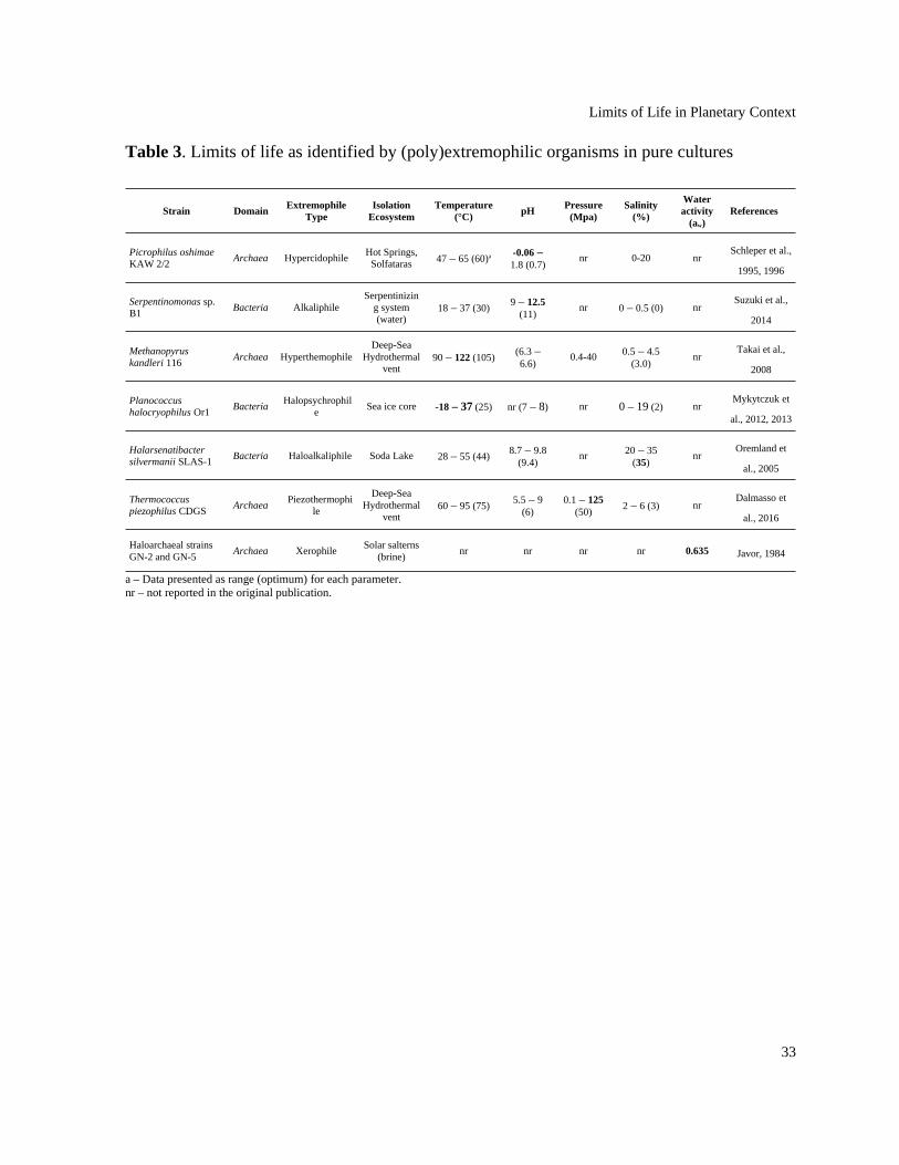
Limits of Life in Planetary Context
Table 3. Limits of life as identified by (poly)extremophilic organisms in pure cultures
Strain DomainExtremophile
TypeIsolation
EcosystemTemperature
(°C)pH
Pressure(Mpa)
Salinity(%)
Wateractivity
(aw)References
Picrophilus oshimae KAW 2/2
Archaea HypercidophileHot Springs,
Solfataras 47 – 65 (60)a -0.06 –1.8 (0.7)
nr 0-20 nrSchleper et al.,
1995, 1996
Serpentinomonas sp. B1
Bacteria AlkaliphileSerpentinizin
g system(water)
18 – 37 (30)9 – 12.5
(11)nr 0 – 0.5 (0) nr
Suzuki et al.,
2014
Methanopyrus kandleri 116
Archaea HyperthemophileDeep-Sea
Hydrothermalvent
90 – 122 (105)(6.3 –6.6)
0.4-40 0.5 – 4.5(3.0)
nrTakai et al.,
2008
Planococcus halocryophilus Or1
Bacteria Halopsychrophil
eSea ice core -18 – 37 (25) nr (7 – 8) nr 0 – 19 (2) nr
Mykytczuk et
al., 2012, 2013
Halarsenatibacter silvermanii SLAS-1
Bacteria Haloalkaliphile Soda Lake 28 – 55 (44)8.7 – 9.8
(9.4)nr 20 – 35
(35)nr
Oremland et
al., 2005
Thermococcus piezophilus CDGS
Archaea Piezothermophi
le
Deep-SeaHydrothermal
vent60 – 95 (75)
5.5 – 9(6)
0.1 – 125(50)
2 – 6 (3) nrDalmasso et
al., 2016
Haloarchaeal strains GN-2 and GN-5
Archaea XerophileSolar salterns
(brine)nr nr nr nr 0.635 Javor, 1984
a – Data presented as range (optimum) for each parameter.nr – not reported in the original publication.
33

Limits of Life in Planetary Context
Table 4. Examples of notable Polyextremophiles and their physiological requirements.
Strain Domain Extremophile typeIsolation
EcosystemTemperatur
e (°C)pH
Pressure(MPa)
Salinity(%)
References
Acidianus infernus So4a Archaea Acidothermophile Solfatara Crater 65 – 96 (90)
1 – 5.5 (2)
na0.2 (na)
Segerer et
al., 1986
Colwellia piezophila ATCC BAA-637
Bacteria Piezopsychrophile Deep-sea 4 – 15 (10)
7 (na) 40 – 80 (60)
na (3)
Nogi et al.,
2004
Halomonas campisalis MCM B-365
Bacteria Hyperalkaliphile Soda Lake 4 – 50 (30)
6 – 12 (9.5)
na 1.1 – 26.3(8.9)
Aston and
Peyton, 2007
Oceanobacillus iheyensis HTE831
BacteriaAlkaliphile,
Piezotolerant andHalotolerant
Deep-sea (mud) 15 – 42 (30)
6.5 – 10 (7 – 9.5)
0.1 – 300 – 21
(3)
Lu et al.,
2001
Anoxybacillus pushchinensis K1
Bacteria Alkalithermophile Manure 37 – 66 (62)
8 – 10.5 (9.5)
na< 3 (na)
Pikuta et al.,
2000
Actinopolyspora righensisH23
Bacteria Halophile Saline soil 20 – 40 (28 – 32)
5 – 8 (6 – 7)
na10 – 30
(15 – 25)
Meklat et al.,
2013
“Geothermobacterium ferrireducens” FW-1a
Bacteria HyperthermophileObsidian Pool,YellowstoneNational Park
65 – 100 (85)
na na 0 (na)Kashefi et
al., 2002
Shewanella piezotolerans WP3
Bacteria Piezophile Deep-sea 0 – 28 (15 – 20)
6 – 8 (7)
0.1 – 50 (20)
1 – 7.2 (3 – 4)
Xiao et al.,
2007
Colwellia sp. MT-41 Bacteria Piezopsychrophile Deep-sea2
(na)6.8 (na)
51.8 – 103.5(69)
naYayanos et
al., 1981
Pedobacter arcticus A12 Bacteria Psychrophile Tundra (soil) 4 – 25 (18)
6 – 9 (7)
na 0 – 2 (0)
Zhou et al.,
2012
Thermococcus gammatolerans EJ3
ArchaeaThermophile andRadiation-tolerant
HydrothermalVent (chimney)
55 – 95 (88)
na (5.5 – 6.5)
na (20)Jolivet et al.,
2003
Deinococcus radiodurans R1
BacteriaVacuum- and
Radiation-tolerantSpoiled canned
meatMars-like conditions, Vacuum, UV & Space Radiation
De Vera et
al., 2012
Cryomyces antarcticus MA5682
FungiVacuum- and
Radiation-tolerantAntarctica Mars-like conditions, Vacuum, UV & Space Radiation
De Vera et
al., 2012
Deinococcus geothermalisDSM 11300
Bacteria Xerotolerant Hot Spring 30 – 55 (47)
5 – 8 (6.5)
na naFrösler et al.,
2017
Halobacterium salinarum NRC-1
ArchaeaXerotolerant,Vacuum- and
Radiation-tolerant
Bore core from asalt mine
42 (na)
na na 25Kottemann
et al., 2005
34

Limits of Life in Planetary Context
35

Limits of Life in Planetary Context
Table 5. Boundary conditions for different planetary bodies of astrobiological interest(compared to Earth), split into atmosphere, surface, and subsurface layers. The observed orputative geochemistry as well as other potential influences are also listed.
PlanetaryBody
Type LayerTemperatur
e (ºC)pH
Pressure (MPa)
Salinity (% NaCl)
Geochemistry References
Earth planet
Atmosphere -100 – 40
Neutral,local acidicconditions
possible dueto volcanismand humanactivities
0.0001 – 0.1 0
8.1% N2, 21% O2, 9340 ppm Ar, 400 ppmCO2
18.2 ppm Ne, 5.2 ppm He, 1.7 ppm CH4,1.1 ppm Kr, 0.6 ppm H2, variable H2O
Hans Wedepohl, 1995;McDonough and Sun, 1995;
Wayne, 2000 Surface -98.6 – 464 -3.6 – 13.3 0.003 – 112 0 – saturation
Soils and sediments of varyinglithologies, siliceous crust, ranging frommafic to felsic composition. Extensive
ocean (70% planet surface), with 4,000 maverage depth, 4°C and 3.5% averagetemperature and salinity respectively
Subsurface 3.25 – <400 ~1 – 12.8 <8000.05 –
saturation
Soils and sediments of varyinglithologies, siliceous crust, ranging frommafic to felsic composition, ultramafic
mantle
Venus planet
Atmosphere -40 – 482a 0b 0.1 – 9.3c nr96.5% CO2, 3.5% N2; small quantities of
CO, SO2, HCl, HF, HDO, and H2O;H2SO4 condensates
Airey et al., 2017; Basilevskyand Head, 2003; Bertaux et al.,2007; Cockell, 1999; Lang andHansen, 2006; Schulze-Makuch
et al., 2004
Surface 377 – 482 nr 4.5 – 9.3c nrRocks are similar to tholeiitic and alkaline
basalts; no liquid water
Subsurface nr nr nr nr Fluid channels; volcanism
Mars planet
Atmosphere -138 – 35d nr0.0001–0.0009
nr95.3% CO2, 2.7% N2, 1.6% Ar, 0.13% O2,
0.08% CO; trace amounts of H2O, NO,Ne, Kr, Xe
Fairén et al., 2004; Hecht et al.,2009; Johnson et al., 2011; Jones
et al., 2011; Longstaff, 2014;Michalski et al., 2013; NASA;
Nicholson and Schuerger, 2005;Sinha et al., 2017; Smith et al.,
2009; Varnes et al., 2003;
Wordsworth, 2016
Surface -138 – 30 7.7e 0.0004–0.0009
5.2 – 5.8
Basaltic, Fe-/Mg-rich phyllosilicates,perchlorate salts, Al-rich clays, sulphates,
chlorides, calcite, and silicas; potentialcryosphere
Subsurface 55g 4.96 – 9.13h 10 – 303g Cl-rich brinesPotential groundwater; basalt crust;
possible serpentinization
Enceladus icy moon
Plume jets 0 ~8.5 – 9High velocity
jets> 0.5
90–99% H2O, ≤0.61–4.27% N2, 0.3–5.3%CO2, 0.1–1.68% CH4, 0.4–0.9% NH3, 0.4–39% H2, trace amounts of hydrocarbons;
high mass organic cations, silicates,sodium, potassium, carbonates Gioia et al., 2007; Glein et al.,
2015; Holm et al., 2015; Hsu etal., 2015; Postberg et al., 2009,
2018; Taubner et al., 2018;Waite et al., 2009; Zolotov et al.,
2011
Icy shell(~10 km thick)
-233 – -23 nr nrMay have
ammonia brinepockets
May have tectonics
Subsurfaceglobal ocean(~0–170 km
depth)
<90 8.5 – 12.2k 1 – 8 0.45 – < 4 Possible serpentinization
Titan icy moon
Atmosphere -183 – -73j nr > 0.01 – 0.15 nr
98.4% N2, 1.4% CH4, 0.2% H2, tracehydrocarbons and organics; 95% N2, 5%CH4, 0.1% H2; ~50 ppmv CO and ~15
ppbv CO2; C2H3CN; cloudsBaland et al., 2014; Brassé et al.,
2017; Cordier et al., 2017; deKok et al., 2007; Fulchignoni etal., 2005; Jennings et al., 2016;
Mastrogiuseppe et al., 2014;McKay, 2016; Mitchell and
Lora, 2016; Mitri et al., 2014;
Norman, 2011; Sohl et al., 2014
Surface -183 – -179 nr 0.15–0.35i nr
Lakes and sea have CH4, C2H4, anddissolved nitrogen; dunes of solid organic
material; low-latitude deserts and high-latitude moist climates
Subsurface -18 11.8l 50–300m
Likely densesubsurface
ocean (≤1,350kg m-3)
suggesting highsalinity
CH4 and C2H6
Ceresdwarfplanet
Atmosphere nr nr nr nrTransient atmosphere with possible water
vapor Castillo-Rogez et al., 2018;Fanale and Salvail, 1989; Hayneand Aharonson, 2015; Hendrix
et al., 2016; Küppers et al., 2014; Surface (-157– -30)n 9.7–11.3n nr <10n Surface clays; (Mg, Ca)-carbonates; (Mg,
NH4)-phyllosilicates; Fe-rich clays; salt
36
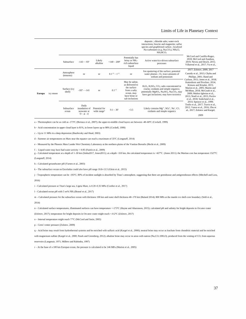
Limits of Life in Planetary Context
deposits ; chloride salts; water-rockinteractions; brucite and magnetite; sulfurspecies and graphitized carbon ; localized
Na-carbonates (e.g.,Na2CO3), NH4Cl,NH4HCO3
McCord and Castillo-Rogez,2018; McCord and Zambon,
2019; Neveu and Desch, 2015;Villarreal et al., 2017; Vu et al.,
2017; Zolotov, 2009, 2017
Subsurface -143 – -93o Likelyalkaline
<140 – 200p
Potentially hasbriny or NH3-
rich subsurfaceliquid
Active water/ice‐driven subsurfaceprocesses
Europa icy moon
Atmosphere(tenuous)
nr nr 0.1-12 – 1-12 nrIon sputtering of the surface; potentialwater plumes ; O2; trace amounts of
sodium and potassiumCassidy et al., 2013; Chyba and
Phillips, 2001; Hand andCarlson, 2015; Jones et al., 2018;Kattenhorn and Prockter, 2014;
Kimura and Kitadai, 2015;Marion et al., 2005; Martin andMcMinn, 2018; McGrath et al.,
2009; Muñoz-Iglesias et al.,2013; Noell et al., 2015; Pavlov
et al., 2018; Soderlund et al.,2014; Spencer et al., 1999;
Teolis et al., 2017; Travis et al.,2012; Vance et al., 2016; Zhu etal., 2017; Zolotov and Kargel,
2009
Surface (icyshell)
-187 – -141 nr 0.1-12
May be saline,as delivered to
the surfacefrom a saltyocean, may
have brine orsalt inclusions
H2O2, H2SO4, CO2; salts concentrated incracks; oxidants and simple organics;
potentially MgSO4, Na2SO4, Na2CO3, mayhave gas inclusions; may have tectonics
Subsurfaceocean
Dailyinundation ofseawater atT= -4 – 0
Potential forwide rangeq
0.1 – 30r <3.5 Likely contains Mg2+, SO4
2-, Na+, Cl-;oxidants and simple organics
a – Thermosphere can be as cold as -173ºC (Bertaux et al., 2007); the upper-to-middle cloud layers are between -40–60ºC (Cockell, 1999)
b – Acid concentration in upper cloud layer is 81%, in lower layers up to 98% (Cockell, 1999)
c – Up to 11 MPa in a deep depression (Basilevsky and Head, 2003)
d – Summer air temperatures on Mars near the equator can reach a maximum of 35ºC (Longstaff, 2014)
e – Measured by the Phoenix Mars Lander Wet Chemistry Laboratory at the northern plains of the Vastitas Borealis (Hecht et al., 2009)
f – Liquid water may have had water activity > 0.95 (Fairén et al., 2009) g – Calculated temperature at a depth of 1–30 km (Sinha2017, Jones2011); at a depth ~310 km, the calculated temperature is <427ºC (Jones 2011); the Martian core has temperature 1527ºC
(Longstaff, 2014)
h – Calculated groundwater pH (Varnes et al., 2003)
k – The subsurface ocean on Enceladus could also have pH range 10.8–13.5 (Glein et al., 2015)
j – Tropospheric temperature can be -193ºC; 80% of incident sunlight is absorbed by Titan’s atmosphere, suggesting that there are greenhouse and antigreenhouse effects (Mitchell and Lora,
2016)
i – Calculated pressure at Titan's large sea, Ligeia Mare, is 0.20–0.35 MPa (Cordier et al., 2017)
l – Calculated ocean pH with 5 wt% NH3 (Brassé et al., 2017)
m – Calculated pressure for the subsurface ocean with thickness 100 km and outer shell thickness 40–170 km (Baland 2014); 800 MPa at the mantle ice shell-core boundary (Sohl et al.,
2014)
n – Calculated surface temperatures, illuminated surfaces can have temperature <-173ºC (Hayne and Aharonson, 2015); calculated pH and salinity for bright deposits in Occator crater
(Zolotov, 2017); temperature for bright deposits in Occator crater might reach <-0.2ºC (Zolotov, 2017)
o – Internal temperature might reach 77ºC (McCord and Sotin, 2005)
p – Ceres' center pressure (Zolotov, 2009)
q – Acid brine may result from hydrothermal systems and be enriched with sulfuric acid (Kargel et al., 2000); neutral brine may occur as leachate from chondritic material and be enriched
with magnesium sulfate (Kargel et al., 2000; Pasek and Greenberg, 2012); alkaline brine may occur in areas with natron (Na2CO3.10H2O), produced from the venting of CO2 from aqueous
reservoirs (Langmuir, 1971; Millero and Rabindra, 1997)
r – At the base of a 100 km Europan ocean, the pressure is calculated to be 146 MPa (Marion et al., 2005)
37

Limits of Life in Planetary Context
Figures
Figure 1. Representative idealized cross section of Earth’s crust showing the diversity of extreme environments and their approximate location.
38

Limits of Life in Planetary Context
Figure 2. The temperature, pressure, pH, and salinity boundaries observed for life on Earth compared to the phase space observed on planetary bodies discussed in the main text. Polygoncharts are designed to represent ranges in multidimensional space. Each edge represents the range for the specific variables Single values (e.g., when min = max) are represented by a single vertex on an axis, while missing values (e.g., NA or NR) are represented by the absenceof the corresponding polygon edge on the corresponding axis.
39