localising the lesion: “where in the cns” · web viewfibres from the thalamus pass to the...
TRANSCRIPT

Localising the lesion: “where in the CNS”
Learning objectives
Definition of CNS and PNS
Definition of UMN and LMN
Function of each of the cerebral lobes
The homunculus
Circle of willis and blood supply to the cerebral hemispheres
Motor tracts – lateral corticospinal
Sensory tracts – lateral spinothalamic and dorsal columns
Stroke syndromes
Clinical case scenarios
Definitions
CNS = Brain and spinal cord
PNS = anything outside brain and spinal cord
Also include autonomic nervous system and cranial nerves
Motor control systems
Corticospinal (pyradmial)
Skilled, intricate, strong and organised movements
Defectiveness loss of skilled voluntary movement, spasticity and reflex changes
Such as hemiparesis, hemiplegia or paraparesis
Extrapyradimal system
Fast, fluid movements that the corticospinal system has generated
Defectiveness bradykinesia, rigidity, tremor, chorea
Such as huntingtons
The cerebellum
Co-ordinating smooth and learned movement initiated by the pyradimal system and in posture and balance control
Defectiveness ataxia, past pointing, action tremor and incoordination
The motor system
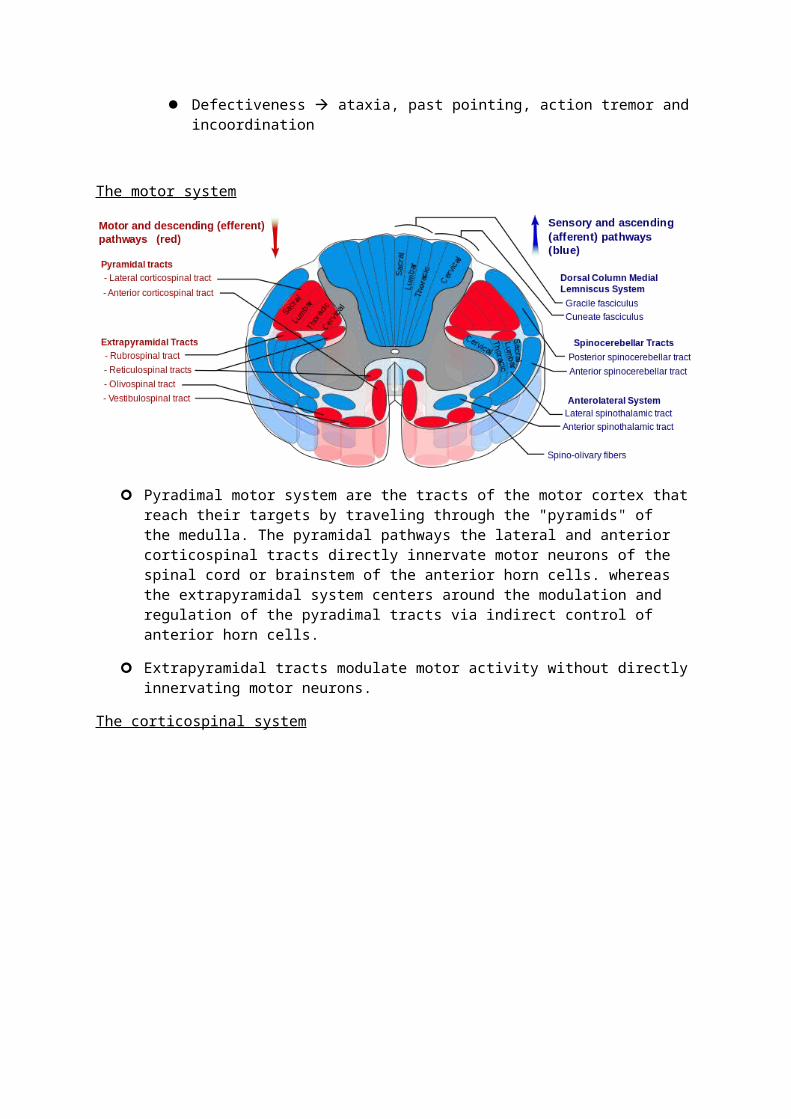
Pyradimal motor system are the tracts of the motor cortex that reach their targets by traveling through the "pyramids" of the medulla. The pyramidal pathways the lateral and anterior corticospinal tracts directly innervate motor neurons of the spinal cord or brainstem of the anterior horn cells. whereas the extrapyramidal system centers around the modulation and regulation of the pyradimal tracts via indirect control of anterior horn cells.
Extrapyramidal tracts modulate motor activity without directly innervating motor neurons.
The corticospinal system

The homunculus

UMN vs LMN
UMN LMN
Wasting no yes
Fasciculation no yes
Tone increased decreased
Power decreased decreased
Reflexes increased decreased
Plantars up going down going
Sensory pathways

Peripheral nerves carry sensation from dorsal roots to the cord
Posterior columns (dorsal columns)
Vibration, joint position, light touch and point discrimination
Cross in the brainstem passing to the thalamus
Spinothalamic tracts
Pain and temperature
Cross within the cord and pass in the spinothalamic tracts to the thalamus and reticular formation
Sensory cortex
Fibres from the thalamus pass to the parietal region sensory cortex and motor cortex
Cortical functions
Frontal lobe
Reasoning, planning, parts of speech, movement, emotions and problem solving
Left frontal = broccas area (aphasia)
Parietal lobe
Movement, orientation, recognition, perception of stimuli

Occipital lobe
Visual processing
Temporal lobe
Perception and recognition of auditory stimuli, memory and speech
Left temporal = wernicke’s area
Cerebellum
Balance and co-ordination
Basal ganglia
Initiation and inhibition of movement
Wernickes area – like broccas area is it the understanding of written and spoken speech
Circle of Willis
Internal carotid artery supplies brain
External carotid artery supplies face
Middle cerebral artery supplies 1/3rd of brain
Vertebral arteries join to form the basillar artery which join at the base of the brain

Stroke
TACS – All three of
Hemiplegia or hemi sensory loss
Visual field defect
Disturbance of higher function
Dysphasia
Dysphagia
PACS – 2 out of 3
LACS – blockage of small branch of big artery
No visual field defect
Pure motor stroke
Pure sensory
Sensory motor
Ataxia
POCS – brain stem, cerebellum, cranial nerves
Bilateral motor or sensory
Conjugate eye movement disorder

Cerebeller dysfunction
Hemiplegia or cortical blindness
Acute occlusion of blood vessel leading to hypoxia and infarction
Risk factors
DM, hypertension, smoking, hypercholesterolemia, FHx, AF
Investigations
bloods, CT, MRI, carotid dopplers, Echo, ECG, 24 hour tape
Treatment in ischaemic stroke
Aspirin
Clopidogrel
Supportive management
In ischaemic stroke you have in ischaemic penumbra which is the area of the brain which is damaged during ischaemia in order to reduce the effects from this you need to optimise conditions – temp, BP, glucose
Cerebellar syndrome
Causes
Vascular lesion
Alcohol
Demyelination
Tumours
Hypothyroidism
Metabolic disorders
Signs “DANISH”
Dysdiadochokinesis
Ataxia
Nystagmus
Intention tremor
Slurred speech, dysarthria
Hpyotonia, hyporeflexia
Multiple Sclerosis
Areas of demyelination and perivascular inflammation (white plaques)

Disseminated in time and occurring anywhere within CNS
Aetiology - ?autoimmune ?vitamin D deficiency
Classification
Benign -little disease activity for many years, minimal disability
Relapse remitting - most common, repeated attacks with periods of recovery
Secondary chronic progressive - continuous progression of symptoms following an initial relapsing and remitting disease course
Primary progressive - accumulation of pernament disability over time with superimposed relapses
Investigations
LP – increased protein, increased immunoglobulin, oligoclonal bands
Visual evoked potentials
MRI
On examination
Unsteady gait
Reduced proprioception
Brisk reflexes
Brown-sequard syndrome
Loss of movement on same side as damage
Loss of pain and temp and sensation on opposite side
spinal cord lesion where there is an incomplete lesion characterized by loss of motor function loss of vibration sense and fine touch, loss of proprioception and signs of weakness on the same side of the spinal injury. This is a result of a lesion affecting the dorsal column and the corticospinal tract. On the contralateral side of the lesion, there will be a loss of pain and temperature sensation and crude touch 1 or 2 segments below the level of the lesion
Management
Symptoms control (tremors, pain, muscle spasms)
Steroids - severe relapses to speed up any recovery with will occur naturally. A severe relapse is usually classed as one that has significantly affected activities of daily living
Beta-inferons and Glatiramer - reduce rates of relapses by 30% and is only used in relapsing and remitting or relapsing progressive disease

IV natalizumab - is a newer monoclocal antibody treatment used in patients with very severe active disease that can reduce relapses by 80%, cost, practical consideration and complications limits its use.
Motor neurone disease
Degeneration of upper and lower motor neurones of unknown cause 5-10% autosomal dominant Types
o Spinal muscular atrophy – limb weakness due to involvement of spinal cord anterior horn cells
o Primary later sclerosis – spastic limb weakness due to UMN involvement of the spinal cord
o Progressive bulbar palsy – involvement of bulbar motor neurones, progressive disease
o Amyotrophic lateral sclerosis – mixture of all the above Investigations
o Diagnosed clinically after other causes excludedo EMG confirms fasciculation's and fibrillations
Management – symptom control Fatal within 3-5 years Cardiac and smooth muscle aren’t involved and ocular muscle very rarely Autonomic dysfunction occurs late Signs
o Dysarthria, brisk jaw reflexo Fasciculation/wasting in deltoids, biceps, quadriceps and in tongueo Weakness in all4 limbs, brisk reflexes in arms, absent in legs
Combination of UMN and LMN
Clinical case 1
23, female presents to her GP with a 2 week history of bilateral leg weakness having started with pins and needles and numbness in her hands and feet. She has had a few days of urinary incontinence which has resolved. 2 years ago she had an episode of blurred vision and pain in the right eye which lasted a month and fully resolved
Diagnosis – MS
Visual – optic neuritis, diplopia, nystagmus, internuclear opthalmoplegia, dysarthria, dysphagia, weakness, muscle spasms, ataxia, pain, paraesthesis, fatigue, cognitive impairment, depression, unstable mood
Uhthoff’s phenomenum – the worsening of neurological symptoms after periods of exercise and increased body heat
Lhermittes sign – an electrical sensation that spreads from the back into the limbs on neck flexion and or extension
Bowel problems – incontinence, diarrhoea, constipation
Urinary – incontinence, frequency, urinary retention
Plaques of demyelination within the CNS caused by an inflammatory process. Different areas of the CNS are involved over time

LP – cell count, protein, glucose and oligoclonal bands. WCC less than 50/mm3
MRI
Visual evoked potentions – show delayed conduction between the retina and the occipital cortex
There is no curative treatment
Multidisciplinary team
Symptomatic – spasticity, pain, fatigue, depression, continence
Steroids, beta interferon, glatiramer, natalizumab
Clinical case 2
61 female
Becoming increasingly weak on her right side over a one week period. She is unable to walk and has slurred speech and right side of her face is drooping
Past history of breast cancer
o/e – right facial weakness, grade 4/5 weakness of the right arm and leg, right homonymous hemianopia and some difficulty naming objects and reflexes are brisk on the right side and her right plantar response is upgoing
diagnosis = Cerebral maetastases from carcinoma of the breast
CT head shows extensive oedema surrounding the subtle impression of a ring enhanced lesion in the left frontal lobe, extending into the left parietal lobe. There is associated mass effect displacing the lateral ventricle
Features of raised intracranial pressure it is likely the oedema around the tumour has increased or bleedin has occurred within the tumour
Features of raised ICP – visual loss, seizures and focal neurological deficit such as third and 6th cranial nerve palsies
Multidisciplary team, neurosurgery, corticosteroids, radiotherapy, chemotherapy
Case 3
76 male Background of AF (on warfarin) has 2 hour history of severe global right sided weakness. He
is eye-opening to painful stimuli and is moving his left side spontaneously. When questioned he seems confused
12/15 E2, V4, M6 Left hemisphere primary intracerebral haemorrhage causing right sided hemiparesis Bloods tests, CXR, head CT head CT should have been performed within 24 hours or immediately in patients
presenting with acute stroke if any of the following apply to them. – on anticoagulation treatment, known bleeding tendancy, decreased consciousness, papliodemea, neckstiffness or fever, severe headache with sudden onset,
Ultrasound doppler, cerebral angiography, echocardiography

Risk factors – hypertension, smoking, DM, FH, increasing age, previous strokes, vascular disease, hyperlipidaemia, hypercoagulable state, alcohol abuse, malignancy
In ishcamic stroke – thrombolysis three hours from obsets are elegible Aspirin, lipid lowering drugs, anticoagulation if patient has AF or other source of embolus Haemorrhaging stroke – supprotive care. neurosurgery
Case 4
56 male
6 month history of progressive weakness of his right hand. Also had problems with swallowing and has choked whilst eating on several occasions
o/e he has wasting of his upper and lower limbs and some fasciculation's were noted his right plantar was up going and his reflexes were generally brisk
Motor neurone disease
MRI – to exclude local brainstem pathology
EMG – acute denervation of the lower motor neurones
Cases were the diagnosis isn’t clear – LP to exclude MS, muscle biopsy to exclude muscle disease. Blood tests for other conditions