lopamidol and metrizamide in cervical myelography: side … · lopamidol and metrizamide in...
TRANSCRIPT
848
lopamidol and Metrizamide in Cervical Myelography: Side Effects, EEG, and CSF Changes L. Moschini,' O. Manara,' G. Bonaldi,' V. Cassinari, 2 N. Quilici,' and G. Belloni'
Two nonionic contrast media, iopamidol and metrizamide (Amipaque), were used for cervical myelography (C1-C2 puncture) in 95 consecutive patients. Both contrast media gave excellent radiographic results. Headache and vagal symptoms were similar in both groups, whereas metrizamide produced more electroencephalographic changes and epileptic seizures. Meningeal irritation occurred in both groups and was severe in three cases. Cerebrospinal fluid showed protein and cellular changes of inflammatory type in both groups. lopamidol is considered to be the more suitable contrast medium for cervical myelography despite its slight neurotoxicity.
Nonionic water-soluble contrast media are now common ly used to study the spinal cord as well as the subarachnoid spaces . Most of the early reports of these contrast media were extremely favorable; recently , reports of adverse and serious side effects have been published more and more frequently [1-5). In order to evaluate the neurotoxic ity of these contrast media more precisely, we have been studying iopamidol and metrizamide (Amipaque). In addition to side effects, we have observed the changes on the electroencephalogram (EEG) and of the cerebrospinal fluid (CSF) at 24 and 48 hr, respectively, after cervical myelography.
Subjects and Methods
The study, still in progress, involves 95 patients who underwent cervical myelography with C1-C2 puncture . Subjects were divided into three groups: 55 received injection of 10 ml iopamidol 300; 25 were given 10 ml of Amipaque with a concentration of 300 mg I/ ml ; and in th e last 15 subjects the same quantity and concentration of Amipaque was aspirated into the syringe by microfilter 1 hr before the myelographic examination [6). Fifty-six subjects were male, 39 female. The mean age of the subjects was 47 years (range, 27-71). The pathologies most frequently observed were disk prolapses (25%) and degenerative changes of the cervical spine (44%). In 22 cases no pathology was found . The iopamidol and Amipaque groups were comparable in gender distribution , mean age, and pathology, thus enabling stat istical comparison to be made.
All subjects were premedicated with 5 mg Valium. No patients referred had histories of epileptic seizures or allergic reactions. The most important blood parameters (azotemia, glycemia, electrophoresis, etc.) were checked before the examination and 48 hr later. Blood pressure , heart rate, and resp iratory rate were monitored during the procedure. Subjects were allowed no food or liquids the night before the examination . After the procedure 1,000-1 ,500 ml
of isotonic solution was infused intravenously. After the examination all patients remained in a half-sitting position for 6 hr, then lay supine for up to 24 hr.
An EEG was obtained before and 24 hr after the examination in 89 subjects; CSF was collected during the examination at C1-C2 level and 48 hr after by the lumbar route in 65 subjects. Total proteins, protein electrophoresis, and the cellu lar content were evaluated.
Results
Quality of the Films
Over 90% of the myelograms were of excellent quality with both contrast media. The root sleeves in particular were very clearly shown on regular films, while the spinal cord was better demonstrated by sagittal tomography .
Clinical Controls
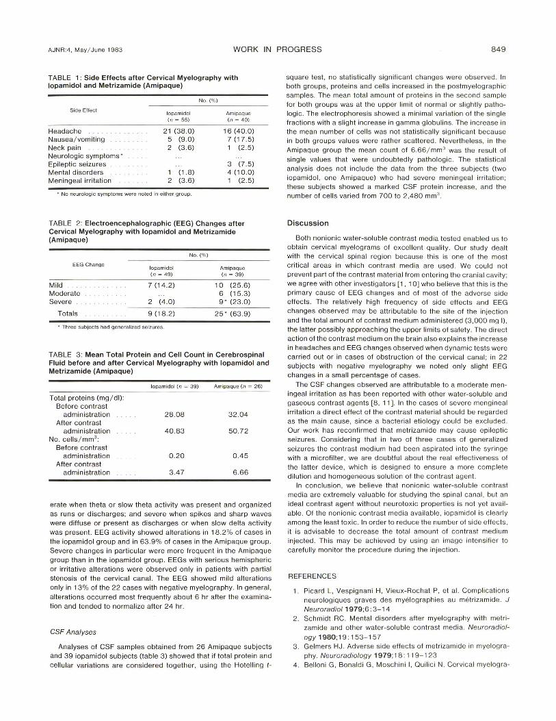
In agreement with investigators who have been using iopamidol in angiographies [7), we observed no significant changes in heart and respiratory functions or in blood parameters. Among the side effects (table 1), headache was the most frequent symptom [8 , 9); it had an early onset (about 6 hr after the examination), moderate intensity, and disappeared within 24 hr in most cases. Headache increased in over 50% of cases, with no gender differential, when functional tests were performed. Generalized seizures were observed in three subjects in the Amipaque group, beginning about 1 hr after the examination and disappearing after adequate therapy. All three subjects had a spondylotic stenosis of the cervical canal , which probably caused more contrast medium than usual to enter the cranial cavity. Among the other side effects, five cases (four with Amipaque and one with iopamidol) of temporospatial disorientation were observed; in these events also, there was early onset and rapid resolution without any particular therapy. Soon after the examination three subjects (two with iopamidol and one with Amipaque) experienced severe meningeal irritation with headache, neck pain , and hyperpyrexia; one had a temporary loss of consciousness. In all three cases the CSF sampled at the same time proved to be sterile but showed a noticeable increase of proteins and cells.
EEG Parameters
The measured EEG changes (table 2) were considered mild when a minimal disorganization of background activity was present; mod-
, Neuroradiology Service. Ospedali Riuniti , Bergamo. Italy. Address reprint requests to L. Moschini. 2 Department of Neurosurgery . Ospedali Riuniti , Bergamo, Italy.
AJNR 4:848-850, May/ June 1983 0 195-6108/ 83 / 0403-0848 $00.00 © American Roentgen Ray Society
AJNR:4, May / June 1983 WORK IN PROGRESS 849
TABLE 1 : Side Effects after Cervical Myelography with lopamidol and Metrizamide (Amipaque)
Side Effect
Headache Nausea/ vomiting Neck pain Neuro logic symptoms' Epileptic seizures Menta l disorders Meningeal irritation
lopamidol (n = 55)
2 1 (38.0) 5 (9.0) 2 (3 .6)
1 (1 .8) 2 (3. 6)
. No neurologic symptoms were noted in either group.
No. (%)
Amipaque (n = 40)
16 (40.0) 7 (1 7. 5) 1 (2.5)
3 (7 .5) 4 (10 .0) 1 (2.5)
TABLE 2: Electroencephalographic (EEG) Changes after Cervical Myelography with lopamidol and Metrizamide (Amipaque)
No. (% )
EEG Change topam idot Amipaque (n = 49) (n = 39)
Mild 7 (14 .2 ) 10 (25 .6) Moderate 6 (15 .3) Severe 2 (4 .0) 9' (23.0 )
Tota ls 9 (18.2) 2 5 " (63.9)
. Three subjects had generalized seizures.
TABLE 3: Mean Total Protein and Cell Count in Cerebrospinal Fluid before and after Cervical Myelography with lopamidol and Metrizamide (Amipaque)
topamidot (n = 39) Amipaque (n = 26)
Total proteins (mg / dl): Before contrast
admin istration 28. 08 32.04 A fter contrast
administration 40.83 50.72 No. ce lls / mm3
:
Before contrast administration 0 .20 0.45
After contrast administration 3. 47 6 .66
erate when theta or slow theta activity was present and organized as run s or di scharges; and severe when spikes and sharp waves were diffuse or present as discharges or when slow delta ac tivity was present. EEG activity showed alterations in 18.2% of cases in the iopamid ol group and in 63. 9% o f cases in th e Amipaque g roup. Severe changes in particu lar were more frequent in the Amipaq ue group than in th e iopamidol group. EEGs with serious hemispheri c
or irritative alterations were observed only in patients w ith parti a l stenosis of the cervica l canal. Th e EEG showed mild a lterations only in 13% of the 22 cases w ith negati ve myelography. In general,
alterations occurred most frequently about 6 hr after the examination and tended to normalize after 24 hr.
CSF Analyses
Analyses of CSF samples obta ined from 26 Amipaque subjects and 39 iopamidol subjects (table 3 ) showed that if total pro tei n and ce llular vari ation s are considered togeth er, using the Hote lling t-
square test , no stati stica lly significant changes were observed . In both groups, proteins and ce lls increased in the postmyelographic samples. The mean total amount of proteins in the second sample for both groups was at the upper limit of normal or slightly pathologic. The electrophoresis showed a minimal variation of the sing le fractions with a slight inc rease in gamma globulins. The increase in the mean number of ce lls was not stati sti ca lly significant because in both groups values were rather scattered. Nevertheless, in the
Amipaque group th e mean count of 6.66 / mm 3 was the resu lt of
single va lues that were undoubtedly patho logic. Th e stati sti ca l analysis does not inc lude the data from the three subjects (two iopamidol, one Amipaque) who had severe meningeal irritat ion ; these subjects showed a marked CSF protein increase, and the number of cells varied from 700 to 2,480 mm3
.
Discussion
Both non ionic water-soluble contrast med ia tested enabled us to obtain cervica l myelog rams of excellent quality. Our study dealt with the cervica l spinal reg ion because this is one of the most c riti cal areas in which contrast med ia are used . We could not prevent part of the contrast materi al from entering the c ran ial cavity; we ag ree with other investigators [1 , 10] who believe that this is the primary cause of EEG c hanges and of most of the adverse side effec ts. The relati ve ly high frequency of side effec ts and EEG changes observed may be attributable to the site of th e injection
and the total amount of contrast med ium administered (3,000 mg I) , the latter possibly approaching th e upper limits of safety . The d irec t ac tion of the contrast med ium on th e brain at so explains the inc rease in headaches and EEG changes observed when dynamic tests were carried out or in cases of obstruc tion of th e cervica l canal; in 22 subjec ts with negative myelog raphy we noted only slight EEG changes in a small percentage of cases.
The CSF changes observed are attributable to a moderate meningeal irritation as has been reported with other water-soluble and gaseous contrast agents [8, 11]. In the cases of severe mengineal irritati on a d irec t effect of th e contrast material shou ld be regarded as the main cause, s ince a bacterial etio logy could be excluded . Our work has reconfirmed that metrizamide may cause epileptic seizures. Considering that in two of three cases of generalized seizures th e contrast med ium had been aspirated into the syr inge
w ith a mic rof ilter , we are doubtful about th e real effect iveness of the latter device, which is designed to ensure a more comp lete
dilution and homogeneous solution of the con trast agent. In conc lu sion, we be lieve that non ionic water-soluble contrast
media are extremely va luable for studying the spinal canal, bu t an ideal contrast agent without neurotoxic properties is not yet avai l
able. Of the non ioni c contrast media available, iopamidol is c learl y among the least tox ic. In order to reduce the number of s ide effec ts, it is advisable to decrease th e total amount of contrast med iu m injected . Th is may be achieved by using an image intensifie r to carefull y monitor th e proced ure during th e injection.
REFERENCES
1. Picard L , Vespignani H , Vieux-Rochat P, et al. Comp lications neuro log iq ues g raves des myelog raphies au metri zamide. J Neuroradio/ 1979 ;6: 3- 1 4
2 . Schmidt RC. Men tal d isorders after myelog raphy with met ri zamide and other water-soluble contrast med ia. Neuroradiol
ogy 1980; 19 : 153 - 157 3. Gelmers HJ . Adverse side effec ts of metrizamide in myelogra
phy . Neuroradiology 1979; 18: 11 9-1 23 4 . Belloni G , Bonald i G , Moschin i I, Quilic i N. Cervical myelogra-
850 WORK IN PROGRESS AJNR:4 . May / June 1983
phy with iopamidol. Neuroradiology 1981 ;21 : 97 -99 5. Quilici N. Belloni G, Bonaldi G, Cassinari V, Moschini L. Cis
ternografia opaca con iopamidolo. Radiol Med (Torino) 1981 ;67: 965-970
6. Legre J, Lavieille J, Debaene A, Tapias PL, Vaillant J. Prevention des accidents de la myelographie cervica le a l 'Amipaque . J Neuroradio/1981 ;8: 353- 36 1
7. Gambardella A, Latessa G, Meoli S, Rotondo A. Valutazione comparativa dello iopamidolo e della iotalamato di metilglucamina in angiografi a cerebrale. Presented at th e meeting of the Italian Association for Medical Rad iology and Nuc lear Medi-
c ine, Naples, September 1980 8. Skalpe 10, Amundsen P. Thoracic and cervica l myelography
with metri zam ide. Radiology 1975;116: 1 01 -1 06 9. Hammer B, Lackner W. lopamidol , a new non- ionic hydrosol
uble contrast medium for neuroradiology. Neuroradio logy 1980;19 : 11 9 -1 21
10. Caille JM, Guibert-Trainer F, Howa JM , Billerey J , Calabet A, Piton J. Contamination encephalique par la metrizamide apres myelog raphie . J Neuroradio l 1980;7 : 3 -1 2
11 . Bickerstaff ER. Changes in the cerebrospinal fluid after pneumo-encephalography. Lancet 1951;1 :1209-1 2 1 0