loss of drosophila a-type lamin c initially causes tendon ... · genomes and developmental control...
TRANSCRIPT

Developmental Biology 373 (2013) 216–227
Contents lists available at SciVerse ScienceDirect
Developmental Biology
0012-16
http://d
n Corr
E-m
journal homepage: www.elsevier.com/locate/developmentalbiology
Genomes and Developmental Control
Loss of Drosophila A-type lamin C initially causes tendon abnormalityincluding disintegration of cytoskeleton and nuclear laminain muscular defects
Ryo Uchino a, Yu-ki Nonaka a, Tuneyoshi Horigome a, Shin Sugiyama b, Kazuhiro Furukawa a,n
a Department of Chemistry, Faculty of Science, Niigata University, Niigata 950-2181, Japanb Division of Biological Science, Graduate School of Science, Nagoya University, Nagoya 464-8602, Japan
a r t i c l e i n f o
Article history:
Received 1 May 2012
Received in revised form
19 July 2012
Accepted 1 August 2012Available online 13 September 2012
Keywords:
A-type lamin
Muscular dystrophy
Cytoskeleton
Nuclear lamina
Tendon cells
06/$ - see front matter & 2012 Elsevier Inc. A
x.doi.org/10.1016/j.ydbio.2012.08.001
esponding author. Fax: þ81 25 262 6163.
ail address: [email protected]
a b s t r a c t
Lamins are the major components of nuclear envelope architecture, being required for both the
structural and informational roles of the nuclei. Mutations of lamins cause a spectrum of diseases in
humans, including muscular dystrophy. We report here that the loss of the A-type lamin gene, lamin C
in Drosophila resulted in pupal metamorphic lethality caused by tendon defects, matching the
characteristics of human A-type lamin revealed by Emery–Dreifuss muscular dystrophy (EDMD).
In tendon cells lacking lamin C activity, overall cell morphology was affected and organization of the
spectraplakin family cytoskeletal protein Shortstop which is prominently expressed in tendon cells
gradually disintegrated, notably around the nucleus and in a manner correlating well with the
degradation of musculature. Furthermore, lamin C null mutants were efficiently rescued by restoring
lamin C expression to shortstop-expressing cells, which include tendon cells but exclude skeletal
muscle cells. Thus the critical function of A-type lamin C proteins in Drosophila musculature is to
maintain proper function and morphology of tendon cells.
& 2012 Elsevier Inc. All rights reserved.
Introduction
Lamins belong to the type-V family of intermediate filamentproteins that share the same tripartite structure; an a-helical roddomain flanked by N- and C-terminal head and tail domains(Gruenbaum et al., 2003; Dechat et al., 2010). They are the basicsubunits for assembling the stable lamina located between theinner nuclear membrane of the nuclear envelope and peripheralchromatin, and bind to chromosomes/chromatin, DNA, andvarious proteins of the inner nuclear membrane and nuclear porecomplex to establish and maintain nuclear architecture. Laminsare also present inside the nucleus and interact with nuclearproteins involved in regulation of gene expression and DNAreplication (Gruenbaum et al., 2003; Dechat et al., 2010; Wilsonand Foisner, 2010). Recent studies show that lamins further linkto the cytoskeleton through the Linker of Nucleoskeleton andCytoskeleton (LINC) complex, thus they are also thought to berequired for architectural functions in the cytoplasm (Houbenet al., 2007; Razafsky and Hodzic, 2009; Roux and Burke, 2007;Wilson and Foisner, 2010).
ll rights reserved.
(K. Furukawa).
Lamins are present in all metazoans but not in unicellularorganisms and plants and have been classified into A-type whichis expressed in a subset of differentiated tissues and B-type which isconstitutive during development (Gruenbaum et al., 2003; Dechatet al., 2010). Studies demonstrate that mutations in both lamintypes cause debilitating diseases in human, and developmentaldisorders in model animals (Emery, 2000; Dechat et al., 2010;Rankin and Ellard, 2006; Roux and Burke, 2007; Worman et al.,2010), thus lamins have become recognized as having significantfunctions in tissue formation and maintenance during development.
Various mutations of the A-type lamin gene (LMNA) result inboth dominant and recessive phenotypes that are associated with anumber of different human disorders including both Hutchinson–Gilford premature aging syndrome and Emery–Dreifuss musculardystrophy (EDMD) (Rankin and Ellard, 2006; Roux and Burke,2007; Worman et al., 2010). Analysis of mutated domains in theA-type lamin proteins provides some insights on how it functionsin tissue organization, but the relationship between mutations anddefective phenotypes of A-type lamin is very complex. In the micemodel, LMNA null mutants show severe postnatal growth retarda-tion and muscular dystrophy resembling EDMD, suggesting it willbe a useful tool for the study of muscular dystrophy (Dechat et al.,2010; Rankin and Ellard, 2006; Roux and Burke, 2007; Wormanet al., 2010), however, the actual functions of A-type lamin inmusculature have remained unclear.

R. Uchino et al. / Developmental Biology 373 (2013) 216–227 217
Drosophila also has both types of lamins, which are the B-typelamin Dm0 and the A-type lamin C. Whereas in mammals, twotypes of B-type lamin genes, LMNB1 and LMNB2 are reported,Drosophila only has a single B-type lamin gene. Although bothmammals and Drosophila have single A-type lamin genes, multipletranslation products are generated by alternative splicing frommammalian LMNA, while Drosophila produces a single lamin Cprotein (Riemer et al., 1995; Gruenbaum et al., 2003; Melcer et al.,2007; Dechat et al., 2010). Lamin Dm0 is ubiquitously expressed inalmost all cell-types, lamin C is detected in differentiated tissuesin a manner similar to LMNA in mammals (Riemer et al., 1995;Melcer et al., 2007). Drosophila A-type lamin C has been shown tobe an essential gene, and mutation of lamin C was reported tocause nuclear and muscle defects, similar to those abnormalitiesin a mutation of mammalian LMNA (Schulze et al., 2005, 2009;Dialynas et al., 2010). Although the exoskeleton system ofDrosophila is different from the internal skeleton of vertebrates,adult Drosophila myogenesis is known to lead to the formation ofmulti-fiber muscles similar to those in vertebrates (reviewed inMaqbool and Jagla, 2007) and the myotendinous systems arecomparable between vertebrates and Drosophila (Schweitzeret al., 2010). Thus we have utilized Drosophila to elucidate theconsequences of the loss of A-type lamin function in musculature.
In previous genetic and molecular analyses of lamin C (Schulzeet al., 2005, 2009) detailed developmental studies on musculatureformation has so far not been done. In this study, we haveproduced new null alleles of lamin C and studied the developmentof those alleles in addition to previous ones, finding that the lossof the lamin C resulted in pupal metamorphic lethality. Thisabnormality can be rescued by inducing the expression of laminC proteins in tendon cells, but not by expression in muscle cells.Thus the presence of lamin C proteins in musculature is anabsolute requirement in tendon cells. The implications of ourfindings concerning the roles of A-type lamins in muscle forma-tion and muscle dystrophy are discussed.
Materials and methods
Drosophila strains
The following transgenic fly lines were obtained from theBloomington Drosophila Stock Center (Bloomington, USA): ttv00681
(Stock Center No. 10949), UAS-shortstop(A)-GFP (Stock Center No.29044), integrin-linked kinase::GFP (Stock Center No. 6831) (Morinet al., 2001), UAS-actin-GFP (Stock Center No. 7311), UAS-tubulin-
GFP (Stock Center No. 7373), UAS-GFPnls (Stock Center No. 4776),UAS-dsRednls (UAS-RedStinger; Stock Center No. 8545), UAS-RFP
(UAS-RFP.W; Stock Center No. 31417), how24B-GAL4 driver (StockCenter No. 1767), and heatshock-GAL4 (Stock Center No. 1799).Shortstop-GAL4NP3315 (Stock Center No. 104450) and shortstop-
GAL4NP3076 (Stock Center No. 104365) were obtained from theDrosophila Genetic Resource Center (Kyoto Japan). UAS-lamin C
RNAi lines (Stock Center Nos. 10119R-1 and 10119R-2) wereobtained from the National Institute of Genetics Fly Stock Center(Mishima, Japan). Stripe-GAL4 and MHC-GFP were obtained fromDr. G. Morata (CSIC-UAM, Madrid, Spain) and Drs. E.N. Olson andE. Chen (University of Texas, TX, USA), respectively. LamCEX187,LamCEX296 and UAS-lamin C were obtained from Dr. L. Wallrath(Univ. of Iowa, IA, USA). UAS-GFP-tagged-lamin C lines wereconstructed in our lab by standard methods. Fly lines that wereused in this experiment are also listed in Table 1.
Overexpression of lamin C using artificial lamin C expressionlines has been reported to cause abnormal effects in Drosophila
development (Stuurman et al., 1999; Gurudatta et al., 2010).However, moderate lamin C expression with the previously
produced UAS-lamin C line has since been shown to be viablewith various GAL4 drivers (Schulze et al., 2009); we thereforeused this UAS-lamin C line in our analysis, and confirmed that allGAL4 drivers used in our analysis caused no-abnormal effects inwild type development (data not shown; Schulze et al., 2009).
Production of lamin C null mutants
To produce LamCLC58 and LamCLC10 null alleles, the integratedp{PPT-GB} in G00158 (Morin et al., 2001) was excised by matingwith a Drosophila line carrying a D2–3 transposase. Lines isolatedbased on reversion to white eye color of the red-eye colordependent on the p{PPT-GB} construct were complementationtested with LamCEX296 and toutvelu00681. Since lamin C is nestedwithin toutvelu (ttv) (Schulze et al., 2005) which is an essentialgene, lines which complemented ttv00681 but not LamCEX296 wereselected. The genomic sequences flanking the p{PPT-GB} insertionsite were determined for the novel alleles LamCLC58 and LamCLC10
by PCR amplification of the Lamin C region using the primersTCACTCTCATTGCGATTCTC and GTTGTTTTGTTTGCGTTCTA,followed by sequencing using the internal genomic primersAAGTGGAAAAGCTAAGCAAC and TTCATTGTGGTCGCTTTTCA, andthe p{PPT-GB} primer CACAACCTTTCCTCTCAACA (Fig. 1). P-ele-ment-mediated rescue experiments were performed with lamin C
cDNA. LamCLC58 lines carrying shortstop-GAL4NP3315 and shortstop-
GAL4NP3076 were produced by meiotic recombination. We obtainedsimilar results with both shot-GAL4 drivers, and the resultsdescribed in the text were obtained with shortstop-GAL4NP3076.
Modified Drosophila culture conditions
We found that lamin C null alleles were sensitive to moisture-conditions and bacterial and fungal infections. Thus, flies andlarvae were grown on sterilized corn-meal medium at 24 1C.To avoid insufficient or excess humidity during pupal stages,pre-pupae were collected and were placed on sterilized 20% agarplates without nutriment to analyze development. White1/white1
animals were used as wild type controls.
Immunohistological analyses
For staining with antibodies, larval tissues were fixed for15 min in 3.7% formaldehyde, permeablized for 10 min in 0.2%Triton X-100, and then blocked for 1 h in PBS with 3% goat serum.The fixed larval tissues were treated for 12 h at 4 1C with primaryantibodies. Antibodies used were, ADL 84 Drosophila LaminDm0 mouse monoclonal antibody (Riemer et al., 1995), LC28Drosophila Lamin C mouse monoclonal antibody (Riemer et al.,1995), Shortstop rabbit polyclonal antibodies (Frommer et al.,1996), MSP-300 rabbit polyclonal antibodies (Volk, 1992), andMef rabbit polyclonal antibodies (Dr. B.M. Paterson, the Labora-tory of Biochemistry, NCI, USA). Subsequently, larval tissues wereincubated with Cy2 or Cy3-conjugated secondary antibodies(Jackson Immuno Research), and then mounted in 90% glycerol.All immunofluorescence images for fixed tissues were capturedusing a confocal microscope (Radiance 200; Bio-Rad Labora-tories). For detection of fluorescence proteins in live animals,larvae were anesthetized with ether and were mounted on slideglasses with water, Phosphate buffered saline, or 90% glycerol inH2O. Results were similar, but clearer images were obtained with90% glycerol. Pupae were put on 20% agar on a slide glass withoutanesthesia. Live fluorescence images were captured with a digitalcamera on a fluorescence microscope. For western blotting, totalextracts of third instar larvae were prepared from mutant andcontrol animals were subjected to SDS-PAGE, blotted, and probed

Table 1Phenotypes of various lamin C mutants and rescue of the LamCLC58allele using different GAL4 drivers.
Allele (number
of animals)
GAL4 line used to
drive UAS-lamin
C
Tissue specificity of
GAL4 line
Larval
lethal
(%)
Early
pupal
lethal (%)
Head
eversion
defect (%)
Head everted
but
pupal lethal (%)
Emerging
as
adults (%)
LamCEX187a,c (154) – – 5 25 70 0 0
LamCEX296a,c (217) – – 9 20 71 0 0
LamCLC10a (146) – – 3 13 84 0 0
LamCLC58a (180) – – 5 12 83 0 0
LamCLC58FRTa,d
(87)
– – 6 15 79 0 0
LamCLC58a (104) Act5Cg Ubiquitous 10 19 54 13 4
LamCLC58a (117) Armg Ubiquitous 17 26 55 2 0
LamCLC58a (129) Stripeh Tendon cells 8 27 8 56 1
LamCLC58a (110) How24Bg Mesoderm, muscle, cardia, and tendon cells 1 8 0 3 89
LamCLC58b (71) 48Yg Endoderm nte nt þf 0 0
LamCLC58b (68) 69Bg Epiderm and imaginal discs nt nt þ 0 0
LamCLC58b (60) Dppe Dorsal ectoderm nt nt þ 0 0
LamCLC58b (55) Twig Mesoderm and myoblasts nt nt þ 0 0
LamCLC58b (60) 1151i Adepithelial cells and myoblasts nt nt þ 0 0
LamCLC58b (50) 5053Ag Adult longitudinal muscles, and some cells in gut nt nt þ 0 0
LamCLC58b (60) C23g Embryonic transverse muscle nt nt þ 0 0
LamCLC58b (55) C179g Mesoderm and larval muscles nt nt þ 0 0
LamCLC58b (58) DJ631j Adult longitudinal muscles, cardia and testes nt nt þ 0 0
LamCLC58b (52) DJ715j Adult brain, muscles, and retina nt nt þ 0 0
LamCLC58b (58) DJ752j Adult brain, muscles, and cardia nt nt þ 0 0
LamCLC58b (92) DJ757j Adult muscles, nt nt þ 0 0
LamCLC58b (65) NP0049j Adult muscle subsets nt nt þ 0 0
LamCLC58b (95) NP1029i Embryonic muscle, and larval cardia nt nt þ 0 0
LamCLC58b (75) NP3239j Adult abdomen and thoracic muscle nt nt þ 0 0
LamCLC58b (60) NP6657j Adult epiepithelium and muscle of thoracic and
abdomen
nt nt þ 0 0
LamCLC58b (102) Tink Cardia, visceral and somatic muscles nt nt þ 0 0
LamCLC58b (55) Mefg Muscles nt nt þ 0 0
LamCLC58b (63) MHCl Muscles nt nt þ 0 0
Adult body formation was observed in most homozygous LamCEX187, LamCEX296, LamCLC10, and LamCLC58 animals, but none enclosed, and they showed head eversion defects.
The animals rescued with the How24B-GAL driver were strong and phenotypically indistinguishable from w1 control animals, whereas animals rescued to adult stage with
other GAL4 drivers tended to be weak and short-lived. Wild type animals expressing lamin C proteins by various GAL4 drivers were viable and showed no-abnormal
phenotypes in development (data not shown; Schulze et al., 2009).a Animals were collected as young larvae.b Animals were collected as prepupae or early-pupae.c The isolation and phenotypes of LamCEX187, and LamCEX296 are also described elsewhere (Schulze et al., 2005).d LamCLC58 FRT refers to results of germ line mosaic analysis of the LamCLC58 allele. Its phenotype is similar to that of LamCLC58 homozygotes, thus lamin C is not
critically required maternally.e nt Is not tested.fþ Indicates that a head eversion defect was detected.
g Obtained from the Bloomington Drosophila Stock Center (Indiana University, USA).h Obtained from Dr. G. Morata (CSIC-UAM, Spain).I Obtained from Dr. T.J. Fuwa (Soka University, Japan).j Obtained from Drosophila Genetic Resource Center (KIT, Japan).k Obtained from Dr. M. Frasch (University of Erlangen-Nuremberg, Germany).l Obtained from Dr. A. DiAntonioa (Washington University, USA).
R. Uchino et al. / Developmental Biology 373 (2013) 216–227218
with either monoclonal anti-lamin C antibody or monoclonalanti-tubulin a (12G10; from DSHB, USA).
Results
Lethality of lamin C null alleles is associated with muscle defects
during pupal metamorphic stages
The previously reported pre-pupal lethality of the Drosophila
A-type lamin (lamin C) null alleles (Schulze et al., 2005) wasextended to late pupal stage by modifying culture conditions(see Section 2; Table 1). These pupae underwent adult pigmenta-tion and bristle formation but without the formation of visiblehead structures (Fig. 1). Dissection revealed inverted heads with acharacteristic head integument and CNS in their thoraces (datanot shown), which is typical of a head-eversion failure known as acryptocephal phenotype (Fristrom, 1965). Germ line mosaic
analysis on lamin C null alleles also showed head-eversion failureas similar to the lamin C null mutations (Table 1). Lamin C is notpresent maternally (Riemer et al., 1995); thus, mutant pheno-types are not maternal-effect.
Head eversion normally starts around 12 h APF (After PupaFormation) induced by peristaltic contractions of the abdominalmusculature. Thus, we analyzed abdominal muscular morphologyand its formation from pre-pupal stage (0 h APF) in live animalswith a GFP reporter driven by the myosin heavy chain promoter(MHC-GFP). In wild type animals, an ordered pattern of remaininglarval muscles was observed at 0 h APF; their structure becameirregular due to metamorphic histolysis around 10 h APF, andsubsequently, stereotypic imaginal muscle formation was initiatedand proceeded until 100 h APF (Fig. 2A, Wt). In the lamin C nullallele; lamin CLC58 (LamCLC58), the overall patterning of larvalmuscles was similar to wild type at 0 h APF, although the shapeof each muscle was observed as being slightly different from thoseof wild type. In contrast, adult muscle development was obviously

Fig. 1. Phenotypes and molecular characterization of lamin C null mutants. (A) Pharate adult morphology of homozygotes of the previously isolated LamCEX296 allele
and the newly isolated LamCLC58 allele compared with a wild type animal (Wt). Both mutant alleles never enclosed, and are shown removed from their pupal cases. Top row
is dorsal view and bottom row ventral view. Note that head structures are not visible externally in mutants. (B) Schematic representation showing the intron–exon
organization of wild type (Wt), the G00158 Flytrap line, LamCLC10, and LamCLC58 alleles. Exons are indicated by closed boxes, and nucleotide positions are indicated as
distance in base pairs from the transcription initiation site. The open reading frame starts at position 1312 and the ends at 5770. The insertion point of p{PPT-GB} in
G00158 at position 2369 is indicated by an arrow. In LamCLC10, an 850 bp fragment of p{PPT-GB} remains inserted at position 2369 (indicated by open box). In LamCLC58,
sequences from position 1118 to 2368 are replaced with a 277 bp fragment of p{PPT-GB} (indicated by open box). (C) Total extracts of third instar larvae were prepared
from LamCLC10 and LamCLC58 mutant animals and wild type (Wt) control animals. They were subjected to SDS-PAGE, blotted, and probed with either mouse monoclonal
anti-lamin C antibody, or polyclonal anti-lamin C antibodies (data not shown). Bands detected by monoclonal anti-tubulin a (12G10; from DSHB, USA) are shown at
bottom and serve as loading controls. Numbers to the right indicate molecular weight (kd). Lamin C proteins (marked by LC) were not detected in the extracts from
LamCLC58 and LamCLC10 animals. In addition, no expression of lamin C proteins has been observed in imunohistochemical analyses of LamCLC10 and LamCLC58 animals
(data not shown).
R. Uchino et al. / Developmental Biology 373 (2013) 216–227 219
abnormal and at 10–25 h APF the morphology of the myotubes inLamCLC58 was indistinct when compared to wild type (Fig. 2A). Inthe following stages, rounded myotubes appeared instead ofelongated myotubes and became randomly dispersed in the abdo-men of some mutant animals by 50 and 70 h APF, and none wereelongated even at 125 h APF (Fig. 2A, middle row). In otherspecimens, elongated myotubes were detected, but myotubenumbers were reduced and their positions were abnormal(Fig. 2A, lower row). In the elongated but mislocated myotubes,sarcomeric organization was similar to those of wild type animals(Supplementary Fig. 1). Thus these results suggest that laminC is involved in processes required for the correct formation ormaintenance of musculature patterning rather than musclecell differentiation. These muscle pattern abnormalities arethought to disrupt the abdominal peristaltic contractions thatcause the elevation of pressure in hemolymph necessary for headevagination.
The primary defect of loss of lamin C is abnormality of the tendon
cells
To determine the cell-types affected by the loss of lamin C,a UAS controlled lamin C transgene was expressed in LamCLC58
mutants using 23 different GAL4 transgenes (GAL4 drivers)including 17 muscle cell specific and one tendon cell specificGAL4 driver (Table 1).
The ubiquitously expressed Arm-GAL4 restored head eversion,whereas Act5C-GAL4 restored both head eversion and viability toLamCLC58 mutant animals, but all at low frequencies (Table 1).Surprisingly however, almost all the muscle-expressed drivers didnot show any rescue of the musculature defects caused by the
loss of lamin C. The exception was that viable adults frequentlyappeared with the how24B-GAL4 driver (Table 1), which isexpressed in tendon cells in addition to muscle cells (Zaffranet al., 1997). Interestingly, when the stripe-GAL4 driver was used,although only about 1% of animals were rescued to adults, theprominent head eversion defect was rescued. In these pupae,overall muscle morphology was rescued to normal (Fig. 2B), eventhough stripe expression is reported to be specific in tendon cellsbut not in muscle cells nor in their precursors (Frommer et al.,1996; Volk, 1999) as was confirmed by our observations withUAS-controlled nuclear localization signal (nls) possessing dsRed
reporter (UAS-dsRednls) protein driven by stripe-GAL4 driver(Figs. 3 and 4A and B). On the other hand, tendon cell specificdepletion of lamin C by stripe-GAL4/UAS-lamin C RNAi(Supplementary Fig. 2A), caused induction of the head eversiondefect (Table 2). Similar defects were observed with the how24B-
GAL4 driver also. In these RNAi phenocopies, mislocated muscleswere observed as in the LamCLC58 allele (Fig. 2C).
In LamC null alleles, severe fragmentation and lobulation ofmuscle cell nuclei were observed at the late larval stages(Supplementary Fig. 3A) (Schulze et al., 2009). However, at125 h APF, many of the imaginal muscle cell nuclei had relativelynormal morphology (Supplementary Fig. 3B and C), and thealtered morphology was most often observed in the endorepli-cated large-sized nuclei of the elongated and mislocated myo-tubes (Supplementary Fig. 3B). Tendon specific lamin C RNAi didnot cause conspicuous changes in nuclear morphology in larvalmuscle cell nuclei (Supplementary Fig. 2B), although it did inducehead eversion defects (Fig. 2C; Table 2). In contrast, in thecomplementary experiment where the musculature of LamCLC58
mutants had been rescued by lamin C expression driven by the
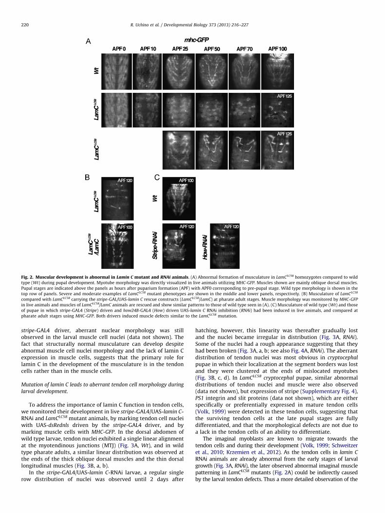
Fig. 2. Muscular development is abnormal in Lamin C mutant and RNAi animals. (A) Abnormal formation of musculature in LamCLC58 homozygotes compared to wild
type (Wt) during pupal development. Myotube morphology was directly visualized in live animals utilizing MHC-GFP. Muscles shown are mainly oblique dorsal muscles.
Pupal stages are indicated above the panels as hours after puparium formation (APF) with APF0 corresponding to pre-pupal stage. Wild type morphology is shown in the
top row of panels. Severe and moderate examples of LamCLC58 mutant phenotypes are shown in the middle and lower panels, respectively. (B) Musculature of LamCLC58
compared with LamCLC58 carrying the stripe-GAL/UAS-lamin C rescue constructs (LamCLC58/LamC) at pharate adult stages. Muscle morphology was monitored by MHC-GFP
in live animals and muscles of LamCLC58/LamC animals are rescued and show similar patterns to those of wild type seen in (A). (C) Musculature of wild type (Wt) and those
of pupae in which stripe-GAL4 (Stripe) driven and how24B-GAL4 (How) driven UAS-lamin C RNAi inhibition (RNAi) had been induced in live animals, and compared at
pharate adult stages using MHC-GFP. Both drivers induced muscle defects similar to the LamCLC58 mutation.
R. Uchino et al. / Developmental Biology 373 (2013) 216–227220
stripe-GAL4 driver, aberrant nuclear morphology was stillobserved in the larval muscle cell nuclei (data not shown). Thefact that structurally normal musculature can develop despiteabnormal muscle cell nuclei morphology and the lack of lamin Cexpression in muscle cells, suggests that the primary role forlamin C in the development of the musculature is in the tendoncells rather than in the muscle cells.
Mutation of lamin C leads to aberrant tendon cell morphology during
larval development.
To address the importance of lamin C function in tendon cells,we monitored their development in live stripe-GAL4/UAS-lamin C-RNAi and LamCLC58 mutant animals, by marking tendon cell nucleiwith UAS-dsRednls driven by the stripe-GAL4 driver, and bymarking muscle cells with MHC-GFP. In the dorsal abdomen ofwild type larvae, tendon nuclei exhibited a single linear alignmentat the myotendinous junctions (MTJ) (Fig. 3A, Wt), and in wildtype pharate adults, a similar linear distribution was observed atthe ends of the thick oblique dorsal muscles and the thin dorsallongitudinal muscles (Fig. 3B, a, b).
In the stripe-GAL4/UAS-lamin C-RNAi larvae, a regular singlerow distribution of nuclei was observed until 2 days after
hatching, however, this linearity was thereafter gradually lostand the nuclei became irregular in distribution (Fig. 3A, RNAi).Some of the nuclei had a rough appearance suggesting that theyhad been broken (Fig. 3A, a, b; see also Fig. 4A, RNAi). The aberrantdistribution of tendon nuclei was most obvious in cryptocephal
pupae in which their localization at the segment borders was lostand they were clustered at the ends of mislocated myotubes(Fig. 3B, c, d). In LamCLC58 cryptocephal pupae, similar abnormaldistributions of tendon nuclei and muscle were also observed(data not shown), but expression of stripe (Supplementary Fig. 4),PS1 integrin and slit proteins (data not shown), which are eitherspecifically or preferentially expressed in mature tendon cells(Volk, 1999) were detected in these tendon cells, suggesting thatthe surviving tendon cells at the late pupal stages are fullydifferentiated, and that the morphological defects are not due toa lack in the tendon cells of an ability to differentiate.
The imaginal myoblasts are known to migrate towards thetendon cells and during their development (Volk, 1999; Schweitzeret al., 2010; Krzemien et al., 2012). As the tendon cells in lamin C
RNAi animals are already abnormal from the early stages of larvalgrowth (Fig. 3A, RNAi), the later observed abnormal imaginal musclepatterning in LamCLC58 mutants (Fig. 2A) could be indirectly causedby the larval tendon defects. Thus a more detailed observation of the

Fig. 3. Tendon cell development is affected in lamin C RNAi animals. The effect of stripe-GAL4-driven UAS-lamin C RNAi (RNAi) on tendon cell development in live
animals during larval development shown by fluorescence microscopy (A) and at the pharate adult stages by confocal microscopy (B). Tendon cell nuclei (red) were labeled
with stripe-GAL4/UAS-dsRednls and muscles (green) were labeled with MHC-GFP. (A) The regular distribution of tendon cells observed in wild type (Wt) larvae is no longer
observed lamin C RNAi-induced larvae (RNAi) after 2 days. Note that tendon nuclei increase in size by endoreplication during larval development. Boxed sectors of the 5
days-RNAi sample are shown below at higher magnification (a, b). Arrows indicate nuclei that have been damaged and which were never observed in wild type. (B) Control
pharate adults (Wt) and lamin C RNAi-induced cryptocephal pupae (RNAi) showing oblique dorsal muscles (a, c), and dorsal longitudinal muscles (b, d). In RNAi-induced
pupae, the oblique dorsal muscles show rounded morphology and tendon cells are distributed at their edges (arrows in c) and not at the segment borders as in wild type
controls (a, b). A mislocated dorsal longitudinal muscle parallel to the segment border is indicated (left side of white dot in c), as well as a muscle in normal orientation
(arrowhead in c). Both the tendon and muscle cells of the dorsal longitudinal musculature are gone from their normal locations with a few exceptions (arrowhead in d).
Scale bars: 100 mm in panels (A); 50 mm in (B).
R. Uchino et al. / Developmental Biology 373 (2013) 216–227 221
MTJ of lateral traverse muscles was performed in live larvae. Whenthe tendon cell nuclei at the MTJ of the rod-like traverse muscles ofwild type 3rd instar larvae were studied, they were observed to beround in shape and uniform in size (Fig. 4a). However, in stripe-
GAL4/UAS-lamin C RNAi animals at the same stage, the nuclei of thetendon cells that associated with aberrant highly-stretched muscleswere changed in shape, size and position (Fig. 4b) as in the dorsaltendon nuclei of Lamin C RNAi (Fig. 3A, a, b), suggesting the nuclei are

Table 2Phenotypes induced by lamin C RNAi using various GAL4 drivers.
GAL4 driver
(number of animals)
Tissue specificity of GAL4 driver Early pupal
lethal (%)
Head eversion
defect (%)
Head everted but
pupal lethal (%)
Emerging as
adults (%)
� (104) – 1 0 0 99
1151 (100) Adepithelial cells and myoblasts 1 0 3 96
NP1029 (112) Embryonic muscle, and larval cardia 0 0 1 98
NP3239 (102) Adult abdomen and thoracic muscle 3 0 2 95
NP6657 (100) Adult epithelium and thoracic and
abdominal muscles
0 0 3 97
Tin (100) Cardia, visceral and somatic muscles 4 0 3 93
MHC (115) Muscles 3 0 2 96
Stripe (140) Tendon cells 17 73 6 4
How24B (137) mesoderm, muscle, cardia, and
tendon cells
12 88 0 0
Head eversion defects were observed when UAS-lamin C RNAi was combined with how24B-GAL4 and stripe-GAL4 drivers ,but not with the other muscle-cell expressing
GAL4 drivers tested.
R. Uchino et al. / Developmental Biology 373 (2013) 216–227222
distorted by muscle tension. When the cytoplasm of tendon cells waslabeled with an RFP reporter to monitor cell morphology in livelarvae, the characteristic crescent-shaped morphology of wild typetendon cells (Fig. 4d) was not seen in mutant tendon cells (Fig. 4e–g).At the ends of contracting muscles, tendon cells were detached fromcuticle (Fig. 4f), and extremely elongated tendon cells were alsodetected (Fig. 4g). As highly contracted muscles (Fig. 4c) andmislocated tendon cells with deformed nuclei (Figs. 3A, a, b and 5D),were observed, those tendon cells are assumed to have detachedfrom cuticle or muscle attachment sites, or to possibly have beenruptured by muscle tension. Thus, the destruction of tendon cells andchanges in their distribution suggest that lamin C loss in tendon cellsmake them more vulnerable to increases in tension from thecontractile forces of the muscles exerted during larval movements.
As formation of the imaginal muscle pattern depends on boththe larval muscle pattern and the positions of the tendon cells atthe epidermis (Bate, 1993; Volk, 1999; Schweitzer et al., 2010;Krzemien et al., 2012), the defects in larval tendon cells in lamin C
mutants cause larval muscle patterning abnormalities whichwould then lead to abnormal pupal muscle patterns. Whereasthe loss of tendon cells cause deletion of muscles, the survivingtendon cells in ectopic positions seem to induce mislocation ofmyotubes during pupal development (Figs. 2A and 3B, c).
Tendon cells lacking lamin C proteins exhibit changes in the
organization of cytoskeleton
The cytoskeleton and the extracellular matrix (ECM) play majorroles in maintaining tendon cell integrity against muscular forces(Roper et al., 2002; Grevengoed and Peifer, 2003; Delon and Brown,2007). Thus their organization was visualized at the MTJ withtendon cell-specific stripe-GAL4-driven UAS-GFP-tagged genes suchas actin, tubulin, or shortstop (shot) for the cytoskeleton and anintegrin-linked kinase (ILK) fly trap line (Morin et al., 2001) for theECM and was observed in combination with the cytoplasmic RFP
reporter in live larvae.A short, compact filament array composed of F-actin, micro-
tubules, and Shot which has been referred to as a ‘‘cytoskeletalbelt’’ (Subramanian et al., 2003; Alves-Silva et al., 2008) isobserved in tendon cells and is visible as a thin layer when themuscles are viewed laterally at low magnification (Wt in Fig. 4i–k).The region of the tendon cell containing the cytoskeletal belt isextremely shortened baso-apically, and the cytoskeletal belt isinterpreted to securely bridge the MTJ at the basal side to thecuticle at the apical side. It was this belt-like structure ofcytoskeleton proteins, that was dramatically changed in lamin C
RNAi larvae (RNAi in Fig. 4i–k). ILK which is localized in the musclecells at the interface with the ECM of the MTJ in wild type (Fig. 4h,
Wt) (Delon and Brown, 2007) was not affected (Fig. 4h, RNAi). Themicrotubules and F-actin which are normally concentrated in thecytoskeletal belt (Wt in Fig. 4i, j) initially appeared to be diffusedthroughout the cytoplasm (RNAi in Fig. 4i, j), whereas the dis-tribution of Shot which is normally localized at the cytoplasmicbelt (Wt in Fig. 4k) was ‘‘split’’, showing cytoskeletal belts at boththe basal MTJ side and apical cuticle-attached side of the tendoncells in lamin C RNAi larvae (RNAi in Fig. 4k, n). Some of the Shotprotein also showed a diffused cytoplasmic distribution. However,the diffused-appearing distributions of F-actin, microtubules andShot were found to be composed of apical–basal oriented fila-ments when observed at higher magnification (Fig. 4D). Thesefilamentous structures were no longer seen in elongated tendoncells (Fig. 4g; also see Fig. 5D). The obtained results indicate thatcytoskeleton organization in tendon cells, is affected by the loss oflamin C, whereas an ECM associated protein in the muscle cells isnot affected.
Disintegration of shortstop structure precedes nuclear lamina
deformation
Shot is a spectraplakin family protein and directly connectswith actin filaments, microtubules, and also cytoplasmic inter-mediate filaments (IF) to provide cells with structural strength(Roper et al., 2002; Grevengoed and Peifer, 2003; Sonnenberg andLiem, 2007). Reduction of Shot proteins in tendon cells induced byshot RNAi has been reported to result in abnormal elongated cellmorphology with stretched filament arrays of F-actin and micro-tubules and a split distribution of Shot proteins to both the apicaland basal ends (Subramanian et al., 2003; Alves-Silva et al., 2008).This interesting abnormal distribution of cytoskeletal structures issimilar to our observation in the LamC RNAi tendon cells (Fig. 4k, n).The fluorescence intensity of GFP-shot fusion proteins expressedwith stripe-GAL4 driver did not differ between wild type, LamC RNAiand LamCLC58 tendon cells. In addition, forced expression of GFP-shotproteins by the combination of UAS-GFP-shot and stripe-GAL4 driverused in this study have already been reported to rescue the tendoncell defects in shot mutants (Bottenberg et al., 2009), indicating thatstripe-GAL4 induced GFP-shot proteins are expressed in a form andat a level that can effectively function in tendon cells. However, theydid not rescue the tendon cell phenotypes of LamC RNAi (Fig. 4k, n)and the LamCLC58 mutant (Fig. 5C and D). These results suggest thatthe effect on Shot protein distribution that was observed in tendoncells by reducing lamin C protein was not caused by reducing Shotexpression levels. Therefore, distribution of Shot and lamin proteinsin tendon cells was further analyzed in LamCLC58 animals. Lamin C isnot present maternally (Riemer et al., 1995), it was confirmed by

Fig. 4. Morphology of the myotendinous junction is affected in lamin C RNAi larvae. Tendon cells of stripe-GAL4 driven lamin C RNAi (RNAi) animals were compared
with wild type (Wt) using various fluorescent cell markers. Muscle morphology was simultaneously visualized using MHC-GFP (green). (A) Morphology and positioning of
tendon cell nuclei associated with the lateral traverse muscles of larvae were analyzed with stripe-GAL4/UAS-dsRednls (red) in control Wt (a) and RNAi-induced (b, c)
animals. Altered morphology and positioning of muscle cells and tendon nuclei are easily recognized in lamin C RNAi larvae (b). The ends of stretched and detached
muscles are marked by arrowheads. A highly contracted muscle is indicated by an asterisk (c). (B) Tendon cell morphology was analyzed with the stripe-GAL4/UAS-RFP
(red) in control Wt (d) and RNAi-induced (e–g) animals. Morphology of a slightly deformed tendon cells is shown in panel RNAi (e). A tendon cell detaching from cuticle
(f) and an extremely stretched tendon cell (g) are also shown. (C and D) Morphologies of cytoskeletal and extracellular matrix structures of tendon cells in larvae were
analyzed. Integrin-linked kinase (h, GFP-ILK, green), tubulin (i, GFP-Tub, green), actin (j, GFP-Act, green), and shot (k, GFP-Shot, green) proteins were induced in tendon
cells using the stripe-GAL4 driver. Overall tendon cell morphology was visualized using stripe-GAL4/UAS-RFP (red) (C), and high-resolution images of cytoskeletal
structures are presented below (D). Cytoskeletal belts are marked by small arrows in (C). The split localization of Shot is indicated by large arrows (k, n). Analyses were
carried out with live animals by fluorescence microscopy in (A)–(C) and by confocal microscopy in (D). The scale bars each represent 10 mm.
R. Uchino et al. / Developmental Biology 373 (2013) 216–227 223
germ line mosaic analysis (Table 1), thus the LamCLC58 animals thatwe analyzed completely lacked Lamin C during development.
In wild type animals, Shot was detected as a layer surroundingthe nuclear envelope in addition to a cytoplasmic filament mesh-work beside the MTJ similar to previous observations (Fig. 5A and B)(Subramanian et al., 2003). At higher magnification, a dense andcontinuous layer of Shot protein was easily seen lining the nuclearenvelope (Fig. 5f2, i2). In the tendon cells of young LamCLC58 mutantlarvae, these shot structures started to be broken (Fig. 5k) and thecontinuous layer at the nuclear periphery became fragmented(Fig. 5n), although there was a continuous layer of lamin Dm0 atthe nuclear envelope (Fig. 5j, m). In the tendon cells with severelydeformed nuclei showing highly abnormal lamin Dm0 distribution(Fig. 5p), Shot did not form an obvious meshwork and was muchmore diffused (Fig. 5q). As the breakdown of Shot structures in
tendon cells lacking lamin C preceded or was in parallel to the lossof integrity in the lamin Dm0 lamina layer, these observations implythe lamin C has a critical role in maintaining Shot dependentcytoskeletal structures.
The requirement for lamin C is in cells that express shortstop
In the musculature, Shot is required for muscle cell-dependenttendon cell differentiation, and is expressed in tendon cells but notin muscle cells (Strumpf and Volk, 1998; Volk, 1999). To testwhether the requirement for lamin C is in the same cell-typesof both lamin C mutant and wild type animals, UAS-lamin Cexpression was driven with the shot-GAL4 driver, which is anenhancer trap line. We first confirmed protein expression andspecificity of shot-GAL driver with UAS-GFP-lamin C or UAS-lamin

Fig. 5. Shot distribution is abnormal in tendon cells of LamC mutant larvae. The morphology of cytoskeletal structures composed of Shot in wild type (A, B) and
LamCLC58 mutant (C, D) tendon cells were studied by confocal microscopy after fixation. Both lower magnification images of the entire nucleus (1) and higher magnification
images of the nuclear periphery (2) are shown. Endogenous Shot was detected with indirect immunofluorescence using highly specific Shot antibodies (Shot; b) or directly
as GFP-tagged Shot (GFP-Shot; e, h, k, n and q) driven by stripe-GAL4 driver. Nuclear envelope morphology was monitored with stripe-GAL4 driven GFP-tagged lamin C
(GFP-LamC; a) or monoclonal lamin Dm0 antibody labeling (LamDm; d, g, j, m, and p). A cytoplasmic meshwork and a layer of Shot at the nuclear periphery are seen in wild
type animals (b, e, and h). These structures appear to start to breakdown in tendon cells of young LamCLC58 larvae (k and n). At higher magnification, broken Shot structures
are easily detected at the nuclear periphery (k2 and n2). The perinuclear Shot layer is fragmented (n2), although an altered but continuous lamin Dm0 layer is observed in
(m2). Organized Shot structures are completely lost in the tendon cells (q), which have heavily distorted nuclei (p). Scale bars: 10 mm, all panels.
R. Uchino et al. / Developmental Biology 373 (2013) 216–227224
C using antibodies and a GFP reporter. The fluorescence intensityof LamCLC58 mutants showed no difference in comparison to thoseof wild types (Fig. 6A and B), and similar results were alsoobtained with other UAS-fluorescence reporters (data not shown).Shot-GAL4 driver induced lamin C signals were detected in tendoncells but never observed in larval and pupal abdominal musclenuclei (Fig. 6A and B), nor in the heart and thoracic muscle cells(data not shown). The mutant larval muscle nuclei showedobviously abnormal nuclear morphology (Fig. 6A; SupplementaryFig. 3A) that was similar to the phenotype in LamCLC58 mutants.
Although the shot-GAL4 is functional in the tendon cells but notin the muscle cells, the gross developmental defects of LamCLC58
mutants were rescued by shot-GAL4/UAS-lamin C, with 90% oflarvae forming pupal cases, 83% having everted head structures,and 60% emerging as adult flies (Table 3). In the enclosed adults,although 23% of these animals exhibited severe defects in walkingand flying following enclosure, others were superficially indis-tinguishable from wild type animals and could produce offspring,demonstrating that the expression of lamin C in the same cells asShot is sufficient to rescue development to imagoes.
The null allele of shot is embryonic lethal and it is essential forepidermal integrity, nerve growth, and tracheal formation inaddition to the formation of normal musculature (Roper et al.,2002). Thus lamin C could be required not only in tendon cells butalso in other Shot-expressing cells.
Discussions
Drosophila lamin C alleles have been described to showrecessive pre-pupal lethality accompanied by aberrant nuclearmorphology in imaginal discs and muscle cells (Schulze et al.,2005, 2009). However, the direct cause of the lethality resultingfrom the loss of lamin C function had not been determined.We here report the phenotypes of newly isolated and previously
reported lamin C alleles. Under our modified culture conditions(see Section 2), they were found to develop to pharate adultstages and showed head eversion defects. Although head eversionis known to be dependent on peristaltic contraction of theabdominal muscles (Fristrom, 1965) and the abdominal musclepatterns in lamin C mutants were obviously abnormal, this defectwas not rescued by lamin C transgene expression in muscle cells.However it was rescued by expression in tendon cells, indicatingthat lamin C is critical in the tendons rather than the muscles.
In contrast to our results, a direct effect of lamin C on musclecells has been previously speculated, based on the observationthat the expression of N-terminal truncated lamin C in musclecells induced lethality accompanied by altered nuclear structuresincluding lamin C aggregation (Schulze et al., 2009; Dialynaset al., 2010). Similar head domain-less A-type lamins have beenreported to aggregate in nucleoplasm in vertebrates. Theseaberrant lamin structures have been shown to disrupt the nuclearrim-distribution of endogenous lamins including B-type lamins,by causing them to co-aggregate, and result in the inhibition ofreplication and transcription (Spann et al., 1997; Moir et al., 2000;Spann et al., 2002). As N-terminal truncated A-type laminsinterfere with lamin-nuclear function through an artificial domi-nant effect, the defects caused by their expression in Drosophilamuscle cells could have been caused by their disruption of thenormal localization of lamin Dm0 (B-type lamin). As endogenouslamin Dm0 retains localization at the nuclear rim in the lamin C
null mutants (see in Fig. 5C), the recessive phenotypes caused bylamin C loss that we describe here are probably different from,but not in conflict with the dominant negative phenotypesreported to be induced by head domain-less lamin C expression.
The shot gene is essential in tendon cell differentiation. Itsexpression is detected in the tendon cells but not in the musclecells from around stages 11 onward of embryonic development.Mutations of shot are reported to affect the EGF-receptor signalingpathway by inducing mislocalization of Vein activator at the MTJ

Fig. 6. Characterization of the tissue specificity of the shot-GAL4 driver. Expression of GFP-tagged lamin C or lamin C was induced with the shot-GAL4 driver in LamCLC58
and wild type (Wt) animals. (A) Distribution of GFP-LamC (green) in larval musculature was compared with lamin Dm0 (LamDm, red). Lamin Dm0 was detected with
specific antibody, and GFP was directly visualized. GFP-lamin C expression driven by shot-GAL4 was only found some nuclei (presumably tendon cells), which were not in
lines of nuclei (presumably myotubes) in LamCLC58 and Wt larval musculature. In LamCLC58 panels, nuclear morphology of the aligned nuclei was severely fragmented and
lobulated as similar to LamCLC58 muscle nuclei in Supplementary Fig. 3A. Distribution of LamC (green) in adult dorsal muscles was also compared with distribution of
F-actin detected with phalloidin dye (red in B) and Mef nuclear proteins detected with specific antibodies (red in C). In wild type expressing UAS-lamin C under the control
of shot-GAL4 driver, the combined signals of endogenous and exogenous lamin C were detected in nuclei of the muscle cells and cells of the muscle-attachment sites
(tendon cells). In LamCLC58 carrying the same transgenes, exogenous lamin C is only detected in nuclei of muscle-attachment sites (lower row in B). To more precisely
determine the lack of expression of exogenous lamin C in muscle nuclei, the nuclei were marked by the muscle-specific nuclear protein, Mef (red) and compared with
lamin C signals in (C). The scale bars in (A and B) and (C) each represent 10 and 5 mm, respectively.
Table 3Phenotypes of lamin CLC58 mutants rescued by shot-GAL4/UAS-lamin C.
Shot-GAL4
(number of
animals)
Larval
lethal
(%)
Early
pupal
lethal (%)
Head
eversion
defect (%)
Head everted
but pupal lethal
(%)
Emerging
as adults
(%)
� (180) 5 12 82 0 0
þ (145) 10 4 3 23 60
The shot-GAL driver which is expressed in the epidermis, nervous system, and
tendon cells was used to induce UAS-lamin C in LamCLC58 mutants. The character-
istic head eversion defect was rescued, and pupal lethality was greatly decreased.
R. Uchino et al. / Developmental Biology 373 (2013) 216–227 225
and result in the disruption of somatic muscle pattern at stage 16of embryonic development (Strumpf and Volk, 1998; Volk, 1999).Shot belongs to the spectraplakin family of cytoskeletal linkerproteins and has also been shown to contribute to the mechanicalstrength of tendon cells (Subramanian et al., 2003; Alves-Silvaet al., 2008). Thus Shot has both structural and informationalfunctions (Roper et al., 2002).
In our analyses, expression of a lamin C transgene with a shot-promoter driven GAL4 transgene was restricted to tendon cells inmusculature, and its expression levels were similar betweenLamCL58 and wild type animals. Further, shot-GAL4 driver inducedlamin C proteins were capable of effectively rescuing lamin C null
mutants to adults. Although loss of lamin C induced disintegra-tion of cytoskeletal and nuclear lamina structures in the tendoncells, they were relatively normal at the early larval stages incontrast to the muscular abnormality which occurs in shot allelesat embryonic stages. In lamin C null mutants, disintegrationof the cytoskeleton which includes Shot was initiated beforenuclear lamina deformation and progressed gradually duringlarval growth. This alteration eventually caused destruction oftendon cells and muscle detachment from epidermis in a mannersimilar to shot mutant phenotypes (Strumpf and Volk, 1998;Subramanian et al., 2003; Alves-Silva et al., 2008). A-type laminis known to be major contributor to the mechanical properties ofnuclei as well as the cytoskeleton and cytoskeleton-basedprocesses, whereas B-type lamin contributes to nuclear integrity(Houben et al., 2007; Schape et al., 2009; Lammerding et al., 2006;Lee et al., 2007). Thus, our findings with lamin C alleles arecoincident with these interpretations and strongly suggest thatlamin C null mutation directly induces mechanical instability ofthe cytoskeleton and nuclear structures, which probably leads toweakening of tendon cell strength and function.
Interactions between the cytoskeleton and the nuclear laminain which A-type lamins are localized are generally mediatedthrough the double layer lipid nuclear membrane by LINCcomplexes formed by Klarsicht/ANC-1/Syne homology (KASH)domain proteins directly linked to Sad1/UNC-84 (SUN) domain

R. Uchino et al. / Developmental Biology 373 (2013) 216–227226
proteins in the nuclear envelope. In mammals, KASH domainproteins are divided into the three major groups by associationwith actin filaments, microtubules, and intermediate filaments(IF) (Roux and Burke, 2007; Razafsky and Hodzic, 2009; Roperet al., 2002; Wilson and Foisner, 2010).
In Drosophila, the nesprin-ortholog MSP-300 associated withactin filaments and Klarsicht associated with microtubules are theknown KASH domain proteins (Mosley-Bishop et al., 1999;Starr and Han, 2002), and Klaroid and testis specific Spag4 arethe known SUN domain proteins (Kracklauer et al., 2007, 2010).However, KASH domain deleted MSP-300 alleles and klarsicht,spag4 and klaroid null alleles have been reported not to result inlethality (Mosley-Bishop et al., 1999; Kracklauer et al., 2007,2010; Xie and Fischer, 2008; Technau and Roth, 2008). Further,when the distribution of MSP-300 was studied in wild type andLamCLC58 muscles, it was detected in the sarcomere of both aspreviously described (Volk, 1992). In tendon cells its distributioncould not be evaluated because of its low expression (data notshown). When the distributions of Klarsicht and Klaroid werestudied, they were detected in the region of the nuclear envelope(NE) in wild type, and their localizations were similar to thedistribution of lamin Dm0 at the NE in the tendon cells ofLamCLC58 mutant larvae (data not shown; see lamin Dm0 distribu-tion in Fig. 5). Thus there was no indication that these knownLINC complex proteins play a central role in the lamin C-depletedphenotypes of tendon cells.
In humans, an IF-associating KASH domain protein; Nesprin3a has been also reported in addition to actin and microtubuleassociating KASH domain proteins, and its IF-binding is mediatedby plectin protein containing a plakin (or plectin) repeat domainwhich can directly bind IF proteins (Roux and Burke, 2007;Sonnenberg and Liem, 2007; Razafsky and Hodzic, 2009; Roperet al., 2002; Wilson and Foisner, 2010). Although, interactionbetween Lamin C and Spectraplakin-related Shot which also has aplakin repeat domain has not been proven and an IF-associatingtype of KASH domain protein has not yet been reported inDrosophila, the mammalian spectraplakin; Bpag1/dystoninisoform has recently been reported to also interact with Nesprin3a (Young and Kothary, 2008). Furthermore, for interaction ofShot with the nucleus, the actin–plakin and plakin domains ofShot have been shown to be targeted to the nucleus and topredominantly concentrate at the nuclear periphery in tendoncells (Subramanian et al., 2003). Thus, it is possible that lamin Cconnects to Shot mediated cytoskeletal complexes through anovel LINC complex protein with characteristics similar toNesprin 3a and protects tendon cell and nuclei from forcesproduced by the contractions of the muscles.
In human EDMD, mutations of A-type lamins are characterizedby early onset contractures of the Achilles tendons and tendons ofthe elbows and neck, whereas muscle weakness and wastingshows slow progression compared to other types of musculardystrophy (Emery, 2000; Rankin and Ellard, 2006; Roux andBurke, 2007; Worman et al., 2010). Although we are not awareof any detailed histological studies of the tendon cells in theprogression of EDMD, in LMNA null model mice, the interdigita-tion between muscle and tendon is reduced, and the musclenuclei show abnormal morphology and tend to cluster at the MTJ(Sullivan et al., 1999; Mittelbronn et al., 2008; Gnocchi et al.,2011). Thus tendon defects are strongly implicated in musculardystrophy resulting from the loss of A-type lamin in mammals,which could be analogous to what we report here in Drosophila.
In vertebrates, tendons are morphologically diverse tissuesand their strength depend mainly on extracellular collagen fibrils(Kannus, 2000; Banos et al., 2008), however, well developed actinand myosin bundles are also observed in cytoplasm and aroundthe nucleus in rabbit tendon cells (Ippolito et al., 1977;
Postacchini et al., 1978). In Drosophila, the spectraplakin familyprotein Shot has a major role in formation and maintenance ofcytoskeletal organization in tendon cells (Subramanian et al.,2003; Alves-Silva et al., 2008), but the mammalian spectraplakins,Bpag1/dystonin and MACF1, which are Shot orthologs, have notyet been analyzed in tendon cells. However, Bpag1 isoforms areknown to be expressed in musculature (Sonnenberg and Liem,2007; Boyer et al., 2010), and show nuclear envelope targeting,nesprin 3a interaction, and functions in organizing the actincytoskeleton around the nucleus in myogenic and other culturecells (Young et al., 2003; Young and Kothary, 2006, 2008). Asmature mammalian tendon cells are relatively large cells, withlengths up to 300 mm, and their volume is almost entirely takenup by a large nucleus (Ippolito et al., 1980), these cytoskeletalproteins might similarly protect the mammalian tendon cell andnucleus against mechanical stress from muscle contraction. Thefact that the nuclear defects observed in LMNA model mice wereconcentrated at the MTJ structures (Sullivan et al., 1999;Mittelbronn et al., 2008; Gnocchi et al., 2011) suggest that thedefects in the mouse and fly musculatures by loss of A-typelamins may be initially caused by similar mechanisms at sites ofmechanical stress; namely tendon cells.
We hypothesize that loss of A-type lamins cause a reductionin spectraplakin mediated cytoskeleton-integrity and cytoskeleton-based processes that perturbs tendon cell morphology and functionleading to symptoms seen in human autosomal recessive EDMD.Further studies on the involvement of tendon cell-cytoskeletalstructures in the progression of EDMD in both vertebrates andDrosophila are therefore warranted.
Acknowledgments
We wish to thank Dr. G. Morata, Dr. L. Wallrath, Drs. E.N. Olsonand E. Chen, Dr. A. DiAntonioa, Dr. M. Frasch, Dr. T.J. Fuwa forproviding fly lines, T. Volk for providing the Stripe, Shot, andMSP-300 antibodies, Drs. M. Kracklauer and M. Welte for provid-ing the Klaroid and Klarsicht antibodies, Dr. B.M. Paterson forproviding the Mef antibodies, Dr. P.A. Fisher for providing laminantibodies, and Dr. Y. Hayashi for discussions concerning theclinical aspects of human EDMD.
Appendix A. Supplementary materials
Supplementary data associated with this article can be foundin the online version at http://dx.doi.org/10.1016/j.ydbio.2012.08.001.
References
Alves-Silva, J., Hahn, I., Huber, O., Mende, M., Reissaus, A., Prokop, A., 2008.Prominent actin fiber arrays in Drosophila tendon cells represent architecturalelements different from stress fibers. Mol. Biol. Cell. 19, 4287–4297.
Banos, C.C., Thomas, A.H., Kuo, C.K., 2008. Collagen fibrillogenesis in tendondevelopment: current models and regulation of fibril assembly. Birth DefectsRes. C Embryo Today 84, 228–244.
Bate, M., 1993. The mesoderm and it derivatives. In: Bate, M., Arias, A.M. (Eds.),The Development of Drosophila melanogaster, vol. II. CSHL Press, New York,pp. 1013–1090.
Bottenberg, W., Sanchez-Soriano, N., Alves-Silva., J., Hahn, I., Mende, M., Prokop, A.,2009. Context-specific requirements of functional domains of the SpectraplakinShort stop in vivo. Mech. Dev. 126, 489–502.
Boyer, J.G., Bernstein, M.A., Boudreau-Larivi �ere, C., 2010. Plakins in striatedmuscle. Muscle Nerve 41, 299–308.
Dechat, T., Adam, S.A., Taimen, P., Shimi, T., Goldman, R.D., 2010. Nuclear lamins.Cold Spring Harbor Perspect. Biol. 2, a000547.
Delon, I., Brown, N.H., 2007. Integrins and the actin cytoskeleton. Curr. Opin.Cell Biol. 19, 43–50.

R. Uchino et al. / Developmental Biology 373 (2013) 216–227 227
Dialynas, G., Speese, S., Budnik, V., Geyer, P.K., Wallrath, L.L., 2010. The role ofDrosophila Lamin C in muscle function and gene expression. Development137, 3067–3077.
Emery, A.E.H., 2000. Emery–Dreifuss muscular dystrophy—a 40 year retrospective.Neuromuscular Disord. 10, 228–232.
Fristrom, J.W., 1965. Development of the morphological mutant cryptocephal ofDrosophila melanogaster. Genetics 52, 297–318.
Frommer, G., Vorbruggen, G., Pasca, G., Jackle, H., Volk, T., 1996. Epidermal egr-likezinc finger protein of Drosophila participates in myotube guidance. EMBO J.15, 1642–1649.
Gurudatta, B.V., Shashidhara, L.S., Parnaik, V.K., 2010. Lamin C and chromatinorganization in Drosophila. J. Genet. 89, 37–49.
Gnocchi, V.F., Scharner, J., Huang, Z., Brady, K., Lee, J.S., White, R.B., Morgan, J.E.,Sun, Y.B., Ellis, J.A., Zammit, P.S., 2011. Uncoordinated transcription andcompromised muscle function in the lmna-null mouse model of Emery–Dreifuss muscular dystrophy. PLoS One 6, e16651.
Grevengoed, E.E., Peifer, M., 2003. Cytoskeletal connections: building strong cellsin new ways. Curr. Biol. 13, R568–R570.
Gruenbaum, Y., Goldman, R.D., Meyuhas, R., Mills, E., Margalit, A., Fridkin, A.,Dayani, Y., Prokocimer, M., Enosh, A., 2003. The nuclear lamina and itsfunctions in the nucleus. Int. Rev. Cytol. 226, 1–62.
Houben, F., Ramaekers, F.C., Snoeckx, L.H., Broers, J.L., 2007. Role of nuclearlamina–cytoskeleton interactions in the maintenance of cellular strength.Biochim. Biophys. Acta 1773, 675–686.
Ippolito, E., Natali, P.G., Postacchini, F., Accinni, L., DeMartino, C., 1977. Ultra-structural and immunochemical evidence of actin in the tendon cells. Clin.Orthop. Relat. Res. 126, 282–284.
Ippolito, E., Natali, P.G., Postacchini, F., Accinni, L., DeMartino, C., 1980. Morpho-logical, immunochemical, and biochemical study of rabbit achilles tendon atvarious ages. J. Bone Jt. Surg. Am. 62, 583–598.
Kannus, P., 2000. Structure of the tendon connective tissue. Scand. J. Med. Sci.Sports 10, 312–320.
Kracklauer, M.P., Banks, S.M., Xie, X., Wu, Y., Fischer, J.A., 2007. Drosophila klaroidencodes a SUN domain protein required for Klarsicht localization to thenuclear envelope and nuclear migration in the eye. Fly 1, 75–85.
Kracklauer, M.P., Wiora, H.M., Deery, W.J., Chen, X., Bolival Jr., B., Romanowicz, D.,Simonette, R.A., Fuller, M.T., Fischer, J.A., Beckingham, K.M., 2010. TheDrosophila SUN protein Spag4 cooperates with the coiled-coil protein YuriGagarin to maintain association of the basal body and spermatid nucleus. J.Cell Sci. 123, 2763–2772.
Krzemien, J., Fabre, C.C., Casal, J., Lawrence, P.A., 2012. The muscle pattern of theDrosophila abdomen depends on a subdivision of the anterior compartment ofeach segment. Development 139, 75–83.
Lammerding, J., Fong, L.G., Ji, J.Y., Reue, K., Stewart, C.L., Young, S.G., Lee, R.T., 2006.Lamins A and C but not lamin B1 regulate nuclear mechanics. J. Biol. Chem.281, 25768–25780.
Lee, J.S., Hale, C.M., Panorchan, P., Khatau, S.B., George, J.P., Tseng, Y., Stewart, C.L.,Hodzic, D., Wirtz, D., 2007. Nuclear lamin A/C deficiency induces defects in cellmechanics, polarization, and migration. Biophys. J. 93, 2542–2552.
Maqbool, T., Jagla, K., 2007. Genetic control of muscle development: learning fromDrosophila. J. Muscle Res. Cell Motil. 28, 397–407.
Melcer, S., Gruenbaum, Y., Krohne, G., 2007. Invertebrate lamins. Exp. Cell Res. 313,2157–2166.
Mittelbronn, M., Sullivan, T., Stewart, C.L., Bornemann, A., 2008. Myonucleardegeneration in LMNA null mice. Brain Pathol. 18, 338–343.
Moir, R.D., Spann, T.P., Herrmann, H., Goldman, R.D., 2000. Disruption of nuclearlamin organization blocks the elongation phase of DNA replication. J. Cell Biol.149, 1179–1192.
Morin, X., Daneman, R., Zavortink, M., Chia, W., 2001. A protein trap strategy todetect GFP-tagged proteins expressed from their endogenous loci inDrosophila. Proc. Natl. Acad. Sci. USA 98, 15050–15055.
Mosley-Bishop, K.L., Li, Q., Patterson, L., Fischer, J.A., 1999. Molecular analysis ofthe Klarsicht gene and its role in nuclear migration within differentiating cellsof the Drosophila eye. Curr. Biol. 9, 1211–1220.
Postacchini, F., Accinni, L., Natali, P.G., Ippolito, E., DeMartino, C., 1978. Regenerationof rabbit calcaneal tendon: a morphological and immunochemical study. CellTissue 195, 81–97.
Rankin, J., Ellard, S., 2006. The laminopathies: a clinical review. Clin. Genet. 70,261–274.
Razafsky, D., Hodzic, D.A., 2009. Bringing KASH under the SUN: the many faces ofnucleo-cytoskeletal connections. J. Cell Biol. 186, 461–472.
Riemer, D., Stuurman, N., Berrios, M., Hunter, C., Fisher, P.A., Weber, K., 1995.Expression of Drosophila lamin C is developmentally regulated: analogies withvertebrate A-type lamins. J. Cell Sci. 108, 3189–3198.
Roux, K.J., Burke, B., 2007. Nuclear envelope defects in muscular dystrophy.Biochim. Biophys. Acta 1772, 118–127.
Roper, K., Gregory, S.L., Brown, N.H., 2002. The ‘spectraplakins’: cytoskeletal giantswith characteristics of both spectrin and plakin families. J. Cell Sci. 115,4215–4225.
Schape, J., Prausse, S., Radmacher, M., Stick, R., 2009. Influence of lamin A on themechanical properties of amphibian oocyte nuclei measured by atomic forcemicroscopy. Biophys. J. 96, 4319–4325.
Schulze, S.R., Curio-Penny, B., Li, Y., Imani, R.A., Rydberg, L., Geyer, P.K., Wallrath, L.L.,2005. Molecular genetic analysis of the nested Drosophila melanogaster lamin Cgene. Genetics 71, 185–196.
Schulze, S.R., Curio-Penny, B., Speese, S., Dialynas, G., Cryderman, D.E.,McDonough, C.W., Nalbant, D., Petersen, M., Budnik, V., Geyer, P.K., Wallrath.,L.L., 2009. A comparative study of Drosophila and human A-type lamins. PLoSOne 26, e7564.
Schweitzer, R., Zelzer, E., Volk, T., 2010. Connecting muscles to tendons: tendonsand musculoskeletal development in flies and vertebrates. Development 137,334–337.
Sonnenberg, A., Liem, R.K., 2007. Plakins in development and disease. Exp. Cell Res.313, 2189–2203.
Stuurman, N., Delbecque, J.P., Callaerts, P., Aebi, U., 1999. Ectopic overexpression ofDrosophila lamin C is stage-specific lethal. Exp. Cell Res. 248, 350–357.
Spann, T.P., Moir, R.D., Goldman, A.E., Stick, R., Goldman, R.D., 1997. Disruption ofnuclear lamin organization alters the distribution of replication factors andinhibits DNA synthesis. J. Cell Biol. 136, 1201–1212.
Spann, T.P., Goldman, A.E., Wang, C., Huang, S., Goldman, R.D., 2002. Alterationof nuclear lamin organization inhibits RNA polymerase II-dependenttranscription. J. Cell Biol. 156, 603–608.
Starr, D.A., Han, H., 2002. Role of ANC-1 in tethering nuclei to the actincytoskeleton. Science 298, 406–409.
Strumpf, D., Volk, T., 1998. Kakapo, a novel cytoskeletal-associated protein isessential for the restricted localization of the neuregulin-like factor, vein, atthe muscle–tendon junction site. J. Cell Biol. 143, 1259–1270.
Subramanian, A., Prokop, A., Yamamoto, M., Sugimura, K., Uemura, T., Betschinger,J., Knoblich, J.A., Volk, T., 2003. Shortstop recruits EB1/APC1 and promotesmicrotubule assembly at the muscle–tendon junction. Curr. Biol. 13,1086–1095.
Sullivan, T., Escalante-Alcalde, D., Bhatt, H., Anver, M., Bhat, N., Nagashima, K.,Stewart, C.L., Burke, B., 1999. Loss of A-type lamin expression compromisesnuclear envelope integrity leading to muscular dystrophy. J. Cell Biol. 147,913–920.
Technau, M., Roth, S., 2008. The Drosophila KASH domain proteins Msp-300 andKlarsicht and the SUN domain protein Klaroid have no essential functionduring oogenesis. Fly 2, 82–91.
Volk, T., 1992. A new member of the spectrin superfamily may participate in theformation of embryonic muscle attachments in Drosophila. Development 116,721–730.
Volk, T., 1999. Singling out Drosophila tendon cells: a dialogue between twodistinct cell types. Trends Genet. 15, 448–453.
Wilson, K.L., Foisner, R., 2010. Lamin-binding proteins. Cold Spring HarborPerspect. Biol. 2, a000554.
Worman, H.J., Ostlund, C., Wang, Y., 2010. Diseases of the nuclear envelope.Cold Spring Harbor Perspect. Biol. 2, a000760.
Xie, X., Fischer, J.A., 2008. On the roles of the Drosophila KASH domain proteinsMsp-300 and Klarsicht. Fly 2, 74–81.
Young, K.G., Pool, M., Kothary, R., 2003. Bpag1 localization to actin filaments and tothe nucleus is regulated by its N-terminus. J. Cell Sci. 116, 4543–4555.
Young, K.G., Kothary, R., 2006. A Bpag1 isoform involved in cytoskeletal organiza-tion surrounding the nucleus. Exp. Cell Res. 312, 121–134.
Young, K.G., Kothary, R., 2008. Dystonin/Bpag1 is a necessary endoplasmicreticulum/nuclear envelope protein in sensory neurons. Exp. Cell Res. 314,2750–2761.
Zaffran, S., Astier, M., Gratecos, D., Semeriva, M., 1997. The held out wings (how)Drosophila gene encodes a putative RNA-binding protein involved in thecontrol of muscular and cardiac activity. Development 124, 2087–2098.