loss of dystrobrevin causes muscle degeneration and a
TRANSCRIPT

http://wjst.wu.ac.th Health Sciences
Walailak J Sci & Tech 2018; 15(9): 659-667.
Loss of Dystrobrevin Causes Muscle Degeneration and a Short Lifespan in Caenorhabditis elegans Prapaporn JATTUJAN, Krai MEEMON and Worawit SUPHAMUNGMEE* Department of Anatomy, Faculty of Science, Mahidol University, Ratchathevi, Bangkok 10400, Thailand (*Corresponding author’s e-mail: [email protected]) Received: 4 May 2017, Revised: 25 October 2017, Accepted: 4 November 2017 Abstract
Duchene’s muscular dystrophy (DMD) is an inherited disorder in an X-linked recessive manner. Lack of dystrophin causes progressive muscle degeneration. Dystrophin structurally connects to actin filaments at the N-terminus while the C-terminus interacts with an integral domain of the dystroglycan complex. Among the associated molecules, dystrobrevin acts as a modulator protein exerting dystrophin's function for strengthening the cell stability. Previous data has reported the delayed muscle degeneration following an overexpression of the dystrobrevin (dyb) gene in the DMD-phenotype C. elegans, whose dystrophin (dys) gene was missing. This finding indicates the role of a modulator protein, rather than dystrophin, to maintain the cell integrity. The present study aims to investigate the phenotypes of C. elegans, due to dyb- or dys-deficiency. When compared with the wild-type, the dyb-deficient worms exhibited uncoordinated locomotion and lysis of the muscular layer in the body wall and internal organs as well as those observed in the dys-deficient worms. The ultrastructure of both mutant worms appeared severe muscle degeneration, decrease of the mitochondria, and replacement of fibrotic tissue, particularly the dys-mutant which was even more severe. Additionally, a shorter lifespan was observed with a 17 % reduction (p < 0.05) in dyb-deficient worms and 27 % reduction (p < 0.05) in dys-deficient worms when compared to wild-type. It is speculated that dystrobrevin may stabilize the cell through interaction with other protein complexes at the plasma membrane while it also binds to dystrophin. Therefore, the loss of dystrobrevin is also sufficient to disrupt the signaling pathway and causes muscle degeneration.
Keywords: Muscular dystrophy, muscle degeneration, C. elegans, dystrobrevin, dystrophin Introduction
Muscular dystrophy is a group of inherited muscle diseases clinically determined as non-inflammatory muscular disorders. The disease diagnosis is uniquely defined as progressive degeneration of muscle fibers, particularly of the skeletal muscles without functional abnormality on the neuronal control or major alteration of the myofilament organization. Among several types of the disease, Duchene muscular dystrophy (DMD) is the major disorder underlying rapid progressive muscle weakness and atrophy [1,2]. DMD usually affects the male with a worldwide record of 1 in every 3500 newborn boys [3]. The affected boy has the X-linked recessive chromosome received from the mother and the father's Y chromosome. While in the female, a pair of the X-linked recessive chromosome and the normal chromosome received from parents is likely to make the disease mild. Instead, the affected female becomes a carrier and is able to pass over the gene to a child. In regard to DMD, muscle weakness often occurs from proximal to distal direction so that the major muscle groups of back, girdles and limbs are affected. Consequently, the weakness of such muscles results in movement disability, failure of respiratory functions, and eventually heart diseases.

Dystrobrevin and Muscular Dystrophy Prapaporn JATTUJAN et al. http://wjst.wu.ac.th
Walailak J Sci & Tech 2018; 15(9) 660
The key molecules involved in the mechanism of muscular dystrophy have been extensively studied, including dystrophin (DYS) and other protein complexes connecting the cytoskeleton to the plasma membrane and to the extracellular matrix [4,5]. A partial lack of the dystrophin gene causes Becker muscular dystrophy (BMD), which is characterized by an abnormal expression of DYS and its muscle-regulated proteins. Absence of the complete gene, in turn, causes DMD [6]. Although both DMD and BMD patients show similar characteristics of the disorders, the different severity of the diseases is rather dependent on the amount of loss of the dystrophin (dys) gene. The dys gene is very well-known as a major gene responsible for the DMD. It is considered as one of the largest identified genes in the human X-chromosome [7]. A full length of its protein with a molecular weight of 427 kDa is largely expressed in the skeletal muscle, the cardiac muscle and the smooth muscle of mammals. The structure of the DYS protein consists of the actin binding sites at the N-terminal domain, a central rod domain organized by 24 spectrin helical coils responsible for structural flexibility, and the cysteine-rich C-terminal domain [8]. At the C-terminus of DYS, there are binding sites for dystroglycan, a region that associates with dystrobrevin and syntrophin [9]. The dystrobrevin (DYB) protein is believed to regulate muscle function through the signaling pathway in a costamere, which are specialized domains located at the junction of the plasma membrane and the sarcomere of myofibrils [10]. DMD-like appearance of the dys-knockout mice has been evident previously [11]. In relation to recent findings, it is worth asking whether the interaction between DYB and other signaling molecules plays an important role in stabilizing the integrity of the muscle cell. Absence of DYB may disrupt the cascade interaction of the syncoilin which then breaks a connection between the protein complex at the plasma membrane and desmin intermediate filaments [12]. Whatever alters the interactions of these muscle-regulated proteins during the loss of DYB should be speculated. Several studies have been conducted in animal models such as in C. elegans, zebrafish and mice, in order to elucidate the molecular mechanism and roles of DYS and DYB for maintaining the cell integrity [11,13-15]. In human tissue, the disruption of the dys gene decreases the localization of α-dystrobrevin at the plasma membrane. Besides, the disruption of the sarcoglycan complex whose transmembrane regions are structurally connected to DYS exhibits a similar result [16]. The decreasing localization to DYB at the plasma membrane of mice, however, seemed to be affected only by the disruption of DYS, not loss of the sarcoglycan complex [17]. Hence, the function of DYB can be potentially regulated by DYS rather than dystroglycan or sarcoglycan complexes. Dystroglycan and sarcoglycan complexes are therefore important for preventing membrane damage, especially during the muscle cell contraction. A major loss of DYS causes muscle degeneration through impaired function between the cytoskeletal compartment and the rest of the dystrophin-binding molecules, such as the dystrophin-glycoprotein complex and its regulatory proteins connected via the extracellular matrix [18]. Severe degrees of muscular dystrophy have been observed in animal models when lacking both DYS and DYB. Meanwhile, a linkage between these 2 proteins seems probable as their regulating mechanism to maintain the normal function of muscle and non-muscle tissues [16,19]. It has not yet been verified whether a loss of DYB alone is sufficient to affect the muscle degeneration process in humans. The objective of the present study is to investigate a role of DYB whether it could facilitate the function of DYS in order to stabilize the muscle cell integrity by using dys -mutated or dyb-mutated C. elegans. Both mutated worms have demonstrated similar phenotypes and locomotion patterns indicating that DYB could act independent from DYS for regulating the cell signaling pathway. The worm's lifespan assay regarding the severity of muscle degeneration caused by the individual gene mutation is also addressed. Materials and methods
Worm cultivation C. elegans strains were provided from Caenorhabditis Genetics Center (University of Minnessota, USA): double mutations of dys-1 (cx18) and hlh-1 (cx561) (strain LS587), double mutations of dyb-1 (cx36) and hlh-1 (cx561) (strain LS590), and the wild-type (strain N2 Bristol). All strains were maintained at 15 °C. Worms were grown on nematode growth medium (NGM) agar plates seeded with Escherichia coli OP50 bacteria.

Dystrobrevin and Muscular Dystrophy Prapaporn JATTUJAN et al. http://wjst.wu.ac.th
Walailak J Sci & Tech 2018; 15(9)
661
Worm locomotion All worms were grown until reaching the adult stage in NGM plates at 15 °C. Individual worm of each strain was picked onto a new plate laid over with bacterial lawn. To examine locomotion, each worm left on the plate was set on a microscope stage, located at the center field of the objective lens. Movies and a series of photographs were taken on a 10× objective lens of a stereomicroscope (Olympus Macro View MVX10 Zoom Microscope, Japan) in order to obtain the worm movement. Ultrastructural study A synchronous population of the adult worms from each strain was fixed in 2.5 % glutaraldehyde, 4 % paraformaldehyde in 0.01 M phosphate buffer, pH 7.2. The worms were then placed in 1 % osmium tetroxide. Following dehydration, they were embedded in Araldite 502 resin which later polymerized in 60 °C oven. Resin blocks were carried out to the ultramicrotome (Leica EM UC6) with regard to cutting a thin section (60 - 90 nm thick) and placing onto a 300-meshed copper grid. After metal staining of the thin section, the ultrastructure of the worms was visualized by an electron microscope (Hitachi H-7100 TEM, Hitachi Co. Ltd., Japan) operating at 100 kV. Lifespan assay Synchronized L1 larvae of all strains were transferred to 5-Fluoro-2'-deoxyuridine (FUDR) containing NGM plates, which were seeded with OP50 bacteria. Worms were maintained at 15 °C. Survival, dead and censor worms were counted daily until all animals died. The survival curves of each strain were plotted using Kaplan-Meier survival curves and analyzed by log-rank test using GraphPad Prism (GraphPad Software, Inc., La Jolla, CA). A significant difference among all groups was determined at p value < 0.05.
Statistical analysis The means and standard deviations of data were calculated and expressed by using Microsoft Office Professional plus 2010 Excel (version 14.0.7208.5000). Statistical analysis between group of data was processed using GraphPad Prism software (GraphPad version 5) and one-way analysis of variance (ANOVA) followed by Dunnett’s test. A p value < 0.05 was considered for a significant difference. Results and discussion
Most of the developmental processes of the internal organs in C. elegans were completed at L4 larvae, except for the matured gonad that was clearly visible when the worm reached the adult stage. A transformation of L4 larvae into the adult occurred rapidly. The length of the adult worm was 800 - 1000 μm and the width was about 60 μm, respectively. A waveform locomotion pattern was observed in the wild-type (strain N2) worm indicating the normal characteristics of this phenotype (Figure 1). Movement of the worms appeared to be in either trunk-forward or trunk-backward direction. The latter seemed to be less common. As the worm strain N2 moved on the agar plate generating a waveform tract, a regular interval between each turn was noted being set for a straight-forward locomotion. In this study, double mutants were used, particularly in a combination of a specific mutant (dys-1 or dyb-1 gene) with a mild mutation in myoD (hlh-1) gene in order to obtain the aggravated phenotype [20]. Therefore, these double mutant phenotypes would have no potential for muscle cell differentiation. Uncoordinated locomotion was monitored in the dys-1 worms (strain LS587) whose slow movement due to lack of the muscle tone located at the middle of the trunk. This observation provides supportive data that the complete lack of DYS impairs muscle function. Weakness of the body wall muscles and the intestinal tract seemed to be obvious, as musculature sites. Slow, uncoordinated locomotion also appeared in the dyb-1-deficient worms (strain LS590). However, partial weakness of the body wall muscles was observed implying that lack of DYB leads to severe muscle degeneration, but less susceptible than lack of DYS. It is noteworthy that several parts of the visceral organs of the mutant worms, including the intestines and the gonads were poorly defined, especially the unhealthy, damaged eggs. Muscle atrophy is consequently involved in the abnormal development of dys-1- or dyb-1-deficient worms. Despite the lack of clear data showing how

Dystrobrevin and Muscular Dystrophy Prapaporn JATTUJAN et al. http://wjst.wu.ac.th
Walailak J Sci & Tech 2018; 15(9) 662
the gastrointestinal tract alteration is involved in muscular dystrophy, Alves et al. [21] has reported decreased thicknesses of the muscular layer and mucosal layer of the intestine in dystrophic mdx mice. The gonadal atrophy is expected to present a similar phenomenon leading to non-synchronous stages or rupture of the matured eggs in C. elegans. Hence, a loss of either dys or dyb gene impairs the movement of worms which may indicate the dynamic roles of them in response to cell signaling pathway (e.g. a hypersensitivity of a cholinergic synapse transmission in the dys-1 or dyb-1 mutated worms) [22,23].
Figure 1 Worm phenotypes and their locomotion on NGM agar plate. The wild-type worm (strain N2) represents a smooth surface with a regular thickness of the entire skin and the body wall. The worm movement is indicated as coordinated locomotion in mostly a straight-forward direction. The dys-1/hlh-1 worms (strain LS587) with double mutants of dystrophin homologue (dys-1) and hlh-1 genes represent irregular thickness of the body wall and the poorly defined visceral organs caused by muscle degeneration. Slow, uncoordinated locomotion of the mutated worms are noticeable as well as the disorientation of the eggs possibly caused by partially atrophic gonad. The dyb-1/hlh-1 worms (strain LS590) with double mutants of dystrobrevin homologue (dys-1) and hlh-1 genes exhibit similar appearance of muscle degeneration in the body wall and the viscera. Uncoordinated locomotion of this phenotype on the agar plate is also observed. Images in the top row represent the worm phenotypes. Images in the bottom row represent locomotion tracts of the worm on NGM agar plates. Bar = 100 μm.
Dystrobrevin and Muscular Dystrophy Prapaporn JATTUJAN et al. http://wjst.wu.ac.th
Walailak J Sci & Tech 2018; 15(9)
663
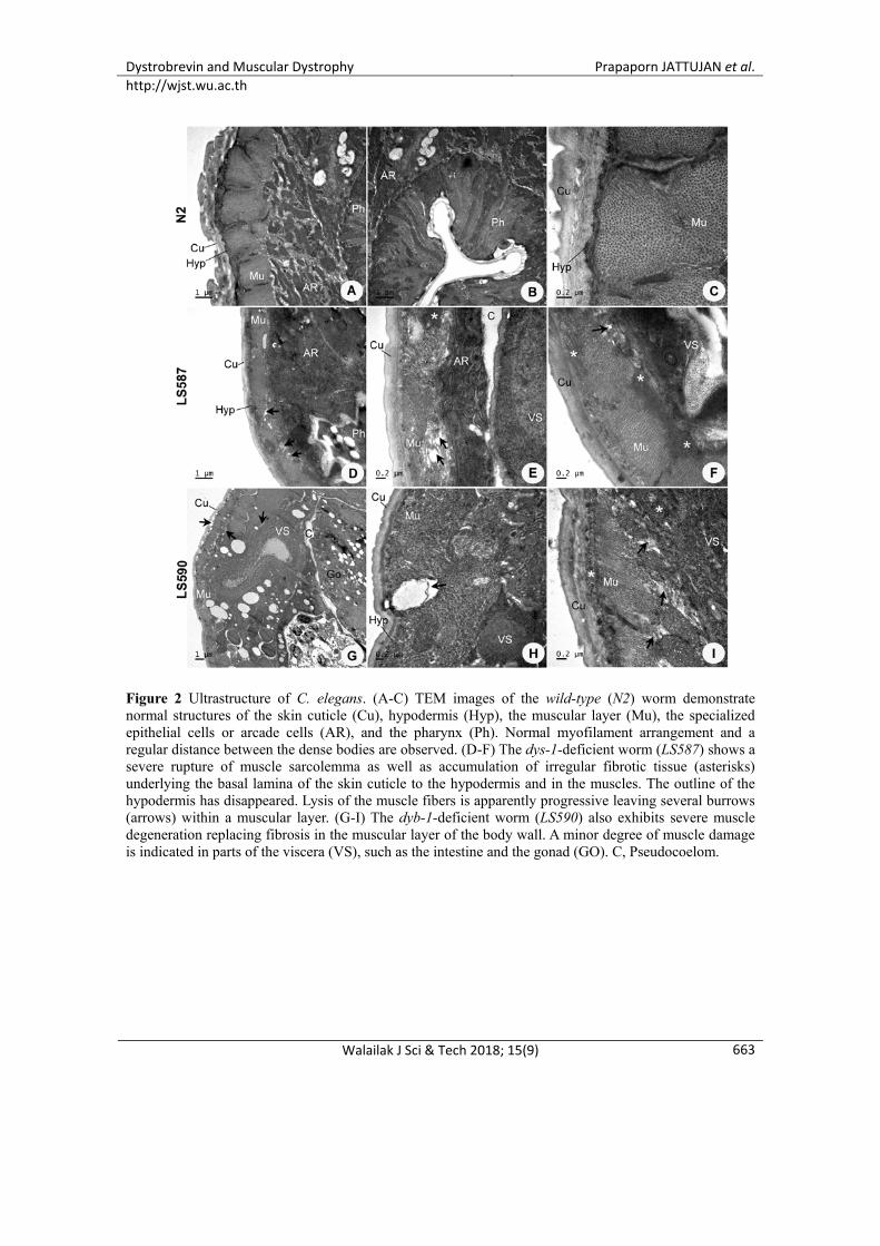
Figure 2 Ultrastructure of C. elegans. (A-C) TEM images of the wild-type (N2) worm demonstrate normal structures of the skin cuticle (Cu), hypodermis (Hyp), the muscular layer (Mu), the specialized epithelial cells or arcade cells (AR), and the pharynx (Ph). Normal myofilament arrangement and a regular distance between the dense bodies are observed. (D-F) The dys-1-deficient worm (LS587) shows a severe rupture of muscle sarcolemma as well as accumulation of irregular fibrotic tissue (asterisks) underlying the basal lamina of the skin cuticle to the hypodermis and in the muscles. The outline of the hypodermis has disappeared. Lysis of the muscle fibers is apparently progressive leaving several burrows (arrows) within a muscular layer. (G-I) The dyb-1-deficient worm (LS590) also exhibits severe muscle degeneration replacing fibrosis in the muscular layer of the body wall. A minor degree of muscle damage is indicated in parts of the viscera (VS), such as the intestine and the gonad (GO). C, Pseudocoelom.

Dystrobrevin and Muscular Dystrophy Prapaporn JATTUJAN et al. http://wjst.wu.ac.th
Walailak J Sci & Tech 2018; 15(9) 664
Figure 3 The mean lifespan of C. elegans. (A-C) The mean lifespan (at 50 % of survival rate in A.) of the dys-1/hlh-1 (strain LS587) and the dyb-1/hlh-1 (strain LS590) mutated worms shows a significant decrease in a comparison with the wild-type (strain N2). The LS587 worms reveal ~27 % decrease of the mean lifespan, while the LS590 worms reveal ~17 % decrease of their mean lifespan, when compared to that of N2 (p < 0.05). The actual function of the DYB protein is not yet elucidated, although it is likely involved in the intracellular signaling for the long-term stability of the muscle cell. Parts of DYB interact with syntrophins and nitric oxide synthase, while it also binds to desmuslin, an intermediate filament connecting extracellular matrix [24]. A previous result regarding delayed progression of muscle degeneration caused by an overexpression of the dyb gene into dys-lacking C. elegans provides strong evidence to support a synergistic action of their intracellular signaling [25]. Our results also agree with a previous report demonstrating the less severe phenotypes found in worms lacking DYB rather than DYS [20]. We speculate that the role of DYB is as a signaling pathway modulator in facilitating a bridging connection between the cytoskeleton, plasma membrane (via DYS) and extracellular matrix (via several molecules, including dystroglycan complex). Lack of DYS may disrupt this bridging interaction directly, while lack of DYB disconnects some modulator molecules adjacent to the plasma membrane. Recent data from Oh et al. [26] has described the possible structural elements between DYB and a cytoskeletal protein, α-catulin for stabilizing the plasma membrane. Progression of the skeletal muscle degeneration is one of the crucial defects in DMD symptoms. Our result in C. elegans ultrastructure showed that lacking of dys gene disrupted the stability of muscle fibers due to its progressive damage, not the alteration of the myofilament arrangement (Figure 2). The dissociation of degenerated muscle fibers was clearly demonstrated in the TEM images taken from the body wall of both LS587 and LS590 worms as well as partial atrophy of the pharyngeal muscles and the intestines. Distances between the dense bodies of the myofilaments in comparison between the mutants and the wild-type worms appeared regular. Sarcolemmal breakdown with scattering glycogens in the
Dystrobrevin and Muscular Dystrophy Prapaporn JATTUJAN et al. http://wjst.wu.ac.th
Walailak J Sci & Tech 2018; 15(9)
665
entire muscular layer of the body wall were pronounced in both the dys-1- and the dyb-1-deficient worms. The atrophic muscular layer of the worm's body wall was periodically replaced by a fibrotic tissue. Beside the muscular layer of the body wall, replacing fibrosis was also obvious in most areas underlying the hypodermis. However, lack of these genes showed less effect on the non-muscle cells, such as the arcade cells that were located in between the muscular layer and the visceral organs. As arcade cells were involved in the formation of specialized structures in the cuticle and support the neuronal receptor in the body wall, these supportive structures seemed to be intact. In clinical diagnosis of the DMD patients, fibrosis is a prominent pathological feature usually obtained from the biopsies of particular skeletal muscles [27]. The amount of fibrotic tissue may indicate the severity of the disease as it causes muscle dysfunction and eventually contributes to a lethal condition [28]. It has long been known that lack of dys-1, in C. elegans, leads to the disruption of sarcomeric anchoring structures and the latter affects the muscle cell death [29]. Our ultrastructural results indicate that the absence of dyb-1 could potentially alter the subcellular control, as deteriorated muscle fibers were observed in the electron micrographs. Many ongoing studies focus on exploring the therapeutic targeting molecules to stop a progression of fibrotic scars in muscle tissues of the DMD patients. As for a consequence of DMD, patients usually suffer from muscle atrophy causing a failure of many organ functions and being a cause of death at a young age. This study compared the worm lifespan between LS587 and LS590 to the wild-type (Figure 3). Reduction of the mean lifespan was clear with death due to the pathology at 25.24±6.81 days for LS587, 28.79±7.22 days for LS590, and 34.92±9.21 days for wild-type. This represents a 27 % decrease for the dys-1-deficient C. elegans when compared to the wild-type. A similar result was also obtained for the dyb-1-deficient worms with 17 % decrease. According to Gieseler et al. [30], the mild, mutated worms with a single mutation to the hlh-1 gene presented a normal phenotype, locomotion as expected for a normal lifespan. Double mutation of hlh-1 with dys-1 or dyb-1 rather produced massive degrees of deterioration in the worm phenotypes than a single mutation. Apparently, a severe degree of muscle degeneration due to lack of either DYS or DYB was the major cause of the worm’s death. The animals' gene that turns into DMD phenotype does have a certain impact on their living habit. Not only the disability to move, but a failure of the gastrointestinal tract could also shorten the worm lifespan. Conclusions
Absence of dystrobrevin in C. elegans exhibits a severe degree of muscle degeneration and subsequently shortens the worm lifespan. In this regard, dystrobrevin may act to stabilize the cell membrane and regulate the cytoskeleton, especially during contraction of the skeletal muscles. At the upregulation level of the signaling pathway, dystrobrevin functions to be associated with dystroglycans to maintain the cell integrity, and thereafter connected to the extracellular matrix. Dystrobrevin is surely the key molecule involved in the cause of muscular dystrophy if missing. A loss of dystrobrevin leads to partial disruption of the signaling pathway modulator so that could disconnect the regulating cascade between the cytoskeleton (through one of the binding sites of dystrophin) and the transmembrane proteins (associated complexes of dystroglycans and sarcoglycans). For future insight into DMD treatment, it is necessarily to understand the benefit of the key regulators and those of associated proteins involved in the mechanism of muscular dystrophy. Acknowledgements
This work was supported by the Central Instrumentation Facilities (CIF) grant, Faculty of Science, and the Talent Management Program at Mahidol University. Strains of C. elegans were provided by the CGC, which is funded by the NIH Office of Research Infrastructure Programs (P40 OD010440).
Dystrobrevin and Muscular Dystrophy Prapaporn JATTUJAN et al. http://wjst.wu.ac.th
Walailak J Sci & Tech 2018; 15(9) 666
References
[1] E Mercuri and F Muntoni. Muscular dystrophies. Lancet 2013; 381, 845. [2] F Rahimov and LM Kunkel. The cell biology of disease: Cellular and molecular mechanisms
underlying muscular dystrophy. J. Cell Biol. 2013; 201, 499-510. [3] AEH Emery and F Muntoni. Duchenne Muscular Dystrophy. 3rd ed. Oxford University Press,
Oxford, UK, 2003. [4] HR Fuller, L Graham, M Llavero Hurtado and T Wishart. Understanding the molecular
consequences of inherited muscular dystrophies: Advancements through proteomic experimentation. Expert. Rev. Proteomics 2016; 13, 659-71.
[5] R Turk, JJ Hsiao, MM Smits, BH Ng, TC Pospisil, KS Jones, KP Campbell and ME Wright. Molecular signatures of membrane protein complexes underlying muscular dystrophy. Mol. Cell Proteomics 2016; 15, 2169-85.
[6] EP Hoffman, LM Kunkel, C Angelini, A Clarke, M Johnson and JB Harris. Improved diagnosis of Becker muscular dystrophy by dystrophin testing. Neurology 1989; 39, 1011-7.
[7] H Nishio, Y Takeshima, N Narita, H Yanagawa, Y Suzuki, Y Ishikawa, Y Ishikawa, R Minami, H Nakamura and M Matsuo. Identification of a novel first exon in the human dystrophin gene and of a new promoter located more than 500kb upstream of the nearest known promoter. J. Clin. Invest. 1994; 94, 1037-42.
[8] M Koenig and LM Kunkel. Detailed analysis of the repeat domain of dystrophin reveals four potential hinge segments that may confer flexibility. J. Biol. Chem. 1990; 265, 4560-6.
[9] HM Sadoulet-Puccio, M Rajala and LM Kunkel. Dystrobrevin and dystrophin: An interaction through coiled-coil motifs. Proc. Natl. Acad. Sci. USA 1997; 94, 12413-18.
[10] M Nakamori and MP Takahashi. The role of alpha-dystrobrevin in striated muscle. Int. J. Mol. Sci. 2011; 12, 1660-71.
[11] RM Grady, RW Grange, KS Lau, MM Maimone, MC Nichol, JT Stull and JR Sanes. Role for alpha-dystrobrevin in the pathogenesis of dystrophin-dependent muscular dystrophies. Nat. Cell Biol. 1999; 1, 215-20.
[12] E Poon, EV Howman, SE Newey and KE Davies. Association of syncoilin and desmin: Linking intermediate filament proteins to the dystrophin-associated protein complex. J. Biol. Chem. 2002; 277, 3433-9.
[13] K Gieseler, M Abdel-Dayem and L Ségalat. In vitro interactions of Caenorhabditis elegans dystrophin with dystrobrevin and syntrophin. Federat. Eur. Biochem. Soc. Lett. 1999; 461, 59-62.
[14] S Bohm, H Jin, SM Hughes, RG Roberts and Y Hinits. Dystrobrevin and dystrophin family gene expression in zebrafish. Gene Expr. Patterns 2008; 8, 71-78.
[15] JW McGreevy, CH Hakim, MA McIntosh and D Duan. Animal models of Duchenne muscular dystrophy: From basic mechanisms to gene therapy. Dis. Model Mech. 2015; 8, 195-213.
[16] L Metzinger, DJ Blake, MV Squier, LV Anderson, AE Deconinck, R Nawrotzki, D Hilton-Jones and KE Davies. Dystrobrevin deficiency at the sarcolemma of patients with muscular dystrophy. Hum. Mol. Genet. 1997; 6, 1185-91.
[17] M Durbeej, RD Cohn, RF Hrstka, SA Moore, V Allamand, BL Davidson, RA Williamson and KP Campbell. Disruption of the beta-sarcoglycan gene reveals pathogenetic complexity of limb-girdle muscular dystrophy type 2E. Mol. Cell. 2000; 5, 141-51.
[18] M Durbeej and KP Campbell. Muscular dystrophies involving the dystrophin-glycoprotein complex: An overview of current mouse models. Curr. Opin. Genet. Dev. 2002; 12, 349-61.
[19] ML Ree, CF Lien and DC Górecki. Dystrobrevins in muscle and non-muscle tissues. Neuromuscul. Disord. 2007; 17, 123-34.
[20] K Gieseler, MC Mariol, C Bessou, M Migaud, CJ Franks, L Holden-Dye and L Ségalat. Molecular, genetic and physiological characterisation of dystrobrevin-like (dyb-1) mutants of caenorhabditis elegans. J. Mol. Biol. 2001; 307, 107-17.
[21] GA Alves, LR Silva, EF Rosa, J Aboulafia, E Freymueller-Haapalainen, C Souccar and VLA Nouailhetas. Intestine of dystrophic mice presents enhanced contractile resistance to stretching
Dystrobrevin and Muscular Dystrophy Prapaporn JATTUJAN et al. http://wjst.wu.ac.th
Walailak J Sci & Tech 2018; 15(9)
667
despite morphological impairment. Am. J. Physiol. Gastrointest. Liver Physiol. 2014; 306, G191-G199.
[22] K Gieseler, C Bessou and L Ségalat. Dystrobrevin- and dystrophin-like mutants display similar phenotypes in the nematode Caenorhabditis elegans. Neurogenetics 1999; 2, 87-90.
[23] J Giugia, K Gieseler, M Arpagaus and L Ségalat. Mutations in the dystrophin-like dys-1 gene of Caenorhabditis elegans result in reduced acetylcholinesterase activity. Federat. Eur. Biochem. Soc. Lett. 1999; 463, 270-2.
[24] Y Mizuno, TG Thompson, JR Guyon, HGW Lidov, M Brosius, M Imamura, E Ozawa, SC Watkins and LM Kunkel. Desmuslin an intermediate filament protein that interacts with alpha-dystrobrevin and desmin. Proc. Natl. Acad. Sci. USA 2001; 98, 6156-61.
[25] K Gieseler, K Grisoni, MC Mariol and L Ségalat. Overexpression of dystrobrevin delays locomotion defects and muscle degeneration in a dystrophin-deficient Caenorhabditis elegans. Neuromuscul. Disord. 2002; 12, 371-7.
[26] HJ Oh, LS Abraham, J van Hengel, C Stove, TJ Proszynski, K Gevaert, JX DiMario, JR Sanes, F van Roy and H Kim. Interaction of α-catulin with dystrobrevin contributes to integrity of dystrophin complex in muscle. J. Biol. Chem. 2012; 287, 21717-28.
[27] I Desguerre, M Mayer, F Leturcq, JP Barbet, RK Gherardi and C Christov. Endomysial fibrosis in Duchenne muscular dystrophy: A marker of poor outcome associated with macrophage alternative activation. J. Neuropathol. Exp. Neurol. 2009; 68, 762-73.
[28] W Klingler, K Jurkat-Rott, F Lehmann-Horn and R Schleip. The role of fibrosis in Duchenne muscular dystrophy. Acta Myol. 2012; 31, 184-95.
[29] N Brouilly, C Lecroisey, E Martin, L Pierson, MC Mariol, H Qadota, M Labouesse, N Streichenberger, N Mounier and K Gieseler. Ultra-structural time-course study in the C. elegans model for Duchenne muscular dystrophy highlights a crucial role for sarcomere-anchoring structures and sarcolemma integrity in the earliest steps of the muscle degeneration process. Hum. Mol. Genet. 2015; 24, 6428-45.
[30] K Gieseler, K Grisoni and L Ségalat. Genetic suppression of phenotypes arising from mutations in dystrophin-related genes in Caenorhabditis elegans. Curr. Biol. 2000; 10, 1092-7.