lossofthe suv39h histone methyltransferases impairs ... cell... · lossofthesuv39h histone...
TRANSCRIPT
Cell, Vol. 107, 323–337, November 2, 2001, Copyright 2001 by Cell Press
Loss of the Suv39h Histone MethyltransferasesImpairs Mammalian Heterochromatinand Genome Stability
ing centromeres and telomeres (Karpen and Allshire,1997) and to facilitate the extensive reorganization ofchromosomes during meiosis (Karpen et al., 1996; Dern-burg et al., 1996). Although alterations in chromatinstructure can be induced by sequence-specific recruit-
Antoine H.F.M. Peters,1,5 Donal O’Carroll,1,5,6
Harry Scherthan,2,6 Karl Mechtler,1
Stephan Sauer,1 Christian Schofer,3
Klara Weipoltshammer,3 Michaela Pagani,1
Monika Lachner,1 Alexander Kohlmaier,1
ment of transcription factors and chromatin remodelingSusanne Opravil,1 Michael Doyle,1,6 Maria Sibilia,1,6
complexes (Kingston and Narlikar, 1999), or by the clus-and Thomas Jenuwein1,4
tering of DNA repeats (Csink and Henikoff, 1998), DNA1 Research Institute of Molecular Pathology (IMP)sequence per se appears not to be sufficient to mediateVienna Biocenterthe establishment of distinct chromosomal subdomainsDr. Bohrgasse 7(Murphy and Karpen, 1998; Henikoff et al., 2001). Fur-A-1030 Viennather, while changes in DNA methylation patterns corre-Austrialate with distinct epigenetic states (Bird and Wolffe,2 Department of Human Biology1999), DNA methylation is virtually absent in S. pombeUniversity of Kaiserslauternand rare in Drosophila (Lyko, 2001), despite the epige-Erwin-Schrodingerstrasse, Geb. 14netic control operating in these organisms.D-67663 Kaiserslautern
Recently, a “histone code” hypothesis has been sug-Germanygested (Strahl and Allis, 2000), which predicts that differ-3 Institute of Histology and Embryologyent modifications (e.g., acetylation, phosphorylation,University of Viennamethylation) of histone N termini are interdependent andSchwarzspanierstraße 17represent an evolutionarily conserved mechanism thatA-1090 Viennacan induce and stabilize functionally distinct chromo-Austriasomal subdomains (Jenuwein and Allis, 2001). For ex-ample, general underacetylation of histones (Jeppesenet al., 1992; Ekwall et al., 1997) and phosphorylation of
Summaryhistone H3 (Hendzel et al., 1997; Wei et al., 1999) arerequired for faithful chromosome segregation, presum-
Histone H3 lysine 9 methylation has been proposed ably by preserving the more compacted chromatinto provide a major “switch” for the functional organi- structure of pericentric heterochromatin. Furthermore,zation of chromosomal subdomains. Here, we show the discovery of mammalian histone H3 lysine 9 specificthat the murine Suv39h histone methyltransferases histone methyltransferases (Suv39h HMTases) (Rea et(HMTases) govern H3-K9 methylation at pericentric al., 2000), which are heterochromatin-enriched enzymesheterochromatin and induce a specialized histone transiently accumulating around centromeres during mi-methylation pattern that differs from the broad H3-K9 tosis (Aagaard et al., 2000), revealed a regulatory mecha-methylation present at other chromosomal regions. nism in which the selective methylation of histone H3Suv39h-deficient mice display severely impaired via- at lysine 9 (H3-K9) creates a high-affinity binding sitebility and chromosomal instabilities that are associated for the heterochromatic HP1 proteins (Lachner et al.,with an increased tumor risk and perturbed chromo- 2001; Bannister et al., 2001; Nakayama et al., 2001).some interactions during male meiosis. These in vivo Gain- and loss-of-function studies for Suv39h enzymesdata assign a crucial role for pericentric H3-K9 methyl- in mammalian cell lines (Melcher et al., 2000; Rea etation in protecting genome stability, and define the al., 2000) suggest a major role for the SUV39H1-HP1Suv39h HMTases as important epigenetic regulators methylation system in chromosome segregation. In ad-for mammalian development. dition, the SUV39H1 HMTase is also involved in local
gene repression (Firestein et al., 2000), and is targetedto specific cell cycle genes through the tumor suppres-Introductionsor Rb (Nielsen et al., 2001).
In S. pombe, the clr4 and swi6 genes, which encodeChromatin represents the physiological template of thethe fission yeast homologs of SUV39H1 and HP1, aregenetic information in all eukaryotic cells, and is func-required for heterochromatic gene silencing (Thon ettionally divided into euchromatic and heterochromatical., 1994; Allshire et al., 1995; Ivanova et al., 1998) andregions. This functional distinction has been proposedcentromere function (Ekwall et al., 1996). Disruption ofto be crucial for epigenetic control of gene expressionthe HMTase activity of Clr4 prevents pericentric associa-programs (Turner, 2000; Jenuwein and Allis, 2001), totion of Swi6 protein (Bannister et al., 2001) and impairsunderlie the specialized chromatin structure surround-H3-K9 methylation (Nakayama et al., 2001). Althoughclr4 and swi6 null mutants are viable, mutation of either
4 Correspondence: [email protected] locus leads to high rates of chromosome missegrega-5 These authors contributed equally to this work
tion (Ekwall et al., 1996). By contrast, HP1 null mutants6 Present addresses: (D.O’C.) The Rockefeller University, New York;are lethal in Drosophila (Eissenberg et al., 1992) and(H.S.) Max Planck Institute for Molecular Genetics, Berlin; (M.D.)display a more severe phenotype, including aberrantInstitute of Botany, Vienna University; (M.S.) Institute of Dermatol-
ogy, Vienna University chromosome segregation and telomeric fusions (Kellum
Cell324
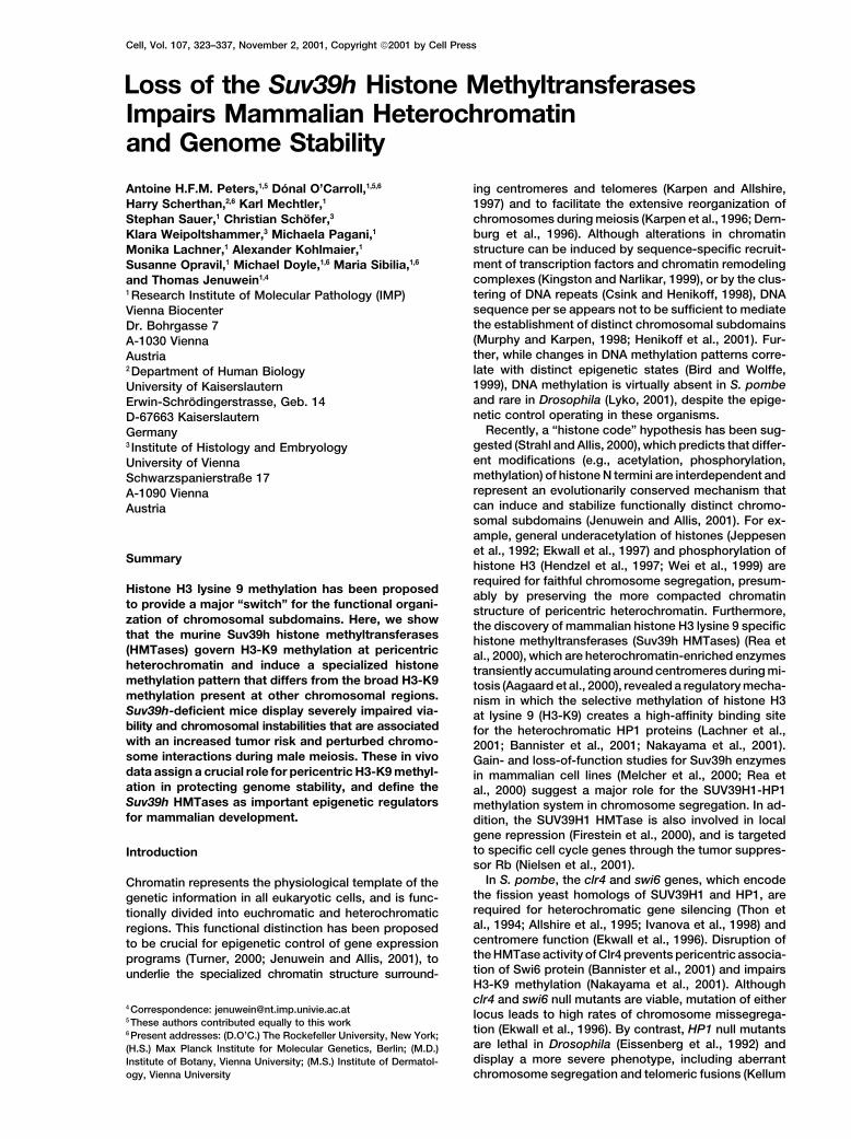
Figure 1. Targeting and Genotyping of Suv39h1- and Suv39h2-Deficient Mice
(A) Diagrammatic representation of the Suv39h1 and Suv39h2 genomic loci, the replacement vectors and the targeted alleles. Exons areindicated by black boxes with numbers referring to the starting amino acid positions of the respective exons (O’Carroll et al., 2000). Alsoshown are the diagnostic restriction sites and the external probes used for Southern blot analyses.(B) Southern blot analyses of PvuII- or HindIII-digested DNA isolated from offspring of Suv39h1�/� or Suv39h2�/� intercrosses.(C) Protein blot analyses of testis nuclear extracts from wild-type (wt), Suv39h1�/� (Suv1�/�), and Suv39h2�/� (Suv2�/�) mice with�-Suv39h1 and �-Suv39h2 antibodies.(D) Suv39h double null (dn) mice are growth retarded at birth and during adulthood.
and Alberts, 1995; Fanti et al., 1998). Surprisingly, Suv39h1 and Suv39h2 gene loci in the mouse. In addi-tion, we developed �-methH3-K9 antibodies which se-Su(var)3-9 mutants (Tschiersch et al., 1994) have so
far not revealed chromosome segregation defects in lectively recognize H3-K9 methylation in heterochro-matic subdomains.Drosophila.
A genetic analysis of mammalian heterochromatin and Here, we demonstrate that the Suv39h HMTases regu-late H3-K9 methylation at pericentric heterochromatin.its role in epigenetic gene control and chromosome sta-
bility has been lacking. The murine Suv39h HMTases Combined disruption of both Suv39h HMTase genesseverely impairs viability and induces chromosomal in-are encoded by two loci, Suv39h1 and Suv39h2, which
show overlapping expression profiles during em- stabilities plus an increased risk of tumorigenesis. Inaddition, Suv39h double null male mice display com-bryogenesis, while in adult mice Suv39h2 is mainly ex-
pressed in testes (O’Carroll et al., 2000). These data plete spermatogenic failure that is largely caused bynonhomologous chromosome associations. Together,suggest redundant roles for the Suv39h HMTases during
embryonic development and a putative additional func- these in vivo results assign a direct role for Suv39h-dependent H3-K9 methylation at pericentric heterochro-tion in the male germline. To address the functional
consequence(s) of Suv39h-dependent H3-K9 methyla- matin in protecting genome stability during mammaliandevelopment.tion during mammalian development, we disrupted the
Suv39h HMTases and Genome Stability325
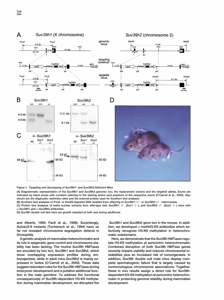
Figure 2. Chromosomal Instabilities inSuv39h dn PMEFs
(A) Relative doubling rates of wild-type (wt)and Suv39h dn (double null) PMEFs deter-mined in a 3T3 protocol over the first 20 pas-sages.(B) DNA contents of wt and Suv39h dn PMEFmass cultures at passage 3 and passage 8.(C) Statistical karyotype analysis with two wtand two Suv39h dn PMEF cultures at passage8. For each culture, 45 metaphases were eval-uated. Insets display metaphase spreadsshowing a diploid number (n � 40) of chromo-somes for wt and a hyper-tretraploid number(n � 82) of chromosomes for Suv39h dnPMEFs.
Results in embryonic stem (ES) cells using a conventional tar-geting approach that replaces parts of the evolutionarilyconserved chromo domain with the bacterial LacZ geneTargeted Disruption of the Suv39h1 and Suv39h2
Gene Loci in the Mouse Germline and an RSV-neomycin selecion cassette (Figure 1A).These targeting strategies produce in-frame fusion pro-Murine Suv39h HMTases are encoded by two loci which
have been mapped to centromere-proximal positions teins of the first 40 amino acids of Suv39h1 or of the first113 amino acids of Suv39h2 with lacZ, which maintainin the X chromosome (Suv39h1) or in chromosome 2
(Suv39h2) (O’Carroll et al., 2000). Both gene loci were �-galactosidase activities (data not shown). Success-fully targeted ES cell clones were used to generate chi-independently disrupted by homologous recombination
Cell326
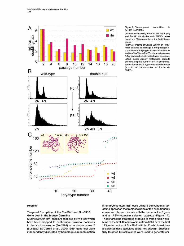
Table 1. Incidence of B Cell Lymphomas in Mice with Reduced Suv39h Gene Dosage
Suv39h # of Mice Total # % of MiceGenotype Gene dosage with Tumor of Mice with Tumor
W1W2 3 0 57 0W1H2, W1N2, H1N2 0–2 1 22 4.6H1W2, N1W2 2–3 8 26 30.8H1H2, N1H2 1–2 20 72 27.8N1N2 0 2 6 33.3
Abbreviations: W1N2: Suv39h1�/�; Suv39h2�/�; H1H2: Suv39h1�/�; Suv39h2�/�; N1H2: Suv39h1�/�; Suv39h2�/�
meric mice that transmitted the mutated Suv39h1 or To further characterize these genomic instabilities, weperformed karyotype analyses with passage 8 PMEFs.Suv39h2 alleles through the germline (Figure 1B). Protein
blot analyses of testis nuclear extracts from wild-type, We examined 45 karyotypes each for two independentwt and Suv39h dn PMEF cultures. As shown in FigureSuv39h1-, and Suv39h2-deficient mice with �-Suv39h1
and �-Suv39h2 specific antibodies (O’Carroll et al., 2000) 2C, a major fraction of the wt karyotypes are nondiploid,with chromosome numbers ranging from 25 to 82. Aneu-indicated absence of the respective proteins, demon-
strating that we had generated null alleles for both genes ploidies were significantly increased in Suv39h dn karyo-types and comprised chromosome numbers from 38 to(Figure 1C).162. Notably, whereas wt PMEFs display a random arrayof aneuploid karyotypes, Suv39h dn PMEFs are largelySeverely Impaired Viability of Suv39h Doublehypo-tetraploid or hypo-octaploid. Chromosomes inNull MiceSuv39h dn PMEFs appear of normal morphology andMice deficient for either Suv39h1 or Suv39h2 displaywe did not observe Robertsonian fusions. We concludenormal viability and fertility (data not shown), and dothat the absence of Suv39h gene function induces geno-not exhibit apparent phenotypes. Therefore, we inter-mic instability, primarily by impairing segregation of thecrossed Suv39h1�/� and Suv39h2�/� mice to gener-entire set of chromosomes.ate compound Suv39h mutants that were then used to
derive Suv39h double null (dn) mice. Suv39h dn miceobtained from several different intercrosses are born at Development of B Cell Lymphomas in Suv39honly sub-Mendelian ratios, are growth retarded (Figure Mutant Mice1D), and are characterized by hypogonadism in males. We next analyzed Suv39h mutant mice for tumor inci-For example, from a total of 197 mice, 46 mice would dence. Because the majority of Suv39h dn mice arehave been expected to be double null, but only 15 nonviable, we examined distinct Suv39h genotypes thatSuv39h dn mice (�33%) were born. Analysis of mouse differ in their gene dosage for either Suv39h1 or Suv39h2.embryogenesis indicated normal development of Suv39h For example, we expected that random X-inactivationdn fetuses until day E12.5, whereas at later stages, of the X-linked Suv39h1 gene could increase the tumorSuv39h dn fetuses are smaller and display an increased risk in Suv39h1�/� mice, even in the presence of arate of resorptions and prenatal lethality (data not functional copy of Suv39h2, which is significantly down-shown). Together, these results demonstrate that the regulated in most adult tissues (O’Carroll et al., 2000).Suv39h genes are required for normal viability, and for Examination of 99 mice either heterozygous (het) or nullpre- and postnatal development. for the Suv39h1 locus indicated an �28% penetrance
of tumor formation with an onset between 9 and 15months of age (Table 1). These tumors are B cell lympho-Chromosome Missegregation in Suv39h dn
Embryonic Fibroblasts mas (Figure 3A) which, according to FACS profiling (seeExperimental Procedures), resemble slowly progressingTo examine the Suv39h-dependent defects in more de-
tail, we derived primary mouse embryonic fibroblasts non-Hodgkin lymphomas in humans (Foon and Gale,1995). The tumor incidence for late onset B cell lympho-(PMEFs) from day E12.5 fetuses. Comparative growth
curves between wild-type (wt) and Suv39h dn PMEFs mas was �33% in the few viable Suv39h dn mice (n �6). By contrast, Suv39h2�/� or Suv39h2�/� mice de-in a 3T3 protocol over the first 20 passages indicated
that Suv39h dn PMEFs displayed a higher doubling rate veloped B cell lymphomas at only � 5% penetrance(n � 21), and we did not observe tumor formation inuntil passage 12 (Figure 2A). At later passages, the
Suv39h dn PMEFs appear to have a slightly reduced control wt mice (n � 57).Immunohistochemistry for H3-K9 methylation with aproliferative potential which further declines upon con-
tinued cultivation. DNA profiles of passage 3 and pas- newly developed antibody (�-4x-methH3-K9, see below)on tissue sections from wt mice indicated broad stainingsage 8 wt and Suv39h dn PMEFs show that wt PMEFs
appear genomically stable at passage 3, whereas in spleen and lymph nodes (Figure 3B, left panel). Thestaining was somewhat reduced within germinal centersSuv39h dn PMEFs already contain cells with a greater
than 4N DNA material (Figure 2B, top panels). At passage which contain unstimulated centroblasts. Importantly,H3-K9 methylation was lost in Suv39h dn sections (data8, wt PMEFs are largely senesced. By contrast, Suv39h
dn PMEFs continue to proliferate, although many cells not shown) and was also largely absent in tumor tissuesfrom Suv39h1�/�, Suv39h2�/� (null1/het2) mice. Indisplay tetraploid and even octaploid DNA contents (Fig-
ure 2B, lower panels). these tumor sections, only a few areas of methH3-K9
Suv39h HMTases and Genome Stability327
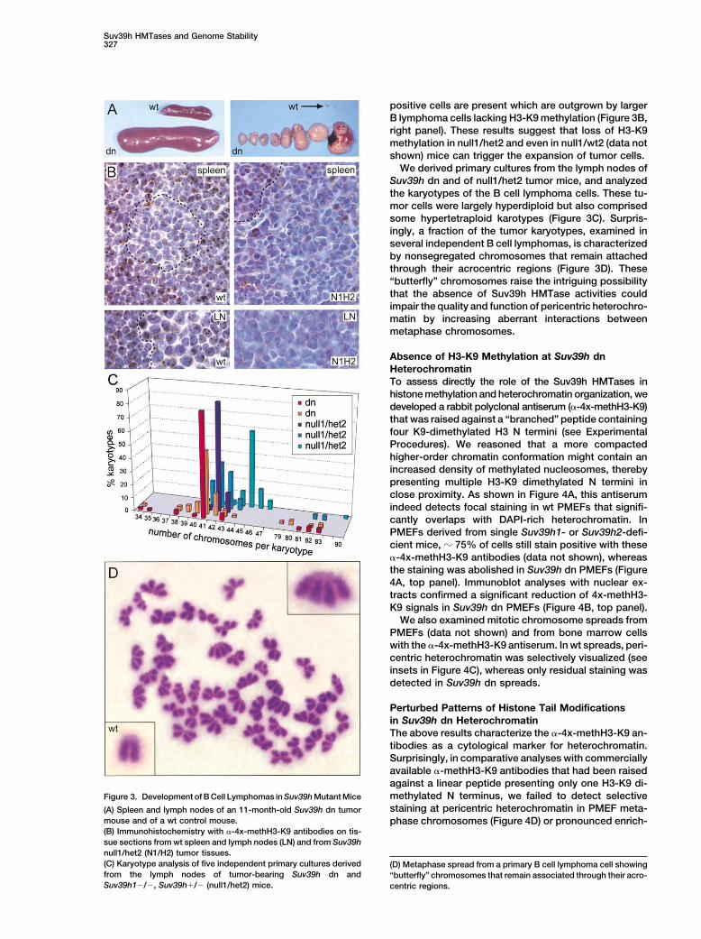
positive cells are present which are outgrown by largerB lymphoma cells lacking H3-K9 methylation (Figure 3B,right panel). These results suggest that loss of H3-K9methylation in null1/het2 and even in null1/wt2 (data notshown) mice can trigger the expansion of tumor cells.
We derived primary cultures from the lymph nodes ofSuv39h dn and of null1/het2 tumor mice, and analyzedthe karyotypes of the B cell lymphoma cells. These tu-mor cells were largely hyperdiploid but also comprisedsome hypertetraploid karotypes (Figure 3C). Surpris-ingly, a fraction of the tumor karyotypes, examined inseveral independent B cell lymphomas, is characterizedby nonsegregated chromosomes that remain attachedthrough their acrocentric regions (Figure 3D). These“butterfly” chromosomes raise the intriguing possibilitythat the absence of Suv39h HMTase activities couldimpair the quality and function of pericentric heterochro-matin by increasing aberrant interactions betweenmetaphase chromosomes.
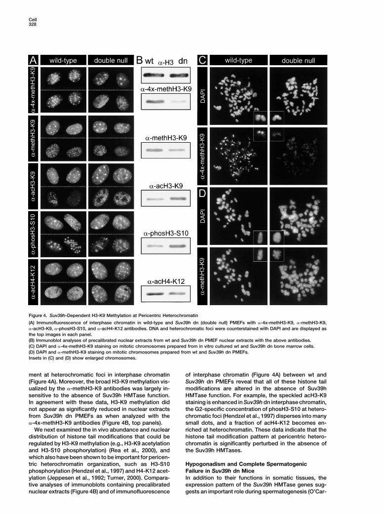
Absence of H3-K9 Methylation at Suv39h dnHeterochromatinTo assess directly the role of the Suv39h HMTases inhistone methylation and heterochromatin organization, wedeveloped a rabbit polyclonal antiserum (�-4x-methH3-K9)that was raised against a “branched” peptide containingfour K9-dimethylated H3 N termini (see ExperimentalProcedures). We reasoned that a more compactedhigher-order chromatin conformation might contain anincreased density of methylated nucleosomes, therebypresenting multiple H3-K9 dimethylated N termini inclose proximity. As shown in Figure 4A, this antiserumindeed detects focal staining in wt PMEFs that signifi-cantly overlaps with DAPI-rich heterochromatin. InPMEFs derived from single Suv39h1- or Suv39h2-defi-cient mice, � 75% of cells still stain positive with these�-4x-methH3-K9 antibodies (data not shown), whereasthe staining was abolished in Suv39h dn PMEFs (Figure4A, top panel). Immunoblot analyses with nuclear ex-tracts confirmed a significant reduction of 4x-methH3-K9 signals in Suv39h dn PMEFs (Figure 4B, top panel).
We also examined mitotic chromosome spreads fromPMEFs (data not shown) and from bone marrow cellswith the �-4x-methH3-K9 antiserum. In wt spreads, peri-centric heterochromatin was selectively visualized (seeinsets in Figure 4C), whereas only residual staining wasdetected in Suv39h dn spreads.
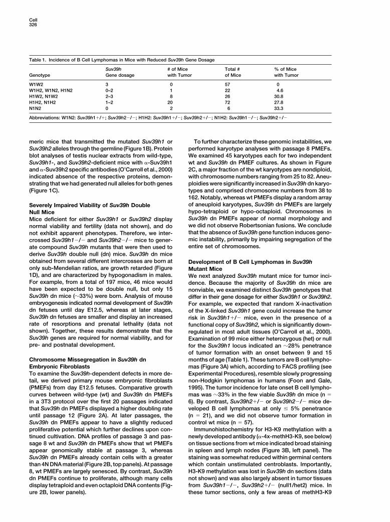
Perturbed Patterns of Histone Tail Modificationsin Suv39h dn HeterochromatinThe above results characterize the �-4x-methH3-K9 an-tibodies as a cytological marker for heterochromatin.Surprisingly, in comparative analyses with commerciallyavailable �-methH3-K9 antibodies that had been raisedagainst a linear peptide presenting only one H3-K9 di-methylated N terminus, we failed to detect selectiveFigure 3. Development of B Cell Lymphomas in Suv39h Mutant Micestaining at pericentric heterochromatin in PMEF meta-(A) Spleen and lymph nodes of an 11-month-old Suv39h dn tumor
mouse and of a wt control mouse. phase chromosomes (Figure 4D) or pronounced enrich-(B) Immunohistochemistry with �-4x-methH3-K9 antibodies on tis-sue sections from wt spleen and lymph nodes (LN) and from Suv39hnull1/het2 (N1/H2) tumor tissues.(C) Karyotype analysis of five independent primary cultures derived (D) Metaphase spread from a primary B cell lymphoma cell showingfrom the lymph nodes of tumor-bearing Suv39h dn and “butterfly” chromosomes that remain associated through their acro-Suv39h1�/�, Suv39h�/� (null1/het2) mice. centric regions.
Cell328
Figure 4. Suv39h-Dependent H3-K9 Methylation at Pericentric Heterochromatin
(A) Immunofluorescence of interphase chromatin in wild-type and Suv39h dn (double null) PMEFs with �-4x-methH3-K9, �-methH3-K9,�-acH3-K9, �-phosH3-S10, and �-acH4-K12 antibodies. DNA and heterochromatic foci were counterstained with DAPI and are displayed asthe top images in each panel.(B) Immunoblot analyses of precalibrated nuclear extracts from wt and Suv39h dn PMEF nuclear extracts with the above antibodies.(C) DAPI and �-4x-methH3-K9 staining on mitotic chromosomes prepared from in vitro cultured wt and Suv39h dn bone marrow cells.(D) DAPI and �-methH3-K9 staining on mitotic chromosomes prepared from wt and Suv39h dn PMEFs.Insets in (C) and (D) show enlarged chromosomes.
ment at heterochromatic foci in interphase chromatin of interphase chromatin (Figure 4A) between wt andSuv39h dn PMEFs reveal that all of these histone tail(Figure 4A). Moreover, the broad H3-K9 methylation vis-
ualized by the �-methH3-K9 antibodies was largely in- modifications are altered in the absence of Suv39hHMTase function. For example, the speckled acH3-K9sensitive to the absence of Suv39h HMTase function.
In agreement with these data, H3-K9 methylation did staining is enhanced in Suv39h dn interphase chromatin,the G2-specific concentration of phosH3-S10 at hetero-not appear as significantly reduced in nuclear extracts
from Suv39h dn PMEFs as when analyzed with the chromatic foci (Hendzel et al., 1997) disperses into manysmall dots, and a fraction of acH4-K12 becomes en-�-4x-methH3-K9 antibodies (Figure 4B, top panels).
We next examined the in vivo abundance and nuclear riched at heterochromatin. These data indicate that thehistone tail modification pattern at pericentric hetero-distribution of histone tail modifications that could be
regulated by H3-K9 methylation (e.g., H3-K9 acetylation chromatin is significantly perturbed in the absence ofthe Suv39h HMTases.and H3-S10 phosphorylation) (Rea et al., 2000), and
which also have been shown to be important for pericen-tric heterochromatin organization, such as H3-S10 Hypogonadism and Complete Spermatogenic
Failure in Suv39h dn Micephosphorylation (Hendzel et al., 1997) and H4-K12 acet-ylation (Jeppesen et al., 1992; Turner, 2000). Compara- In addition to their functions in somatic tissues, the
expression pattern of the Suv39h HMTase genes sug-tive analyses of immunoblots containing precalibratednuclear extracts (Figure 4B) and of immunofluorescence gests an important role during spermatogenesis (O’Car-
Suv39h HMTases and Genome Stability329
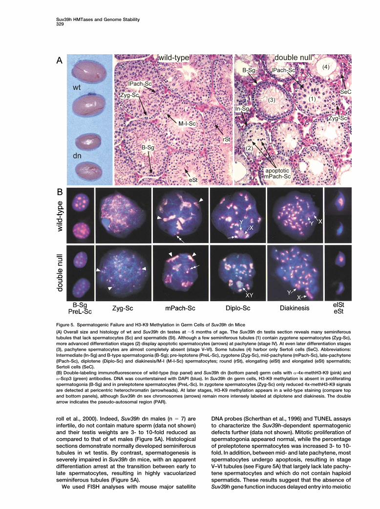
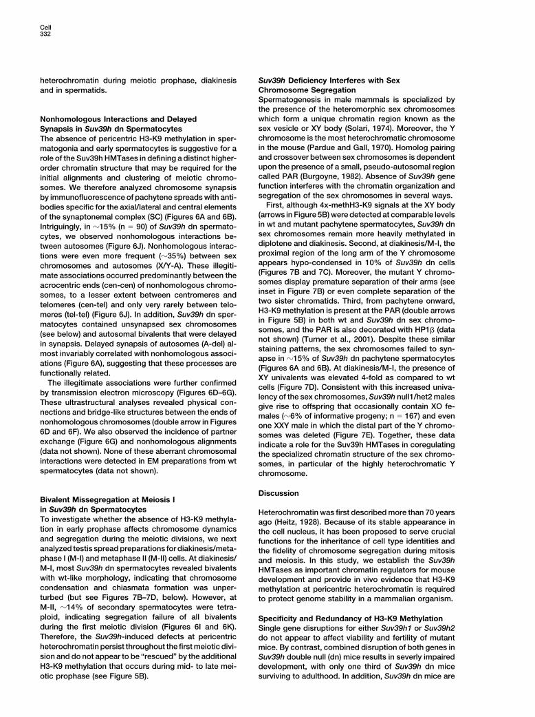
Figure 5. Spermatogenic Failure and H3-K9 Methylation in Germ Cells of Suv39h dn Mice
(A) Overall size and histology of wt and Suv39h dn testes at �5 months of age. The Suv39h dn testis section reveals many seminiferoustubules that lack spermatocytes (Sc) and spermatids (St). Although a few seminiferous tubules (1) contain zygotene spermatocytes (Zyg-Sc),more advanced differentiation stages (2) display apoptotic spermatocytes (arrows) at pachytene (stage IV). At even later differentiation stages(3), pachytene spermatocytes are almost completely absent (stage V–VI). Some tubules (4) harbor only Sertoli cells (SeC). Abbreviations:Intermediate (In-Sg) and B-type spermatogonia (B-Sg); pre-leptotene (PreL-Sc), zygotene (Zyg-Sc), mid-pachytene (mPach-Sc), late-pachytene(lPach-Sc), diplotene (Diplo-Sc) and diakinesis/M-I (M-I-Sc) spermatocytes; round (rSt), elongating (elSt) and elongated (eSt) spermatids;Sertoli cells (SeC).(B) Double-labeling immunofluorescence of wild-type (top panel) and Suv39h dn (bottom panel) germ cells with �-4x-methH3-K9 (pink) and�-Scp3 (green) antibodies. DNA was counterstained with DAPI (blue). In Suv39h dn germ cells, H3-K9 methylation is absent in proliferatingspermatogonia (B-Sg) and in preleptotene spermatocytes (PreL-Sc). In zygotene spermatocytes (Zyg-Sc) only reduced 4x-methH3-K9 signalsare detected at pericentric heterochromatin (arrowheads). At later stages, H3-K9 methylation appears in a wild-type staining (compare topand bottom panels), although Suv39h dn sex chromosomes (arrows) remain more intensely labeled at diplotene and diakinesis. The doublearrow indicates the pseudo-autosomal region (PAR).
roll et al., 2000). Indeed, Suv39h dn males (n � 7) are DNA probes (Scherthan et al., 1996) and TUNEL assaysto characterize the Suv39h-dependent spermatogenicinfertile, do not contain mature sperm (data not shown)
and their testis weights are 3- to 10-fold reduced as defects further (data not shown). Mitotic proliferation ofspermatogonia appeared normal, while the percentagecompared to that of wt males (Figure 5A). Histological
sections demonstrate normally developed seminiferous of preleptotene spermatocytes was increased 3- to 10-fold. In addition, between mid- and late pachytene, mosttubules in wt testis. By contrast, spermatogenesis is
severely impaired in Suv39h dn mice, with an apparent spermatocytes undergo apoptosis, resulting in stageV–VI tubules (see Figure 5A) that largely lack late pachy-differentiation arrest at the transition between early to
late spermatocytes, resulting in highly vacuolarized tene spermatocytes and which do not contain haploidspermatids. These results suggest that the absence ofseminiferous tubules (Figure 5A).
We used FISH analyses with mouse major satellite Suv39h gene function induces delayed entry into meiotic
Cell330
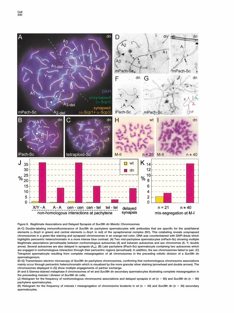
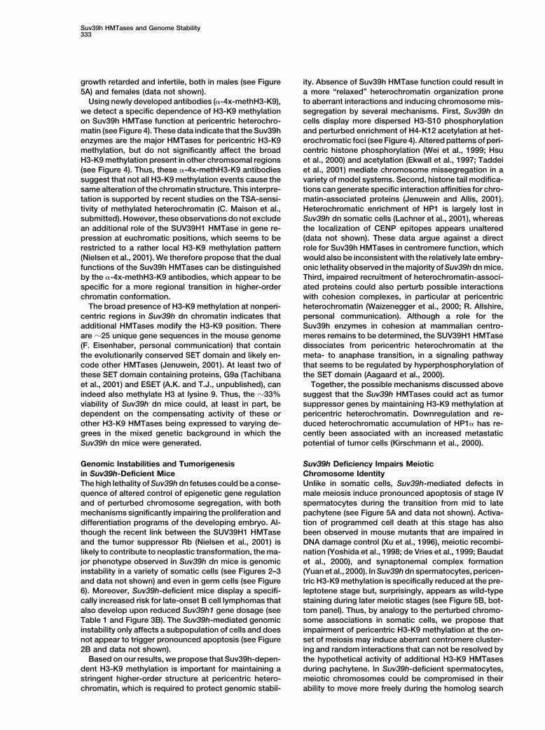
Figure 6. Illegitimate Associations and Delayed Synapsis of Suv39h dn Meiotic Chromosomes
(A–C) Double-labeling immunofluorescence of Suv39h dn pachytene spermatocytes with antibodies that are specific for the axial/lateralelements (�-Scp3 in green) and central elements (�-Scp1 in red) of the synaptonemal complex (SC). This colabeling reveals unsynapsedchromosomes in a green-like staining and synapsed chromosomes in an orange-red color. DNA was counterstained with DAPI (blue) whichhighlights pericentric heterochromatin in a more intense blue contrast. (A) Two mid-pachytene spermatocytes (mPach-Sc) showing multipleillegitimate associations (arrowheads) between nonhomologous autosomes (A) and between autosomes and sex chromomes (X, Y; doublearrow). Several autosomes are also delayed in synapsis (Adel). (B) Late pachytene (lPach-Sc) spermatocyte containing two autosomes whichare engaged in nonhomologous interaction through their pericentric regions (arrowhead). In addition, the sex chromosomes failed to pair. (C)Tetraploid spermatocyte resulting from complete missegregation of all chromosomes in the preceding mitotic division of a Suv39h dnspermatogonium.(D–G) Transmission electron microscopy of Suv39h dn pachytene chromosomes, confirming that nonhomologous chromosome associationsmainly occur through pericentric heterochromatin which is visualized by the more granular silver staining (arrowhead and double arrows). Thechromosomes displayed in (G) show multiple engagements of partner exchange.(H and I) Giemsa-stained metaphase II chromosomes of wt and Suv39h dn secondary spermatocytes illustrating complete missegregation inthe preceeding meiosis I division of Suv39h dn cells.(J) Histogram for the frequency of nonhomologous chromosome associations and delayed synapsis in wt (n � 80) and Suv39h dn (n � 90)pachytene spermatocytes.(K) Histogram for the frequency of meiosis I missegregation of chromosome bivalents in wt (n � 40) and Suv39h dn (n � 30) secondaryspermatocytes.
Suv39h HMTases and Genome Stability331
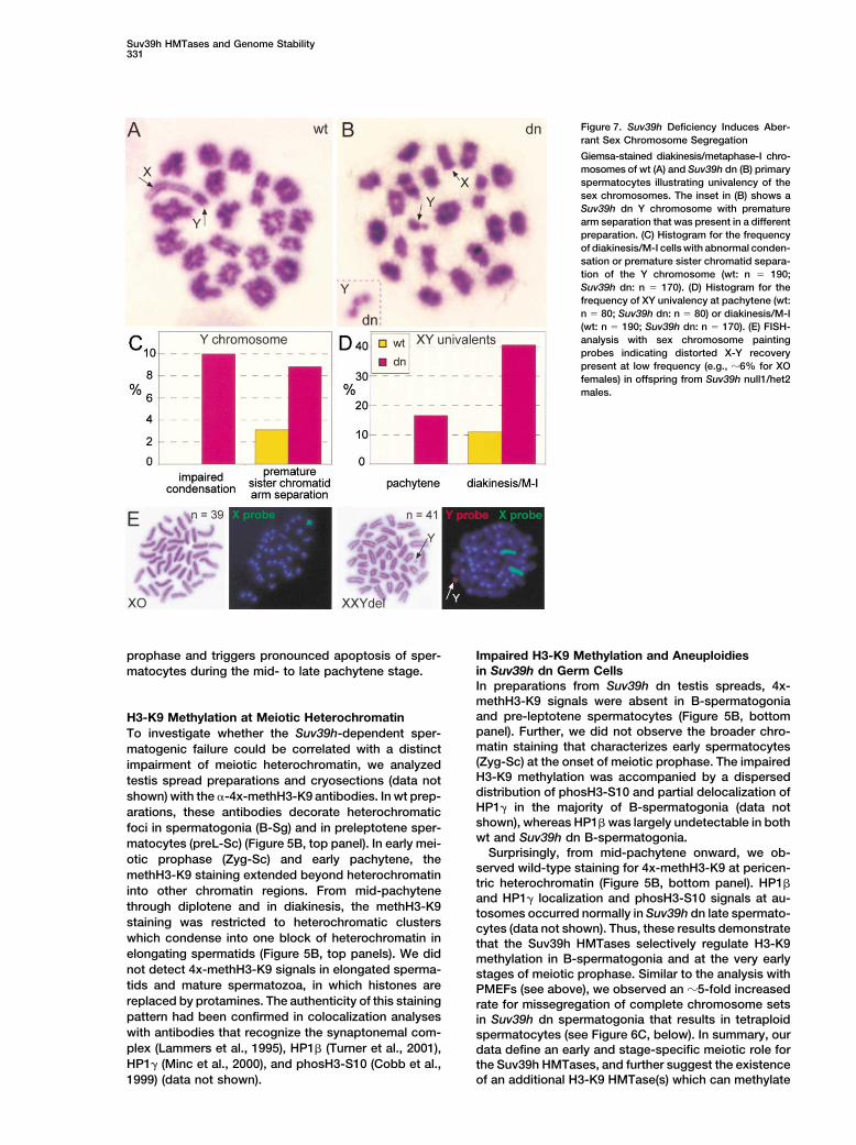
Figure 7. Suv39h Deficiency Induces Aber-rant Sex Chromosome Segregation
Giemsa-stained diakinesis/metaphase-I chro-mosomes of wt (A) and Suv39h dn (B) primaryspermatocytes illustrating univalency of thesex chromosomes. The inset in (B) shows aSuv39h dn Y chromosome with prematurearm separation that was present in a differentpreparation. (C) Histogram for the frequencyof diakinesis/M-I cells with abnormal conden-sation or premature sister chromatid separa-tion of the Y chromosome (wt: n � 190;Suv39h dn: n � 170). (D) Histogram for thefrequency of XY univalency at pachytene (wt:n � 80; Suv39h dn: n � 80) or diakinesis/M-I(wt: n � 190; Suv39h dn: n � 170). (E) FISH-analysis with sex chromosome paintingprobes indicating distorted X-Y recoverypresent at low frequency (e.g., �6% for XOfemales) in offspring from Suv39h null1/het2males.
prophase and triggers pronounced apoptosis of sper- Impaired H3-K9 Methylation and Aneuploidiesin Suv39h dn Germ Cellsmatocytes during the mid- to late pachytene stage.In preparations from Suv39h dn testis spreads, 4x-methH3-K9 signals were absent in B-spermatogoniaand pre-leptotene spermatocytes (Figure 5B, bottomH3-K9 Methylation at Meiotic Heterochromatinpanel). Further, we did not observe the broader chro-To investigate whether the Suv39h-dependent sper-matin staining that characterizes early spermatocytesmatogenic failure could be correlated with a distinct(Zyg-Sc) at the onset of meiotic prophase. The impairedimpairment of meiotic heterochromatin, we analyzedH3-K9 methylation was accompanied by a dispersedtestis spread preparations and cryosections (data notdistribution of phosH3-S10 and partial delocalization ofshown) with the �-4x-methH3-K9 antibodies. In wt prep-HP1� in the majority of B-spermatogonia (data notarations, these antibodies decorate heterochromaticshown), whereas HP1� was largely undetectable in bothfoci in spermatogonia (B-Sg) and in preleptotene sper-wt and Suv39h dn B-spermatogonia.matocytes (preL-Sc) (Figure 5B, top panel). In early mei-
Surprisingly, from mid-pachytene onward, we ob-otic prophase (Zyg-Sc) and early pachytene, theserved wild-type staining for 4x-methH3-K9 at pericen-methH3-K9 staining extended beyond heterochromatintric heterochromatin (Figure 5B, bottom panel). HP1�
into other chromatin regions. From mid-pachyteneand HP1� localization and phosH3-S10 signals at au-
through diplotene and in diakinesis, the methH3-K9 tosomes occurred normally in Suv39h dn late spermato-staining was restricted to heterochromatic clusters cytes (data not shown). Thus, these results demonstratewhich condense into one block of heterochromatin in that the Suv39h HMTases selectively regulate H3-K9elongating spermatids (Figure 5B, top panels). We did methylation in B-spermatogonia and at the very earlynot detect 4x-methH3-K9 signals in elongated sperma- stages of meiotic prophase. Similar to the analysis withtids and mature spermatozoa, in which histones are PMEFs (see above), we observed an �5-fold increasedreplaced by protamines. The authenticity of this staining rate for missegregation of complete chromosome setspattern had been confirmed in colocalization analyses in Suv39h dn spermatogonia that results in tetraploidwith antibodies that recognize the synaptonemal com- spermatocytes (see Figure 6C, below). In summary, ourplex (Lammers et al., 1995), HP1� (Turner et al., 2001), data define an early and stage-specific meiotic role forHP1� (Minc et al., 2000), and phosH3-S10 (Cobb et al., the Suv39h HMTases, and further suggest the existence
of an additional H3-K9 HMTase(s) which can methylate1999) (data not shown).
Cell332
heterochromatin during meiotic prophase, diakinesis Suv39h Deficiency Interferes with SexChromosome Segregationand in spermatids.Spermatogenesis in male mammals is specialized bythe presence of the heteromorphic sex chromosomeswhich form a unique chromatin region known as theNonhomologous Interactions and Delayedsex vesicle or XY body (Solari, 1974). Moreover, the YSynapsis in Suv39h dn Spermatocyteschromosome is the most heterochromatic chromosomeThe absence of pericentric H3-K9 methylation in sper-in the mouse (Pardue and Gall, 1970). Homolog pairingmatogonia and early spermatocytes is suggestive for aand crossover between sex chromosomes is dependentrole of the Suv39h HMTases in defining a distinct higher-upon the presence of a small, pseudo-autosomal regionorder chromatin structure that may be required for thecalled PAR (Burgoyne, 1982). Absence of Suv39h geneinitial alignments and clustering of meiotic chromo-function interferes with the chromatin organization andsomes. We therefore analyzed chromosome synapsissegregation of the sex chromosomes in several ways.by immunofluorescence of pachytene spreads with anti-
First, although 4x-methH3-K9 signals at the XY bodybodies specific for the axial/lateral and central elements(arrows in Figure 5B) were detected at comparable levelsof the synaptonemal complex (SC) (Figures 6A and 6B).in wt and mutant pachytene spermatocytes, Suv39h dnIntriguingly, in �15% (n � 90) of Suv39h dn spermato-sex chromosomes remain more heavily methylated incytes, we observed nonhomologous interactions be-diplotene and diakinesis. Second, at diakinesis/M-I, thetween autosomes (Figure 6J). Nonhomologous interac-proximal region of the long arm of the Y chromosometions were even more frequent (�35%) between sexappears hypo-condensed in 10% of Suv39h dn cellschromosomes and autosomes (X/Y-A). These illegiti-(Figures 7B and 7C). Moreover, the mutant Y chromo-mate associations occurred predominantly between thesomes display premature separation of their arms (seeacrocentric ends (cen-cen) of nonhomologous chromo-inset in Figure 7B) or even complete separation of thesomes, to a lesser extent between centromeres andtwo sister chromatids. Third, from pachytene onward,telomeres (cen-tel) and only very rarely between telo-H3-K9 methylation is present at the PAR (double arrowsmeres (tel-tel) (Figure 6J). In addition, Suv39h dn sper-in Figure 5B) in both wt and Suv39h dn sex chromo-matocytes contained unsynapsed sex chromosomessomes, and the PAR is also decorated with HP1� (data(see below) and autosomal bivalents that were delayednot shown) (Turner et al., 2001). Despite these similar
in synapsis. Delayed synapsis of autosomes (A-del) al-staining patterns, the sex chromosomes failed to syn-
most invariably correlated with nonhomologous associ-apse in �15% of Suv39h dn pachytene spermatocytes
ations (Figure 6A), suggesting that these processes are(Figures 6A and 6B). At diakinesis/M-I, the presence of
functionally related. XY univalents was elevated 4-fold as compared to wtThe illegitimate associations were further confirmed cells (Figure 7D). Consistent with this increased univa-
by transmission electron microscopy (Figures 6D–6G). lency of the sex chromosomes, Suv39h null1/het2 malesThese ultrastructural analyses revealed physical con- give rise to offspring that occasionally contain XO fe-nections and bridge-like structures between the ends of males (�6% of informative progeny; n � 167) and evennonhomologous chromosomes (double arrow in Figures one XXY male in which the distal part of the Y chromo-6D and 6F). We also observed the incidence of partner somes was deleted (Figure 7E). Together, these dataexchange (Figure 6G) and nonhomologous alignments indicate a role for the Suv39h HMTases in coregulating(data not shown). None of these aberrant chromosomal the specialized chromatin structure of the sex chromo-interactions were detected in EM preparations from wt somes, in particular of the highly heterochromatic Yspermatocytes (data not shown). chromosome.
DiscussionBivalent Missegregation at Meiosis Iin Suv39h dn Spermatocytes Heterochromatin was first described more than 70 yearsTo investigate whether the absence of H3-K9 methyla- ago (Heitz, 1928). Because of its stable appearance intion in early prophase affects chromosome dynamics the cell nucleus, it has been proposed to serve crucialand segregation during the meiotic divisions, we next functions for the inheritance of cell type identities andanalyzed testis spread preparations for diakinesis/meta- the fidelity of chromosome segregation during mitosisphase I (M-I) and metaphase II (M-II) cells. At diakinesis/ and meiosis. In this study, we establish the Suv39hM-I, most Suv39h dn spermatocytes revealed bivalents HMTases as important chromatin regulators for mousewith wt-like morphology, indicating that chromosome development and provide in vivo evidence that H3-K9condensation and chiasmata formation was unper- methylation at pericentric heterochromatin is requiredturbed (but see Figures 7B–7D, below). However, at to protect genome stability in a mammalian organism.M-II, �14% of secondary spermatocytes were tetra-ploid, indicating segregation failure of all bivalents Specificity and Redundancy of H3-K9 Methylationduring the first meiotic division (Figures 6I and 6K). Single gene disruptions for either Suv39h1 or Suv39h2Therefore, the Suv39h-induced defects at pericentric do not appear to affect viability and fertility of mutantheterochromatin persist throughout the first meiotic divi- mice. By contrast, combined disruption of both genes insion and do not appear to be “rescued” by the additional Suv39h double null (dn) mice results in severly impairedH3-K9 methylation that occurs during mid- to late mei- development, with only one third of Suv39h dn mice
surviving to adulthood. In addition, Suv39h dn mice areotic prophase (see Figure 5B).
Suv39h HMTases and Genome Stability333
growth retarded and infertile, both in males (see Figure ity. Absence of Suv39h HMTase function could result ina more “relaxed” heterochromatin organization prone5A) and females (data not shown).
Using newly developed antibodies (�-4x-methH3-K9), to aberrant interactions and inducing chromosome mis-segregation by several mechanisms. First, Suv39h dnwe detect a specific dependence of H3-K9 methylation
on Suv39h HMTase function at pericentric heterochro- cells display more dispersed H3-S10 phosphorylationand perturbed enrichment of H4-K12 acetylation at het-matin (see Figure 4). These data indicate that the Suv39h
enzymes are the major HMTases for pericentric H3-K9 erochromatic foci (see Figure 4). Altered patterns of peri-centric histone phosphorylation (Wei et al., 1999; Hsumethylation, but do not significantly affect the broad
H3-K9 methylation present in other chromsomal regions et al., 2000) and acetylation (Ekwall et al., 1997; Taddeiet al., 2001) mediate chromosome missegregation in a(see Figure 4). Thus, these �-4x-methH3-K9 antibodies
suggest that not all H3-K9 methylation events cause the variety of model systems. Second, histone tail modifica-tions can generate specific interaction affinities for chro-same alteration of the chromatin structure. This interpre-
tation is supported by recent studies on the TSA-sensi- matin-associated proteins (Jenuwein and Allis, 2001).Heterochromatic enrichment of HP1 is largely lost intivity of methylated heterochromatin (C. Maison et al.,
submitted). However, these observations do not exclude Suv39h dn somatic cells (Lachner et al., 2001), whereasthe localization of CENP epitopes appears unalteredan additional role of the SUV39H1 HMTase in gene re-
pression at euchromatic positions, which seems to be (data not shown). These data argue against a directrole for Suv39h HMTases in centromere function, whichrestricted to a rather local H3-K9 methylation pattern
(Nielsen et al., 2001). We therefore propose that the dual would also be inconsistent with the relatively late embry-onic lethality observed in the majority of Suv39h dn mice.functions of the Suv39h HMTases can be distinguished
by the �-4x-methH3-K9 antibodies, which appear to be Third, impaired recruitment of heterochromatin-associ-ated proteins could also perturb possible interactionsspecific for a more regional transition in higher-order
chromatin conformation. with cohesion complexes, in particular at pericentricheterochromatin (Waizenegger et al., 2000; R. Allshire,The broad presence of H3-K9 methylation at nonperi-
centric regions in Suv39h dn chromatin indicates that personal communication). Although a role for theSuv39h enzymes in cohesion at mammalian centro-additional HMTases modify the H3-K9 position. There
are �25 unique gene sequences in the mouse genome meres remains to be determined, the SUV39H1 HMTasedissociates from pericentric heterochromatin at the(F. Eisenhaber, personal communication) that contain
the evolutionarily conserved SET domain and likely en- meta- to anaphase transition, in a signaling pathwaythat seems to be regulated by hyperphosphorylation ofcode other HMTases (Jenuwein, 2001). At least two of
these SET domain containing proteins, G9a (Tachibana the SET domain (Aagaard et al., 2000).Together, the possible mechanisms discussed aboveet al., 2001) and ESET (A.K. and T.J., unpublished), can
indeed also methylate H3 at lysine 9. Thus, the �33% suggest that the Suv39h HMTases could act as tumorsuppressor genes by maintaining H3-K9 methylation atviability of Suv39h dn mice could, at least in part, be
dependent on the compensating activity of these or pericentric heterochromatin. Downregulation and re-duced heterochromatic accumulation of HP1� has re-other H3-K9 HMTases being expressed to varying de-
grees in the mixed genetic background in which the cently been associated with an increased metastaticpotential of tumor cells (Kirschmann et al., 2000).Suv39h dn mice were generated.
Genomic Instabilities and Tumorigenesis Suv39h Deficiency Impairs MeioticChromosome Identityin Suv39h-Deficient Mice
The high lethality of Suv39h dn fetuses could be a conse- Unlike in somatic cells, Suv39h-mediated defects inmale meiosis induce pronounced apoptosis of stage IVquence of altered control of epigenetic gene regulation
and of perturbed chromosome segregation, with both spermatocytes during the transition from mid to latepachytene (see Figure 5A and data not shown). Activa-mechanisms significantly impairing the proliferation and
differentiation programs of the developing embryo. Al- tion of programmed cell death at this stage has alsobeen observed in mouse mutants that are impaired inthough the recent link between the SUV39H1 HMTase
and the tumor suppressor Rb (Nielsen et al., 2001) is DNA damage control (Xu et al., 1996), meiotic recombi-nation (Yoshida et al., 1998; de Vries et al., 1999; Baudatlikely to contribute to neoplastic transformation, the ma-
jor phenotype observed in Suv39h dn mice is genomic et al., 2000), and synaptonemal complex formation(Yuan et al., 2000). In Suv39h dn spermatocytes, pericen-instability in a variety of somatic cells (see Figures 2–3
and data not shown) and even in germ cells (see Figure tric H3-K9 methylation is specifically reduced at the pre-leptotene stage but, surprisingly, appears as wild-type6). Moreover, Suv39h-deficient mice display a specifi-
cally increased risk for late-onset B cell lymphomas that staining during later meiotic stages (see Figure 5B, bot-tom panel). Thus, by analogy to the perturbed chromo-also develop upon reduced Suv39h1 gene dosage (see
Table 1 and Figure 3B). The Suv39h-mediated genomic some associations in somatic cells, we propose thatimpairment of pericentric H3-K9 methylation at the on-instability only affects a subpopulation of cells and does
not appear to trigger pronounced apoptosis (see Figure set of meiosis may induce aberrant centromere cluster-ing and random interactions that can not be resolved by2B and data not shown).
Based on our results, we propose that Suv39h-depen- the hypothetical activity of additional H3-K9 HMTasesduring pachytene. In Suv39h-deficient spermatocytes,dent H3-K9 methylation is important for maintaining a
stringent higher-order structure at pericentric hetero- meiotic chromosomes could be compromised in theirability to move more freely during the homolog searchchromatin, which is required to protect genomic stabil-
Cell334
fetuses as described (O’Carroll et al., 2000). To analyze their prolifer-or, alternatively, lack a stringent higher-order chromatinative potential, PMEFs were seeded onto 10 cm2 dishes. Over thestructure that appears to be required for partner recogni-next 30 passages, 3 � 105 cells were continually reseeded everytion (Karpen et al., 1996; Dernburg et al.,1996; McKeethird day onto a new 10 cm2 dish (3T3 protocol), and their doubling
et al., 2000). Our data characterize Suv39h-dependent rates determined. The DNA profiles of passage 3 and passage 8H3-K9 methylation as one of the earliest requirements PMEF cultures were obtained by FACS of ethanol-fixed and propid-
ium-iodide stained cells, using chicken erythrocyte nuclei (Bectonfor successful meiosis. Because the failure to resolveDickinson) as an internal standard.nonhomologous interactions results in delayed synapsis
or incomplete pairing (see Figure 6A), it triggers apo-ptosis by activating the pachytene checkpoint (de Vries Immunohistochemistry of Tumor Sections
Spleen and lymph nodes from wt and tumor mice were fixed O/Net al., 1999), thereby protecting the male germline fromin 2% p-FA, and 5 m sections were processed for immunohisto-accumulating aneuploidies.chemistry with �-4x-methH3-K9 antibodies (1:5,000 dilution inFinally, Suv39h deficiency induces univalency of theblocking solution) as described (Czvitkovich et al., 2001).sex chromosomes from pachytene to diakinesis that is
further illustrated by distorted X-Y recovery in offspringBone Marrow Culture and FACS Analysis of B Cellfrom Suv39h null1/het2 males (see Figure 7). Intriguingly,Lymphoma CellsHP1� (Turner et al., 2001) and the Suv39h2 HMTaseBone marrow cells from wt and Suv39h dn mice were cultivated for(O’Carroll et al., 2000) localize to the specialized chroma-two weeks in StemPro-34 SFM medium (Life Technologies) supple-
tin structure of the sex chromosomes in the XY body. mented with IL-3 (10 ng/ml), IL-6 (5 ng/ml), SCF (100 ng/ml), FLT 3Although XY body formation appears normal in early/ ligand (20 ng/ml), GM-CSF (1 ng/ml) (all from R&D Systems), 10 M
dexamethasone (Sigma) and IGF-1 (40 ng/ml) (Sigma). Cultures weremid pachytene of Suv39h dn spermatocytes, Suv39hgrown at densities of �3 � 106 cells per ml, and purified fromdeficiency prolongs H3-K9 methylation (see arrows indifferentiated and dead cells by Ficoll-Paque gradient centrifugationFigure 5B) and induces hypocondensation of the Y chro-(Pharmacia).mosome (see Figures 7B and 7C). These results impli-
Primary lymphoma cells were obtained from spleen and lymphcate the Suv39h HMTase activities in the definition of nodes using a 70 m Nylon Cell Strainer (Becton Dickinson), andthe heterochromatic identity of the Y chromosome and cultivated in Iscove’s modified Dulbecco’s medium (IMDM) supple-
mented with 5% heat-inactivated fetal calf serum, 2 mM glutaminesuggest that Suv39h-mediated H3-K9 methylation mayand 1% penicillin-streptomycin (all Gibco-BRL). Single cells suspen-indirectly promote and/or stabilize homolog pairing ofsions were grown O/N in medium additionally containing 50 Mthe heteromorphic sex chromosomes, probably by rein-�-mercaptoethanol and 5% conditioned supernatant from rIL-7 pro-forcing the association potential of the pseudo-autoso-ducing J558L cells.
mal region (Turner et al., 2001). The identity of the tumor cells was determined by FACS analysesusing antibodies (all from Pharmingen) that detect specific cell sur-face markers. All tumor cells were double positive for the B cellExperimental Proceduresmarkers B220-low (RA3-6B2) and CD19 (1D3), but negative for theT cell markers CD3 (145-2C11), CD4 (RM4-5), CD8 (53-6.7), or forGeneration and Genotyping of Suv39h1-
and Suv39h2-Deficient Mice the granulocyte/ macrophage markers Gr-1 (RB6-8C5), Mac-1 (M1/70) and for a marker of the eythroid lineage, Ter-119. The majorityPartial genomic clones of the Suv39h1 locus (X chromosome) and
of the Suv39h2 locus (chromosome 2) (O’Carroll et al., 2000) were of the B cell lymphoma cells were also double positive for CD43 (S7)and IgM (R6-60.2), while some clonal cultures displayed reactivityused to generate short and long arms of homology, in a strategy to
produce in-frame fusion proteins of the first 40 amino acids of toward CD5 (53-7.3).Suv39h1 or of the first 113 amino acids of Suv39h2 with �-galactosi-dase (LacZ) modified with a nuclear localization signal (nls) (see
Chromosome Spreads and Karyotype AnalysesFigure 1A). The pGNA-derived targeting cassettes contained anPMEF and tumor cell karyotypes were analyzed on colchicine-RSV-neomycin (neo) gene for positive selection and two polyadenyl-arrested and Giemsa-stained metaphase chromosome spreads asation sites. The diphtheria toxin A (DTA) gene under the control ofdescribed (Czvitkovich et al., 2001).the MCI promoter (provided by T. Kallunki and M. Karin, San Diego,
Metaphase spreads of spermatogonia and spermatocytes wereCA) was used to select against random integration and was insertedprepared from isolated seminiferous tubule fragments which had3 of the long arms of homology.been hypotonically swollen with 1% sodium citrate for 10 min at RTG418-resistant embryonic stem cell (R1 and E14.1) colonies wereand fixed O/N at 4�C with Carnoy’s solution (75% methanol, 25%screened for homologous recombination by nested PCR using prim-acetic acid). After incubation of seminiferous fragments in 60% ace-ers external to the short arms of Suv39h1 (PCR1: 5-ATGGGGGCAGtic acid for 2 min, a single cell suspension was generated by re-GGTTTTCGGGTAGAC, PCR2: 5-AAATGGTATTTGCAGGCCACTTCpeated pipetting, transferred onto a pre-heated (60�C) glass slide,TTG) or of Suv39h2 (PCR1: 5-GAAAAGGTTGTTCTCCAGCTC,and cells were spread by mechanical shearing with a glass hockeyPCR2: 5-GGATGGGATGGTGGAATGGTTTTTAT) and primers withinstick.the lacZ gene (lacZ-PCR1: 5-AACCCGTCGGATTCTCCGTGGG
The karyotype of XO females and XXY males was determined byAAC, lacZ-PCR2: 5-CTCAGGAAGATCGCACTCCAGCC). Success-fluorescent in situ hybridization using X and Y chromosome paintingful targeting was confirmed by Southern blot analysis of PvuII-probes (Cambio) on metaphase spreads that had been prepareddigested ES cell DNA with an �500 bp external Suv39h1 intronfrom peripheral lymphocytes after a three day stimulation with LPSprobe, or of HindIII-digested ES cell DNA with an �500 bp externaland concavalin A.Suv39h2 exon/intron probe.
All mice described in this study were maintained on a mixedgenetic background of 129/Sv and C57Bl/6J origin. Protein blot Generation and Purification of �-4x-methH3-K9 Antibodiesanalysis of nuclear extracts from mouse testes with �-Suv39h1 and A hexameric peptide, -TARK(Me)2ST-, containing a dimethylated�-Suv39h2 antibodies was performed as described (O’Carroll et al., lysine (Bachem) was linked via C-terminal lysine residues to gener-2000). ate a “branched” peptide that consists of four -TARK(Me)2ST- “fin-
gers.” The sequence of this branched peptide is [TARK(Me)2ST]4-K2-K-cys. Crude antisera from two positive rabbits (#2233 andGrowth Curves and FACS Analyses of PMEFs
Mouse primary embryonic fibroblasts (PMEFs) were prepared from #2236) were batch-absorbed against a branched, but unmodifiedcontrol peptide, followed by affinity purification against the dimethy-day E12.5 wild-type, Suv39h1�/�, Suv39h2�/�, and Suv39h dn
Suv39h HMTases and Genome Stability335
lated branched antigen that had been crosslinked to a Poros� col- technical assistance, Christa Heyting (Wageningen University, TheNetherlands) for �-Scp antibodies, Christian Seiser (Vienna Bio-umn (Lachner et al., 2001). Bound antibodies were eluted with 100
mM glycine pH 2.5 and neutralized with 1/10 vol. of 2 M Hepes center) for �-acH4-K12 antibodies, and Robin Allshire (MRC, Edin-burgh) for communicating unpublished results. We also thank PeterpH 7.9. The methyl-specificity of the antibodies was confirmed on
slotblots presenting unmodified or K9-dimethylated histone H3 pep- de Boer (Wageningen University, the Netherlands), Dieter Schweizerand an anonymous reviewer for helpful comments on the manu-tides. The affinity-purified �-4x-methH3-K9 antibodies (concentra-
tion � 0.6 mg/ml) detect H3-K9 methylation in a wide variety of script. This work was supported by the IMP through BoehringerIngelheim and by grants from by the Austrian Research Promotionspecies (D. Schweizer, unpublished) and can be used at 1:1,000
dilution for protein blot analysis or at 1:1,000 to 1: 5,000 dilutions Fund and the Vienna Economy Promotion Fund to T.J., and by theDeutsche Forschungsgemeinschaft (grant number DFG #350/8-3)for indirect immunofluorescence.to H.S.
Protein Blot Analysis for Histone Tail ModificationsReceived May 21, 2001; revised September 27, 2001.Nuclear extracts were prepared from early passage wild-type and
Suv39h dn PMEFs and precalibrated with �-H3 (Santa Cruz SC-References8654) and �-HP1� (Serotec MAC353) antibodies. Approximately 10
g of total nuclear extract was then separated by SDS-PAGE andAagaard, L., Schmid, M., Warburton, P., and Jenuwein, T. (2000).analyzed with �-4x-methH3-K9, �-methH3-K9 (Upstate 07-212),Mitotic phosphorylation of SUV39H1, a novel component of active�-acH3-K9 (Upsate 06-942), �-phosH3-S10 (Hendzel et al., 1997),centromeres, coincides with transient accumulation at mammalianand �-acH4-K12 (Taplik et al., 1998) antibodies.centromeres. J. Cell Sci. 113, 817–829.
Allshire, R.C., Nimmo, E.R., Ekwall, K., Javerzat, J.P., and Cranston,Immunofluorescence of Interphase ChromatinG. (1995). Mutations derepressing silent centromeric domains inand Metaphase Chromosomesfission yeast disrupt chromosome segregation. Genes Dev. 9,Immunofluorescence of interphase chromatin and metaphase chro-218–233.mosomes has been done as described (Aagaard et al., 2000; Melcher
et al., 2000). Bannister, A.J., Zegerman, P., Partridge, J.F., Miska, E.A., Thomas,J.O., Allshire, R.C., and Kouzarides, T. (2001). Selective recognitionof methylated lysine 9 on histone H3 by the HP1 chromo domain.Testis HistologyNature 410, 120–124.Testes were dissected from adult mice, fixed in Bouins fluid (75%Baudat, F., Manova, K., Yuen, J.P., Jasin, M., and Keeney, S. (2000).saturated picric acid, 5% glacial acetic acid, 9.3% formaldehyde)Chromosome synapsis defects and sexually dimorphic meiotic pro-and stained with hematoxylin/eosine. FISH analyses with mousegression in mice lacking spo11. Mol. Cell 6, 989–998.major satellite DNA probes were done as described (Scherthan et
al., 1996), and Tunel assays were performed using the DeadEnd Bird, A., and Wolffe, A.P. (1999). Methylation-induced repression—apoptosis detection system (Promega). In addition, testis cryosec- belts, braces and chromatin. Cell 99, 451–454.tions (O’Carroll et al., 2000) were also analyzed by immunofluores- Burgoyne, P.S. (1982). Genetic homology and crossing over in thecence with �-Scp, �-HP1, �-phosH3-S10 and �-4x-meth H3-K9 anti- X and Y chromosomes of mammals. Hum. Genet. 61, 85–90.bodies (data not shown).
Cobb, J., Miyaike, M., Kikuchi, A., and Handel, M.A. (1999). Meioticevents at the centromeric heterochromatin: histone H3 phosphoryla-
Immunofluorescence of Germ Cells and Meiotic tion, topoisomerase II alpha localization and chromosome conden-Chromosome Spreads sation. Chromosoma 108, 412–425.Chromosome spreads of spermatogenic cells were prepared ac-
Csink, A.K., and Henikoff, S. (1998). Something from nothing: thecording to Peters et al. (1997) with some minor modifications. Dou-evolution and utility of satellite repeats. Trends Genet. 14, 200–204.ble-labeling immunofluorescence of germ cell preparations wasCzvitkovich, S., Sauer, S., Peters, A.H.F.M., Deiner, E., Wolf, A.,performed by sequential incubation with rabbit polyclonal �-4x-Laible, G., Opravil, S., Beug, H., and Jenuwein, T. (2001). Overex-methH3-K9 antibodies and with goat �-rabbit Alexa568-conjugatedpression of the SUV39H1 histone methyltransferase induces alteredsecondary antibodies. After a brief fixation in 1% p-FA, samplesproliferation and differentiation in transgenic mice. Mech. Dev. 107,were incubated with rabbit polyclonal �-Scp3 antibodies (Lammers141–153.et al., 1995) that were visualized with goat �-rabbit Alexa488-conju-
gated secondary antibodies. In addition, costainings were also done Dernburg, A.F., Sedat, J.W., and Hawley, R.S. (1996). Direct evidencewith �-Scp3 and �-Scp1 (Scherthan et al., 1996), and �-Scp3 and of a role for heterochromatin in meiotic chromosome segregation.HP1� (Serotec MAC353), and �-Scp3 and �-phosH3-S10 (Hendzel Cell 86, 135–146.et al., 1997) antibodies (data not shown). de Vries, S.S., Baart, E.B., Dekker, M., Siezen, A., de Rooij, D.G., de
Boer, P., and te Riele, H. (1999). Mouse MutS-like protein Msh5 isEM Analysis required for proper chromosome synapsis in male and female meio-Preparation and silver staining of SC complexes from spread germ sis. Genes Dev. 13, 523–531.cells was performed according to Peters et al. (1997). Slides were Eissenberg, J.C., Morris, G.D., Reuter, G., and Hartnett, T. (1992).incubated with 50% AgNO3 under a nylon mesh for 1 hr at 60�C and The heterochromatin-associated protein HP-1 is an essential proteinthen for another 1 hr at 50�C. After four washes in double-distilled in Drosophila with dosage-dependent effects on position-effect var-water and thorough air-drying, slides were coated with a film pre- iegation. Genetics 131, 345–352.pared from a 1% (w/v) Falcon plastic solution in chloroform, and
Ekwall, K., Nimmo, E.R., Javerzat, J.P., Borgstrom, B., Egel, R.,overlaid with copper grids. Visually preselected nuclei were releasedCranston, G., and Allshire, R. (1996). Mutations in the fission yeastfrom the glass slide to the film-coated copper grids by careful addi-silencing factors clr4� and rik1� disrupt the localisation of thetion of 2.5% hydrofluoric acid. Grids were analyzed on a Jeol 1200chromo domain protein Swi6p and impair centromere function. J.EKII transmission electron microscope.Cell Sci. 109, 2637–2648.
Ekwall, K., Olsson, T., Turner, B.M., Cranston, G., and Allshire, R.C.Acknowledgments
(1997). Transient inhibition of histone deacetylation alters the struc-tural and functional imprint at fission yeast centromeres. Cell 91,
We are particularly grateful to Dieter Schweizer (Institute of Botany,1021–1032.
Vienna University) for advice and help during the initial analysis of theFanti, L., Giovinazzo, G., Berlocco, M., and Pimpinelli, S. (1998). Thespermatogenic defects in Suv39h-deficient mice. We thank Frankheterochromatin protein 1 prevents telomere fusions in Drosophila.Eisenhaber and Alexander Schleiffer for database searches on SETMol. Cell 2, 527–538.domain sequences, Lukas Kenner for advice on the characterization
of tumor sections, Martin Jerratsch (University of Kaiserslautern) for Firestein, R., Cui, X., Huie, P., and Cleary, M.L. (2000). SET domain-
Cell336
dependent regulation of transcriptional silencing and growth control a dominant role in heterochromatin organization, chromosome seg-regation, and mitotic progression. Mol. Cell. Biol. 20, 3728–3741.by SUV39H1, a mammalian ortholog of Drosophila Su(var)3-9. Mol.
Cell. Biol. 20, 4900–4909. Minc, E., Courvalin, J.C., and Buendia, B. (2000). HP1 gamma associ-ates with euchromatin and heterochromatin in mammalian nucleiFoon, K.A., and Gale, R.P. (1995). Chronic Lymphoid Leukemias.and chromosomes. Cytogenet. Cell Genet. 90, 279–284.In Blood: Principles and Practice of Hematology, R.I. Handin, T.P.
Stossel, and S.E. Lux, eds. (Philadelphia: J.B. Lippincott Company), Murphy, T.D., and Karpen, G. (1998). Centromeres take flight: alphapp. 783–811. satellite and the quest for the human centromere. Cell 93, 317–320.Heitz, E. (1928). Das Heterochromatin der Moose. Jhrb. Wiss. Bo- Nakayama, J., Rice, J.C., Strahl, B.D., Allis, C.D., and Grewal, S.I.S.tanik 69, 762–818. (2001). Role of histone H3 lysine 9 methylation in epigenetic control
of heterochromatin assembly. Science 292, 110–113.Hendzel, M.J., Wei, Y., Mancini, M.A., Van Hooser, A., Ranalli, T.,Brinkley, B.R., Bazett-Jones, D.P., and Allis, C.D. (1997). Mitosis- Nielsen, S.J., Schneider, R., Bauer, U.-M., Bannister, A.J., Morrison,specific phosphorylation of histone H3 initiates primarily within peri- A., O’Carroll, D., Firestein, R., Cleary, M., Jenuwein, T., Herrera, R.E.,centromeric heterochromatin during G2 and spreads in an ordered and Kouzarides, T. (2001). Rb targets histone H3 methylation andfashion coincident with mitotic chromosome condensation. Chro- HP1 to promoters. Nature 412, 561–565.mosoma 106, 348–360.
O’Carroll, D., Scherthan, H., Peters, A.H.F.M., Opravil, S., Haynes,Henikoff, S., Ahmad, K., and Malik, H.S. (2001). The centromere A.R., Laible, G., Rea, S., Schmid, M., Lebersorger, A., Jerratsch, M.,paradox: stable inheritance with rapidly evolving DNA. Science 293, et al. (2000). Isolation and characterization of Suv39h2, a second1098–1102. histone H3 methyltransferase gene that displays testis-specific ex-
pression. Mol. Cell. Biol. 20, 9423–9433.Hsu, J.Y., Sun, Z.W., Li, X., Reuben, M., Tatchell, K., Bishop, D.K.,Grushcow, J.M., Brame, C.J., Caldwell, J.A., Hunt, D.F., et al. (2000). Pardue, M.L., and Gall, J.G. (1970). Chromosomal localization ofMitotic phosphorylation of histone H3 is governed by Ipl1/aurora mouse satellite DNA. Science 168, 1356–1358.kinase and Glc7/PP1 phosphatase in budding yeast and nematodes.
Peters, A.H.F.M., Plug, A.W., van Vugt, M.J., and de Boer, P. (1997).Cell 102, 279–291.
A drying-down technique for the spreading of mammalian meiocytesIvanova, A.V., Bonaduce, M.J., Ivanov, S.V., and Klar, A.J. (1998). from the male and female germline. Chromosome Res. 5, 66–68.The chromo and SET domains of the Clr4 protein are essential for Rea, S., Eisenhaber, F., O’Carroll, D., Strahl, B.D., Sun, Z.W., Schmid,silencing in fission yeast. Nat. Genet. 19, 192–195. M., Opravil, S., Mechtler, K., Ponting, C.P., Allis, C.D., and Jenuwein,Jenuwein, T. (2001). Re-SET-ting heterochromatin by histone meth- T. (2000). Regulation of chromatin structure by site-specific histoneyltransferases. Trends Cell Biol. 11, 266–273. H3 methyltransferases. Nature 406, 593–599.
Jenuwein, T., and Allis, C.D. (2001). Translating the histone code. Scherthan, H., Weich, S., Schwegler, H., Heyting, C., Harle, M., andScience 293, 1074–1080. Cremer, T. (1996). Centromere and telomere movements during early
meiotic prophase of mouse and man are associated with the onsetJeppesen, P., Mitchell, A., Turner, B.M., and Perry, P. (1992). Anti-of chromosome pairing. J. Cell Biol. 134, 1109–1125.bodies to defined histone epitopes reveal variations in chromatin
conformation and underacetylation of centromeric heterochromatin Solari, A.J. (1974). The behavior of the XY pair in mammals. Int. Rev.in human metaphase chromosomes. Chromosoma 101, 322–332. Cytol. 38, 273–317.
Karpen, G.H., and Allshire, R.C. (1997). The case for epigenetic Strahl, B.D., and Allis, C.D. (2000). The language of covalent histoneeffects on centromere identity and function. Trends Genet. 13, modifications. Nature 403, 41–45.489–496. Tachibana, M., Sugimoto, K., Fukushima, T., and Shinkai, Y. (2001).Karpen, G.H., Le, M.H., and Le, H. (1996). Centric heterochromatin SET-domain containing protein, G9a, is a novel lysine-preferringand the efficiency of achiasmate disjunction in Drosophila female mammalian histone methylthansferase with hyperactivity and spe-meiosis. Science 273, 118–122. cific selectivity to lysines 9 and 27 of histone H3. J. Biol. Chem. 276,
25309–25317.Kellum, R., and Alberts, B.M. (1995). Heterochromatin protein 1 isrequired for correct chromosome segregation in Drosophila em- Taddei, A., Maison, C., Roche, D., and Almouzni, G. (2001). Revers-bryos. J. Cell Sci. 108, 1419–1431. ible disruption of pericentric heterochromatin and centromere func-
tion by inhibiting deacetylases. Nat. Cell Biol. 3, 114–120.Kingston, R.E., and Narlikar, G.J. (1999). ATP-dependent remodelingand acetylation as regulators of chromatin fluidity. Genes Dev. 13, Taplik, J., Kurtev, V., Lagger, G., and Seiser, C. (1998). Histone H42339–2352. acetylation during interleukin-2 stimulation of mouse T-cells. FEBS
Lett. 436, 349–352.Kirschmann, D.A., Lininger, R.A., Gardner, L.M., Seftor, E.A., Odero,V.A., Ainsztein, A.M., Earnshaw, W.C., Wallrath, L., and Hendrix, Thon, G., Cohen, A., and Klar, A.J.S. (1994). Three additional linkageM.J. (2000). Down-regulation of HP1Hs alpha expression is associ- groups that repress transcription and meiotic recombinaiton in theated with the metastatic phenotype in breast cancer. Cancer Res. mating-type region of Schizosaccharomyces pombe. Genetics 138,60, 3359–3363. 29–38.
Lachner, M., O’Carroll, D., Rea, S., Mechtler, K., and Jenuwein, T. Tschiersch, B., Hofmann, A., Krauss, V., Dorn, R., Korge, G., and(2001). Methylation of histone H3 lysine 9 creates a binding site for Reuter, G. (1994). The protein encoded by the Drosophila position-HP1 proteins. Nature 410, 116–120. effect variegation suppressor gene Su(var)3-9 combines domains
of antagonistic regulators of homeotic gene complexes. EMBO J.Lammers, J.H., van Aalderen, M., Peters, A.H.F.M., van Pelt, A.A.,13, 3822–3831.de Rooij, D.G., de Boer, P., Offenberg, H.H., Dietrich, A.J., and Heyt-
ing, C. (1995). A change in the phosphorylation pattern of the 30,000- Turner, B.M. (2000). Histone acetylation and an epigenetic code.33,000 Mr synaptonemal complex proteins of the rat between early Bioassays 22, 836–845.and mid-pachytene. Chromosoma 104, 154–163. Turner, J.M., Burgoyne, P.S., and Singh, P.B. (2001). M31 andLyko, F. (2001). DNA methylation learns to fly. Trends Genet. 17, macroH2A1.2 colocalize at the pseudoautosomal region during169–172. mouse meiosis. J. Cell Sci., in press.
McKee, B.D., Hong, C.S., and Das, S. (2000). On the roles of hetero- Waizenegger, I.C., Hauf, S., Meinke, A., and Peters, J.M. (2000). Twochromatin and euchromatin in meiosis in Drosophila: mapping chro- distinct pathways remove mammalian cohesin from chromosomemosomal pairing sites and testing candidate mutations for effects arms in prophase and from centromeres in anaphase. Cell 103,on X-Y nondisjunction and meiotic drive in male meiosis. Genetica 399–410.109, 77–93. Wei, Y., Yu, L., Bowen, J., Gorovsky, M.A., and Allis, C.D. (1999).
Phosphorylation of histone H3 is required for proper chromosomeMelcher, M., Schmid, M., Aagaard, L., Selenko, P., Laible, G., andJenuwein, T. (2000). Structure-function analysis of SUV39H1 reveals condensation and segregation. Cell 97, 99–109.

Suv39h HMTases and Genome Stability337
Xu, Y., Ashley, T., Brainerd, E.E., Bronson, R.T., Meyn, M.S., andBaltimore, D. (1996). Targeted disruption of ATM leads to growthretardation, chromosomal fragmentation during meiosis, immunedefects, and thymic lymphoma. Genes Dev. 10, 2411–2422.
Yoshida, K., Kandoh, G., Matsuda, Y., Habu, T., Nishimune, Y., andMorita, T. (1998). The mouse RecA-like gene Dmc1 is required forhomologous chromosome synapsis during meiosis. Mol. Cell 1,707–718.
Yuan, L., Liu, J.G., Zhao, J., Brundell, E., Daneholt, B., and Hoog, C.(2000). The murine Scp3 gene is required for synaptonemal complexassembly, chromosome synapsis, and male fertility. Mol. Cell 5,73–83.