low-intensity pulsed ultrasound rescues … article open access low-intensity pulsed ultrasound...
TRANSCRIPT

RESEARCH ARTICLE Open Access
Low-intensity pulsed ultrasound rescuesinsufficient salivary secretion inautoimmune sialadenitisMinami Sato1, Shingo Kuroda1, Karima Qurnia Mansjur1, Ganzorig Khaliunaa1, Kumiko Nagata1, Shinya Horiuchi1,Toshihiro Inubushi2, Yoshiko Yamamura3, Masayuki Azuma3 and Eiji Tanaka1,4*
Abstract
Introduction: Low-intensity pulsed ultrasound (LIPUS) has been known to promote bone healing by nonthermaleffects. In recent studies, LIPUS has been shown to reduce inflammation in injured soft tissues. Xerostomia is oneof the most common symptoms in Sjögren syndrome (SS). It is caused by a decrease in the quantity or quality ofsaliva. The successful treatment of xerostomia is still difficult to achieve and often unsatisfactory. The aim of thisstudy is to clarify the therapeutic effects of LIPUS on xerostomia in SS.
Methods: Human salivary gland acinar (NS-SV-AC) and ductal (NS-SV-DC) cells were cultured with or without tumornecrosis factor-α (TNF-α; 10 ng/ml) before LIPUS or sham exposure. The pulsed ultrasound signal was transmitted at afrequency of 1.5 MHz or 3 MHz with a spatial average intensity of 30 mW/cm2 and a pulse rate of 20 %. Cell number,net fluid secretion rate, and expression of aquaporin 5 (AQP5) and TNF-α were subsequently analyzed. Inhibitory effectsof LIPUS on the nuclear factor κB (NF-κB) pathway were determined by Western blot analysis. The effectiveness ofLIPUS in recovering salivary secretion was also examined in a MRL/MpJ/lpr/lpr (MRL/lpr) mouse model of SS withautoimmune sialadenitis.
Results: TNF-α stimulation of NS-SV-AC and NS-SV-DC cells resulted in a significant decrease in cell number and netfluid secretion rate (p < 0.01), whereas LIPUS treatment abolished them (p < 0.05). The expression changes of AQP5and TNF-α were also inhibited in LIPUS treatment by blocking the NF-κB pathway. Furthermore, we found thatmRNA expression of A20, a negative feedback regulator, was significantly increased by LIPUS treatment after TNF-α orinterleukin 1β stimulation (NS-SV-AC, p < 0.01; NS-SV-DC, p < 0.05). In vivo LIPUS exposure to MRL/lpr mice exhibited asignificant increase in both salivary flow and AQP5 expression by reducing inflammation in salivary glands (p < 0.01).
Conclusions: These results suggest that LIPUS upregulates expression of AQP5 and inhibits TNF-α production. Thus,LIPUS may restore secretion by inflamed salivary glands. It may synergistically activate negative feedback of NF-κBsignaling in response to inflammatory stimulation. Collectively, LIPUS might be a new strategic therapy for xerostomiain autoimmune sialadenitis with SS.
* Correspondence: [email protected] of Orthodontics and Dentofacial Orthopedics, Institute ofBiomedical Sciences, Tokushima University Graduate School, Tokushima,Japan4Department of Orthodontics, Faculty of Dentistry, King Abdulaziz University,Jeddah, Saudi ArabiaFull list of author information is available at the end of the article
© 2015 Sato et al. Open Access This article is distributed under the terms of the Creative Commons Attribution 4.0International License (http://creativecommons.org/licenses/by/4.0/), which permits unrestricted use, distribution, andreproduction in any medium, provided you give appropriate credit to the original author(s) and the source, provide a link tothe Creative Commons license, and indicate if changes were made. The Creative Commons Public Domain Dedication waiver(http://creativecommons.org/publicdomain/zero/1.0/) applies to the data made available in this article, unless otherwise stated.
Sato et al. Arthritis Research & Therapy (2015) 17:278 DOI 10.1186/s13075-015-0798-8

IntroductionSjögren syndrome (SS) is one of the most commonchronic autoimmune diseases. It is characterized bylymphocytic infiltrates and destruction of salivary andlacrimal glands [1, 2]. Patients with SS manifest pro-gressive dryness of the mouth and eyes due to insuf-ficient salivary and lacrimal secretions [3].Currently, the cause of dry mouth (xerostomia) experi-
enced by patients with SS remains unknown. However,dynamic expression of various cytokines has been de-tected in the salivary glands of humans, as well as in ex-perimental animals, during the development of SS [4, 5].In particular, expression of tumor necrosis factor (TNF)-αhas been strongly associated with decreased salivary flowin patients with SS [6]. It is indicated that TNF-α inhibitssalivary secretion due to its neurotoxic effect on sympa-thetic nerves. Aquaporin 5 (AQP5), a water channel pro-tein, facilitates the rapid transcellular movement of waterin response to osmotic and/or hydrostatic pressure gradi-ents [7]. On the basis of its reduced expression and abnor-mal distribution in the salivary and lacrimal glands ofpatients with SS, a potential role of AQP5 is proposed.The treatment of SS remains a challenge because most
of the randomized controlled trials done to date havefailed to demonstrate efficacy of the treatments evaluated.Hydroxychloroquine (HCQ), one of the most frequentlyproposed treatments for SS, is currently used on the basisof data obtained from an observational study [8] and acrossover study [9]. However, in a more recent ran-domized controlled study, the primary endpoint wasnot achieved [10]. In a recent clinical report regardingthe chimeric anti-TNF-α antibody infliximab, patientswith SS were found to exhibit a dramatic improvementin salivary flow [11], though there have been a fewscattered reports of anti-TNF-α treatment inefficacy[12, 13]. Furthermore, Yamamura et al. [3] found thatTNF-α stimulation dramatically decreased water flowrate (e.g., salivary flow) in cultured acinar cells of humansalivary glands, supporting the effectiveness of infliximab.Thus, accumulating evidence suggests that anticytokinetherapy that includes targeted inhibition of TNF-α activitymay represent a treatment for xerostomia caused by auto-immune sialadenitis in SS. However, a significant numberof patients taking HCQ may be at an increased risk forretinal toxicity [14], and anti-TNF-α treatment has manyside effects, including anaphylaxis [15], and increased riskfor infections, such as tuberculosis and demyelination,aplastic anemia, intestinal perforation, lymphoma, andcongestive heart failure [16]. In other words, the estab-lishment of a treatment with a better side effect profileis urgently needed.Low-intensity pulsed ultrasound (LIPUS) has been used
extensively as a therapeutic, operative, and diagnostic toolin medicine. Previous studies have demonstrated that
LIPUS can promote bone repair and regeneration, acceler-ate bone fracture healing [17, 18], and enhance osteogen-esis at the distraction site [19, 20]. Therefore, LIPUS iswell-accepted as a noninvasive and safe therapeutic toolfor the treatment of bone fractures [21].Recently, the effect of ultrasound on soft tissues has
received much attention. It has been reported that LIPUSpromotes cell proliferation and the synthesis of extracellu-lar matrix in fibroblasts and myoblasts [22–24]. Further-more, ultrasound has been shown to reduce inflammationand promote regeneration in various injured soft tissues[23, 25, 26]. Thus, ultrasound therapy may have consider-able clinical potential for shortening the healing timeof injured or pathological soft tissues. However, littleinformation is available regarding the effect of LIPUSon salivary glands and the acinar and ductal cells thatcompose them. Accordingly, the aim of this study was toexamine the inhibitory effects of LIPUS on cell prolifer-ation, net fluid secretion rate, and AQP5 expression ofTNF-α-stimulated normal human salivary gland acinarand ductal cells in vitro. In addition, we analyzed the effectof LIPUS on the nuclear factor κB (NF-κB) signaling path-way in the acinar and ductal cells stimulated with TNF-α.We also evaluated improvement of the effects of LIPUSon xerostomia in a mouse model of SS.
MethodsCell cultureImmortalized clones of human salivary gland acinar(NS-SV-AC) and ductal (NS-SV-DC) cells have beengenerated [27]. These cells were cultured at 37 °C inserum-free keratinocyte medium (SFKM; Gibco/ThermoFisher Scientific, Grand Island, NY, USA) in an atmospherecontaining 5 % CO2.
AnimalsA total of 10 C57BL/6 and 20 MRL/MpJ/lpr/lpr (MRL/lpr)female mice were purchased from Japan SLC (Hamamatsu,Japan) and were used as the control and experimentalgroups, respectively. Mice were kept at a constant ambienttemperature (22–24 °C) with a 12-h/12-h light/dark cycleand received a solid diet ad libitum in the animal facility ofTokushima University under specific pathogen-free condi-tions. The experimental protocol described below wasapproved by the ethics committee of Tokushima University(permit number 13015). The control groups included6-week-old, 12-week-old, 20-week-old, and 24-week-oldmice (n = 5 for each). The experimental groups included12-week-old and 20-week-old mice (n = 5 for each).
LIPUSLIPUS was applied by a modified version of the ST-Sonicclinical device (ITO Co., Tokyo, Japan). The modifiedsystem consisted of a 5.0-cm2 circular surface transducer
Sato et al. Arthritis Research & Therapy (2015) 17:278 Page 2 of 12
and a cell culture plate. The ultrasound head had a meanbeam nonuniformity value of 2.7 and an effective radiatingarea of 4.1 cm2. An ultrasound signal was transmitted at afrequency of 3 MHz in vitro and 1.5 MHz in vivo. It had aspatial average intensity value of 30 mW/cm2 and a pulserate of 1:4 (2 ms on and 8 ms off). A six-well plate wasmaintained in vitro with its top above water level in afoam-fronted plastic sliding assembly containing an aper-ture of dimensions matching to the monolayer (Additionalfile 1: Figure S1). The distance between the transducerand the cells was approximately 1 mm. The cell cultureswere treated with 20 min of a single ultrasound ex-posure. The tank water was maintained at 37 ± 0.5 °C.The LIPUS exposure assembly was maintained in ahumidified atmosphere of 5 % CO2 at 37 °C during allexperiments, and an electronic control panel activated analert signal if the coupling gel or liquid was depleted. Con-trol samples were treated in parallel, although LIPUS wasnot applied. The in vivo submandibular glands of the micereceived 20 min of LIPUS per day for 14 days.
Cell proliferationCells were grown in 96-well microplates (2 × 10⁴ cells/well) in SFKM. After the appropriate incubation period,the number of attached cells was counted using a Z1COULTER COUNTER (Beckman Coulter, Fullerton, CA,USA). Moreover, the 2-(2-meth-oxy-4-nitrophenyl)-3-(4-nitrophenyl)-5-(2,4-disulfophenyl)-2H-tetrazolium mono-sodium salt assay was performed using Cell CountReagent SF (Nacalai Tesque, Kyoto, Japan). The resultswere obtained at day 0 as a baseline reading. Subse-quently, the culture medium was removed and the cellswere stimulated with or without TNF-α (10 ng/ml). Fourhours later, the cultured cells were exposed to LIPUS orsham exposure. After 1 day of culturing, the cell numberswere counted again. Cell proliferation was evaluated basedon the ratio of treated cells to untreated control cells.
ImmunofluorescenceCells grown on a coverslip were treated with or withoutTNF-α (10 ng/ml) for 4 h, then received LIPUS or shamexposure. Twenty-four hours later, the cells were washedtwice with phosphate-buffered saline (PBS), fixed in 4 %paraformaldehyde in PBS for 20 min, and then incu-bated for 1 h at 37 °C with goat antihuman AQP5 anti-body (1:100, sc-28628; Santa Cruz Biotechnology, SantaCruz, CA, USA). After three washes with PBS contain-ing 1 % bovine serum albumin, the cells were incubatedfor 1 h with Alexa Flour 488–conjugated secondary anti-body (1:1000; Cell Signaling Technology, Danvers, MA,USA) at room temperature (RT) in the dark. After theunbound antibodies were washed away, coverslips weremounted using fluorescence mounting medium (Dako,Glostrup, Denmark). Bound antibody was observed using
a fluorescence microscope (BZ-9000; KEYENCE, Osaka,Japan), and the fluorescence intensity was quantified byusing the BZ analyzer (KEYENCE).
Net fluid secretion rate measurementsThe net fluid secretion rates for NS-SV-AC and NS-SV-DC cells treated with or without TNF-α (10 ng/ml), aswell as those that additionally received LIPUS or shamexposure, were measured using a modified method de-scribed previously [28]. Briefly, 4 h after LIPUS or shamexposure, the liquid on the apical side was collected andits volume was measured using a calibrated pipette.
RNA isolation and real-time polymerase chain reactionanalysisCultured NS-SV-AC and NS-SV-DC cells were treatedwith or without TNF-α (10 ng/ml) or interleukin (IL)-1β(1 ng/ml) and received LIPUS or sham exposure. After4 h, total cellular RNA was extracted using NucleoSpinRNA (MACHEREY-NAGEL, Düren, Germany). First-strand cDNA was synthesized from total RNA (1000 ng)using a High Capacity RNA-to-cDNA Kit (AppliedBiosystems, Foster City, CA, USA). Using real-time PCRanalysis with StepOnePlus (Applied Biosystems) andTaqMan Fast Advanced Master Mix (Applied Biosystems),mRNA levels of AQP5, TNF-α, and A20 were examined.The following TaqMan probe mixtures were used: TaqMangene expression assays; AQP5, Hs00387048_m1; TNF-α,Hs01113624_g1; A20, Hs00234713_m1; and β-actin,Hs01060665_g1 (Applied Biosystems). The cycling condi-tions included 20 s at 95 °C, 40 cycles of 1 s at 95 °C, and20 s at 60 °C. Detection of β-actin was used as an internalcontrol. Expression of AQP5, TNF-α, and A20 were calcu-lated using the cycle threshold method.
Western blot analysisCultured in vitro NS-SV-AC and NS-SV-DC cells weretreated with or without TNF-α (10 ng/ml) or IL-1β(1 ng/ml) and then received LIPUS or sham exposure.After 24 h, the cells were precipitated and lysed withM-PER Mammalian Protein Extraction Reagent (ThermoFisher Scientific, Waltham, MA, USA). Salivary glandswere homogenized in vivo with T-PER Mammalian Pro-tein Extraction Reagent (Thermo Fisher Scientific). Thesamples were centrifuged, and the protein concentrationof each supernatant was measured using a bicinchoninicacid protein assay kit (Thermo Fisher Scientific) andmicroplate reader (Corona Electric, Hitachinaka, Japan).SDS-PAGE was used to separate each 20-μg sample invitro and each 40-μg sample in vivo, and the separatedproteins were then transferred electrophoretically ontopolyvinylidene difluoride membranes (EMD Millipore,Billerica, MA, USA). The membranes were blocked for1 h at RT with 0.1 % Tris-buffered saline with Tween
Sato et al. Arthritis Research & Therapy (2015) 17:278 Page 3 of 12
20 (TBS-T) containing 3 % skim milk, then incubatedovernight at 4 °C with antihuman AQP5 antibody(1:500; Santa Cruz Biotechnology), anti-TNF-α antibody(1:500, catalog number 3707; Cell Signaling Technology),phosphorylated inhibitor of nuclear factor of κ lightpolypeptide gene enhancer in B cells, α subunit (phospho-IκBα) antibody (1:1000, catalog number 9246; Cell Signal-ing Technology), IκBα antibody (1:1000, catalog number9242; Cell Signaling Technology), phospho-NF-κB p65antibody (1:1000, catalog number 3033; Cell SignalingTechnology), NF-κB p65 antibody (1:1000, catalognumber 8242; Cell Signaling Technology), phosphorylatedinhibitor of nuclear factor κB kinase subunit β (phospho-IKKβ) antibody (1:1000, catalog number 2697; Cell Signal-ing Technology), IKKβ antibody (1:1000, catalog number2678; Cell Signaling Technology), interleukin 1 receptor-associated kinase 1 (IRAK1) antibody (1:1000, catalognumber 4504; Cell Signaling Technology), or anti-β-actinantibody (1:1000, catalog number 4967; Cell SignalingTechnology) in TBS-T. The membranes were washedthree times with TBS-T for 15 min and then incubatedfor 1 h with the appropriate secondary antibodies con-jugated to horseradish peroxidase (HRP). Bound anti-bodies were visualized using a Western blot detectionsystem with LumiGLO reagent (Cell Signaling Technol-ogy) according to the manufacturer’s instructions. Pro-tein bands were quantitated in vivo by densitometricanalysis using image analysis software (CS Analyzer;ATTO, Tokyo, Japan).
Fluid secretion measurementsControl mice (aged 6, 12, 20, and 24 weeks old) and ex-perimental mice (12 and 20 weeks old) had their salivaryvolumes measured following LIPUS treatment. A modifiedmeasurement method described previously was used [29].Briefly, an intramuscular injection of pilocarpine (5 mg/kg) was administered without anesthesia. The total vol-ume of saliva was then determined gravimetrically after a20-minute collection period according to a method usedin a Saxon test for the diagnosis of patients with SS [30].
HistologyAfter measuring fluid secretion, all salivary glands wereresected, fixed with 4 % phosphate-buffered formaldehyde(pH 7.2), and prepared for histological examination.Formalin-fixed tissue sections (6 μm) were then subjectedto hematoxylin and eosin staining, and three pathologistsindependently evaluated the histology without being in-formed of the condition of each mouse.Histological grading was performed according to a previ-
ously proposed method [31]. Briefly, longitudinal sectionsof all glands were examined at × 150 magnification andscored for the degree of inflammatory infiltrate observed.Scoring ranged from 1 to 4 and was used to indicate that
1= 1–5 foci of mononuclear cells were observed amongmore than 20 cells; 2= more than 5 such foci were observedwithout significant parenchymal destruction; 3= multipleconfluent loci were observed with moderate degenerationof parenchymal tissue; and 4= extensive infiltration of theglands with mononuclear cells and extensive parenchymaldestruction were observed, respectively.
Immunohistochemical staining of AQP5Sections of salivary glands were deparaffinized and rehy-drated in a xylene-ethanol series. After the endogenous per-oxidases in each section were blocked, the sections wereincubated overnight with an antihuman AQP5 antibody(1:200 in an antibody solution buffer; Santa Cruz Biotech-nology) at 4 °C. After the sections were washed three timeswith PBS, they were incubated with EnVision + Rabbit/HRP(Dako) as a secondary antibody. Immunoreactivity was de-tected using diaminobenzidine (Dako), and each section wascounterstained with Mayer’s hematoxylin.
Statistical analysisMean and standard deviation values were calculated. Sig-nificant differences in experimental data were analyzed byone-way analysis of variance, followed by the Tukey–Kramer test and the Bonferroni–Dunn test as a posthoc test to examine mean differences at the 5 % level ofsignificance.
ResultsEffects of LIPUS on cell proliferationCompared with untreated control cells, cells stimulatedwith TNF-α showed a significant decrease in the numberand proliferation of adherent NS-SV-AC and NS-SV-DCcells that were detected (p < 0.01) (Fig. 1a, b). In con-trast, the subsequent LIPUS treatment induced a signifi-cant increase in proliferation for both NS-SV-AC andNS-SV-DC cells stimulated with TNF-α (p < 0.01). Treat-ment with LIPUS alone had no catabolic effect on cellproliferation.
AQP5 expression in NS-SV-AC and NS-SV-DC cellsUntreated NS-SV-AC cells exhibited intense AQP5expression compared with untreated NS-SV-DC cells(Fig. 1c, Additional file 2: Figure S2). However, followingtreatment with TNF-α, AQP5 expression markedly de-creased, especially in the NS-SV-AC cells. When NS-SV-AC and NS-SV-DC cells were treated with TNF-α andthen LIPUS, AQP5 levels increased to match the baselinelevel of AQP5 expression exhibited by untreated cells. Incontrast, LIPUS treatment alone had no effect on AQP5expression.
Sato et al. Arthritis Research & Therapy (2015) 17:278 Page 4 of 12
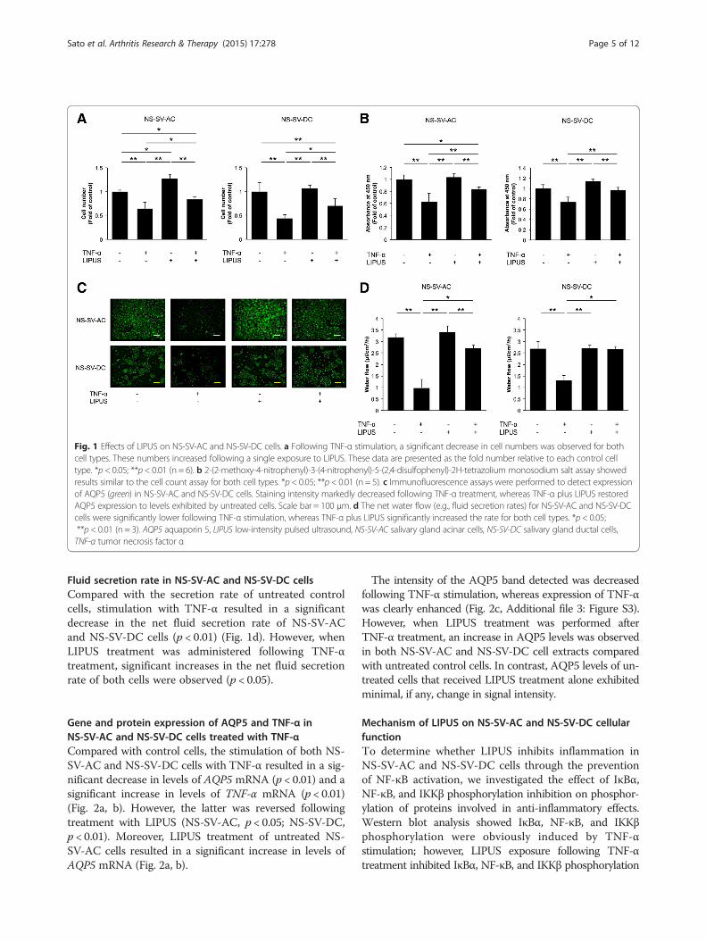
Fluid secretion rate in NS-SV-AC and NS-SV-DC cellsCompared with the secretion rate of untreated controlcells, stimulation with TNF-α resulted in a significantdecrease in the net fluid secretion rate of NS-SV-ACand NS-SV-DC cells (p < 0.01) (Fig. 1d). However, whenLIPUS treatment was administered following TNF-αtreatment, significant increases in the net fluid secretionrate of both cells were observed (p < 0.05).
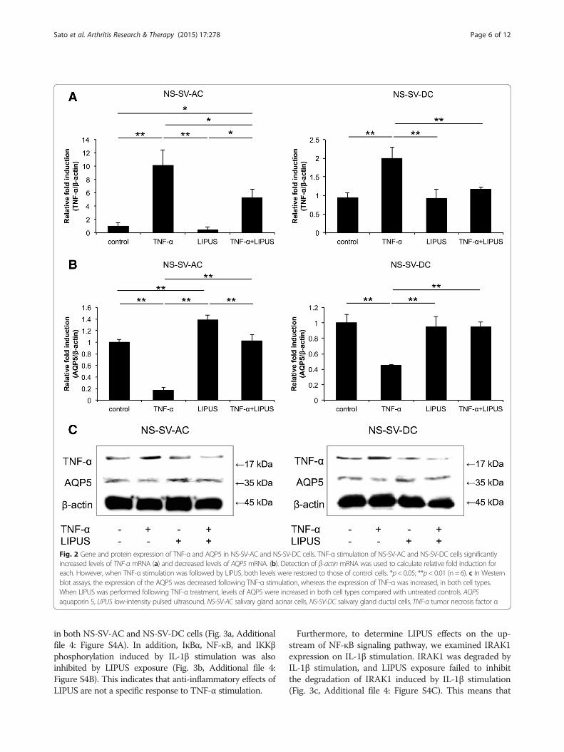
Gene and protein expression of AQP5 and TNF-α inNS-SV-AC and NS-SV-DC cells treated with TNF-αCompared with control cells, the stimulation of both NS-SV-AC and NS-SV-DC cells with TNF-α resulted in a sig-nificant decrease in levels of AQP5 mRNA (p < 0.01) and asignificant increase in levels of TNF-α mRNA (p < 0.01)(Fig. 2a, b). However, the latter was reversed followingtreatment with LIPUS (NS-SV-AC, p < 0.05; NS-SV-DC,p < 0.01). Moreover, LIPUS treatment of untreated NS-SV-AC cells resulted in a significant increase in levels ofAQP5 mRNA (Fig. 2a, b).
The intensity of the AQP5 band detected was decreasedfollowing TNF-α stimulation, whereas expression of TNF-αwas clearly enhanced (Fig. 2c, Additional file 3: Figure S3).However, when LIPUS treatment was performed afterTNF-α treatment, an increase in AQP5 levels was observedin both NS-SV-AC and NS-SV-DC cell extracts comparedwith untreated control cells. In contrast, AQP5 levels of un-treated cells that received LIPUS treatment alone exhibitedminimal, if any, change in signal intensity.
Mechanism of LIPUS on NS-SV-AC and NS-SV-DC cellularfunctionTo determine whether LIPUS inhibits inflammation inNS-SV-AC and NS-SV-DC cells through the preventionof NF-κB activation, we investigated the effect of IκBα,NF-κB, and IKKβ phosphorylation inhibition on phosphor-ylation of proteins involved in anti-inflammatory effects.Western blot analysis showed IκBα, NF-κB, and IKKβphosphorylation were obviously induced by TNF-αstimulation; however, LIPUS exposure following TNF-αtreatment inhibited IκBα, NF-κB, and IKKβ phosphorylation
Fig. 1 Effects of LIPUS on NS-SV-AC and NS-SV-DC cells. a Following TNF-α stimulation, a significant decrease in cell numbers was observed for bothcell types. These numbers increased following a single exposure to LIPUS. These data are presented as the fold number relative to each control celltype. *p < 0.05; **p < 0.01 (n = 6). b 2-(2-methoxy-4-nitrophenyl)-3-(4-nitrophenyl)-5-(2,4-disulfophenyl)-2H-tetrazolium monosodium salt assay showedresults similar to the cell count assay for both cell types. *p < 0.05; **p < 0.01 (n = 5). c Immunofluorescence assays were performed to detect expressionof AQP5 (green) in NS-SV-AC and NS-SV-DC cells. Staining intensity markedly decreased following TNF-α treatment, whereas TNF-α plus LIPUS restoredAQP5 expression to levels exhibited by untreated cells. Scale bar = 100 μm. d The net water flow (e.g., fluid secretion rates) for NS-SV-AC and NS-SV-DCcells were significantly lower following TNF-α stimulation, whereas TNF-α plus LIPUS significantly increased the rate for both cell types. *p < 0.05;**p < 0.01 (n = 3). AQP5 aquaporin 5, LIPUS low-intensity pulsed ultrasound, NS-SV-AC salivary gland acinar cells, NS-SV-DC salivary gland ductal cells,TNF-α tumor necrosis factor α
Sato et al. Arthritis Research & Therapy (2015) 17:278 Page 5 of 12
in both NS-SV-AC and NS-SV-DC cells (Fig. 3a, Additionalfile 4: Figure S4A). In addition, IκBα, NF-κB, and IKKβphosphorylation induced by IL-1β stimulation was alsoinhibited by LIPUS exposure (Fig. 3b, Additional file 4:Figure S4B). This indicates that anti-inflammatory effects ofLIPUS are not a specific response to TNF-α stimulation.
Furthermore, to determine LIPUS effects on the up-stream of NF-κB signaling pathway, we examined IRAK1expression on IL-1β stimulation. IRAK1 was degraded byIL-1β stimulation, and LIPUS exposure failed to inhibitthe degradation of IRAK1 induced by IL-1β stimulation(Fig. 3c, Additional file 4: Figure S4C). This means that
Fig. 2 Gene and protein expression of TNF-α and AQP5 in NS-SV-AC and NS-SV-DC cells. TNF-α stimulation of NS-SV-AC and NS-SV-DC cells significantlyincreased levels of TNF-α mRNA (a) and decreased levels of AQP5 mRNA. (b). Detection of β-actin mRNA was used to calculate relative fold induction foreach. However, when TNF-α stimulation was followed by LIPUS, both levels were restored to those of control cells. *p< 0.05; **p< 0.01 (n = 6). c In Westernblot assays, the expression of the AQP5 was decreased following TNF-α stimulation, whereas the expression of TNF-α was increased, in both cell types.When LIPUS was performed following TNF-α treatment, levels of AQP5 were increased in both cell types compared with untreated controls. AQP5aquaporin 5, LIPUS low-intensity pulsed ultrasound, NS-SV-AC salivary gland acinar cells, NS-SV-DC salivary gland ductal cells, TNF-α tumor necrosis factor α
Sato et al. Arthritis Research & Therapy (2015) 17:278 Page 6 of 12

Fig. 3 (See legend on next page.)
Sato et al. Arthritis Research & Therapy (2015) 17:278 Page 7 of 12

the effect point of LIPUS on the NF-κB signaling pathwayis downstream of IRAK1. Next, we examined A20 (tumornecrosis factor-α-induced protein 3 [TNFAIP3]) mRNAexpression using real-time PCR. A20 is known as theintracellular ubiquitin-editing protein and a key player inthe negative feedback of NF-κB signaling in response tomultiple stimulation [32]. Compared with untreatedcontrol cells, the stimulation of both NS-SV-AC andNS-SV-DC cells with TNF-α or IL-1β resulted in asignificant increase in levels of A20 mRNA (p < 0.01).Furthermore, A20 mRNA expression was more in-creased by treatment with LIPUS after TNF-α or IL-1βstimulation (NS-SV-AC, p < 0.01; NS-SV-DC, p < 0.05)(Fig. 3d). These data show that LIPUS activates A20,which produces negative feedback of NF-κB signalingin response to inflammatory stimulation, resulting ininhibition of inflammation in salivary gland cells.
Changes in fluid secretion volumes with agingThe average salivary volumes for 12-week-old, 20-week-old, and 24-week-old MRL/lpr mice were signifi-cantly lower than those obtained from 6-week-old mice(p < 0.01) (Fig. 4a). Moreover, compared with C57BL/6mice, the average salivary secretion volumes for theMRL/lprmice were significantly lower at all ages (p < 0.01).
In vivo effect of LIPUS on salivary secretion in MRL/lpr miceTo examine the effect of LIPUS on salivary secretion inMRL/lpr mice, secreted saliva volumes were measuredusing a modified method employed in the Saxon test[21]. Following LIPUS treatment, salivary secretion wasfound to be restored in both 12-week-old and 20-week-old MRL/lpr mice to a degree comparable to that ofyounger C57BL/6 mice (Fig. 4b). Furthermore, both 12-week-old and 20-week-old MRL/lpr mice exhibited asignificant increase in salivary secretion following LIPUStreatment compared with untreated MRL/lpr mice.
Anti-inflammatory effect of LIPUS on salivary glands inMRL/lpr miceMRL/lpr mice treated with LIPUS showed a marked re-duction in histological damage, such as the lymphocyteinfiltration of surrounding duct and the destruction ofgland tissue, compared with untreated MRL/lpr mice
(Additional file 5: Figure S5A). Moreover, the histo-logical score for the inflammatory lesions of the sub-mandibular glands of MRL/lpr mice was significantlyimproved following LIPUS (p < 0.05) (Fig. 4c).
AQP5 expression in salivary glandsAs shown in Fig. 4d, submandibular gland acinar cells ofuntreated MRL/lpr mice showed weak staining for AQP5expression (brown stain) in both the serous and mucousacini of the submandibular glands. In contrast, strongexpression of AQP5 was observed at apical sites in sub-mandibular gland acinar cells of MRL/lpr mice treatedwith LIPUS.
Induction of AQP5 and TNF-α expression in salivaryglandsFollowing LIPUS exposure, significantly lower levels ofTNF-α (p < 0.05) and significantly higher levels of AQP5(p < 0.05) were detected in the salivary glands of treatedMRL/lpr mice compared with untreated MRL/lpr mice(Fig. 4e, Additional file 5: Figure S5B).
DiscussionTo our knowledge, the effect of LIPUS on salivary glandcells has not previously been investigated. Thus, thepresent study would be the first to investigate the effectof LIPUS on cell proliferation and the synthesis of AQP5in normal human salivary gland acinar and ductal cellsstimulated with TNF-α. The present results indicate thatLIPUS is able to upregulate cell proliferation and AQP5expression in both of these stimulated cell types. Fur-thermore, using a mouse model of autoimmune sialade-nitis in SS, LIPUS treatment was found to improvesecretion from salivary glands in both age-related and SSxerostomia experiments.Although the mechanisms mediating LIPUS-stimulated
tissue repair have not yet been elucidated, it is hypothe-sized that the anabolic biophysical effects of ultrasoundare due to mechanical stress and/or the impact of fluidmicrostreaming on the cellular plasma membrane, focaladhesions, and cytoskeletal structures. As a result, intra-cellular signal transduction is activated and gene tran-scription is affected [33]. There is evidence to suggest thatLIPUS activates integrins on the cell surface that act as
(See figure on previous page.)Fig. 3 Anti-inflammatory mechanism of LIPUS on NS-SV-AC and NS-SV-DC cellular function. a Western blot analysis showed IκBα phosphorylationwas obviously induced by TNF-α stimulation; however, when LIPUS treatment was administered following TNF-α treatment, IκBα, NF-κB, and IKKβphosphorylation were inhibited. b Similar inhibitory effects of LIPUS were observed in IL-1β stimulation. c In Western blot analysis, IRAK1 wasdegraded after IL-1β stimulation, and LIPUS exposure failed to inhibit it. d The stimulation of both NS-SV-AC and NS-SV-DC cells with TNF-α orIL-1β resulted in a significant increase in levels of A20 mRNA, and A20 mRNA expression was further increased following treatment with LIPUSafter TNF-α or IL-1β stimulation. *p < 0.05; **p < 0.01 (n = 6). A20 tumor necrosis factor-α-induced protein 3 (TNFAIP3), IKKβ inhibitor of nuclearfactor κB kinase subunit β, IL-1β interleukin 1β, IRAK1 interleukin 1 receptor-associated kinase 1, IκBα inhibitor of nuclear factor of κ lightpolypeptide gene enhancer in B cells, α subunit, LIPUS low-intensity pulsed ultrasound, NF-κB nuclear factor κB, NS-SV-AC salivary gland acinarcells, NS-SV-DC salivary gland ductal cells, TNF-α tumor necrosis factor α
Sato et al. Arthritis Research & Therapy (2015) 17:278 Page 8 of 12

mechanoreceptors to promote the attachment of variousfocal adhesion adaptor proteins [34]. In addition, levels ofphosphorylated focal adhesion kinase (FAK) in synovialcells were found to increase following LIPUS treatment[35, 36]. Furthermore, LIPUS treatment of cementoblastshas been shown to enhance expression of extracellularsignal-regulated kinase 1/2 [35]. Because the proliferationof various cell types is mediated by growth factorsor cytokine-induced mitogen-activated protein kinase(MAPK) proteins [37], the exposure of salivary glandcells to LIPUS may lead to specific activation of anintegrin/FAK/MAPK pathway.In human salivary glands, AQP5 localizes to the apical
membranes of acinar cells and stimulates the outflow ofwater into the acinar lumen [38]. In patients with SS,the salivary and lacrimal glands exhibit a marked reduc-tion in AQP expression in the plasma membrane [39],as well as a delocalization of AQP5 to the basal mem-brane [40]. Correspondingly, reduced salivary gland se-cretion has been observed in mice harboring a mutantAQP5 channel [41]. Therefore, although the molecular
mechanisms by which AQP5 dysfunction is induced inthe salivary and lacrimal glands of patients with SS remainsunknown, it appears that enhancement of AQP5 expres-sion in salivary acinar cells may be a critical aspect. In thepresent study, expression of AQP5 in NS-SV-AC andNS-SV-DC cells was inhibited by TNF-α treatment, whereasit was recovered following LIPUS exposure. Furthermore,AQP5 synthesis and expression in the salivary glands ofMRL/lpr mice was restored following LIPUS treatment,resulting in the restoration of salivary flow to normal levels.In this study, we hypothesize that this event is due to
the anti-inflammatory effects mediated by LIPUS treat-ment through the prevention of NF-κB activation. NF-κB is composed of homo- and heterodimeric complexesof members of the Rel protein family. NF-κB normallyresides in the cytoplasm, where it is retained by associ-ation with IκB protein, an endogenous inhibitor. Variousextracellular stimuli trigger the degradation of IκB bythe proteasome pathway. Subsequently, NF-κB releasedfrom IκB translocates into the nucleus, binds to theregulatory element of the target genes, and controls their
Fig. 4 In vivo effects of LIPUS on MRL/lpr mice. a The average salivary volumes of 12-week-old, 20-week-old, and 24-week-old MRL/lpr mice werelower those of 6-week-old MRL/lpr mice. The average salivary secretion of the MRL/lpr mice at each age was also significantly lower than that forC57BL/6 mice at the same ages. **p< 0.01; ‡p< 0.01 compared with 6-week-old C57BL/6 mice; ##p< 0.01 compared with 6-week-old MRL/lpr mice (n = 5).b Both 12-week-old and 20-week-old MRL/lprmice showed a significant increase in salivary flow following LIPUS treatment. **p< 0.01 (n = 5). c Histologicalscores for inflammatory lesions present in the salivary glands of untreated versus LIPUS-treated MRL/lprmice. LIPUS treatment significantly improved thehistological score. *p< 0.05 (n = 5). d In both the serous and mucous acini of the submandibular glands, acinar cells of untreated MRL/lprmice exhibitedweak staining for AQP5, whereas intense staining of AQP5 was localized to apical sites in acinar cells of MRL/lpr mice treated with LIPUS (arrows).Scale bar = 100 μm. e In Western blot assays, levels of TNF-α were significantly lower, and levels of AQP5 were significantly higher, following LIPUStreatment compared with untreated MRL/lpr samples. Detection of β-actin was used to calculate relative fold induction for each. *p < 0.05 (n = 5).AQP5 aquaporin 5, LIPUS low-intensity pulsed ultrasound, MRL/lpr MRL/MpJ/lpr/lpr, TNF-α tumor necrosis factor α
Sato et al. Arthritis Research & Therapy (2015) 17:278 Page 9 of 12

transcription [42]. Our results demonstrated that theanti-inflammatory effects of LIPUS in NS-SV-AC andNS-SV-DC cells were involved in the inhibition of NF-κB signaling pathway. Interestingly, LIPUS activated theintracellular ubiquitin-editing protein A20 in inflamma-tory stimulation. A20, a cytoplasmic zinc finger protein,was originally identified as a TNF-inducible protein. Ithas been characterized as a dual inhibitor of NF-κBactivation and cell death [31] and functions as a negativefeedback regulator of NF-κB activation via multiplemechanisms [43]. Furthermore, A20 inhibits TNF- andIL-1-induced NF-κB activation in 293 cells [44], andderegulated Toll-like receptor signaling in response tocommensal bacteria was shown to be responsible for themultiorgan inflammation and premature death of A20-knockout mice [45]. Thus, LIPUS may inhibit the NF-κBpathway by activating a negative feedback system in thesalivary gland acinar and ductal cells.In IL-1β-stimulated synovial membrane cells, increased
expression of cyclooxygenase 2 (Cox-2), a NF-κB-responsible gene, was found to be significantly inhib-ited by LIPUS in vitro [24]. Expression of Cox-2 in theknee joints of MRL/lpr mice was also found to be mark-edly reduced following daily treatments with LIPUS [25],whereas inhibition of Cox-2 was found to reduce theproliferation and induce the apoptosis of human chol-angiocarcinoma QBC939 cells via inhibition of prosta-glandin E2 production [46]. On the basis of these data,it is hypothesized that inhibition of Cox-2 expression byLIPUS restores salivary secretion in MRL/lpr mice as asecondary effect.It is generally accepted that salivary secretion volume
decreases with age. The prevalence of xerostomia alsoincreases with age and affects approximately 30 % ofpeople aged 65 years or older [47]. In contrast, the pro-duction and composition of saliva remains largely inde-pendent of age in healthy individuals [48, 49]. Recently,Yamamura et al. [50] postulated that age-related hyper-methylation of the AQP5 gene could account for thedownregulation of AQP5 expression that is observed inthe salivary glands of patients with SS. Correspondingly,demethylation of the AQP5 promoter in salivary glandcells by 5-aza-2′-deoxycytidine (decitabine) could poten-tially restore salivary flow in aged mice. The results ofthe present study demonstrate that the average salivaryvolumes of the 12-week-old and older MRL/lpr andwild-type mice were significantly lower than those of the6-week-old mice. In addition, LIPUS treatment restoredthe salivary secretion volume of both the 12-week-oldand 20-week-old MRL/lpr mice to the salivary flow levelobserved in the younger wild-type mice. Thus, it appearsthat reduced salivary flow in older MRL/lpr mice is aconsequence of autoimmune disease as well as the agingprocess, and LIPUS treatment can compensate for these
processes. However, further studies are needed to iden-tify the effect of LIPUS on age-related xerostomia tounderstand how LIPUS affects salivary glands and theircells.In recent years, LIPUS has been paid attention as a
physical therapy that has insignificant side effects. In thisstudy, we investigated the effectiveness of LIPUS on xer-ostomia associated with SS, and the results suggest thatLIPUS might rescue salivary secretion volume in an SSmouse model by their anti-inflammatory effect in thesalivary gland tissue. Furthermore, LIPUS has much po-tential for clinical application because it can be used incombination with conventional pharmacotherapy. Our re-sults also suggest that further studies to determine clinicalefficacy, safety, and response duration are warranted.
ConclusionsLIPUS treatment was found to increase cell proliferationand AQP5 expression in salivary gland cells pretreated withTNF-α in vitro. Moreover, LIPUS activates the intracellularubiquitin-editing protein A20, which produces negativefeedback of NF-κB signaling in response to inflammatorystimulation. LIPUS exposure also restored salivary glandsecretion volumes in older MRL/lpr mice in vivo, therebypromoting an anti-inflammatory response and improvingAQP5 dysfunction. Therefore, LIPUS stimulation may rep-resent a treatment strategy for inflammatory diseases ofsalivary glands, including xerostomia in SS.
Additional files
Additional file 1: Figure S1. Schematic representation of the in vitroLIPUS system used. A cell culture plate with medium was placed in theultrasound field at a distance of about 1 mm from the transducer tooptimize beam uniformity across the target region. (TIFF 1093 kb)
Additional file 2: Figure S2. Quantification of the fluorescenceintensity in Fig. 1C. **p < 0.01 (n = 4). (TIFF 897 kb)
Additional file 3: Figure S3. Bands of Western blot analysis for thecropped images in Fig. 2c are provided in this file. (TIFF 1856 kb)
Additional file 4: Figure S4. Bands of Western blot analysis for thecropped images in Fig. 3a (A), 3b (B) and 3c (C) are provided in this file.(TIFF 5449 kb)
Additional file 5: Figure S5. (A) MRL/lpr mice treated with LIPUSexhibited a marked reduction in histological damage compared withuntreated MRL/lpr mice. Scale bar = 100 μm. (B) Bands of Western blotanalysis for the cropped images in Fig. 4e are provided in this file.(TIFF 5901 kb)
AbbreviationsA20: tumor necrosis factor-α-induced protein 3 (TNFAIP3); AQP5: aquaporin 5;Cox-2: cyclooxygenase 2; FAK: focal adhesion kinase;HCQ: hydroxychloroquine; HRP: horseradish peroxidase; IKKβ: inhibitor ofnuclear factor κB kinase subunit β; IL-1β: interleukin 1β; IRAK1: interleukin 1receptor-associated kinase 1; IκBα: inhibitor of nuclear factor of κ lightpolypeptide gene enhancer in B cells, α subunit; LIPUS: low-intensity pulsedultrasound; MAPK: mitogen-activated protein kinase; MRL/lpr: MRL/MpJ/lpr/lpr;NF-κB: nuclear factor κB; NS-SV-AC: salivary gland acinar cells; NS-SV-DC: salivarygland ductal cells; PBS: phosphate-buffered saline; RT: room temperature;
Sato et al. Arthritis Research & Therapy (2015) 17:278 Page 10 of 12

SFKM: serum-free keratinocyte medium; SS: Sjögren syndrome; TBS-T:Tris-buffered saline with Tween 20; TNF-α: tumor necrosis factor α.
Competing interestsThe authors declare that they have no competing interests.
Authors’ contributionsMS carried out all of studies, performed the statistical analysis, and draftedthe manuscript. SK performed the statistical analysis and helped to revise themanuscript. KQM carried out the histological analysis and helped to revisethe manuscript. GK carried out the immunohistochemical analysis andhelped to revise the manuscript. KN participated in the design of the study,carried out the immunoassays, and helped to revise the manuscript. SHcarried out the study of cell metabolism, analyzed the data, and helped torevise the manuscript. TI participated in the design of the study and studycoordination and drafted the manuscript. YY provided the salivary glandcells, performed technical support, participated in the design of the study,and helped to revise the manuscript. MA provided the salivary gland cells,participated in the design of the study, and helped to revise the manuscript.ET participated in the design of the study and study coordination anddrafted the manuscript. All authors read and approved the final manuscript.
AcknowledgmentsThis research was supported in part by Grant-in-Aid 26293436 (to ET) for scienceresearch from the Ministry of Education, Culture, Sports, Science and Technology,Japan. The funder had no role in study design, data collection and analysis,decision to publish, or preparation of the manuscript. Furthermore, the authorsthank Nobuyasu Yamanaka, Atsushi Chuma, and Akira Tabata for technicalassistance.
Author details1Department of Orthodontics and Dentofacial Orthopedics, Institute ofBiomedical Sciences, Tokushima University Graduate School, Tokushima,Japan. 2Genetic Disease Program, Sanford Children’s Health Research Center,Sanford-Burnham Medical Research Institute, La Jolla, CA, USA. 3Departmentof Oral Medicine, Institute of Biomedical Sciences, Tokushima UniversityGraduate School, Tokushima, Japan. 4Department of Orthodontics, Faculty ofDentistry, King Abdulaziz University, Jeddah, Saudi Arabia.
Received: 2 June 2015 Accepted: 24 September 2015
References1. Fox RI. Sjögren’s syndrome. Lancet. 2005;366:321–31.2. Daniels TE. Labial salivary gland biopsy in Sjögren’s syndrome: assessment as a
diagnostic criterion in 362 suspected cases. Arthritis Rheum. 1984;27:147–56.3. Yamamura Y, Motegi K, Kani K, Takano H, Momota Y, Aota K, et al. TNF-α
inhibits aquaporin 5 expression in human salivary gland acinar cells viasuppression of histone H4 acetylation. J Cell Mol Med. 2012;16:1766–75.
4. Hamano H, Saito I, Haneji N, Mitsuhashi Y, Miyasaka N, Hayashi Y.Expressions of cytokine genes during development of autoimmunesialadenitis in MRL/lpr mice. Eur J Immunol. 1993;23:2387–91.
5. Fox RI, Kang HI, Ando D, Abrams J, Pisa E. Cytokine mRNA expression insalivary gland biopsies of Sjögren’s syndrome. J Immunol. 1994;152:5532–9.
6. Soliven B, Wang N. Tumor necrosis factor-α regulates nicotinic responses inmixed cultures of sympathetic neurons and nonneuronal cells. JNeurochem. 1995;64:883–94.
7. King LS, Yasui M. Aquaporins and disease: lessons from mice to humans.Trends Endocrinol Metab. 2002;13:355–60.
8. Fox RI, Dixon R, Guarrasi V, Krubel S. Treatment of primary Sjögren’ssyndrome with hydroxychloroquine: a retrospective, open-label study.Lupus. 1996;5 Suppl 1:S31–6.
9. Kruize AA, Hené RJ, Kallenberg CG, van Bijsterveld OP, van der Heide A,Kater L, et al. Hydroxychloroquine treatment for primary Sjögren’ssyndrome: a two year double blind crossover trial. Ann Rheum Dis.1993;52:360–4.
10. Gottenberg JE, Ravaud P, Puéchal X, Le Guern V, Sibilia J, Goeb V, et al.Effects of hydroxychloroquine on symptomatic improvement in primarySjögren syndrome: the JOQUER randomized clinical trial. JAMA.2014;312:249–58.
11. Steinfeld SD, Demols P, Salmon I, Kiss R, Appelboom T. Infliximab in patients withprimary Sjögren’s syndrome: a pilot study. Arthritis Rheum. 2001;44:2371–5.
12. Mariette X, Ravaud P, Steinfeld S, Baron G, Goetz J, Hachulla E, et al.Inefficacy of infliximab in primary Sjögren’s syndrome: results of therandomized, controlled Trial of Remicade in Primary Sjögren’s Syndrome(TRIPSS). Arthritis Rheum. 2004;50:1270–6.
13. Sankar V, Brennan MT, Kok MR, Leakan RA, Smith JA, Manny J, et al.Etanercept in Sjögren’s syndrome: a twelve-week randomized, double-blind,placebo-controlled pilot clinical trial. Arthritis Rheum. 2004;50:2240–5.
14. Flach AJ. Improving the risk-benefit relationship and informed consent forpatients treated with hydroxychloroquine. Trans Am Ophthalmol Soc.2007;105:191–7.
15. Desai D, Goldbach-Mansky R, Milner JD, Rabin RL, Hull K, Pucino F, et al.Anaphylactic reaction to anakinra in a rheumatoid arthritis patient intolerant tomultiple nonbiologic and biologic disease-modifying antirheumatic drugs. AnnPharmacother. 2009;43:967–72.
16. Antoni C, Braun J. Side effects of anti TNF therapy: current knowledge. Clin ExpRheumatol. 2002;20(6 Suppl 28):S152–7.
17. Gebauer D, Correll J. Pulsed low-intensity ultrasound: a new salvageprocedure for delayed unions and nonunions after leg lengthening inchildren. J Pediatr Orthop. 2005;25:750–4.
18. Heckman JD, Ryaby JP, McCabe J, Frey JJ, Kilcoyne RF. Acceleration of tibialfracture-healing by non-invasive, low-intensity pulsed ultrasound. J BoneJoint Surg Am. 1994;76:26–34.
19. Azuma Y, Ito M, Harada Y, Takagi H, Ohta T, Jingushi S. Low-intensity pulsedultrasound accelerates rat femoral fracture healing by acting on the variouscellular reactions in the fracture callus. J Bone Miner Res. 2001;16:671–80.
20. Chan CW, Qin L, Lee KM, Zhang M, Cheng JC, Leung KS. Low intensitypulsed ultrasound accelerated bone remodeling during consolidation stageof distraction osteogenesis. J Orthop Res. 2006;24:263–70.
21. Warden SJ, Bennell KL, McMeeken JM, Wark JD. Acceleration of freshfracture repair using the Sonic Accelerated Fracture Healing System (SAFHS):a review. Calcif Tissue Int. 2000;66:157–63.
22. Zhou S, Schmelz A, Seufferlein T, Li Y, Zhao J, Bachem MG. Molecularmechanisms of low intensity pulsed ultrasound in human skin fibroblasts.J Biol Chem. 2004;279:54463–9.
23. Nagata K, Nakamura T, Fujihara S, Tanaka E. Ultrasound modulates theinflammatory response and promotes muscle regeneration in injuredmuscles. Ann Biomed Eng. 2013;41:1095–105.
24. Nakamura T, Fujihara S, Katsura T, Yamamoto K, Inubushi T, Tanimoto K, etal. Effects of low-intensity pulsed ultrasound on the expression and activityof hyaluronan synthase and hyaluronidase in IL-1β-stimulated synovial cells.Ann Biomed Eng. 2010;38:3363–70.
25. Nakamura T, Fujihara S, Yamamoto-Nagata K, Katsura T, Inubushi T, TanakaE. Low-intensity pulsed ultrasound reduces the inflammatory activity ofsynovitis. Ann Biomed Eng. 2011;39:2964–71.
26. Takakura Y, Matsui N, Yoshiya S, Fujioka H, Muratsu H, Tsunoda M, et al.Low-intensity pulsed ultrasound enhances early healing of medial collateralligament injuries in rats. J Ultrasound Med. 2002;21:283–8.
27. Azuma M, Tamatani T, Kasai Y, Sato M. Immortalization of normal humansalivary gland cells with duct-, myoepithelial-, acinar-, or squamousphenotype by transfection with SV40 ori- mutant deoxyribonucleic acid. LabInvest. 1993;69:24–42.
28. Neufeld TK, Grant ME, Grantham JJ. A method to measure the rate of netfluid secretion by monolayers of cultured renal epithelial cells. J Tiss CultMeth. 1991;13:229–34.
29. Delporte C, O’Connell BC, He X, Lancaster HE, O’Connell AC, Agre P, et al.Increased fluid secretion after adenoviral-mediated transfer of the aquaporin-1cDNA to irradiated rat salivary glands. Proc Natl Acad Sci U S A. 1997;94:3268–73.
30. Kohler PF, Winter ME. A quantitative test for xerostomia: the Saxon test, anoral equivalent of the Schirmer test. Arthritis Rheum. 1985;28:1128–32.
31. White SC, Casarett GW. Damage of rat thyroid by 131I and evidence againstimmunologic transferability. Radiat Res. 1974;57:288–99.
32. Vereecke L, Beyaert R, van Loo G. The ubiquitin-editing enzyme A20 (TNFAIP3)is a central regulator of immunopathology. Trends Immunol. 2009;30:383–91.
33. Romano CL, Romano D, Logoluso N. Low-intensity pulsed ultrasound forthe treatment of bone delayed union or nonunion: a review. UltrasoundMed Biol. 2009;35:529–36.
34. Lal H, Verma SK, Smith M, Guleria RS, Lu G, Foster DM, et al. Stretch-inducedMAP kinase activation in cardiac myocytes: differential regulation throughβ1-integrin and focal adhesion kinase. J Mol Cell Cardiol. 2007;43:137–47.
Sato et al. Arthritis Research & Therapy (2015) 17:278 Page 11 of 12

35. Sato M, Nagata K, Kuroda S, Horiuchi S, Nakamura T, Karima M, et al.Low-intensity pulsed ultrasound activates integrin-mediatedmechanotransduction pathway in synovial cells. Ann Biomed Eng.2014;42:2156–63.
36. Tanaka E, Kuroda S, Horiuchi S, Tabata A, El-Bialy T. Low-intensity pulsedultrasound in dentofacial tissue engineering. Ann Biomed Eng. 2015;43:871–86.
37. Cowan KJ, Storey KB. Mitogen-activated protein kinases: new signalingpathways functioning in cellular responses to environmental stress. J ExpBiol. 2003;206:1107–15.
38. Aqre P, Brown D, Nielsen S. Aquaporin water channels: unansweredquestions and unresolved controversies. Curr Opin Cell Biol. 1995;7:472–83.
39. Tsubota K, Hirai S, King LS, Agre P, Ishida N. Defective cellular trafficking oflacrimal gland aquaporin-5 in Sjögren’s syndrome. Lancet. 2001;357:688–9.
40. Steinfeld S, Cogan E, King LS, Agre P, Kiss R, Delporte C. Abnormaldistribution of aquaporin-5 water channel protein in salivary glands fromSjögren’s syndrome patients. Lab Invest. 2001;81:143–8.
41. Ma T, Song Y, Gillespie A, Carlson EJ, Epstein CJ, Verkman AS. Defectivesecretion of saliva in transgenic mice lacking aquaporin-5 water channels. JBiol Chem. 1999;274:20071–4.
42. Barnes PJ, Karin M. Nuclear factor-κB: a pivotal transcription factor in chronicinflammatory diseases. N Engl J Med. 1997;336:1066–71.
43. Catrysse L, Vereecke L, Beyaert R, van Loo G. A20 in inflammation andautoimmunity. Trends Immunol. 2014;35:22–31.
44. Song HY, Rothe M, Goeddel DV. The tumor necrosis factor-inducible zincfinger protein A20 interacts with TRAF1/TRAF2 and inhibits NF-κB activation.Proc Natl Acad Sci U S A. 1996;93:6721–5.
45. Turer EE, Tavares RM, Mortier E, Hitotsumatsu O, Advincula R, Lee B, et al.Homeostatic MyD88-dependent signals cause lethal inflammation in theabsence of A20. J Exp Med. 2008;205:451–64.
46. Wu GS, Zou SQ, Liu ZR, Tang ZH, Wang JH. Celecoxib inhibits proliferationand induces apoptosis via prostaglandin E2 pathway in humancholangiocarcinoma cell lines. World J Gastroenterol. 2003;9:1302–6.
47. Ship JA, Pillemer SR, Baum BJ. Xerostomia and the geriatric patient. J AmGeriatr Soc. 2002;50:535–43.
48. Wu AJ, Atkinson JC, Fox PC, Baum BJ, Ship JA. Cross-sectional andlongitudinal analyses of stimulated parotid salivary constituents in healthy,different-aged subjects. J Gerontol. 1993;48:M219–24.
49. Ship JA, Nolan NE, Puckett SA. Longitudinal analysis of parotid andsubmandibular salivary flow rates in healthy, different-aged adults. JGerontol A Biol Sci Med Sci. 1995;50:M285–9.
50. Yamamura Y, Aota K, Yamanoi T, Kani K, Takano H, Momota Y, et al. DNAdemethylating agent decitabine increases AQP5 expression and restoressalivary function. J Dent Res. 2012;91:612–7.
Submit your next manuscript to BioMed Centraland take full advantage of:
• Convenient online submission
• Thorough peer review
• No space constraints or color figure charges
• Immediate publication on acceptance
• Inclusion in PubMed, CAS, Scopus and Google Scholar
• Research which is freely available for redistribution
Submit your manuscript at www.biomedcentral.com/submit
Sato et al. Arthritis Research & Therapy (2015) 17:278 Page 12 of 12