low resolution refinement - phenix · low resolution refinement ... delta l a u d si re-30 -15 0...
TRANSCRIPT

Low Resolution Refinement
Paul AdamsLawrence Berkeley Laboratory and
Department of Bioengineering UC Berkeley
Macromolecular Crystallography SchoolMadrid, May 2017

Macromolecular Crystallography
PDB ID: 3k7aResolution: 3.80Å
PDBID: 2gkgResolution: 1.00Å
• Many challenges, but low resolution data is increasingly an issue:
• How to interpret “featureless” maps (pattern matching, chemical constraints)
• How to optimize models with sparse data (prior information)

The Challenge of Too Few Data
• With only low resolution data we typically have too many parameters to optimize
• Atomic coordinates, displacement parameters
• Underdetermined optimization problems lead to overfitting of the data
• To help address overfitting we can:
• Add prior information to reduce the number of effective parameters
• Remove parameters
• Current refinement methods do not define a reasonable chemical result in the absence of data

New Methods in Phenix for Improving Models
• Using prior structural knowledge as additional restraints:
• Secondary structure
• Protein mainchain conformations (Ramachandran)
• Related high resolution structures as restraints
• Multiple copies of the same molecule as restraints (c.f. local NCS restraints in SHELX)
• Automated correction of models during refinement using prior knowledge of stereochemistry:
• Fixing of rotamers
• Flipping of side chains
Reference model restraints(Jeff Headd)

1GTX and 1OHV
4-aminobutyrate-aminotransferase
1GTX: 3.0 Å
1OHV: 2.3 Å

1GTX and 1OHV
1GTX: 3.0 Å1OHV: 2.3 Å
4-aminobutyrate-aminotransferase

• Pre-correct rotamer outliers• Set rotamer outliers in the model to match the torsion angles of the
reference model if the reference model has an acceptable rotamer at that position and there is no significant clash or density mismatch
• Generate reference torsion restraints• Restrain each torsion angle in the working model to the
corresponding torsion angle in the reference model• Chains are aligned using SSM alignment to allow for sequence differences
• Restraints take the form of a modified harmonic ‘top-out’ potential that allows for structural differences
Reference Model Restraints
Combines two concepts:
Headd JJ et al., 2012, Acta Cryst. D68:381-390

Etotal =
n!
i=1
Ei
where σ is the ESD, Δ is the difference between the model dihedral and reference dihedral, and l is a ‘limit’ parameter that limits how far the model dihedral may vary from the reference dihedral before being shut off.
Reference model restraints
‘Top-out’ potential:
Ei = w∆2
iSimple harmonic potential:
Ei = τ(1.0− e
−∆2
i
l2 )
τ = wl2
w =1
σ2
"top-out" function
delta
residual
-30 -15 0 15 30
025
5075
100
125
150
175
200
225
250
default: limit = 15.0°developed by Ralf Grosse-Kunstleve
Similar potentials are used in REFMAC5 and BUSTER - Geman-McClure robust estimator function

The ‘limit’ parameter
>> limit,no restraint
< limit, restrain all dihedrals to reference
default: limit = 15.0°
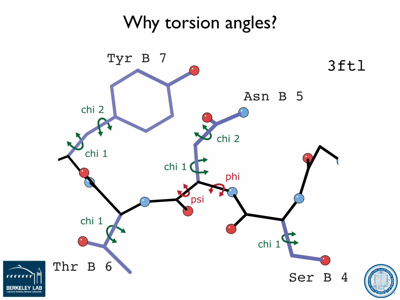
Why torsion angles?
3ftl
Asn B 5
Tyr B 7
Thr B 6Ser B 4
chi 1phi
psi
chi 2chi 1
chi 1
chi 1
chi 2

1GTX/1OHV reference example
5 macrocycles of phenix.refinew/ reference restraints Rfree: 0.2379 ! 0.2186
ΔR: 0.833 ! 0.60MolProbity: 64th ! 96th
outlier
tp rotamer
outlier correction restrained refinement
1GTX (3.0Å) 1OHV (2.3Å) 1GTX w/ 1OHV reference
Leu A 34 Glu A 41
mt-10rotamer
tprotamer

Practical Example
cAMP bound: 2.49Å
cGMP bound: 3.20Å
APO form: 2.69Å
Cyclic GMP-dependent protein kinases (PKG’s)
JJ Kim et al. (2011) Crystal structures of PKG Iβ (92-227) with cGMP and cAMP reveal the molecular details of cyclic nucleotide binding. PLoS ONE.

Cyclic GMP-dependent protein kinase
cAMP bound: 2.49Å
cGMP bound: 3.20Å
APO form: 2.69Å
16.53
2.61%
0.00%
98.80%
2.04
0.00%
0.00%
cAMP bound
0
95th
81st
0.1960
0.2264
Clashscore, all atoms:
Poor rotamers:
Rama outliers:
Rama favored:
Cβ dev. > 0.25Å:
MolProbity score:
Res w/ bad bonds:
Res w/ bad angles:
All-Atom Contacts
ProteinGeometry
R-work
R-free
Clashscore percentile
MP score percentile
Validation Criteria
Residual
56.57
18.58%
2.02%
85.48%
3.84
2.38%
5.95%
cGMP bound
23
12th
15th
0.2102
0.2582
24.56
4.00%
0.40%
96.00%
2.61
0.00%
1.18%
cGMP bound
0
96th
87th
0.1980
0.2397
28.52
10.53%
3.19%
89.02%
3.29
0.79%
0.98%
APO
3
12th
46th
0.2205
0.2612
19.5
3.66%
0.60%
96.61%
2.43
0.00%
0.20%
APO
0
89th
15th
0.2166
0.2525

Sources of Prior Information
Images from PumMa web site (http://www.pumma.nl)
Mainchain distributions
Sidechain distributions
Covalent geometry
Related structures
Secondary structure
Internal symmetry

Torsion space NCS restraints(Jeff Headd)

rotamer outlier correction
Leu B 180outlier
Leu B 180tp rotamer
Leu B 180 Leu B 180tp rotamer
1b04: 2.8 ÅDNA ligase
1. Identify rotamer outlier
2. correct to corresponding rotamer in NCS-related chain by matching χ angles
3. ‘backrub’search, thenlimited χ angletorsion search
4. verify rotamer is still correct match
“backrub”
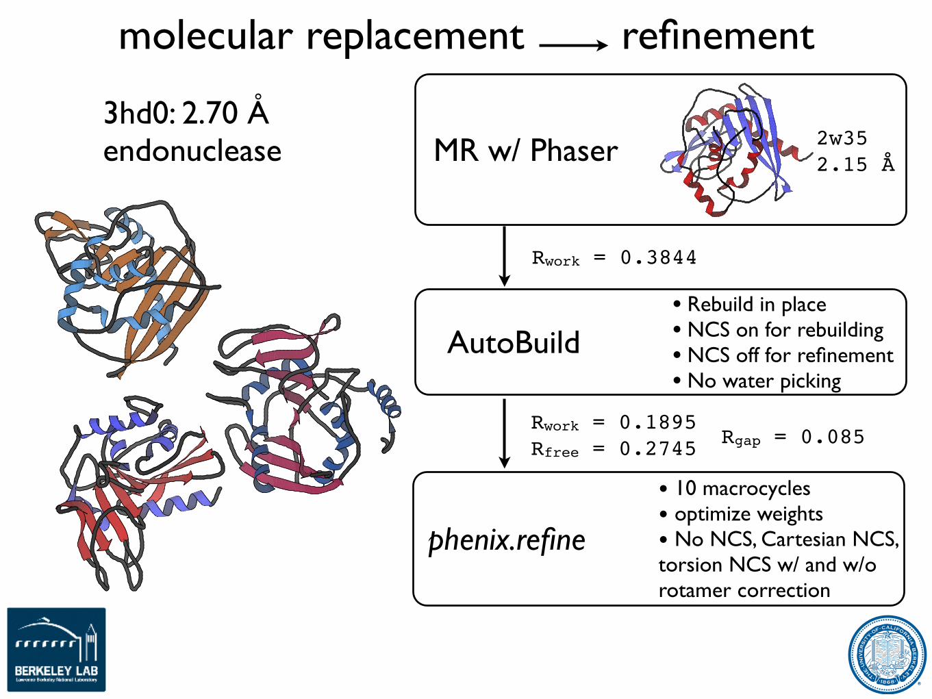
molecular replacement refinement
3hd0: 2.70 Åendonuclease
Rwork = 0.3844
Rwork = 0.1895Rfree = 0.2745
MR w/ Phaser 2w352.15 Å
AutoBuild• Rebuild in place• NCS on for rebuilding• NCS off for refinement• No water picking
phenix.refine
• 10 macrocycles• optimize weights• No NCS, Cartesian NCS, torsion NCS w/ and w/o rotamer correction
Rgap = 0.085

0.25%
0.26%
0.27%
0.28%
0.29%
0.3%
0.31%
0.32%
0.33%
0.34%
1% 2% 3% 4% 5% 6% 7% 8% 9% 10% 11%
macrocycle
1 2 3 4 5 6 7 8 9 10
Rfr
ee3hd0 refinement
no NCS Cartesian NCS torsion NCS torsion NCS w/ rotamer correction
Rwork = 0.2040Rfree = 0.2606
torsion NCS w/ rotamer correction Rgap = 0.056

Sources of Prior Information
Images from PumMa web site (http://www.pumma.nl)
Mainchain distributions
Sidechain distributions
Covalent geometry
Related structures
Secondary structure
Internal symmetry

More Prior Information
• As the number of observations decreases we need to increase the amount of prior information we include (or the number of constraints we apply)
• At the extreme - what if we had no data?
• Other fields have been trying to address this problem:
• Structure prediction
• Homology modelling
• Protein folding
From: Kryshtafovych & Fidelis, Drug Discovery Today, 2009, 14:386–393
http://www.predictioncenter.org

Physically Realistic Potentials (Rosetta)(Nat Echols & Frank DiMaio)

Rosetta
• ab initio model generation and model optimization
• Requires extensive computational sampling
Black - Rosetta ab initio models, Red - Crystal structure after Relax protocol

Why Rosetta
• Designed to recognize near-native structures among many possible models; combines empirical and physical potentials
• All-atom force field, incorporates solvation effects, attractive forces, hydrogen bonds, knowledge-based dihedral restraints
• Can yield chemically realistic ab initio models without experimental data to guide assembly
• Occasionally good enough for molecular replacement
• Shown to be useful for NMR structure determination with sparse data (CS-Rosetta), MR solution improvement (MR-Rosetta), RNA structure refinement (ERRASER)
Kuhlman et al. (2003) Science 302:1364-8Rohl et al. (2004) Methods Enzymol. 383:66-93Keedy et al. (2009) Proteins 77:29-49 https://www.rosettacommons.org

Complementary Algorithms
Rosetta
• Physically realistic potentials
• Repacking to remove steric clashes and building rotameric sidechains
• Torsion-angle minimization
• Real-space target (refinement against electron density)
• Fragment-based rebuilding (optional, not currently used)
Phenix
• Reciprocal space X-ray target functions (ML, MLHL, LS-twin)
• Bulk solvent correction
• B-factor refinement (including TLS)
• Map calculation
• Density modification (using RESOLVE)
Python/C++ architecture facilitates combination

Low Resolution Protocol
• Sidechain repacking (using density)
• Coordinate refinement (reciprocal space torsion angle minimization and reduced nonbonded penalty)
• B-factor refinement
• Sidechain repacking (using density)
• Coordinate refinement (real space and reciprocal space torsion angle minimization)
• B-factor refinement
• Sidechain repacking (using density)
• Coordinate refinement (reciprocal space minimization with restrained bonds and angles)
• B-factor refinement
3 Cycles 5 Cycles 2 Cycles
Protocol run 5 times in parallel and the best model selected based on R-free

Test Cases
3fps (3.2Å) 3k07 (3.52Å) 2x79 (3.8Å) 1isr (4.0Å)
Membrane Proteins
3pwy (3.5Å) 3idq (3.7Å) 3a8n (4.5Å)
Solved using homologous proteins
2j5f (3.0Å) 1bke (3.15Å) 3mtt (3.3Å) 1kct (3.46Å) 3snh (3.7Å) 2vaf (3.8Å) 3rzf (4.0Å)
Solved by molecular replacement with same protein from another deposition at higher resolution

Calcium ATPase - phenix.refine
R R-free mp score RMSD
start 0.47 0.51 3.21 6.1
phenix 0.43 0.48 2.66 6.2

Calcium ATPase - DEN
R R-free mp score RMSD
start 0.47 0.51 3.21 6.1
DEN 0.38 0.44 3.79 6.1

Calcium ATPase - Phenix-Rosetta
R R-free mp score RMSD
start 0.47 0.51 3.21 6.1
Rosetta 0.24 0.28 1.55 1.7

Calcium ATPase - Detail
• Phenix-Rosetta model is very close to the deposited structure (even at the level of side chains) with better fit to density

0"
4"
8"
12"
16"Start" Phenix"DEN" Rose8a"
0.0" 6.0"molprobity" 0.0" 6.0"RMSd" 0.20" 0.55"Rfree"
freq
uency"
Improved Models
• Phenix-Rosetta typically has improved fit to the crystallographic data and models are closer to the known structure
• Phenix-Rosetta always has improved model quality, as judged by Molprobity
• Generally similar to DEN results but with much improved geometry, and generally faster
0"
4"
8"
12"
16"Start" Phenix"DEN" Rose8a"
0.0" 6.0"molprobity" 0.0" 6.0"RMSd" 0.20" 0.55"Rfree"
freq
uency"
DiMiao et al., 2013, Nature Methods 10:1102-1104

Acknowledgments
• CCI• Lawrence Berkeley Laboratory
• Pavel Afonine, Youval Dar, Nat Echols, Jeff Headd, Richard Gildea, Ralf Grosse-Kunstleve, Dorothee Liebschner, Nigel Moriarty, Nader Morshed, Billy Poon, Ian Rees, Nicholas Sauter, Oleg Sobolev, Peter Zwart
• Los Alamos National Laboratory
• Tom Terwilliger, Li-Wei Hung
• Funding:
• NIH/NIGMS:
• P01GM063210, P50GM062412, P01GM064692, R01GM071939
• Lawrence Berkeley Laboratory
• PHENIX Industrial Consortium
• Cambridge University
• Randy Read, Airlie McCoy, Laurent Storoni, Gabor Bunkoczi, Robert Oeffner
• Duke University
• Jane Richardson & David Richardson, Ian Davis, Vincent Chen, Jeff Headd, Christopher Williams, Bryan Arendall, Laura Murray, Gary Kapral, Dan Keedy, Swati Jain, Bradley Hintze, Lindsay Deis, Lizbeth Videau
• Others
• Alexandre Urzhumtsev & Vladimir Lunin
• Garib Murshudov & Alexi Vagin
• Kevin Cowtan, Paul Emsley, Bernhard Lohkamp
• David Abrahams
• PHENIX Testers & Users: James Fraser, Herb Klei, Warren Delano, William Scott, Joel Bard, Bob Nolte, Frank von Delft, Scott Classen, Ben Eisenbraun, Phil Evans, Felix Frolow, Christine Gee, Miguel Ortiz-Lombardia, Blaine Mooers, Daniil Prigozhin, Miles Pufall, Edward Snell, Eugene Valkov, Erik Vogan, Andre White, and many more
• Oak Ridge National Laboratory
• Marat Mustyakimov, Paul Langan
• University of Washington
• Frank DiMaio, David Baker