lowering inflammation level by lp-pla2 inhibitor ... · background: type 2 diabetes melitus ... rat...
TRANSCRIPT
J Cardiovasc Disease Res., 2017; 8(2):50-55A Multifaceted Peer Reviewed Journal in the field of Cardiologywww.jcdronline.org | www.journalonweb.com/jcdr
50 Journal of Cardiovascular Disease Research, Vol 8, Issue 2, Apr-Jun, 2017
Original Article
INTRODUCTIONCardiovascular disease (CVD) has become the leading cause of death in both developed and developing countries. The global mortality rate of cardiovascular disease has reached 13.4 millions since 2007 and is ex-pected to increase to 23.6 millions in 2030.1 Atherosclerosis is the most common cause of CVD.2
Type 2 Diabetes mellitus (T2DM) is one of major classical risk factors for atherosclerosis.3 Insulin resistance in T2DM causes extensive tissue damage due to vascular inflammation and oxidative stress.4 Hyperglyce-mic environment caused by T2DM increased Reactive Oxygen Species (ROS) production.5
Enzymatic activity and expression of lipoprotein associated phospho-lipase A2 (Lp-PLA2) was studied as a biomarker of CVD.6 Lp-PLA2’s anti-inflammatory role demonstrated by the ability of this enzyme to hydrolyze atherogenesis mediators such as oxidized LDL (oxLDL) and platelet activating factor (PAF). But, the products of these molecules such as lysophosphatidylcholine (lysoPC) and oxidized fatty acid (oxFA) have pro-inflammatory, proliferative and pro-atherogenic effect.A new pharmacological therapy or atherosclerosis that selectively inhibit Lp-PLA2 was being developed substrate. Darapladib reduce caspase-3 and caspase-8 activity and inhibits macrophage apoptosis induced by ox-LDL.Several studies stated that darapladib showed significant results in in-hibiting atherosclerosis process.7,8,9,10 However, there are several opinions
about darapladib usefulness.11 So, this study aims to determine epression of inflammation marker of T2DM in vivo model with darapladib treat-ment.
MATERIAL AND METHODSStudy groupThis study used 4 weeks old male Sprague Dawley rats and weighted about 150-200 grams. They were obtained from Bogor Agricultural Uni-versity, Indonesia. 30 rats were divided into three groups; normal (N); T2DM fed with High Fat Diet (HFD) and low dose Streptozotocin (STZ) intraperitoneal injection of 35 mg/kg BW, and T2DM with darapladib administration group (DMDP). Each group was divided into two serial times: 8 weeks (early phase) and 16 weeks (late phase). So there are 6 groups total with 5 rats in each group. Darapladib was obtained from Glaxo Smith Kline. Samples were given Darapladib orally 20mg/Kg body weight once daily according the time serial groups given. The normal rats food contained total energy Calories (kcal/g) of 3.43 (67% carbohydrate, 21% protein and 12% fat), while the HFD contained total calorie energy of 5.29 (58% fat, 17% carbohydrate and 25 % pro-tein). Measurements of parameters were done at the Central Laboratory of Biological Sciences, Brawijaya University, Malang, Indonesia. Slicing and staining samples were done at Pathological Anatomy Laboratory, Faculty of Medicine University of Brawijaya, Malang, Indonesia
Lowering Inflammation Level by Lp-PLA2 Inhibitor (Darapladib) in Early Atherosclerosis Development: in vivo Rat Type 2 Diabe-tes Mellitus Model
ABSTRACTBackground: Type 2 Diabetes Melitus (T2DM) is a condition of insulin resistance that causes extensive tissue damage due to vascular inflammation and oxidative stress. Lipoprotein-associated phospholipase A2 (Lp-PLA2) has anti-inflam-matory role as it hydrolyzes atherogenesis mediators such as oxidized LDL (Ox LDL) and platelet activating factor (PAF) but in contrast, it has pro inflammatory effect as it produced lysophosphatidylcholine (lysoPC) and oxidized fatty acid (oxFA). Methods and Results: This study aimed to measure inflammation marker of T2DM in vivo model with Lp-PLA2 selective inhibitor (darapladib) treatment. It used true experimental laboratory and post test only with control group de-sign using 30 spraque dowley rats that is divided into 3 main groups: normal, T2DM, and T2DM with darapladib (DMDP) administration group. Each group are divided into 2 serial treatment times: 8-weeks and 16-weeks intervention. So, there are 6 groups total with 5 rat in each group. Parameter measured was ox-LDL, tumor necrosis factor-α (TNF-α), inducible nitric oxide synthase (iNOS), interleukin 6 (IL-6), PAF, perivascular adipose tissue (PAT) thickness and also blood glucose, lipid profile, and insulin plasma level. ANOVA test result showed that darapladib were significantly (p: 0.000) lowering tissue and blood ox-LDL level, iNOS, PAF, IL-6 and PAT thickness on T2DM in vivo model. Conclusions: Darapladib proved to have anti inflammation role on T2DM model.Key words: Type 2 diabetes mellitus, Inflammation, Oxidative stress, Lipoprotein-associated phospholipase A2, Darap-ladib.
Heriansyah Teuku,1 Wihastuti Titin Andri,2 Bambang Budi Siswanto,3 Anwar Santoso,3 Renan Sukmawan,3 Djanggan Sar-gowo,4 Imam Subekti,5 Aulanni’am,6 Nurjati Chairani Siregar,7 Saptawati Bordosono8
1Department of Cardiology and Vascular Medicine, Faculty of Medicine, Syiah Kuala University, Banda Aceh, INDONESIA.2Department of Biomedicine, Faculty of Medicine, Brawijaya University, Malang, INDONESIA.3Department of Cardiology and Vascular Medicine, Faculty of Medicine, University of Indonesia, Jakarta, INDONESIA.4Department of Cardiology and Vascular Medicine, Faculty of Medicine, University of Brawijaya, Malang, INDONESIA.5Department of Internal Medicine, Division of Metabolic and Endocrinology, Faculty of Medicine, University of Indonesia, Jakarta, INDONESIA.6Department of Biochemistry, Faculty of Veterinary Medicine, University of Brawijaya, Malang, INDONESIA.7Department of Pathology Anatomy, Faculty of Medicine, University of Indonesia, Jakarta, INDONESIA.8Department of Nutrition, Faculty of Medicine, University of Indonesia, Jakarta, INDONESIA.
CorrespondenceDr. Titin Andri Wihastuti
Veteran street 1, Malang East Java Indonesia
Ph.no: +628105101827 (62): (341)(564755)
E-mail address: [email protected]
Submission Date: 28-09-2016; Revision Date: 07-10-2016;
Accepted Date: 26-10-2016.DOI : 10.5530/jcdr.2017.2.12
Wihastuti et al.: The Level of Inflammation Marker by Lp-PLA2 Inhibitor (Darapladib)
Journal of Cardiovascular Disease Research, Vol 8, Issue 2, Apr-Jun, 2017 51
Blood glucose measurements.A dose of 20 mg/kg body weight STZ was administered prior to the first measurement of blood glucose to induce T2DM. T2DM was diagnosed after measurement of the blood glucose level with a GlucoDR blood glu-cose test meter (All Medicus Co. Ltd, Dongan-gu, Anyang-si, Korea). T2DM was diagnosed after obtaining random blood glucose levels >200 mg/dL.Measurement of lipid profile levelsLipid profiles were measured using rat blood serum and calculate total cholesterol, HDL (High Density Lipoprotein), and LDL (Low Density Lipoprotein) using EnzyChrom™ kit which is a Bioassay system.Measurement of insulin resistanceInsulin in rat’s blood plasma was measured using sandwich ELISA by us-ing Rat INS (insulin) ELISA kit (Cat. No. E-EL-R 2466). The results were expressed by ng/mL units. Forthe conversion of plasma insulin levels of ng/mL to IU/L, used WHO formula by dividing the result by 0.0347, as 1 IU is equivalent to 0.0347 mg/L.13 Measurements of insulin resistance can be performed with HOMA-IR (homeostatic model assessment-insu-lin resistance) formula especially in rat, which require data from fasting glucose and plasma insulin levels by the following formula:14
Explanation:HOMA-IR : Homeostatic Model Assessment-Insulin ResistanceFBG : Fasting Blood Glucose (mmol/L)FINS : Fasting Insulin Plasma (μU/mL)The interpretation from HOMA-IR calculation is if the result>1,716 then it can be categorized as insulin resistance with a sensitivity of 83.87% and a specificity of 80.56% (95%confidence interval).15
Biochemical testsInterleukin 6 (IL-6), tumor necrosis factor α (TNF-α) and inducible ni-tric oxide synthase (iNOS) level were measured by immunofluorescence of aortic tissues that were previously fixated with PHEMO buffer (68 mM PIPES, 25 mM, HEPES, pH 6.9, 15 mMEGTA, 3 mM MgCl2, 10% [v/v] dimethyl sulfoxide containing 3.7% formaldehyde and 0.05% glutaral-
dehyde) and were processed by imumunofluoresence labeling with anti-rat antibody Lp-PLA2 using rhodamin secondary antibody and anti-rat antibody IL-6 using fluorescein isothiocyanate (FITC) secondary anti-body (BIOS Inc., Boston, MA, USA). These parameters were observed with confocal laser scanning microscopy (Olympus Corporation, Tokyo, Japan) and were quantitatively analyzed using Olympus Fluo View soft-ware (version 1.7A; Olympus Corporation).Ox-LDL was measured using rat’s aorta tissue as samples whereas PAF measurement used blood plasma sample. Rats had been fasted a day before the sample obtained. Ox-LDL level was measured by Sandwich ELISA method using Rat ox-LDL ELISA kit (Cat. No. E-EL-R0710). PAF ELISA kit use Cat. No.MBS722041.EthicsWe obtained ethical approval for the animal treatment and experimental processes in this study from the Animal Care and Use Committee Brawi-jaya University Number 229-KEP-UBStatistical AnalysisThe one-way analysis of variance (ANOVA) test was used to determine the effects of time series factor, treatment of darapladib and the interac-tion of time and treatment Darapladib on OxLDL, PAF, IL-6, perivascu-lar adiopse tissue (PAT) and TNF-α level in T2DM Spraque Dowley rats. This analysis was followed by the post hoc test using Duncan method to detect differences in parameters of each intervention. SPSS software version 20 (IBM Corporation, New York, NY, USA) was used for data analysis.
RESULTSLipid profile and fasting glucose level are shown in table 1. There are significant effects of darapladib administration effect in lowering total cholesterol, HDL cholesterol, non HDL cholesterol and fasting blood glucose level at both 2 serial time treatments.Inflammation marker result are shown in table 2. DMDP group has lower Ox-LDL level (in blood and tissue) than T2DM group at both 8 weeks and 16 weeks serial time treat-ments. One way ANOVA test shows a significant value (p<0.05; p=0.000) for darapladib in lowering OxLDL level in T2DM model at 2 serial time treatments.TNF-α and iNOS expression at 8 weeks show higher level of TNF-α and iNOS in DMDP group than DM group. 8 weeks treatment of darapladib does not significantly (p>0.05) decrease TNF-α and iNOS level at T2DM
Table 1: Lipid profile and fasting glucose level
Variable Group 8 weeksMean± SD
16 weeksMean± SD
Total cholesterol(mg/dL)
NormalDM
DMDP
72.80 ± 4.05b
123.00 ± 2.86d
97.96 ± 1.70c
56.56 ± 5.43a
111.72 ± 7.30cd
98.85 ± 3.25c
HDL (mg/dL) NormalDM
DMDP
34.74 ± 8.31f
4.96 ± 0.41a
15.94 ± 1.21bcd
35.77 ± 1.68f
13.96 ± 0.87bc
20.79 ± 2.76e
Non HDL (mg/dL) NormalDM
DMDP
49.83 ± 5.06b
95.53 ± 8.66de
85.92 ± 6.84d
19.24 ± 3.67a
88.25 ± 6.23de
61.52 ± 6.03c
Fasting blood glucose(mg/dL)
NormalDM
DMDP
91.60 ± 7.16ab
128.00 ± 15.02cd
103.60 ± 13.72abc
79.60 ± 14.64a
147.80 ± 58.23d
101.80 ± 19.07abc
Note: Trials with duncan multiple range test (DMRT). Multiple similar letter notation show the difference between two mean value and not significant at α = 0.05
Wihastuti et al.: The Level of Inflammation Marker by Lp-PLA2 Inhibitor (Darapladib)
52 Journal of Cardiovascular Disease Research, Vol 8, Issue 2, Apr-Jun, 2017
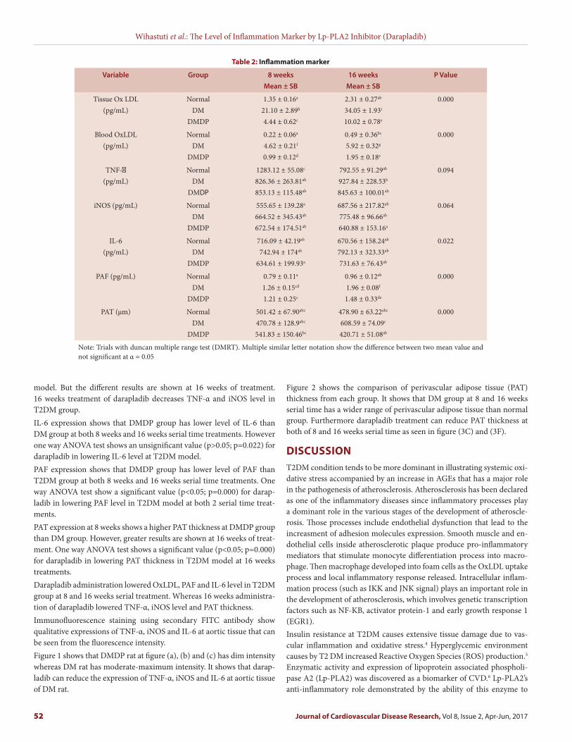
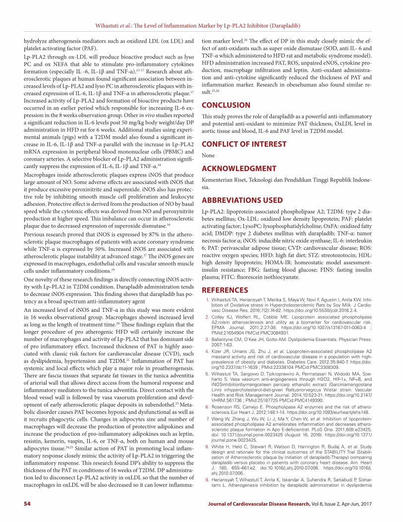
model. But the different results are shown at 16 weeks of treatment. 16 weeks treatment of darapladib decreases TNF-α and iNOS level in T2DM group.IL-6 expression shows that DMDP group has lower level of IL-6 than DM group at both 8 weeks and 16 weeks serial time treatments. However one way ANOVA test shows an unsignificant value (p>0.05; p=0.022) for darapladib in lowering IL-6 level at T2DM model.PAF expression shows that DMDP group has lower level of PAF than T2DM group at both 8 weeks and 16 weeks serial time treatments. One way ANOVA test show a significant value (p<0.05; p=0.000) for darap-ladib in lowering PAF level in T2DM model at both 2 serial time treat-ments.PAT expression at 8 weeks shows a higher PAT thickness at DMDP group than DM group. However, greater results are shown at 16 weeks of treat-ment. One way ANOVA test shows a significant value (p<0.05; p=0.000) for darapladib in lowering PAT thickness in T2DM model at 16 weeks treatments.Darapladib administration lowered OxLDL, PAF and IL-6 level in T2DM group at 8 and 16 weeks serial treatment. Whereas 16 weeks administra-tion of darapladib lowered TNF-α, iNOS level and PAT thickness.Immunofluorescence staining using secondary FITC antibody show qualitative expressions of TNF-α, iNOS and IL-6 at aortic tissue that can be seen from the fluorescence intensity. Figure 1 shows that DMDP rat at figure (a), (b) and (c) has dim intensity whereas DM rat has moderate-maximum intensity. It shows that darap-ladib can reduce the expression of TNF-α, iNOS and IL-6 at aortic tissue of DM rat.
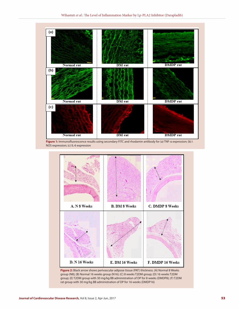
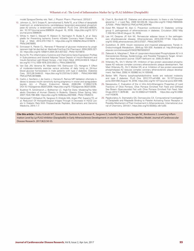
Figure 2 shows the comparison of perivascular adipose tissue (PAT) thickness from each group. It shows that DM group at 8 and 16 weeks serial time has a wider range of perivascular adipose tissue than normal group. Furthermore darapladib treatment can reduce PAT thickness at both of 8 and 16 weeks serial time as seen in figure (3C) and (3F).
DISCUSSIONT2DM condition tends to be more dominant in illustrating systemic oxi-dative stress accompanied by an increase in AGEs that has a major role in the pathogenesis of atherosclerosis. Atherosclerosis has been declared as one of the inflammatory diseases since inflammatory processes play a dominant role in the various stages of the development of atheroscle-rosis. Those processes include endothelial dysfunction that lead to the increasment of adhesion molecules expression. Smooth muscle and en-dothelial cells inside atherosclerotic plaque produce pro-inflammatory mediators that stimulate monocyte differentiation process into macro-phage. Then macrophage developed into foam cells as the OxLDL uptake process and local inflammatory response released. Intracellular inflam-mation process (such as IKK and JNK signal) plays an important role in the development of atherosclerosis, which involves genetic transcription factors such as NF-KB, activator protein-1 and early growth response 1 (EGR1).Insulin resistance at T2DM causes extensive tissue damage due to vas-cular inflammation and oxidative stress.4 Hyperglycemic environment causes by T2 DM increased Reactive Oxygen Species (ROS) production.5
Enzymatic activity and expression of lipoprotein associated phospholi-pase A2 (Lp-PLA2) was discovered as a biomarker of CVD.6 Lp-PLA2’s anti-inflammatory role demonstrated by the ability of this enzyme to
Table 2: Inflammation marker
Variable Group 8 weeksMean ± SB
16 weeksMean ± SB
P Value
Tissue Ox LDL(pg/mL)
NormalDM
DMDP
1.35 ± 0.16a
21.10 ± 2.89h
4.44 ± 0.62c
2.31 ± 0.27ab
34.05 ± 1.93i
10.02 ± 0.78e
0.000
Blood OxLDL(pg/mL)
NormalDM
DMDP
0.22 ± 0.06a
4.62 ± 0.21f
0.99 ± 0.12d
0.49 ± 0.36bc
5.92 ± 0.32g
1.95 ± 0.18e
0.000
TNF-(pg/mL)
NormalDM
DMDP
1283.12 ± 55.08c
826.36 ± 263.81ab
853.13 ± 115.48ab
792.55 ± 91.29ab
927.84 ± 228.53b
845.63 ± 100.01ab
0.094
iNOS (pg/mL) NormalDM
DMDP
555.65 ± 139.28a
664.52 ± 345.43ab
672.54 ± 174.51ab
687.56 ± 217.82ab
775.48 ± 96.66ab
640.88 ± 153.16a
0.064
IL-6(pg/mL)
NormalDM
DMDP
716.09 ± 42.19ab
742.94 ± 174ab
634.61 ± 199.93a
670.56 ± 158.24ab
792.13 ± 323.33ab
731.63 ± 76.43ab
0.022
PAF (pg/mL) NormalDM
DMDP
0.79 ± 0.11a
1.26 ± 0.15cd
1.21 ± 0.25c
0.96 ± 0.12ab
1.96 ± 0.08f
1.48 ± 0.33de
0.000
PAT (µm) NormalDM
DMDP
501.42 ± 67.90abc
470.78 ± 128.9abc
541.83 ± 150.46bc
478.90 ± 63.22abc
608.59 ± 74.09c
420.71 ± 51.08ab
0.000
Note: Trials with duncan multiple range test (DMRT). Multiple similar letter notation show the difference between two mean value and not significant at α = 0.05
Wihastuti et al.: The Level of Inflammation Marker by Lp-PLA2 Inhibitor (Darapladib)
Journal of Cardiovascular Disease Research, Vol 8, Issue 2, Apr-Jun, 2017 53
Figure 1: Immunofluorescence results using secondary FITC and rhodamin antibody for (a) TNF-α expression; (b) i NOS expression; (c) IL-6 expression
Figure 2: Black arrow shows perivascular adipose tissue (PAT) thickness. (A) Normal 8 Weeks group (N8); (B) Normal 16 weeks group (N16); (C) 8 weeks T2DM group; (D) 16 weeks T2DM group; (E) T2DM group with 30 mg/kg BB administration of DP for 8 weeks (DMDP8); (F) T2DM rat group with 30 mg/kg BB administration of DP for 16 weeks (DMDP16)
Wihastuti et al.: The Level of Inflammation Marker by Lp-PLA2 Inhibitor (Darapladib)
54 Journal of Cardiovascular Disease Research, Vol 8, Issue 2, Apr-Jun, 2017
hydrolyze atherogenesis mediators such as oxidized LDL (ox LDL) and platelet activating factor (PAF). Lp-PLA2 through ox-LDL will produce bioactive product such as lyso PC and ox NEFA that able to stimulate pro-inflammatory cytokines formation (especially IL -6, IL-1β and TNF-α).13-17 Research about ath-erosclerotic plaques at human found significant association between in-creased levels of Lp-PLA2 and lyso PC in atherosclerotic plaques with in-creased expression of IL-6, IL-1β and TNF-α in atherosclerotic plaque.17 Increased activity of Lp-PLA2 and formation of bioactive products have occurred in an earlier period which responsible for increasing IL-6 ex-pression in the 8 weeks observation group. Other in vivo studies reported a significant reduction in IL-6 levels post 50 mg/kg body weight/day DP administration in HFD rat for 6 weeks. Additional studies using experi-mental animals (pigs) with a T2DM model also found a significant in-crease in IL-6, IL-1β and TNF-α parallel with the increase in Lp-PLA2 mRNA expression in peripheral blood mononuclear cells (PBMC) and coronary arteries. A selective blocker of Lp-PLA2 administration signifi-cantly suppress the expression of IL-6, IL-1β and TNF-α.18
Macrophages inside atherosclerotic plaques express iNOS that produce large amount of NO. Some adverse effects are associated with iNOS that it produce excessive peroxinitrite and superoxide. iNOS also has protec-tive role by inhibiting smooth muscle cell proliferation and leukocyte adhesion. Protective effect is derived from the production of NO by basal speed while the cytotoxic effects was derived from NO and peroxynitrite production at higher speed. This imbalance can occur in atherosclerotic plaque due to decreased expression of superoxide dismutase.16
Previous research proved that iNOS is expressed by 87% in the athero-sclerotic plaque macrophages of patients with acute coronary syndrome while TNF-α is expressed by 50%. Increased iNOS are associated with atherosclerotic plaque instability at advanced stage.17 The iNOS genes are expressed in macrophages, endothelial cells and vascular smooth muscle cells under inflammatory conditions.19
One novelty of these research findings is directly connecting iNOS activ-ity with Lp-PLA2 in T2DM condition. Darapladib administration tends to decrease iNOS expression. This finding shows that darapladib has po-tency as a broad spectrum anti-inflammatory agentAn increased level of iNOS and TNF-α in this study was more evident in 16 weeks observational group. Macrophages showed increased level as long as the length of treatment time.20 These findings explain that the longer procedure of pro atherogenic HFD will certainly increase the number of macrophages and activity of Lp-PLA2 that has dominant side of pro inflammatory effect. Increased thickness of PAT is highly asso-ciated with classic risk factors for cardiovascular disease (CVD), such as dyslipidemia, hypertension and T2DM.21 Inflammation of PAT has systemic and local effects which play a major role in proatherogenesis. There are fascia tissues that separate fat tissues in the tunica adventitia of arterial wall that allows direct access from the humoral response and inflammatory mediators to the tunica adventitia. Direct contact with the blood vessel wall is followed by vasa vasorum proliferation and devel-opment of early atherosclerotic plaque deposits in subendothel.23 Meta-bolic disorder causes PAT becomes hypoxic and dysfunctional as well as it recruits phagocytic cells. Changes in adipocytes size and number of macrophages will decrease the production of protective adipokines and increase the production of pro-inflammatory adipokines such as leptin, resistin, kemerin, vaspin, IL-6, or TNF-α, both on human and mouse adipocytes tissue.24,25 Similar action of PAT in promoting local inflam-matory response closely mimic the activity of Lp-PLA2 in triggering the inflammatory response. This research found DP’s ability to suppress the thickness of the PAT in conditions of 16 weeks of T2DM. DP administra-tion led to disconnect Lp-PLA2 activity in oxLDL so that the number of macrophages in oxLDL will be also decreased so it can lower inflamma-
tion marker level.26 The effect of DP in this study closely mimic the ef-fect of anti-oxidants such as super oxide dismutase (SOD, anti IL- 6 and TNF-α which administered to HFD rat and metabolic syndrome model). HFD administration increased PAT, ROS, unpaired eNOS, cytokine pro-duction, macrophage infiltration and leptin. Anti-oxidant administra-tion and anti-cytokine significantly reduced the thickness of PAT and inflammation marker. Research in obesehuman also found similar re-sult.25,26
CONCLUSIONThis study proves the role of darapladib as a powerful anti-inflammatory and potential anti-oxidant to minimize PAT thickness, OxLDL level in aortic tissue and blood, IL-6 and PAF level in T2DM model.
CONFLICT OF INTERESTNone
ACKNOWLEDGMENTKementerian Riset, Teknologi dan Pendidikan Tinggi Republik Indone-sia.
ABBREVIATIONS USEDLp-PLA2: lipoprotein-associated phospholipase A2; T2DM: type 2 dia-betes mellitus; Ox-LDL: oxidized low density lipoprotein; PAF: platelet activating factor; LysoPC: lysophosphatidylcholine; OxFA: oxidized fatty acid; DMDP: type 2 diabetes mellitus with darapladib; TNF-α: tumor necrosis factor α; iNOS: inducible nitric oxide synthase; IL-6: interleukin 6; PAT: perivascular adipose tissue; CVD: cardiovascular disease; ROS: reactive oxygen species; HFD: high fat diet; STZ: streotozotocin; HDL: high density lipoprotein; HOMA-IR: homeostatic model assessment-insulin resistance; FBG: fasting blood glucose; FINS: fasting insulin plasma; FITC: fluorescein isothiocyanate.
REFERENCES1. Wihastuti TA, Heriansyah T, Merika S, Maya W, Novi F, Agustin I, Anita KW. Inhi-
bition of Oxidative stress in Hypercholesterolemic Rats by Soy Milk. J Cardio-vasc Disease Res. 2016;7(2):74-82. https://doi.org/10.5530/jcdr.2016.2.4.
2. Colley KJ, Wolfert RL, Cobble ME. Lipoprotein associated phospholipase A2:rolein atherosclerosis and utility as a biomarker for cardiovascular risk. EPMA Journal. 2011;2:27-38. https://doi.org/10.1007/s13167-011-0063-4 ; PMid:21654904 PMCid:PMC3084931.
3. Ballantyne CM, O’Kee JH, Gotto AM. Dyslipidemia Essentials. Physician Press. 2007:1-63.
4. Kizer JR, Umans JG, Zhu J, et al. Lipoprotein-associated phospholipase A2 massand activity and risk of cardiovascular disease in a population with high-prevalence of obesity and diabetes. Diabetes Care. 2012;35:840-7. https://doi.org/10.2337/dc11-1639 ; PMid:22338104 PMCid:PMC3308309.
5. Wihastuti TA, Sargowo D, Tjokroprawiro A, Permatasari N, Widodo MA, Soe-harto S. Vasa vasorum anti-angiogenesis through H2O2, HIF-1α, NF-κB, and iNOSinhibitionbymangosteen pericarp ethanolic extract (Garciniamangostana Linn) inhypercholesterol-diet-given Rattusnorvegicus Wistar strain. Vascular Health and Risk Management Journal. 2014;10:523-31. https://doi.org/10.2147/VHRM.S61736 ; PMid:25187725 PMCid:PMC4149390.
6. Rosenson RS, Camejo E. Phospholipase A2 enzymes and the risk of athero-sclerosis.Eur Heart J. 2012;148:1-14. https://doi.org/10.1093/eurheartj/ehs148.
7. Wang W, Zhang J, Wu W, Li J, Ma Y, Chen W, et al. Inhibition of lipoprotein-associated phospholipase A2 ameliorates inflammation and decreases athero-sclerotic plaque formation in Apo E-deficientrat. PLoS One. 2011;6(8):e23425, doi: 10.1371/journal.pone.0023425 (August 16, 2016). https://doi.org/10.1371/journal.pone.0023425.
8. White H, Held C, Stewart R, Watson D, Harrington R, Budaj A, et al. Study design and rationale for the clinical outcomes of the STABILITY Trial (Stabili-zation of Atherosclerotic plaque by Initiation of darapladib Therapy) comparing darapladib versus placebo in patients with coronary heart disease. Am. Heart J. 160, 655–661.e2. doi:10.1016/j.ahj.2010.07.006. https://doi.org/10.1016/j.ahj.2010.07.006.
9. Heriansyah T, Wihastuti T, Anita K, Iskandar A, Suhendra R, Setiabudi P, Sishar-tami L. Atherogenesis inhibition by darapladib administration in dyslipidemia
Wihastuti et al.: The Level of Inflammation Marker by Lp-PLA2 Inhibitor (Darapladib)
Journal of Cardiovascular Disease Research, Vol 8, Issue 2, Apr-Jun, 2017 55
model Sprague-Dawley rats. Natl. J. Physiol. Pharm. Pharmacol. 2015;6:7.10. Johnson JL, Shi Y, Snipes R, Janmohamed S, Rolfe TE, et al. Effect of darapladib
treatment on endarterectomy carotidplaque lipoprotein-associated phospholi-pase A2 activity: a randomized, controlledtrial. PLoS One. 2014;9(2):e89034, doi: 10.1371/journal.pone.0089034 (August 16, 2016). https://doi.org/10.1371/journal.pone.0089034.
11. White H, Held C, Stewart R, Watson D, Harrington R, Budaj A, et al. Dara-pladib for Preventing Ischemic Events inStable Coronary Heart Disease. N. Engl. J. Med. 2014;370:1702-11. https://doi.org/10.1056/NEJMoa1315878 ; PMid:24678955.
12. Srinivasan K, Patole CL, Ramarao P. Reversal of glucose intolerance by piogli-tazonein high fat diet-fed rat. Methods Find Exp Clin Pharmacol. 2004;26(5):327-33. https://doi.org/10.1358/mf.2004.26.5.831322 ; PMid:15319810.
13. Burns TA. Pro inflammatory Cytokine and Chemokine Gene Expression Profiles in Subcutaneous and Visceral Adipose Tissue Depots of Insulin-Resistant and Insulin-Sensitive Light Breed Horses. J Vet Intern Med. 2010;24:932-9. https://doi.org/10.1111/j.1939-1676.2010.0551.x; PMid:20649750.
14. Van Dijk JW, Venema M, Mechelen VW, Stehouwer CD, Hartgens F. Effect of moderate-intensity exercise versus activities of daily living on 24-hour blood glucose homeostasis in male patients with type 2 diabetes. Diabetes Care. 2013;36:3448-53. https://doi.org/10.2337/dc12-2620 ; PMid:24041682 PMCid:PMC3816888.
15. Cacho J, Sevillano J, de Castro J, Herrera E, Ramos MP. Validation ofsimple in-dexes to assess insulin sensitivity during pregnancy in wistar and spraguedaw-leyrats. Am. J. Physiol. Endocrinol. Metab. 2008;295: E1269-E1276. DOI:10.1152/ajpendo.90207.2008. https://doi.org/10.1152/ajpendo.90207.2008.
16. Buettner R, Schölmerich J, Bollheimer LC. High-Fat Diets: ModelingThe Met-abolic Disorders of Human Obesity In Rodents. Obesity (Silver Spring, Md.) 2007;15(4);798–808. https://doi.org/10.1038/oby.2007.608 ; PMid:17426312.
17. Heriansyah T, Wihastuti TA, Sargowo D, Widodo MA, Anjani PM, Suparno TD, et al. Reduction Of Histopathological Images Through A Decrease In H2O2 Lev-els In Diabetic Rats With Polysaccharide Peptides. Biomarkers and Genomic Medicine, 2014;1–7.
18. Chait A, Bornfeldt KE. Diabetes and atherosclerosis: Is there a role forhyper-glycemia ?. J Lipid Res. 2009 50:335-39. https://doi.org/10.1194/jlr.R800059-JLR200 ; PMid:19029122 PMCid:PMC2674740.
19. Eckel RH, Wassef M, Alan C. Prevention conference VI: Diabetes: writing-group II: pathogenesis of atherosclerosis in diabetes. Circulation 2002 May 7;105(18):e138-43 (August 16, 2016).
20. Lee HY, Despres JP, Koh KK. Perivascular adipose tissue in the pathogen-esis ofcardiovascular disease. Atherosclerosis. 2013;230:177-84. https://doi.org/10.1016/j.atherosclerosis.2013.07.037 ; PMid:24075741.
21. Gustafson, B. 2015. Insulin resistance and impaired adipogenesis. Trends in-Endocrinology& Metabolism, 26(4):pp.193–200. Available at: http://linkinghub.elsevier.com/retrieve/pii/S1043276015000107.
22. Zalewski A, Macphee C. Role of Lipoprotein-Associated Phospholipase A2 in A therosclerosis Biology, Epidemiology, and Possible Therapeutic Target. Ameri-can Heart Association journal. DGAT1-deficient rat. 2005;25:482-6.
23. Wilensky RL, Shi Y, Mohler ER. Inhibition of lipo protein associated phospho-lipase A2 reduces complex coronary atherosclerotic plaque development. Nat Med. Wilensky RL, Shi Y, Mohler ER, et al. Inhibition of lipo protein associated phospholipase A2 reduces complex coronary atherosclerotic plaque develop-ment. Nat Med. 2008;14:105966.Vol;14:1059-66.
24. Barber MN. Plasma lysophosphatidylcholine levels are reduced inobesity and type 2 diabetes. PLoS One. 2012;7(7):e41456. doi: 10.1371/journal.pone.0041456 (August 16, 2016). https://doi.org/10.1371/journal.pone.0041456.
25. Nasopoulou C. Evaluation of the in Vitro Anti-Atherogenic Properties of Lipid Fractions of Olive Pomace, Olive Pomace Enriched Fish Feed and Gilthead Sea Bream (Sparusaurata) Fed with Olive Pomace Enriched Fish Feed. Mar. Drugs.2013;11:3676-88; doi:10.3390/md11103676. https://doi.org/10.3390/md11103676.
26. Papakyriakou A, Stamatakis GA, Demopoulos CA. Computational Investigation of Darapladib and Rilapladib Binding to Platelet Activating Factor Receptor. A Possible Mechanism of Their Involvement in Atherosclerosis. International Jour-nal of Chemistry. 2014;6:1. https://doi.org/10.5539/ijc.v6n1p50.
Cite this article : Teuku H,Andri WT, Siswanto BB, Santoso A, Sukmawan R, Sargowo D, Subekti I, Aulanni’am, Siregar NC, Bordosono S. Lowering Inflam-mation Level by Lp-PLA2 Inhibitor (Darapladib) in Early Atherosclerosis Development: in vivo Rat Type 2 Diabetes Mellitus Model. Journal of Cardiovascular Disease Research. 2017;8(2):50-55.