lp(a) and calcific aortic valve disease: a novel drug ... · lipoprotein(a) in calcific aortic...
TRANSCRIPT

Lipoprotein(a) in Calcific Aortic Valve Disease:
From genomics to novel drug target for aortic stenosis
George Thanassoulis MD MSC FRCPC1
1Preventive and Genomic Cardiology, McGill University Health Center and Research Institute
and Department and Department of Medicine, McGill University, Montreal, QC
Word count: 179 (abstract); 4,054 (main text)
Address Correspondence to:
G. Thanassoulis MD MSc FRCPC
Director, Preventive and Genomic Cardiology
McGill University Health Center, D5-5120
1001 Decarie Blvd, Montreal, QC
H4A 3J1
514-934-1934 x35465
by guest, on June 27, 2018w
ww
.jlr.orgD
ownloaded from
2
Abstract
Calcific aortic stenosis (AS) is the most common form of valve disease in the western
world and affects over 2.5 million individuals in North America. Despite the large burden of
disease, there are no medical treatments to slow the development of AS, due at least in part to
our incomplete understanding of its causes. The CHARGE extra-coronary calcium consortium
reported a genome-wide association study (GWAS) demonstrating that genetic variants in LPA
are strongly associated with aortic valve (AV) calcium and clinical AS. Using a Mendelian
randomization study design, it was demonstrated that the effect of this genetic variant is
mediated by plasma lipoprotein(a) (Lp[a]), directly implicating elevations in Lp(a) as a cause of
AV calcium and progression to AS. This discovery has sparked intense interest in Lp(a) as a
modifiable cause for aortic valve disease. Herein, we will review the mounting epidemiological
and genetic findings in support of Lp(a) mediated valve disease, discuss potential mechanisms
underlying this observation and outline the steps to translate this discovery to a much needed
novel preventive and/or therapeutic strategy for aortic valve disease.
by guest, on June 27, 2018w
ww
.jlr.orgD
ownloaded from
3
Calcific Aortic Valve Disease – Large burden of disease without known medical treatment
Calcific aortic valve disease (CAVD) is currently the third most common cardiovascular
disease after ischemic heart disease and hypertension. CAVD encompasses all forms of calcific
aortic valve disease, including subclinical forms, such as the presence of aortic valve calcium
(AVC) identified by computed tomographic (CT) imaging and aortic sclerosis (i.e. thickening
and calcification) identified by echocardiography, as well as, the clinical disease of aortic
stenosis. In North America, CAVD afflicts over 5 million individuals1 and is a common cause of
death, disability and health care costs with over 15,000 deaths annually in the US alone2. Aortic
stenosis (AS), which is defined as a progressive narrowing of the aortic valve (i.e. the valve
through which blood is ejected from the left ventricle into the systemic circulation), affects over
2.5 million people3 and leads to severe impairment in cardiac function consisting of heart failure,
angina and syncope when stenosis becomes severe (see Table 1). Age is the most important risk
factor, with 2% of the population over 65 having AS, a figure that rises to 12% (95% CI 7-18%)
after 75 years of age4. Current treatment consists solely of valve replacement, with nearly
100,000 such procedures performed yearly in the US5, frequently late in the disease process
when patients are generally old and frail, with the attendant high costs and complications
associated with intervention at this late stage. Despite the huge burden of disease, there are
currently no medical treatments to prevent or retard the progression of this disease and reduce
the need for valve replacement.
The natural history of CAVD consists of a long clinically silent phase of valve
calcification and hardening (“sclerosis”) which lasts generally at least a decade and heralds the
clinical disease. AV sclerosis is exceedingly common with a prevalence of 26% after 65 years of
age, 40% after 75 years of age and 75% after 85 years of age6. AV sclerosis, which was long
by guest, on June 27, 2018w
ww
.jlr.orgD
ownloaded from
4
deemed a benign consequence of ageing, is also now known to confer a 40% increase in the risk
of death and a 66% increase in the risk of CV death, independent of age and CV risk factors7.
This lesion, which appears to track with vascular disease risk factors, has led to a misconception
that aortic sclerosis and vascular atherosclerosis represent the same disease. Although there is
significant overlap in the early initiating lesion and certain shared risk factors8-11
, the available
evidence indicates major differences in the underlying pathophysiology of these two diseases.
First, histopathological evidence demonstrates a more prominent early mineralization phase and
a paucity of smooth muscle cells in aortic sclerosis, as compared to vascular atherosclerotic
lesions9. Second, calcification pathways appear to predominate early in valve disease and appear
to be independent of the atherosclerotic process as opposed to vascular disease where
calcification develops much later and in parallel with atherosclerosis12
. Third, among individuals
undergoing AV replacement for (non-congenital) AS, only 40% have significant CAD requiring
bypass13
suggesting unique pathological processes. Fourth, lipid lowering agents, which have
been remarkably effective for preventing atherosclerosis have not demonstrated any benefit in
randomized trials for advanced AV disease, in older patient populations, highlighting the major
differences in pathophysiology and treatment of these diseases14-16
.
Lp(a) is associated with Calcific Aortic Valve Disease
A major limitation in developing treatments for CAVD stems in large part from our poor
understanding of the mediators and causes of this disease. Seminal early pathologic studies
performed by Otto et al. clearly demonstrated the importance of lipids in the early initiating
lesion of CAVD. They demonstrated that the early sclerotic lesions of aortic valves were
characterized by lipid pools that were absent in adjacent normal valve tissue, implicating lipids
in the pathological process9. In addition, these lipids were likely derived from plasma, as plasma
by guest, on June 27, 2018w
ww
.jlr.orgD
ownloaded from
5
carrier molecules for cholesterol (apoB and apo[a]) were also present in these pools17
. These
cholesterol particles co-localized with early foci of calcification in valve tissue providing the first
evidence that blood-borne cholesterol, either from LDL and/or Lp(a), were intimately involved
in the early calcification of the aortic valve.
Subsequent epidemiologic studies (summarized in the Table 2) provided further evidence
in support of the lipid hypothesis for CAVD. Stewart et al, using cross-sectional baseline data
from the 5201 participants who underwent echocardiography as part of the Cardiovascular
Health Study, demonstrated that LDL cholesterol and Lp(a) were associated with a 12% (95% CI
3-23%) and 23% increase (95% CI 14-32%), respectively, in the presence of AS10
. Gotoh et al
also found that Lp(a) was associated with CAVD in a small cross-sectional case-control study18
.
Among individuals with Lp(a) > 30 mg/dL, 36% had aortic sclerosis, as compared to only 13%
with Lp(a) < 30 mg/dL (p<0.001). More recently, using a case-control design, Bozbas et al,
demonstrated a slightly higher Lp(a) level (27.4 mg/dL) in 112 cases with aortic sclerosis, as
compared to 19.9 mg/dL among controls19
. Finally, Glader et al also found a significantly higher
Lp(a) among AS cases than controls20
.
Although the above studies support an association between plasma Lp(a) and CAVD, the
observational nature of these studies could not establish whether Lp(a) was, in fact, causal for
CAVD or simply a bystander in the calcifying process, a critical consideration in determining
whether Lp(a) represents a therapeutic target. However, recent genetic studies have provided
important insights into the causal nature of these associations.
Genetic Associations between LPA variants, Lp(a) and CAVD
by guest, on June 27, 2018w
ww
.jlr.orgD
ownloaded from
6
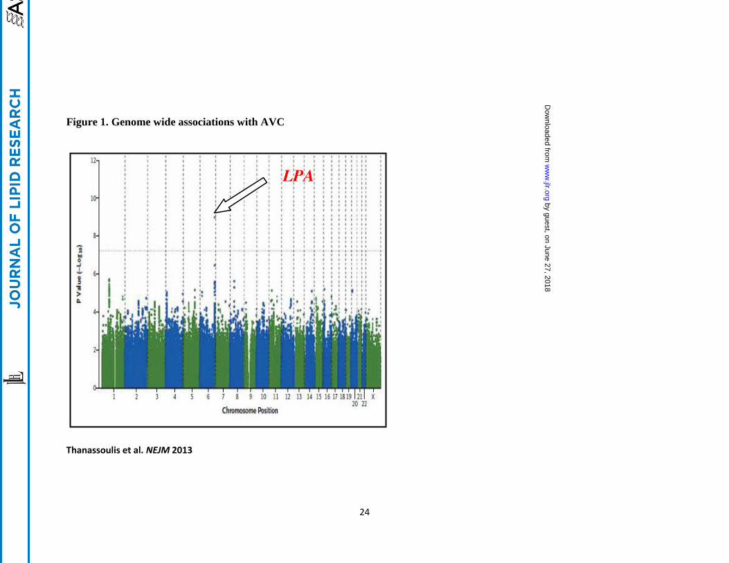
The Cohorts for Heart and Aging Research in Genetic Epidemiology (CHARGE) extra-
coronary calcium working group recently reported the first genome-wide association study of
valve calcium as detected by computed tomography21
(Figure 1). Valve calcium represents a
“deep phenotype” that is more proximal to the biological process (i.e. calcification) and reduces
phenotypic heterogeneity, as compared to a clinical phenotype such as aortic stenosis, allowing
for improved resolution of genetic signals22
. In the discovery stage, 6942 participants were
included from the Framingham Heart Study, the Multi-Ethnic Study of Atherosclerosis and the
Age Gene-Environment Susceptibility-Reykjavik Study, with data on presence or absence of
aortic valve calcium (AVC). A single nucleotide polymorphism (SNP) (rs10455872) was
identified in the LPA gene that conferred a 2-fold increase in the odds of aortic valve
calcification (odds ratio, 2.05; p=9.0×10⁻¹⁰
). Subsequent independent replication in the Heinz-
Nixdorf Study confirmed this 2-fold increase in the odds of valve calcium (odds ratio 2.04;
p=0.018). In a joint meta-analysis combining the discovery and replication cohorts, the
rs10455872 SNP in LPA was associated with a doubling in the odds of AVC (p=2.8×10⁻¹¹
),
strongly supporting an important association with AVC. Other LPA SNPs, including rs3798220,
were also associated with AVC but did not achieve genomewide significance due to lower minor
allele frequence or smaller effect sizes on Lp(a) levels. To demonstrate relevance with clinical
AS, validation in the 28,193 participants from the Malmo Diet Cancer Study (MDCS) was
performed. In the MDCS, the rs10455872 was clearly associated with future incident AS (HR
1.68, 95% CI 1.32 to 2.15) and aortic valve replacement for AS (HR 1.54, 95% 1.05 to 2.27).
These results were also independently confirmed in the Copenhagen City Heart Study in which
the rs10455872 was associated with both incident aortic stenosis and aortic valve replacement21
.
In both cohorts, the association persisted after adjustment for several risk factors and after
by guest, on June 27, 2018w
ww
.jlr.orgD
ownloaded from
7
exclusion of myocardial infarction. To further support the specificity of this association with
valve disease rather than vascular atherosclerosis, additional analyses were performed in
CHARGE participants discordant for AVC and coronary artery calcium. The rs10455872
tracked consistently with AVC rather than coronary calcium demonstrating that the association
was not an epiphenomenon of the known Lp(a) association with coronary artery disease.
The rs10455872 has previously been strongly associated with Lp(a) levels and with
myocardial infarction in several studies, which provided the impetus to evaluate the potential
role of circulating Lp(a) in valve calcification and CAVD. Given that this SNP correlates with
fewer kringle type-IV repeats, which are believed to regulate Lp(a) production and increase
Lp(a) levels23
, this SNP provides an excellent opportunity to evaluate the causal nature of life-
long genetic elevations in Lp(a) using Mendelian randomization. Mendelian randomization uses
the randomization of genetic material at conception to provide largely unconfounded and
unbiased inferences for the role of a circulating biomarker on disease outcomes24, 25
. Since
genetic associations cannot be confounded by other factors (i.e. lifestyle, diet, etc) and
necessarily precede the development of the outcome, Mendelian randomization provides a more
robust measure of the causal association between a genetically elevated biomarker, in this case
Lp(a) and the disease of interest, CAVD, and can provide strong supportive evidence that a
biomarker may be relevant as a therapeutic target. Based on the known associations between
rs10455872 and Lp(a), this SNP was used as a proxy to estimate the association with AVC based
on the observed difference in Lp(a) levels among rs10455972 carriers, as compared to non-
carriers, using an instrumental variable analysis. Based on this analysis, it was found that for
each unit increment in log lp(a), there was a ~60% increase in AV calcium, strongly supporting a
by guest, on June 27, 2018w
ww
.jlr.orgD
ownloaded from

8
causal role for Lp(a) and suggesting that lowering of Lp(a) levels may represent a strategy to
prevent valve calcification21
.
Following the publication of these findings, several additional Mendelian randomization
studies have been reported that are largely in agreement with the findings of the CHARGE
consortium and have extended these analyses to clinical aortic stenosis. In combined analyses of
the Copenhagen City Heart Study and the Copenhagen General Population studies, comprising
77,680 individuals with 454 occurrences of aortic stenosis, Kamstrup et al., demonstrated a
strong graded relationship between Lp(a) levels and AS corresponding to a 60% increase in the
risk of AS among individuals with Lp(a) between 20-64 mg/dL, a 100% increase when Lp(a)
was 65-90 mg/dL and a 200% increase when Lp(a) was over 90 mg/dL26
. Using Mendelian
randomization by comparing carriers of any genotype that raises Lp(a) levels (including
rs10455872, rs3798220 and low number of KIV-2 repeats) to those without these genotypes,
they estimated a 60% increase for each 10-fold increment in Lp(a). Similarly, Arsenault et al.,
provided further independent replication in the 17,553 participants of the prospective EPIC-
Norfolk study27
. Compared to individuals with no copies of the G risk allele (i.e. Lp(a)
increasing), individuals heterozygous and homozygous for the G allele had a hazard a hazard
ratio of 1.78 (95% CI 1.11-2.87) and 4.83 (95% CI 1.77-13.20), respectively, for incident aortic
stenosis. They also provided replication in a case-control study demonstrating that carriers of the
G allele had a 57% increase in the odds of AS, as compared to those without the G allele.
Recently, Capoulade et al28
, have shown that in 220 participants of the Aortic Stenosis
Progression Observation: Measuring Effects of Rosuvastatin (ASTRONOMER) trial that
participants in the top Lp(a) tertile (Lp[a] > 58.5 mg/dL) had a faster rate of progression than
individuals in the lower tertiles. Interestingly, this effect was more pronounced in younger (≤57
by guest, on June 27, 2018w
ww
.jlr.orgD
ownloaded from

9
years of age) than in older participants indicating an important age x Lp(a) interaction (p= 0.04).
In multivariable models, Lp(a) > 58.5 mg/dL was associated with a 2.6 fold increase in the odds
of being a rapid progressor, as defined by an annualized change in maximum aortic velocity >
0.2/ms/year. Importantly, in those ≤57 years, the odds ratio for rapid progression was 4.9 (95%
CI 1.8-3.7). These results demonstrate robust evidence in support of elevated Lp(a) as a marker
of rapid progression of aortic stenosis, especially in younger individuals, and provide strong
evidence to target Lp(a) lowering to this group of patients.
Potential mechanisms of Lp(a)-mediated calcific aortic valve disease
Due to the recent discoveries linking LPA variants with aortic valve disease, there are
currently limited molecular and animal studies exploring the mechanisms underlying this
association, which represents an important knowledge gap that will need to be addressed in the
near future. Nonetheless, Lp(a) has several important properties that could promote AV disease
(Figure 2):
First, Lp(a) is involved in tissue repair. Lp(a), via its apo(a) protein moiety, is comprised of
lysine bindings sites that can bind to areas of damaged and denuded endothelium29
. Although
this may have an important role for maintenance of vascular integrity and possibly for delivery
of cholesterol to sites of injury, the AV leaflets may be particularly prone to cellular damage due
to the repetitive mechanical stress imposed during the cardiac cycle, in concert with high
pressures and shear stress, leading to excessive Lp(a) binding, especially under conditions of
Lp(a) excess.
Second, Lp(a) particles can promote foam cell formation and inflammation in early valve
lesions. Although pathophysiologically distinct in their mature forms, AV sclerosis and vascular
atherosclerosis share a similar initiating phase consisting of subintimal deposition of cholesterol-
by guest, on June 27, 2018w
ww
.jlr.orgD
ownloaded from
10
laden lipoproteins. Native unmodified lipoproteins are poorly internalized by tissue
macrophages. However, their oxidation results in unregulated lipoprotein uptake into
macrophages, via scavenger receptors (i.e as CD36 and SR-A)30
, leading to foam cell formation,
fatty streaks and atheroma. In appropriate settings, Lp(a), and its associated phosphocholine
(PC) containing oxidized phospholipids (OxPL), also triggers apoptosis in macrophages, a key
process that likely contributes to early valvular lesion progression31
. Furthermore, it was recently
shown that Lp(a) bind monocyte chemoattractant protein-1 (MCP-1) in vitro and in vivo.32
The
association of MCP-1 may play a role in modulating monocyte trafficking during early valve
sclerosis. Lp(a) may also promote inflammation via its apoB component. Recent work has
demonstrated that apoB derived peptides (i.e. apoBDS-1) are danger-associated peptides that are
potent promoters of pro-inflammatory cytokines (such as Il-8, Il-6 and CCL2) and other pro-
inflamatory mediators (e.g. PGE2)33
. Whether such properties contribute to Lp(a)-mediated
valve disease, via the apoB component of Lp(a), remains to be seen.
Third, Lp(a) is the primary carrier of OxPL in plasma. This property has emerged as a key
mediator of Lp(a) pathogenicity and likely plays a major role in calcific aortic valve disease. It
has been hypothesized that a physiologic role for Lp(a) may be to bind and transport pro-
atherogenic OxPL in plasma, targeting them for removal from the circulation.34
Although OxPL
have been found on several plasma proteins, including LDL and plasminogen, Lp(a) carries the
largest proportion (~85%) of circulating OxPL on plasma lipoproteins35
. Lp(a) levels are highly
correlated with OxPL/apoB levels (r = 0.85), but this correlation is not consistent for all Lp(a)
particles, with highest correlations observed among the smallest, most atherogenic Lp(a)
particles36-38
. OxPL appear to be “danger-associated molecular patterns” which are potent
stimulators of innate immunity by interacting with various pattern recognition receptors12, 39
40
.
by guest, on June 27, 2018w
ww
.jlr.orgD
ownloaded from
11
In addition, bacterial PC (not as a phospholipid) has molecular identity with PC-containing
OxPL40
. OxPL stimulated endothelial cells have been shown to (1) upregulate expression of
chemoattractants and cell adhesion molecules for inflammatory cell recruitment41, 42
, (2) increase
production of reactive oxygen species43
, and (3) increase pro-inflammatory gene expression (IL-
1, IL-6, Il-8)40
. This pro-inflammatory phenotype is likely important in early phases of valve
sclerosis and likely contributes to the stiffening of the leaflets via recruitment of immune cells
and deposition of extracellular matrix proteins.
Finally, Lp(a) is likely capable of inducing valvular calcification, which is the predominant
biological feature of AV disease and is also likely mediated, in large part, by OxPL but also by
native cholesterol. Increased cholesterol delivery, via Lp(a), to the valve leaflets, can lead to
cholesterol microcrystals which act as nidi for calcification44
. Lp(a), via OxPL, is also a source
of oxidative stress, which is a known promoter of CV calcification45
. Oxidant levels have been
found to be elevated around calcified deposits in human valves and also to co-localize with
signals of active calcium remodeling46
. Furthermore, oxidative stress induces osteogenic
differentiation of endothelial cells by upregulation of runt-related transcription factor-2 (runx-2),
a master regulatory transcription factor involved in bone formation47
. OxPLs have also been
shown to induce, in a dose dependent manner, osteoblastic differentiation and production of
mineralized matrix (i.e. hydroxyapatite) in vascular cells48
in vitro. This property may be
mediated by lysophophosphotidylcholine (lysoPC), derived from OxPL, which is released by
phospholipases with preferences for short-chain oxidized sn2 fatty acids (e.g. sPLA2 and Lp-
PLA2), which are found in high levels in explanted AVs49
. However, the role of these
phosholipases is controversial as several pathological mechanisms of Lp(a) appear to be
mediated by an intact OxPL component (e.g. inflammatory gene expression, chemokine
by guest, on June 27, 2018w
ww
.jlr.orgD
ownloaded from

12
expression and apoptosis)) and recent randomized trials in coronary disease with phospholipase
inhibitors failed to demonstrate benefit (and actually showed evidence of harm). Furthermore,
OxPL are also ligands for toll-like receptors (TLR), including TLR-2 and TLR-4,40, 50
which are
major promoters of osteogenic differentiation via bone-morphogenetic protein-2 (BMP-2), a
critical activator of bone and cartilage formation51
. Importantly, TLR-4 is expressed at much
higher levels on left-sided valves and may explain, in addition to the higher hemodynamic stress,
the increased predilection for OxPL to induce AV calcification51
. Recently, Bouchareb et al52
,
have also implicated autotaxin (ATX) in Lp(a)-mediated calcification. ATX activity was 60%
higher in calcified aortic valves than controls and co-localized with OxPL and apo(a).
Interestingly, ATX appeared to be transported by Lp(a) into aortic valve tissue but also locally
secreted by valve interstitial cells. ATX was shown to generate lysophosphatidic acid from
available lysoPC, which promoted valve inflammation and calcification via an NF-κB/IL-
6/BMP-dependent pathway.
Based on these properties, Lp(a) may mediate AV disease by binding to areas of denuded
valvular tissue, leading to excessive deposition of Lp(a), thus delivering apoB, cholesterol and
highly atherogenic, pro-calcifying OxPL to the leaflet surface culminating in a pro-inflammatary
and pro-calcifying cascade at the valve leaflets (see Figure 2). This process is likely magnified
several fold at the AV and at other areas of high hemodynamic stress (i.e. vascular branch
points), which may also explain the higher predilection of calcification at these sites as compared
to the remainder of the cardiovascular system.
Future Directions: Can Aortic Stenosis be prevented by Lp(a) targeted therapy?
by guest, on June 27, 2018w
ww
.jlr.orgD
ownloaded from
13
A preventative treatment for CAVD could markedly improve quality of life for older
patients, limit the number of valve replacements with a significant cost savings for our health
care system and reduce death and disability in this vulnerable age group. Although the failure of
previous randomized trials to prevent aortic stenosis using lipid-lowering therapies15, 16, 47
has
dampened enthusiasm for prevention, CAVD has attractive characteristics for this approach: an
easily identified sub-clinical phase that characterizes at risk individuals; a slow regulated process
that begins many years prior to clinical manifestations and provides a time window for
intervention; and, at the very least, a partially shared pathophysiology with atherosclerosis, a
highly preventable disease, that can be successfully managed with pharmacotherapy.
To date, the only major strategy for the prevention of CAVD has been the lowering of
low-density lipoprotein using statins (and ezetimibe in one trial). Four randomized trials of LDL
lowering have been completed and the results have been overwhelmingly negative – statins by
lowering LDL do not appreciably reduce the progression of aortic valve disease15, 16, 47
. These
results were confirmed by a recent meta-analysis,53
which reported that despite marked lowering
of LDL (35-55%) for more than 2 years (2.5-4 years in the different trials) in these trials, there
was no evidence for a reduction in echocardiographic parameters in aortic stenosis progression
or in clinical outcomes (i.e. death, aortic valve replacement, hospitalization for AS etc.).
Nonetheless, these trials had major limitations. They were, in large part, performed late in the
disease process, could not be performed in individuals with an indication for statin therapy (i.e.
many individuals with high cholesterol could not be ethically randomized to placebo) and used
statin therapy as the preferred treatment approach, despite some evidence that statins in advanced
vascular disease could promote calcification (which may stabilize vascular lesions but worsen
AS)13
. Although the relevance of Lp(a) was not known at the time of these trials, statins also do
by guest, on June 27, 2018w
ww
.jlr.orgD
ownloaded from
14
not have any appreciable lowering effect on Lp(a) levels, and may even increase Lp(a), which
may have also contributed to this failure. New evidence from Mendelian randomization has
shown that LDL is likely causal in valve disease, despite the failure of statins in these RCTs, and
has highlighted the potential role of earlier intervention prior to the development of advanced
valve disease54
.
Although the evidence in support of Lp(a) mediated aortic valve calcification and
stenosis are compelling with genetic data strongly supporting a causal association, the true test of
Lp(a) as a causal factor for AS will require a randomized trial targeting Lp(a). Although limited
therapeutic options are currently available to lower Lp(a) clinically (e.g. niacin), several new
agents are in development. Proprotein convertase subtilisin/kexin type 9 (PCSK9) inhibitors,
new generation CETP inhibitors and specific apo(a) antisense molecules are all currently in
various stages of development and have been shown in early clinical studies to have Lp(a)
lowering properties55, 56
. Of particular interest, the apo(a) antisense molecules, by virtue of their
specificity for Lp(a) lowering, will eventually allow for a clear test of the role of Lp(a) lowering
in aortic valve disease and other CV diseases57
. Currently, niacin, in its extended release
formulation, remains one of the few agents that is currently available that lowers Lp(a) in a dose-
dependent fashion. Using extended release niacin (ER-niacin) in individuals with early valve
disease and high Lp(a), The Early Aortic Valve Lipoprotein(a) Lowering (EAVaLL) pilot
randomized trial will provide a preliminary test of the Lp(a) lowering hypothesis in aortic valve
disease. If successful, this trial will pave the way for future trials using newer and more targeted
Lp(a) lowering agents58
. In EAVaLL, 238 participants with Lp(a) greater than 50 mg/dL and
aortic sclerosis or mild AS will be randomized to ER-niacin or placebo for 2 years. The primary
by guest, on June 27, 2018w
ww
.jlr.orgD
ownloaded from

15
outcome will be reduction in aortic valve calcium by CT. Secondary outcomes will include
echocardiographic parameters of progression as well as tolerability and safety.
Recently, an apo(a) antisense developed by ISIS pharmaceuticals has been shown to lead
to a marked dose-dependent decrease in circulating Lp(a), up to 90%, without affecting other
lipid markers59
. Such profound and specific Lp(a)-lowering activity has not been previously
observed with other agents and represents a unique opportunity to test the Lp(a) hypothesis in
CV disease. Such an agent will provide, the much needed “magic bullet” to confirm the
hypothesis that Lp(a) is causally mediating aortic valve disease as well as demonstrating that
specific Lp(a) lowering could slow disease progression and prevent AS. Due to the profound
Lp(a) lowering achievable, such a trial should focus on young patients with marked elevations of
Lp(a) with early valve disease, where disease progression is expected to be highest and most
closely related to ongoing Lp(a) deposition (rather than other factors, e.g. hemodynamic stress
may predominate at later stages) and therefore, where greatest benefit could be demonstrated.
Given the profound Lp(a) lowering observed with the apo(a) antisense, and the post-hoc
ASTRONOMER analysis demonstrating that Lp(a) predicted both progression and clinical
outcomes, and that this was most pronounced in the younger population, one might envision a
trial targeting younger individuals (i.e. < 70 years of age) with moderately advanced disease (i.e.
mild and/or moderate aortic stenosis) with high Lp(a), e.g. > 50 mg/dL. Given the expected rates
of progression in this group of patients, echocardiographic parameters such as valve area (as well
as maximum aortic velocity) would be appropriate end-points with change in valve calcium
score as potentially useful secondary outcomes. It would be particularly interesting to also
consider state-of-the-art molecular imaging techniques using novel positron emission
tomography with sodium fluoride-18, as pioneered by Dweck et al60, 61
, which measures active
by guest, on June 27, 2018w
ww
.jlr.orgD
ownloaded from
16
osteoblast activity and ongoing calcium deposition at the aortic valve. Validating such an
endpoint would also provide new effective approaches to accelerate the testing of potentially
novel therapies for AV disease.
In summary, the recent identification of genetic variants in LPA as being strongly
associated with calcific aortic valve disease, and the Mendelian randomization studies
demonstrating that this genetic association is mediated by circulating Lp(a), as well as our
increased understanding of the mechanisms of Lp(a) and their potential role in atherogenesis and
cardiovascular calcification, has reignited intense interest in Lp(a) in general, but also as a
specific therapeutic target for calcific aortic valve disease. It is hoped that lowering of Lp(a) will
demonstrate reduced progression of aortic valve disease and that targeted therapies directed at
Lp(a) will become the preferred treatment for aortic valve disease in at risk individuals with high
Lp(a) and, ultimately, will reduce the need for valve replacement and the costs and complications
of CAVD.
References
1. Bach DS, Radeva JI, Birnbaum HG, Fournier AA and Tuttle EG. Prevalence, referral
patterns, testing, and surgery in aortic valve disease: leaving women and elderly patients behind?
The Journal of heart valve disease. 2007;16:362-9.
2. Mozaffarian D, Benjamin EJ, Go AS, Arnett DK, Blaha MJ, Cushman M, de Ferranti S,
Despres JP, Fullerton HJ, Howard VJ, Huffman MD, Judd SE, Kissela BM, Lackland DT,
Lichtman JH, Lisabeth LD, Liu S, Mackey RH, Matchar DB, McGuire DK, Mohler ER, 3rd,
Moy CS, Muntner P, Mussolino ME, Nasir K, Neumar RW, Nichol G, Palaniappan L, Pandey
DK, Reeves MJ, Rodriguez CJ, Sorlie PD, Stein J, Towfighi A, Turan TN, Virani SS, Willey JZ,
Woo D, Yeh RW and Turner MB. Heart disease and stroke statistics--2015 update: a report from
the American Heart Association. Circulation. 2015;131:e29-322.
3. Yutzey KE, Demer LL, Body SC, Huggins GS, Towler DA, Giachelli CM, Hofmann-
Bowman MA, Mortlock DP, Rogers MB, Sadeghi MM and Aikawa E. Calcific aortic valve
disease: a consensus summary from the Alliance of Investigators on Calcific Aortic Valve
Disease. Arteriosclerosis, thrombosis, and vascular biology. 2014;34:2387-93.
4. Osnabrugge RL, Mylotte D, Head SJ, Van Mieghem NM, Nkomo VT, LeReun CM,
Bogers AJ, Piazza N and Kappetein AP. Aortic stenosis in the elderly: disease prevalence and
by guest, on June 27, 2018w
ww
.jlr.orgD
ownloaded from

17
number of candidates for transcatheter aortic valve replacement: a meta-analysis and modeling
study. Journal of the American College of Cardiology. 2013;62:1002-12.
5. Go AS, Mozaffarian D, Roger VL, Benjamin EJ, Berry JD, Blaha MJ, Dai S, Ford ES,
Fox CS, Franco S, Fullerton HJ, Gillespie C, Hailpern SM, Heit JA, Howard VJ, Huffman MD,
Judd SE, Kissela BM, Kittner SJ, Lackland DT, Lichtman JH, Lisabeth LD, Mackey RH, Magid
DJ, Marcus GM, Marelli A, Matchar DB, McGuire DK, Mohler ER, 3rd, Moy CS, Mussolino
ME, Neumar RW, Nichol G, Pandey DK, Paynter NP, Reeves MJ, Sorlie PD, Stein J, Towfighi
A, Turan TN, Virani SS, Wong ND, Woo D and Turner MB. Heart disease and stroke statistics--
2014 update: a report from the American Heart Association. Circulation. 2014;129:e28-e292.
6. Lindroos M, Kupari M, Heikkila J and Tilvis R. Prevalence of aortic valve abnormalities
in the elderly: an echocardiographic study of a random population sample. J Am Coll Cardiol.
1993;21:1220-5.
7. Otto CM, Lind BK, Kitzman DW, Gersh BJ and Siscovick DS. Association of aortic-
valve sclerosis with cardiovascular mortality and morbidity in the elderly. N Engl J Med.
1999;341:142-7.
8. O'Brien KD, Reichenbach DD, Marcovina SM, Kuusisto J, Alpers CE and Otto CM.
Apolipoproteins B, (a), and E accumulate in the morphologically early lesion of 'degenerative'
valvular aortic stenosis. Arterioscler Thromb Vasc Biol. 1996;16:523-32.
9. Otto CM, Kuusisto J, Reichenbach DD, Gown AM and O'Brien KD. Characterization of
the early lesion of 'degenerative' valvular aortic stenosis. Histological and immunohistochemical
studies. Circulation. 1994;90:844-53.
10. Stewart BF, Siscovick D, Lind BK, Gardin JM, Gottdiener JS, Smith VE, Kitzman DW
and Otto CM. Clinical factors associated with calcific aortic valve disease. Cardiovascular
Health Study. J Am Coll Cardiol. 1997;29:630-4.
11. Thanassoulis G, Massaro JM, Cury R, Manders E, Benjamin EJ, Vasan RS, Cupple LA,
Hoffmann U, O'Donnell CJ and Kathiresan S. Associations of long-term and early adult
atherosclerosis risk factors with aortic and mitral valve calcium. J Am Coll Cardiol.
2010;55:2491-8.
12. Demer L and Tintut Y. The roles of lipid oxidation products and receptor activator of
nuclear factor-kappaB signaling in atherosclerotic calcification. Circulation research.
2011;108:1482-93.
13. Mundy G, Garrett R, Harris S, Chan J, Chen D, Rossini G, Boyce B, Zhao M and
Gutierrez G. Stimulation of bone formation in vitro and in rodents by statins. Science.
1999;286:1946-9.
14. Chan KL, Teo K, Dumesnil JG, Ni A, Tam J and Investigators A. Effect of Lipid
lowering with rosuvastatin on progression of aortic stenosis: results of the aortic stenosis
progression observation: measuring effects of rosuvastatin (ASTRONOMER) trial. Circulation.
121:306-14.
15. Cowell SJ, Newby DE, Prescott RJ, Bloomfield P, Reid J, Northridge DB, Boon NA,
Scottish Aortic S and Lipid Lowering Trial IoRI. A randomized trial of intensive lipid-lowering
therapy in calcific aortic stenosis. N Engl J Med. 2005;352:2389-97.
16. Rossebo AB, Pedersen TR, Boman K, Brudi P, Chambers JB, Egstrup K, Gerdts E,
Gohlke-Barwolf C, Holme I, Kesaniemi YA, Malbecq W, Nienaber CA, Ray S, Skjaerpe T,
Wachtell K, Willenheimer R and Investigators S. Intensive lipid lowering with simvastatin and
ezetimibe in aortic stenosis. N Engl J Med. 2008;359:1343-56.
by guest, on June 27, 2018w
ww
.jlr.orgD
ownloaded from

18
17. O'Brien KD, Reichenbach DD, Marcovina SM, Kuusisto J, Alpers CE and Otto CM.
Apolipoproteins B, (a), and E Accumulate in the Morphologically Early Lesion of `Degenerative'
Valvular Aortic Stenosis. Arteriosclerosis, Thrombosis, and Vascular Biology. 1996;16:523-532.
18. Gotoh T, Kuroda T, Yamasawa M, Nishinaga M, Mitsuhashi T, Seino Y, Nagoh N,
Kayaba K, Yamada S, Matsuo H and et al. Correlation between lipoprotein(a) and aortic valve
sclerosis assessed by echocardiography (the JMS Cardiac Echo and Cohort Study). Am J
Cardiol. 1995;76:928-32.
19. Bozbas H, Yildirir A, Atar I, Pirat B, Eroglu S, Aydinalp A, Ozin B and Muderrisoglu H.
Effects of serum levels of novel atherosclerotic risk factors on aortic valve calcification. J Heart
Valve Dis. 2007;16:387-93.
20. Glader CA, Birgander LS, Soderberg S, Ildgruben HP, Saikku P, Waldenstrom A and
Dahlen GH. Lipoprotein(a), Chlamydia pneumoniae, leptin and tissue plasminogen activator as
risk markers for valvular aortic stenosis. Eur Heart J. 2003;24:198-208.
21. Thanassoulis G, Campbell CY, Owens DS, Smith JG, Smith AV, Peloso GM, Kerr KF,
Pechlivanis S, Budoff MJ, Harris TB, Malhotra R, O'Brien KD, Kamstrup PR, Nordestgaard BG,
Tybjaerg-Hansen A, Allison MA, Aspelund T, Criqui MH, Heckbert SR, Hwang SJ, Liu Y,
Sjogren M, van der Pals J, Kalsch H, Muhleisen TW, Nothen MM, Cupples LA, Caslake M, Di
Angelantonio E, Danesh J, Rotter JI, Sigurdsson S, Wong Q, Erbel R, Kathiresan S, Melander O,
Gudnason V, O'Donnell CJ and Post WS. Genetic associations with valvular calcification and
aortic stenosis. N Engl J Med. 2013;368:503-12.
22. Tracy RP. 'Deep phenotyping': characterizing populations in the era of genomics and
systems biology. Current opinion in lipidology. 2008;19:151-7.
23. Clarke R, Peden JF, Hopewell JC, Kyriakou T, Goel A, Heath SC, Parish S, Barlera S,
Franzosi MG, Rust S, Bennett D, Silveira A, Malarstig A, Green FR, Lathrop M, Gigante B,
Leander K, de Faire U, Seedorf U, Hamsten A, Collins R, Watkins H and Farrall M. Genetic
variants associated with Lp(a) lipoprotein level and coronary disease. N Engl J Med.
2009;361:2518-28.
24. Thanassoulis G. Mendelian randomization: how genetics is pushing the boundaries of
epidemiology to identify new causes of heart disease. The Canadian journal of cardiology.
2013;29:30-6.
25. Thanassoulis G and O'Donnell CJ. Mendelian randomization: nature's randomized trial in
the post-genome era. JAMA : the journal of the American Medical Association. 2009;301:2386-
8.
26. Kamstrup PR, Tybjaerg-Hansen A and Nordestgaard BG. Elevated lipoprotein(a) and risk
of aortic valve stenosis in the general population. J Am Coll Cardiol. 2014;63:470-7.
27. Arsenault BJ, Boekholdt SM, Dube MP, Rheaume E, Wareham NJ, Khaw KT, Sandhu
MS and Tardif JC. Lipoprotein(a) Levels, Genotype and Incident Aortic Valve Stenosis: A
Prospective Mendelian Randomization Study and Replication in a Case-Control Cohort.
Circulation Cardiovascular genetics. 2014.
28. Capoulade R, Chan KL, Yeang C, Mathieu P, Bosse Y, Dumesnil JG, Tam JW, Teo KK,
Mahmut A, Yang X, Witztum JL, Arsenault BJ, Despres JP, Pibarot P and Tsimikas S. Oxidized
Phospholipids, Lipoprotein(a), and Progression of Calcific Aortic Valve Stenosis. Journal of the
American College of Cardiology. 2015;66:1236-46.
29. Nielsen LB, Stender S, Kjeldsen K and Nordestgaard BG. Specific accumulation of
lipoprotein(a) in balloon-injured rabbit aorta in vivo. Circulation research. 1996;78:615-26.
by guest, on June 27, 2018w
ww
.jlr.orgD
ownloaded from

19
30. Miller YI, Choi S-H, Wiesner P, Fang L, Harkewicz R, Hartvigsen K, Boullier A, Gonen
A, Diehl CJ, Que X, Montano E, Shaw PX, Tsimikas S, Binder CJ and Witztum JL. Oxidation-
Specific Epitopes Are Danger-Associated Molecular Patterns Recognized by Pattern Recognition
Receptors of Innate Immunity. Circulation Research. 2011;108:235-248.
31. Seimon TA, Nadolski MJ, Liao X, Magallon J, Nguyen M, Feric NT, Koschinsky ML,
Harkewicz R, Witztum JL, Tsimikas S, Golenbock D, Moore KJ and Tabas I. Atherogenic lipids
and lipoproteins trigger CD36-TLR2-dependent apoptosis in macrophages undergoing
endoplasmic reticulum stress. Cell metabolism. 2010;12:467-82.
32. Wiesner P, Tafelmeier M, Chittka D, Choi SH, Zhang L, Byun YS, Almazan F, Yang X,
Iqbal N, Chowdhury P, Maisel A, Witztum JL, Handel TM, Tsimikas S and Miller YI. MCP-1
Binds to Oxidized LDL and is Carried by Lipoprotein(a) in Human Plasma. J Lipid Res. 2013.
33. Ketelhuth DF, Rios FJ, Wang Y, Liu H, Johansson ME, Fredrikson GN, Hedin U,
Gidlund M, Nilsson J, Hansson GK and Yan ZQ. Identification of a danger-associated peptide
from apolipoprotein B100 (ApoBDS-1) that triggers innate proatherogenic responses.
Circulation. 2011;124:2433-43, 1-7.
34. Tsimikas S, Brilakis ES, Miller ER, McConnell JP, Lennon RJ, Kornman KS, Witztum
JL and Berger PB. Oxidized Phospholipids, Lp(a) Lipoprotein, and Coronary Artery Disease.
New England Journal of Medicine. 2005;353:46-57.
35. Bergmark C, Dewan A, Orsoni A, Merki E, Miller ER, Shin MJ, Binder CJ, Horkko S,
Krauss RM, Chapman MJ, Witztum JL and Tsimikas S. A novel function of lipoprotein [a] as a
preferential carrier of oxidized phospholipids in human plasma. Journal of lipid research.
2008;49:2230-9.
36. Tsimikas S, Clopton P, Brilakis ES, Marcovina SM, Khera A, Miller ER, de Lemos JA
and Witztum JL. Relationship of Oxidized Phospholipids on Apolipoprotein B-100 Particles to
Race/Ethnicity, Apolipoprotein(a) Isoform Size, and Cardiovascular Risk Factors: Results From
the Dallas Heart Study. Circulation. 2009;119:1711-1719.
37. Tsimikas S, Kiechl S, Willeit J, Mayr M, Miller ER, Kronenberg F, Xu Q, Bergmark C,
Weger S, Oberhollenzer F and Witztum JL. Oxidized phospholipids predict the presence and
progression of carotid and femoral atherosclerosis and symptomatic cardiovascular disease: five-
year prospective results from the Bruneck study. J Am Coll Cardiol. 2006;47:2219-28.
38. Tsimikas S, Clopton P, Brilakis ES, Marcovina SM, Khera A, Miller ER, de Lemos JA
and Witztum JL. Relationship of oxidized phospholipids on apolipoprotein B-100 particles to
race/ethnicity, apolipoprotein(a) isoform size, and cardiovascular risk factors: results from the
Dallas Heart Study. Circulation. 2009;119:1711-9.
39. Matzinger P. The Danger Model: A Renewed Sense of Self. Science. 2002;296:301-305.
40. Miller YI, Choi SH, Wiesner P, Fang L, Harkewicz R, Hartvigsen K, Boullier A, Gonen
A, Diehl CJ, Que X, Montano E, Shaw PX, Tsimikas S, Binder CJ and Witztum JL. Oxidation-
specific epitopes are danger-associated molecular patterns recognized by pattern recognition
receptors of innate immunity. Circulation research. 2011;108:235-48.
41. Leitinger N, Tyner TR, Oslund L, Rizza C, Subbanagounder G, Lee H, Shih PT,
Mackman N, Tigyi G, Territo MC, Berliner JA and Vora DK. Structurally similar oxidized
phospholipids differentially regulate endothelial binding of monocytes and neutrophils.
Proceedings of the National Academy of Sciences of the United States of America.
1999;96:12010-5.
42. Subbanagounder G, Leitinger N, Schwenke DC, Wong JW, Lee H, Rizza C, Watson AD,
Faull KF, Fogelman AM and Berliner JA. Determinants of bioactivity of oxidized phospholipids.
by guest, on June 27, 2018w
ww
.jlr.orgD
ownloaded from

20
Specific oxidized fatty acyl groups at the sn-2 position. Arterioscler Thromb Vasc Biol.
2000;20:2248-54.
43. Bae YS, Lee JH, Choi SH, Kim S, Almazan F, Witztum JL and Miller YI. Macrophages
generate reactive oxygen species in response to minimally oxidized low-density lipoprotein: toll-
like receptor 4- and spleen tyrosine kinase-dependent activation of NADPH oxidase 2.
Circulation research. 2009;104:210-8, 21p following 218.
44. Hirsch D, Azoury R, Sarig S and Kruth HS. Colocalization of cholesterol and
hydroxyapatite in human atherosclerotic lesions. Calcified tissue international. 1993;52:94-8.
45. Mody N, Parhami F, Sarafian TA and Demer LL. Oxidative stress modulates osteoblastic
differentiation of vascular and bone cells. Free Radical Biology and Medicine. 2001;31:509-519.
46. Liberman M, Bassi E, Martinatti MK, Lario FC, Wosniak J, Jr., Pomerantzeff PM and
Laurindo FR. Oxidant generation predominates around calcifying foci and enhances progression
of aortic valve calcification. Arterioscler Thromb Vasc Biol. 2008;28:463-70.
47. Ducy P, Zhang R, Geoffroy V, Ridall AL and Karsenty G. Osf2/Cbfa1: a transcriptional
activator of osteoblast differentiation. Cell. 1997;89:747-54.
48. Parhami F, Morrow AD, Balucan J, Leitinger N, Watson AD, Tintut Y, Berliner JA and
Demer LL. Lipid Oxidation Products Have Opposite Effects on Calcifying Vascular Cell and
Bone Cell Differentiation: A Possible Explanation for the Paradox of Arterial Calcification in
Osteoporotic Patients. Arteriosclerosis, Thrombosis, and Vascular Biology. 1997;17:680-687.
49. Mahmut A, Boulanger MC, El Husseini D, Fournier D, Bouchareb R, Despres JP, Pibarot
P, Bosse Y and Mathieu P. Elevated expression of lipoprotein-associated phospholipase A2 in
calcific aortic valve disease: implications for valve mineralization. J Am Coll Cardiol.
2014;63:460-9.
50. Kadl A, Sharma PR, Chen W, Agrawal R, Meher AK, Rudraiah S, Grubbs N, Sharma R
and Leitinger N. Oxidized phospholipid-induced inflammation is mediated by Toll-like receptor
2. Free radical biology & medicine. 2011;51:1903-9.
51. Yang X, Fullerton DA, Su X, Ao L, Cleveland Jr JC and Meng X. Pro-Osteogenic
Phenotype of Human Aortic Valve Interstitial Cells Is Associated With Higher Levels of Toll-
Like Receptors 2 and 4 and Enhanced Expression of Bone Morphogenetic Protein 2. Journal of
the American College of Cardiology. 2009;53:491-500.
52. Bouchareb R, Mahmut A, Nsaibia MJ, Boulanger MC, Dahou A, Lepine JL, Laflamme
MH, Hadji F, Couture C, Trahan S, Page S, Bosse Y, Pibarot P, Scipione CA, Romagnuolo R,
Koschinsky ML, Arsenault BJ, Marette A and Mathieu P. Autotaxin Derived From
Lipoprotein(a) and Valve Interstitial Cells Promotes Inflammation and Mineralization of the
Aortic Valve. Circulation. 2015;132:677-90.
53. Teo KK, Corsi DJ, Tam JW, Dumesnil JG and Chan KL. Lipid Lowering on Progression
of Mild to Moderate Aortic Stenosis: Meta-analysis of the Randomized Placebo-Controlled
Clinical Trials on 2344 Patients. Canadian Journal of Cardiology. 2011;27:800-808.
54. Smith JG, Luk K, Schulz CA, Engert JC, Do R, Hindy G, Rukh G, Dufresne L, Almgren
P, Owens DS, Harris TB, Peloso GM, Kerr KF, Wong Q, Smith AV, Budoff MJ, Rotter JI,
Cupples LA, Rich S, Kathiresan S, Orho-Melander M, Gudnason V, O'Donnell CJ, Post WS and
Thanassoulis G. Association of low-density lipoprotein cholesterol-related genetic variants with
aortic valve calcium and incident aortic stenosis. Jama. 2014;312:1764-71.
55. Merki E, Graham M, Taleb A, Leibundgut G, Yang X, Miller ER, Fu W, Mullick AE,
Lee R, Willeit P, Crooke RM, Witztum JL and Tsimikas S. Antisense oligonucleotide lowers
by guest, on June 27, 2018w
ww
.jlr.orgD
ownloaded from
21
plasma levels of apolipoprotein (a) and lipoprotein (a) in transgenic mice. J Am Coll Cardiol.
2011;57:1611-21.
56. Raal FJ, Giugliano RP, Sabatine MS, Koren MJ, Langslet G, Bays H, Blom D, Eriksson
M, Dent R, Wasserman SM, Huang F, Xue A, Albizem M, Scott R and Stein EA. Reduction in
lipoprotein(a) with PCSK9 monoclonal antibody evolocumab (AMG 145): a pooled analysis of
more than 1,300 patients in 4 phase II trials. J Am Coll Cardiol. 2014;63:1278-88.
57. Graham MJ, Viney N, Crooke R and Tsimikas S. Antisense Inhibition of
Apolipoprotein(a) to Lower Plasma Lipoprotein(a) Levels in Humans. Journal of lipid research.
2015 Nov 4. pii: jlr.R052258. [Epub ahead of print]
58. Early Aortic Valve Lipoprotein(a) Lowering Trial.
https://clinicaltrials.gov/ct2/show/NCT02109614. Accessed Dec 11th
2015.
59. Tsimikas S, Viney NJ, Hughes SG, Singleton W, Graham MJ, Baker BF, Burkey JL,
Yang Q, Marcovina SM, Geary RS, Crooke RM and Witztum JL. Antisense therapy targeting
apolipoprotein(a): a randomised, double-blind, placebo-controlled phase 1 study. Lancet.
2015;386:1472-83.
60. Dweck MR, Jenkins WS, Vesey AT, Pringle MA, Chin CW, Malley TS, Cowie WJ,
Tsampasian V, Richardson H, Fletcher A, Wallace WA, Pessotto R, van Beek EJ, Boon NA,
Rudd JH and Newby DE. 18F-sodium fluoride uptake is a marker of active calcification and
disease progression in patients with aortic stenosis. Circulation Cardiovascular imaging.
2014;7:371-8.
61. Dweck MR, Jones C, Joshi NV, Fletcher AM, Richardson H, White A, Marsden M,
Pessotto R, Clark JC, Wallace WA, Salter DM, McKillop G, van Beek EJ, Boon NA, Rudd JH
and Newby DE. Assessment of valvular calcification and inflammation by positron emission
tomography in patients with aortic stenosis. Circulation. 2012;125:76-86. by guest, on June 27, 2018w
ww
.jlr.orgD
ownloaded from

22
Table 1. Aortic Valve Disease Severity
Mean Transvalvular
Gradient
Peak Aortic Velocity Aortic Valve
Area Features
Aortic Sclerosis <20 mm Hg <2.5 m/s >2.0 m2 Echogenic foci of calcification; leaflet thickening
Aortic Stenosis
Mild <25 mm Hg 2.6-2.9 m/s 1.5-2.0 cm2
Moderate 25-40 mm Hg 3.0-4.0 m/s 1.0-1.5 cm2 Increasing obstruction to flow with progressive calcification and fibrosis
Severe > 40 mm Hg >4.0 m/s <1.0 cm2
by guest, on June 27, 2018w
ww
.jlr.orgD
ownloaded from

23
Table 2. Epidemiologic and Genetic Associations Implicating Lp(a) and LPA variants with calcific aortic valve disease
Author Year Study Design N Results
Epidemiologic associations
Gotoh 1995 Cross-sectional 784 (n=160 with aortic sclerosis) 36.1% AScl in Lp(a) ≥ 30 mg/dL vs 12.7% in Lp(a) < 30 mg/dL
Stewart 1997 Cross-sectional 5201 (n= 1405 with sclerosis/stenosis) OR 1.23 (95% CI 1.14, 1.32) for top Lp(a) quartile vs lowest
Glader 2003 Case-control 202 (n=101 with AS) OR 1.7 (95% CI 0.8-2.9) for Lp(a) > 30 mg/dL and 3.4 (95%CI
1.1-11.2) for Lp(a) > 48 mg/dL
Bozbas 2007 Case-control 285 (n=112 with AVC) Lp(a) 27.4 mg/dL in cases versus 19.9 mg/dL in controls
Capoulade 2015 Cohort 220 (with mild to moderate AS)
followed for 3.5 ± 1.2 years
Lp(a) >58.5 mg/dL was associated with 2.6-fold (95% CI 1.4–
5.0; p= 0.003) increase in odds of rapid AS progression.
Genetic Associations
Thanassoulis 2013 GWAS (AVC) and
prospective cohort
(AS)
CHARGE: 6942 (n= 2245 with AVC)
MDCS: 28,193 (n=308 with AS)
CCHS: 10,400 (n=192 with AS)
For AVC, OR per G allele = 2.05 (95% CI 1.66–2.53) for
rs10455872 in LPA gene
For AS, HR per allele in MDCS, 1.68 (95% CI 1.32-2.15) and
HR per allele 1.54 (95% CI 1.05-2.27) in CCHS
Kamstrup 2014 Prospective cohort
(AS)
77680 (n= 454 with AS) combined
CCHS and CGHS
HR 1.6 (95% CI: 1.2 to 2.1) for a 10-fold genetic Lp(a) increase
Arsenault 2014 Prospective cohort
and case-control
replication (AS)
17553 (n=118 with AS) in EPIC-
Norfolk
In incident analysis, HR=1.78 [1.11-2.87] and HR=4.83 [1.77-
13.20], respectively for 1 or 2 copies of the rs10455872 G allele;
in case-control, OR 1.57 [95% CI 1.10-2.26)
by guest, on June 27, 2018w
ww
.jlr.orgD
ownloaded from
24
Figure 1. Genome wide associations with AVC
Thanassoulis et al. NEJM 2013
by guest, on June 27, 2018w
ww
.jlr.orgD
ownloaded from

25
Figure 2. Proposed Model of Lp(a)-mediated CAVD
A. Lp(a) is a cholesterol rich lipoprotein in which apoB is covalently
linked to apo(a).
B. Lp(a) carries oxidized phospholipids (OxPL) in plasma (Bergmark
et al. J Lipid Research 2008)35
.
C. Lp(a) binds avidly to areas of endothelial injury (Neilsen et al.
Circ Research 1998)29
. The aortic valves (in blue) are particularly
susceptible to denudation (denoted by X in the figure) due to
hemodynamic stress.
D. Deposition of Lp(a) leads to delivery of oxPL , a known promoter
of osteoblastic differentiation (and calcification) (Parhami et al.
ATVB 1997)48
.
by guest, on June 27, 2018w
ww
.jlr.orgD
ownloaded from