luminescent mycena: new and noteworthy species
TRANSCRIPT

Luminescent Mycena: new and noteworthy species
Dennis E. Desjardin1
Brian A. PerryDepartment of Biology, San Francisco State University,1600 Holloway Avenue, San Francisco, California94132
D. Jean LodgeUSDA-Forest Service, Northern Research Station, P.O.Box 1377, Luquillo, Puerto Rico 00773-1377
Cassius V. StevaniInstituto de Quımica da Universidade de Sao Paulo,Caixa Postal 26077, 05599-970, Sao Paulo, SP, Brazil
Eiji NagasawaTottori Mycological Institute, 211, Kokoge,Tottori 689-1125, Japan
Abstract: Seven species of Mycena are reported asluminescent, representing specimens collected inBelize, Brazil, Dominican Republic, Jamaica, Japan(Bonin Islands), Malaysia (Borneo) and Puerto Rico.Four of them represent new species (Mycena luxae-terna, M. luxarboricola, M. luxperpetua, M. silvaelu-cens) and three represent new reports of lumines-cence in previously described species (M. aff.abieticola, M. aspratilis, M. margarita). Mycenasubepipterygia is synonymized with M. margarita, andM. chlorinosma is proposed as a possible synonym.Comprehensive descriptions, illustrations, photo-graphs and comparisons with phenetically similarspecies are provided. A redescription of M. chlorophos,based on analyses of type specimens and recentlycollected topotypical material, is provided. Theaddition of these seven new or newly reportedluminescent species of Mycena brings the total to 71known bioluminescent species of fungi.
Key words: Agaricales, bioluminescence, My-cenaceae, mycenoid fungi, taxonomy
INTRODUCTION
Sixty-four species of lamellate and poroid basidiomy-cetous fungi (Agaricales) are currently recognized asbioluminescent (Desjardin et al. 2008), representingfour distinct lineages belonging to the Omphalota-ceae (Omphalotus lineage, 12 spp.), Physalacriaceae(Armillaria lineage, five spp.), Mycenaceae (Mycenoidlineages, 45 spp.) and an unnamed lineage (includ-
ing Gerronema viridilucens Desjardin, Capelari &Stevani and Mycena lucentipes Desjardin, Capelari &Stevani; Perry and Desjardin unpubl). Our continuedsearch for bioluminescent fungi has yielded anadditional four new species belonging to genusMycena and three previously published speciesheretofore unknown as luminescent. These taxa aredescribed formally or redescribed herein. A singlespecimen from Sao Paulo state, Brazil, mistakenlyreported among collections of M. fera Maas Geest. &de Meijer by Desjardin et al. (2008), shows affinities toM. abieticola Singer, a species described from Mexicogrowing on the bark of Abies religiosa. We report thisspecimen as representing a luminescent Mycenaspecies and provide a comprehensive descriptionbut do not formally describe it as new until additionalmaterial becomes available. Mycena aspratilis MaasGeest. & de Meijer, described recently from materialcollected in Parana, Brazil, and the sole member ofsect. Aspratiles, is reported herein as luminescentbased on new material from Puerto Rico. In additionM. margarita (Murr.) Murr., a species widespread inthe Caribbean region, is reported for the first time asfrequently luminescent, and we recognize M. sub-epipterygia (Murr.) Murr. as a synonym and M.chlorinosma Singer as a possible synonym. Thebinomial Mycena chlorophos (Berk. & M.A. Curtis)Sacc., typified by material collected in the 1850s fromthe Bonin Islands, Japan, has been used for biolumi-nescent agarics occurring in southern Asia, southeast-ern Asia, the Pacific, Australia and Brazil. Unfortu-nately published descriptions of this species thatdocument taxonomically informative features arebased on material collected from Sri Lanka, Malaysia,Borneo and Brazil, not from the Bonin Islands. Tostabilize the epithet, allow for accurate comparisonsof New World and Old World specimens commonlyreferred to M. chlorophos and to help determinewhether the species is pantropical or representsseveral distinct geographically isolated species weprovide a comprehensive description derived fromtopotypical material and designate an epitype speci-men. The addition of these seven new or newlyreported luminescent Mycena species brings the totalto 71 known bioluminescent species of fungi.
MATERIALS AND METHODS
Color terms and notations in parentheses in lowercase arethose of Kornerup and Wanscher (1978), while capitalizednames are those of Ridgway (1912) as reproduced by
Submitted 3 Aug 2009; accepted for publication 27 Aug 2009.1 Corresponding author. E-mail: [email protected]
Mycologia, 102(2), 2010, pp. 459–477. DOI: 10.3852/09-197# 2010 by The Mycological Society of America, Lawrence, KS 66044-8897
459

Smithe (1975). The term ‘‘inamyloid’’ means not reactivein Melzer’s reagent (neither amyloid nor dextrinoid). Wedefine ‘‘pileus marginal cells’’ as terminal cells of pileipellishyphae found on the margin of the pileus, often represent-ed macromorphologically as a ciliate pileus margin. Allmeasurements and colors reported for microscopic featureswere observed from dried material rehydrated in 100%
ethanol followed by distilled water, 3% potassium hydroxide(KOH) or Melzer’s reagent. Basidiospores were measuredin Melzer’s reagent after rehydration with 100% ethanol.Spore statistics include: xm, the arithmetic mean of thespore length by spore width (6 standard deviation) for nspores measured in a single specimen; xmr, the range ofspore means, and xmm, the mean of spore means (6 SD)when more than one specimen is available; Q, the quotientof spore length by spore width in any one spore, indicatedas a range of variation in n spores measured; Qm, the meanof Q values in a single specimen; Qmr, the range of Qm
values and Qmm, the mean of Qm values where more thanone specimen is available; n, the number of spores measuredper specimen; s, the number of specimens involved.Herbarium acronyms (F, FH, FLAS, LE, NY, SFSU, SP,TFM, TMI, ZT) are from Holmgren et al. (1990). Photo-graphs of luminescing basidiomes were taken with a NIKOND50 digital camera equipped with a Nikkor AF Zoom 28–105 mm lens with exposure 90–120 s or a Nikon D200 digitalcamera with exposure 480–720 s. We follow the taxonomicconstructs of Matheny et al. (2006) at the family rank andMaas Geesteranus (1992, 1997) at the infrageneric rank.
TAXONOMY
Mycena silvaelucens B.A. Perry et Desjardin, sp. nov.(FIGS. 1, 9)
MycoBank MB 515159Pileus 4–18 mm diam, forma hemisphaerii, tum plano-
convexus vel planus, in centro brunneus et ad marginemcineraceus vel fere candidus; superficies non viscida, nonsulcata. Lamellae subdecurrentes, intervenosae, incanae,atrantes vel fumeae, maturentes cum margine palescente.Stipes 4–14 3 0.5–1 mm, cineraceus, laevis, affixus peralbum adpressum mycelialem pulvinum. Odor indistinctus;sapor fungoideus. Basidiosporae 7–9.5 3 5–6.5 mm, valdeamyloideae. Cheilocystidia 27–56 3 10–17.5 mm, fusiformesventricosa, apice rotundato-capitato vel acuminato etelongato, 20 mm longo et interdum furcato. Pleurocystidiacarentia. Pileipellis hyphae sparsim nodulosa, non spinu-losa. Pileus et stipes flavovirentem lucem emittentes, stipesmagis intense luminescens. Fungi gregarii in corticedipterocarpacearum arborearum, non montanus in sylvaborneensi. Holotypus hic designatus: Malaysia, Borneo,Sabah, Sepilok, 12 Dec 2007, B.A. Perry 568 (SFSU).
Etymology. silvae 5 forest (L.), lucens 5 light (L.),referring to the light emitted by the forest-dwellingbasidiomes.
Pileus (FIG. 1) 4–18 mm diam, hemispherical tobroadly convex, soon expanding to convex-applanateor applanate; margin entire, pellucid-striate; surfacemoist to dry (not viscid), glabrous, when young, disc
dark grayish brown (6D-E3-4) and pale gray toward themargin, in age becoming grayish brown (5C3-4) with awhitish gray margin, hygrophanous, becoming palegray with nearly white margin with moisture loss.Context thin (, 1 mm), concolorous with pileussurface. Lamellae subdecurrent, subdistant (12–15)with 1–2 series of lamellulae, narrow, intervenose,grayish white when young, darkening to grayish brown(5C3) with pallid edges in age. Stipe 4–14 3 0.5–1 mm,central in most, slightly eccentric in some, terete,cylindrical or enlarged slightly toward the base, smooth,glabrous, slightly polished, pale grayish white to palegray overall, arising from a flattened pad of whiteradiating mycelium. Odor not distinctive. Taste fungoid.
Luminescence. Pileus, lamellae and stipe emit yel-lowish green light, stipe more intensely luminescentthan the pileus and lamellae; mycelium luminescenceundetermined.
Basidiospores (FIG. 9a) (7–)7.5–9.5 3 5–6.5 mm [xm
5 8.3 6 0.6 3 5.8 6 0.5 mm, Q 5 1.3–1.6, Qm 5 1.4 6
0.08, n 5 25], broadly ellipsoid, smooth, hyaline,strongly amyloid, thin-walled. Basidia (FIG. 9b) 21–303 8–9 mm, clavate, 4-spored, clamped. Basidiolesclavate. Lamellar edge sterile; cheilocystidia (FIG. 9c)27–51 (–56) 3 10–17.5 mm, fusoid-ventricose, apexconstricted or with a short, broad mucro, or with anelongated or forked, irregular projection up to 20 mmlong, hyaline, inamyloid, thin-walled, nongelatinous.Pleurocystidia absent. Subhymenium nongelatinous.Lamellar trama regular, hyphae 6–22 mm diam,cylindrical to inflated, hyaline, strongly dextrinoid,thin-walled. Pileipellis a cutis of loosely interwoven,repent hyphae 2–4 mm diam, cylindrical to sinuous,branched, smooth or a few with short knob-likebranchlets, nonspinulose, nonincrusted, hyaline,weakly dextrinoid, nongelatinous, thin-walled, as athin tissue layer overlaying the pileus tramal hyphaeor slightly interspersed with them; pileocystidia absent;terminal cells undifferentiated, cylindrical, obtuse.Hypodermium undifferentiated from the tramal tissue.Pileus trama composed of two types of looselyinterwoven hyphae: (i) narrowly cylindrical hyphae3–8 mm diam resembling those of the pileipellis; (ii)broad, cylindrical to inflated hyphae 12–24(–40) mmdiam, hyaline, dextrinoid, smooth or incrusted.Stipitipellis a cutis with scattered, erect terminal cells(caulocystidia); cortical hyphae 4–8 mm diam, parallel,cylindrical, smooth (nonspinulose), nonincrusted,hyaline, dextrinoid, nongelatinous, thin-walled; med-ullary hyphae similar but up to 20 mm diam. Caulocys-tidia scattered, 16–40 3 2.5–4.5 mm, cylindrical,smooth, obtuse, hyaline, inamyloid, nongelatinous,thin-walled. Basal pad composed of clusters of tightlyadherent hyphae 2.5–4 mm diam, cylindrical, smooth,hyaline, weakly dextrinoid, nongelatinous, thin-walled
460 MYCOLOGIA
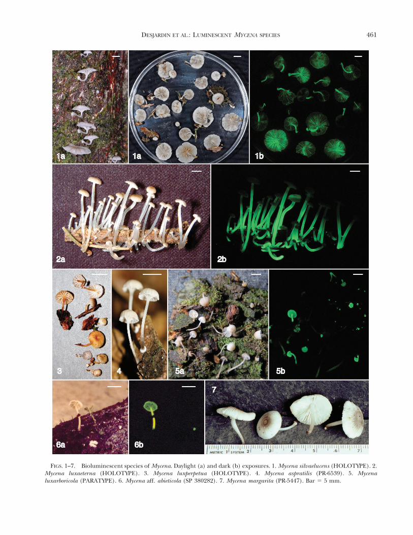
FIGS. 1–7. Bioluminescent species of Mycena. Daylight (a) and dark (b) exposures. 1. Mycena silvaelucens (HOLOTYPE). 2.Mycena luxaeterna (HOLOTYPE). 3. Mycena luxperpetua (HOLOTYPE). 4. Mycena aspratilis (PR-6539). 5. Mycenaluxarboricola (PARATYPE). 6. Mycena aff. abieticola (SP 380282). 7. Mycena margarita (PR-5447). Bar 5 5 mm.
DESJARDIN ET AL.: LUMINESCENT MYCENA SPECIES 461

or with walls up to 0.8 mm thick; terminal cells repentor slightly recurved, like the caulocystidia. Clampconnections present in all tissues.
Habit, habitat and known distribution. Gregarious,abundant on bark of standing dipterocarpous tree inlowland dipterocarp forest. Borneo. Known only fromthe holotype.
Holotype. MALAYSIA. Borneo, Sabah, Sepilok, Sepi-lok Orang-Utan Rehabilitation Center, MangroveTrail, N05u51.7329, E117u56.9489, 12 Dec 2007, col-lected by B.A. Perry, BAP 568 (SFSU).
Commentary. Mycena silvaelucens is characterized bymoderate size basidiomes with grayish brown to pale
gray, hygrophanous, dry pilei, subdecurrent, interve-nose lamellae, a pallid stipe that arises from a pad ofwhite radiating mycelium, broadly ellipsoid, amyloidbasidiospores with mean 8.3 3 5.8 mm, abundantfusoid-ventricose cheilocystidia, a pileipellis and stipiti-pellis of repent, cylindrical, nonspinulose, nongelati-nous hyphae, lignicolous habit, and luminescent pileusand stipe. The new species is allied with a small group ofspecies in sect. Fragilipedes that have grayish brownpilei, nonnitrous odors, smooth cheilocystidia, andnonspinulose, nongelatinous pileipellis and stipitipellishyphae. Mycena silvaelucens is most phenetically similarto two Australian species, M. illiria Grgurinovic and M.
FIG. 8. Mycena chlorophos fruited in culture from inoculum isolated from topotypical material (TFM-M-M512). a.Basidiomes formed 9 Nov 2006. b. 13 Nov 2006. c. 18 Dec 2006. d. 13 Nov 2006. Bar 5 10 mm.
462 MYCOLOGIA

australiana Cleland. Mycena illiria differs in forming abroadly conical to umbonate, dark brown to nearlyblack pileus, adnexed and nonintervenose lamellae,smaller cheilocystidia (mean 32 3 8.4 mm), stipitipellishyphae with nodulose excrescences, and habit on litter(Grgurinovic 2002). Mycena australiana differs informing a conico-campanulate, wood brown pileus,adnate and nonintervenose lamellae, a mealy stipe baselacking radiating mycelium, narrower cheilocystidia(mean width 8.3 mm), and stipitipellis hyphae withnodulose excrescences (Grgurinovic 2002). Neither ofthese Australian species was reported as luminescent.
Mycena luxaeterna Desjardin, B.A. Perry et Stevani,sp. nov. (FIGS. 2, 10)
MycoBank MB 515160Pileus 4–8(–17) mm diam, plano-convexus, umbilicatus,
pellucidus et sulcato-striatus, viscidus; fuscus juventute cum
margine candida, postea albo-helvola. Lamellae horizon-tales, juventute adnatae vel subdecurrentes, postea magisdecurrentes, subdistantes, incanae, margine in vivo con-colori, in sicco aurantiaco et resinoso. Stipes 15–45 3 1–2 mm, albus, glutinosus, affixus ad ligneum frustrum velcoriaceum folium per radiatum album mycelialem pulvi-num. Odor leniter raphanoideus; sapor leviter amarus velraphanoideus. Basidiosporae 6.5–8.3 3 3.2–4.0 mm, valdeamyloideae. Cheilocystidia 27–45 3 5.4–7.5, subcylindracea,subfusiformes vel anguste fusiformes ventricosa; apexobtusus cum adhaerenti aureo exsudato. Pleurocystidiacarentia. Pileipellis ixotrichodermium 80–120 mm crassum,partibus terminalibus dense diverticulatis cum cylindraceis
FIG. 9. Mycena silvaelucens (BAP 568 – HOLOTYPE). a.Basidiospores. b. Basidia. c. Cheilocystidia. Bar 5 10 mm.
FIG. 10. Mycena luxaeterna (DED 8087 – HOLOTYPE).a. Basidiospores. b. Basidia and basidiole. c. Cheilocystidia.d. Pileipellis terminal cells. e. Caulocystidia. Bar 5 10 mm.
DESJARDIN ET AL.: LUMINESCENT MYCENA SPECIES 463
vel obtusis interdum furcatis projecturis. Stipes flavoviren-tem validam lucem emittens. Fungi dense gregarii inramunculo vel raro folio arborearum in silva atlantica,provincia brasiliense Sancti Pauli. Holotypus hic designatus:Brazil, Sao Paulo state, Mun. Iporanga, Parque EstadualTurıstico do Alto Ribeira, 19 Mar 2007, collected by C.V.Stevani, D.E. Desjardin 8087 (SP 381953).
Etymology. lux 5 light (L.), aeterna 5 eternal (L.),referring to the constant light emitted by thebasidiomes. The epithet was inspired by and borrowedfrom Mozart’s Requiem (Communio).
Pileus (FIG. 2) 4–8(–17) mm diam, convex to plano-convex with a central umbilicus, striate to sulcate,glabrous, viscid, shiny; when young dark grayishbrown (6F4-5), fading with age to grayish yellow(5D3-4) or pale grayish brown (6D3), with a palegrayish white ring at the edge. Lamellae horizontal,adnate to subdecurrent when young, subdecurrent todecurrent in age, subdistant (13–18) with two series oflamellulae, pale grayish white, nonmarginate whenfresh, edges orange and resinous in dried material.Stipe 15–30(–45) 3 1–1.5(–2) mm, central, terete,cylindrical, equal, hollow, pliant to subcartilaginous,arising from white radiating basal mycelium, white totranslucent-hyaline and coated overall with a thick,clear gel. Odor weakly raphanoid. Taste slightly bitteror raphanoid.
Luminescence. Stipe strongly yellowish green lumi-nescent, strongest near the base; elsewhere nonlumi-nescent. Mycelium luminescent in culture.
Basidiospores (FIG. 10a) 6.5–8.3 3 3.2–4.0 mm [xmr
5 6.8–7.5 3 3.5 mm, xmm 5 7.2 6 0.5 3 3.5 6
0.04 mm, Q 5 1.9–2.3, Qmr 5 2.0–2.1, Qmm 5 2.05 6
0.1, n 5 16–22, s 5 2], ellipsoid to amygdaliform,smooth, hyaline, strongly amyloid, thin-walled. Basid-ia (FIG. 10b) 22–25 3 6.4–7.0 mm, clavate, hyaline, 4-spored, rarely 2-spored, clamped. Basidioles clavate,hyaline. Lamellar edge sterile, golden orange in waterand 3% KOH from a coating of plaque-like resinousexudates; cheilocystidia (FIG. 10c) abundant, 27–45 3
5.4–7.5 mm, subcylindrical to subfusoid or narrowlyfusoid-ventricose, obtuse, hyaline, inamyloid, nonge-latinous, thin-walled. Pleurocystidia absent. Subhyme-nium nongelatinous. Lamellar trama regular, com-posed of subparallel hyphae 2.5–12 mm diam,cylindrical, hyaline, dextrinoid, nongelatinous, thin-walled. Pileipellis an ixotrichodermium 85–120 mmthick, composed of erect, subparallel to interwovenhyphae with diverticulate terminal cells; hyphae 1.5–8 mm diam, cylindrical to slightly irregular in outline,smooth, hyaline, inamyloid, thin-walled, embedded ina gelatinous matrix; terminal cells (FIG. 10d) erect torepent, densely diverticulate, 1.5–5 mm diam; diver-ticula 1.5–6.5 3 1.2–2.2 mm, cylindrical, obtuse,simple or forked, hyaline, inamyloid, thin-walled.
Hypodermium undifferentiated. Pileus trama interwo-ven, composed of hyphae 2–7 mm diam, irregular inoutline, hyaline, strongly dextrinoid, nongelatinous,thin-walled. Stipitipellis an ixocutis composed ofrepent, parallel hyphae and caulocystidia embeddedin a thick gelatinous matrix; cortical hyphae 2–5 mmdiam, cylindrical, smooth or with scattered diverticu-la, hyaline, inamyloid, thin-walled; medullary hyphae3–16(–20) mm diam, cylindrical, smooth, hyaline,strongly dextrinoid, nongelatinous, thin-walled; cau-locystidia (FIG. 10e) scattered, 18–26 3 2.5–6 mm,simple, cylindrical to subfusoid, obtuse, or irregularin outline and diverticulate like the pileipellisterminal cells. Clamp connections common in alltissues.
Habit, habitat and known distribution. Densely gre-garious, in clusters of 2–20 basidiomes, mostly onsticks, rarely on leaves of undetermined dicotyledon-ous trees in primary Atlantic Forest habitat. Sao Paulostate, Brazil. Known only from a single site.
Holotype. BRAZIL. Sao Paulo state, Mun. Iporanga,Parque Estadual Turıstico do Alto Ribeira, Poco daViuva, S24u35.2209, W48u37.8409, 19 Mar 2007, collect-ed by C.V. Stevani, D.E. Desjardin 8087 (SP 381953;Isotype: SFSU).
Paratype. BRAZIL. Sao Paulo state, Mun. Iporanga,Parque Estadual Turıstico do Alto Ribeira, Poco daViuva, S24u35.2209, W48u37.8409, 23 Feb 2007, C.V.Stevani 2-23-07 (SP 381962, SFSU).
Commentary. Distinctive features of Mycena luxae-terna include: a small, plano-convex, umbilicate, viscid,grayish brown to grayish yellow pileus; subdecurrent,pallid lamellae that develop orange-resinous edgeswhen dried; a translucent, white, thickly gelatinousstipe that strongly emits yellowish green light whenobserved in the dark; a lignicolous habit on smallsticks; strongly amyloid, moderate size basidiospores;narrowly fusoid, obtuse cheilocystidia and a lack ofpleurocystidia; nongelatinous subhymenium; a thickixotrichodermium pileipellis with diverticulate termi-nal cells that is not readily separable when fresh; nodifferentiated hypodermium; strongly dextrinoid tra-mal tissues; a thin ixocutis stipitipellis embedded in athick gelatinous matrix that is not readily separablewhen fresh; and caulocystidia similar in morphology tothe cheilocystidia and the pileipellis terminal cells.
In the field M. luxaeterna is strongly reminiscent ofRoridomyces (Mycena) roridus (Scop.) Rexer; howeverthe hymeniform pileipellis of the latter removesRoridomyces from consideration. The thick ixotricho-dermium-type pileipellis of M. luxaeterna is similar tothose of members of sect. Hygrocyboideae (Fr.) Sing.(e.g. M. epipterygia [Scop. : Fr.] S.F. Gray and allies).In the latter species however the pileipellis is readilyseparable and the lamellae edges are separable as an
464 MYCOLOGIA

elastic thread formed from clavate, apically diverticu-late cheilocystidia embedded in a gelatinous matrix. Ifone overlooks the thick ixotrichodermium pileipellisand thick layer of gelatin on the stipe surface M.luxaeterna shares many similarities with members ofsect. Insignes Maas. Geest., such as M. pseudoclavicu-laris A.H. Sm. from Oregon and M. roriduliformis(Murr.) Dennis from Jamaica. Mycena pseudoclavicu-laris differs in forming a lubricous (not gelatinous)stipe with densely diverticulate cortical hyphae, largerbasidiospores (8.1–9.8 3 4.5–5.6 mm), broader chei-locystidia (6.5–10 mm), has pleurocystidia, and growson debris of yellow pines (Maas Geesteranus 1992).Mycena roriduliformis differs in forming a lubricous(not gelatinous) stipe, smaller basidiospores (4.5–4.93 2.5–3.0 mm), fusiform to subclavate cheilocystidia7–17 mm diam that are often apically lobed, and acutis-type pileipellis (Maas Geesteranus 1992). FinallyM. luxaeterna shares many features with members ofsect. Euspeireae Maas Geest., although M. luxaeternalacks a gelatinous subhymenium, lacks pleurocystidiaand lacks separable pileus and stipe pellicles. Withinsect. Euspeireae the most phenetically similar species isM. euspeirea (Berk. & M.A. Curtis) Sacc. knowncurrently from Cuba, Honduras and Venezuela.Mycena euspeirea differs in having a pileus and stipewith separable pellicles, smaller basidiospores (5.8–6.1 3 2.7–2.9 mm), broader cheilocystidia (9–10 mm),distinct pleurocystidia, a gelatinous subhymeniumand smooth pileipellis hyphae (Maas Geesteranus1992). Four new species belonging to sects. Insignes(viz. M. conspersa Maas Geest. & de Meijer, M. demissaMaas Geest. & de Meijer, M. surculosa Maas Geest. &de Meijer) and Euspeireae (M. tapeina Maas Geest. &de Meijer) recently were described from the southernstate of Parana, adjacent to Sao Paulo state (MaasGeesteranus and de Meijer 1997). All four speciesdiffer from M. luxaeterna in pileipellis and stipitipellisanatomy, cheilocystidia shape, basidiospore size, andlamellar spacing. None of the above mentionedspecies that are phenetically similar to M. luxaeternahave been reported as luminescent. Because M.luxaeterna shares features with members of sects.Hygrocyboideae, Insignes and Euspeireae placement inany currently accepted infrageneric taxon is problem-atical. Multiple gene sequences should help clarifythis issue (to be published elsewhere).
Mycena luxperpetua Desjardin, B.A. Perry et Lodge,sp. nov. (FIGS. 3, 11)
MycoBank MB 515161Pileus 2–5.5 mm diam, juventute forma hemisphaerii,
plano-convexus, postea centro leviter indentato, pellucido-striatus vel subsulcatus, subviscidus, juventute stramineustinctus, postea ochraceus. Lamellae arcuato-decurrentes,
distantes, albae, postea cremeae, margine concolori. Stipes4–8 3 0.5 mm, subviscidus, luteus vel pallidior, posteaochraceus. Basidiosporae 7–8(–8.7) 3 4–5.5(–5.8) mm,amyloideae. Cheilocystidia abundantis, 38–52 3 3.8–5.5 mm, cylindracea vel subcylindracea, late obtusa; capitulaplerumque subcapitata; cheilocystidia similaria pleurocystid-iorum. Pileipellis ixocutiformis vel ixotrichodermiformis vel200 mm crassa cum cellulis terminalibus dense diverticulatis;hypodermium constatum e late inflatis hyphis 10–30 mmdiam, fusiformibus vel subglobosis, contento hyalino velspadiceo; trama et subhymenium valde dextrinoidea, nongelatinosa. Stipitipellis ixocutis constata e laevibus velsparsim diverticulatis hyphis. In cultura totus fungusflavovirentem lucem emittens. Fungi dispersi in erectismortuis arboribus in silva subtropica humida porto-ricense. Holotypus hic designatus: Puerto Rico, El Verderesearch area, 15 Jan 2007, D.J. Lodge and M. RichardsonPR-6463 (SFSU).
FIG. 11. Mycena luxperpetua (PR-6463 – HOLOTYPE). a.Basidiospores. b. Basidium and basidioles. c. Cheilocystidia.d. Pileipellis terminal cells. e. Caulocystidia. Bar 5 10 mm.
DESJARDIN ET AL.: LUMINESCENT MYCENA SPECIES 465

Etymology. lux 5 light (L.), perpetua 5 perpetual(L.), referring to the constant light emitted by thebasidiomes. The epithet was inspired by and borrowedfrom Mozart’s Requiem (Communio).
Pileus (FIG. 3) 2–5.5 mm diam, convex to hemi-spherical when young, becoming broadly convex andshallowly depressed in age, pellicid-striate to striate orsubsulcate, glabrous, shiny, slightly viscid; Drab Graywith Straw Yellow tints when young, becoming YellowOcher to Clay Color in age. Lamellae arcuate-decurrent, distant with one series of lamellulae,moderately broad (0.5–0.8 mm), white to CreamColor, nonmarginate, drying white with concolorousedges. Stipe 4–8 3 0.3–0.7 mm, central, cylindrical orslightly expanded at the apex and base, apexpruinose, glabrous elsewhere, shiny, slightly to mod-erately viscid, white to Spectrum Yellow when young,becoming Yellow Ocher to Clay Color in ageespecially near the base, attached by a hyalinemycelial pad. Odor and taste not recorded.
Luminescence. yellowish green in all parts of thebasidiome and mycelium in culture.
Basidiospores (FIG. 11a) 7–8(–8.7) 3 4–5.5(–5.8)mm [xm 5 7.6 6 0.7 3 4.7 6 0.5 mm, Q 5 1.1–1.9, Qm
5 1.8 6 0.1, n 5 25], ellipsoid, smooth, hyaline,amyloid, thin-walled. Basidia (FIG. 11b) (20–)27–323 6–8.5 mm, clavate, 4-spored, rarely 2-spored, withsterigmata up to 7 mm long, clamped. Basidiolesclavate. Lamellar edge mostly sterile, with scatteredbasidia, nongelatinized; cheilocystidia (FIG. 11c) abun-dant, 38–52 3 3.8–5.5 mm, cylindrical to subcylin-drical, broadly obtuse and mostly subcapitate, capit-ulum 5.5–7.5 mm diam, hyaline, inamyloid, slightlyrefractive, nonexudative. Pleurocystidia scattered, sim-ilar to cheilocystidia in shape and size. Subhymeniumnongelatinous. Lamellar trama regular, composed ofsubparallel hyphae 4.5–13 mm diam, cylindrical toinflated, hyaline, dextrinoid, nongelatinous, thin-walled. Pileipellis an ixocutis to an ixolattice up to200 mm thick, composed of subparallel and mostlyrepent or slightly interwoven hyphae with diverticu-late terminal cells; hyphae 1.5–4 mm diam, cylindrical,branched, smooth, hyaline, inamyloid, thin-walled,embedded in a gelatinous matrix; terminal cells(FIG. 11d) repent, densely diverticulate, 1.5–3.2 mmdiam; diverticula 1.5–8 3 0.5–2 mm, cylindrical,obtuse, simple or forked, hyaline, inamyloid, thin-walled. Hypodermium composed of broadly swollenhyphae 10–30 mm diam, fusoid to subglobose, con-tents hyaline to pale grayish brown, dextrinoid,nongelatinous to subgelatinous, thin-walled. Pileustrama interwoven, composed of hyphae 5–16 mmdiam, cylindrical to inflated, hyaline, strongly dextri-noid, nongelatinous, thin-walled. Stipitipellis an ix-ocutis composed of repent, parallel hyphae and
caulocystidia; cortical hyphae 3–8 mm diam, cylindrical,smooth or with scattered diverticula, hyaline, inamy-loid, gelatinous, thin-walled; medullary hyphae 5.5–20 mm diam, cylindrical or sometimes inflated, hya-line, strongly dextrinoid, nongelatinous, thin-walled;caulocystidia (FIG. 11e) scattered, 16–24 3 3.2–5.5 mm, simple, clavate, obtuse, or a few irregular inoutline and diverticulate like the pileipellis terminalcells. Clamp connections common in all tissues, someof the medallion-type in the pileus ixolattice.
Habit, habitat and known distribution. Lignicolous,scattered on standing dead undetermined dicotyle-donous tree. Puerto Rico. Known only from theholotype.
Holotype. PUERTO RICO. El Verde research area,almost to the ridge above footbridge over the Q.Sonadora, N18u199240, W65u499250, 360 m, 15 Jan2007, collected by D.J. Lodge and M. Richardson, PR-6463 (SFSU).
Commentary. Distinctive features of Mycena luxper-petua include: a small, convex-depressed, viscid, DrabGray to Clay Color pileus; arcuate-decurrent, pallid,nonmarginate lamellae; a latently ochraceous to ClayColor, viscid (not thickly gelatinous) stipe; entirelyluminescent basidiomes; broadly ellipsoid basidio-spores; cylindrical, subcapitate cheilocystidia andpleurocystidia; nongelatinous subhymenium; an ixo-cutis pileipellis with diverticulate terminal cells thatmight represent a separable pellicle; a differentiatedhypodermium of fusoid to subglobose cells often withgrayish brown contents; strongly dextrinoid tramaltissues; a thin ixocutis stipitipellis of weakly gelatinizedhyphae; and caulocystidia similar in morphology to thecheilocystidia and the pileipellis terminal cells. Myce-lial cultures are strongly luminescent (FIG. 1a, asMycena aff. euspeirea, Desjardin et al. 2008).
Many features of M. luxperpetua are similar to thoseof M. luxaeterna, and in the field they would be nearlyindistinguishable except for size. Mycena luxaeternahowever differs in forming larger basidiomes (pileus4–17 mm diam; stipe 15–45 3 1–2 mm), lamellaredges with orange resinous exudates when dried, awhite (non-yellow) stipe covered with a thick hyalinegel, basidiospores with mean range 6.8–7.5 3 3.5 mm(Qmm 5 2.0), fusoid-ventricose cheilocystidia, anabsence of pleurocystidia, and no differentiatedhypodermium. In comparison M. luxperpetua formssmaller basidiomes (pileus 2–5.5 mm diam; stipe 4–83 0.3–0.7 mm), lamellar edges lacking orange resin-ous exudates, a yellow stipe that is merely viscid (notglutinous), broader basidiospores with mean 7.6 3
4.7 mm (Qm 5 1.8), cylindrical-subcapitate cheilocys-tidia and pleurocystidia, and a differentiated hypo-dermium. As with M. luxaeterna, M. luxperpetuashares some features with members of sects. Insignes
466 MYCOLOGIA

and Euspeireae but differs from all described speciesby the unique combination of characters itemizedabove.
Mycena luxarboricola Desjardin, B.A. Perry et Stevani,sp. nov. (FIGS. 5, 12)
MycoBank MB 515162Pileus 3–5 mm diam, convexus vel obtuse conicus,
striatus, glaber, cum disco spadiceo vel ochraceo, marginealutaceo vel albido. Lamellae arcuatae, subdistantes, modicelatae, albae vel alutaceae, non-marginatae. Stipes 8–12 3
0.75–1 mm, ad centrum, cylindraceus, siccus, ubiquepruinosus, basi fibrillis albis; apex albus vel alutaceus; basisochracea vel spadicea. Basidiosporae 7–10.2 3 7–9.3 mm,amyloideae. Cheilocystidia abundantia, 22–38(–52) 3 (5–)7–17 mm, subcylindracea vel late clavata, dense spinulosa indimidio distale; spinulae 1.5–3.5 3 0.5–1.5 mm, cylindraceaevel bacilliformes. Pleurocystidia carentes. Pileipellis cutisconstata e dense spinulosis hyphis 3–13 mm diam, hyalinavel cum brunneo globulari contento, non-amyloidea, non-gelatina; spinulae 1.5–4(–6) 3 0.5–1.5 mm, cylindraceae velbacilliformes; hypodermium constatum e hyphis inflatis, vel 5 to 25 mm diam, valde dextrinoideum, non-gelatinosum. Stipitipellis cutis constata e dense spinulosishyphis et dispersis caulocystidiis subcylindraceis vel clavatis,similibus cheilocystidiorum. Totum basidiome claram fla-vovirentem lucem emittens. Fungi gregarii in corticeviventium arborum in silva riparia in provincia brasilienseParanae. Holotypus hic designatus: Brazil, Parana state,Mun. Jacarezinho, between Fazenda Sao Joao and FazendaCalifornia, 27 Dec 2005, Luiz Fernando Mendes s.n. (SP380286).
Etymology. lux 5 light (L.), arboricola 5 dwelling ina tree (L.), referring to the luminescent arborealbasidiomes.
Pileus (FIG. 5) 3–5 mm diam, convex to obtuselyconical, striate, dull, dry, glabrous, disc pale brown toochraceous, margin pale yellowish white to nearlywhite. Lamellae arcuate, subdistant (8–10) with oneseries of lamellulae, moderately broad, white to paleyellowish white, nonmarginate. Stipe 8–12 3 0.75–1 mm, central, terete, cylindrical, pliant, hollow, dull,dry, pruinose overall, base with white fibrils; apexwhite to pale yellowish white, base ochraceous to palebrown. Odor and taste not recorded.
Luminescence. Entire basidiome emits bright yellow-ish green light.
Basidiospores (FIG. 12a) 7–10.2 3 7–9.3 [xmr 5 8.53 7.8–8.2 mm, xmm 5 8.5 6 0.0 3 8.0 6 0.3 mm, Q 5
1.0–1.2, Qmr 5 1.03–1.08, Qmm 5 1.05 6 0.03, n 5 20,s 5 2], globose to subglobose, smooth, hyaline,strongly amyloid, thin-walled. Basidia (FIG. 12b) 22–28 3 8.3–10 mm, clavate to broadly clavate, hyaline, 4-spored, clamped. Basidioles clavate. Cheilocystidia(FIG. 12c) abundant, lamellar edge sterile, 22–38(–52) 3 (5–)7–17 mm, subcylindrical to broadly
clavate, densely spinulose over the upper half,hyaline, inamyloid, nongelatinous, thin-walled; spinu-lae 1.5–3.5 3 0.5–1.5 mm, cylindrical to rod-like,obtuse, hyaline, thin-walled. Pleurocystidia absent.Subhymenium nongelatinous. Lamellar trama regular,of dextrinoid hyphae. Pileipellis a cutis of repent,densely spinulose hyphae (FIG. 12d); hyphae 3–13 mmdiam, cylindrical to inflated, hyaline or with brownglobular contents, inamyloid, nongelatinous, thin-walled; spinulae 1.5–4(–6) 3 0.5–1.5 mm, cylindricalto rod-like, obtuse, hyaline. Hypodermium pseudopar-enchymatous, composed of short, inflated hyphae upto 25 mm diam, hyaline, strongly dextrinoid, non-
FIG. 12. Mycena luxarboricola (SP 380286 – HOLO-TYPE). a. Basidiospores. b. Basidia and basidiole. c.Cheilocystidia. d. Pileipellis hyphae. e. Stipitipellis hyphae.f. Caulocystidia. Bar 5 10 mm.
DESJARDIN ET AL.: LUMINESCENT MYCENA SPECIES 467

gelatinous, thin-walled. Pileus trama hyphae interwo-ven, cylindrical, nongelatinous to subgelatinous,dextrinoid. Stipitipellis a cutis of repent, denselyspinulose hyphae plus scattered caulocystidia; stipiti-pellis hyphae (FIG. 12e) (2–)5–10 mm diam, cylindricalto slightly inflated, hyaline, inamyloid, nongelatinous,thin-walled, covered with spinulae 1.5–3.2 3 0.5–1.5 mm, cylindrical to rod-like; cortical hyphae 2.5–4 mm diam, parallel, cylindrical, smooth, hyaline,strongly dextrinoid, nongelatinous, thin-walled; med-ullary hyphae 6–12 mm diam, cylindrical, parallel,hyaline, dextrinoid, nongelatinous, thin-walled; cau-locystidia (terminal cells, FIG. 12f) scattered, subcy-lindrical to clavate, similar to the cheilocystidia.Clamp connections common in all tissues.
Habit, habitat and known distribution. Solitary, scat-tered to gregarious on bark of standing, living tree in ariparian forest. Parana, Brazil. Known only from twospecimens within a small area.
Holotype. BRAZIL. Parana state, Mun. Jacarezinho,between Fazenda Sao Joao and Fazenda California, 27Dec 2005, collected by Luiz Fernando Mendes s.n. (SP380286).
Paratype. BRAZIL. Parana state, Mun. Jacarezinho,Sao Joao, Fazenda California, 20 Dec 2006, collectedby Luiz Fernando Mendes s.n. (SP 381961, SFSU).
Commentary. Distinctive features of Mycena luxar-boricola include: a small, pale brown to ochraceous,dry pileus; arcuate, pallid, nonmarginate lamellae; asmall, dry, pruinose stipe; lignicolous basidiomesthat are entirely luminescent and grow on the bark ofliving trees; globose to subglobose, strongly amyloidbasidiospores with mean 8.5 3 8.0 mm; broadlyclavate, densely spinulose cheilocystidia; an absenceof pleurocystidia; nongelatinous, cutis-type pileipellisand stipitipellis of densely spinulose hyphae; anddextrinoid, clamped tramal tissues. In combinationthese features indicate placement in sect. Supinae,where it is allied with M. fera Maas Geest. & deMeijer, M. globulispora Maas Geest. & de Meijer, andM. recessa Maas Geest. & de Meijer, all describedrecently from the Brazilian state of Parana. Mycenafera differs in forming larger basidiospores (mean10.5 3 10.2 mm), cheilocystidia with fewer (only 3–6)and longer diverticulae (5–24 3 2.5–10 mm), andlacks caulocystidia (Maas Geesteranus and de Meijer1997, Desjardin et al. 2007). Mycena globulisporadiffers in forming a dark grayish brown pileus, whitestipe, larger basidiospores 9.8–10.7 3 8.9–10.3 mm,cheilocystidia with few apical spinulae, sparse pleur-ocystidia, and pileipellis and stipitipellis hyphaecovered with few, relatively long and coarse diverti-culae (Maas Geesteranus and de Meijer 1997).Mycena recessa differs in forming a stipe only up to3 mm long, smaller basidiospores 6.3–7.2 3 4.6–
5.5 mm, cheilocystidia with few apical spinulae,smooth stipitipellis hyphae, and growth on bambootwigs (Maas Geesteranus and de Meijer 1997).
Singer (1969, 1973) described two species fromSouth America and one from Mexico referable tosect. Supinae, and they differ from M. luxarboricola asfollows. Mycena hypsizyga Singer, from Argentina,differs in forming a stipe 4–5 mm long, largerbasidiospores 9.5–13 3 8–11 mm, bisporic basidia,narrower cheilocystidia (3–7.5 mm diam), and lacksclamp connections (Singer 1969). Mycena melinoce-phala Singer also from Argentina differs in forming ayellow, umbilicate pileus, more subglobose to broadlyellipsoid basidiospores 9–10.5 3 7–9.5 mm, andpileipellis hyphae with longer diverticulae (, 10 mmlong) (Singer 1973). Mycena abieticola, from Mexico,differs in forming ascending and broadly adnatelamellae, larger basidiospores 10.5–13.5 3 8.5–12 mm,apparently 2-spored basidia lacking clamp connec-tions, cheilocystidia with longer diverticulae(, 12 mm long), predominantly unclamped hyphae,and grows on conifers (Abies religiosa) (Singer 1973).Of the known European and North American speciesof sect. Supinae, M. luxarboricola comes closest to M.supina (Fr.) Kummer. Mycena supina however differsin forming dark brown to dark sepia pilei, morenumerous lamellae (11–17) and more densely prui-nose stipes with abundant caulocystidia (Maas Gees-teranus 1992, Robich 2003). Of the above mentionedphenetically similar species only M. fera has beenreported as luminescent (Desjardin et al. 2007).
Mycena aff. abieticola Singer, Beih. Sydowia 7:37. 1973.(FIGS. 6, 13)
Pileus (FIG. 6) about 5 mm diam, obtusely conical,striate, dull, dry, glabrous to minutely pruinose, paleyellowish brown. Lamellae adnate with a decurrenttooth to arcuate, distant, broad, white, nonmarginate.Stipe about 10 3 0.5 mm, terete, cylindrical, dull, dry,glabrous, apex pale yellowish brown, base brown,noninsititious, arising from white tomentum. Odorand taste not recorded.
Luminescence. Entire basidiome emitting a bright,greenish-yellow light.
Basidiospores (FIG. 13a) 10.2–14 3 (9.5–)10.2–13 mm [xm 5 12.5 6 1.1 3 11.8 6 1.0 mm, Q 5 1.0–1.2, Qm 5 1.05 6 0.05, n 5 25], subglobose toglobose, smooth, hyaline, amyloid, thin-walled. Basid-ia (FIG. 13b) 20–26 3 11.2–13.2 mm, broadly clavate,hyaline, thin-walled, 2-spored with sterigmata up to9.5 mm long, unclamped. Basidioles broadly clavate.Cheilocystidia (FIG. 13c) abundant, 24–30 3 6.5–8.5 mm, subcylindrical to clavate, with few apicalspinulae, hyaline, inamyloid, nongelatinous, thin-
468 MYCOLOGIA

walled; spinulae 1–2.5 3 0.5–1 mm, rod-like, obtuse,hyaline. Pleurocystidia absent. Subhymenium nongela-tinous. Lamellar trama regular, dextrinoid. Pileipellis acutis of repent, hyphae 3–10 mm diam, cylindrical toinflated, densely spinulose, hyaline, inamyloid toweakly dextrinoid, nongelatinous; terminal cells(FIG. 13d) 24–45 3 4.5–9.5 mm, repent, subclavate toclavate, densely spinulose over the outer surface,hyaline; spinulae 1–3 3 0.5–1 mm, rod-like, obtuse,unevenly distributed. Hypodermium of inflated hyphaeup to 20 mm diam, fusoid to ovoid, hyaline, dextri-noid, nongelatinous, thin-walled. Pileus trama inter-woven, composed of hyphae 3–12 mm diam, cylindri-cal, hyaline, dextrinoid, nongelatinous, thin-walled.Stipitipellis a cutis; cortical hyphae 3–8 mm diam,cylindrical, densely spinulose like the pileipellishyphae, hyaline, inamyloid, nongelatinous, thin-walled; medullary hyphae 3–10 mm diam, cylindrical,smooth, hyaline, dextrinoid, nongelatinous, thin-walled. Caulocystidia absent. Clamp connections absentin all tissues.
Habit, habitat and known distribution. Solitary onbark of dead undetermined dicotyledonous tree. SaoPaulo state, Brazil.
Material examined. BRAZIL. Sao Paulo state, Mun.Iporanga, Parque Estadual Turıstico do Alto Ribeira,Poco do Veado, Poco da Viuva, 26 Mar 2006, C.V.Stevani 26.03.06.02 (SP 380282).
Commentary. The specimen described above waserroneously reported as Mycena fera Maas Geest. & deMeijer by Desjardin et al. (2007). DNA sequences ofthis specimen obtained as part of our project on thephylogeny of Mycena sensu lato (Desjardin and Perryunpubl) indicated that the specimen was not conspe-cific with M. fera and warranted closer scrutiny. Re-examination of the material indeed indicated that itdid not represent M. fera but was most pheneticallysimilar to M. abieticola Singer, a species described fromMexico growing on the bark of Abies religiosa and notreported to be luminescent. The Brazilian specimenmatches M. abieticola in macromorphology, basidio-spore size, 2-spored basidia, cheilocystidia size, absenceof clamp connections, and spinulose, nongelatinouspileipellis and stipitipellis hyphae. However M.abieticola forms a minutely pilose pileus with pileocys-tidia 70 3 3 mm, a pruinose stipe with fascicles of hair-like caulocystidia, longer spinulae (, 12 mm), andgrows on bark of Abies (fide Singer 1973). TheBrazilian specimen forms glabrous pilei and stipeslacking cystidia, has spinulae only up to 3 mm long,and grows on bark of an undetermined dicotyledon-ous tree. Because the specimen consists of only a fewbasidiomes that lack detailed macromorphologicalnotes we are hesitant to describe it as a new species.Mycena fera, although macromorphologically similar,bioluminescent and present at the same site, differs informing smaller basidiospores (mean range 10.3–10.63 10.1–10.3 mm), 4-spored basidia, cheilocystidia with3–6 apical appendages 5–22 3 2.5–4.5 mm, coarserspinulae (5–20 3 1–4 mm) on pileipellis and stipiti-pellis hyphae, and clamp connections in all tissues(Desjardin et al. 2007).
Mycena aspratilis Maas Geest. & de Meijer, Verh. Kon.Ned. Acad. Wetensch., Afd. Natuurk., TweedeReeks 97:44. 1997. (FIG. 4)An excellent, comprehensive description and illus-
trations are provided in the protologue (MaasGeesteranus and de Meijer 1997). To those data weadd the following.
Luminescence. Hymenophore emitting a bright,greenish-yellow light; other parts of the basidiomenonluminescent.
Habit, habitat and known distribution. Scattered onsmall twigs (Puerto Rico) or leaves (Brazil) of deadundetermined dicotyledonous trees. Parana state,Brazil, Puerto Rico.
Material examined. PUERTO RICO. El Yunque
FIG. 13. Mycena aff. abieticola (SP 380282). a. Basidio-spores. b. Basidia and basidiole. c. Cheilocystidia. d.Pileipellis terminal cells. Bar 5 10 mm.
DESJARDIN ET AL.: LUMINESCENT MYCENA SPECIES 469

National Forest, Wilderness Area, El Toro Trail,N18u17900, W65u519150, 650 m, 20 Feb 2009, collectedby S.A. Cantrell, D.J. Lodge and L. Millman, PR-6539(SFSU).
Commentary. Mycena aspratilis is reminiscent of aRoridomyces species in the field, characterized by aglabrous, striate, centrally depressed pileus, long-decurrent lamellae, and thickly gelatinous stipesurface. However the presence of thick-walled(, 3 mm), spinulose and long-pedicellate cheilocysti-dia and pleurocystidia indicate a unique taxonomicposition within Mycena. Maas Geesteranus and deMeijer (1997) erected the monotypic sect. Aspratiles toaccommodate this unusual species and suggested thatit might be allied with sect. Polyadelphia Singer exMaas Geest. Desjardin and Braga-Neto (2007) suggest-ed that M. lacrimans Singer, a putatively rareluminescent species from Amazonia, Brazil, might bea second member of sect. Aspratiles, although M.lacrimans forms a nongelatinous stipe and has thin-walled hymenial cystidia. We report for the first timethat the lamellae of M. aspratilis emit a stronggreenish-yellow light, and we extend the knowndistribution of the species northward from southernBrazil to Puerto Rico.
Mycena margarita (Murr.) Murr, Mycologia 8:220.1916. (FIGS. 7, 14)
Synonyms:5 Prunulus margarita Murr., N. Amer. Flora 9:340. 1916.5 Prunulus subepipterygius Murr., Bull. Torrey Bot. Club
67:233. 1940.5 Mycena subepipterygia (Murr.) Murr., Bull. Torrey Bot.
Club 67:235. 1940.Possible synonym:
5 Mycena chlorinosma Singer, Revue Mycol. 2:232. 1937.
Pileus (FIG. 7) (8–)10–18 mm diam, when youngconical to parabolic, becoming convex-hemispherical,then broadly convex with a low flattened umbo thatbecomes slightly depressed in the center at maturity,pellucid-striate to striate or sulcate, glabrous, shiny,pearly opalescent, subviscid to viscid; Hair Brown toSayal Brown or Drab on the disc, radially streakedwith Light Drab striae, nearly white on the margin.Lamellae adnexed, subdistant to distant with twoseries of lamellulae, moderately broad (22 mm),white, nonmarginate. Stipe 10–20 3 1–2 (–3) mm,central, terete, cylindrical, equal, hollow, arising froma cupulate basal disc with internal radial fibrils (notregularly striate); surface dry, pruinose overall, whiteat the apex grading to Drab Gray at the base, somestaining Yellowish Buff at the base. Odor strongly ofchlorine, or weakly of chlorine or not distinctive insome populations; taste distinct but unidentifiable.
Luminescence. yellowish green light in all parts of
FIG. 14. Mycena margarita. a–b. Basidiospores. c. Basidiaand basidiole. d–e. Cheilocystidia. f–g. Pileipellis terminalcells. h. Pileus marginal cells. i. Caulocystidia. (a, d, f.HOLOTYPE of Prunulus margarita; b, e, g. PR-5447; c, i. PR-3292; h. BZ-4303). Bar 5 10 mm.
470 MYCOLOGIA

the basidiome, or nonluminescent in some popula-tions.
Basidiospores (FIG. 14a–b) 6–8.5 3 4–5(–5.5) mm[xmr 5 6.3–7.6 3 4.1–4.8 mm, xmm 5 6.9 6 0.5 3 4.46 0.3 mm, Q 5 1.3–1.8, Qmr 5 1.5–1.7, Qmm 5 1.57 6
0.07, n 5 15–40, s 5 6], ellipsoid, smooth, hyaline,amyloid, thin-walled. Basidia (FIG. 14c) 14.5–19 3
7.5–9.5 mm, clavate, 4-spored, clamped. Basidiolesclavate. Lamellar edge mostly sterile, nongelatinized;cheilocystidia (FIG. 14d–e) abundant, 30–60 3 (6.5–)9–18(–24) mm, fusoid-ventricose to broadly clavate-rostrate, rostrum narrowly conical and simple orrarely forked, smooth, hyaline, inamyloid, thin-walled.Pleurocystidia absent. Subhymenium nongelatinous togelatinous. Lamellar trama regular, composed ofinflated, short-celled hyphae up to 32 mm diam,dextrinoid, thin-walled, nongelatinous. Pileipellis anixocutis up to 200 mm thick composed of repent,radially aligned, widely spaced hyphae with repent tosuberect terminal cells; hyphae 1.5–5 mm diam,cylindrical, branched, smooth or sparsely spinulose,hyaline, inamyloid, thin-walled, with normal or loop-like clamp connections, interspersed with narrowerhyphae (1–2 mm diam) with numerous needle-likeoutgrowths 2–10 3 0.5–1 mm; terminal cells (FIG. 14f–g) 22–48(–75) 3 (5–)8–13 mm, subcylindrical toclavate, often long-pedicellate, densely spinulose,hyaline, inamyloid, thin-walled; hyphae and terminalcells embedded in a gelatinous matrix; spinulae 1.0–3.5 3 0.5–1.2 mm, cylindrical (rod-like), obtuse,unevenly distributed. Pileus marginal cells (FIG. 14h)abundant, 22–48 3 7–16 mm, similar to the cheilocys-tidia, smooth or sparsely spinulose, apex narrowlyconical and acute, not forked. Hypodermium ofinflated, short- to long-celled hyphae (8–)12–24(–32) mm diam, hyaline, strongly dextrinoid, non-gelatinous, thin-walled. Pileus trama interwoven,hyphae 4–12 mm diam, cylindrical to inflated, hyaline,dextrinoid, nongelatinous, thin-walled. Stipitipellis acutis of repent hyphae plus scattered to clusteredcaulocystidia; cortical hyphae 3–8 mm diam, parallel,cylindrical, smooth, hyaline, inamyloid to weaklydextrinoid, nongelatinous to subgelatinous, thin-walled; medullary hyphae 6.5–24 mm diam, cylindricalto inflated, hyaline, strongly dextrinoid, nongelati-nous, thin-walled; caulocystidia (FIG. 14i) (35–)60–110(–150) 3 8–16(–18) mm, fusoid to lanceolate,hyaline, inamyloid, nongelatinous, thin-walled. Basalcup composed of parallel hyphae (2.5–)3.5–10 mmdiam, cylindrical, smooth, hyaline, inamyloid toweakly dextrinoid, nongelatinous, thick-walled (0.5–1.5 mm); internal radial fibrils of hyphae 2.5–5 mmdiam, cylindrical, smooth, hyaline, inamyloid, non-gelatinous, thick-walled (0.5–1 mm); terminal cellsundifferentiated or subcylindrical to subclavate,
broadly obtuse, smooth. Clamp connections commonin all tissues.
Habit, habitat and known distribution. Solitary orgregarious on rotten logs. Belize, Dominican Republic,Jamaica, Puerto Rico, United States (Florida).
Material examined. BELIZE. Stann Creek Dist.,Doyle’s Delight, north trail along stream bank, 22Aug 2007, collected by C. Young, ledger D.J. Lodge,#BZ-4303 (SFSU). DOMINICAN REPUBLIC. Prov.Santiago, Parque Armando Bermudez, Anton SapeBueno, N19u1297.40, W70u599, 980 m, 11 Jan 2003,collected by E. Grand, ledger D.J. Lodge, #DR-2652(SFSU). JAMAICA. Morce’s Gap, 5000 ft., very wetmountainous region, tree ferns and filmy ferns inabundance, 29, 30 Dec, 2 Jan 1908–1909, W.A. andEdna Murrill, (HOLOTYPE of M. margarita: NY).PUERTO RICO. Cordillero Central, Mun. de Maricao,Maricao Community Forest, behind fish hatchery,N18u119320, W66u599360, 400 m, 25 Jun 1996, collectedby E. Horak, #PR-3292 (ZT) and D.J. Lodge and T.J.Baroni #PR-3293 (SFSU); Sierra de Cayey, Mun.Patillas, Carite Commonwealth Forest, Charco Azul, 1Aug 1998, collected by S.A. Cantrell, #PR-5447(SFSU).
Analysis of the holotype specimen.—The materialconsists of one-half of one pileus with badly frag-mented lamellae, few with intact edges, plus one stipecut in half; all in poor condition. As dried: Pileus5 mm diam, plano-convex (no apparent umbo),striate, cream colored, with adherent debris suggest-ing originally viscid. Lamellae subfree, lacking apseudocollarium, subdistant, broad, off-white, non-marginate. Stipe 9 3 0.5 mm, cylindrical, glabrous,pale brown, arising from a distinct cup-shaped basaldisc.
Basidiospores (FIG. 14a) 6.7–8.5 3 4.3–5.5 mm [xm
5 7.6 6 0.5 3 4.7 6 0.2 mm, Q 5 1.4–1.8, Qm 5 1.646 0.07, n 5 25], broadly ellipsoid, smooth, hyaline,weakly amyloid, thin-walled. Basidia not observed.Basidioles clavate. Cheilocystidia (FIG. 14d) abundant,lamellar edge mostly sterile, 37–54 3 12.5–18 mm,fusoid-ventricose to broadly clavate-mucronate, sim-ple (nonspinulose), hyaline, inamyloid, thin-walled,nongelatinous. Pleurocystidia absent. Lamellar tramadextrinoid. Pileipellis an ixocutis of hyphae embeddedin a thick gelatinous matrix; hyphae difficult todifferentiate, narrow (1.5–5 mm), smooth for most oftheir length, inamyloid; terminal cells (FIG. 14f)inflated to clavate, up to 12 mm diam, long-pedicel-late, with scattered spinulae; spinulae 1–2 3 0.5 mm,cylindrical, unevenly distributed; spinulate cells inter-spersed with narrow hyphae with spine-like out-growths. Pileus trama dextrinoid, nongelatinous; nohypodermium observed, material poorly reviving.
DESJARDIN ET AL.: LUMINESCENT MYCENA SPECIES 471

Stipitipellis a cutis; cortical hyphae 3–6 mm diam,cylindrical, smooth, hyaline, weakly dextrinoid, non-gelatinous, thin-walled; medullary hyphae 5–12(–18) mm, dextrinoid, otherwise similar to corticalhyphae. Caulocystidia scattered, fusoid-ventricose,simple, similar to the cheilocystidia. Clamp connec-tions present.
Commentary. The protologue of Prunulus margaritadescribed a single basidiome with convex-umbonatepileus, lamellae attached to a collarium, a cylindricalstipe, with no mention of odor. Examination of theholotype specimen revealed a convex pileus lacking anumbo (at least when dried), subfree lamellae lacking acollarium, and a stipe that arises from a cup-like basaldisc. All latter features are diagnostic of the freshmaterial herein recognized as M. margarita. The shapeof the pileus in Puerto Rico populations was found tochange with age and included a low umbonate stageduring early expansion. Murrill (1916) did not reportan odor, although he seldom reported odors in hisdescriptions, but the collection from Belize clearlylacked an odor, suggesting this might be a variablecharacter. Smith (1947) studied the holotype speci-men of P. margarita and documented a few micro-morphological features congruent with our findings,although he reported the basidiospores as slightlybroader. Of diagnostic significance were basidiospores7–8 3 5–6 mm (4.3–5.5 mm diam in our analysis), agelatinized pileipellis 150–200 mm thick, nongelati-nous lamellar edges formed from broadly fusoid-ventricose cheilocystidia 12–20 mm broad, and anongelatinous stipitipellis. Dennis (1951) describedand illustrated (PL. 23, FIG. 10) specimens fromTrinidad and Venezuela that he identified as M.margarita that strongly resemble ours. AlthoughDennis reported basidiospores narrower (3.5–4 mmdiam) than those reported by Smith (1947), DJLmeasured basidiospores of RWG Dennis #357 fromVenezuela (4.6–5.5 mm diam) and found that they fellwithin the range of our collections and Smith’s typestudy. The Venezuelan collection however differed inhaving a strongly lamellate basal disc and a narrower(14 mm) ixolattice on the pileus, suggesting that itmight not represent M. margarita. Pegler (1983)provided a description of the species that he reportedas common in Guadeloupe, Dominica and Trinidad,that differs only in his observation that the pileipellis isformed from narrow hyphae 1–3 mm diam (nomention whether they were smooth or spinulose). Nopublished accounts report the species as luminescent.
We accept M. subepipterygia (Murr.) Murr. as asynonym of M. margarita and M. chlorinosma Singer asa possible synonym. Maas Geesteranus (1989) provid-ed details and illustrations of the micromorphology ofthe holotype specimen of Prunulus subepipterygius
collected at Planera Hammock, Florida, and his dataare of sufficient quality that we did not need to re-examine the limited material in the holotype specimenat FLAS. He listed M. subepipterygia as a tentativesynonym of M. chlorinosma, described from materialcollected in a greenhouse in Leningrad, but noted afew differences between the species. Repeated requeststo LE to study the holotype specimen of M. chlorinosmawent unanswered. Mycena chlorinosma was reported bySinger (1937) to have a dry stipe and a strong odor ofchlorine, and Maas Geesteranus (1989) observedsmooth pileipellis hyphae and no terminal cells onthe holotype specimen. In comparison M. subepipter-ygia was reported by Murrill (1940) to have a viscidstipe, no odor was reported, and Maas Geesteranus(1989) observed spinulae on the pileipellis hyphae andterminal cells. A specimen from Monteverde, CostaRica (#B-14427) determined by Singer as M. chlor-inosma and accompanied with notes on macro- andmicromorphology (deposited in F) suggests a speciesidentical to our material described above, althoughSinger provided no details of pileipellis anatomystating ‘‘epicutis little developed.’’ Indeed Singerannotated the holotype specimen of P. subepipterygiusas M. chlorinosma.
Morphologically our specimens share features withboth M. chlorinosma and M. subepipterygia. Like M.chlorinosma our material has a dry stipe and usually astrong chlorine odor but matches M. subepipterygia inhaving spinulose pileipellis hyphae and terminal cells.Smith (1947) published a type study of P. subepipter-ygius and provided some details in disagreement withthe latter type study of Maas Geesteranus (1989). Forexample Smith noted inamyloid basidiospores and agelatinous stipitipellis and subhymenium whereasMaas Geesteranus reported amyloid basidiospores,illustrated subgelatinous stipitipellis hyphae and didnot comment on the subhymenium. Because themicromorphological features of our material closelymatch those from the holotype of P. margarita (theoldest epithet) and match the holotype of P.subepipterygius as reported and illustrated by MaasGeesteranus (1989), we accept our material asrepresenting M. margarita with M. subepipterygia asa synonym. Our circumscription broadens the cir-cumscription of M. margarita to include a dry tosubviscid stipe (i.e. nongelatinous to subgelatinousstipitipellis), a usually strong odor of chlorine, apigmented stipe base arising from a cupulate basaldisc and bioluminescent properties.
Basidiospores from the holotype specimen areslightly larger (xm 5 7.6 3 4.8 mm; Qm 5 1.6) thanthose from all other specimens examined, althoughthe shape is identical: e.g. xm 5 6.3 3 4.3 mm, Qm 5
1.5 from Belize (#BZ-4303); xm 5 6.7 3 4.2 mm, Qm
472 MYCOLOGIA

5 1.6 from Dominican Republic (#DR-2652); xm 5
7.4 3 4.5 mm, Qm 5 1.6 from Puerto Rico (#PR-5447). The specimens from Belize, Dominican Re-public and Puerto Rico have identical nLSU andmtSSU sequences (unpubl data), although there issome variability in micromorphology, odor andluminescence. The Belize specimen forms broadlyclavate-rostrate cheilocystidia, distinctly gelatinoussubhymenial tissue, has a stipe surface described asslightly viscid, although the stipitipellis is nongelati-nous to subgelatinous, and the basidiomes werereported as nonluminescent. The Puerto Rico spec-imens have consistently fusoid-ventricose cheilocys-tidia, nongelatinous subhymenial and stipitipellistissues, and the basidiomes are luminescent, whereasthe Dominican Republic specimen has micromor-phology indistinguishable from the Puerto Ricanspecimens, but unfortunately the luminescent prop-erties were not recorded. Because the molecular dataare invariable among the specimens we consider themall to represent the same species. As in Panellusstipticus, this species may have luminescent andnonluminescent populations, or luminescence inthe Belize specimens was so weak it was not easilyobserved by the naked eye.
Murrill (1916) and Smith (1947) did not place M.margarita in any infrageneric group, whereas Pegler(1983) included the species in sect. Fragilipedes.Maas Geesteranus (1992) established sect. IngrataeMaas Geest. with M. chlorinosma as type species andstirps Subepipterygia A.H. Sm. (type species: M.subepipterygia) as a synonym. Although he refrainedfrom formally accepting M. subepipterygia as asynonym of M. chlorinosma he implicitly recognizedboth epithets as belonging to his sect. Ingratae. Acareful comparison of the circumscriptions of sect.Ingratae and sect. Exornatae (Maas Geesteranus1982b; type species, Hiatula boninensis Berk. &M.A. Curtis, accepted as a synonym of M. chlorophos;cf. Maas Geesteranus 1992) reveal that the onlydifference is loop-like clamp connections on thepileipellis hyphae in sect. Ingratae and normal clampconnections in sect. Exornatae. Indeed M. margaritais strongly suggestive of M. chlorophos, and we had atfirst identified our material as representing thelatter species, especially because M. chlorophos isstrongly luminescent. Even Maas Geesteranus andde Meijer (1997:140) recognized the similaritieswhen they suggested, ‘‘It is plausible, therefore, thatthe viscid-stemmed M. subepipterygia, found inFlorida, is just another name of this wide-spreadspecies’’ (viz. M. chlorophos). Loop-like clampconnections are found in a number of Mycenaspecies spread among different infrageneric groups,and we do not accept the character as sufficiently
significant to distinguish sections. We thereforeaccept M. margarita as a member of sect. Exornataewhere it is closely allied with M. chlorophos, thusreducing sect. Ingratae to synonymy with sect.Exornatae.
Mycena chlorophos (Berk. & M.A. Curtis) Sacc. wasdescribed in 1860 from material collected by CharlesWright in the Bonin Islands, Japan, as part of the U.S.North Pacific Exploring Expedition (1853–1856)under Commanders Ringgold and Rodgers (cf.Pfister 1978). The species has been reported in theOld World from as far north as Hachijo Island, Japan,(Corner 1954) and southward to Sri Lanka (Pegler1986), peninsular Malaysia (Corner 1954, 1994),Borneo (Sabah, Maas Geesteranus 1992), Micronesia(Ito and Imai 1939, Corner 1954), Papua New Guinea(Horak and Kobayasi 1978, Maas Geesteranus andHorak 1995), and Australia, in field guides such asAberdeen (1979), Shepherd and Totterdell (1988),Young (1982). In the New World the species has beenreported from Brazil (Manaus and Rio de Janeiro,Corner 1954; Parana, Maas Geesteranus and deMeijer 1997). Based on these reports the species iscurrently recognized as pantropical. Early descrip-tions of specimens from the Bonin Islands providedfew micromorphological details. At best data wereprovided on basidiospores and cheilocystidia. Forexample Ito and Imai (1939) and Hongo (1977)reported the basidiospores as 6–8 3 4–6 mm and 6.5–93 4.5–6 mm respectively and the cheilocystidia as 35–66 3 10–22, broadly fusoid to ventricose with the apexdrawn out to a point (Hongo 1977). Only MaasGeesteranus (1982a) provided a relatively detailedmicromorphological analysis based on a type study ofthe poorly preserved lectotype at K (mistakenlyreported by Maas Geesteranus as holotype). Moredetailed micromorphological analyses reported byCorner (1954, 1994), Pegler (1986), Maas Geester-anus (1992) and Maas Geesteranus and de Meijer(1997) were based respectively on specimens frompeninsular Malaysia, Sri Lanka, Borneo and Brazil. Nocomprehensive study of the micromorphologicalcharacters of good quality topotypical material hasyet been published. To stabilize the epithet, allow foraccurate comparisons of New World and Old Worldspecimens commonly referred to M. chlorophos and tohelp determine whether the species is pantropical orrepresents several geographically isolated distinctspecies, we provide data from topotypical material,including analyses of an isolectotype specimen ofAgaricus chlorophos (FH), an isolectotype of Agaricuscyanophos (FH), and recently collected and culturedmaterial from the Bonin Islands. In addition wedesignate an epitype specimen (affixed to thelectotype at K) of M. chlorophos.
DESJARDIN ET AL.: LUMINESCENT MYCENA SPECIES 473

Mycena chlorophos (Berk. & M.A. Curtis) Sacc., Syllog.Fung. 5:301. 1887. (FIGS. 8, 15)Basionym: Agaricus chlorophos Berk. & M.A. Curtis,
Proc. Amer. Acad. Arts Sci. 4:113. 1860. Lectotypedesignated here, Japan, Bonin Islands, on hillside ondead logs, 21 Oct 1854 (K). For an analysis of thisspecimen see Maas Geesteranus (1982a).
Isolectotype study of Agaricus chlorophos based onmaterial at FH.—The material consists of fourbasidiomes pressed flat and glued to paper, eachcovered with a fibrous layer of thin paper andinfected with molds. One dried pileus 25 mm diam;lamellae subdistant, narrow, nearly free. One driedand flattened stipe 18 3 2 mm. Material too poor for
FIG. 15. Mycena chlorophos. a–c. Basidiospores (a. ISOTYPE of Agaricus chlorophos; b. ISOTYPE of Agaricus cyanophos; c.EPITYPE). d. Basidium and basidiole. e. Cheilocystidia. f. Pileipellis terminal cells. g. Pileus marginal cells. h. Caulocystidia.(d–h. Nagasawa 06-379). Bar 5 10 mm.
474 MYCOLOGIA

a comprehensive micromorphological analysis, but afew details were retrieved as follows. Basidiospores(FIG. 15a) 7.0–9.0 3 5.0–6.0 mm [xm 5 7.9 6 0.4 3 5.56 0.3 mm, Q 5 1.3–1.6, Qm 5 1.5 6 0.11, n 5 20],broadly ellipsoid, smooth, hyaline, amyloid, thin-walled. Basidia 4-spored. Basidioles clavate. Pleurocys-tidia absent. Lamellar trama dextrinoid. Clampconnections present. Other data unretrievable.
Berkeley and Curtis (1860) described Agaricuscyanophos Berk & M.A. Curtis based on materialcollected from near the same site 2 wk later anddistinguished the two species on pileus shape,lamellar attachment and color of emitted light.Agaricus chlorophos had a convex-depressed pileus,slightly decurrent lamellae and emitted green light,whereas A. cyanophos had a campanulate pileus, freelamellae and emitted pale blue light. Ito and Imai(1939) studied a large number of specimens collectedfrom the Bonin Islands and grown in culture, andthey reported that they were variable in shape andlamellar attachment. Accordingly they accepted thetwo as conspecific and accepted the epithet Mycenachlorophos to represent the species. Maas Geesteranus(1982a) reported that the holotype specimen of A.cyanophos was absent from K. We located a syntypespecimen of A. cyanophos at FH and herein designateit as the lectotype. A type study of this specimen ispresented here.
Lectotype study of Agaricus cyanophos based onmaterial at FH.—Japan, Bonin Islands, on decayedwood in shady ravines, 1 Nov 1854 (LECTOTYPEdesignated here). The lectotype specimen consists ofthree basidiomes pressed flat between scraps of loosepaper, in poor condition. As dried: Pileus 12–20 mmdiam, striate. Lamellae free, subdistant with two seriesof lamellulae. Stipe 10–15 3 1.5 mm, pruinose at theapex, noninsititious. Material too poor for a compre-hensive micromorphological analysis. Only data onbasidiospores was retrievable. Basidiospores (FIG. 15b)7.5–8.5 3 5.0–5.5 mm [xm 5 8.0 6 0.4 3 5.3 6
0.2 mm, Q 5 1.5–1.6, Qm 5 1.5 6 0.06, n 5 6],broadly ellipsoid, smooth, hyaline, amyloid, thin-walled.
Although few details were recoverable from thelectotype specimen, we concur with Ito and Imai(1939) and accept A. cyanophos as a synonym of A.chlorophos.
Description of topotypical material of Mycena chlor-ophos.—Pileus (FIG. 8) 10–27 mm diam, obtuselyconical to campanulate when young, expanding inage to broadly convex or plano-convex, with a smallumbo or centrally depressed, striate to rugulose-striate or sulcate, margin crenulate, viscid when moist,white-pruinose overall; disc light yellowish brown or
yellowish brown (Tawny Olive to Isabella Color; 5D-E4-7) to grayish brown (Hair Brown to Snuff Brown;6D-E3-4), paler to the nearly white margin. Lamellaenarrowly adnexed to free, close to subdistant withtwo series of lamellulae, sometimes intervenose,broad (, 3 mm), white, nonmarginate. Stipe 15–253 1–2(–3) mm, central, terete or compressed andonce-cleft, equal above the basal disc, pliant, hollow,dull, dry, white-pruinose overall, white to grayishwhite overall or with a white apex and gradingdownward to brown (6E8) at the base; basal disc 3–5 mm diam, cushion-shaped, striate, brownish yellow(5C7). Odor strongly nitrous-like or of chlorine.
Luminescence. Pileus and lamellae strongly lumines-cent, stipe weakly luminescent, emitting yellowishgreen light. Mycelium luminescent in culture.
Basidiospores (FIG. 15c) (6.8–)7.0–9.0 3 (4.8–)5.0–6.0 mm [xmr 5 7.7–8.0 3 5.3–5.5 mm, xmm 5 7.8 6 0.13 5.4 6 0.1 mm, Q 5 1.3–1.6, Qmr 5 1.4–1.5, Qmm 5
1.46 6 0.06, n 5 20–25, s 5 6], broadly ellipsoid,smooth, hyaline, amyloid, thin-walled. Basidia(FIG. 15d) 22–30 3 7.5–8.5 mm, clavate, 4-spored,clamped. Basidioles clavate. Lamellar edge fertile butmostly sterile; cheilocystidia (FIG. 15e) abundant, 44–100 3 (9.5–)11–22 mm (shorter and narrower onesnearest the stipe, longer and broader ones nearest thepileus margin), fusoid to fusoid-ventricose, apicallynarrowed to an acute tip, cells near pileus marginwith tip often long and narrow (4–22 3 1.5–4 mm),often strangulate and sometimes bifid, hyaline,inamyloid, nongelatinous, thin-walled. Pleurocystidiaabsent. Subhymenium nongelatinous. Lamellar tramaregular, composed of dextrinoid hyphae. Pileipellis anixocutis up to 80 mm thick composed of looselyinterwoven to radially aligned, repent hyphae withrepent to suberect terminal cells; hyphae 2–6.5 mmdiam, cylindrical, smooth or sparsely spinulose,hyaline, inamyloid; terminal cells (FIG. 15f) 10–50(–65) 3 (5–)10–25 mm, clavate to broadly clavate,densely spinulose, hyaline, inamyloid, thin-walled;hyphae and terminal cells embedded in a thingelatinous matrix; spinulae 1.5–4(–6) 3 0.5–2 mm,cylindrical to rod-like, obtuse, unevenly distributed.Pileus marginal cells (FIG. 15g) abundant, 44–100 3
12.5–23 mm, fusoid to fusoid-ventricose, apex acute orobtuse, often bifid, mostly with sparse spinulae overthe central portion, seldom entirely smooth, apexalways smooth, hyaline, inamyloid, thin-walled. Hypo-dermium a mixture of cylindrical, much-branchedhyphae 3–8 mm diam and inflated, vesiculose to fusoidcells up to 22 mm diam, hyaline, strongly dextrinoid,nongelatinous, thin-walled. Pileus trama hyphae 3–8 mm diam, subparallel, radially arranged, cylindrical,smooth, hyaline to pale yellowish gray, dextrinoid,nongelatinous, thin-walled. Stipitipellis a cutis of
DESJARDIN ET AL.: LUMINESCENT MYCENA SPECIES 475

repent hyphae plus scattered to clustered caulocystid-ia; cortical hyphae 3–10 mm diam, parallel, cylindrical,smooth, hyaline, dextrinoid, nongelatinous, thin-walled; medullary hyphae 10–28 mm diam, cylindrical,hyaline, dextrinoid, nongelatinous, thin-walled; cau-locystidia (FIG. 15h) 72–116 3 12–20 mm, fusoid tofusoid-ventricose, hyaline, inamyloid, thin-walled.Clamp connections common in all tissues.
Habit and habitat. Gregarious on fallen branches,logs and trunks.
Material examined. JAPAN. Bonin Islands, Haha-jima, Ogasawara, Igumadani, 30 Oct 2005, H. Neda(TFM-M-M512). The following six specimens werecultivated in the laboratory of the Tottori MycologicalInstitute, Tottori City, Kokoge, from inoculum isolatedfrom TFM-M-M512: cultivated on organic medium(peat moss + rice bran), 19 Oct 2006, E. Nagasawa 06-356 (EPITYPE designated here affixed to the lectotypeof A. chlorophos in K: TMI; ISOEPITYPE: SFSU); 30Oct 2006, E. Nagasawa 06-374NRB (TMI); cultivatedon organic medium (coconut shells processed forgardening use + peat moss + Ebiosu), 7 Oct 2006,E. Nagasawa 06-288 (TMI); 1 Nov 2006, E. Nagasawa06-374NRA2 (TMI); 8 Nov 2006, E. Nagasawa 06-379 (TMI); 2 Feb 2007, E. Nagasawa 07-0001NR(TMI).
Commentary. The Neda specimen (TFM-M-M512)from Hahajima provided the inoculum from whichnumerous specimens were grown on organic mediumat the Tottori Mycological Institute. The cultivatedbasidiomes matched field collected specimens and theprotologue in all pertinent macro- and micromorpho-logical details. Diagnostic features include: a yellowishbrown to grayish brown, campanulate to convex-depressed, striate to sulcate, viscid pileus; adnexed tofree, subdistant, broad, white and nonmarginatelamellae; a dry, pruinose stipe arising from a cush-ion-shaped, striate basal disc; amyloid basidiosporeswith mean 7.7–8.0 3 5.3–5.5 mm; fusoid-ventricosecheilocystidia 44–100 3 11–22 mm with a narrow, acuteand often bifid apical appendage; no pleurocystidia;an ixocutis-type pileipellis with densely spinuloseterminal cells and marginal cells like the cheilocystidiaexcept centrally spinulose; dextrinoid trama tissues;and caulocystidia similar to the cheilocystidia that arisefrom smooth stipe cortical hyphae. It should be notedthat measurements of fresh basidiospores obtainedfrom spore prints of basidiomes fruited in culture atdifferent times from the same inoculum differed inmean length by up to 1 mm and in mean width by upto 0.5 mm.
Mycena chlorophos is similar to M. margarita and M.discobasis Metrod. Mycena margarita differs in form-ing smaller basidiospores (mean 6.9 3 4.4 mm),smaller cheilocystidia (30–60 3 9–18 mm), smaller
pileus marginal cells (22–48 3 7–16 mm) that aremostly smooth, and many loop-like clamp connec-tions. Mycena discobasis differs in forming paler pilei(white to grayish white), larger basidiospores (mean9.9 3 6.7 mm), and the cheilocystidia that lackelongated, often bifid apical appendages characteris-tic of M. chlorophos (Desjardin et al. 2007).
A KEY TO HELP DISTINGUISH THE LUMINESCENT MEMBERS
OF SECT. EXORNATAE (M. MARGARITA, M. CHLOROPHOS,
M. DISCOBASIS METROD)
1. Basidiospores 8.5–11 3 6–7.5 (mean 9.9 3 6.7) mm;pileus white overall or with a hint of pale gray in theumbilicus . . . . . . . . . . . . . . . . . . . . . . . M. discobasis
1. Basidiospores 6–9 3 4–6 (mean 6.9–7.8 3 4.4–5.4)mm; pileus mostly grayish brown with pallid margin . . . 2
2. Cheilocystidia 44–100 mm long, fusoid to fusoid-ventricose, apically narrowed to an acute tip, thosenear pileus margin with tip often bifid, long andnarrow (4–22 3 1.5–4 mm; pileus marginal cells 44–100 3 12.5–23 mm; basidiospores 7–9 3 5–6 (mean7.8 3 5.4) mm . . . . . . . . . . . . . . . . . . . M. chlorophos
2. Cheilocystidia 30–60 mm long, fusoid-ventricose tobroadly clavate-rostrate, rostrum short, narrowlyconical, those near pileus margin lacking a long,narrow projection; pileus marginal cells 22–48 3 7–16 mm; basidiospores 6–8.5 3 4–5.5 (mean 6.9 3
4.4) mm . . . . . . . . . . . . . . . . . . . . . . . . M. margarita
ACKNOWLEDGMENTS
This research was financed in part by National ScienceFoundation grants to D.E. Desjardin and J.-M. Moncalvo(DEB-0542445), to D.J. Lodge and T.J. Baroni (DEB-9525902 and DEB-0103621), to D.J. Lodge among others(DEB-0218039), and a National Geographic Society grant(No. 8240-07) to T.J. Baroni for collections from the MayaMountains in Belize. We thank FAPESP (Grant No. 06/53628-3) for providing financial support to C.V. Stevani forfieldwork in Brazil. We are most grateful for the help ofHitoshi Neda (Forestry and Forest Products ResearchInstitute, Tsukuba, Japan) who kindly provided the epitypeculture of M. chlorophos to EN for basidiome production.Drs Andrew W. Wilson, M. Cathrine Aime, Timothy J.Baroni, Egon Horak, Colin Young and Mike Richardson arethanked for their contributions and companionship in thefield. The manuscript benefited from prereviews by Drs T.J.Baroni, T. Læssøe and S. Redhead.
LITERATURE CITED
Aberdeen JEC. 1979. An introduction to the mushroom,toadstools and larger fungi of Queensland. QueenslandNaturalists’ Club. 206 p.
Berkeley MJ, Curtis MA. 1860. Characters of new fungicollected in the North Pacific Exploring Expedition byCharles Wright. Proc Am Acad Arts Sci 4:111–130.
476 MYCOLOGIA

Corner EJH. 1954. Further descriptions of luminous agarics.Trans Br Mycol Soc 37:256–271.
———. 1994. Agarics in Malesia I. Tricholomatoid II.Mycenoid. Beih Nova Hedwig 109:1–271.
Dennis RWG. 1951. Some Agaricaceae of Trinidad andVenezuela. Leucosporae I. Trans Br Mycol Soc 34:411–482.
Desjardin DE, Capelari M, Stevani CV. 2007. Biolumines-cent Mycena species from Sao Paulo, Brazil. Mycologia99:317–331.
———, Oliveira AV, Stevani CV. 2008. Fungi biolumines-cense revisited. Phytochem Photobiol Sci 7:170–182.
Grgurinovic CA. 2002. The genus Mycena in South-EasternAustralia. Hong Kong: Fungal Diversity Press. 329 p.
Holmgren PK, Holmgren NH, Barnett LC. 1990. IndexHerbariorum I: The herbaria of the world. RegnumVeg 120:1–693.
Hongo T. 1977. Higher fungi of the Bonin Islands I. MemNat Sci Mus Tokyo 10:31–41.
Horak E, Kobayasi Y. 1978. List of New Guinea species ofAgaricales s.l. Trans Mycol Soc Japan 19:103–107.
Ito S, Imai S. 1939. Fungi of the Bonin Islands III. TransSapporo Nat Hist Soc 16:9–20.
Kornerup A, Wanscher JH. 1978. Methuen handbook ofcolour. 3rd Ed. London: Eyre Methuen. 252 p.
Maas Geesteranus RA. 1982a. Studies in Mycenas 59.Berkeley’s fungi referred to Mycena – 1. Proc K NedAkad Wet (Ser C) 85:273–285.
———. 1982b. Studies in Mycenas 72. Berkeley’s fungireferred to Mycena – 2. Proc K Ned Akad Wet (Ser C)85:527–539.
———. 1989. Conspectus of the Mycenas of the northernhemisphere 12. Proc K Ned Akad Wet (Ser C) 92:331–365.
———. 1992. Mycenas of the northern hemisphere II.Conspectus of the Mycenas of the northern hemi-sphere. Verh Kon Ned Acad Wetensch, Afd Natuurk,Tweede Reeks 90:1–493.
———, de Meijer AAR. 1997. Mycenae paraenses. Verh Kon
Ned Acad Wetensch, Afd Natuurk, Tweede Reeks 97:1–164.
———, Horak E. 1995. Mycena and related genera fromPapua New Guinea and New Caledonia. Biblio Mycol159:143–229.
Matheny PB, Curtis JM, Hofstetter V, Aime MC, MoncalvoJ-M, Ge Z-W, Yang Z-L, Slot JC, Ammirati JF, Baroni TJ,Bougher NL, Hughes KW, Lodge DJ, Kerrigan RW,Seidl MT, Aanen DK, DeNitis M, Daniele GM,Desjardin DE, Kropp BR, Norvell LL, Parker A,Vellinga EC, Vilgalys R, Hibbett DS. 2006. Major cladesof Agaricales: a multilocus phylogenetic overview.Mycologia 98:982–995.
Murrill WA. 1916. Prunulus. N Am Flora 9(5):319–344.———. 1940. Additions to Florida fungi IV. Bull Torrey Bot
Club 67:227–235.Pegler DN. 1986. Agaric flora of Sri Lanka. Kew Bull Add
Ser 12:1–519.Pfister D. 1978. Cryptogams of the United States North
Pacific Exploring Expedition, 1853–1856. Massachu-setts: Harvard University, Farlow Reference Library andHerbarium of Cryptogamic Botany. 196 p.
Ridgway R. 1912. Standards and color nomenclature.Washington, D.C., 44 p, I-LIII pl.
Robich G. 2003. Mycena d’Europa. Trento, Italy: Associa-zione Micologica Bresadola. 728 p.
Shepherd CJ, Totterdell CJ. 1988. Mushrooms and toad-stools of Australia. Melbourne: Inkata Press. 162 p.
Singer R. 1937. Notes sur quelques Basidiomycetes. RevMycol 2:226–242.
———. 1969. Mycoflora australis. Beih Nova Hedwig 29:1–405.
———. 1973. Diagnoses fungorum novorum AgaricaliumIII. Beih Sydowia 7:1–106.
Smith AH. 1947. North American species of Mycena. AnnArbor: Univ Michigan Press. 521 p.
Smithe FB. 1975. Naturalist’s color guide. New York:American Museum of Natural History. 29 p.
Young TAM. 1982. Common Australian fungi. Kensington:New South Wales Univ Press. 151 p.
DESJARDIN ET AL.: LUMINESCENT MYCENA SPECIES 477