lung cancer what the surgeon assessment of needs to know
TRANSCRIPT
REVIEW ARTICLE
Lung cancerwhat the surgeon
needs to know
Martyn PayneMB ChB, FFRad (D) (SA)
Levin and PannersConstantiaberg Mediclinic
Cape Town
Anthony G LinegarMB ChB, FCS (SA)
N1 City HospitalCape Town
IntroductionLung cancer is the commonest
cause of death from cancer, account-ing for over 900 000 of the estimated5.2 million cancer-related deathsworldwide.' South Africa is the onlydeveloping country with a cancerdeath rate approximately the same asthat of the developed world."
Approximately 80% of lung can-cers are categorised as non-small celltumours (NSCLCA) and the rest(20%) are small cell tumours.NSCLCA can be cytologically subdi-vided, with adenocarcinoma beingthe commonest (40%) followed bysquamous cell carcinoma (30%) andlarge cell undifferentiated carcinomas(10%). There are also tumours withmixed cell types where two differenttumour cell types are identified in asingle tumour (e.g. adenosquamouscarcinoma).
Primary surgery is universallyaccepted as the treatment of choice inearly stage (cStage I and cStage II)
NSCLCA in selected patients with thephysiological reserves to undergo theoperation and maintain a reasonablequality of life thereafter. The purposeof surgery is curative, and this requirescareful patient selection if fruitlessinterventions are to be avoided. Theoverall cure rate after complete resec-tion is 13%.3
StagingStaging the tumour is crucial to
planning of treatment, prognostica-tion, and the final interpretation ofthe results of therapy. Non-invasiveimaging techniques (i.e. spiral CTscanning with contrast enhancement)are used to determine the anatomicalextent of the disease and thereby toenable clinical staging according to thetumour node metastasis (TNM) stag-ing system.' Clinical staging refers tostaging prior to surgery and is prefixedby the letter 'c' (cTNM) whilst patho-logical staging (pTNM) refers to thepathological staging achieved by sys-tematic surgical exploration.
Internationalstaging systemThe TNM system (Table I) pro-
vides a reproducible description of theanatomical extent of the disease andwas most recently updated in 1997.4
The T descriptor refers to tumoursize, anatomical extent and relation-ship to adjacent structures. The Ndescriptor refers to extent of nodal
30 SA JOURNAL OF RADIOLOGY • February 2003
disease. The M descriptor refers topresence or absence of metastases.
Imagingassessment of
StageThe chest radiograph (high kVp
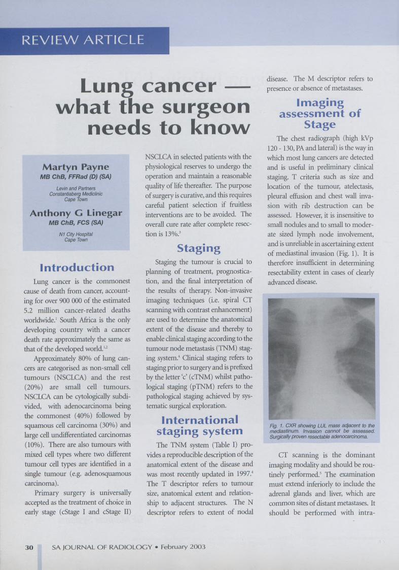
120 - 130,PAand lateral) is the way inwhich most lung cancers are detectedand is useful in preliminary clinicalstaging. T criteria such as size andlocation of the tumour, atelectasis,pleural effusion and chest wall inva-sion with rib destruction can beassessed. However, it is insensitive tosmall nodules and to small to moder-ate sized lymph node involvement,and is unreliable in ascertaining extentof mediastinal invasion (Fig. I). It istherefore insufficient in determiningresectability extent in cases of clearlyadvanced disease.
Fig. 1. CXR showing LUL mass adjacent to themediastinum. Invasion cannot be assessed.Surgically proven resectable adenocarcinoma.
CT scanning is the dominantimaging modality and should be rou-tinely performed.' The examinationmust extend inferiorly to include theadrenal glands and liver, which arecommon sites of distant metastases. Itshould be performed with intra-

REVIEW ARTICLE
Table I. TNM staging systemThmour status (T descriptor)Tx Primary tumour cannot be assessed, or tumour proven by malignant
cells in sputum but not seen on imaging or bronchoscopyTO No evidence of primary tumourTis Carinoma in situTl Tumour 3 em or less in greatest dimension, surrounded by lung or vis-
ceral pleura, without bronchoscopie evidence of invasion more proximalthan the lobar bronchus (i.e. not in the main bronchus)"
T2 Tumour with any of the following features of size or extent:More than 3 em in greatest dimensionInvolves the main bronchus, 2 em or more distal to the carinaInvades the visceral pleuraAssociated with atelectasis or obstructive pneumonitis that extendsto the hilar region and does not involve the entire lung
T3 Tumour of any size that directly invades any of the following:Chest wall (including superior sulcus tumours), diaphragm,mediastinal pleura, parietal pericardiumOr tumour in the main bronchus < 2 em distal to the carina, butwithout involvement of the carinaOr atelectasis or obstructive pneumonitis of the entire lung
Tumour of any size that invades any of the following:Mediastinum, heart, great vessels, trachea, carina, oesophagus,vertebral bodyOr tumour with a malignant pleural or perieardial effusion'Or with satellite nodule(s) within the primary tumour lobe of thelung
T4
Regional lymph nodes (N descriptor)Nodes cannot be assessedNo lymph node metastasesMetastases to ipsilateral peribronchial and or ipsilateral hilar lymph-nodes, and intrapulmonary nodes involved by direct extension of theprimary tumourMetastases to ipsilateral mediastinal and or subcarinal lymph nodesMetastases to contralateral mediastinal, hilar or scalene orsupraclavicular nodes
Distant metastasis status (M descriptor)Mx Cannot be assessed
NxNONl
N2N3
MOMl
No distant metastasesDistant metastases present"
"The uncommon superficial tumour of nny size with its invasive component limited to the bronchial wall, which may extend proximal to themain bronchus, is also classified ac;TI.[Most pleura] effusions in association with a cancer are malignant and caused by the tumour. However. Il few patjent'S have multiple cytologyexaminations of the pleural fluid that are negative for tumour. In these cases the fluid is ncr bloody and is nol an exudate. When these elementsand clinical judgement dictate that the effusiou is not related to the turnour.the effusion should be excluded as a staging clement. and the patientstaged Tl, 2 or 3, Pericardlal effusion is classified according to tJ1C same rules,tSeparnte metastatic turnour nodulels) in the ipsilateral non-primary tumour lobe of the lung are also classified Ml.
venous contrast to allow accurate dif-ferentiation of mediastinal and hilarvascular structures from nodal tissueand tumour. The scan must also bedisplayed with lung and soft tissue set-tings.
CT assessment ofT descriptor
Staging of the T descriptor has anaccuracyof85% (Fig.2).6Grossmedi-astinal invasion can be confidently
31 SAJOURNAL OF RADIOLOGY • February2003
Fig. 2. CT of Tt NOadenocarcinoma
Fig. 3. CT showing obvious mediastinal invasion.
diagnosed, but it can be difficult todistinguish between tumour contigu-ity and extension into mediastinalstructures (Fig. 3). Intact fat plane,contact of 3 cm or less with the medi-astinum and less than 90° contactwith the aorta suggest no invasion,whereas greater than 180° contactwith the mediastinal structures usual-ly indicates invasion.'
CT assessment ofN descriptor
N assessment relies on node sizewith a short axis diameter of greaterthan 1 cm being considered positivefor nodal metastases (Fig. 4). How-ever, small nodes can contain micro-scopic tumour deposits (producing afalse-negative assessment in 20% ofcases) and conversely large nodes maybe clear of metastases (producing afalse-positive assessment in 20% ofcases for glands greater than 2 cm)," A

REVI'EW ARTICLE
Fig. 4. T2N2: lymph nodes measure greater than1 em In short axis.
systematic approach using one of theaccepted lymph node maps should beutilised to describe disease in hilar(Nl), ipsilateral mediastinal (N2) andcontralateral mediastinal (N3) sta-tions."
The profoundly negative impacton survival of nodal metastases is welldocumented, but the identification ofhilar nodal (Nl) disease remains aweakness in the clinical assessment.
CT assessment ofM descriptor
The search for metastases is initial-ly limited to the chest and the upperabdomen unless clinical informationindicates the need for brain and orbone scanning which are not routine-ly performed. Low density liverlesions are frequently encountered.These are most often benign cysts.Ultrasound can be useful in differenti-ating cystic from solid lesions.Isolated adrenal masses are usuallybenign adenomas. Unenhanced CTattenuation of 10 HU or less, or morethan 50% washout on ID-minutedelayed enhanced CT are highly spe-cific for benign adrenal adenomas. to
MR.This is of similar accuracy to CT in
assessing the T and M descriptors, butmay offer an advantage over CT inassessing mediastinal and chest wallinvasion, particularly in superior sul-cus tumours.' Chemical shift MR!accurately differentiates metastasesfrom benign adrenal adenomas. I I
Positron emissiontomography
PET scanning relies on theincreased metabolism of glucose intumour cells to characterise glandsand masses. Its role in routine pre-operative staging is yet to be fullydefined, but may prove to be useful inenhancing the CT assessment, inexamining the Nl hilar nodes, and forevaluation of any contralateralparenchymal nodules."
Stage groupingStage groups were developed on
retrospective survival data.'
5-YearlNMsubset survival (%)
Ca- in situTlNOMO 60T2NOMO 38TlNIMO 34T2NIMO 24T3NOMO 22T3NIMO 9
TlN2MO l3T2N2MO l3T3N2MO l3T4anyN 7N3anyT 3Ml anyT,anyN
Stage
oIAIBIIAlIB
IlIA
IIIB
N
32 SAJOURNAL OF RADIOLOGY. February 2003
The major prognostic factor is theability to achieve a complete resection(RO). Limited invasion of the medi-astinum or of the chest wall (T3) doesnot prohibit complete resection, andprovided the mediastinal glands arenot involved S-year survivals of up to40% can be achieved.
Pleural effusions in the presence ofa lung tumour are almost alwaysmalignant and should be consideredto be so unless careful cytologicalanalysis of the fluid as well as thor-ough thoracoscopic examinationreveals no malignancy in the pleuralspace. This is unlikely in advanced Tand N categories. Malignant effusionsconfer a T4 status on the tumour andthese patients are not surgical candi-dates.
The purpose of detecting medi-astinal nodal disease preoperatively isto avoid unnecessary operations thatwill not favourably influence survival.Where large mediastinal glands arerecognised on the CT scan, biopsy bymediastinoscopy and or mediastin-otomy is undertaken, where theseglands are within reach of the medi-astinoscope. Mediastinoscopy willproduce a false-negative result in 25%of cases."
Clinical staging under stages thefull extent of the disease in at least25% of cases which explains why theoverallS-year survival in TINO is only60% (i.e only 60% were in fact TINOand the rest had more advanced dis-ease).
Additionalinformation
Chest radiograph and CT scanningwill provide other valuable informa-tion that may influence surgical plan-

REVIE\N ARTICLE
ning. Background lung disease suchas COPD, scarring, likely presence ofadhesions from previous pleural dis-ease, and cardiac disease can beassessed and factored into the overallrisk assessment for surgery (Fig. 5).The site and size of the tumour andsome anatomical factors identified onthe scan can assist in estimating thelikely extent of the resection necessaryto achieve a complete resection (i.e.lobectomy or pneumonectomy)(Fig.6).
Fig. 5. TI tumour, but note background of COPOwhich may complicate surgical approach.
Fig. 6. Small tumour LLL but close to LULbronchus therefore pneumonectomy rather thanlobectomy required.
ConclusionThe radiological assessment of
lung cancer requires an understand-ing of the biological behaviour of thedisease, and a knowledge of the meth-ods available to study it. Critical infor-mation required by the surgeonrevolves around staging the primarytumour and assessing its resectabilitybased on the information providedabove. Accurate interpretation of theinformation requires practice andteam work between the radiologistand the thoracic surgeon.
Although clinical staging understages the disease in approximately25% of cases, it is not an unacceptablesituation, as it means that with thecurrent system no patient withresectable disease will be denied apotentially curative resection.
Standardised reporting of theinformation needs to include adetailed description of the tumourbased on the TNM system, and usingan agreed upon nomenclature for thelymph nodes based on one of theavailable lymph node maps. It shouldalso include any co-existing diseasethat may be used in the overall riskassessment for any planned therapy.Finally,the data should be recorded asaccurately as possible for the purposeoflater comparisons, used in assessingthe response to therapy or the accura-cy of clinical staging.
References1. Pisani p, Parkin OM, Bray F,Ferlay J. Estimates
of the worldwide mortality from 25 cancers in1990. Int J Cancer 1999;83: 18-29.
2. Travis WD, Lubin J, rues L, Devesa S. UnitedStates lung carcinoma incidence trends: declin-ing for most histologic cell types among males,increasing among females. Cancer 1996; 77:2464-2470.
3. Martini N, Bains S, Burt M, et al. Incidence oflocal recurrence and second primary tumoursin resected stage I lung cancer. J ThoracCardiovasc Surg 1995; 109: 120-128.
4. Mountain CF. Revisions in the internationalsystem for staging lung cancer. Chest 1997; Ill:1710-1717.
5. Webb WR, Gatsonis C, Zerhouni EA, et a/. CTand MR! imaging in staging non-small cellbronchogenic carcinomas: report of theRadiologic Diagnostic Oncology Group.Radiology 1991; 178: 705-713.
6. Isbicki JR, Thetter 0, Karg 0, et al. Accuracy ofcomputed tomographic scan and surgicalassessment for the staging of bronchial carcino-ma. A prospective study. J Thoroe CardiovascSurg 1992; 104: 413-420.
7. Herman SJ,Winton TL, Weisbrod GL, TowersMJ, Mentzer SJ.Mediastinal invasion by bron-chogenic carcinoma: CT signs. Radiology 1994;190: 841-846.
8. Naruke T, Suemasu K, Ishikawa S. Lymph nodemapping and curability at various levels ofmetastasis in resected lung cancer. J ThoracicCardiovasc Surg 1978;76: 833-839.
9. Mountain CF, OresIer CM. Regional lymphnode classification for lung cancer staging.Chestl997; 111: 1718-1723.
lO. Korobkin M. CT characterisation of adrenalmasses: the time has come. Radiology2000; 217:629-632.
Il. Miller DG, CroveJlo M, Mattteucci T, PetersenRO , Miettinen MM. Benign adrenocorticalmasses: diagnosis with chemical shift MR!imaging. Radiology 1992; 185: 345-351.
12. Lowe vr, Naunheim KS. Positron emissiontomography in lung cancer. Ann Thorac Surg1998;65: 1821-1829.
13. Goldstraw P, Mannam G, Kaplan 0, Michail P.Surgical management of non-small cell lungcancer with ipsilateral mediastinal nodal metas-tasis (N2 disease). J Thorac Cardiovasc Surg1994; 107: 19-27.
33 SA JOURNAL OF RADIOLOGY. February 2003