lymphatic system
TRANSCRIPT

LYMPH &
LYMPHOID
ORGANS
Sunday, January 18, 2015 1


The lymphatic system represents an accessory route through which fluid
can flow from the interstitial spaces into the blood. Most important, the
lymphatics can carry proteins and large particulate matter away from the
tissue spaces, neither of which can be removed by absorption directly into
the blood capillaries.
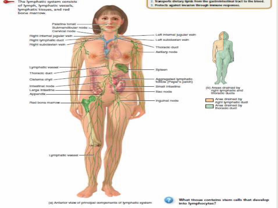
The lymphatic system (lim-FAT-ik) consists of a fluid called lymph,
vessels called lymphatic vessels that transport the lymph, a number of
structures and organs containing lymphatic tissue (lymphocytes within
a filtering tissue), and red bone marrow.
The lymphatic system assists in circulating body fluids and helps defend
the body against disease-causing agents.
Sunday, January 18, 2015 3

Most components of blood plasma filter through blood capillary walls
to form interstitial fluid. After interstitial fluid passes into lymphatic
vessels, it is called lymph (LIMF clear fluid).
Lymphatic tissue is a specialized form of reticular connective tissue
that contains large numbers of lymphocytes, These lymphocytes are
Agranular white blood cells participate in immune responses: B
cells and T cells.
The major difference between interstitial fluid and lymph is location:
Interstitial fluid is found between cells, and lymph is located within
lymphatic vessels and lymphatic tissue.
This return of proteins to the blood from the interstitial spaces is an
essential function without which we would die within about 24 hours.
Sunday, January 18, 2015 4

FUNCTIONS OF THE LYMPHATIC SYSTEM
The lymphatic system has three primary functions:
1. Drains excess interstitial fluid. Lymphatic vessels drain excess
Interstitial fluid from tissue spaces and return it to the Blood.Without
this function, the maintenance of circulating blood volume would not be
possible.
2. Transports dietary lipids. Lymphatic vessels transport lipids lipid-
soluble vitamins (A, D, E, and K) absorbed by the Gastrointestinal tract.
3. Carries out immune responses. Lymphatic tissue initiates Highly specific
responses directed against particular microbes or abnormal cells.
Sunday, January 18, 2015 5
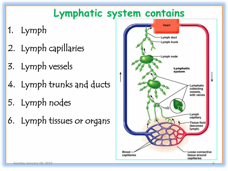
Lymphatic system contains
1. Lymph
2. Lymph capillaries
3. Lymph vessels
4. Lymph trunks and ducts
5. Lymph nodes
6. Lymph tissues or organs
Sunday, January 18, 2015 6

FORMATION OF LYMPH.
Lymph is a clear , watery appearing fluid found in the lymphatic vessels.
Plasma gets filtered into the interstitianal space across the capillary wall.
This will be re absorbed , leaving a small amount of fluid behind.
In glands, various substances like proteins ,fats, are also enters into the
interstitial space.
Various organic substances from degenerating cells are also enters in the
inetrstitial space.
The left out fluid and other substances enter the lymph vessels and
constitute lymph.
Small pressure favours the formation of more fluid than they absorbed.this
fluid enters the lymphatic vessels.
Sunday, January 18, 2015 7
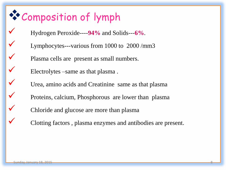
Composition of lymph
Hydrogen Peroxide----94% and Solids---6%.
Lymphocytes---various from 1000 to 2000 /mm3
Plasma cells are present as small numbers.
Electrolytes –same as that plasma .
Urea, amino acids and Creatinine same as that plasma
Proteins, calcium, Phosphorous are lower than plasma
Chloride and glucose are more than plasma
Clotting factors , plasma enzymes and antibodies are present.
Sunday, January 18, 2015 8

Factors which influence formation of lymph
Increase in hydrostatic pressure increases the lymph formation.
Increase in capillary surface.
Increase in capillary in capillary permeability… hypoxia, ADP, hydrogen
ions, Bradykinin, histamine.
Increased functional activity.
Lymph flow is aided by
Presence of valves in the lymphatic vessels
Movement of muscles
Pressure gradient , respiratory pump during inspiration
An increase in lymph formation.
Sunday, January 18, 2015 9

Functions of lymph Returns of protein, electrolytes and water to blood from the tissue spaces.
Helps in the redistribution of body water.
Removes the particulate matter and bacteria.
Maintain the interstitial fluid levels
Body defense by lymphocytes and antibodies.
Fat absorption from intestine by he lacteals.
Transport antibodies and other drugs injected by intra muscularly.
Lymph flow is responsible for structural and functional integrity of tissues.
Supplies nutrients and oxygen to those parts where blood cannot reached
Lymph flow in kidney helps in formation of concentrated urine by
maintaining osmotic pressure gradient
Any factor which increases lymph formation is called
lymphatogogue. Ex: sunlight, Histamine….
Sunday, January 18, 2015 10

Sunday, January 18, 2015 11
• Blood capillaries
• Interstitial spaces
• Lymphatic capillaries
• Lymph vessels
• Lymph ducts
• Junction of the internal jugular and subclavian veins

The two “pumps” that aid the return of venous blood to
1. Skeletal muscle pump. The “milking action” of skeletal muscle
Contractions compresses lymphatic vessels (as well as veins) and forces
lymph toward the junction of the internal jugular and subclavian veins.
2. Respiratory pump. Lymph flow is also maintained by pressure
Changes that occur during inhalation (breathing in). Lymph flows from
the abdominal region, where the pressure is higher, toward the thoracic region,
where it is lower.
When the pressures reverse during exhalation (breathing out), the valves
in lymphatic vessels Prevent backflow of lymph. In addition, when a lymphatic
vessel distends, the smooth muscle in its wall contracts, which helps move lymph
from one segment of the vessel to the next.
Sunday, January 18, 2015 12

LYMPHATIC CAPILLARIES
Lymphatic capillaries have greater permeability than blood capillaries
and thus can absorb large molecules such as proteins and lipids.
Lymphatic capillaries are also slightly larger in diameter than blood
capillaries and have a unique one-way structure that permits interstitial
fluid to flow into them but not out.
The ends of endothelial cells that make up the wall of a lymphatic
capillary overlap.
When pressure is greater in the interstitial fluid than in lymph, the
cells separate slightly, like the opening of a one-way swinging
door, and interstitial fluid enters the lymphatic capillary.
When pressure is greater inside the lymphatic capillary, the cells
adhere more closely, and lymph cannot escape back into interstitial
fluid.Sunday, January 18, 2015 13

The pressure is relieved as lymph moves further down the lymphatic
capillary.
Attached to the lymphatic capillaries are anchoring filaments, which
contain elastic fibers.
They extend out from the lymphatic capillary, attaching lymphatic
endothelial cells to surrounding tissues.
When excess interstitial fluid accumulates and causes tissue swelling, the
anchoring filaments are pulled, making the openings between cells even
larger so that more fluid can flow into the lymphatic capillary.
In the small intestine, specialized lymphatic capillaries called lacteals carry
dietary lipids into lymphatic vessels and ultimately into the blood .
The presence of these lipids causes the lymph draining from the small
intestine to appear creamy white; such lymph is referred to as CHYLE ,
Elsewhere, lymph is a clear, pale-yellow fluid.
Sunday, January 18, 2015 14

Sunday, January 18, 2015 15

Lymphatic Vessels and Lymph Circulation
1. Most of the fluid filtering from the arterial ends of blood capillaries
flows among the cells and finally is reabsorbed back into the venous ends
of the blood capillaries.
2. But on the average, about one tenth of the fluid instead enters the
lymphatic capillaries and returns to the blood through the lymphatic
system rather than through the venous capillaries.
3. The total quantity of all this lymph is normally only 2 to 3 liters each day
4. Lymphatic vessels begin as lymphatic capillaries.These capillaries,
which are located in the spaces between cells, are closed at one end.
5. Just as blood capillaries converge to form venules and then veins,
lymphatic capillaries unite to form larger lymphatic vessels, which
resemble small veins in structure but have thinner walls and more valves.
Sunday, January 18, 2015 16

5. At intervals along the lymphatic vessels, lymph flows through lymph nodes,
encapsulated bean-shaped organs consisting of masses of B cells and T cells.
6. In the skin, lymphatic vessels lie in the subcutaneous tissue and generally
follow the same route as veins;
7. Tissues that lack lymphatic capillaries include a vascular tissues (such as
cartilage, the epidermis, and the cornea of the eye), the central nervous system,
portions of the spleen, and red bone marrow.
Sunday, January 18, 2015 17

Lymph Trunks and Ducts
As you have already learned, lymph passes from lymphatic capillaries into
lymphatic vessels and then through lymph nodes.
As lymphatic vessels exit lymph nodes in a particular region of the body,
they unite to form lymph trunks.The principal trunks are the
lumbar, intestinal, bronchomediastinal, subclavian, and jugular
trunks .
The lumbar trunks drain lymph from the lower limbs, the wall and
viscera of the pelvis, the kidneys, the adrenal glands, and the abdominal
wall.
The Intestinal trunk drains lymph from the stomach, intestines,
pancreas, Spleen, and part of the liver.
The Bronchomediastinal trunks drain lymph from the thoracic
wall, lung, and heart.Sunday, January 18, 2015 18
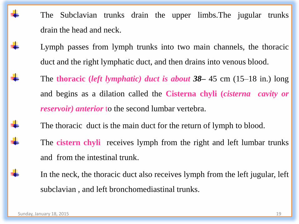
The Subclavian trunks drain the upper limbs.The jugular trunks
drain the head and neck.
Lymph passes from lymph trunks into two main channels, the thoracic
duct and the right lymphatic duct, and then drains into venous blood.
The thoracic (left lymphatic) duct is about 38– 45 cm (15–18 in.) long
and begins as a dilation called the Cisterna chyli (cisterna cavity or
reservoir) anterior to the second lumbar vertebra.
The thoracic duct is the main duct for the return of lymph to blood.
The cistern chyli receives lymph from the right and left lumbar trunks
and from the intestinal trunk.
In the neck, the thoracic duct also receives lymph from the left jugular, left
subclavian , and left bronchomediastinal trunks.
Sunday, January 18, 2015 19

The thoracic duct receives lymph from the left side of the head,
neck, and chest, the left upper limb, and the entire body inferior to the
ribs .
The thoracic duct in turn drains lymph into venous blood at the junction of
the left internal jugular and left subclavian veins.
The right lymphatic duct is about 1.2 cm (0.5 in.) long and receives
lymph from the right jugular, right subclavian, and right
bronchomediastinal trunks.
The right lymphatic duct receives lymph from the upper right side of the
body. From the right lymphatic duct, lymph drains into venous blood at
the junction of the right internal jugular and right Subclavian veins.
Sunday, January 18, 2015 20

Sunday, January 18, 2015 21

Lymphatic Organs and Tissues
The tissue responsible for immunity is aggregated separately into
Primary and Secondary lymphoid organs.
Primary organs are BONE MARROW & THYMUS.
Seconday Organs Are SPLEEN, LYMPH NODES, PAYER’S
PATCHES OF GUT, & WALDEYER’S RING (Tonsils and Adenoids).
WALDEYER’S ring of lymph consists of five tonsils.
Pharyngeal tonsil or adenoid is embedded in the posterior wall of the
Nasopharynx
The two palatine tonsils lie at the posterior region of the oral cavity, one
on either side, these can be removed by tonsillectomy.
The paired lingual tonsils are located at the base of the tongue.
Sunday, January 18, 2015 22

The primary lymphoid organs develop firstly and provide proper
microenvironment for differentiation of immature precursors into
functionally matured lymphocytes.
The secondary lymphoid organs provide an optional microenvironment for
attacking Ag specific lymphocytes directing their final maturation and
effecting the distribution of effectors into all parts of the body.
THYMUS
The thymus is a Bilobed organ located in the mediastinum between the
sternum and the aorta.
An enveloping layer of connective tissue holds the two lobes closely
together, but connective tissue capsule separates the two. Extensions of
the capsule, called trabeculae (tra-BEK-uˉ-leˉ little beams), penetrate
inward and divide each lobe into lobules.Sunday, January 18, 2015 23

Each thymic lobule consists of a deeply staining outer cortex and a
lighter- staining central medulla.
The cortex is composed of large numbers of T cells and scattered
dendritic cells, epithelial cells, and macrophages.
Immature T cells (pre-T cells) migrate from red bone marrow to the
cortex of the thymus, where they proliferate and begin to mature.
Dendritic cells (den-DRIT-ik; dendr- a tree), which are derived from
monocytes assist the maturation process.
Dendritic cells in other parts of the body, such as lymph nodes, play
another key role in immune responses. Each of the specialized epithelial
cells in the cortex has several long processes that surround and
serve as a frame work for as many as 50 T cells.
Sunday, January 18, 2015 24

These epithelial cells help “educate” the pre-T cells in a process known
as positive selection.
Additionally, they produce Thymic hormones that are thought to aid in
the maturation of T cells. Only about 2% of developing T cells survive in
the cortex.The remaining cells die via apoptosis.
Thyamic macrophages help clear out the debris of dead and dying
cells. The surviving T cells enter the medulla.
The MEDULLA consists of widely scattered, more mature T
cells, epithelial cells, dendritic cells, and macrophages
Some of the epithelial cells become arranged into concentric layers of flat
cells that degenerate and become filled with keratohyalin granules and
keratin.
Sunday, January 18, 2015 25

Sunday, January 18, 2015 26

These clusters are called thymic corpuscles or Hassall's corpuscles.
their role is uncertain, they may serve as sites of T cell death in
the medulla.
T cells that leave the thymus via the blood migrate to lymph nodes, the
spleen, and other lymphatic tissues, where they colonize parts of these
organs and tissues.
Because of its high content of lymphoid tissue and a rich blood supply,
the thymus has a reddish appearance in a living body. With age,
fatty infiltrations replace the lymphoid tissue and the thymus takes on
more of the yellowish color of the invading fat, giving the false
impression of reduced size.
Sunday, January 18, 2015 27

The actual size of the thymus, defined by its connective tissue capsule,
does not change. In infants, the thymus has a mass of about 70 g.
It is after puberty that adipose and areolar connective tissue begin to
replace the thymic tissue. By the time a person reaches maturity, the
functional portion of the gland is reduced considerably, and in old age
the functional portion may weigh only 3 g.
Before the thymus atrophies, it populates the secondary lymphatic
organs and tissues with T cells Some T cells continue to
proliferate in the thymus throughout an individual’s lifetime, but this
number decreases with age.
Sunday, January 18, 2015 28

Lymph Nodes Located along lymphatic vessels are about 600 bean-shaped lymph
nodes. They are scattered throughout the body, both superficially
and deep, and usually occur in groups .
Large groups of lymph nodes are present near the mammary glands and
in the axillae and groin.
Lymph nodes are 1–25 mm long and, like the thymus, are covered by a
capsule of dense connective tissue that extends into the node.
The capsular extensions, called trabeculae, divide the node into
compartments, provide support, and provide a route for blood vessels into
the interior of a node.
Internal to the capsule is a supporting network of reticular fibers and
fibroblasts. The capsule, trabeculae, reticular fibers, and fibroblasts
constitute the stroma (supporting framework of connective tissue) of
a lymph node.
Sunday, January 18, 2015 29

The parenchyma (functioning part) of a lymph node is divided into a
superficial cortex and a deep medulla. The cortex consists of an outer
cortex and an inner cortex.
Within the outer cortex are egg-shaped aggregates of B cells called
lymphatic nodules (follicles).
A lymphatic nodule consisting chiefly of B cells is called a primary
lymphatic nodule.
After B cells in a primary lymphatic nodule recognize an antigen, the
primary lymphatic nodule develops into a secondary lymphatic nodule.
Most lymphatic nodules in the outer cortex are secondary lymphatic
nodules which form in response to an antigen and are sites of plasma
cell and memory B cell formation.
Sunday, January 18, 2015 30

The center of a secondary lymphatic nodule contains a region of light-
staining cells called a germinal center. In the germinal center are B cells,
follicular dendritic cells, and macrophages.
When follicular dendritic cells “present” an antigen , B cells proliferate
and develop into antibody-producing plasma cells or develop into
memory B cells.
Memory B cells persist after an initial immune response and “remember”
having encountered a specific antigen. B cells that do not develop
properly undergo apoptosis and are destroyed by macrophages.
The region of a secondary lymphatic nodule surrounding the germinal
center is composed of accumulations of B cells that have migrated away
from their site of origin within the nodule.
Sunday, January 18, 2015 31

The inner cortex does not contain lymphatic nodules. It consists
mainly of T cells and dendritic cells that enter a lymph node from other
tissues.
The dendritic cells present antigens to T cells, causing their
proliferation.
The newly formed T cells then migrate from the lymph node to areas of
the body where there is antigenic activity.
The medulla of a lymph node contains B cells, antibody producing
plasma cells that have migrated out of the cortex into the medulla, and
macrophages.
lymph flows through several afferent lymphatic vessels which
penetrate the convex surface of the node at several points.
Sunday, January 18, 2015 32

Sunday, January 18, 2015 33

The afferent vessels contain valves that open toward the center of the
node, directing the lymph inward. Within the node, enters sinuses, a
series of irregular channels that contain branching reticular fibers,
lymphocytes, and macrophages.
From the afferent lymphatic vessels, lymph flows into the subcapsular
Sinus,from here the lymph flows through trabecular sinuses,into
medullary sinuses.The medullary sinuses drain into one or two efferent
lymphatic vessels
Efferent lymphatic vessels. which are wider and fewer in number than
afferent vessels. They contain valves that open away from the center of
the lymph node to convey lymph, antibodies secreted by plasma cells,
and activated T cells out of the node. Efferent lymphatic vessels emerge
from one side of the lymph node at a slight depression called a hilum
Blood vessels also enter and leave the node at the hilum
Sunday, January 18, 2015 34

Lymph nodes function as a type of filter. As lymph enters one end of a
lymph node, foreign substances are trapped by the reticular fibers within
the sinuses of the node.Then macrophages destroy some foreign
substances by phagocytosis, while lymphocytes destroy others by
immune responses.
The filtered lymph then leaves the other end of the lymph node. There are
many afferent lymphatic vessels that bring lymph into a lymph node and
only one or two efferent lymphatic vessels that transport lymph out of a
lymph node, the slow flow of lymph within the lymph nodes allows
additional time for lymph to be filtered. Additionally,
All lymph flows through multiple lymph nodes on its path through the
lymph vessels. This exposes the lymph to multiple filtering events
before returning to the blood.Sunday, January 18, 2015 35

Sunday, January 18, 2015 36

SPLEEN
The oval spleen is the largest single mass of lymphatic tissue in the
body, measuring about 12 cm (5 in.) in length.
It is located in the left hypochondriac region between the stomach and
diaphragm. The superior surface of the spleen is smooth and convex and
conforms to the concave surface of the diaphragm.
Neighbouring organs make indentations in the visceral surface of the
spleen—the gastric impression (stomach), the renal impression (left
kidney), and the colic impression (left colic flexure of large intestine).
Like lymph nodes, the spleen has a hilum. Through it pass the splenic
artery, splenic vein, and efferent lymphatic vessels.
A capsule of dense connective tissue surrounds the spleen
Sunday, January 18, 2015 37
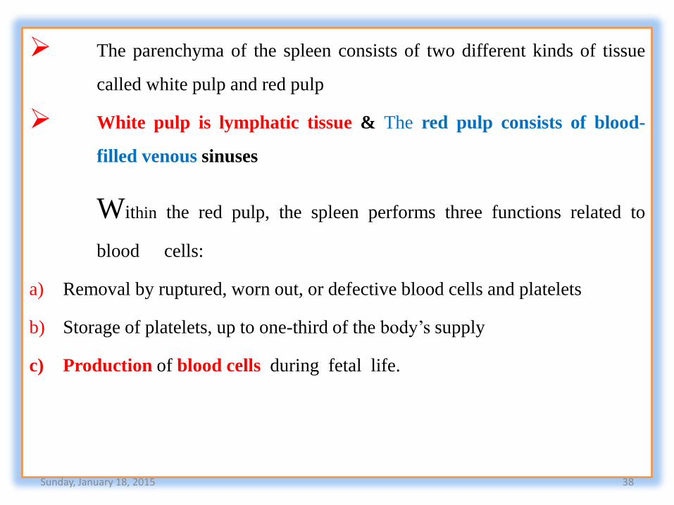
The parenchyma of the spleen consists of two different kinds of tissue
called white pulp and red pulp
White pulp is lymphatic tissue & The red pulp consists of blood-
filled venous sinuses
Within the red pulp, the spleen performs three functions related to
blood cells:
a) Removal by ruptured, worn out, or defective blood cells and platelets
b) Storage of platelets, up to one-third of the body’s supply
c) Production of blood cells during fetal life.
Sunday, January 18, 2015 38

Sunday, January 18, 2015 39

Sunday, January 18, 2015 40
BLOOD GROUPS