lysophosphatidylcholine as a death effector in ... · lysophosphatidylcholine as a death effector...
TRANSCRIPT

Lysophosphatidylcholine as a Death Effector
in Lipoapoptosis of Hepatocytes
Myoung Sook Han1, Sun Young Park1, Koei Shinzawa2, Sunshin Kim1,
Kun Wook Chung1, Ji-Hyun Lee3, Choon Hyuck Kwon4, Kwang-Woong Lee4,
Joon-Hyoek Lee1, Cheol Keun Park5, Woo Jin Chung6, Jae Seok Hwang6, Ji-Jing Yan7,
Dong-Keun Song7, Yoshihide Tsujimoto2, and Myung-Shik Lee1
Departments of 1Medicine, 4Surgery, and 5Pathology, Samsung Medical Center,
Sungkyunkwan University School of Medicine, Seoul 135-710, Korea; 2Laboratory of
Molecular Genetics, Department of Medical Genetics, Osaka University Medical
School and SORST of the Japan Science and Technology Corporation (JST), Osaka
565-1871, Japan; 3Samsung Biomedical Research Institute, Seoul 135-710, Korea;
6Department of Medicine, Keimyung University School of Medicine; Daegu 700-712,
Korea; 7Department of Pharmacology, College of Medicine, Hallym University,
Chunchon, Gangwon 200-702, Korea.
Running title: Lysophosphatidylcholine and Lipoapoptosis
Address correspondence to: Myung-Shik Lee, Dept. of Medicine, Samsung Medical
Center, 50 Irwon-dong Kangnam-ku, Seoul 135-710, Korea. Tel: 82-2-3410-3436; Fax:
82-2-3410-0388; E-mail: [email protected]
The first 2 authors contributed equally to this work.
1
by guest, on August 26, 2018
ww
w.jlr.org
Dow
nloaded from
Abbreviations: NASH, nonalcoholic steatohepatitis; PA, palmitic acid; PLA2,
phospholipase A2; LPC, lysophosphatidylcholine; siRNA, small interfering RNA;
iPLA2, Ca2+-independent PLA2; PTX, pertussis toxin; GPCR, G-protein coupled
receptor; OA, oleic acid; TG, triglyceride; NAFLD, nonalcoholic fatty liver disease;
T2D, type 2 diabetes; PACOCF3, palmitoyl trifluoromethyl ketone; MAFP, methyl
arachidonyl fluorophosphonate; MTT, 3-[4,5-dimethylthiazol-2-yl]-2,5-
diphenyltetrazolium bromide; PI, propidium iodide; LDH, lactate dehydrogenase; NAO,
10-N-nonyl acridine orange; ORO, Oil Red O; AST, aspartate aminotransferase; ALT,
alanine aminotransferase; PEPCK, phosphoenolpyruvate carboxykinase; TDO2,
tryptophan 2,3-dioxygenase; AAT, α-1-antitrypsin; CK8, cytokeratin 8; DAG,
diacylglycerol; PC, phosphatidylcholine; AA, aristolochic acid; BEL, bromoenol
lactone; cPLA2, cytoplasmic PLA2; RNAi, RNA interference; LPE,
lysophosphatidylethanolamine; LPG, lysophosphatidylglycerol; LPI,
lysophosphatidylinositol; LPS, lysophosphatidylserine; ROS, reactive oxygen species;
LPA, lysophosphatidic acid.
2
by guest, on August 26, 2018
ww
w.jlr.org
Dow
nloaded from
ABSTRACT
Pathogenesis of nonalcoholic steatohepatitis (NASH) is unclear, despite epidemiological
data implicating FFAs. We studied the pathogenesis of NASH employing lipoapoptosis
models. Palmitic acid (PA) induced classical apoptosis of hepatocytes. PA-induced
lipoapoptosis was inhibited by acyl-CoA synthetase inhibitor but not by ceramide
synthesis inhibitors, suggesting conversion product(s) other than ceramide are involved.
Phospholipase A2 (PLA2) inhibitors blocked PA-induced hepatocyte death, suggesting
an important role for PLA2 and its product lysophosphatidylcholine (LPC). Small
interfering RNA (siRNA) for Ca2+-independent PLA2 (iPLA2) inhibited lipoapoptosis of
hepatocytes. PA increased LPC content, which was reversed by iPLA2 inhibitors.
Pertussis toxin (PTX) or dominant-negative Gαi mutant inhibited hepatocyte death by
PA or LPC acting through G-protein coupled receptor (GPCR)/Gαi. PA decreased
cardiolipin content, and induced mitochondrial potential loss and cytochrome c
translocation. Oleic acid (OA) inhibited PA-induced hepatocyte death by diverting PA to
triglyceride (TG) and decreasing LPC content, suggesting FFAs lead to steatosis or
lipoapoptosis according to the abundance of saturated/unsaturated FFAs. LPC
administration induced hepatitis in vivo. LPC content was increased in the liver
specimens from NASH patients. These results demonstrate that LPC is a death effector
in lipoapoptosis of hepatocytes and suggest potential therapeutic values of PLA2
inhibitors or GPCR/Gαi inhibitors in NASH.
Key Words: fatty acids, PLA2, steatohepatitis, triglyceride, ceramide.
3
by guest, on August 26, 2018
ww
w.jlr.org
Dow
nloaded from

INTRODUCTION
Nonalcoholic fatty liver disease (NAFLD) is characterized by lipid deposition in
hepatocytes without alcohol abuse (1). NASH is a severe form of NAFLD accompanied
by necrosis, inflammation and fibrosis (2). While steatosis alone is non-progressive,
20% of NASH progress to cirrhosis (1). The pathogenesis of NAFLD is not clearly
understood, and may entail multiple injuries from increased FFAs, oxidative stress, lipid
peroxidation and TNFα (1,3).
NAFLD is frequently associated with type 2 diabetes (T2D), obesity and insulin
resistance that constitute important components of metabolic syndrome (2). FFAs
released from visceral fat of obese subjects are strong culprits in insulin resistance.
Recent papers suggested a potential role for FFAs in insulin deficiency as well as in
insulin resistance, which is consistent with FFA-mediated injury of pancreatic β-cells
and several other tissues (4).
FFAs may be related to NAFLD through their increased flux from visceral fat to
the liver. Because of strong epidemiological and in vivo data suggesting the relationship
between FFAs and NAFLD, several in vitro experiments studying hepatocyte injury by
FFAs have been conducted, which suggested possible role for mitochondrial pathway,
lysosomal pathway, ER stress and JNK activation in lipoapoptosis of hepatocytes (5-9).
However, the intracellular metabolites of FFAs that are directly responsible such
intracellular events are not clearly identified. We found evidences supporting the role
for LPC produced by PLA2 that catalyzes the hydrolysis of the fatty acyl ester bond at
the sn-2 position of glycerophospholipids and has been implicated in several types of
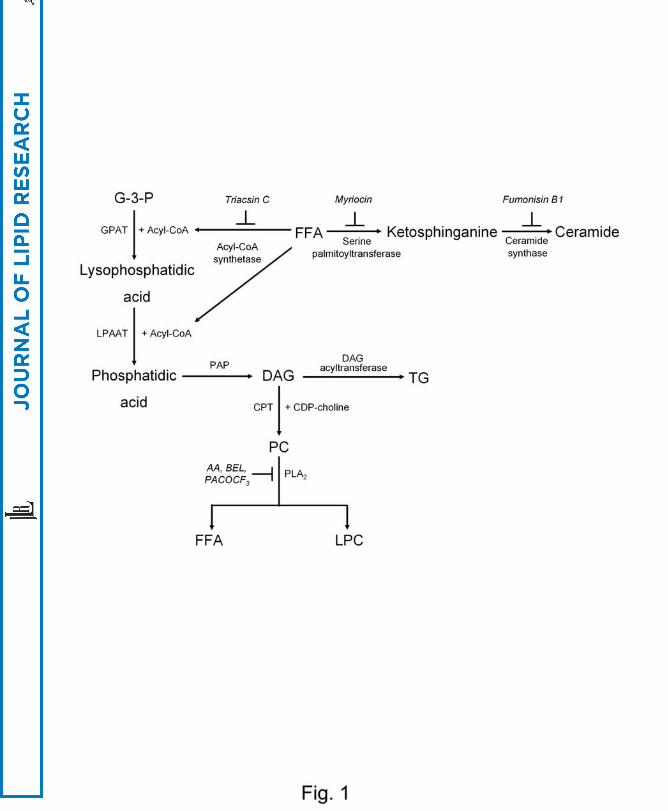
cell death (10-13) as an effector in the lipoapoptosis of hepatocytes (Fig. 1).
4
by guest, on August 26, 2018
ww
w.jlr.org
Dow
nloaded from
MATERIALS AND METHODS
Reagents
Palmitoyl trifluoromethyl ketone (PACOCF3) and methyl arachidonyl
fluorophosphonate (MAFP) were from Calbiochem. zVAD-fmk was from Enzyme
Systems (Livermore). All other chemicals were obtained from Sigma unless stated
otherwise.
Cell Death Assay
Chang cells grown in DMEM-10% FBS were treated with FFAs or other reagents for 24
h unless stated otherwise. We chose 24 h treatment protocol because our time-course
study revealed that cell death above 50% with full-blown features of apoptosis such as
nuclear condensation/fragmentation and sub-G1 DNA peak occurred 24 h after
treatment. PA solution was made according to a previously published protocol with
modifications (14). Briefly, PA stock solution (50 mM) was prepared by dissolving in
70% ethanol and heating at 50°C. Working PA solution was made by diluting stock
solution in 2% fatty acid-free BSA-DMEM-10% FBS because the concentration of FFA,
particularly that of PA, is quite low in 10% FBS while cells were healthier with serum
for prolonged experiment. 3-[4,5-dimethylthiazol-2-yl]-2,5-diphenyltetrazolium
bromide (MTT) assay, Hoechst 33258/propidium iodide (PI) staining and DNA ploidy
analysis were conducted as described (15). We employed MTT assay throughout the
study because the results from MTT assay were consistent with those of other cell death
assays such as Hoechst /PI staining or DNA ploidy analysis in our pilot study. In some
5
by guest, on August 26, 2018
ww
w.jlr.org
Dow
nloaded from
experiments, cells were pretreated with inhibitors for 1 h before PA treatment. To assess
the death of primary murine hepatocytes, lactate dehydrogenase (LDH) release assay
was employed (12). %cell death was calculated as (experimental release – spontaneous
release)/(total release – spontaneous release) x 100.
Ammonia Removal
Ammonia concentration in the culture supernatant was measured using a commercial kit
(Asan Pharmaceuticals). Briefly, cells were incubated in a medium containing test
reagents and 2 mM NH4Cl for 48 h. The absorption at 630 nm by indophenol generated
from ammonia was measured. %removal was calculated as (1 – (A630 with added
ammonia to the cells – basal A630 of the cells)/(A630 with added ammonia to the blank –
basal A630 of the blank)).
siRNA and RT-PCR
siRNAs for human iPLA2β (AAT TGC GCG GAG AAC GAG GAG), iPLA2γ (AAA
ATG AAC ATT TCC GGG ACA) and an irrelevant siRNA purchased from Invitrogen
was transfected using Oligofectamine (Invitrogen) according to the manufacturer’s
protocol. Expression of iPLA2β and iPLA2γ at the RNA level was determined by RT-
PCR using specific primer sets. Expression of markers for hepatocytes was also tested
by RT-PCR using specific primer sets (16).
Measurement of LPC
Intracellular LPC content was measured using an enzymatic assay previously reported
(17). Briefly, sample was added to the mixture of 100 mM Tris-HCl, pH 8.0, 0.01%
6
by guest, on August 26, 2018
ww
w.jlr.org
Dow
nloaded from

Triton X-100, 1 mM CaCl2, 3 mM N-ethyl-N-[2-hydroxy-3-sulfopropyl]-3-
methylaniline, 10 U/mL peroxidase, 0.1 U/mL glycerophosphorylcholine
phosphodiesterase and 10 U/mL choline oxidase. After incubation at 37°C for 5 min,
reagent mixture containing 100 mM Tris-HCl, pH 8.0, 5 mM 4-aminoantipyrine,
0.01% Triton X-100 and 20 U/mL lysophospholipase was added. After incubation for
another 5 min, A570 was measured using LPC 16:0 as a standard.
Transfection
Chang cells stably expressing a dominant-negative Gαi2 mutant (αi2G203T) (18) or
wild-type Gαi2 (UMR cDNA Resource Center) were produced by transfection with
FuGENE 6 (Roche) and selection with G418 for 3 weeks.
Mitochondrial Events
After incubating cells with 0.4 μM 10-N-nonyl acridine orange (NAO) (Molecular
Probes) at 37°C, NAO fluorescence was measured by flow cytometry (19).
Mitochondrial potential was measured by calculating A590/A530 after incubating cells
with 10 μg/mL JC-1 (Molecular Probes) for 10 min (20).
Cell Fractionation
Cells lysed in an isotonic buffer were fractionated as described (20). Heavy membrane
fraction and cytosolic fraction were subjected to Western blotting using specific
antibodies against cytochrome c (PharMingen) or Bid (Santa Cruz) (15). The purity of
the heavy membrane and cytosolic fraction was confirmed by Western blotting using
anti-Cox4 and -IκB antibody, respectively. Expression of phosphorylated JNK and total
7
by guest, on August 26, 2018
ww
w.jlr.org
Dow
nloaded from

JNK in the whole cell lysate was studied by Western blotting using specific antibodies
(Cell Signaling).
Oil Red O Staining
Cells were fixed with 10% formaldehyde for 1 h. After staining with 3 μg/mL Oil Red
O (ORO) solution for 15 min, dye was extracted by isopropanol and A540 measured.
Human Liver Biopsy
Human liver tissue was obtained from the non-tumor part of the livers from patients
undergoing hepatic resection for metastatic colon cancer at Samsung Medical Center.
The absence of cancer and cirrhosis was confirmed grossly and microscopically. The
isolation of hepatocytes was performed using two-step collagenase perfusion technique
as previously described (21). After isolation, primary hepatocytes were cultured in a
modified hormonally defined medium (William's E medium supplemented with 20 μg/L
epidermal growth factor, 10 mg/L insulin, 1.7 mg/L hydrocortisone, 24.97 μg/L
CuSO4·5H2O, 14.38 pg/L ZnSO4·7H2O, 3 μg/L H2SeO3, 50 mg/L linoleic acid, 1.05 g/L
NaHCO3 and 1.19 g/L HEPES) (22). Aspartate aminotransferase/alanine
aminotransferase (AST/ALT) levels were measured using Blood Chemistry Analyzer
FPC 3000 (Fuji). Ultrasonography-guided liver biopsy was conducted in patients with
clinical findings compatible with NAFLD without history of alcoholism (alcohol
consumption, < 140 gm/week) using 18-gauge Solco needles inserted through the
intercostals space. All patients had serum AST/ALT levels above 60 U/L and body mass
index above 25 without recent history of diet control or parenteral nutrition. All were
negative for viral markers, autoantibodies related to the liver diseases, clinical evidence
8
by guest, on August 26, 2018
ww
w.jlr.org
Dow
nloaded from

of cirrhosis, lipodystrophy or Wilson’s disease, and were not taking drugs related to
lipid metabolism. Informed consent was obtained from all participants. All human
studies were approved by the IRB of Samsung Medical Center. An experienced
hepatopathologist blinded to the subjects’ detail scored liver biopsy specimens
according to a published classification (2).
TUNEL Staining
TUNEL staining of the mouse liver tissues was conducted as described (15). Briefly,
formalin-fixed sections were deparaffinized and microwaved in 0.01 M sodium citrate
buffer (pH 6.0)-0.1% Triton X-100. They were incubated with TUNEL reagents (Roche
Molecular Biochemicals) at 37°C for 60 min. After washing, they were further incubated
with Convert-POD for 30 min. Diaminobenzidine was used as a color substrate.
Mouse Hepatocytes
Primary murine hepatocytes were isolated from C57BL/6 mice using the retrograde
two-step collagenase perfusion technique (23). LPC (kindly provided by Song K-S,
Doosan Pharmaceutical; purity, > 99%) dissolved in PBS-2% BSA by sonication was
injected into the tail vein of ICR mice. All animal experiments were conducted in
accordance with the institutional guideline of Osaka University Animal Facility and
Samsung Medical Center Animal Facility.
Measurement of iPLA2 Activity
Activity of iPLA2 was measured using a commercial kit (Cayman) with modifications
(24). Briefly, cells were collected with a cell scraper, sonicated and centrifuged at
9
by guest, on August 26, 2018
ww
w.jlr.org
Dow
nloaded from
20,000g at 4°C for 20 min. The supernatant was removed, and the concentration of
proteins was determined. iPLA2 activity was assayed by incubating the samples with
arachidonoyl thio-PC for 1 h at 25°C in a Ca2+-free buffer (300 mM NaCl, 0.5 % Triton
X-100, 60% glycerol, 4 mM EGTA, 10 mM HEPES, pH 7.4, 2 mg/mL BSA). The
reaction was terminated by adding 5,5’-dithio-bis-2-nitrobenzoic acid for 5 min, and
A405 was measured. The specific activity of iPLA2 was calculated and expressed as
absorbance per mg protein. The background activity was subtracted from all readings.
Statistical Analysis
All values were expressed as means ± SD from more than 3 independent experiments
performed in triplicate. Two-tailed Student's t-test was employed to compare values
between two groups. ANOVA was employed for multiple comparisons. Scheffé test
was employed to compare two groups once ANOVA showed significant differences. P
values less than 0.05 were considered to represent statistically significant differences.
RESULTS
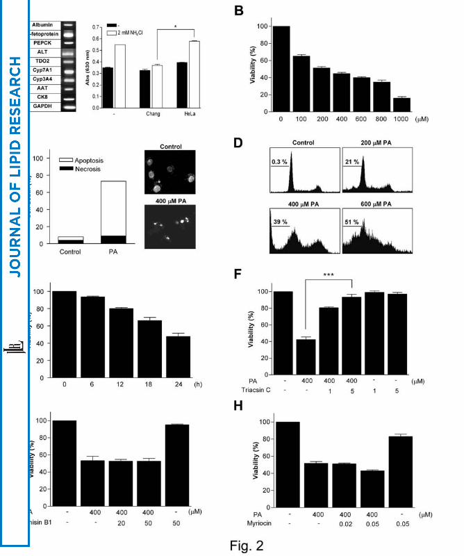
Apoptosis of Chang Liver Cells by FFAs
We studied the effect of palmitic acid (PA), the most abundant saturated fatty acid in
vivo, on Chang ‘normal’ hepatocyte line. We chose Chang cells because they represent
more physiological system as nonmalignant human hepatocytes compared to hepatoma
cells (25). RT-PCR analysis showed that Chang cells expressed markers for hepatocytes
such as α-fetoprotein, ALT, phosphoenolpyruvate carboxykinase (PEPCK), albumin,
tryptophan 2,3-dioxygenase, (TDO2), cytochrome enzymes (Cyp7A1, Cyp3A4), α-1-
10
by guest, on August 26, 2018
ww
w.jlr.org
Dow
nloaded from

antitrypsin (AAT) and cytokeratin 8 (CK8) (Fig. 2A) (16). Furthermore, Chang cells
cleared 78.8% of added NH4Cl which was significantly higher than that by control
HeLa cells (8.0%) (P < 0.05) (Fig. 2A), suggesting that Chang cells represent authentic
hepatocytes. PA induced Chang cell death as assessed by MTT assay (Fig. 2B).
Hoechst/PI staining demonstrated that Chang cell death by PA was a classical apoptosis
characterized by nuclear condensation/fragmentation (Fig. 2C). Sub-G1 DNA peak was
also observed by DNA ploidy analysis (Fig. 2D). Time-course study showed that cell
death above 50% occurred 24 h after treatment (Fig. 2E).
Because lipid intermediates produced from PA such as ceramide are well-known
inducers of apoptosis (26), we investigated whether agents inhibiting conversion of PA
to lipid intermediates could affect Chang cell lipoapoptosis. Triacsin C that inhibits
acyl-CoA synthetase, the first enzyme in the conversion of PA to lipid intermediates
through palmityol-CoA (27) (Fig. 1), effectively blocked Chang cell death by PA,
suggesting that conversion product(s) of PA but not PA itself induce lipoapoptosis (P <
0.000005) (Fig. 2F). We then studied the effect of Fumonisin B1 that blocks ceramide
synthesis by inhibiting sphingosine N-acyltransferase (Fig. 1) (28). Contrary to our
expectation, Fumonisin B1 did not affect Chang cell death by PA (Fig. 2G). Because
this data was inconsistent with previous reports by others employing pancreatic islet
cells (4), we next studied the effect of Myriocin that blocks the synthesis of
ceramide/sphingosine by inhibiting serine palmitoyltransferase (29) (Fig. 1). Myriocin
also did not inhibit Chang cell death by PA (Fig. 2H), suggesting that conversion of
FFAs to lipid intermediates in the sphingolipid-ceramide pathway and finally to
ceramide is not involved in lipoapoptosis of hepatocytes.
We next explored possible involvement of PKC in Chang cell lipoapoptosis, since
11
by guest, on August 26, 2018
ww
w.jlr.org
Dow
nloaded from

recent papers showed a role for PKC activation in apoptosis (30) and PKC activators
such as diacylglycerol (DAG) or LPC can be synthesized from palmitoyl-CoA (31) (Fig.
1). Both Calphostin C and GF109203X, well-known PKC inhibitors, significantly
attenuated Chang cell death by PA, supporting a role for PKC in Chang cell
lipoapoptosis (P < 0.000005 for both) (Supplemental Fig. 1).
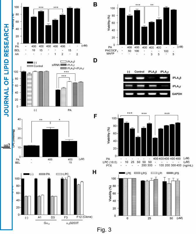
Role of PLA2 and LPC in Lipoapoptosis
We next studied possible role for PLA2 in Chang cell lipoapoptosis because PLA2 is
involved in several cell death models (12,13) and phosphatidylcholine (PC), a
conversion product of DAG, can be a substrate for PLA2, leading to the production of
LPC, another PKC activator (Fig. 1). Intriguingly, Aristolochic acid (AA), a general
inhibitor of PLA2 or bromoenol lactone (BEL), a specific inhibitor of iPLA2 (12,13)
markedly inhibited Chang cell death by PA, suggesting important roles for PLA2 and the
conversion product(s) of PA downstream of PLA2 rather than DAG upstream of PLA2 in
lipoapoptosis and PKC activation (P < 0.0001 and 0.00005, respectively) (Fig. 3A).
Because these results also suggested possible involvement of iPLA2 among diverse
PLA2 members, we employed other PLA2 inhibitors that are specific for each type of
PLA2. PACOCF3, another iPLA2-specific inhibitor of a different class (12,32),
dramatically blocked Chang cell death by PA (P < 0.00005). MAFP, an inhibitor of
cytoplasmic PLA2 (cPLA2) (12), also significantly blocked Chang cell death by PA (P <
0.005); however, the protection by the maximum tolerable MAFP was less compared to
PACOCF3 (P < 0.005), suggesting that iPLA2 rather than cPLA2 is involved in the
lipoapoptosis of hepatocytes (Fig. 3B). Consistent with these pharmacological data,
iPLA2 activity was significantly increased after treatment of Chang cells with PA (P <
12
by guest, on August 26, 2018
ww
w.jlr.org
Dow
nloaded from

0.05), which was inhibited by BEL (P < 0.01) (Supplemental Fig. 2)
To confirm the role for iPLA2, we conducted an RNA interference (RNAi)
experiment. Transfection of siRNA for iPLA2β or iPLA2γ, two major types of iPLA2,
significantly attenuated Chang cell death by PA, substantiating roles for iPLA2 in
lipoapoptosis (P < 0.00005 for both comparisons) (Fig. 3C). Transfection of iPLA2β and
iPLA2γ siRNA markedly decreased the expression of iPLA2β and iPLA2γ at the RNA
level, respectively (Fig. 3D). However, combination of both siRNAs did not induce a
further decrease in lipoapoptosis (Fig. 3C).
Next, we studied whether LPC that is liberated from PC by PLA2 as the most
abundant lysophospholipids in vivo, could be indeed produced from PA. Intracellular
LPC content was significantly increased by PA (P < 0.01), and the LPC content after PA
treatment was significantly attenuated by BEL (P < 0.05) (Fig. 3E). In contrast, LPC
content in the culture supernatant was not increased after treatment of Chang cells with
PA (Supplemental Fig. 3). Treatment of Chang cells with exogenous palmitoyl-LPC (10
~ 50 μM) for 24 h also induced significant cell death in a dose-dependent manner (P <
0.000001) (Fig. 3F).
We next tested the effect of PTX on Chang cell lipoapoptosis, since PTX inhibits
LPC signaling through GPCR/Gαi (33,34). PTX (100 ~ 400 ng/mL) remarkably
inhibited Chang cell death by exogenous LPC or PA (P < 0.00005 and 0.000000005,
respectively) (Fig. 3F). We further studied the role for GPCR/Gαi in lipoapoptosis by
transfecting a dominant-negative mutant of Gαi (αi2G203T) (18). Stable Chang cell
transfectants expressing αi2G203T were resistant to death by PA or LPC, strongly
supporting the role for LPC and GPCR/Gαi in lipoapoptosis of Chang cells (Fig. 3G).
Transfection with a control wild-type Gαi2 did not significantly affect Chang cell death
13
by guest, on August 26, 2018
ww
w.jlr.org
Dow
nloaded from
by PA or LPC, probably because of endogenous expression of Gαi. We next examined
the effect of other lysophospholipids that exist in vivo at much lower concentrations
compared to LPC but could be affected by PLA2 inhibitors.
Lysophosphatidylethanolamine (LPE), lysophosphatidylglycerol (LPG),
lysophosphatidylinositol (LPI) or lysophosphatidylserine (LPS) up to 50 μM that is
much higher than their physiological concentrations induced negligible cell death even
48 h after treatment, suggesting that the effects of PLA2 inhibitors are most likely
through LPC (Fig. 3H).
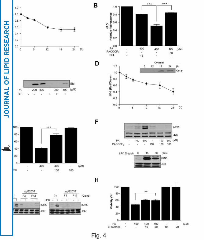
We next studied intracellular events associated with lipoapoptosis. We measured
intracellular content of cardiolipin that is crucial for cytochrome c binding to
mitochondrial inner membrane as a mitochondria-specific phospholipid and could be
affected by FFA treatment. Cardiolipin content measured by NAO fluorescence began
to decrease 3 ~ 6 h after PA treatment and further decreased 12 ~ 24 h after treatment
(Fig. 4A). PA-induced decrease in cardiolipin content was significantly attenuated by
iPLA2 inhibitors such as BEL or PACOCF3, suggesting important roles for PLA2 or
LPC in the decrease of cardiolipin content and lipoapoptosis (P < 0.0001 and 0.0005,
respectively) (Fig. 4B). We also studied whether LPC could directly induce changes
of Bid apart from its receptor-mediated effect because previous papers reported that
Bid could perturb mitochondrial membrane in association with LPC (35). Bid in
mitochondrial fraction was increased by PA treatment which was inhibited by BEL
(Fig. 4C), suggesting a possible role for full-length Bid in cell death by LPC as
reported (35). Probably because of the decrease in cardiolipin content and increase in
Bid in the mitochondria, cytochrome c was translocated from mitochondria to
cytoplasm and mitochondrial potential was decreased between 3 ~ 24 h after PA
14
by guest, on August 26, 2018
ww
w.jlr.org
Dow
nloaded from

treatment (Fig. 4D). Chang cell death by PA was significantly inhibited by a
pancaspase inhibitor, zVAD-fmk, suggesting that effector caspases were activated
downstream of cytochrome c translocation (P < 0.000005) (Fig. 4E). We also studied
whether PA activates JNK because LPC is a well-known activator of JNK (36).
Treatment of Chang cells with PA induced JNK activation which was inhibited by
PACOCF3, suggesting that LPC produced from PA is involved in JNK activation (Fig.
4F). Exogenous LPC also induced JNK activation in Chang cells as expected (Fig.
4F). Stable Chang cell transfectants expressing dominant-negative αi2G203T mutant
showed attenuated JNK activation after treatment with PA or LPC, suggesting that
LPC produced from PA activates JNK through GPCR/Gαi (Fig. 4G). SP600125 (10 ~
20 μM), a JNK inhibitor, induced a small but significant decrease in PA-induced death
(ca. 25%) (P < 0.005), suggesting that JNK activation plays a certain role in
lipoapoptosis of Chang cells (Fig. 4H).
Inhibition of PA-Induced Lipoapoptosis by OA
Because DAG can be converted to TG (Fig. 1), we examined the role of TG in lipid
injury. PA did not increase intracellular TG content estimated by ORO staining
followed by spectrophotometry or microscopic examination. In contrast, OA, the most
abundant unsaturated fatty acid in vivo, markedly increased TG content. Treatment
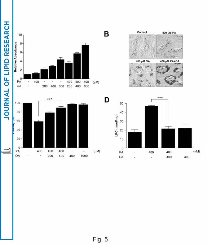
with OA in combination with PA further increased TG content (Fig. 5A, B).
Regarding cell viability, OA did not induce Chang cell death. On the contrary, OA
significantly blocked Chang cell death by PA (P < 0.00005), similar to previous reports
(37) (Fig. 5C). OA also abolished the increase in LPC content by PA treatment (P <
0.00005) (Fig. 5D), suggesting that increased FFAs lead to either steatosis of
15
by guest, on August 26, 2018
ww
w.jlr.org
Dow
nloaded from

hepatocytes due to increased TG content or death of hepatocytes due to increased LPC
content depending on the absolute/relative abundance of saturated/unsaturated fatty
acids (Fig. 1).
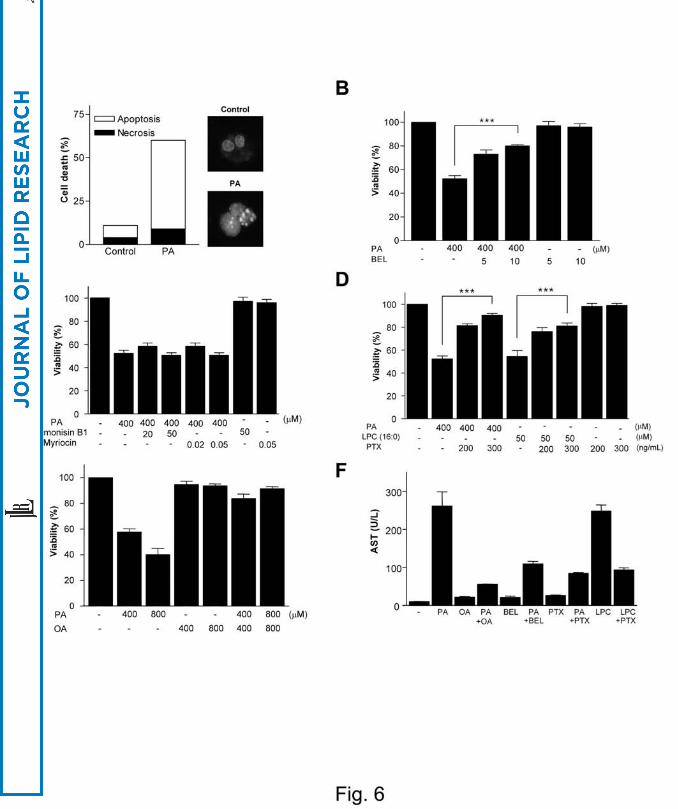
Effect of PA on Primary Hepatocytes
We next employed primary hepatocytes instead of cell lines. Hoechst/PI staining
showed that PA induced death of primary human hepatocytes as well, which was a
classical apoptosis characterized by nuclear condensation/fragmentation (Fig. 6A).
Lipoapoptosis of primary human hepatocytes by PA was inhibited by BEL (P < 0.0005)
but not by Fumonisin B1 or Myriocin, suggesting that iPLA2/LPC pathway rather than
ceramide pathway is crucial, similar to Chang cells (Fig. 6B, C). Exogenous LPC also
induced death of primary human hepatocytes, which was inhibited by PTX (P < 0.0005)
(Fig. 6D). PTX also significantly blocked death of primary human hepatocytes by PA,
suggesting that endogenous LPC induces death of primary human hepatocytes through
GPCR/Gαi (P < 0.000001) (Fig. 6D). OA attenuated death of primary human
hepatocytes by PA, supporting our hypothesis that unsaturated fatty acids divert
saturated fatty acids into TG pathway and attenuate primary human hepatocyte death by
saturated fatty acids (Fig. 6E). Consistent with the death of primary human hepatocytes,
PA induced release of AST from cultured primary human hepatocytes, which was
inhibited by BEL, PTX or OA. LPC also induced AST release, which was blocked by
PTX (Fig. 6F). PA induced death of primary murine hepatocytes also, as assessed by
LDH release assay. Lipoapoptosis of primary murine hepatocytes by 750 μM PA (33.9 ±
3.8%) was significantly inhibited by 30 μM BEL (26.4 ± 3.6%), 100 μM PACOCF3
(20.5 ± 3.1%) or 300 ng/mL PTX (26.9 ± 1.4%) (P < 0.05, 0.01 and 0.05, respectively).
16
by guest, on August 26, 2018
ww
w.jlr.org
Dow
nloaded from

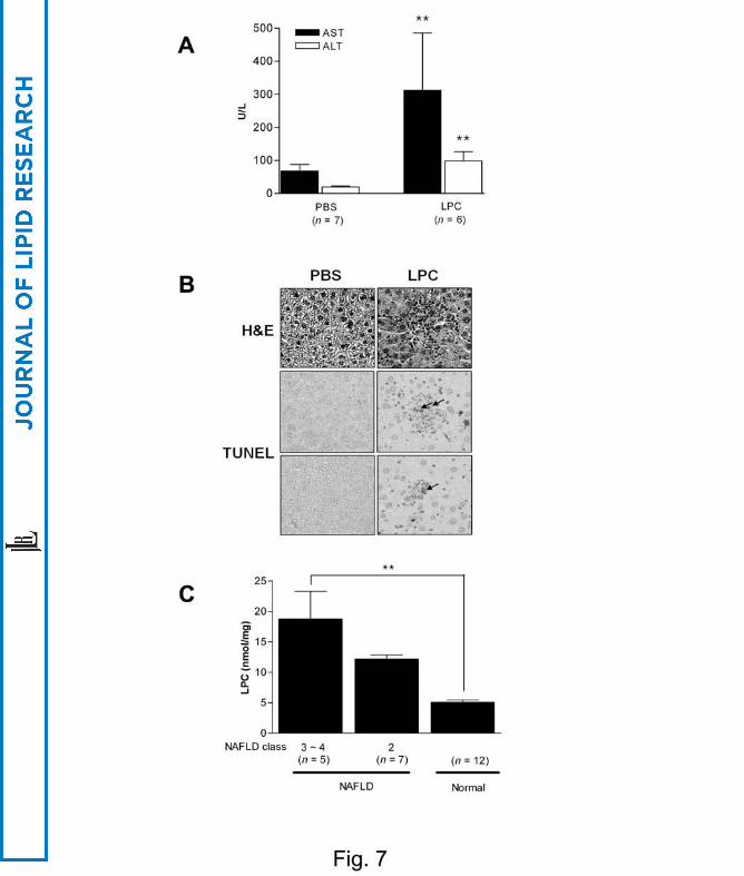
LPC Content in NAFLD Liver Specimens
We next studied if LPC is able to induce liver injury by directly injecting LPC in vivo.
When 60 mg/Kg LPC was administered to ICR mice through tail veins, AST/ALT levels
were notably increased 24 h after injection (P < 0.005 for both comparisons) (Fig. 7A).
H&E and TUNEL staining on histological sections obtained 3 days after LPC injection
showed lobular hepatitis with a histological score of 1 (focal lytic necrosis, one focus or
less per x 100 field) without evidence of steatosis (38) and TUNEL+ apoptotic cells in
the area of hepatitis, respectively, supporting our hypothesis that LPC plays a role as a
mediator of hepatocyte injury in vivo (Fig. 7B).
We finally measured LPC content in the liver biopsy specimens. ANOVA revealed
that LPC content in the liver specimens from patients with NAFLD was significantly
higher than that in the non-tumor part of the liver from patients with metastatic colon
cancer (P < 0.005). LPC content in the liver specimens from patients with NASH
(NAFLD class 3 ~ 4) appears to be higher compared to those from patients with
NAFLD class 2 (lobular hepatitis only), however, the difference was not statistically
significant by Scheffé test (P > 0.1) (Fig. 7C).
DISCUSSION
We observed that PA induces apoptosis of hepatocytes through conversion to LPC that
led to activation of GPCR, mitochondrial events and JNK (Fig. 8). While Chang cells
and primary hepatocytes from mice or human all underwent apoptosis after treatment
with PA, the susceptibility to lipoapoptosis was different, which could be due to the
17
by guest, on August 26, 2018
ww
w.jlr.org
Dow
nloaded from

difference in cell types, species or the method of cell death assay. Although the identity
of Chang cells has been questioned, recent papers have demonstrated hepatocyte nature
of Chang cells based on expression of α-fetoprotein and albumin (39,40), which was
again supported by our results showing expression of various markers for hepatocytes
and removal of ammonia by Chang cells. In contrast to our expectation, conversion of
PA to ceramide/sphingosine did not play a role in Chang cell lipoapoptosis because both
Fumonisin B1 and Myriocin inhibiting ceramide synthesis at 2 different steps did not
block Chang cell lipoapoptosis. These results differ from previous papers suggesting
essential roles for ceramide in the lipoapoptosis of pancreatic islet cells or
hematopoietic cells (4,41). However, papers reporting ceramide-independent
lipoapoptosis of various types of cells including hepatocytes have also been published
(5,7,42,43). These discrepancies might be due to the differences in cell types or
experimental conditions.
Instead of the ceramide pathway, conversion to DAG, PC and then LPC appears to
play important roles in Chang cell lipoapoptosis. Palmitoyl-CoA can be incorporated to
phosphatidic acid that can be converted to DAG, a well-known PKC activator (30)
(Fig. 1). Downstream of DAG, we noticed that DAG could be converted to PC and then
to LPC, another PKC activator, by PLA2 (44). Because PLA2 participates in several cell
death models, (12,13) we investigated whether PLA2 is involved in lipoapoptosis of
hepatocytes. Our results showing a remarkable inhibition of Chang cell lipoapoptosis by
BEL or PACOCF3 suggest the involvement of iPLA2 and LPC rather than DAG
upstream of PLA2. While BEL has been reported to inhibit phosphatidic acid
phosphohydrolase in addition to iPLA2 (24), our result that another iPLA2-specific
inhibitor PACOCF3 that is structurally unrelated to BEL (24) dramatically inhibited
18
by guest, on August 26, 2018
ww
w.jlr.org
Dow
nloaded from

Chang cell death by PA, suggests that iPLA2 plays a critical role in lipoapoptosis.
Moreover, LPC content was significantly increased after PA treatment of Chang cells
and reverted to normal level by BEL, suggesting an important role of LPC as a death
effector in PA-induced lipoapoptosis. However, it is not clear which among several
types of iPLA2 that could be inhibited by BEL (45,46) are responsible for the
lipoapoptosis of hepatocytes. Because we observed significant decreases in Chang cell
lipoapoptosis by either iPLA2β or iPLA2γ siRNA, a single type of iPLA2 may not be
fully responsible for the production of LPC from FFAs. Further works will be necessary
to elucidate the roles for specific iPLA2 type in lipoapoptosis.
The role for PLA2 in cell death/survival has been reported in diverse models of
hypoxia, reperfusion injury and reactive oxygen species (ROS) damage (12,13,47,48).
Our results are similar to previous papers showing significant role of iPLA2 in hypoxic
cell injury (12,47), however, are different from those results in that LPC appears to be
an effector downstream of iPLA2 activation. Our data that iPLA2 activity was
significantly (nearly 2-fold) increased by PA and the increased activity was completely
reversed by BEL are also similar to previous data that iPLA2 activity was increased in
the course of hypoxic cell death (12). The mechanism of the activation of iPLA2 activity
by PA is not clearly understood and may involve translocation to target organelles,
displacement of inhibitor molecules by FFA-induced ER stress or ROS (12,24,49).
Incomplete protection of PA-induced hepatocyte death by combination of iPLA2β and
iPLA2γ siRNAs or iPLA2 inhibitors such as BEL or PACOCF3 suggests potential role of
iPLA2-independent mechanism in lipoapoptosis of hepatocytes. For instance, role of
fatty acid metabolites other than LPC such as DAG or ceramide cannot be totally
eliminated because we employed mostly pharmacological inhibitors that could have
19
by guest, on August 26, 2018
ww
w.jlr.org
Dow
nloaded from

overlapping substrate specificities.
Exogenous LPC has been reported to induce apoptosis (44,50) and stimulate
inflammatory cells (17). However, proapoptotic role of endogenous LPC has not been
reported to our knowledge. As iPLA2 does not have a specificity toward PC, the roles of
other lysophospholipids cannot be completely eliminated. However, in vivo
concentrations of other phospholipids such as phosphatidylethanolamine,
phosphatidylglycerol, phosphatidylinositol or phosphatidylserine are around or below
1/10 of that of PC (51,52). Much higher in vivo concentration and much stronger
apoptotic activity of LPC compared to LPE, LPG, LPI or LPS essentially eliminates the
possibility that other minor lysophospholipids play significant roles in lipoapoptosis.
Previous papers also showed obvious apoptotic activity of LPC, while that of other
lysophospholipids has not been clearly demonstrated (44,50). Products of PLA2 released
from PC together with LPC such as arachidonic acid or its derivatives (10,11) have
diverse effects in vivo. While dipalmitoyl-phosphatidic acid is predominantly produced
by PA treatment (53), a certain amount of arachidonic acid may be produced by repeated
deacylation and reacylation at the sn-2 position and may contribute the phenotypes
associated with FFA excess. However, a recent paper showed much weaker activation of
stress molecules such as JNK by a variety of unsaturated FFAs including arachidonic
acid compared to saturated FFAs (54), consistent with our results suggesting that
saturated FFAs exert much stronger cytotoxicity compared to unsaturated FFAs.
Intracellular events associated with LPC-induced apoptosis are not clearly defined.
Recent papers reported that GPCRs mediate various actions of LPC (33,34), while it
remains controversial whether GPCRs are LPC receptors or indirectly mediate LPC
actions. The inhibition of PA- or LPC-induced Chang cell apoptosis by PTX or a
20
by guest, on August 26, 2018
ww
w.jlr.org
Dow
nloaded from

dominant-negative Gαi mutant (18) suggests that certain GPCR/Gαi is involved in
apoptosis by endogenous or exogenous LPC (55). The effect of dominant-negative Gαi
mutant observed in this study is not mediated by lysophosphatidic acid (LPA) that could
be produced from LPC by autotaxin (56) because Chang cell death by PA or LPC was
not inhibited by Ki16425, an LPA1/LPA3 receptor antagonist (56) and LPA up to 50 μM
did not induce Chang cell death (Han MS et al. unpublished data). Furthermore, LPA is
a well-known survival or growth factor and has been shown to protect cells from
apoptosis (57).
Because GPCRs recognize extracellular ligands, LPC might be released to the
extracellular space and bind to GPCRs (58). Instead, LPC might enter the receptor-
binding site in a lateral fashion between transmembrane regions of the receptor without
leaving membrane (59), which is more consistent with the absence of the increase of
LPC content in the culture supernatant after PA treatment. However, it is not proven
whether such a model that is proposed for LPA is valid for LPC binding to its receptors.
Downstream of receptor binding, we observed decreased cardiolipin content after PA
treatment, consistent with a previous report (53). PA-induced decrease in cardiolipin
content could be due to the inhibition of CDP-DAG synthase, the rate-limiting step in
cardiolipin biosynthesis, by LPC as previously suggested (60). While a different
mechanism such as poor substrate availability of dipalmitoyl-phosphatidic acid for
CDP-DAG synthase has been proposed (53), the inhibition of lipoapoptosis and the
reversal of the decrease in cardiolipin content by PLA2 inhibitors downstream of
phosphatidic acid support a role for LPC rather than dipalmitoyl-phosphatidic acid
upstream of PLA2. The loss of cardiolipin, a mitochondria-specific phospholipid that
maintains mitochondrial membrane stability by binding to cytochrome c (61), is
21
by guest, on August 26, 2018
ww
w.jlr.org
Dow
nloaded from
probably responsible for cytochrome c translocation, loss of mitochondrial potential and
cell death after PA treatment. Besides receptor binding, LPC might directly induce
mitochondrial events by recruiting Bid, a well-known initiator of apoptosis, to the
mitochondrial membrane (35). JNK activation by saturated FFAs may also contribute to
the cytochrome c release and lipoapoptosis in hepatocytes, while the contribution of
JNK activation in lipoapoptosis of Chang cells does not appear to be dominant. While
we focused on the mitochondrial events associated with LPC accumulation in the course
of hepatocyte death by PA, previous papers showed involvement of other organelles
such as ER and lysosome in lipoapoptosis of hepatocytes (5-8). Further works will be
required to address the potential relationship between iPLA2/LPC pathway and ER
stress or lysosomal permeabilization (7,8,24).
Inability of OA to induce lipoapoptosis is consistent with JNK activation by
saturated FFA but not by unsaturated fatty acids (54). The increased TG content by OA
but not by PA and further increase by combined OA/PA treatment suggest that an
increased channeling of PA into TG plays a protective role against PA-induced
lipoapoptosis. While channeling into TG protects against lipotoxicity, excessive TG
may impose deleterious effects on cellular function such as impaired insulin production
in pancreatic β-cells. An increased TG content in the liver leads to steatosis that is one
of the two main features of NAFLD. Preferential increase of TG content by unsaturated
fatty acids might be due to increased stability of lipid droplets containing a higher
percentage of unsaturated acyl chains (37).
Death of hepatocytes constitutes another main feature of NAFLD, leading to
inflammation, fibrosis and ultimately cirrhosis (NASH). Cells may enter the steatosis
track if the DAG -> TG pathway prevails or instead the death track if the DAG -> PC ->
22
by guest, on August 26, 2018
ww
w.jlr.org
Dow
nloaded from

LPC pathway predominates, which might depend on the amount and types of available
FFAs or the “second hit” such as inflammatory cytokines (62,63) that may determine
the conversion of steatosis to hepatitis (Fig. 8). Consistent with our in vitro models, we
observed increased LPC content in patients with NAFLD depending on the severity of
the disease, while we cannot completely eliminate the possibility that the difference in
the methods of tissue sampling between NAFLD tissue and control tissue might affect
the results. The development of hepatitis and elevation of liver enzymes after LPC
administration also provides another line of in vivo evidence supporting the role of LPC
in the liver injury.
Our results demonstrate that FFAs lead to steatosis or lipoapoptosis according to
the absolute/relative abundance of FFAs, consistent with previous epidemiological data
reporting more prevalent NASH in patients with a high intake of saturated fatty acids
(64). Our data suggest the potential therapeutic values of the modulation of dietary fatty
acids and inhibitors of PLA2 or GPCR/Gαi in the prevention/treatment of hepatocyte
injury in NASH. However, caution is advised for PLA2 or GPCR/Gαi inhibitors as
therapeutic agents because they could have diverse biological effects on various tissues.
23
by guest, on August 26, 2018
ww
w.jlr.org
Dow
nloaded from

ACKNOWLEDGEMENTS
This work was supported by the Nano/Bio Science Program Grant (2004-00716), and
21C Frontier Functional Proteomics Project from the Korean Ministry of Science &
Technology (FPR05C1-160). Lee M-S is an awardee of the SRC Grants/R01-2005-000-
10326-0 from the Korea Science & Engineering Foundation. We are grateful to Kim JB
at Seoul National University for helpful discussion. We thank Rhee S-D and Cheon H-G
at KRICT for their kind provision of mice.
24
by guest, on August 26, 2018
ww
w.jlr.org
Dow
nloaded from

REFERENCES
1. Mulhall, B.P., J.P. Ong, and Z.M. Younossi. 2002. Non-alcoholic fatty liver
disease:An overview. J. Gastroenterol. Hepatol. 17: 1136-1143.
2. Matteoni, C.A., Z.M. Younossi, T. Gramlich, N. Boparai, Y.C. Liu, and A.J.
McCullough. 1999. Nonalcoholic fatty liver disease: a spectrum of clinical and
pathological severity. Gastroenterology 116: 1413-1419.
3. Schattenberg, J.M., R. Singh, Y. Wang, J.H. Lefkowitch, R.M. Rigoli, P.E.
Scherer, and M. Czaja. 2006. JNK1 but not JNK2 promotes the development of
steatohepatitis in mice. Hepatology 43: 163-172.
4. Shimabukuro, M., Y.T. Zhou, M. Levi, and R.H. Unger. 1998. Fatty acid-
induced β cell apoptosis: a link between obesity and diabetes. Proc. Natl. Acad.
Sci. USA 95: 2498-2502.
5. Wei, Y., D. Wang, F. Topczewski, and M. J. Pagliassotti. 2006. Saturated fatty
acids induce endoplasmic reticulum stress and apoptosis independently of
ceramide in liver cells. Am. J. Physiol. Endocrinol. Metab. 291: 275-281.
6. Ji, J., L. Zhang, P. Wang, Y. M. Mu, X. Y. Zhu, Y. Y. Wu, H. Yu, B. Zhang, S.
M.Chen, and X. Z. Sun. 2005. Saturated free fatty acid, palmitic acid, induces
apoptosis in fetal hepatocytes in culture. Exp. Toxicol. Pathol. 56: 369-376.
7. Feldstein, A. E., N. W. Werneburg, Z. Li, S. F. Bronk, and G. J. Gores. 2006.
Bax inhibition protects against free fatty acid-induced lysosomal
permeabilization. Am. J. Physiol. Gastrointest. Liver Physiol. 290: 1339-1346.
8. Feldstein, A. E., N. W. Werneburg, A. Canbay, M. E. Guicciardi, S. F. Bronk, R.
Rydzewski, L. J. Burgart, and G. J. Gores. 2004. Free fatty acids promote
25
by guest, on August 26, 2018
ww
w.jlr.org
Dow
nloaded from

hepatic lipotoxicity by stimulating TNF-alpha expression via a lysosomal
pathway. Hepatology 40: 185-194.
9. Malhi, H., S.F. Bronk, N.W. Werneburg, and G.J. Gores. 2006. Free fatty acids
induce JNK-dependent hepatocyte lipoapoptosis. J. Biol. Chem. 281: 12093-
12101.
10. Schmid, P. C., E. Deli, and H. H. Schmid. 1995. Generation and remodeling of
phospholipid molecular species in rat hepatocytes. Arch. Biochem. Biophys.
319: 168-176.
11. Schmid, P. C., I. Spimrova., and H. H. Schmid. 1995. Incorporation of
exogenous fatty acids into molecular species of rat hepatocyte
phosphatidylcholine. Arch. Biochem. Biophys. 322: 306-312.
12. Shinzawa, K., and Y. Tsujimoto. 2003. PLA2 activity is required for nuclear
shrinkage in caspase-independent cell death. J. Cell Biol. 163: 1219-1230.
13. Cauwels, A., B. Janssen, A. Waeytens, C. Cuvelier, and P. Brouckaert. 2003.
Caspase inhibition causes hyperacute tumor necrosis factor-induced shock via
oxidative stress and phospholipase A2. Nat. Immunol. 4: 387-393.
14. Cnop, M., J.C. Hannaert, A. Hoorens, D.L. Elizirik, and D.G. Pipeleers. 2001.
Inverse relationship between cytotoxicity of free fatty acids in pancreatic islet
cells and cellular triglyceride accumulation. Diabetes 50: 1771-1777.
15. Suk, K., S. Kim, Y.-H. Kim, K.-A. Kim, I. Chang, H. Yagita, M. Shong, and
M.-S. Lee. 2001. IFNγ/TNFα synergism as the final effector in autoimmune
diabetes: A key role for STAT1/IRF-1 in pancreatic β-cell death. J. Immunol.
166: 4481-4489.
16. Cai, J., Y. Zhao, Y. Liu, F. Ye, Z. Song, H. Qin, S. Meng, Y. Chen, R. Zhou, X.
26
by guest, on August 26, 2018
ww
w.jlr.org
Dow
nloaded from

Song, Y. Guo, M. Ding, and H. Deng. 2007. Directed differentiation of human
embryonic stem cells into functional hepatic cells. Hepatology 45: 1229-1239.
17. Yan, J.J., J.S. Jung, J.E. Lee, J. Lee, S.O. Huh, H.S. Kim, K.C. Jung, J.Y. Cho,
J.S. Nam, H.W. Suh, Y.H. Kim, and D.K. Song. 2004. Therapeutic effects of
lysophosphatidylcholine in experimental sepsis. Nat. Med. 10: 161-167.
18. Winitz, S., S.K. Gupta, N.-X. Qian, L.E. Heasley, R.A. Nemenoff, and G.L.
Johnson. 1994. Expression of a mutant Gi2α subunit inhibits ATP and thrombin
stimulation of cytoplasmic phospholipase A2-mediated arachidonic acid release
independet of Ca2+ and mitogen-activated protein kinase regulation. J. Biol.
Chem. 269: 1889-1895.
19. Polyak, K., Y. Xia, J.L. Zweier, K.W. Kinzler, and B. Vogelstein. 1997. A
model for p53-induced apoptosis. Nature 389: 300-305.
20. Chang, I., N. Cho, S. Kim, J.Y. Kim, E. Kim, J.-E. Woo, J.H. Nam, S.J. Kim,
and M.-S. Lee. 2004. Role of calcium in pancreatic islet cell death by
IFNγ/TNFα. J. Immunol. 172: 7008-7014.
21. Lee, K.W., J.B. Park, J.J. Yoon, J.H. Lee, S.Y. Kim, H.J. Jung, S.K. Lee, S.J.
Kim, H.H. Lee, D.S. Lee, and J.W. Joh. 2004. The viability and function of
cryopreserved hepatocyte spheroids with different cryopreservation solutions.
Transplant. Proc. 36: 2462-2463.
22. Tong, J.Z., O. Bernard, and F. Alvarez. 1990. Long-term culture of rat liver
cells spheroids in hormonally defined media. Exp. Cell Res. 189: 87-92.
23. Nakagawa, T., S. Shimizu, T. Watanabe, O. Yamaguchi, K. Otsu, H. Yamagata,
H. Inohara, T. Kubo, and Y. Tsujimoto. 2005. Cyclophilin D-dependent
mitochondrial permeability transition regulates some necrotic but not apoptotic
27
by guest, on August 26, 2018
ww
w.jlr.org
Dow
nloaded from
cell death. Nature 434: 652-658.
24. Smani, T., S. I. Zakharov, P. Csutora, E. Leno, E. S. Trepakova, and V. M.
Bolotina. 2004. A novel mechanism for the store-operated calcium influx
pathway. Nat. Cell Biol. 6: 113-120.
25. Joshi, S.S., C.A. Kuszynski, M. Bakchi, and D. Bagchi. 2000.
Chemopreventive effects of grage seed proanthocyanidin extract of Chang liver
cells. Toxicology 155: 83-90.
26. Obeid, L.M., C.M. Linardic, L.A. Karolak, and Y.A. Hannun. 1993.
Programmed cell death induced by ceramide. Science 259: 1769-1771.
27. Knoll, L.J., O.F. Schall, I. Suzuki, G.W. Gokel, and J.I. Gordon. 1995.
Comparison of the reactivity of tetradecanoic acids, a triacsin, and unsaturated
oximes with four purified Saccharomyces cerevisiae fatty acid activation
proteins. J. Biol. Chem. 270: 20090-20097.
28. Tonnetti, L., M.C. Veri, E. Bonvini, and L. D'Adamio. 1999. A role for neutral
sphingomyelinase-mediated ceramide production in T cell receptor-induced
apoptosis and mitogen-activated protein kinase-mediated signal transduction. J.
Exp. Med. 189: 1581-1589.
29. Ogretmen, B., B.J. Pettus, M.J. Rossi, R. Wood, J. Usta, Z. Szulc, A. Bielawska,
L.M. Obeid, and Y.A. Hannun. 2002. Biochemical mechanisms of the
generation of endogenous long chain ceramide in response to exogenous short
chain ceramide in the A549 human lung adenocarcinoma cell line. J. Biol.
Chem. 277: 12960-12969.
30. Eitel, K., H. Staiger, J. Rieger, H. Mischak, H. Brandhorst, M.D. Brendel, R.G.
Bretzel, H.U. Haring, and M. Kellerer. 2003. Protein kinase C delta activation
28
by guest, on August 26, 2018
ww
w.jlr.org
Dow
nloaded from

and translocation to the nucleus are required for fatty acid-induced apoptosis of
insulin-secreting cells. Diabetes 52: 991-997.
31. Finney, R.E., E. Nudelman, T. White, S. Bursten, P. Klein, L.L. Leer, N. Wang,
D. Waggoner, J.W. Singler, and R.A. Lewis. 2000. Pharmacological inhibition
of phosphatidylcholine biosynthesis is associated with induction of
phosphatidylinositol accumulation and cytolysis of neoplastic lines. Cancer
Res. 60: 5204-5213.
32. Balsinde, J., and E.A. Dennis. 1996. Bromoenol lactone inhibits magnesium-
dependent phosphatidate phosphohydrolase and blocks triacylglycerol
biosynthesis in mouse P388D1 macrophages. J. Biol. Chem. 271: 31937-31941.
33. Kabarowski, J.H.S., K. Zhu, L.Q. Le, O.N. Witte, and Y. Xu. 2001.
Lysophosphatidylcholine as a ligand for the immunoregulatory receptor G2A.
Science 293: 702-705.
34. Zhu, K., L.B. Baudhuin, G. Hong, F.S. Williams, K.L. Cristina, J.H.S.
Kabarowski, O.N. Witte, and Y. Xu. 2001. Sphingosylphosphorylcholine and
lysophosphatidylcholine are ligands for the G protein-coupled receptor GPR4.
J. Biol.Chem. 276: 41325-41335.
35. Goonesinghe, A., E. S. Mundy, M. Smith, R. Khosravi-Far, J. C. Martinou,
and M. D. Esposti. 2005. Pro-apoptotic Bid induces membrane perturbation by
inserting selected lysolipids into the bilayer. Biochem. J. 387: 109-118.
36. Fang, X., S. Gibson, M. Flowers, T. Furui, R.C. Bast, and G.B. Mills. 1997.
Lysophosphatidylcholine stimulates activator protein 1 and the c-Jun N-
terminal kinase activity. J. Biol. Chem. 272: 13688-13689.
37. Listenberger, L.L., X. Han, S.E. Lewis, S. Cases, R.V. Farese, D.S. Ory, and
29
by guest, on August 26, 2018
ww
w.jlr.org
Dow
nloaded from
J.E. Schaffer. 2003. Triglyceride accumulation protects against fatty acid-
induced lipotoxicity. Proc. Natl. Acad. Sci. USA 100: 3077-3082.
38. Ishak, K., A. Baptista, L. Bianchi, F. Callea, J.D. Groote, F. Gudat, H. Denk, V.
Desmet, G. Korb, R.N.M. MacSween, M.J. Phillips, B.G. Portmann, H. Poulsen,
P.J. Scheuer, M. Schmid and H. Thaler. 1995. Histological grading and staging
of chronic hepatitis. J. Hepatol. 22: 696-699.
39. Lee, J.-S., and S.S. Thorgeirsson. 2002. Functional and genomic implications
of global gene expression profiles in cell lines from human hepatocellular
cancer. Hepatology 35: 1134-1143.
40. Fernandez-Martiniz, A., B. Molla, R. Mayoral, L. Boaca, M. Casado, and P.
Martin-Sanz. 2006. Cyclo-oxygenase 2 expression impairs serum-withdrawal-
induced apoptosis in liver cells. Biochem. J. 398: 371-380.
41. Paumen, M.B., Y. Ishida, M. Muramatsu, M. Yamamoto, and T. Honjo. 1997.
Inhibition of carnitine palmitoyltransferase I augments sphingolipid synthesis
and palmitate-induced apoptosis. J. Biol. Chem. 272: 3324-3329.
42. Listenberger, L.L., D.S. Ory, and J.E. Shaffer. 2001. Palmitate-induced
apoptosis can occur through a ceramide-independent pathway. J. Biol. Chem.
276: 14890-14895.
43. Hardy, S., W. El-Assaad, E. Przybytkowski, E. Joly, M. Prentki, and Y.
Langelier. 2003. Saturated fatty acid-induced apoptosis in MDA-MB-231
breast cancer cells. J. Biol. Chem. 278: 31861-31870.
44. Takahashi, M., H. Okazaki, Y. Ogata, K. Takeuchi, U. Ikeda, and K. Shimada.
30
by guest, on August 26, 2018
ww
w.jlr.org
Dow
nloaded from
2002. Lysophosphatidylcholine induces apoptosis in human endothelial cells
through a p38-mitogen-activated protein kinase-dependent mechanism.
Atherosclerosis 161: 387-394.
45. Kinsey, G.R., B.S. Cummings, C.S. Beckett, G. Saavedra, W. Zhang, J.
McHowat, and R.G. Schnellmann. 2005. Identification and distribution of
endoplasmic reticulum iPLA2. Biochem. Biophys. Res. Com. 327: 287-293.
46. Jenkins, C.M., D.J. Mancuso, W. Yan, H.F. Sims, B. Gibson, and R.W. Gross.
2004. Identification, cloning, expression, and purification of three novel human
calcium-independent phospholipase A2 family members possessing
triacylglycerol lipase and acylglycerol transacylase activities. J. Biol. Chem.
279: 48968-48975.
47. Williams, S. D., and R. A. Gottlieb. 2002. Inhibition of mitochondrial calcium-
independent phospholipase A2 (iPLA2) attenuates mitochondrial phospholipid
loss and is cardioprotective. Biochem. J. 362: 23-32.
48. Yellaturu, C. R., and G. N. Rao. 2003. A requirement for calcium-independent
phospholipase A2 in thrombin-induced arachidonic acid release and growth in
vascular smooth muscle cells. J. Biol. Chem. 278: 43831-43837.
49. Martinez, J., and J. J. Moreno. 2001. Role of Ca2+-independent phospholipase
A2 on arachidonic acid release induced by reactive oxygen species. Arch.
Biochem. Biophys. 392: 257-262.
50. Masamune, A., Y. Sakai, A. Satoh, M. Fujita, M. Yoshida, and T. Shimosegawa.
2001. Lysophosphatidylcholine induces apoptosis in AR42J cells. Pancreas 22:
75-83.
51. Philips, G.B., and J.T. Dodge. 1967. Composition of phospholipids and of
31
by guest, on August 26, 2018
ww
w.jlr.org
Dow
nloaded from
phospholipid fatty acids of human plasma. J. Lipid Res. 8: 676-681.
52. Okita, M., D.C. Gaudette, G.B. Mills, and B.J. Holub. 1997. Elevated levels
and altered fatty acid composition of plasma lysophosphatidylcholine (lysoPC)
in ovarian cancer patients. Int. J. Cancer 71: 31-34.
53. Ostrander, D.B., G.C. Sparagna, A.A. Amoscato, J.B. McMillin, and W.
Dowhan. 2001. Decreased cardiolipin synthesis corresponds with cytochrome c
release in palmitate-induced cardiomyocyte apoptosis. J. Biol. Chem. 276:
38061-38067.
54. Solinas, G., W. Naugler, F. Galimi, M.-S. Lee, and M. Karin. 2006. Saturated
fatty acids inhibit induction of insulin gene transcription via JNK-mediated
phosphorylation of insulin receptor substrates. Proc. Natl. Acad. Sci. USA 103:
16454-16459.
55. Lin, P., and R.D. Ye. 2003. The lysophospholipid receptor G2A activates a
specific combination of G proteins and promotes apoptosis. J. Biol. Chem. 278:
14379-14386.
56. Hama, K., J. Aoki, M. Fukaya, Y. Kishi, T. Sakai, R. Suzuki, H. Ohta, T.
Yamori, M. Watanabe, J. Chun, and H. Arai. 2004. Lysophosphatidic acid and
autotaxin stimulate cell motility of neoplastic and non-neoplastic cells through
LPA1. J. Biol. Chem. 279: 17634-17639.
57. Umezu-Goto, M., Y. Kishi, A.Taira, K. Hama,. N. Dohmae, K. Takio, T.
Yamori, G. B. Mills, K. Inoue, J. Aoki, and H. Arai. 2002. Autotaxin has
lysophospholipase D activity leading to tumor cell growth and motility by
lysophosphatidic acid production. J. Cell Biol. 158: 227-233.
58. van den Besselaar, A.M.H.P., B. de Kruijff, H. van den Bosch, and L.L.M. van
32
by guest, on August 26, 2018
ww
w.jlr.org
Dow
nloaded from
Deenen. 1979. Transverse distribution and movement of
lysophosphatidylcholine in sarcoplasmic reticulum membranes as determined
by 13C-NMR and lysophospholipase. Biochim. Biophys. Acta. 555: 193-199.
59. Xie, Y., T.C. Gibbs, and K.E. Meier. 2002. Lysophosphatidic acid as an
autocrine and paracrine mediator. Biochim. Biophys. Acta. 1582: 270-281.
60. Xu, F.Y., W.A. Taylor, and G.M. Match. 1998. Lysophosphatidylcholine
inhibits cardiolipin biosynthesis in H9c2 cardiac myoblast cells. Arch. Biochem.
Biophys. 349: 341-348.
61. Ott, M., J.D. Robertson, V. Gogvadze, B. Zhivotovsky, and S. Orrenius. 2002.
Cytochrome c release from mitochondrial proceeds by a two-step process. Proc.
Natl. Acad. Sci. USA 99: 1259-1263.
62. Ryden, M., E. Arvidsson, L. Blomqvist, L. Perbeck, A. Dicker, and P. Arner.
2004. Targets for TNF-α-induced lipolysis in human adipocytes. Biochem.
Biophys. Res. Com. 318: 168-175.
63. Yang, S.Q., H.Z. Lin, M.D. Lane, M. Clemens, and A.M. Diehl. 1997. Obesity
increases sensitivity to endotoxin liver injury: Implications for the pathogenesis
of steatohepatitis. Proc. Natl. Acad. Sci. USA 94: 2557-2562.
64. Musso, G., R. Gambino, F. De Michieli, M. Cassader, M. Rizzetto, M. Durazzo,
E. Faga, B. Silli, and G. Pagano. 2003. Dietary habits and their relations to
insulin resistance and postprandial lipemia in nonalcoholic steatohepatitis.
Hepatology 37: 909-916.
33
by guest, on August 26, 2018
ww
w.jlr.org
Dow
nloaded from

FIGURE LEGENDS
Fig. 1. Metabolism of FFAs.
FFAs may be incorporated into ceramide that could be an important death effector or
into DAG that could be converted to LPC or TG. (G-3-P, glycerol-3-phosphate; GPAT,
glycerophosphate acyltransferase; LPAAT, lysophosphatidic acid acyltransferase; PAP,
phosphatidic acid phosphohydrolase; CPT, CDP-choline:1,2-DAG phosphocholine
transferase)
Fig. 2. Apoptosis of Chang liver cells by PA.
(A) RT-PCR was conducted using RNA extracted from Chang ‘normal’ liver cells and
primers specific for various hepatocyte markers. Removal of exogenous ammonia by
Chang cells or control HeLa cells was measured. (B) Chang cells were treated with PA
for 24 h, and MTT assay was conducted. (C) After the same treatment as in B,
Hoechst/PI double staining was performed, and the number of cells showing nuclear
condensation/fragmentation was counted. (D) After the same treatment as in B, DNA
ploidy assay was conducted employing PI staining followed by flow cytometric
analysis. The percentage of cells showing sub-G1 peak was calculated as a measure of
nuclear fragmentation. (E) Chang cells were treated with PA for the indicated time
periods, and MTT assay was performed. More than 50% of the cells became dead after
treatment with 400 μM PA for 24 h. (F) After pretreatment with Triacsin C, an inhibitor
of acyl-CoA synthetase, Chang cells were treated with PA for MTT assay. (G, H) After
pretreatment with Fumonisin B1 or Myriocin, inhibitors of ceramide synthesis, Chang
cells were treated with PA (*, P < 0.05; **, P < 0.01; ***, P < 0.001).
34
by guest, on August 26, 2018
ww
w.jlr.org
Dow
nloaded from

Fig. 3. Role of PLA2/LPC pathway in lipoapoptosis of hepatocytes.
(A) After pretreatment with AA, a general inhibitor of PLA2 or BEL, an inhibitor of
iPLA2, Chang cells were treated with PA. After 24 h of incubation, MTT assay was
done. (B) Chang cells were pretreated with PACOCF3, another iPLA2-specific inhibitor
or MAFP, a cPLA2-specific inhibitor, before treatment with PA. The inhibition of
lipoapoptosis by MAFP was less pronounced compared to PACOCF3. (C) Chang cells
were transfected with iPLA2β and/or iPLA2γ siRNA before treatment with PA. An
irrelevant siRNA served as a control. (D) Expression of iPLA2β and iPLA2γ in siRNA-
transfected cells was examined by RT-PCR. (E) After PA treatment with or without BEL
pretreatment, intracellular LPC content in Chang cells was measured by an enzymatic
assay. (F) Chang cells were treated with exogenous LPC or PA with or without PTX
pretreatment, and cell viability was measured by MTT assay. (G) Stable transfectants
expressing wild-type Gαi2 (A1, D3) or a dominant-negative Gαi2 mutant (F3, F12) were
produced by transfection with FuGENE 6 and G418 selection. They were treated with
PA or LPC, and MTT assay was done. (H) Chang cells were treated minor products of
iPLA2 for 48 h (*, P < 0.05; **, P < 0.01; ***, P < 0.001).
Fig. 4. Intracellular events associated with lipoapoptosis of hepatocytes.
(A) Chang cells were treated with PA for the indicated time periods, and NAO
fluorescence was determined by flow cytometry as a measure of mitochondrial
cardiolipin content. (B) Chang cells were treated with PA for 24 h after pretreatment
with BEL or PACOCF3, and NAO fluorescence was measured as in A. (C) Chang cells
were treated with PA with or without BEL pretreatment. After cell fractionation, the
presence of Bid in the heavy membrane fraction was examined by Western blotting. (D)
35
by guest, on August 26, 2018
ww
w.jlr.org
Dow
nloaded from
Chang cells were treated with PA for the indicated time periods, and then incubated with
JC-1 for 10 min. Emission at 530 and 590 nm was measured, and A590/A530 was
calculated as an indicator of mitochondrial potential. Treated cells were fractionated,
and the presence of cytochrome c in the cytosolic fraction was examined by Western
blotting (inset). (E) Chang cells were treated with PA after pretreatment with zVAD-fmk,
and MTT assay was done. (F) Chang cells were treated with PA for 24 h with or without
PACOCF3 pretreatment, and cell lysate was subjected to Western blotting using
antibodies specific for phosphorylated JNK or total JNK (upper panel). Chang cells
were also treated with LPC for the indicated time, and cell lysate was subjected to
Western blotting (lower panel). (G) Stable transfectants expressing dominant-negative
Gαi2 mutant were treated with PA or LPC, and cell lysate was subjected to Western
blotting as in F. (H) Chang cells were treated with PA after pretreatment with SP600125,
a JNK inhibitor, and MTT assay was done (*, P < 0.05; **, P < 0.01; ***, P < 0.001).
Fig. 5. Effect of unsaturated fatty acids on lipoapoptosis of hepatocytes.
(A) Chang cells were treated with various combinations of saturated fatty acid (PA) and
unsaturated fatty acid (OA). After ORO staining and isopropanol extraction, absorbance
at 540 nm was measured to estimate intracellular TG content. (B) After the same
treatment and ORO staining as in A, cells were observed on a light microscope.
Treatment with OA but not with PA increased intracellular TG content. Combined
OA/PA treatment further increased TG content. (C) After treatment of Chang cells with
various combinations of PA and OA, MTT assay was conducted. (D) After the same
treatment as in C, intracellular LPC content was measured by an enzymatic assay (*, P
< 0.05; **, P < 0.01; ***, P < 0.001).
36
by guest, on August 26, 2018
ww
w.jlr.org
Dow
nloaded from
Fig. 6. Death of primary hepatocytes by PA.
(A) Primary human hepatocytes isolated by two-step collagenase perfusion were treated
with PA for 24 h, and Hoechst/PI staining was conducted as in Fig. 2C. (B) Primary
human hepatocytes pretreated with BEL were incubated with PA as in Fig. 3A, and
MTT assay was done. (C) Primary human hepatocytes pretreated with Fumonisin B1 or
Myriocin were incubated with PA as in Fig. 2G, H, and MTT assay was conducted. (D)
After pretreatment with PTX, primary human hepatocytes were treated with PA or
exogenous LPC as in Fig. 3F. (E) After treatment of primary human hepatocytes with
various combinations of PA and OA, MTT assay was conducted as in Fig. 5C. (F) After
treatment of human primary hepatocytes with PA in the presence or absence of BEL,
PTX or OA, AST level in the supernatant was measured using a blood chemistry
analyzer (*, P < 0.05; **, P < 0.01; ***, P < 0.001).
Fig. 7. LPC, liver injury in vivo and NAFLD.
(A) LPC was administered into the tail vein of ICR mice, and AST/ALT levels were
measured. (B) Histological grading of the liver injury was assessed according to a
published criteria (38) (x 100). TUNEL staining was done on the adjacent sections to
identify apoptotic hepatocytes in the area of hepatitis. (C) LPC content was measured in
the liver biopsy specimens from patients with NAFLD (NAFLD) and non-tumoral part
of the liver from patients undergoing hepatic resection due to metastatic colon cancer
(Normal). Severity of NAFLD was assessed according to a published classification (2)
(*, P < 0.05; **, P < 0.01; ***, P < 0.001).
37
by guest, on August 26, 2018
ww
w.jlr.org
Dow
nloaded from

Fig. 8. Proposed model for NAFLD.
DAG produced by incorporation of FFA into glycerol backbone may be converted to
LPC or TG depending on the absolute/relative abundance of saturated/unsaturated fatty
acids. Steatosis and hepatitis may occur when TG pathway and LPC pathway prevail,
respectively. LPC may induce apoptosis through activation of GPCR, mitochondrial
events and JNK.
38
by guest, on August 26, 2018
ww
w.jlr.org
Dow
nloaded from