lysozyme is an inducible marker of macrophage activation in
TRANSCRIPT

Lysozyme Is an Inducible Marker of Macrophage Activation in Murine Tissues as Demonstrated by In Situ Hybridization By Satish Keshav, Ping Chung , Genevieve Milon,* and Siamon Gordon
From the Sir William Dunn School of Pathology, University of Oxford, Oxford OXI 3RE, United Kingdom and the *Institut Pasteur, Paris 75724, France
S i l m m a r y
This study demonstrates the induction of lysozyme mRNA expression in situ in tissue macrophages (MR) of mice following in vivo stimulation. The resting resident tissue MR of most tissues do not contain enough lysozyme mRNA to be detected by in situ hybridization using 3sS-labeled RNA probes. Following Bacille Calmette Guerin or Plasmodium yoelli infection, however, MR recruited to liver and spleen hybridize strongly to the lysozyme probe. Within 24 h of infection, cells found in the marginal zone of the spleen begin to produce lysozyme mRNA. This response is also evoked by a noninfectious agent (intravenously injected sheep erythrocytes), and is possibly the result of an early phagocytic interaction. Later in the infection, other cells in the red and white pulp of the spleen, and cells in granulomas in the liver, become lysozyme-positive. Kupffer cells are rarely lysozyme-positive. Lysozyme mKNA levels in liver granulomas remain relatively constant during the infection, and lysozyme is produced by most granuloma cells. This contrasts with tumor necrosis factor ol (TNFot) mRNA, which is produced by fewer cells in the granuloma, and which can be massively induced by lipopolysaccharide administration. The production of lysozyme, previously considered a constitutive function of MR, is therefore an indicator of MR activation in vivo, where immunologically specific and nonspedfic stimuli both stimulate lysozyme production at high levels in subpopulations of cells occupying discrete anatomical locations.
M acrophages (M~) 1 in tissue culture are an important source of physiologically important secretory products
such as cytokines, growth factors, complement and clotting proteins, proteases and other enzymes (1). There is, however, little information on their actual secretory function in vivo. Studies based solely on the intracellular localization of pro- teins in tissue sections may be misleading, because MR do not store large amounts of protein before exocytosis, and secreted proteins may also be found in actively endocytic cells without having been synthesized there (2, 3). The availability of nucleic acid probes for various MR products now allows specific mKNA species to be localized in situ, giving an in- dication of biosynthetic activity in vivo.
Lysozyme is an abundant protein product of myelomono- cytic cells and is generally regarded as the prototypical con- stitutive marker of MR biosynthetic activity (2). The con- stitutive and uniform production of lysozyme by unstimulated and stimulated MR in vitro contrasts with that of most other
~ Abbreviations used in this paper: BCG, Bacille Calmette Guefin; Mq~, macrophage(s); PPD, purified protein derivative.
MR secretory proteins, whose synthesis and secretion are regu- lated by inflammatory stimuli (1). Resident tissue MR, how- ever, are heterogeneous in their production of lysozyme in vivo; for instance, while lamina propria MR in the small in- testine are lysozyme-negative, alveolar MR are strongly positive (4). In addition, in granulomatous disorders such as sarcoid- osis, or in monocytic leukaemias, where the number of MR in the body is increased, serum and urine lysozyme levels are raised, and may be used as indicators of disease activity (5). These observations suggest that while lysozyme production in vitro may be constitutive, lysozyme production in vivo is regulated. The human and mouse lysozyme cDNA's have recently been cloned and sequenced, and shown to crosshybri- dize readily (4, 6). We have therefore used radio-labeUed KNA probes of high specific activity to localize the production of lysozyme mRNA in murine tissues.
BaciUe Calmette Guerin (BCG) infection produces a chronic granulomatous disease in which large numbers of MR are recruited to the liver, spleen and lungs, and we have exam- ined the regulation of lysozyme mRNA levels in Mq~ in normal and BCG infected mice, as well as in mice treated with other stimuli (7, 8). TNF has been implicated in the
1049 J. Exp. Med.�9 The Rockefeller University Press �9 0022-1007/91/11/1049/10 $2.00 Volume 174 November 1991 1049-1058
on January 10, 2019jem.rupress.org Downloaded from http://doi.org/10.1084/jem.174.5.1049Published Online: 1 November, 1991 | Supp Info:

pathogenesis of murine BCG granuloma formation (9). We therefore compared the patterns of lysozyme and TNF m R N A distribution in infected tissues. The results of these studies show that lysozyme production in the resident tissue M ~ of the liver and spleen is not constitutive, and that a variety of stimuli can induce lysozyme production in subpopulations of M~. At the same time the regulation of lysozyme syn- thesis could be distinguished from that of the prototypical inflammatory marker TNF.
Materials and Methods Mice. C57/B16 and Balb/c mice were bred and housed in the
Sir William Dunn School of Pathology. Athymic nude C57/B16 mice and euthymic littermates were obtained from Harlan/Olac (Bicester, UK).
Bacille Calmette Guerin (BCG) Infection. Stocks of frozen BCG organisms (I>90% viable) were obtained from the Pasteur Insti- tute (Paris, France). Mice were injected intravenously with + 107 viable organisms each in 500 #1 of sterile PBS. To obtain lymph node cells sensitized to BCG, mice were injected with _+ 107 or- ganisms in 50 #1 into each hind footpad, and the draining lymph nodes (popliteal, inguinal, and para-aortic) collected 7 d later. LPS challenge was performed by injection of 25 #g of Escherichia coli LPS (Sigma Chemical Co., St. Louis, MO) in 100 #1 of saline into the peritoneal cavity of infected mice. The mice were killed 2 h later.
Adoptive Transfer. Single cell suspensions were prepared from lymph nodes of infected and uninfected mice 7 d after infection. Lymph node cells were prepared by homogenizing the lymph nodes in a loose fitting glass homogenizer and washing twice in PBS to remove fibrous debris. 1.6-1.8 x 10⁷ sensitized cells or 2 x 10⁷ unsensitized cells were injected intraperitoneally into naive mice with or without 50 #g purified protein derivative (PPD) (kindly prepared by F. Romain, Pasteur Institute). Peritoneal cells were col- lected from these mice 48 h later, counted and cytospun onto poly- l-lysine coated slides.
Other Treatments. Nonlethal Plasmodium yoelii infection was es- tablished by injecting 100 #1 of infected mouse blood (stored at -70~ into the peritoneal cavity of mice (10). Sheep erythrocytes were injected intravenously in a volume of 100 #1. Resident, LPS and thioglycollate elicited peritoneal M~ were obtained by sterile peritoneal lavage from untreated animals or 4 d after intraperitoneal injection of 25 #g of LPS or 1 ml of thioglycollate broth.
Tissue Processing. Mice were killed with CO2 and perfused with 10 ml of heparinized PBS (50 U/ml) followed by 50 ml of 4% paraformaldehyde in PBS. Small blocks of tissue were dissected and either fixed in 4% paraformaldehyde in PBS or immediately frozen in liquid nitrogen in OCT embedding medium (Miles Scientific, Naperville, IL). Tissue placed in paraformaldehyde was subsequently transferred to a sterile solution of 0.5 M sucrose in PBS and left overnight at 4~ before freezing in OCT. Tissue sec- tions of 5-10 #m were cut onto poly-blysine coated microscope slides. Isolated cells were cytospun onto poly-l-lysine coated slides and fixed and processed as for the tissues.
In Situ Hybridization. Sections or cell preparations were fixed in 4% paraformaldehyde in PBS for 20 min at room temperature, washed three times in PBS, then dehydrated through graded eth- anol solutions and stored at -20~ for up to 2 mo.
Slides for in situ hybridization were processed by a modification of the method of Hogan et al. (11). Sections were rehydrated in distilled water, treated with 0.2 M HC1 for 20 min, rinsed with 10 mM "Iris, 1 mM EDTA buffer then treated with 20 #g/ml Pro-
teinase K in the same buffer for 10 min. Proteinase K digestion was stopped by incubating the slides with 0.2% (w/v) glycine in water for 30 s. Following this the slides were post-fixed in 4% paraformaldehyde in PBS for 20 rain, washed three times in PBS then dehydrated through graded ethanol baths and air-dried for approximately I h. All steps were carried out at room temperature.
Hybridization mixture with 105 cpm/#l of 35S-labeled RNA probe was pipetted onto dry slides and covered with clean siliconized glass coverslips. The hybridization mixture consisted of 50% for- mamide, 10% dextran sulphate, 0.01% BSA, 0.01% Ficoll 4000, 0.01% polyvinylpyrollidone, 1 #g/#l tRNA, 10 mM Tris-HC1 pH 7.4, 1 mM EDTA, 1 mM pyrophosphate, 10 mM dithiothreitol. Hybridization was carried out for 12 to 24 h at 37~ in a 50% formamide/H~O saturated environment.
After hybridization, coverslips were removed by incubating the slides in a solution with the same composition as the hybridization mixture, but without dextran sulphate, tKNA, or radiolabeled probe, at 37~ for 1 to 2 h. The slides were washed in 0.5 M NaC1, 10 mM Tris-HC1 pH 7.4, 1 mM EDTA (NTE buffer) for 15 rain then digested with 20 #g/ml RNAse A in NTE buffer for 30 min at 37~ The slides were rinsed once with NTE buffer then trans- ferred to 2xSSC (20xSSC = 3 M NaC1, 0.3 M tri-sodium citrate) at 37~ for 1 h followed by 0.2xSSC at 45~ for another hour. Mouse lysozyme mKNA was detected by cross-species hybridiza- tion at low stringency; for TNF mRNA the hybridization and first wash were carried out at 50~ and the final SSC washes at 45~ (2xSSC) and 50~ (0.2xSSC). After these washes the slides were dehydrated through graded ethanol baths containing 0.3 M NH4- Acetate, air-dried and processed for autoradiography.
Radio-labeled Probes. The 640 bp long BamHI/RsaI restriction fragment of the human lysozyme cDNA was subcloned in both directions into the HinclI site of the pGEM3 expression vector plasmid (Pharmacia, Uppsala, Sweden) (4). The 330 bp long SacI/XbaI fragment of the murine TNF cDNA was subcloned into the multiple cloning site of the pT7T3 vector (Pharmacia) (12). 35S-labeled lysozyme RNA probes were prepared using T7 poly- merase on HindlII linearized plasmid templates, and TNF KNA probes were prepared using T7 and T3 polymerase on HindlII and EcoKI linearized plasmid templates. 3zP-labeled DNA probes for Northern blots were prepared by the random oligonucleotide primer method (13). The BamHl/Ddel fragment of the human lysozyme cDNA and XbaI/SacI fragment of the murine TNF cDNA were used (4, 12).
Dipping andAutoradiography. Dried hybridized slides were dipped in Ilford K5 emulsion and exposed in the dark at 4~ for 3 to 6 d. Autoradiographs were developed for 5 rain in Kodak D19 de- veloper at 17~ and counter-stained with Mayer's haematoxylin or haematoxylin and eosin.
Northern Blotting. KNA was prepared from whole cells and snap- frozen organs after the method of Chirgwin et al. (14). Agarose- formaldehyde gels were run and processed by standard methods. Nitrocellulose filters were hybridized to 3zp-labeled probes at 42~ and washed at a final stringency of 0.1x SSC at 60~ (15).
Results Lysozyme Expression In Situ In Normal Tissues. We exam-
ined a variety of tissues from normal adult and neonatal mice by in situ hybridization to establish the extent of lysozyme gene expression in tissue McI, defined previously by im- munochemical localization of F4/80 antigen (16). Unlike al- veolar Mff', the majority of tissue McI, did not contain de-
1050 Lysozyme and Macrophage Activation In Situ
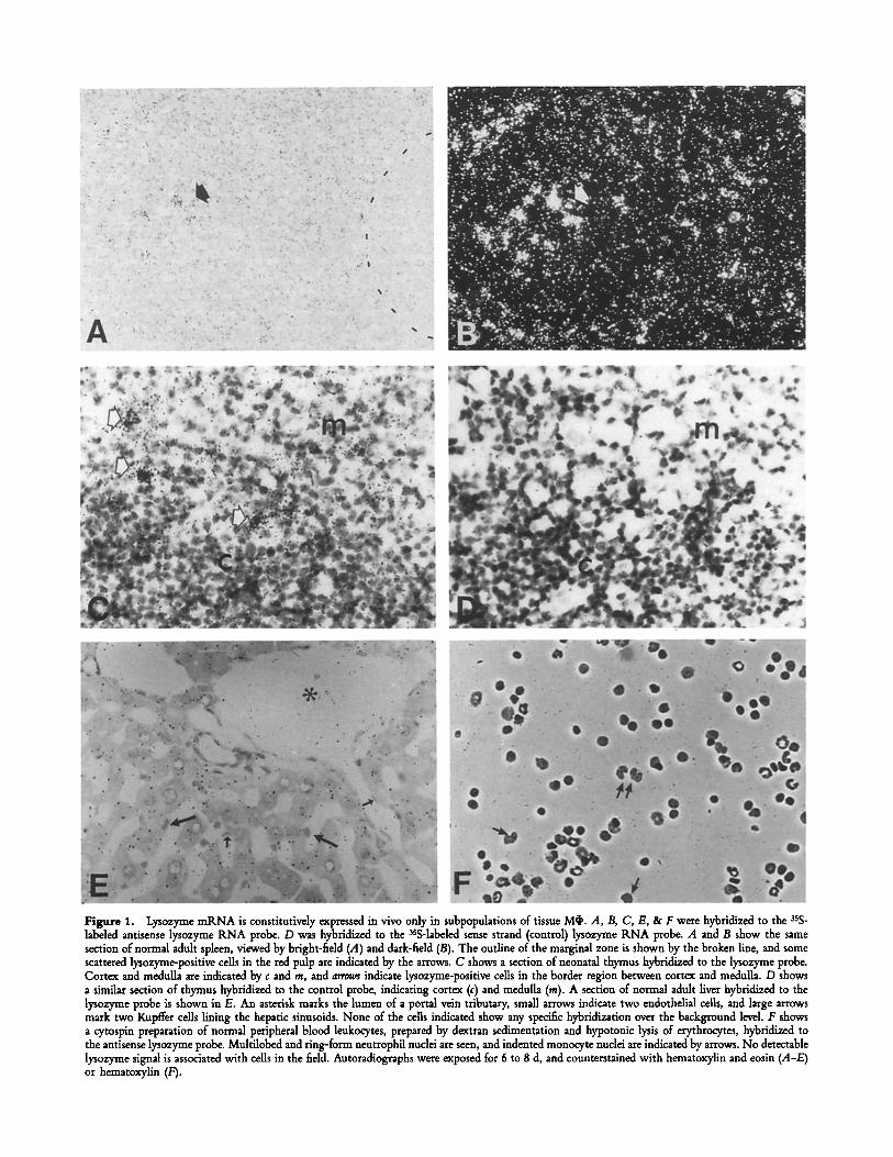
Figure 1. Lysozyme mRNA is constitutively expressed in vivo only in suhpopulations of tissue M~. A, B, C, E, & F were hybridized to the sss- labeled antisense lysozyme RNA probe. D was hybridized to the aSS-labeled sense strand (control) lysozyme KNA probe. A and B show the same section of normal adult spleen, viewed by bright-field (A) and dark-field (B). The outline of the marginal zone is shown by the broken line, and some scattered lysozyme-positive cells in the red pulp are indicated by the arrows. C shows a section of neonatal thymus hybridized to the lysozyme probe. Cortex and medulla are indicated by c and m, and arrows indicate lysozyme-positive cells in the border region between cortex and medulla. D shows a similar section of thymus hybridized to the control probe, indicating cortex (c) and medulla (m). A section of normal adult liver hybridized to the lysozyme probe is shown in E. An asterisk marks the lumen of a portal vein tributary, small arrows indicate two endothelial cells, and large arrows mark two Kupffer cells lining the hepatic sinusoids. None of the cells indicated show any specific hybridization over the background level. F shows a cytospin preparation of normal peripheral blood leukocytes, prepared by dextran sedimentation and hypotonic lysis of erythrocytes, hybridized to the antisense lysozyme probe. Multilobed and ring-form neutrophil nuclei are seen, and indented monocyte nuclei are indicated by arrows. No detectable lysozyme signal is associated with cells in the field. Autoradiographs were exposed for 6 to 8 d, and counterstained with hematoxylin and eosin (A-E) or hematoxylin (F).

tectable lysozyme mRNA (4). Low levels were found in the red pulp of the spleen (Fig. 1 A and B), scattered positive cells were found in the cortico-medullary region of the neo- natal thymus (Fig. 1 C) and a few positive cells were found in the subcapsular area of mesenteric lymph nodes. The dis- tribution of these cells was compatible with their being McI,, but the majority of F4/80 + cells were negative.
Prominent M~ populations such as Kupffer cells of the liver, lamina propria Mq' in the intestine, microglia in the brain and resident M~ in the leptomeninges and choroid plexus were uniformly negative (Fig. 1 E). Peripheral blood mono- cytes and neutrophils were generally lysozyme mRNA nega- tive, with only occasional cells showing weak hybridization (Fig. 1 E). Apart from strongly positive Paneth cells in the small intestine (4), no lysozyme producing epithelial cells were detected in the gastrointestinal tract, male urogenital tract, kidney or resting, gestating or lactating female genital tract and breast. F4/80 + cells in these organs also lacked lyso- zyme mRNA. No other cell types, including tear gland and salivary gland cells, were lysozyme-positive. Although human tears and saliva are lysozyme-rich, mouse saliva collected after pilocarpine stimulation had a much lower concentration of lysozyme activity, while mouse tears contained no detectable activity (not shown).
Bacille Calmette Guerin (BCG) Infection Induces Lysozyme Extrression in Macrophages (M~) In Viva BCG infection results in the recruitment of large numbers of immunologically ac- tivated M6 to granulomas in the organs of infected mice (7, 8). We therefore infected mice with live BCG organisms and examined the liver and spleen by in situ hybridization for lysozyme mKNA. As shown in Fig. 2 A, lysozyme-positive cells appeared in the marginal zone of the spleen within I d of intravenous infection. By day 7 of the infection, the signal in the spleen was greatly increased but was still localized mainly to the marginal zone, with some positive cells also appearing in the white pulp (Fig. 2, C and D). The intensity and extent of labeling increased further during the course of infection and in addition to strong labeling of marginal zone cells, foci of labeled cells were found in the red and white pulp. By 21 d of infection the architecture of the spleen was disrupted and collections of lysozyme-positive cells were found throughout the organ (Fig. 2, E and F). Control sections hybridized to the sense strand probe showed no specific signal.
Lysozyme-positive granulomas in the liver developed more slowly. Unlike the spleen, there was no induction of lyso- zyme mP, NA 24 h after infection. The majority of cells in the granulomas which eventually develop are F4/80 + MO (S. Rabinowitz, unpublished observations), so the extent of labeling of lysozyme-positive cells is consistent with their being MO (Fig. 3 A). Although a few sinusoidal cells, which may be Kupffer cells or migrating monocytes, were labeled, the majority of Kupffer cells were lysozyme-negative even in infected animals (Fig. 3 B). Further studies examined the role of T cells in the induction of lysozyme expression in situ. BCG infection in congenitally athymic (nude) mice resulted in small foci of lysozyme-positive cells appearing in the liver and spleen. The number and size of granulomas was
less than in euthymic littermates, but the intensity of the lyso- zyme signal in M~ was comparable (not shown).
To examine the effect of specific antigen challenge, PPD was injected intraperitoneally into BCG infected mice and lysozyme mRNA levels in peritoneal cells determined by in situ hybridization to cytospin preparations of cells recovered 24 h later. The combination of infection and antigen chal- lenge yielded strongly lysozyme-positive M~ which were shown in separate cytospin preparations to be F4/80 + and MHC dass II +. Peritoneal cells from infected mice without PPD challenge or uninfected mice with PPD challenge were weakly positive. In adoptive transfer experiments, sensitized lymphocytes and PPD injected together into the peritoneal cavity of naive mice yielded a population of peritoneal cells with increased lysozyme expression (data not shown).
Modulation of Lysozyme Gene Expression In Macrophages (M~) by Other Stimuli. In view of the localized and rapid appear- ance of the lysozyme-positive cells in the spleen after BCG infection, we examined the effect of another infectious agent, P. yoelii, which is also known to activate M~ function in the spleen and liver (10). Fig. 4 shows that marked induction of lysozyme gene expression is seen in the marginal zone of the spleen within 1 d of intraperitoneal injection of P. yoelii parasitized mouse erythrocytes (Fig. 4 A). In the course of the following 21 d, lysozyme-positive cells were concentrated in the white pulp, and red pulp labeling was less evident than in the case of BCG infection. Scattered lysozyme-positive cells were also seen in the liver, but these were only a proportion of the heavily pigment-laden phagocyte population. To ex- plore the early induction of lysozyme mRNA in marginal zone M~, sheep erythrocytes were injected intravenously into normal mice. This too resulted in the rapid appearance of lysozyme-positive cells in the marginal zone (Fig. 4 B). Un- like the injection of live infectious agents, however, injection of sheep erythrocytes generated a short-lived effect which dis- appeared within 7 d.
Regulation of Lysozyme Expression In Vivo Is Distinct from that of Tumor Necrosis Factor (TNF). Since lysozyme expres- sion was inducible rather than constitutive in most M~ in vivo, it was of interest to compare its induction with that of a induced inflammatory product such as TNF (17). It is well known that TNF mRNA and protein expression are increased in vitro in Mq~ which have been treated with LPS. In addition, it has been shown that TNF is present in granu- loma M~ in vivo, and that the production of TNF is essen- tial for granuloma formation in response to BCG infection (9). In Fig. 5, we demonstrate the presence of TNF mRNA in granuloma cells by in situ hybridization to the routine TNF probe. In contrast to lysozyme mRNA, which was uniformly distributed among the granuloma cells, only a proportion of M~ in the granuloma contained TNF mP, NA (Fig. 3 and 5). Furthermore, when BCG infected mice were injected with LPS, TNF mRNA expression was massively induced in granu- loma cells as well as in other cells in the hepatic sinusoids. Lysozyme mRNA levels, in contrast, were not enhanced by LPS treatment. This was confirmed by Northern blot anal- ysis, which showed clear elevation of TNF mP, NA levels by
1052 Lysozyme and Macrophage Activation In Situ

Figure 2. Lysozyme mtLNA is progressively induced in MO subpopulations in BCG infected spleen. All sections were hybridized to the antisense 3sS-labeled lysozyme probe. The left hand photographs show bright field views and the right hand photographs show the corresponding dark-field illumination view. Arrows indicate the position of the marginal zone. (.,4 and B) Spleen from an animal injected 24 h previously with 107 BCG or- ganisms. There is a ring of positive cells in the marginal zone (arrows), which is more marked than that in the normal spleen (Fig. 1 A and B). (C and D) 7 d after infection the number of positive cells and strength of the signal is increased. The signal is still largely confined to the marginal zone and the interior of the white pulp (arrows). (E and F) 21 d after infection lysozyme-positive cells are scattered throughout the spleen in small granuloma- like foal. Autoradiographs were exposed for 6 to 8 d and counterstained with hematoxylin and eosin.
1053 Keshav et al.

Figure 3. Lysozyme mRNA is expressed in granuloma M~ and sinusoidal cells in BCG in- fected liver. Liver sections from BCG infected mice 21 d after BCG infection hybridized to the antisense 3sS-labeled lysozyme probe. A shows a low power view of the liver, with lysozyme-positive granu- lomas scattered through the pareuchyma (aster/sks). A few isolated, lysozyme-positive cells may also be seen (arrows). At higher magnification (B), it can be seen that the lysozyme signal is distributed evenly, and is present in the majority of granu- loma cells (asterisk). A few lysozyme-positive cells are also seen in the sinusoids (arrows). At the same time large adherent sinusoidal cells (probably Kupffer cells) indicated by solid arrowheads, are negative, as are the endothelial cells (flattened, darkly stained nuclei, indicated by open arrowheads). Autoradiographs were exposed for 6 d and coun- terstained with haematoxylin and eosin.
BCG infection and further induction of expression in liver, lung and intestine by subsequent LPS challenge (Fig. 6). In the lung, TNF and lysozyme mRNA are detectable in the normal state, although the TNF signal is much less than the lysozyme signal, and both are increased by infection. The hybridization signals in these organs are presumed to arise from McI). In the small intestine there is a clearly detectable lysozyme signal (derived from Paneth cells) in the normal state, while the TNF signal is much weaker.
Discussion
The wide dispersion and diverse phenotype of tissue M~ suggest an active role for these cells in tissue homeostasis and defense. To study the secretory activity of tissue McI ' in vivo more closely, lysozyme was chosen as a well characterized
indicator of M~ secretory potential. Earlier studies had indi- cated that lysozyme production was restricted to only a few cell types and that lysozyme production by M~ in vitro was a constitutive function which could potentially label all differentiated Mr TNF, an inflammatory cytokine which is also produced almost exclusively by M~I', but is dependent on activation for full expression, was studied in parallel.
We noted previously that lysozyme mRNA was produced constitutively by M~ in tissue culture, but that the majority of tissue M~ did not transcribe the gene in vivo in the resting animal (4). The more extensive survey reported here confirms this generalization. In addition, it seems that lysozyme produc- tion in the steady state is correlated with phagocytic activity, either of foreign material (as in alveolar M~I' of the lung) or of endogenous ligands (as in M~ of the neonatal thymus and red pulp of the spleen). Furthermore, a number of the
1054 Lysozyme and Macrophage Activation In Situ

Figure 4. Plasmodium yoelii infection and sheep erythrocytes rapidly in- duce lysozyme mR.NA expression in marginal zone M~ of the spleen. Sections of murine spleen, 24 h after injection of P. yoelii infected erythro- cytes (A) or normal sheep erythrocytes (/3) were hybridized to the 3sS- labeled antisense lysozyme probe. A shows a bright field view, while B is dark field view to enhance the autoradiographic signal. The area of the marginal zone is indicated by arrows. Strongly positive cells are seen in the marginal zone in both sections, while a few positive cells also appear to be present in the white pulp itself in B. This is probably caused by oblique sectioning of a nodule of white pulp. Autoradiographs were ex- posed for 8 d, and stained with haematoxylin.
lysozyme-positive M~ populations lack F4/80 antigen, but do express other Mob antigens such as SEtL and FA-11 (refer- ence 18; S. Rabinowitz, unpublished observations).
Most epithelial cells were likewise negative for lysozyme mRNA, even in organs where lysozyme is secreted (for ex- ample in the salivary gland). This may be due to limitations in the sensitivity of in situ hybridization and Northern blot techniques, and low levels of lysozyme, distributed relatively uniformly among many cells, may in fact be produced in some of the tissues which appear negative. Nevertheless, some of the negative results are noteworthy. The absence of detect- able lysozyme mRNA in kidney contrasts strongly with the
1055 Keshav et al.
abundance of the protein in cells of the proximal convoluted tubule, and demonstrates a potential source of experimental error where proteins produced in one site are sequestered in another. The same phenomenon may occur when paracrine secretions are localized immunochemically on the target cell rather than the source.
In Kupffer cells, which do not contain detectable quanti- ties of lysozyme mRNA, the absence of lysozyme biosyn- thesis may reflect a more general refractory state (19). How- ever BCG infection followed by LPS injection may be sufficient to induce TNF production in some of these cells (Fig. 5 B). Although the present data indicate that newly recruited Mob constitute the major lysozyme expressing population, fur- ther experiments are required to distinguish resident and recruited cells definitively.
In infected animals, lysozyme mRNA production was strongly induced compared to normal controls. Lysozyme production was, however, confined to discrete subpopulations of Mq~, while the majority of tissue Mq~ remained lysozyme- negative. In the spleen, for example, lysozyme gene activa- tion in the early stages of infection was confined to the mar- ginal zone, the area where blood-borne thymus independent carbohydrate antigens are initially trapped and engulfed by M~ (20-22). The marginal zone is also the site of entry of peripheral blood monocytes into the spleen, so that the lyso- zyme signal may result from newly recruited cells stimulated in the circulation. The persistence of the signal, and its ex- tension into the red and white pulp with continuing infec- tion, suggest that the lysozyme-positive cells remain local- ized even if they are initially stimulated in the circulation. In BCG-infected liver, while the majority of Kupffer cells lacked lysozyme mRNA, some monocytes in the sinusoids were lysozyme-positive, and these and other cells presum- ably contribute to the formation of granulomas in which the lysozyme gene is persistently transcribed.
The observation that sheep erythrocytes and P. yoelii in- fected mouse erythrocytes also induced lysozyme gene ex- pression in the marginal zone suggests that lysozyme gene expression by M~ is associated with active phagocytosis. Plas- modium and BCG induced lysozyme gene expression was prolonged, while sheep erythrocyte induced expression was relatively transient, possibly reflecting the persistence of the initiating stimulus.
Activation of M~ in vitro for full bactericidal function requires the participation of T cells, and many nonspecific inflammatory stimuli produce only a partially activated pheno- type (23). The effects of BCG infection in congenitally athymic mice indicate that lysozyme gene expression in Mob can be induced independent of mature T cell function. At the same time it is evident that T cell products may increase M~ lyso- zyme production in peritoneal cells from BCG primed animals challenged with an intraperitoneal dose of PPD.
Although lysozyme gene expression in Mq~ is inducible in vivo, the regulation of lysozyme expression remains dis- tinct from that of an inflammatory cytokine such as TNF. Lysozyme, which is completely nontoxic to the host, and acts as a nonspecific antibacterial defense, is produced rapidly and continuously in response to many stimuli. TNF, on the

Figure 5. TNF mKNA is expressed in BCG granulomas, and further induced by LPS injection. A and B show sections of mouse liver 16 d after infection with BCG, hybridized to the antisense 3sS-labded TNF probe. In A, the granulomas (amaos) show moderate hbeling of most cells, while one granuloma (bottom right corner) is more strongly labeled. Kupffer cells seem to be TNF- negative. In B, after injection with LPS, the in- tensity of the signal, and the number of TNF- positive cells is greatly increased. In the granulomas (solid arrows), the TNF signal is increased in in- dividual cells, and is also greatest in cells on the periphery of the granuloma. In addition, TNF mRNA is induced in cells outside the granulomas (open arrows). Autoradiographs were exposed for 6 d, and stained with haematoxylin and eosin.
other hand, which has complex physiological effects, including definite host toxicity, is tightly regulated. Furthermore, while lysozyme secretion is decreased in vitro by LPS treatment, and unchanged in vivo, TNF production is markedly induced by LPS treatment (24, 25). Lysozyme gene expression is not regulated posttranscriptionally, while TNF production is regu- lated by changes in mRNA stability and translation rate (25). As this can result in the localization of mRNA in cells which are not actually producing the relevant protein, a complete description of in situ secretory activity for proteins such as TNF requires localization ofmRNA and protein. As regula- tory elements of the TNF gene have been recently described, and both mouse and human lysozyme genes cloned, it will be possible to compare lysozyme and TNF gene regulation
at the molecular level (26-28). Additional information re- garding tissue-specific gene regulation may also be obtained by comparing the two mouse lysozyme genes (6).
While cell lines derived from colon carcinomas and hepa- tomas can produce lysozyme, only myelomonocytic cells and specialized exocrine cells (such as Paneth cells) produce lyso- zyme in vivo (29, 30). Lysozyme production could therefore be useful as a marker of myelomonocytic cell activity in dis- ease. Furthermore, as neutrophils contain mainly preformed lysozyme in their granules while mature M~ retain the ability to synthesize new protein, lysozyme mRNA levels in vivo specifically monitor changes in M~ biosynthetic and secre- tory activity induced by phagocytic and T cell derived stimuli.
1056 Lysozyme and Macrophage Activation In Situ

Figure 6. Superinduction of TNF but not lysozyme mRNA by LPS. Northern blot showing hybridization of 3zp-labded TNF and lysozyme probes to RNA from gut (G), lung (Lu) and liver (Li) of normal mice, mice infected with BCG 21 d earlier, and mice infected with BCG and challenged with 25/zg of LPS for 2 h. Each lane was loaded with 20 gg of total RNA, and ethidium bromide staining used to assess the position of ribosomal RNA bands (28s and 18s) and to ensure even loading. The predominant TNF mRNA band migrates at about 1.7 kb, and the lysozyme mRNA band at about 1.3 kb. Autoradiograms were exposed for 24 h before development.
We thank Jim Kent for technical assistance, Les Haven for histology, and Harry Edwards, Stan Bucking- ham, and Cathy Lee for photography.
This work was supported by the Medical Research Council (UK), a Rhodes Scholarship (S. Keshav), and a NAT(9 Fellowship (G. Milon).
Address correspondence to Satish Keshav, Sir William Dunn School of Pathology, University of Oxford, South Parks Road, Oxford OX1 3tLE, U.K.
Received for publication 14 January 1991 and in revised form 30 July 1991.
References 1. Nathan, C.F. 1987. Secretory products ofmacrophages.J. Clin.
Invest. 79:319. 2. Gordon, S., J. Todd, and Z.A. Cohn. 1974. In vitro synthesis
and secretion of lysozyme by mononuclear phagocytes..)'. Exp. Med. 139:1228.
3. Mason, D.Y., and C.R. Taylor. 1975. The distribution of muramidase (lysozyme) in human tissues..]. Clin. Pathol. 28:124.
4. Chung, L.-P., S. Keshav, and S. Gordon. 1988. Cloning of the human lysozyme cDNA: Inverted Alu repeat in the mRNA and in situ hybridization for macrophages and Paneth cells. Proc. Natl. Acad. Sci. USA. 85:6227.
5. Osserman, E.F., and D.P. Lawlor. 1966. Serum and urinary lysozyrne (muramidase) in monocytic and myelomonocytic leu-
kaemia. J. Extx Med. 124:921. 6. Cross, M., I. Mangelsdorf, A. Wedel, and K. Renkawitz. 1988.
Mouse lysozyme M gene: Isolation, characterization, and ex- pression studies. Proc Natl. Acad. Sci. USA. 85:6232.
7. Pelletin, M., A. Forget, D. Bourassa, P. Gros, and E. Skamene. 1982. Immunopathology of BCG infection in genetically resis- tant and susceptible mouse strains. J. Immunol. 129:2179.
8. Nibbering, P.H., G.A. Van der Heide, and R. van Furth. 1989. Immunocytochemical analysis of cellular response to BCG. Clin. Ex F Immunol. 75:147.
9. Kindler, V., A.-P. Sapino, G.E. Grau, P.-F. Piguet, and P. Vas- sail 1989. The inducing role of tumor necrosis factor in the development of bacterial granulomas during BCG infection.
1057 Keshav et al.

Cell. 56:731. 10. Lee, S.-H., P. Crocker, and S. Gordon. 1986. Macrophage
plasma membrane and secretory properties in murine malaria. Effects of Plasmodium yoelii infection on macrophages in the liver, spleen and blood. J. Extx Med. 163:54.
11. Hogan, B., F. Costantini, and E. Lacy. 1986. Manipulating the Mouse Embryo: A Laboratory Manual. Cold Spring Harbor Laboratory, Cold Spring Harbor, NY. pp. 228-242.
12. Pennica, D., J.S. Hayflick, T.S. Bringman, M.A. Palladino, and D.V. Goeddel. 1985. Cloning and expression in Escherichia coli of the cDNA for murine tumor necrosis factor. Prog Natl. Acad. Sci. USA. 82:6060.
13. Feinberg, A., and B. Vogelstein. 1982. A technique for radi- olabeling DNA restriction endonuclease fragments to high specific activity. Anal. Biochem. 132:6.
14. Chirgwin, J.M., A.E. Przybyla, R.J. MacDonald, and W.J. Rutter. 1979. Isolation of biologically-active ribonucleic acid from sources enriched in ribonuclease. Biochemistry. 18:5294.
15. Maniatis, T., E.F. Fritsch, and J. Sambrook. 1982. Molecular Cloning: A Laboratory Manual. Cold Spring Harbor Labora- tory, Cold Spring Harbor, NY. pp. 188-196.
16. Gordon, S., S. Keshav, and L.P. Chung. 1988. Mononuclear phagocytes: tissue distribution and functional heterogeneity. Current Opinion in Immunology. 1:26.
17. Beutler, B., and A. Cerami. 1989. The biology of cachec- tin/TNF - a primary mediator of the host response. Annu. Rev. Immunol. 7:625.
18. Crocker, P.R., and S. Gordon. 1989. Mouse macrophage hae- magglutinin (sheep erythrocyte receptor) with specificity for sialylated glycoconjugates characterized by a monoclonal anti- body. J. Exp. Med. 169:1333.
19. Lepay, D.A., R.M. Steinman, C.F. Nathan, H.W. Murray, and Z.A. Cohn. 1985. Murine Kupffer cells. Mononuclear phago- cytes deficient in the generation of reactive oxygen inter- mediates. J. Exp. Med. 161:1079.
20. Humphrey, J.H., and D. Grennan. 1981. Different macrophage populations distinguished by means of fluorescent polysaccha-
rides. Recognition and properties of marginal-zone macro- phages. Eur. J. Immunol. 11:221.
21. Dijkstra, C.J., E. van Vliet, E.A. Dopp, A.A. Vanderlelij, and G. Kraal. 1985. Marginal zone macrophages identified by a monoclonal antibody: characterization of immunohistochem- ical capacities. Immunology. 55:23.
22. Matsuma, K., H. Fuji, and M. Katani. 1986. Splenic mar- ginal zone macrophages and marginal metallophils in rats and mice. Cell Tissue Res. 246:263.
23. Cohn, Z.A. 1978. The activation of mononuclear phagocytes. Facts, fancy, and future. J. Immunol. 121:813.
24. Warfel, A.H., and D. Zucker-Franklin. 1986. Down-regulation of macrophage lysozyme by lipopolysaccharide and interferon. J. Immunol. 137:651.
25. Beutler, B., N. Krochin, I. Milsark, C. Luedke, and A. Cerami. 1986. Control of cachectin (tumor necrosis factor) synthesis: mechanisms of endotoxin resistance. Science (Wash. DC). 232:977.
26. Jongeneel, C.V., A.N. Sakhov, Nedospasov, and J.-C. Cerot- tini. 1989. Molecular control of tissue-specific expression at the mouse TNF locus. Eur. J. Immunol. 19:549.
27. Shakhov, A.N., M.A. Collart, P. Vassali, S.A. Nedospasov, and C.V. Jongeneel. 1990. kB-type enhancers are involved in LPS-mediated transcriptional activation of the TNFa gene in primary macrophages. J. Exp. Med. 171:35.
28. Peters, C.W., U. Kruse, K. Pollwein, K.-H. Grzeschik, and A.E. Sippel. 1989. The human lysozyme gene: sequence orga- nization and chromosomal localization. Eur.J. Biochem. 182:507.
29. Fett, J.W., D.J. Strydom, R.R. Lobb, E.M. Alderman, B.L. Vallee, P.J. Artymiuk, S. Collett, D.C. Phillips, C.M. Dobson, and C. Redfield. 1985. Lysozyme: a major secretory product of a human colon carcinoma cell line. Biochemistry. 24:965.
30. Knowles, B.B., C.C. Howe, and D.P. Aden. 1980. Human hepatocellutar carcinoma cell lines secrete the major plasma pro- teins and hepatitis B surface antigen. Science (Wash. DC). 209:497.
1058 Lysozyme and Macrophage Activation In Situ