m1 iprgcs influence visual function through retrograde ... · horizontal cells (knapp and dowling,...
TRANSCRIPT

Cellular/Molecular
M1 ipRGCs Influence Visual Function through RetrogradeSignaling in the Retina
X Cameron L. Prigge,1 Po-Ting Yeh,2 X Nan-Fu Liou,2 X Chi-Chan Lee,2 X Shih-Feng You,2 X Lei-Lei Liu,1
X David S. McNeill,3 X Kylie S. Chew,3 Samer Hattar,3 X Shih-Kuo Chen,2,4 and X Dao-Qi Zhang1
1Eye Research Institute, Oakland University, Rochester, Michigan 48309, 2Department of Life Science, National Taiwan University, Taipei, Taiwan 10617,3Department of Biology, Johns Hopkins University, Baltimore, Maryland 21218, and 4Genome and Systems Biology Degree Program, National TaiwanUniversity and Academia Sinica, Taipei, Taiwan 10617
Melanopsin-expressing intrinsically photosensitive retinal ganglion cells (ipRGCs, with five subtypes named M1–M5) are a uniquesubclass of RGCs with axons that project directly to many brain nuclei involved in non-image-forming functions such as circadianphotoentrainment and the pupillary light reflex. Recent evidence suggests that melanopsin-based signals also influence image-formingvisual function, including light adaptation, but the mechanisms involved are unclear. Intriguingly, a small population of M1 ipRGCs haveintraretinal axon collaterals that project toward the outer retina. Using genetic mouse models, we provide three lines of evidence showingthat these axon collaterals make connections with upstream dopaminergic amacrine cells (DACs): (1) ipRGC signaling to DACs is blockedby tetrodotoxin both in vitro and in vivo, indicating that ipRGC-to-DAC transmission requires voltage-gated Na � channels; (2) thistransmission is partly dependent on N-type Ca 2� channels, which are possibly expressed in the axon collateral terminals of ipRGCs; and(3) fluorescence microscopy reveals that ipRGC axon collaterals make putative presynaptic contact with DACs. We further demonstratethat elimination of M1 ipRGCs attenuates light adaptation, as evidenced by an impaired electroretinogram b-wave from cones, whereasa dopamine receptor agonist can potentiate the cone-driven b-wave of retinas lacking M1 ipRGCs. Together, the results strongly suggestthat ipRGCs transmit luminance signals retrogradely to the outer retina through the dopaminergic system and in turn influence retinallight adaptation.
Key words: amacrine cell; dopamine; ganglion cell; melanopsin; retina; vision
IntroductionThe visual system operates over a large range of light levels aslighting conditions change across 10 orders of magnitude from
dim starlight to bright daylight. This process, called light adapta-tion, is commonly thought to occur primarily in the retina.Retinal rod photoreceptors operate in dim light and cone photo-
Received Sept. 17, 2015; revised May 25, 2016; accepted May 26, 2016.Author contributions: C.L.P., S.-K.C., and D.-Q.Z. designed research; C.L.P., P.-T.Y., N.-F.L., C.-C.L., S.-F.Y., L.-L.L.,
S.-K.C., and D.-Q.Z. performed research; D.S.M., K.S.C., and S.H. contributed unpublished reagents/analytic tools;C.L.P., P.-T.Y., N.-F.L., C.-C.L., S.-F.Y., and S.-K.C. analyzed data; C.L.P., S.-K.C., and D.-Q.Z. wrote the paper.
This work was supported by the National Institutes of Health (Grant R01 EY022640 to D.-Q.Z.), Oakland UniversityProvost’s Graduate Student Research Awards (C.L.P.), and the Taiwan Ministry of Science and Technology (GrantMOST-103-2321-B-002-045 to S.-K.C.). We thank Drs. Frank Giblin, Douglas McMahon, Gary Pickard, and BarryWinkler and the anonymous reviewers for their critical comments; Dr. Paulo Kofuji for kindly providing transgenic
mice for our research; and the Technology Commons College of Life Science at National Taiwan University fortechnical assistance with confocal laser scanning microscopy.
The authors declare no competing financial interests.Correspondence should be addressed to either of the following: Dr. Dao-Qi Zhang, Eye Research Institute,
Oakland University, 423 Dodge Hall, Rochester, MI 48309, E-mail: [email protected]; or Dr. Shih-Kuo Chen,Department of Life Science, National Taiwan University, No. 1 Sec. 4 Roosevelt Road, Taipei, Taiwan 10617,E-mail: [email protected].
DOI:10.1523/JNEUROSCI.3500-15.2016Copyright © 2016 the authors 0270-6474/16/367184-14$15.00/0
Significance Statement
Melanopsin-expressing intrinsically photosensitive retinal ganglion cells (ipRGCs) comprise a third class of retinal photorecep-tors that are known to mediate physiological responses such as circadian photoentrainment. However, investigation into whetherand how ipRGCs contribute to vision has just begun. Here, we provide convergent anatomical and physiological evidence that axoncollaterals of ipRGCs constitute a centrifugal pathway to DACs, conveying melanopsin-based signals from the innermost retina tothe outer retina. We further demonstrate that retrograde signals likely influence visual processing because elimination of axoncollateral-bearing ipRGCs impairs light adaptation by limiting dopamine-dependent facilitation of the cone pathway. Our findingsstrongly support the hypothesis that retrograde melanopsin-based signaling influences visual function locally within the retina, anotion that refutes the dogma that RGCs only provide physiological signals to the brain.
7184 • The Journal of Neuroscience, July 6, 2016 • 36(27):7184 –7197

receptors function in bright light. Both photoreceptors can adjusttheir sensitivity to more than seven decimal orders of illumina-tion to continue to respond to brighter illumination (photore-ceptor adaptation; Boynton and Whitten, 1970; Nakatani et al.,1991). In addition, postreceptoral neurons (horizontal, bipolar,amacrine, and ganglion cells) have the ability to adjust their sen-sitivity according to the ambient light condition through changesin their intrinsic membrane properties and modification of theirsynapses with other neurons (network adaptation; Green et al.,1975; Dunn et al., 2006).
Retinal ganglion cells (RGCs), the sole output neurons of thevertebrate retina, receive inputs from rod and cone photorecep-tors through bipolar cells and amacrine cells, which are involvedin network light adaptation. In addition, a small population ofRGCs express the photopigment melanopsin which allows themto respond to light directly (Berson et al., 2002; Hattar et al.,2002). Like rods and cones, intrinsically photosensitive RGCs(ipRGCs) exhibit photoreceptor adaptation (Wong et al., 2005;Do and Yau, 2013). However, it is unclear how photoreceptorand network adaptation of ipRGCs contributes to light adapta-tion of the visual system (Allen et al., 2014). ipRGCs primarilyproject, via the optic nerve, to a variety of brain nuclei involved innon-image-forming visual functions such as circadian photoen-trainment and the pupillary light reflex (Berson et al., 2002; Hat-tar et al., 2003; Panda et al., 2003). Intriguingly, recent studiesdemonstrated that ipRGCs send signals retrogradely to dopami-nergic amacrine cells (DACs) within the retina (Zhang et al.,2008; Zhang et al., 2012; Atkinson et al., 2013; Newkirk et al.,2013). Intraretinal retrograde signal transmission provides a pos-sible mechanism by which ipRGCs may influence light adapta-tion via the retinal dopaminergic system.
DACs are a subpopulation of retinal amacrine cells that con-stitute an intraretinal feedback pathway, transmitting rod andcone signals in the reverse direction from the inner to the outerretina (Dowling and Ehinger, 1975; Dacey, 1988; Witkovsky,2004). With additional excitatory input from ipRGCs, this feed-back pathway is expanded by transmitting melanopsin-basedsignals from the inner to the outer retina, acting as a centrifugalpathway (Zhang et al., 2008). The integrated signals frommelanopsin, rods, and cones provide a driving force that stimu-lates DACs to release the key neurotransmitter dopaminethrough synaptic and extrasynaptic exocytosis volume transmis-sion (Dkhissi-Benyahya et al., 2013). Released dopamine exertsits action through a family of G-protein-coupled dopamine re-ceptors, D1–D5. Activation of these receptors by dopamine canreconfigure retinal circuits according to prevailing illuminationby uncoupling electrical synapses throughout the retina (Lasaterand Dowling, 1985; Mills and Massey, 1995; Krizaj et al., 1998;Mills et al., 2007), modulating excitatory glutamate receptors onhorizontal cells (Knapp and Dowling, 1987), and regulating theactivity of sodium channels of bipolar cells and ganglion cells(Hayashida and Ishida, 2004; Ichinose and Lukasiewicz, 2007).As a result, retina-specific dopamine deletion leads to an impair-ment of light-adapted vision (Jackson et al., 2012).
Despite the importance of signal transmission from ipRGCsto DACs, the exact route and synaptic mechanisms of thisretrograde signaling pathway has been unknown. Axon collater-als have been reported previously in a small subpopulation ofRGCs in mice, monkeys, and humans (Dacey, 1985; Peterson andDacey, 1998; Joo et al., 2013). Our recent study demonstratedthat some, if not all, of these collateral-bearing cells are M1ipRGCs, one of five (M1–M5) subtypes (Joo et al., 2013). Here,
we present convergent in vitro and in vivo physiological and an-atomical evidence that axon collaterals of M1 ipRGCs make con-nections with DACs, forming a centrifugal pathway that conveysretrograde signals from the inner retina to the outer retina topotentially facilitate light-adapted vision.
Materials and MethodsAnimals. Adult mice of both sexes were used in the present study. Allanimals were maintained on a 12:12 h light/dark cycle with ad libitumaccess to food and water. All procedures were conducted according toNational Institutes of Health guidelines for work with laboratory animalsand were approved by the Institutional Animal Care and Use Commit-tees at Johns Hopkins University, National Taiwan University, and Oak-land University.
For ipRGC recordings, a transgenic mouse line with a C57BL/6J back-ground was used; melanopsin (Opn4 )-expressing ipRGCs were labeledusing green fluorescent protein (GFP; provided by Dr. Paulo Kofuji,University of Minnesota). For DAC recordings, two strategies were usedto isolate melanopsin-based signaling to DACs. One strategy involvedusing C3H/HeJ mice homozygous for the Pde6b rd1 mutation (rd1; Jack-son Laboratory) in which rod loss occurs rapidly with onset at postnatalday 8 (P8) and is nearly complete by P21. By P90, virtually all outerphotoreceptors disappear except for �3% of cone somata in the dorsalretina (Carter-Dawson et al., 1978). We crossed this line with our trans-genic mouse in which DACs are genetically labeled using red fluorescentprotein (RFP) under the control of the promoter for the rate-limitingenzyme for dopamine synthesis, tyrosine hydroxylase (TH; C57BL/6Jbackground; Zhang et al., 2004), to obtain rd1 TH::RFP transgenic mice.This mouse line carried a mixed C57BL/6J and C3H background and wasused in the current study and our previous study (Atkinson et al., 2013).The other strategy involved genetically deleting Cnga3, a cone photorecep-tor-specific cyclic nucleotide channel subunit, in our TH::RFP transgenicmice (Biel et al., 1999). We crossed our TH::RFP transgenic mice with a tripleknock-out (Opn4� / � Gnat1� / � Cnga3� / �) mouse line (Hattar et al.,2003) to obtain cnga3 knock-out TH::RFP transgenic mice. This mouse linehad a mixed C57BL/6J and BL6/129 background. For morphologicalexperiments, an Opn4-driven tamoxifen-inducible CreERT2(Opn4 CETL) line was crossed with an alkaline phosphatase reporter(Rosa IAP) line in a mixed BL/6; 129SvJ background (Joo et al., 2013).For our in vivo intraocular tetrodotoxin (TTX) injection experiment,rod- and cone-transducin double-knock-out ( gnat1 � / �; gnat2 cpfl3/
cpfl3) mice were crossed with the Opn4-tau-LacZ mouse line to obtainmelanopsin only (MO) mice (Calvert et al., 2000; Chang et al., 2006).
Generation of Opn4-DTA mouse. To generate Opn4-diphtheria toxin A(DTA) mice, we used the targeting arms and general strategy detailed inHattar et al. (2002) and Guler et al. (2008). The DTA fragment (Maxwellet al., 1986) was inserted into the melanopsin gene locus using homolo-gous recombination. The construct contained a 4.4 kb region immedi-ately 5� of the melanopsin start codon, the DTA fragment, an internalribosomal entry site, tau-LacZ coding sequence, a self-excising neomycinresistance construct (Bunting et al., 1999), and the 1.5 kb 3� homologousarm starting from melanopsin exon 9. Specific targeting of the constructto the melanopsin locus was confirmed by PCR in both heterozygous(Opn4DTA/�) mice. PCR primers were as follows: melanopsin forwardprimer: 5�-GAG CCA CTG AGC ATG TGT AGT C-3�; melanopsin re-verse primer: 5�-CCC CAG GGG ATC TAG TCA AT-3�; DTA forwardprimer: 5�-GAG CCA CTG AGC ATG TGT AGT C-3�; and DTA reverseprimer: 5�-CAT CGC ATC TTG GCC ACG TTT T-3�.
Patch-clamp recordings. To minimize the influence of a circadian ef-fect, all experiments were performed during the day. Mice were adaptedto the dark for 1–2 h before experiments and were then treated by CO2
overdose and cervical dislocation. Eyes were enucleated and hemisectedat the ora serrata under infrared illumination. The cornea and lens wereremoved in a Petri dish filled with oxygenated extracellular solution con-taining the following (in mM): 125 NaCl, 2.5 KCl, 1 MgSO4, 2 CaCl2, 1.25NaH2PO4, 20 D-glucose, and 26 NaHCO3. The retina was separated fromthe sclera and placed ganglion cell side down (rd1 retinas) or photore-ceptor side down (Opn4::GFP, wild-type, and Cnga3 � / � retinas) in a
Prigge et al. • Centrifugal Pathway in the Retina J. Neurosci., July 6, 2016 • 36(27):7184 –7197 • 7185

recording chamber and mounted on the stage of an upright conventionalfluorescence microscope (Axio Examiner; Zeiss) within a light-tightFaraday cage. Oxygenated extracellular medium (pH 7.4 with 95% O2
and 5% CO2) continuously perfused the recording chamber at a rate of2–3 ml per min and the superfusate was maintained at 32°C by atemperature-control unit (TC-344A; Warner Instruments).
The retina was maintained in darkness for �1 h before recording. Cellsand recording pipettes were viewed on a computer monitor connected toa digital camera (AxioCam; Zeiss) mounted on the microscope. TheTH::RFP-expressing cells were identified using fluorescence microscopywith a rhodamine filter set with a brief snapshot of fluorescence excita-tion light (1–5 s). Opn4::GFP-expressing cells were identified using aFITC filter. The identified cells and glass electrode were visualized usinginfrared differential interference contrast (DIC) optics for patch-clamp re-cording. Experiments were performed 10–15 min after fluorescence wasused to locate the cells to allow the retinas to recover from photobleaching(caused by the brief exposure to fluorescence excitation light). The recoverymay have been incomplete during this short dark adaptation period, so ourexperiments were likely performed in a partially dark-adapted state.
Whole-cell voltage-clamp recordings were obtained from the somas ofDACs and ipRGCs using 4 –7 M� electrodes and the signals were ampli-fied using an Axopatch 200B amplifier (Molecular Devices). A holdingpotential of �60 mV was used for all recordings. The intracellular cesiumsolution used for DAC recordings contained the following (in mM): 120Cs-methane sulfonate, 5 NaCl, 5 CsCl, 10 HEPES, 5 EGTA, 0.5 CaCl2, 4Mg-ATP, 0.3 Na-GTP, and 5 lidocaine N-ethyl-chloride (QX-314). QX-314 was used to improve the space clamp quality of the voltage-clampand to block intrinsic action potentials (APs) of DACs, thus highlightingthe extrinsic light-induced inward current of the cells. The pH of theintracellular solution was titrated to 7.2–7.4 with CsOH. Data were notcorrected for a liquid junction potential of 3.5 mV. For whole-cellcurrent-clamp ipRGC recordings, a K �-based intracellular solution wasused that contained the following (in mM): 120 K-gluconate, 5 NaCl, 4KCl, 10 HEPES, 2 EGTA, 4 Mg-ATP, 0.3 Na-GTP, and �0.001% AlexaFluor 488 hydrazide. All chemicals used in the recording solutions werepurchased from Sigma-Aldrich unless otherwise noted. Data were ac-quired using a Digidata 1440A digitizer (Molecular Devices) and ana-lyzed offline using Clampex 10 software (Molecular Devices).
Cadmium chloride and nifedipine were purchased from Sigma-Aldrich. Alexa Fluor 488 hydrazide was obtained from Life Technologies.�-conotoxin GVIA, �-agatoxin IVA, and SNX 482 were purchased fromAbcam and L-AP4, TTX, and mibefradil dihydrochloride were obtainedfrom Tocris Bioscience. Concentrated stock solutions were prepared forall drugs used, which were later diluted in the extracellular medium asrequired, except for nifedipine, which was diluted in DMSO.
Light stimulation. Light stimuli were generated using a 470 nm LED(LED Supply and LC Corporation) to stimulate the melanopsin chro-mophore (peak sensitivity of �480 nm). An LED controller (Mightex)was used to drive the LED and the light intensity was adjusted by varyingthe driving current. The light intensity was measured at the surface of theretina by using an optical power meter (units converted from �W/cm 2 tophotons cm�2s�1; model 843-R; Newport). A light intensity of 4.7 �10 13 photons cm�2s�1 was used for all experiments.
ERG recordings. Electroretinogram (ERG) recordings were all con-ducted at circadian time 6 (CT 6) to minimize circadian influence. On theday of the experiment, mice were kept in darkness. Mice were intraperi-toneally administered with a mixture of ketamine (37.5 mg/kg) and xy-lazine (7.77 mg/kg). The pupils were dilated with 1% atropine and 0.5%proparacaine-hydrochloride was used as a local anesthetic. Eyes werekept moist with 1.5% hydrocellulose eye drops. A silver electrode wasused to record ERG responses and needle electrodes were placed in thecheek and tail as reference and ground leads, respectively. For light-adapted ERGs, a white LED light source was placed in a homemadegaze-field that provides 0.7 log cd s/m 2 light stimulation (above mel-anopsin threshold; Tu et al., 2005) with another 25 cd/m 2 white LED assteady background to saturate the rod response sufficiently. Flash inten-sity and duration were controlled by a Grass Instruments S44 stimulator.During 20 min of light adaptation, 5 ms of flash stimulation was providedat a frequency of 0.75 Hz. Signals were amplified, filtered (bandwidth:
0.3–1000 Hz), and digitized (10 kHz) with a Grass Instruments P5 pre-amplifier. Data were collected in a 2 min averaged bin. B-wave ampli-tudes were analyzed offline. Each trace was digitally filtered with a 60 Hzlow-pass filter to remove the influence of oscillatory potentials. B-waveamplitudes were calculated by subtracting the peak of the b-wave (within200 ms after light stimulation) and the trough of the a-wave. Baselineb-wave was calculated by averaging the first 10 ERG recordings from eachtrial for normalization. PD168077 (2 mg/kg) dissolved in 1� PBS (TocrisBioscience) was injected intraperitoneally just before the ERG record-ings. Data analyses were performed using Prism 6.0 software (GraphPadSoftware).
Immunohistochemistry. For whole-mount staining, mice were per-fused with PBS for 2 min, followed by 4% paraformaldehyde (PFA) for 15min, and then the eyes were enucleated. The entire eye was prefixed for 30min in 4% PFA before the retina was removed and postfixed in 4% PFAfor 30 min. After fixation, retinas were blocked in 5% goat serum, 1%BSA, and 0.2% Triton X-100 in PBS (0.1 M) for 2– 6 h at room tempera-ture. Subsequently, the retina was incubated overnight at 4°C in ablocking solution containing the following antibodies: chicken anti-�-galactosidase (1:500; Abcam), rabbit anti-c-Fos (1:10,000; Millipore),mouse anti-TH (1:1000; Immunostar), chicken anti-synaptophysin (1:1000; Abcam), rabbit anti-PSD95 (1:200; Abcam), and mouse anti-bassoon (1:400; Enzo), as indicated in the main text. After washing inPBS (0.1 M), retinas were incubated with the corresponding secondaryantibodies conjugated with Alexa Fluor 488, 568, 594, or 633 (Invitro-gen) at room temperature for 2 h and mounted with Vectashield (VectorLaboratories) for imaging. For whole-mount immunofluorescencestaining of ipRGC axon collaterals, alkaline phosphatase staining wasperformed first (see below), followed by the antibody-staining protocolsdescribed above. For cross-sectional examination, alkaline phosphatasestaining was performed, the retina was mounted with glycerol, and thelocation of the ipRGC axon collateral was marked in DIC images. Afterimaging, retinas were washed with PBS, placed between two coverslipswith optimal cutting temperature (O.C.T.) compound (Sakura), frozenusing dry ice, and the retina was embedded in an O.C.T. block for cryo-stat sectioning. Next, the sections were air dried and incubated in block-ing solution. After blocking, antibody-staining protocols were followedas indicated above.
Confocal imaging. Immunofluorescence was examined using a ZeissLSM 780 confocal laser scanning microscope. The excitation/detectionwavelength settings were 488 nm/490 –534 nm for c-Fos, 561 nm/564 –614 nm for TH, and 633 nm/622–741 nm for LacZ triple staining andwere visualized with a 20� Plan-Apochromat objective. The excitation/detection wavelength settings were 488 nm/490 –534 nm for bassoon,561 nm/564 – 614 nm for TH, and 633 nm/623– 644 nm for alkalinephosphatase (ipRGC staining) and were visualized with a 100� Plan-Apochromat objective. The excitation/detection wavelength settingswere 594 nm/615– 638 nm for TH, 488 nm/503–547 nm for synaptophy-sin, 561 nm/578 –595 nm for PSD-95, and 633 nm/623– 644 nm foripRGC collateral in quadruple staining and were visualized with a 40�Plan-Apochromat objective. Pseudocoloring was applied for visualiza-tion using Zeiss ZEN blue software. Rendered 3D views were obtainedusing ImageJ software from 29 stacked images with a 0.08 �m intervaltaken using a 100� Plan-Apochromat objective. For all confocal images,the pinhole for all channels was set at 1 arbitrary unit.
In vivo TTX injection. After an overnight dark adaptation, micewere anesthetized, a polished glass needle was used to pierce the eyebehind the ora serrata, and 1 �l of TTX (2 mM in 1� PBS) was injectedinto one eye and 1 �l of PBS (0.1 M) was injected into the other eye asa control using a syringe pump (KD Scientific) with a 10 �l/min flowrate. After injection, mice were placed under 400 lux white fluorescentlight for 90 min and then retinas were obtained from the mice forimmunostaining with TH, c-Fos, and �-galactosidase antibodies.
Tamoxifen injections. Tamoxifen was dissolved in sunflower oil to ob-tain a 10 mg/ml concentration. Labeling intensity was dependent on theamount of tamoxifen injected intraperitoneally and on the efficiency ofexcision from the LoxP regions in the reporter mice. At P28, mice wereinjected with 500 �l of tamoxifen. After 10 d, retinas were dissected outfrom eyeballs of the mice for alkaline phosphatase staining.
7186 • J. Neurosci., July 6, 2016 • 36(27):7184 –7197 Prigge et al. • Centrifugal Pathway in the Retina

Alkaline phosphatase staining. Alkaline phosphatase staining was per-formed as described previously (Chen et al., 2011). In brief, mice wereperfused with 6 ml of PBS and 45 ml of 4% PFA and the retina wasisolated. Whole-mount retinas were fixed for 30 min, heat inactivated at60°C for 45 min, and incubated in an alkaline phosphatase staining so-lution (NBT/BCIP tablet; Roche) overnight in darkness. After staining,retinas were postfixed in 4% PFA for 2 h and mounted in 50% glycerol.
Statistical analyses. Electrophysiological data were analyzed using theClampfit 10.4 and SigmaPlot 12.0 (Systat Software) software packages.To assess the effects of drugs, the significance of the reduction of thelight-induced inward current amplitude was determined using a paired ttest or a Wilcoxon signed-rank test when the data were normally and notnormally distributed, respectively. Values of the normally distributeddata are presented as the mean � SEM. p 0.05 was considered statis-tically significant.
ResultsRetrograde signaling from ipRGCs to DACs is AP dependentWe have proposed previously that ipRGCs send retrograde sig-nals to DACs through dendrodendritic synapses, axon collateralsof ipRGCs, or both (Fig. 1A; Zhang et al., 2012). The intrinsicipRGC light response is characterized by a sustained graded po-tential that is accompanied by an increased firing frequency ofAPs. If signal transmission occurs via the dendrodendritic route,ipRGCs likely drive DACs through the graded potential with thepossibility of increased dendritic spikes (Stuart and Sakmann,1994). If transmission occurs via axon collaterals, signaling islikely mediated by increased AP frequency. APs are generated byvoltage-gated sodium channels and therefore can be abolished bythe specific sodium channel blocker TTX. TTX is unlikely to
affect light-induced graded potentials of ipRGCs because they aregenerated by transient receptor potential channels (Warren et al.,2006; Xue et al., 2011). To test this hypothesis, we used a double-transgenic mouse in which Opn4-expressing RGCs and DACs aregenetically labeled with GFP and RFP, respectively (Zhang et al.,2004; Schmidt et al., 2008). To identify the M1 subtype, we in-cluded Alexa Fluor 488 in the pipette solution for whole-cellrecordings. Alexa Fluor 488 diffused throughout the cell bodyand processes to reveal the entire morphology of the GFP-labeledipRGCs. Of the five subtypes of ipRGCs (M1–M5), M1 ipRGCsare distinguished from other subtypes by their dendrites, whichterminate exclusively in the S1 sublamina of the inner plexiformlayer (IPL; Viney et al., 2007; Ecker et al., 2010). Alexa Fluor488-labeled dendrites costratified with the RFP-labeled dendritesof DACs (Fig. 1C), indicating that the recorded cells were indeedM1 ipRGCs. To reveal the intrinsic photoresponses of M1ipRGCs, rod and cone inputs to ipRGCs were blocked using theON pathway blocker L-AP4 (50 �M; Slaughter and Miller, 1981).Figure 1B illustrates a characteristic light response of a M1 ipRGCto 10 s of 470 nm light (4.7 � 10 13 photons cm�2s�1) with aprolonged membrane depolarization accompanied by an in-creased AP frequency (Fig. 1B, red trace). Application of 1 �M
TTX completely blocked the light-induced APs, but did not affectthe light-induced membrane depolarization (Fig. 1B, blue trace).The effect of TTX was completely reversible. These results wereobserved in all seven M1 cells tested.
Our results demonstrate that the light-induced graded poten-tial of ipRGCs persists in the presence of TTX. If the ipRGCgraded potential could mediate signal transmission to DACs,then the ipRGC-driven responses of DACs should be detectablein the presence of TTX. To test this possibility, we used a trans-genic mouse in which DACs are marked by RFP to facilitate theiridentification for patch-clamp recordings (Zhang et al., 2004; Fig.2A). We included the intracellular sodium channel blocker QX-314 in the intracellular pipette solution for all DAC recordings toensure that voltage-gated Na� channels were already blockedwithin DACs before extracellular application of TTX (Fig. 2B).Therefore, the effect of TTX observed below was not due toblockage of voltage-gated Na� channels within the DACs. Anypossible rod and cone input was blocked using L-AP4 (50 �M).Figure 2C (left) illustrates a wild-type DAC that exhibited a light-induced transient excitatory inward current, followed by a pro-longed desensitizing current that was accompanied by miniatureEPSC events in response to 3 s of 470 nm light (4.7 � 10 13 pho-tons cm�2s�1). In the presence of 1 �M TTX, the transient cur-rent and the miniature EPSC events were abolished (Fig. 2C,center). The effect of TTX was reversible after washout (Fig. 2C,right). The same results were observed in three other wild-typeDACs, suggesting that the TTX-resistant ipRGC graded potential(Fig. 1B) failed to evoke excitatory currents in DACs.
Because L-AP4 only blocks the ON bipolar cell-mediated ex-citatory response of DACs, we sought to further rule out a possi-ble contribution of disinhibitory input from the OFF channel tothe ON response. To do so, we used rd1 mice in which rod andcone photoreceptors degenerate soon after birth, leaving ipRGCsas the only photoreceptive input to DACs (Carter-Dawson et al.,1978; Atkinson et al., 2013). Upon stimulation with 470 nm lightfor 3 s, an rd1 DAC had a strong transient light-induced inwardcurrent of 79.46 pA, followed by a sustained response. During thesustained response, there was a barrage of miniature EPSC events(Fig. 2D, left). Again, the application of 1 �M TTX resulted in thecomplete abolishment of the transient and sustained inward cur-rents (Fig. 2D, center). The abolishment was partially restored
Figure 1. Tetrodotoxin (TTX) does not affect light-evoked graded potentials of M1 ipRGCs whileblocking action potentials (APs). A, Diagram depicting potential dendrodendritic contact (1) and/oraxon collateral-dendrite contact (2) between an ipRGC and a DAC. P, photoreceptors; BC, bipolar cell;ONL,outernuclear layer;OPL,outerplexiformlayer; INL, innernuclear layer;GCL,ganglioncell layer.B,Current-clamp recording of an M1 ipRGC in the presence of 50 �M L-AP4 with a light-induced slowmembrane depolarization accompanied by APs (red trace). Coapplication of 1 �M TTX blocked thelight-induced APs but had no effect on the membrane depolarization (blue trace). Stimulation barshows timing of light pulse (470 nm; 4.7�10 13 photons cm�2s�1). C, Merged image demonstrat-ing that the recorded ipRGC was an M1-type because its Alexa Fluor 488-labeled dendrites (green)costratified with RFP-labeled DAC dendrites (red) in the OFF layer of the IPL.
Prigge et al. • Centrifugal Pathway in the Retina J. Neurosci., July 6, 2016 • 36(27):7184 –7197 • 7187

upon washout (Fig. 2D, right). The results were consistent in allcells tested (n 6).
Our results suggest that ipRGC input to DACs is AP depen-dent. However, because a recent study showed that ipRGC re-sponses can be transmitted through gap junctions to spiking,wide-field amacrine cells (Reifler et al., 2015), it was possible thatipRGCs could be signaling polysynaptically to DACs throughwide-field inhibitory amacrine cells. To test this possibility, weapplied a mixture of antagonists of inhibitory receptors (20 �M
GABAzine for GABAA receptors, 50 �M TPMPA for GABAC re-ceptors, and 1 �M strychnine for glycine receptors). The ipRGC-mediated inward current and miniature EPSCs of an rd1 DACremained intact in the presence of the inhibitory antagonists (Fig.2E). The results were consistent in all cells tested (n 5), suggest-ing that ipRGCs are unlikely to signal to DACs polysynapticallythrough electrically coupled amacrine cells (Muller et al., 2010;Reifler et al., 2015).
To confirm that ipRGC input to DACs is AP dependent invivo, we performed double-immunofluorescence staining withantibodies against TH and c-Fos, an indirect marker of neuronalactivity (Hunt et al., 1987), in whole-mount retinas of Gnat1 andGnat2 double-knock-out MO mice (Gnat1� / � Gnat2 cpfl3/cpfl3;Calvert et al., 2000; Chang et al., 2006). In this transgenic mouseline, phototransduction in rods and cones is genetically elimi-nated by knock-out of the G-protein transducin; therefore, theonly functional photoreceptor in the MO retina is the ipRGC.
To observe c-Fos expression in ipRGCs simultaneously, wecrossed the MO line with an Opn4-tau-LacZ line, in which�-galactosidase is predominantly expressed in M1 ipRGCs(Gnat1� / � Gnat2 cpfl3/cpfl3 Opn4 tau � LacZ/�; Calvert et al., 2000;Chang et al., 2006). As expected, all DACs and M1 ipRGCs werenegative for c-Fos without light exposure (data not shown). Afterlight exposure (400 lux white) for 90 min, most M1 ipRGCs werepositive for c-Fos (Fig. 3E), whereas approximately half of DACswere c-Fos positive in the MO mice (Fig. 3A,C,D). This resultsuggests that ipRGCs can transmit light signals and increase theactivity of DACs independently of functional rods and cones.However, light activation of DACs via ipRGCs, as indicated byc-Fos staining, was blocked by intraocular injection of TTX be-fore light exposure (Fig. 3B–D), whereas most M1 ipRGCs re-mained c-Fos positive (Fig. 3F). This in vivo result is consistentwith the physiological data and suggests that retrograde signalingfrom ipRGCs to DACs is AP dependent. Together, the electro-physiological and immunofluorescence data indicated thatipRGCs convey retrograde light information to DACs throughTTX-sensitive signal transmission, presumably through APspropagated along intraretinal axon collaterals.
Axon collaterals of ipRGCs make potential synaptic contactwith DACsTo further illustrate the connection between ipRGC axon collateralsand DACs, we sparsely labeled ipRGCs in Opn4CETL/� Rosa26 IAP/�
Figure 2. AP-dependent signal transmission from ipRGCs to DACs. A, Whole-cell voltage-clamp recordings were performed on DACs. B, Gray trace showing spontaneous and light-induced actioncurrents (arrow) of a DAC recorded using an intracellular pipette solution without QX-314; the black trace demonstrates spontaneous and light-evoked EPSCs (arrowhead) of a DAC with anintracellular pipette solution containing 5 mM QX-314. For all of the following DAC recordings, 5 mM QX-314 was included in the pipette solution. C, A DAC in a wild-type retina, with rod and cone inputblocked with 50 �M L-AP4, exhibited a light-induced slow inward current superimposed with miniature EPSC events (left). Application of TTX completely blocked the slow inward current andminiature EPSC events (center). TTX’s effect was reversed upon washout (right). The same results were observed from DACs in rd1 retinas; an example is shown in D (left trace, control; center trace,1 �M TTX; and right trace, washout). E, In another rd1 DAC, the ipRGC-mediated inward current and EPSC events were persistent in the presence of inhibitory antagonists (20 �M GABAzine for GABAA
receptors, 50 �M TPMPA for GABAC receptors, and 1 �M strychnine for glycine receptors). Light stimulus duration was 3 s for C–E. Stimulation bar shows timing of light pulse (470 nm; 4.7 � 10 13
photons cm�2s�1).
7188 • J. Neurosci., July 6, 2016 • 36(27):7184 –7197 Prigge et al. • Centrifugal Pathway in the Retina

transgenic mice using alkaline phosphatase staining to identifyipRGC axon collaterals (Fig. 4A). After locating an ipRGC axon col-lateral (Fig. 4B–D, magenta arrowheads), we sectioned the area ofinterest and performed triple immunofluorescence staining with an-tibodies against TH (DACs), synaptophysin (presynaptic marker),and PSD-95 (postsynaptic marker; Fig. 4F–L). Because NBT depo-sition from alkaline phosphatase staining can reflect far-red lightsuch as that from a 633 nm helium-neon laser, we set a confocal
microscope to emit 633 nm helium-neonlaser light and detect the 633 nm reflectionsignal. We adjusted the pinhole to obtain anoptic slice image from the colorimetric alka-line phosphatase staining that was similar tothat obtained from fluorophore-based im-munostaining (Fig. 4E; Jekely and Arendt,2007). Using these methods, we observedmultiple ipRGC axon collaterals colocalizedwith synaptophysin in direct apposition to aDAC dendrites colocalized with PSD-95 inthe OFF layer of the IPL (Fig. 4F–L), indicat-ing that ipRGC axon collaterals form synap-tic connections with DACs. To furtherdemonstrate the close proximity of the pre-synaptic marker and postsynaptic marker,we reconstructed the putative synaptic siteshown in Figure 4F using high-resolutionconfocal images (29 images per stack using a0.08 �m interval) and analyzed it in 3D ro-tation (Henny and Jones, 2006). Contactbetween synaptophysin and PSD-95 (colo-calized with an ipRGC axon collateralshown in Fig. 4F) was shown using threeorthogonal angles (Fig. 5E1–E3), withPSD-95 puncta located on the TH� DACdendrites (Fig. 5B1–B3). To quantify thenumber of putative presynaptic contacts be-tween ipRGC axon collaterals and DACs, weperformed whole-mount staining usingbassoon as a presynaptic marker, TH forDACs, and alkaline phosphatase for ipRGCs(Fig. 6A). Quantitative data show that, at theipRGC-DAC contact sites, the number ofbassoon puncta colocalized with ipRGCaxon collaterals (Fig. 6B, ipRGC axon –ipRGC) was significantly higher than bas-soon puncta colocalized with ipRGC den-drites (Fig. 6B, ipRGC dendrite � ipRGC;p 0.01, n 3). In addition, we rarely ob-served presynaptic marker colocalizationwith DACs in contact with ipRGC axon col-laterals (Fig. 6B, ipRGC axon � DAC; p 0.01, n 3); more frequently, we foundDACs to be presynaptic to ipRGC den-drites, as has been reported previously(Fig. 6B, ipRGC dendrite � DAC; Vu-gler et al., 2007; Van Hook et al., 2012).Our results further support the notionthat ipRGCs are presynaptic to DACsthrough their axon collaterals.
Together, our physiological and ana-tomical data indicate that retrograde sig-naling from axon collateral-bearing M1ipRGCs to DACs is likely mediated by APs
and that the melanopsin-driven ipRGC phototransduction sys-tem is sufficient to modulate DAC activity. These results supportthe hypothesis that ipRGC axon collaterals are the conduit forretrograde signal transmission from ipRGCs to DACs.
Retrograde signal transmission is mediated by VGCCsNext, we sought to determine whether signal transmission fromipRGCs to DACs is mediated by voltage-gated calcium channels
Figure 3. TTX ablates melanopsin-mediated activity of DACs in vivo. A, Whole-mount retina with TH, �-galactosidase, andc-Fos triple immunofluorescence staining from a rod and cone transducin double knock-out and Opn4-tau-LacZ background mouse(Opn4tau�LacZ/� Gnat1�/� Gnat2�/�). At the IPL, some TH� DACs (indicated by white arrowheads) were colabeled with c-Fos(green) after 90 min of white light (400 lux) exposure after intraocular injection of PBS. B, Intraocular injection of TTX before lightexposure blocked c-Fos expression in DACs (indicated by white arrows). C, Illustration depicting the distribution of DACs in thewhole-mount retina after light exposure from a PBS-injected retina (top) and a TTX-injected retina (bottom). Red dots indicatec-Fos-negative DACs, blue dots indicate c-Fos-positive DACs. In the TTX-injected retina, almost all DACs were c-Fos negative.D, Quantification of c-Fos-positive DACs after 90 min white light exposure. The percentage of c-Fos-positive DACs in the TTXinjection group (n 3) was near zero and was significantly lower than in the PBS injection group (n 3). E, F, Most M1 ipRGCs,labeled by �-galactosidase antibody (magenta), are c-Fos (green) positive after 90 min of light exposure (indicated by whitearrows) in both the PBS control group (E) and the TTX intraocular injection group (F ). Scale bars: A, B, 50 �m; C, 1000 �m; E, F, 50 �m.
Prigge et al. • Centrifugal Pathway in the Retina J. Neurosci., July 6, 2016 • 36(27):7184 –7197 • 7189

(VGCCs). Calcium influx in response to membrane depolariza-tion is mediated by VGCCs. When VGCCs open, calcium in theextracellular environment moves down the calcium concentra-tion gradient into the low-calcium cell interior. In a low-calciumscenario, an AP depolarizes the cell terminal and VGCCs open,but few calcium ions are available to enter the cell terminal tofacilitate glutamate release, resulting in reduced signal transmis-sion. Therefore, we tested whether the ipRGC-mediated light-induced inward current of DACs was decreased in a lowextracellular calcium condition. In response to 3 s of 470 nmlight, an rd1 DAC in control extracellular solution (2 mM Ca 2�)exhibited a large ipRGC-mediated inward current (212.4 pA)with miniature EPSC events (Fig. 7A, left). When the extracellu-
lar media was switched to a low calcium solution (0.5 mM Ca 2�
and 1.5 mM Mg 2�; Fig. 7A, center), the amplitude of the peakinward current was attenuated (74.91 pA) and the number of theEPSC events were profoundly decreased. The effect of low extra-cellular calcium was fully reversible upon washout (Fig. 7A,right). Pooled data demonstrate that low extracellular calciumreduced the amplitude of ipRGC-mediated inward current by anaverage of 64.8% (Fig. 7B; n 8; p 0.01, Wilcoxon signed-ranktest).
To further assess the role of Ca 2� channels in retrograde sig-nal transmission, we applied the divalent cation cadmium(Cd 2�) to block all VGCCs nonspecifically (Fig. 7C). In an rd1retina, a control recording shows a DAC with a light-induced
Figure 4. Representative image for colocalization of an ipRGC axon collateral, DAC, presynaptic marker, and postsynaptic marker in the IPL. A, Alkaline phosphatase staining in a whole-mountretina isolated from an Opn4CETL/�;Rosa26IAP/� mouse, in which ipRGCs were sparsely labeled to identify axon collaterals. B–D, Consecutive DIC images of the axon collateral from the red box in Aare shown in high magnification. An ipRGC axon collateral (magenta arrowheads) branched from the primary axon (red arrowheads in B). Dendrites from an ipRGC near the axon collateral areindicated by yellow arrowheads (D). Putative synaptic sites were further investigated by sectioning the retina between the blue lines. E, Bright-field image of NBT/BCIP staining of an ipRGC axoncollateral (top) and a confocal image obtained from the same slice using 633 nm reflection mode (center). The merged image (bottom) shows a similar pattern, although the confocal image wasmuch clearer than the bright-field image. Yellow arrowheads indicate dendrites and magenta arrowheads indicate part of an axon collateral. F–L, High-magnification confocal images from the redsquare in E at the boundary of the IPL. Red arrow indicates the colocalization of the postsynaptic marker PSD-95 (green; I ), a TH � DAC (red; J ), the presynaptic marker synaptophysin (yellow; K ),and the 633 nm reflection signal from NBT deposition of an axon collateral (blue; L). In H, a yellow arrow indicates an ipRGC dendrite and a magenta arrowhead denotes axon collateral. SYN,Synaptophysin; AP, alkaline phosphatase. Scale bars: A, 1000 �m; B–E, 100 �m; F–L, 10 �m.
7190 • J. Neurosci., July 6, 2016 • 36(27):7184 –7197 Prigge et al. • Centrifugal Pathway in the Retina
peak current amplitude of 41.26 pA and miniature EPSCs (Fig.7C, left). Application of 100 �M Cd 2� suppressed the light-induced inward current by 86.4% (5.62 pA; Fig. 7C, center). Theeffect of Cd 2� was partially reversible upon washout (Fig. 7C,right). Average data demonstrate that Cd 2� reduced ipRGC-driven inward current by 79.4% (Fig. 7D; n 9; p � 0.01, Wil-coxon signed-rank test).
To determine whether Cd 2� suppresses ipRGC signaling toDACs by reducing the intrinsic ipRGC light response, we per-formed whole-cell current-clamp recordings of M1 ipRGCs inthe presence of 50 �M L-AP4 (Fig. 7E, left). 100 �M Cd 2� did notalter the amplitude of the light-evoked depolarization, but in-creased the depolarization time course (Fig. 7E, right). The num-ber of light-evoked APs was slightly increased rather than
decreased in the presence of Cd 2� (Fig.7E). Similar results were observed in twoother cells. The results suggested that theobserved effect of Cd 2� on DACs did notresult from the inhibition of intrinsicphotosensitivity of the ipRGCs. To deter-mine whether Cd 2� acted postsynapti-cally on DACs, we assessed its effect on theDAC postsynaptic glutamate response inthe presence of a mixture of synapticblockers (50 �M L-AP4, 20 �M GABAzine,and 1 �M strychnine). Application of 100�M kainate, a nondesensitizing AMPA-type glutamate receptor agonist, in-duced a strong inward current in DACs;coapplication of 100 �M Cd 2� duringthe kainate-induced response did notreduce the amplitude of the inward cur-rent in any cell tested (data not shown;
n 3). These data suggested that Cd 2� did not act postsyn-aptically on DACs.
In summary, these experiments indicated that retrograde sig-nal transmission from ipRGC axon collaterals was mediated byVGCCs located in axon collateral terminals. We next sought todistinguish the types of presynaptic VGCCs involved.
N-type VGCCs significantly contribute to presynapticsignal transmissionVGCCs are classified into two major categories according to theirthreshold for activation by depolarization: high- and low-voltage-activated channels. High-voltage-activated channels can be furthersubdivided into L-, N-, P/Q-, and R-type channels, whereas low-
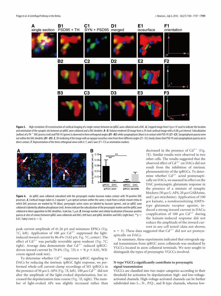
Figure 5. High-resolution 3D reconstruction of confocal imaging of a single contact between an ipRGC axon collateral and a DAC. A, Cropped image from Figure 4F used to indicate the locationand orientation of the synaptic site between an ipRGC axon collateral and a DAC dendrite. B–D, Volume rendered 3D image from a 29 stack confocal image with a 0.08 �m interval. Colocalization(yellow) of a TH � DAC process (red) and PSD-95 (green) is observed in three orthogonal angles (B1–B3) while synaptophysin (blue) is in contact with PSD-95 (C1–C3). Synaptophysin puncta werenot within the DAC dendrite (D1–D3). E, 3D rendering of the image with an opaque isosurface view from three different angles (E1–E3) clearly shows that PSD-95 and synaptophysin puncta are indirect contact. F, Representation of the three orthogonal views with X, Y, and Z axes (F1–F3) as orientation markers.
Figure 6. An ipRGC axon collateral colocalized with the presynaptic marker bassoon makes contact with TH-positive DACprocesses. A, Confocal images taken in 2 separate 1 �m optical sections within the same z-stack from a whole-mount retina inwhich DAC processes are marked by TH (blue), presynaptic active zones are labeled by bassoon (green), and an ipRGC axoncollateral is labeled by alkaline phosphatase (red). Arrows indicate the colocalization of the presynaptic marker and the ipRGC axoncollateral in direct apposition to DAC dendrites. Scale bar, 5 �m. B, Average number and cellular localization of bassoon-positivepuncta at sites of contact between ipRGC axon collaterals and DACs (left bars) and ipRGC dendrites and DACs (right bars). **p 0.01, Tukey’s test (n 3).
Prigge et al. • Centrifugal Pathway in the Retina J. Neurosci., July 6, 2016 • 36(27):7184 –7197 • 7191

voltage-activated channels only comprise T-type channels (Benar-roch, 2010). The calcium channels that mediate Ca2� influx inipRGC axons seem to be distinct from those expressed in ipRGCdendrites and somas. Although light-induced Ca2� influx in iso-lated ipRGCs is mediated primarily by somatic L-type VGCCs(Hartwick et al., 2007), this type of channel does not appear to me-diate glutamate release from retinohypothalamic tract terminalsthrough which ipRGCs send ambient illumination signals to neuraltargets in the brain; instead, neurotransmitter release from ipRGCaxon terminals is mediated by N-, P/Q-, R-, and T-type VGCCs(Mintz et al., 1995; Moldavan et al., 2006).
To determine the relative contribution of each subtype ofVGCC (L-, N-, P/Q-, R-, and T-types) to retrograde synaptictransmission, we used specific antagonists of each channel sub-type. These antagonists do not exert postsynaptic effects onglutamatergic responses; therefore, any effect observed was antic-ipated to be presynaptic (Luebke et al., 1993; Mintz et al., 1995).Of all the VGCC antagonists used, only the N-type blocker�-conotoxin GVIA significantly reduced the ipRGC-mediatedlight-induced inward current of DACs (Fig. 8A). Pooled datafrom rd1 and Cnga3� / � retinas demonstrate that N-type cal-cium channel blockade reduced the DAC inward current by anaverage of 47% (Fig. 8A, n 7; p 0.05, Wilcoxon signed-ranktest). In contrast, other known Ca 2� channel blockers did notaffect ipRGC-driven responses of DACs (Fig. 8B–E). To revealwhether an interaction existed between the VGCCs that couldaccount for the residual current blocked by Cd 2� but not byconotoxin, we used a mixture of all of the VGCC antagonists andretested the ipRGC-mediated DAC light response. The VGCC
blocker mixture contained the following: �-conotoxin GVIA (1�M), nifedipine (10 �M), �-agatoxin IVA (100 nM), SNX 482(150 nM), and mibefradil dihydrochloride (10 �M). On average,the VGCC blocker mixture reduced the peak inward current ofipRGC-driven DAC light responses in rd1 and Cnga3� / � retinasby 46.1% (Fig. 8F, n 6; p 0.05, paired t test). The inhibitoryeffects of the VGCC blocker mixture and conotoxin did not differstatistically (46.1% vs 47%, respectively; p 0.939, t test). Thesedata suggested that the effect of the VGCC blocker mixture wasentirely the result of inhibition of N-type channels by conotoxin.
ipRGCs are involved in light adaptationBecause ipRGCs provide photic input to DACs and dopaminer-gic modulation of the cone pathway is associated with light ad-aptation (Jackson et al., 2012), we next sought to determinewhether the ipRGC circuit is involved in light adaptation. Wemeasured the light-adapted ERG response every 2 min continu-ously for 20 min to assess light-adapted visual function. To ex-clude an effect of circadian modulation on retinal function, allERG recordings were performed on the first day of constant dark-ness (DD) at CT 6. Similar to previous studies (Jackson et al.,2012; Nagaya et al., 2015), in wild-type mice, the b-wave ampli-tude of the light-adapted ERG gradually increased with timeand, after 6 min, was significantly greater than the initiation timepoint (Fig. 9A, top traces, B). However, M1 ipRGC elimination mice(Opn4DTA/�), in which cell loss was confirmed with an antibodyagainst melanopsin (Fig. 9F,G; M1 cells in Opn4DTA/� 54 � 9,control 479 � 13 per retina, n 3, p 0.01, t test), exhibited anunchanged b-wave amplitude even after 20 min of light adaptation
Figure 7. Signal transmission from ipRGCs to DACs is mediated by calcium channels. A, An ipRGC-driven rd1 DAC light response in control extracellular media (2 mM Ca 2�; left) was greatly reducedwhen switched to a low Ca 2� (0.5 mM) extracellular media (center). The response completely recovered when switched back to the control extracellular Ca 2� condition (right). B, Average data showthat low Ca 2� significantly reduced the amplitude of the DAC inward current (Wilcoxon signed-rank test; n 8). C, An rd1 DAC light response (left) was severely attenuated by the application ofCd 2� (100 �M; center). Washout partially restored the response (right). D, Average data show that Cd 2� significantly reduced retrograde signal transmission (Wilcoxon signed-rank test; n 9).Average data are represented as mean � SEM. E, Current-clamp recording from a GFP-labeled M1 ipRGC showing that 100 �M Cd 2� prolonged the plateau phase of the light-induced membranedepolarization and slightly increased the number of light-induced APs. Left trace: control; right trace: Cd 2�. Stimulation bar shows timing of light pulse (3 s; 470 nm; 4.7�10 13 photons cm�2s�1).
7192 • J. Neurosci., July 6, 2016 • 36(27):7184 –7197 Prigge et al. • Centrifugal Pathway in the Retina

(Fig. 9A, traces in second row, D). Furthermore, we repeated theexperiment using Opn4Cre/�;Brn3bZdta/� mice in which mostipRGCs are eliminated, except for M1s that innervate the suprachi-asmatic nucleus and intergeniculate leaflet (Chen et al., 2011). Inthese mice, we observed light-adapted b-wave facilitation that wassimilar to control (Fig. 9A, traces in third row, C), suggesting that M1ipRGCs are sufficient to facilitate light adaptation.
To confirm that the light adaptation phenotype that we ob-served in Opn4DTA/� mice was due to a modulatory effect on thedopaminergic system, we intraperitoneally injected PD168077, aD4 receptor agonist, immediately before the ERG recording. Thisagonist has been shown to restore the light-adapted b-wave am-plitude of mice that lack dopamine in the retina at CT 6 (Jacksonet al., 2012). Furthermore, injection of PD168077 alone did notfurther increase the amplitude of light-adapted b-waves in wild-type mice (two-way ANOVA; Fig. 9A, bottom traces, B). Afterinjection of PD168077, light-adapted b-wave amplitude was re-stored in mice lacking M1 ipRGCs (Fig. 9A, traces in fourth row,D,E). The kinetics of b-wave potentiation in the Opn4DTA/� miceinjected with PD168077 were slower than in the wild-type mice.This could have been a result of the gradual entry of PD168077from the intraperitoneal injection site into the retina. Together,these results demonstrate that retrograde signaling from ipRGCsis involved in light adaptation through modulation of the dopa-minergic system, which facilitates the cone pathway.
DiscussionIn this study, we demonstrated that ipRGCs make anatomicaland physiological connections through their axon collateralswith DACs, forming a centrifugal pathway in the retina. We fur-ther demonstrated that genetic ablation of M1 ipRGCs impairedthe light-adapted ERG in mice and that the impairment could be
restored by a dopamine receptor agonist. The results stronglysuggest that M1 ipRGCs are necessary and sufficient to influencelight adaptation through the centrifugal dopaminergic system.Our findings illustrate a functional role for ipRGCs in vision andelucidate the precise synaptic and circuit mechanisms underlyingipRGC-mediated visual functions.
Route of retrograde signal transmission in the retinaOur data demonstrate that the origin of retrograde signaling ap-pears to be axon collateral-bearing M1 ipRGCs that comprise�10% of all M1 ipRGCs (Joo et al., 2013) and that retrogradesignaling from ipRGCs to DACs is likely routed through in-traretinal axon collaterals. Although axon collaterals have beenreported in a small population of RGCs for decades, it was onlyrecently demonstrated that they are an apparent specializationof ipRGCs (Dacey, 1985; Joo et al., 2013). The axon collateralbranches from the ipRGC’s primary axon before it exits theretina and projects back to the IPL (Joo et al., 2013). Our dataclearly illustrated that the terminals of M1 ipRGC axon collat-erals were colocalized with DAC processes in the outermostIPL, providing a possible site of synaptic contact between thesetwo classes of neurons. We observed that the axon collateralterminals contained the synaptic vesicle protein synaptophy-sin, which was in direct apposition to the postsynapticcomponents revealed by PSD-95 on DAC dendrites. The sig-nificantly higher presynaptic contact between ipRGC axonsand DACs than ipRGC dendrites to DACs indicates that ourobservation was not due to stochastic events. These datastrongly suggest that ipRGC axon collaterals could formaxodendritic synapses with DACs. Although the synapticultrastructure should be further examined with electron mi-croscopy (Pickard and Sollars, 2010), our study is the first to
Figure 8. N-type voltage-gated calcium channels contribute to retrograde signal transmission. A, The N-type VGCC blocker �-conotoxin GVIA (1 �M) significantly suppressed the light-inducedinward currents of rd1 and cnga3 � / � DACs (Wilcoxon signed-rank test; n 7). None of the other antagonists (B: 10 �M nifedipine (L-type), n 5; C: 100 nM �-agatoxin IVA (P/Q-type), n 3;D: 150 nM SNX 482 (R-type), n 3; and E: 10 �M mibefradil (T-type), n 3) significantly reduced signal transmission. F, VGCC blocker mixture containing all of the antagonists (1 �M conotoxin;10 �M nifedipine; 100 nM agatoxin; 150 nM SNX 482, and 10 �M mibefradil) significantly reduced rd1 and cnga3 � / � DAC light-induced responses (paired t test; n 6). Bar charts are average datarepresented as the mean � SEM before and during drug application.
Prigge et al. • Centrifugal Pathway in the Retina J. Neurosci., July 6, 2016 • 36(27):7184 –7197 • 7193

suggest that RGC axon collaterals are presynaptic to upstreamorders of retinal neurons.
Our physiological data directly support the anatomical ev-idence for the retrograde neural pathway in the retina. Axonsare presumed to use APs to transmit synaptic signals. Light-induced responses of ipRGCs have two components: a gradedpotential and APs (Berson et al., 2002). In the presence ofTTX, ipRGC APs were blocked but their graded potentialspersisted. In this condition, the light-induced excitatory in-ward currents of DACs were undetectable, indicating that thegraded potentials of ipRGCs did not generate excitatory cur-rents in DACs. Therefore, we concluded that TTX-sensitiveAPs, not TTX-insensitive graded potentials of ipRGCs, medi-ate retrograde signal transmission. The conclusion derivedfrom our patch-clamp results obtained from isolated retinaswas further validated by our in vivo data: ocular TTX injectionnearly eliminated ipRGC-driven c-Fos expression in DACs,
suggesting that signal transmission from ipRGCs to DA neu-rons is mediated by APs in vivo.
Although our data suggest that APs generated in the ipRGCsoma likely propagate along axon collaterals to mediate retro-grade synaptic transmission to DACs, we cannot completely ruleout other possibilities. First, TTX-sensitive APs initiated in theaxon hillock could back-propagate into the dendritic arbors ofipRGCs, as observed in cortical pyramidal neurons (Stuart andSakmann, 1994), and mediate synaptic transmission via dendro-dendritic synapses with DACs. Second, both dendrites and axonsof ipRGCs express melanopsin and are endogenously photosen-sitive (Berson et al., 2002; Hattar et al., 2002). Melanopsin-basedmembrane depolarization in either of these processes could trig-ger glutamate release locally onto DACs. Although evidence isneeded, these possibilities exist because RGC dendrites containsynaptic vesicles and AMPA receptors of DACs are located inclose proximity to melanopsin-immunopositive dendrites of
Figure 9. M1 ipRGCs modulate the light-adapted ERG b-wave via D4 dopamine receptors. A, Representative light-adapted ERG recording traces after 2 min (black) or 20 min (gray) of lightadaptation in wild-type, Opn4DTA/�, Opn4Cre/�;Brn3bZdta/�, and PD168077-injected Opn4DTA/�, and wild-type mice (from top to bottom). B, Light-adapted ERG b-wave amplitude plotted againstlight adaptation time in wild-type mice (closed circle, n 8). Most of the b-wave amplitudes were significantly higher after 6 min of light adaptation than they were after the first 2 min; the injectionof PD168077 produced similar light adapted b-wave amplitudes as control (open circle, n 5). C, Similar to wild-type, in Opn4-Cre;Brn3b-Zdta mice, all of the b-wave amplitudes were significantlyhigher after 8 min of light adaptation than they were after the first 2 min (n 7). D, B-wave amplitudes from Opn4-DTA mice remained unchanged throughout 20 min of light adaptation (closedsquare, n 8). Ten minuts after intraperitoneal PD168077 injection, the light-adapted ERG b-wave amplitudes were significantly higher than the initial time point from Opn4-DTA mice (opensquare, n 5). E, Paired plot of light-adapted ERG b-wave amplitudes from individual Opn4-DTA mice before and after PD168077 injection after 20 min of light adaptation. F, Whole-mountimmunofluorescence staining with a melanopsin antibody shows a dramatic reduction in the total number of melanopsin-immunopositive ipRGCs in Opn4DTA mice (right) compared with wild-type(left). Only a few weakly stained M1 ipRGCs (arrows), characterized by dendritic stratification in the S1 layer of the IPL, remained in the Opn4-DTA retina (right); an insert (right) displays a magnifiedimage of one such cell (yellow arrow). G, Quantification of melanopsin-immunopositive ipRGCs from WT and Opn4-DTA mice (t test, n 3). For B–D, post hoc Bonferroni’s test compared with 2 minbin. For E, paired t test, n 5. *p 0.05; **p 0.01.
7194 • J. Neurosci., July 6, 2016 • 36(27):7184 –7197 Prigge et al. • Centrifugal Pathway in the Retina

ipRGCs (Sakai et al., 1986; Zhang et al., 2012). Last, ipRGCs couldsignal to DACs polysynaptically through multiple interneurons,although our data ruled out the involvement of ipRGC-coupledspiking amacrine cells (Reifler et al., 2015).
Calcium dependence of presynaptic signal transmissionOur results reveal that signal transmission from ipRGCs to DACsis calcium dependent because low extracellular calcium signifi-cantly reduced the amplitude of the light-induced inward cur-rents of DACs. We postulate that APs generated in an ipRGCpropagate along its axon collateral and depolarize its terminal.This depolarization results in the opening of VGCCs, enabling aninflux of Ca 2� into the terminal. The global Ca 2� channelblocker Cd 2� almost completely blocked the light-induced in-ward currents of DACs. This blockage cannot be attributed to apostsynaptic effect because Cd 2� did not affect the AMPA-receptor-mediated inward current of DACs. Our results furtherreveal that presynaptic Ca 2� influx was partially mediated byN-type VGCCs. It is generally thought that N-type VGCCs areexpressed in axon terminals and are responsible for triggeringneurotransmitter release (Benarroch, 2010), although somestudies showed that they are also expressed on dendrites (Mills etal., 1994; Kisilevsky et al., 2008). Indeed, N-, P/Q-, R-, and T-typechannels are involved in synaptic transmission from ipRGCs toneurons in the suprachiasmatic nucleus, suggesting that ipRGCaxons use these channels for neurotransmitter release (Moldavanet al., 2006). However, we observed that P/Q-, R-, and T-typechannels had no detectable involvement in signal transmissionfrom ipRGCs to DACs. This difference suggests that ipRGC axoncollaterals may not express these subtypes of VGCCs or that theypossess these channels, but they are not involved in synaptictransmission. In addition, L-type VGCCs, which are generallyexpressed in the somas and dendrites of neurons (Hell et al., 1993;Simon et al., 2003), do not appear to mediate synaptic transmis-sion to DACs, further suggesting that ipRGC dendrites are un-likely the conduit for signal transmission. Instead, the datafurther support a model in which AP-driven retrograde signaltransmission partially via conotoxin-sensitive N-type VGCCs,is routed through ipRGC axon collaterals. However, the con-otoxin-resistant transmission that we observed could reflect syn-aptic release triggered by other sources of calcium such as theendoplasmic reticulum.
Potential role for intraretinal retrograde signaling inlight adaptationOur results further demonstrated that retrograde signaling fromM1 ipRGCs to DACs is necessary and sufficient to influence lightadaptation by showing a deficit in the photopic ERG b-waveduring light adaptation in M1 ipRGC elimination mice, but notin non-M1 ipRGC elimination mice. Although the mechanismsresponsible for b-wave light adaptation remain to be determinedfully, cone photoreceptor responses and synaptic plasticity be-tween cones and bipolar cells are likely to play critical roles in thegradual increase of the photopic b-wave during light adaptation(Gouras and MacKay, 1989; Bui and Fortune, 2006; Nagaya et al.,2015). However, facilitation of the photopic b-wave is impairedin retina-specific dopamine depletion mice, suggesting that conesand bipolar cells may modulate the light-adapted b-wave, at leastin part via the dopaminergic system (Jackson et al., 2012). Conescan drive DACs directly through ectopic ON bipolar cell synapsesin the OFF IPL (route 4 in Fig. 10A), which may contribute tob-wave light adaptation through increased levels of retinal dopa-mine (Zhang et al., 2007; Dumitrescu et al., 2009; Hoshi et al.,
2009; Newkirk et al., 2013). Cones may also excite DACs throughipRGCs retrogradely (Zhang et al., 2008; Atkinson et al., 2013;Newkirk et al., 2013) because they can activate ipRGCs throughectopic ON bipolar cell synapses in the OFF IPL (Dumitrescu etal., 2009; Hoshi et al., 2009; route 3 in Fig. 10A). The latter pro-vides an alternative route for cone and bipolar cell contributionto the photopic b-wave through the ipRGC circuit and dopami-nergic system. This route may partially explain why an impairedphotopic b-wave was observed in ipRGC elimination mice.
The impaired photopic b-wave in ipRGC elimination micecould also be the result of a loss of ipRGC network and photore-ceptor adaptation. Like conventional RGCs, ipRGCs undergonetwork adaptation because they receive excitatory input frombipolar cells (Wong et al., 2007; route 3 in Fig. 10A) as well asinhibitory inputs (GABA, glycine, and dopamine) from amacrinecells, including DACs (route 5 in Fig. 10A; Belenky et al., 2003;Viney et al., 2007; Vugler et al., 2007; Wong et al., 2007; VanHook et al., 2012). Unlike regular RGCs, ipRGCs possess thephotopigment melanopsin, which can produce a sustained lightsignal under steady illumination (Wong et al., 2005; Wong, 2012;Do and Yau, 2013). Therefore, ipRGCs can integrate light-adapted cone and melanopsin signals and in turn transmit thesesignals through their axon collaterals to DACs retrogradely(route 1 in Fig. 10A), triggering dopamine release (Dkhissi-Benyahya et al., 2013; Fig. 10B). Increased dopamine release me-diated by ipRGC activity can also act through D4 dopaminereceptors expressed on cones, where they are known to affectcGMP metabolism, gene expression, and rod– cone coupling toregulate cone photoreceptor adaptation (Jackson et al., 2011;route 2 in Fig. 10A); this was demonstrated through the restora-tion of the impaired b-wave with a D4 receptor agonist in ipRGCelimination mice (Fig. 9).
Although cones could signal to DACs through bipolar cells inM1 ipRGC elimination mice (route 4 in Fig. 10A), we failed to
Figure 10. Proposed neural pathways and synaptic mechanisms underlying ipRGC influenceon light adaptation. A, It is proposed that ipRGCs signal to DACs via their axon collaterals toconvey melanopsin-based signals (route 1) from the inner to outer retina (route 2). Rods andcones may use this retrograde signaling pathway as an alternative route to send feedbacksignals through ipRGCs, back to the outer retina (route 3); this route likely complements theprimary feedback pathway in which rod and cone signals are transmitted back to the outerretina via DACs (route 4). Retrograde information transfer from ipRGCs to DACs is reciprocatedby a feedback projection from DACs (route 5). Through this projection, rod and cone signalsare indirectly relayed to ipRGCs via DACs. B, Activation of the ipRGC axon collateral triggersCa 2� influx partially via N-type VGCCs into the collateral terminals, resulting in glutamaterelease onto postsynaptic DACs. Glutamate excites DACs through activation of AMPA-typeglutamate receptors, which depolarizes DACs and increases their AP firing frequency,triggering dopamine release.
Prigge et al. • Centrifugal Pathway in the Retina J. Neurosci., July 6, 2016 • 36(27):7184 –7197 • 7195

detect any light-adapted b-wave using the highest light intensityof our ERG system (Fig. 9D). It appears that cones may requirehigher intensities to mediate light adaptation in the absence ofM1 ipRGCs. Indeed, higher flash intensities can produce greaterlight-adapted b-wave facilitation than the intensity that we usedhere (Jackson et al., 2012). It is worth nothing that the impairedlight-adapted b-wave that we observed without M1 ipRGCs iscontradictory to the previous report showing that selective deple-tion of wavelengths to which melanopsin is most sensitive en-hanced light adaptation (Allen et al., 2014). Future investigationswill need to determine whether reducing melanopsin signalingalone and eliminating the M1 ipRGC circuit may play differentroles in light adaptation of the visual system.
ReferencesAllen AE, Storchi R, Martial FP, Petersen RS, Montemurro MA, Brown TM,
Lucas RJ (2014) Melanopsin-driven light adaptation in mouse vision.Curr Biol 24:2481–2490. CrossRef Medline
Atkinson CL, Feng J, Zhang DQ (2013) Functional integrity and modifica-tion of retinal dopaminergic neurons in the rd1 mutant mouse: roles ofmelanopsin and GABA. J Neurophysiol 109:1589 –1599. CrossRefMedline
Belenky MA, Smeraski CA, Provencio I, Sollars PJ, Pickard GE (2003) Mel-anopsin retinal ganglion cells receive bipolar and amacrine cell synapses.J Comp Neurol 460:380 –393. CrossRef Medline
Benarroch EE (2010) Neuronal voltage-gated calcium channels: brief over-view of their function and clinical implications in neurology. Neurology74:1310 –1315. CrossRef Medline
Berson DM, Dunn FA, Takao M (2002) Phototransduction by retinal gan-glion cells that set the circadian clock. Science 295:1070 –1073. CrossRefMedline
Biel M, Seeliger M, Pfeifer A, Kohler K, Gerstner A, Ludwig A, Jaissle G, FauserS, Zrenner E, Hofmann F (1999) Selective loss of cone function in micelacking the cyclic nucleotide-gated channel CNG3. Proc Natl Acad SciU S A 96:7553–7557. CrossRef Medline
Boynton RM, Whitten DN (1970) Visual adaptation in monkey cones: re-cordings of late receptor potentials. Science 170:1423–1426. CrossRefMedline
Bui BV, Fortune B (2006) Origin of electroretinogram amplitude growthduring light adaptation in pigmented rats. Vis Neurosci 23:155–167.Medline
Bunting M, Bernstein KE, Greer JM, Capecchi MR, Thomas KR (1999) Tar-geting genes for self-excision in the germ line. Genes Dev 13:1524 –1528.CrossRef Medline
Calvert PD, Krasnoperova NV, Lyubarsky AL, Isayama T, Nicolo M, KosarasB, Wong G, Gannon KS, Margolskee RF, Sidman RL, Pugh EN Jr, MakinoCL, Lem J (2000) Phototransduction in transgenic mice after targeteddeletion of the rod transducin alpha-subunit. Proc Natl Acad Sci U S A97:13913–13918. CrossRef Medline
Carter-Dawson LD, LaVail MM, Sidman RL (1978) Differential effect of therd mutation on rods and cones in the mouse retina. Invest OphthalmolVis Sci 17:489 – 498. Medline
Chang B, Dacey MS, Hawes NL, Hitchcock PF, Milam AH, Atmaca-SonmezP, Nusinowitz S, Heckenlively JR (2006) Cone photoreceptor functionloss-3, a novel mouse model of achromatopsia due to a mutation inGnat2. Invest Ophthalmol Vis Sci 47:5017–5021. CrossRef Medline
Chen SK, Badea TC, Hattar S (2011) Photoentrainment and pupillary lightreflex are mediated by distinct populations of ipRGCs. Nature 476:92–95.CrossRef Medline
Dacey DM (1985) Wide-spreading terminal axons in the inner plexiformlayer of the cat’s retina: evidence for intrinsic axon collaterals of ganglioncells. J Comp Neurol 242:247–262. CrossRef Medline
Dacey DM (1988) Dopamine-accumulating retinal neurons revealed by invitro fluorescence display a unique morphology. Science 240:1196 –1198.CrossRef Medline
Dkhissi-Benyahya O, Coutanson C, Knoblauch K, Lahouaoui H, Leviel V,Rey C, Bennis M, Cooper HM (2013) The absence of melanopsin altersretinal clock function and dopamine regulation by light. Cell Mol Life Sci70:3435–3447. CrossRef Medline
Do MT, Yau KW (2013) Adaptation to steady light by intrinsically photo-
sensitive retinal ganglion cells. Proc Natl Acad Sci U S A 110:7470 –7475.CrossRef Medline
Dowling JE, Ehinger B (1975) Synaptic organization of the amine-containing interplexiform cells of the goldfish and Cebus monkey retinas.Science 188:270 –273. CrossRef Medline
Dumitrescu ON, Pucci FG, Wong KY, Berson DM (2009) Ectopic retinalON bipolar cell synapses in the OFF inner plexiform layer: contacts withdopaminergic amacrine cells and melanopsin ganglion cells. J Comp Neu-rol 517:226 –244. CrossRef Medline
Dunn FA, Doan T, Sampath AP, Rieke F (2006) Controlling the gain ofrod-mediated signals in the Mammalian retina. J Neurosci 26:3959 –3970.CrossRef Medline
Ecker JL, Dumitrescu ON, Wong KY, Alam NM, Chen SK, LeGates T, RennaJM, Prusky GT, Berson DM, Hattar S (2010) Melanopsin-expressingretinal ganglion-cell photoreceptors: cellular diversity and role in patternvision. Neuron 67:49 – 60. CrossRef Medline
Gouras P, MacKay CJ (1989) Growth in amplitude of the human cone elec-troretinogram with light adaptation. Invest Ophthalmol Vis Sci 30:625– 630. Medline
Green DG, Dowling JE, Siegel IM, Ripps H (1975) Retinal mechanisms ofvisual adaptation in the skate. J Gen Physiol 65:483–502. CrossRefMedline
Guler AD, Ecker JL, Lall GS, Haq S, Altimus CM, Liao HW, Barnard AR,Cahill H, Badea TC, Zhao H, Hankins MW, Berson DM, Lucas RJ, YauKW, Hattar S (2008) Melanopsin cells are the principal conduits forrod-cone input to non-image-forming vision. Nature 453:102–105.CrossRef Medline
Hartwick AT, Bramley JR, Yu J, Stevens KT, Allen CN, Baldridge WH, Sollars PJ,Pickard GE (2007) Light-evoked calcium responses of isolated melanopsin-expressing retinal ganglion cells. J Neurosci 27:13468–13480. CrossRefMedline
Hattar S, Liao HW, Takao M, Berson DM, Yau KW (2002) Melanopsin-containing retinal ganglion cells: architecture, projections, and intrinsicphotosensitivity. Science 295:1065–1070. CrossRef Medline
Hattar S, Lucas RJ, Mrosovsky N, Thompson S, Douglas RH, Hankins MW,Lem J, Biel M, Hofmann F, Foster RG, Yau KW (2003) Melanopsin androd-cone photoreceptive systems account for all major accessory visualfunctions in mice. Nature 424:76 – 81. Medline
Hayashida Y, Ishida AT (2004) Dopamine receptor activation can reducevoltage-gated Na� current by modulating both entry into and recoveryfrom inactivation. J Neurophysiol 92:3134 –3141. CrossRef Medline
Hell JW, Westenbroek RE, Warner C, Ahlijanian MK, Prystay W, GilbertMM, Snutch TP, Catterall WA (1993) Identification and differentialsubcellular localization of the neuronal class C and class D L-type calciumchannel alpha 1 subunits. J Cell Biol 123:949 –962. CrossRef Medline
Henny P, Jones BE (2006) Innervation of orexin/hypocretin neurons byGABAergic, glutamatergic or cholinergic basal forebrain terminals evi-denced by immunostaining for presynaptic vesicular transporter andpostsynaptic scaffolding proteins. J Comp Neurol 499:645– 661. CrossRefMedline
Hoshi H, Liu WL, Massey SC, Mills SL (2009) ON inputs to the OFF layer:bipolar cells that break the stratification rules of the retina. J Neurosci29:8875– 8883. CrossRef Medline
Hunt SP, Pini A, Evan G (1987) Induction of c-fos-like protein in spinalcord neurons following sensory stimulation. Nature 328:632– 634.CrossRef Medline
Ichinose T, Lukasiewicz PD (2007) Ambient light regulates sodium channelactivity to dynamically control retinal signaling. J Neurosci 27:4756 –4764. CrossRef Medline
Jackson CR, Chaurasia SS, Hwang CK, Iuvone PM (2011) Dopamine D(4)receptor activation controls circadian timing of the adenylyl cyclase 1/cyclic AMP signaling system in mouse retina. Eur J Neurosci 34:57– 64.CrossRef Medline
Jackson CR, Ruan GX, Aseem F, Abey J, Gamble K, Stanwood G, Palmiter RD,Iuvone PM, McMahon DG (2012) Retinal dopamine mediates multipledimensions of light-adapted vision. J Neurosci 32:9359 –9368. CrossRefMedline
Jekely G, Arendt D (2007) Cellular resolution expression profiling usingconfocal detection of NBT/BCIP precipitate by reflection microscopy.Biotechniques 42:751–755. CrossRef Medline
Joo HR, Peterson BB, Dacey DM, Hattar S, Chen SK (2013) Recurrent axon
7196 • J. Neurosci., July 6, 2016 • 36(27):7184 –7197 Prigge et al. • Centrifugal Pathway in the Retina

collaterals of intrinsically photosensitive retinal ganglion cells. Vis Neu-rosci 30:175–182. CrossRef Medline
Kisilevsky AE, Mulligan SJ, Altier C, Iftinca MC, Varela D, Tai C, Chen L,Hameed S, Hamid J, Macvicar BA, Zamponi GW (2008) D1 receptorsphysically interact with N-type calcium channels to regulate channel dis-tribution and dendritic calcium entry. Neuron 58:557–570. CrossRefMedline
Knapp AG, Dowling JE (1987) Dopamine enhances excitatory amino acid-gated conductances in cultured retinal horizontal cells. Nature 325:437– 439. CrossRef Medline
Krizaj D, Gabriel R, Owen WG, Witkovsky P (1998) Dopamine D2receptor-mediated modulation of rod-cone coupling in the Xenopus ret-ina. J Comp Neurol 398:529 –538. Medline
Lasater EM, Dowling JE (1985) Dopamine decreases conductance of theelectrical junctions between cultured retinal horizontal cells. Proc NatlAcad Sci U S A 82:3025–3029. CrossRef Medline
Luebke JI, Dunlap K, Turner TJ (1993) Multiple calcium channel types con-trol glutamatergic synaptic transmission in the hippocampus. Neuron11:895–902. CrossRef Medline
Maxwell IH, Maxwell F, Glode LM (1986) Regulated expression of a diph-theria toxin A-chain gene transfected into human cells: possible strategyfor inducing cancer cell suicide. Cancer Res 46:4660 – 4664. Medline
Mills LR, Niesen CE, So AP, Carlen PL, Spigelman I, Jones OT (1994)N-type Ca2� channels are located on somata, dendrites, and a subpopu-lation of dendritic spines on live hippocampal pyramidal neurons. J Neu-rosci 14:6815– 6824. Medline
Mills SL, Massey SC (1995) Differential properties of two gap junctionalpathways made by AII amacrine cells. Nature 377:734 –737. CrossRefMedline
Mills SL, Xia XB, Hoshi H, Firth SI, Rice ME, Frishman LJ, Marshak DW(2007) Dopaminergic modulation of tracer coupling in a ganglion-amacrine cell network. Vis Neurosci 24:593– 608. Medline
Mintz IM, Sabatini BL, Regehr WG (1995) Calcium control of transmitterrelease at a cerebellar synapse. Neuron 15:675– 688. CrossRef Medline
Moldavan MG, Irwin RP, Allen CN (2006) Presynaptic GABA(B) receptorsregulate retinohypothalamic tract synaptic transmission by inhibitingvoltage-gated Ca2� channels. J Neurophysiol 95:3727–3741. CrossRefMedline
Muller LP, Do MT, Yau KW, He S, Baldridge WH (2010) Tracer coupling ofintrinsically photosensitive retinal ganglion cells to amacrine cells in themouse retina. J Comp Neurol 518:4813– 4824. CrossRef Medline
Nagaya M, Ueno S, Kominami T, Nakanishi A, Koyasu T, Kondo M, Furu-kawa T, Terasaki H (2015) Pikachurin protein required for increase ofcone electroretinogram B-wave during light adaptation. PLoS One 10:e0128921. CrossRef Medline
Nakatani K, Tamura T, Yau KW (1991) Light adaptation in retinal rods ofthe rabbit and two other nonprimate mammals. J Gen Physiol 97:413– 435. CrossRef Medline
Newkirk GS, Hoon M, Wong RO, Detwiler PB (2013) Inhibitory inputstune the light response properties of dopaminergic amacrine cells inmouse retina. J Neurophysiol 110:536 –552. CrossRef Medline
Panda S, Provencio I, Tu DC, Pires SS, Rollag MD, Castrucci AM, PletcherMT, Sato TK, Wiltshire T, Andahazy M, Kay SA, Van Gelder RN, Hogen-esch JB (2003) Melanopsin is required for non-image-forming photicresponses in blind mice. Science 301:525–527. CrossRef Medline
Peterson BB, Dacey DM (1998) Morphology of human retinal ganglion cellswith intraretinal axon collaterals. Vis Neurosci 15:377–387. Medline
Pickard GE, Sollars PJ (2010) Intrinsically photosensitive retinal ganglioncells. Sci China Life Sci 53:58 – 67. CrossRef Medline
Reifler AN, Chervenak AP, Dolikian ME, Benenati BA, Li BY, Wachter RD,Lynch AM, Demertzis ZD, Meyers BS, Abufarha FS, Jaeckel ER, Flannery
MP, Wong KY (2015) All spiking, sustained ON displaced amacrinecells receive gap-junction input from melanopsin ganglion cells. Curr Biol25:2763–2773. CrossRef Medline
Sakai HM, Naka K, Dowling JE (1986) Ganglion cell dendrites are presyn-aptic in catfish retina. Nature 319:495– 497. CrossRef Medline
Schmidt TM, Taniguchi K, Kofuji P (2008) Intrinsic and extrinsic light re-sponses in melanopsin-expressing ganglion cells during mouse develop-ment. J Neurophysiol 100:371–384. CrossRef Medline
Simon M, Perrier JF, Hounsgaard J (2003) Subcellular distribution ofL-type Ca2� channels responsible for plateau potentials in motoneuronsfrom the lumbar spinal cord of the turtle. Eur J Neurosci 18:258 –266.CrossRef Medline
Slaughter MM, Miller RF (1981) 2-amino-4-phosphonobutyric acid: a newpharmacological tool for retina research. Science 211:182–185. CrossRefMedline
Stuart GJ, Sakmann B (1994) Active propagation of somatic action poten-tials into neocortical pyramidal cell dendrites. Nature 367:69 –72.CrossRef Medline
Tu DC, Zhang D, Demas J, Slutsky EB, Provencio I, Holy TE, Van Gelder RN(2005) Physiologic diversity and development of intrinsically photosen-sitive retinal ganglion cells. Neuron 48:987–999. CrossRef Medline
Van Hook MJ, Wong KY, Berson DM (2012) Dopaminergic modulation ofganglion-cell photoreceptors in rat. Eur J Neurosci 35:507–518. CrossRefMedline
Viney TJ, Balint K, Hillier D, Siegert S, Boldogkoi Z, Enquist LW, Meister M,Cepko CL, Roska B (2007) Local retinal circuits of melanopsin-containing ganglion cells identified by transsynaptic viral tracing. CurrBiol 17:981–988. CrossRef Medline
Vugler AA, Redgrave P, Semo M, Lawrence J, Greenwood J, Coffey PJ (2007)Dopamine neurones form a discrete plexus with melanopsin cells in nor-mal and degenerating retina. Exp Neurol 205:26 –35. CrossRef Medline
Warren EJ, Allen CN, Brown RL, Robinson DW (2006) The light-activatedsignaling pathway in SCN-projecting rat retinal ganglion cells. Eur J Neu-rosci 23:2477–2487. CrossRef Medline
Witkovsky P (2004) Dopamine and retinal function. Doc Ophthalmol 108:17– 40. CrossRef Medline
Wong KY (2012) A retinal ganglion cell that can signal irradiance continu-ously for 10 hours. J Neurosci 32:11478 –11485. CrossRef Medline
Wong KY, Dunn FA, Berson DM (2005) Photoreceptor adaptation in in-trinsically photosensitive retinal ganglion cells. Neuron 48:1001–1010.CrossRef Medline
Wong KY, Dunn FA, Graham DM, Berson DM (2007) Synaptic influenceson rat ganglion-cell photoreceptors. J Physiol 582:279 –296. CrossRefMedline
Xue T, Do MT, Riccio A, Jiang Z, Hsieh J, Wang HC, Merbs SL, Welsbie DS,Yoshioka T, Weissgerber P, Stolz S, Flockerzi V, Freichel M, Simon MI,Clapham DE, Yau KW (2011) Melanopsin signalling in mammalian irisand retina. Nature 479:67–73. CrossRef Medline
Zhang DQ, Stone JF, Zhou T, Ohta H, McMahon DG (2004) Characteriza-tion of genetically labeled catecholamine neurons in the mouse retina.Neuroreport 15:1761–1765. CrossRef Medline
Zhang DQ, Zhou TR, McMahon DG (2007) Functional heterogeneity ofretinal dopaminergic neurons underlying their multiple roles in vision.J Neurosci 27:692– 699. CrossRef Medline
Zhang DQ, Wong KY, Sollars PJ, Berson DM, Pickard GE, McMahon DG(2008) Intraretinal signaling by ganglion cell photoreceptors to dopami-nergic amacrine neurons. Proc Natl Acad Sci U S A 105:14181–14186.CrossRef Medline
Zhang DQ, Belenky MA, Sollars PJ, Pickard GE, McMahon DG (2012) Mel-anopsin mediates retrograde visual signaling in the retina. PLoS One7:e42647. CrossRef Medline
Prigge et al. • Centrifugal Pathway in the Retina J. Neurosci., July 6, 2016 • 36(27):7184 –7197 • 7197