madhavi raghu md assistant professor of radiology yale
TRANSCRIPT

Madhavi Raghu MD
Assistant Professor of Radiology
Yale School of Medicine

• None

• Routine 2-D mammography has “structured noise” due to overlapping tissues
• Cancers can be obscured– 20% breast cancers are not detected
by conventional mammography
– The sensitivity of mammography decreases with increasing breast density
• False positives caused by normal superimposed tissues

• DBT is a pseudo-3D breast imaging tool that is gaining widespread adoption
– It has been shown to improve on the limitations of 2D mammography
• Particularly improving the detection of lesions in dense breast tissue.
• FDA approved Feb 2011 and in practice at Yale since August 2011
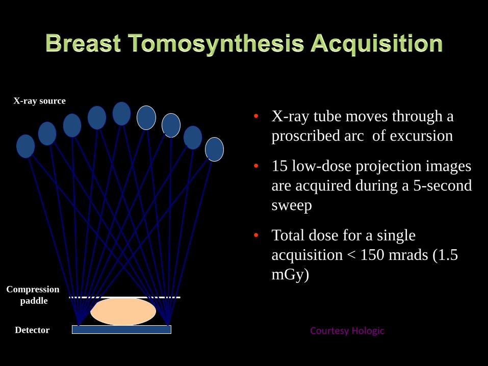
Compression
paddle
• X-ray tube moves through a
proscribed arc of excursion
• 15 low-dose projection images
are acquired during a 5-second
sweep
• Total dose for a single
acquisition < 150 mrads (1.5
mGy)
Detector
X-ray source
Courtesy Hologic

• With tomosynthesis:
– Recalls decreased by 40%
– Increased cancer detection rate at 40%
– Increased detection of invasive cancers: 21%

Recall Rates CDR
Author/year 2D 2D+DBT % change 2D 2D+DBT % change
Ciatto, 2013 5.5 3.5 -17 5.3 8.1 53
Skaane, 2013 6.1 5.3 -15 6.1 8.0 27
Haas, 2013 12 8.4 -30 5.2 5.7 10
Rose, 2013 8.7 5.5 -37 4.0 5.4 35
Friedewald, 2014 10.7 9.1 -15 4.2 5.4 29
McCarthy, 2014 10.4 8.8 -15 4.6 5.5 20
Greenberg, 2014 16.2 13.6 -16 4.9 6.3 29
Durand, 2015 12.3 7.8 -37 5.7 5.9 4
Lourenco, 2015 9.3 6.4 -31 5.4 4.6 -17
Sharpe, 2016 7.5 6.1 -19 3.5 5.4 54

• Decreases the radiation dose
• Preliminary studies demonstrate equivalence with 2D FFDM
2D FFDM 2D+DBT s2D+DBT


• Aujero et al. (2017) Radiology
2D FFDM 2D+DBT s2D+DBT
Recall Rates 8.7% 5.8% 4.3%
Cancer Detection Rate
5.3/1000 6.4/1000 6.1/1000

Tomosynthesis Reconstruction Slices

2D Mammography Tomosynthesis
• Spot compression
• Magnification
• True lateral
• Exaggerated CC
• Rolled CC
• Step Oblique
• Tangential
• Magnification
(for Ca++)
• Spot compression
(fewer)

• Total number of exams performed increased over
time
• % US exams stayed relatively stable
• % diagnostic exams decreased
• % screening exams increased
Screenings Diagnostics US Total
2D: 9/1/10-8/30/11 4914 (34%) 5086 (35%) 4392 (31%) 14413
3D2: 10/1/12-9/30/13 5726 (34%) 5387 (32%) 5711 (34%) 16906
3D3: 10/1/13-9/30/14 5764 (34%) 5048 (30%) 6001 (37%) 16813
3D4: 10/1/14-9/30/15 6710 (41%) 3805 (23%) 5900 (36%) 16415
3D5: 10/1/15-9/30/16 8724 (45%) 4200 (21%) 6622 (34%) 19546
Butler RB, Raghu M et al, RSNA 2016

0.00%
10.00%
20.00%
30.00%
40.00%
50.00%
60.00%
2010-2011 2011-2012 2012-2013 2013-2014 2014-2015 2015-2016
5%8%
12%
21%
39%
52%

2D
Tomo spots
Recalled from a 2D mammogram
USCNB: IDC; ER+, PR+, HER-2+Stage: IB


2D 3D
55 year old woman presents for routine screening

Spot compression views with tomosynthesis and targeted ultrasound was performed
US guided CNBx: Invasive Lobular Carcinoma
DBT: CC spot DBT: ML spot

• Supplemental cancer detection rates: 3.0-4.3/1000
• ACRIN 6666: False positive rate of WBUS was 8.1% (4.4% for mammography– Short term follow up was 8.6% versus 2.2% for
mammography
• Utility of WBUS with DBT:– May be reduced
– If screening is done with DBT and MRI• No incremental benefit with US

• Recommend annual MRI (age 25-30)– Genetics based increased risk
– History of chest radiation
– Calculated lifetime risk of 20% or more
• Recommend annual MRI:– Women with history of breast cancer and dense
breasts
– Diagnosed before 50
• May consider for history of atypia

• Cancer detection rate: 17/1000
• Median size of invasive: 1 cm; 88% node neg
• Sensitivity: 81%; Specificity: 83%

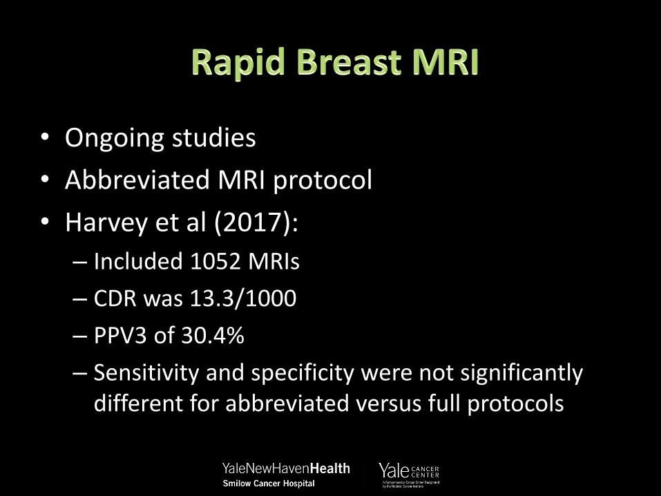
• Ongoing studies
• Abbreviated MRI protocol
• Harvey et al (2017):
– Included 1052 MRIs
– CDR was 13.3/1000
– PPV3 of 30.4%
– Sensitivity and specificity were not significantly different for abbreviated versus full protocols

• Technology evolving
• Applications of the current technology is also changing
• Role of AI may a player in the future
