maggot therapy: the science and implication for cam...
TRANSCRIPT

Advance Access Publication 8 June 2006 eCAM 2006;3(3)303–308
doi:10.1093/ecam/nel022
Review
Maggot Therapy: The Science and Implication for CAMPart II—Maggots Combat Infection
Yamni Nigam1, Alyson Bexfield1,2, Stephen Thomas3 and Norman Arthur Ratcliffe2
1School of Health Science, University of Wales Swansea, Singleton Park, Swansea SA2 8PP, UK,2Department of Biological Sciences, University of Wales Swansea, Singleton Park, Swansea SA2 8PP, UK and3Biosurgical Research Unit (SMTL), Princess of Wales Hospital, Coity Road, Bridgend CF31 1RQ, UK
Maggot therapy employs the use of freshly emerged, sterile larvae of the common green-bottle fly,
Phaenicia (Lucilia) sericata, and is a form of artificially induced myiasis in a controlled clinical
situation. Maggot therapy has the following three core beneficial effects on a wound: debridement,
disinfection and enhanced healing. In part II of this review article, we discuss clinical infections and
the evidence supporting the potent antibacterial action of maggot secretions. Enhancement of wound
healing by maggots is discussed along with the future of this highly successful, often controversial,
alternative treatment.
Keywords: maggot debridement therapy – MRSA – antimicrobial – Lucilia sericata – wounds
Introduction II—The Rebirth ofMaggot Therapy
The recent resurgence and reintroduction of maggot therapy
stems from the steep rise in the emergence of antibiotic-
resistant strains of bacteria, and the need for an effective non-
surgical method of wound debridement. This was begun in
the late 1980s and early 1990s, mainly for treating untreatable
wounds in California (1–3), and was closely followed by
increased use in the UK, Israel and Europe. Currently, several
specialist laboratories produce maggots for clinical and
research purposes. Maggots are supplied to clinical centers
worldwide for the treatment of chronic wounds, such as leg
ulcers, pressure sores, diabetic and necrotic ulcers, as well as
infected surgical wounds, burns and trauma injuries. In the
last decade, thousands of patients all over the world have
had their wounds treated with maggots, so maggot therapy
is well and truly recognized by many clinicians as an impor-
tant adjunct to conventional medicine. In Part II of this
review, we consider how maggots may kill microbial agents
infecting wounds and attempts to isolate antibacterial factors,
including anti-methicillin resistant Staphylococcus aureus
(anti-MRSA) agents, from maggot secretions. Finally, the
role of maggots in stimulating the wound-healing process is
described as are the limitations in the use of maggots in the
clinical situation.
How do Maggots CombatClinical Infections?
How maggots combat clinical infection in wounds has been
studied intensely over the years. Several mechanisms have
been suggested, including simple mechanical irrigation of
the wound by increased exudate, the production of which is
stimulated by larvae ingesting liquefied necrotic tissue, or by
dilution of wound discharge following wound lavage by the
maggots’ own secretions/excretions (4,5). The excretion of
a waste product, ammonia, by Phaenicia sericata was also
believed to be responsible for combating bacterial infections,
since ammonia increases wound pH, resulting in alkaline con-
ditions unfavorable to many bacterial species (6,7). In addi-
tion, larvae of P. sericata carry in their midgut a commensal,
Proteus mirabilis. These commensals produce agents such
as phenylacetic acid (PAA) and phenylacetaldehyde (PAL),
with known antibacterial properties (8). While P. mirabilis
For reprints and all correspondence: Yamni Nigam, University of WalesSwansea, Singleton Park, Swansea SA2 8PP, UK. E-mail:[email protected]
� 2006 The Author(s).This is an Open Access article distributed under the terms of the Creative Commons Attribution Non-Commercial License (http://creative commons.org/licenses/by-nc/2.0/uk/) which permits unrestricted non-commerical use, distribution, and reproduction in any medium, provided the original work is properly cited.
is an endosymbiont of screw worms and several blow flies, and
may contribute to controlling other intestinal and wound
flora (9), the participation of these chemicals in combating
wound infections is unlikely since aseptically raised, sterile
larvae are applied to wounds. In addition, the pH of maggot
secretions is known to be between 8–8.5 (9,10), and Erdmann
(9) revealed that at alkaline pH the antibacterial potential
of PAA is low, while PAL is unstable and therefore limited
as a bactericide. A more likely explanation of how maggots
combat wound infection is that larvae ingest wound bacteria,
which are killed as they pass through the maggot’s digestive
tract. Such destruction of ingested microbes was reported
by Robinson and Norwood (11), who noted that while the
stomach and crop were heavily contaminated with viable
bacteria the hindgut was sterile. This was later confirmed by
Mumcuoglu et al. (12), who followed the fate of ingested
fluorescent Escherichia coli in the alimentary tract of
P. sericata using confocal microscopy.
Maggots Secret Potent Bactericide
In 1935, Simmons led a study of the antibacterial activity
of elimination products from living maggots, revealing the
presence of a potent bactericide present in maggot secre-
tions (13,14). The sterility of the hindgut led Simmons to
believe that this antibacterial activity would be present in
substances excreted with fecal matter. For his study, Simmons
collected externalized secretions from 3-day-old, non-
sterile, P. sericata, reared on decaying beef. After collection,
the material was autoclaved; the thermal stability of the
antibacterial substance permitted the use of non-sterile
larvae. Incidentally, Simmons had tried to work with sterile
larvae, but found excretions from aseptically raised larvae
were much less potent than those collected from non-sterile
maggots. When his autoclaved excretions were tested on
S. aureus, he reported that bacteria exposed to excretions
in vitro for 5–10 min failed to produce colonies on agar
plates, even after a 7 day incubation period, indicating that
the excretions exhibited a strong and rapid disinfection
action (13,14).
In addition to S. aureus, Simmons (13,14) showed in vitro
antibacterial activity against Streptococcus pyogenes,
Streptococcus faecalis, Streptococcus mitor, Clostridium
perfringens, Proteus vulgaris and Eberthella (Salmonella)
typhi. In 1957, Pavillard and Wright (15) confirmed the
presence of antibacterial agents in heat-sterilized maggot
excretions from the blowfly Phormia terraenavae, which
were active against S. aureus, C. perfringens and species of
Streptococcus. In this study, the active fraction was isolated
using paper chromatography, and relatively pure samples
of the antibacterial fraction obtained by using a cellulose
column. The exact nature of this active factor was never
identified, although injections of the fraction into mice pro-
tected them against subsequent intraperitoneal inoculation
with type 1 Pneumococci (15).
More Advanced Technology AnalyzesMethicillin Resistant S. aureus
More recently, using high-performance liquid chromatography,
an antibacterial agent from maggots was partially purified
using Micrococcus luteus as the indicator bacteria (16). This
factor, reported to possess a molecular weight of 6000 Da,
was digested by proteases, caused efflux of potassium ions
from bacterial cells, and exhibited a wide spectrum of anti-
bacterial activity against many resident pathogenic strains
including MRSA. In addition, Thomas et al. (10) conducted
the first study using secretions from aseptically raised larvae
whose immunological profile was unaffected by microbial
contact. In this study, activity was demonstrated against
S. aureus, Pseudomonas aeruginosa, Streptococcus group A
and group B, and a clinical isolate of MRSA. Following the
work of Thomas et al. (10) our own team has further investig-
ated this activity. The choice of antibacterial assay proved vital
in the detection of microbial killing. Bexfield et al. (17)
showed that the use of standard antibacterial assays, such as
the zone of inhibition assay, were ineffective when using mag-
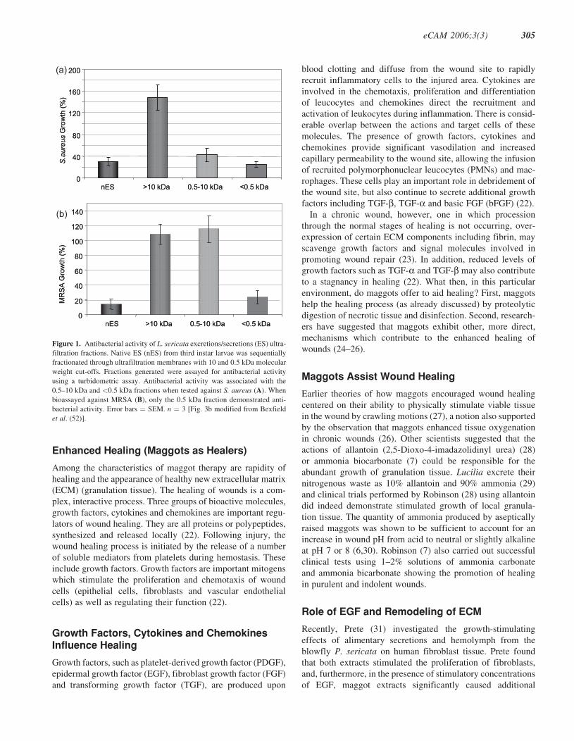
got excretions. Separation of maggot excretions into three
molecular mass fractions (>10 kDa, 0.5–10 kDa and <500
Da) using ultrafiltration revealed the presence of two discrete
antibacterial moieties (17). Significant antibacterial activity
of the 0.5–10 kDa and the <500 Da ultrafiltration fractions
was demonstrated against S. aureus using a turbidometric
assay, whilst the >10 kDa fraction was free of antibacterial
activity (Fig. 1A).
Specificity of the Antibacterial <500 DaFraction and Clinical Implication
When assayed against MRSA, however, antibacterial activity
was detected only within the <500 Da fraction (Fig. 1B), sug-
gesting that this fraction contains the most potent antibacterial
component within maggot secretions/excretions. The identity
of the <500 Da is currently under investigation. Further
analysis of the 0.5–10 kDa fraction indicated that it contains
a heat-stable, protease-sensitive antibacterial molecule of
0.5–3 kDa in size, possibly an antibacterial peptide. Maggots
therefore have a repertoire of externalized defenses against
microbes that we are only just identifying, and whose presence
supports clinical findings and the continued use of maggots.
Isolation, identification and synthesis of maggot-derived
antibacterial compounds may also allow the benefits of
maggot therapy to be applied to wider clinical use, such as
in the treatment of systemic infections. In addition, many
Dipteran species, such as Phormia terranovae (18) are
known to express antimicrobial peptides (AMPs). AMPs, for
example, defensin (18), are small, heat-stable, usually cationic,
peptides induced in response to invading pathogens and
tissue damage (19,20). Defensins are not only involved dir-
ectly in antimicrobial immunity but may also have a role in
wound healing through, for example, cytokine regulation and
enhanced phagocytosis (21).
304 Maggot therapy: Part II
Enhanced Healing (Maggots as Healers)
Among the characteristics of maggot therapy are rapidity of
healing and the appearance of healthy new extracellular matrix
(ECM) (granulation tissue). The healing of wounds is a com-
plex, interactive process. Three groups of bioactive molecules,
growth factors, cytokines and chemokines are important regu-
lators of wound healing. They are all proteins or polypeptides,
synthesized and released locally (22). Following injury, the
wound healing process is initiated by the release of a number
of soluble mediators from platelets during hemostasis. These
include growth factors. Growth factors are important mitogens
which stimulate the proliferation and chemotaxis of wound
cells (epithelial cells, fibroblasts and vascular endothelial
cells) as well as regulating their function (22).
Growth Factors, Cytokines and ChemokinesInfluence Healing
Growth factors, such as platelet-derived growth factor (PDGF),
epidermal growth factor (EGF), fibroblast growth factor (FGF)
and transforming growth factor (TGF), are produced upon
blood clotting and diffuse from the wound site to rapidly
recruit inflammatory cells to the injured area. Cytokines are
involved in the chemotaxis, proliferation and differentiation
of leucocytes and chemokines direct the recruitment and
activation of leukocytes during inflammation. There is consid-
erable overlap between the actions and target cells of these
molecules. The presence of growth factors, cytokines and
chemokines provide significant vasodilation and increased
capillary permeability to the wound site, allowing the infusion
of recruited polymorphonuclear leucocytes (PMNs) and mac-
rophages. These cells play an important role in debridement of
the wound site, but also continue to secrete additional growth
factors including TGF-b, TGF-a and basic FGF (bFGF) (22).
In a chronic wound, however, one in which procession
through the normal stages of healing is not occurring, over-
expression of certain ECM components including fibrin, may
scavenge growth factors and signal molecules involved in
promoting wound repair (23). In addition, reduced levels of
growth factors such as TGF-a and TGF-b may also contribute
to a stagnancy in healing (22). What then, in this particular
environment, do maggots offer to aid healing? First, maggots
help the healing process (as already discussed) by proteolytic
digestion of necrotic tissue and disinfection. Second, research-
ers have suggested that maggots exhibit other, more direct,
mechanisms which contribute to the enhanced healing of
wounds (24–26).
Maggots Assist Wound Healing
Earlier theories of how maggots encouraged wound healing
centered on their ability to physically stimulate viable tissue
in the wound by crawling motions (27), a notion also supported
by the observation that maggots enhanced tissue oxygenation
in chronic wounds (26). Other scientists suggested that the
actions of allantoin (2,5-Dioxo-4-imadazolidinyl urea) (28)
or ammonia biocarbonate (7) could be responsible for the
abundant growth of granulation tissue. Lucilia excrete their
nitrogenous waste as 10% allantoin and 90% ammonia (29)
and clinical trials performed by Robinson (28) using allantoin
did indeed demonstrate stimulated growth of local granula-
tion tissue. The quantity of ammonia produced by aseptically
raised maggots was shown to be sufficient to account for an
increase in wound pH from acid to neutral or slightly alkaline
at pH 7 or 8 (6,30). Robinson (7) also carried out successful
clinical tests using 1–2% solutions of ammonia carbonate
and ammonia bicarbonate showing the promotion of healing
in purulent and indolent wounds.
Role of EGF and Remodeling of ECM
Recently, Prete (31) investigated the growth-stimulating
effects of alimentary secretions and hemolymph from the
blowfly P. sericata on human fibroblast tissue. Prete found
that both extracts stimulated the proliferation of fibroblasts,
and, furthermore, in the presence of stimulatory concentrations
of EGF, maggot extracts significantly caused additional
0
20
40
60
80
100
120
140
nES >10 kDa 0.5-10 kDa <0.5 kDa
MR
SA
Gro
wth
(%
)
0
20
40
60
80
100
120
140
nES >10 kDa 0.5-10 kDa <0.5 kDa
MR
SA
Gro
wth
(%
)
(b)
0
40
80
120
160
200
nES >10 kDa 0.5-10 kDa <0.5 kDa
S.aureus
Gro
wth
(%
)
0
40
80
120
160
200
nES >10 kDa 0.5-10 kDa <0.5 kDa
S.aureus
Gro
wth
(%
)
(a)
Figure 1. Antibacterial activity of L. sericata excretions/secretions (ES) ultra-
filtration fractions. Native ES (nES) from third instar larvae was sequentially
fractionated through ultrafiltration membranes with 10 and 0.5 kDa molecular
weight cut-offs. Fractions generated were assayed for antibacterial activity
using a turbidometric assay. Antibacterial activity was associated with the
0.5–10 kDa and <0.5 kDa fractions when tested against S. aureus (A). When
bioassayed against MRSA (B), only the 0.5 kDa fraction demonstrated anti-
bacterial activity. Error bars ¼ SEM. n ¼ 3 [Fig. 3b modified from Bexfield
et al. (52)].
eCAM 2006;3(3) 305
fibroblast growth. This led to the suggestion that maggot
extracts may either operate to stimulate fibroplasia through a
different mechanism to that of EGF, or may have a synergistic
effect (31).
Other researchers suggest that since fibroblast proliferation
is only one aspect of granulation tissue formation, it seems
likely that additional mechanisms may be involved. Chambers
et al. (32) claim that when maggots are introduced into nec-
rotic wounds, they potentially influence wound healing events
with a combination of excretory/secretory (ES) proteases
which are involved in the remodeling of ECM components.
These workers suggest that proteinases secreted by maggots
cause the lysis of fibrin/ECM, releasing proliferative effectors,
e.g. fibronectin fragments, which cause the enhanced healing
effects seen with maggots. The researchers believe that one
particular type of enzyme with trypsin-like activity may play
a role in protease activated receptor (PAR)-mediated activa-
tion of proliferation or cytokine secretion within a wound (32).
Is There a Role for P. sericata ES Productsand Interaction with ECM?
Recently, a quite exciting and extensive piece of research has
investigated the behavior of human dermal fibroblasts (seeded
on ECM components, e.g. fibronectin or collagen) in the pres-
ence of P. sericata ES products (33). According to these results
maggot ES caused changes in fibroblast adhesion and spread-
ing upon ECM protein surfaces. The authors concluded that
Phaenicia ES affected the integrity of the protein surface,
especially that of fibronectin, whilst maintaining cell viability.
Maggot ES incubated with fibronectin progressively
uncovered or released small, independently bioactive peptides
of fibronectin which modulate fibroblast behavior, prolifera-
tion and migration, thus enhancing new tissue formation and
thereby the acceleration of wound healing (33). More recently,
this team have also shown that P. sericata ES promote fibro-
blast migration on a fibronectin-coated surface, postulating
that a probable mechanism by which maggots enhance tissue
formation may be by the promotion of fibroblast motility (34).
Secretion of Cytokines by Maggots andRole of Growth Factors
Other workers have investigated the potential of maggots
to secrete cytokines in vitro. Mumcuoglu et al. (12) reported
finding high levels of gamma-interferon and interleukin
(IL-10) in maggot secretions, concluding that this could con-
tribute to the increase in granulation observed in debrided tis-
sue in which maggots were present. Most recently, Nibbering
(35) showed that endothelial cells, incubated with maggot
secretions, exhibited an increase in cytokine production,
including IL-8, IL-10 and the growth factor beta-FGF.
A final theory on exactly how maggots stimulate granulation
in a wound implicates growth factors and their undeniable
role in wound healing. Investigations into the presence of
growth factors in invertebrates have revealed considerable
conservation of these molecules throughout evolution, and
the occurrence of several growth factors, e.g. EGF, TGF,
with homology to human growth factors have been demon-
strated (36,37). Since growth factors are involved in inverteb-
rate development, and exhibit homology to human factors,
then are insect growth factors such as those of Phaenicia
involved in the healing of maggot-infested wounds?
Indeed, Livingston (38) stated that ‘maggots (fly embryos)
were of necessity rich in complex organic substances, which,
because of their embryonic nature, are growth-stimulating’.
In a preliminary attempt to examine cross-reactivity of human
growth factors with maggot excretions, we have undertaken a
series of preliminary experiments. Maggot excretions were
screened for the presence of the growth factors such as EGF,
PDGF, TGF-b, FGF and IGF (insulin-like growth factor) using
western blotting. We found specific cross-reactivity between
maggot excretions and the anti-FGF antibody (Tew et al.,
unpublished data). Even though this is a preliminary result, it
is consistent with the maggot extract-mediated stimulation of
fibroplasia reported by (31). Finally, we are clear that maggots
do produce accelerated healing in wounds that have remained
stationary and non-healing for a long time. More work needs
to be undertaken if some of the mystery surrounding this clin-
ical observation is to be uncovered.
Conclusions: Future of Maggot Therapy inRelation to CAM
It is questionable as to whether maggot therapy, a unique,
inexpensive, natural way of attempting to combat wound
infections, will ever obtain the recognition it deserves.
The main problem remains, not patient compliance, but will-
ingness by physicians and surgeons to implement it. Many
doctors see maggot therapy as an ‘antiquated treatment’
(although this treatment is now available on NHS prescriptions
in the UK, and US medical insurance companies do reimburse
patients for maggot therapy (39). Others describe maggot
therapy as a step backwards, which owing to social dis-
appointment, will never regain the popularity of the 1930s
(40). It is, however, interesting that there is no such dis-
approval shown by nursing staff, both district and hospital
based, who find the techniques and application of maggots
on to a wound, easy to learn. There is also no such disapproval
from patients, and often (e.g. in US) there are a lot more
patients requesting maggot therapy than there are practitioners
willing to apply it (39).
Extensions and Applications of InvertebrateImmune Systems: An Approach to CAM
Even though research into the science behind the success
of maggot therapy is proliferating, much more laboratory
evidence into the exact mechanisms of healing and of the
nature of the antibacterial molecules are needed. Only with
this evidence will the cynics and doubters be convinced
that maggots are nature’s remarkable answer to festering,
306 Maggot therapy: Part II

infected, non-healing wounds. Until we know more and
can harness their secrets, maggots will continue in the
face of their critics, to wriggle, wander, debride, cleanse
and heal our wounds. In the larger context, their effects
are related to the emergence of bioprospecting, in particular
the use of natural products from animals (41–43). The
present results from experiments with maggot excretory
products extend our knowledge regarding the range of
invertebrate molecules available for the treatment of human
diseases (44–46).
Acknowledgments
We wish to thank Bro Morgannwg and Action Medical
Research (grant number AP1010) for financial support. The
work was partly funded by the Bro Morgannwg NHS Trust.
References1. Sherman RA, Wyle FA, Vulpe M,Wishnow R, Iturrino J, Watson M, et al.
Maggot therapy for treating pressure sores in spinal cord patients.J Am Paraplegia Soc 1991;14:200 (Abstr.).
2. Sherman RA, Wyle FA, Vulpe M, Levsen L, Castillo L. The utility ofmaggot therapy for treating pressure sores. J Am Paraplegia Soc1993;16:269 (Abstr.).
3. Sherman RA, Wyle F, Vulpe M. Maggot debridement therapy for treatingpressure ulcers in spinal cord injury patients. J Spinal Cord Med 1995;18:71–4.
4. Mumcuoglu KY, Ingber A, Gilead L, Stessman J, Friedman R,Schulman H, et al. Maggot therapy for the treatment of intractablewounds. Int J Dermatol 1999;38:623–7.
5. Sherman RA, Hall MJR, Thomas S. Medicinal maggots: an ancientremedy for some contemporary afflictions. Annu Rev Entomol 2000;45:55–81.
6. Messer FC, McClellan R. Surgical maggots. A study of their functions inwound healing. J Lab Clin Med 1935;20:1219–26.
7. Robinson W. Ammonium bicarbonate secreted by surgical maggotsstimulates healing in purulent wounds. Am J Surg 1940;47:111–5.
8. Erdmann GR, Khalil SKW. Isolation and identification of two anti-bacterial agents produced by a strain of Proteus mirabilis isolatedfrom larvae of the screwworm (Cochliomyia hominivorax) (Diptera:Calliphoridae). J Med Entomol 1986;23:208–11.
9. Erdmann ER. Antibacterial action of myiasis-causing flies. ParasitolToday 1987;3:214–6.
10. Thomas S, Andrews A, Hay P, Bourgoise S. The anti-microbial activityof maggot secretions: results of a preliminary study. J Tissue Viability1999;9:127–32.
11. Robinson W, Norwood VH. Destruction of pyogenic bacteria in thealimentary tract of surgical maggots implanted in infected wounds.J Lab Clin Med 1934;19:581–6.
12. Mumcuoglu K, Miller J, Mumcuoglu M, Friger F, Tarshis M. Destructionof bacteria in the digestive tract of the maggot of Lucilia sericata (Diptera:Calliphoridae). J Med Entomol 2001;38:161–6.
13. Simmons S. A bactericidal principle in excretions of surgical maggotswhich destroys important etiological agents of pyogenic infections.J Bacteriol 1935;30:253–67.
14. Simmons S. The bactericidal properties of excretions of the maggot ofLucilia sericata. Bull Entomol Res 1935;26:559–63.
15. Pavillard ER, Wright EA. An antibiotic from maggots. Nature 1957;180:916–7.
16. Friedman E, Shaharabany M, Ravin S, Golomb E, Gollop N. PartiallyPurified Antibacterial Agent from Maggots Displays a Wide Range ofAntibacterial Activity Presented at 3rd International Conference onBiotherapy. 1998. Jerusalem, Israel.
17. Bexfield A, Nigam Y, Thomas S, Ratcliffe NA. Detection and partialcharacterisation of two antibacterial factors from the excretions/secretionsof the medicinal maggot Lucilia sericata and their activity against
methicillin-resistant Staphylococcus aureus (MRSA). Microbes Infect2004;6:1297–304.
18. Lambert J, Keppi E, Dimarq JL, Wicker C, Reichhart JM, Dunbar B,et al. Insect immunity: isolation from immune blood of the dipteranPhormia terranovae of two insect antibacterial peptides with sequencehomology to rabbit lung macrophage bactericidal peptides. PNAS1989;86:262–6.
19. Vizioli J, Salzet M. Antimicrobial peptides from animals: focus oninvertebrates. Trends Pharmacol Sci 2002;23:494–6.
20. Zasloff M. Antimicrobial peptides of multicellular organisms. Nature2002;415:389–95.
21. Yang D, Biragyn A, Kwak LW, Oppenheim JJ. Mammalian defensinsin immunity: more than just microbicidal. Trends Immunol 2002;23:291–6.
22. Schultz GS, Sibbald RG, Falanga V, Ayello EA, Dowsett C, Harding K,et al. Wound bed preparation: a systematic approach to wound manage-ment. Wound Repair Regen 2003;11:1–28.
23. Falanga V, Grinnell F, Gilchrist B, Maddox YT, Moshell A. Workshopon the pathogenesis of chronic wounds. J Invest Dermatol 1994;102:125–7.
24. Wollina U, Liebold K, Schmidt W-D, Hartmann M, Fassler D. Biosurgerysupports granulation and debridement in chronic wounds—clinical dataand remittance spectroscopy measurement. Int J Dermatol 2002;41:635–9.
25. Sherman RA. Maggot therapy for treating diabetic foot ulcers unrespons-ive to conventional therapy. Diabetes Care 2003;26:446–51.
26. Wollina U, Karte K, Herold C, Looks A. Biosurgery in wound healing—the renaissance of maggot therapy. J Euro Acad Derm 2000;14:285–9.
27. Buchman J, Blair JE. Maggots and their use in the treatment of chronicosteomyelitis. Surg Gynecol Obstet 1932;55:177–90.
28. RobinsonW. Stimulation of healing in non-healing wounds by allantoin inmaggot secretions and of wide biological distribution. J Bone Joint Surg1935;17:267–71.
29. Chapman RF. Chemical communication: pheromones and chemicals withinterspecific significance. In: Chapman RF (ed). The Insects. Structureand Function. Cambridge University Press, 1998, 704–40.
30. Mumcuoglu KY. Clinical applications for maggots in wound care. Am JDerm 2001;2:219–27.
31. Prete PE. Growth effects of Phaenicia sericata larval extracts onfibroblasts: mechanism for wound healing by maggot therapy. Life Sci1997;60:505–10.
32. Chambers L, Woodrow S, Brown AP, Harris PD, Philips D, Hall M, et al.Degradation of extracellular matrix componets by defined proteinasesfrom the greenbottle larva Lucilia sericata used for the clinical debride-ment of non-healing wounds. Br J Dermatol 2003;148:14–23.
33. Horobin AJ, Shakesheff KM, Woodrow S, Robinson C, Pritchard DI.Maggots and wound healing: an investigation of the effects of secretionsfrom Lucilia sericata larvae upon interactions between human dermalfibroblasts and extracellular matrix components. Br J Dermatol2003;148:923–33.
34. Horobin AJ, Shakesheff KM, Pritchard DI. Maggots and wound healing:an investigation of the effects of secretions from Lucilia sericata larvaeupon the migration of human dermal fibroblasts over a fibronectin-coatedsurface. Wound Repair Regen 2005;13:422–33.
35. Nibbering P. Effects of Maggot Excrete on Human Endothelial CellsLivesymposium Biotherape Conference. : 2004Neu-Ulm, Germany.
36. Franchini A, Kletsas D, Ottaviani E. Presence of PDGF and TGF-bimmunoreactive molecules in invertebrate and vertebrate immunocytes:an evolutionary approach. Histochem J 1996;28:599–605.
37. Ottaviani E, Franchini A, Kletsas D. Platelet-derived growth factor andtransforming growth factor-b in invertebrate immune and neuroendocrineinteractions: another sign of conversation in evolution. Comp BiochemPhysiol Part C 2001;129:295–306.
38. Livingston SK. The therapeutic active principle of maggots with adescription of its clinical application in 567 cases. J Bone Joint Surg1936;18:751–6.
39. Bonn D. Maggot therapy: an alternative for wound infection. Lancet2000;356:1174.
40. Bunkis J, Gherini S, Walton RL. Maggot therapy revisited. West J Med1985;142:554–6.
41. Cooper EL. Commentary on traditional and modern biomedicalpropecting: Part II—the benefits by Werner E.G. Muller, Heinz C,Schroder, Matthias Weins, Sanja Perovic-Ottstadt, Renato Batel andIsabel M. Muller. Evid Based Complement Alternat Med 2004;1:207–9.
eCAM 2006;3(3) 307
42. Muller WEG, Batel R, Schroder HC, Muller IM. Traditional and modernbiomedical prospecting: Part I—the history. Evid Based ComplementAlternat Med 2004;1:71–82.
43. Roch P, Beschin A, Bernard E. Antiprotozoan and antiviral activitesof non-cytotoxic truncated and variant analogues of mussel defensin.Evid Based Complement Alternat Med 2004;1:167–74.
44. Ratcliffe NA, Rowley AF, Fitzgerald SW, Rhodes CP. Invertebrateimmunity—basic concepts and recent advances. Int Rev Cytol 1985;97:183–350.
45. Dimarcq J-L. Pharma-entomology: when bugs become drugs. DrugDiscov Today 2003;8:107–10.
46. Whitten MMA, Tew IF, Lee BL, Ratcliffe NA. A novel role for aninsect apolipoprotein (Apolipophorin III) in beta-1,3-glucan patternrecognition and cellular encapsulation reactions. J Immunol 2004;172:2177–85.
Received September 13, 2005; accepted March 23, 2006
308 Maggot therapy: Part II
Submit your manuscripts athttp://www.hindawi.com
Stem CellsInternational
Hindawi Publishing Corporationhttp://www.hindawi.com Volume 2014
Hindawi Publishing Corporationhttp://www.hindawi.com Volume 2014
MEDIATORSINFLAMMATION
of
Hindawi Publishing Corporationhttp://www.hindawi.com Volume 2014
Behavioural Neurology
EndocrinologyInternational Journal of
Hindawi Publishing Corporationhttp://www.hindawi.com Volume 2014
Hindawi Publishing Corporationhttp://www.hindawi.com Volume 2014
Disease Markers
Hindawi Publishing Corporationhttp://www.hindawi.com Volume 2014
BioMed Research International
OncologyJournal of
Hindawi Publishing Corporationhttp://www.hindawi.com Volume 2014
Hindawi Publishing Corporationhttp://www.hindawi.com Volume 2014
Oxidative Medicine and Cellular Longevity
Hindawi Publishing Corporationhttp://www.hindawi.com Volume 2014
PPAR Research
The Scientific World JournalHindawi Publishing Corporation http://www.hindawi.com Volume 2014
Immunology ResearchHindawi Publishing Corporationhttp://www.hindawi.com Volume 2014
Journal of
ObesityJournal of
Hindawi Publishing Corporationhttp://www.hindawi.com Volume 2014
Hindawi Publishing Corporationhttp://www.hindawi.com Volume 2014
Computational and Mathematical Methods in Medicine
OphthalmologyJournal of
Hindawi Publishing Corporationhttp://www.hindawi.com Volume 2014
Diabetes ResearchJournal of
Hindawi Publishing Corporationhttp://www.hindawi.com Volume 2014
Hindawi Publishing Corporationhttp://www.hindawi.com Volume 2014
Research and TreatmentAIDS
Hindawi Publishing Corporationhttp://www.hindawi.com Volume 2014
Gastroenterology Research and Practice
Hindawi Publishing Corporationhttp://www.hindawi.com Volume 2014
Parkinson’s Disease
Evidence-Based Complementary and Alternative Medicine
Volume 2014Hindawi Publishing Corporationhttp://www.hindawi.com