magnesium-containing mixed coatings on zirconia for dental ... · hard tissues and materials...
TRANSCRIPT
Hard Tissues and Materials
Magnesium-containing mixed coatingson zirconia for dental implants:mechanical characterization andin vitro behavior
Karoline Pardun1, Laura Treccani1, Eike Volkmann1,Philipp Streckbein2, Christian Heiss3,4, Juergen W Gerlach5,Stephan Maendl5 and Kurosch Rezwan1
Abstract
An important challenge in the field of dental and orthopedic implantology is the preparation of implant coatings with
bioactive functions that feature a high mechanical stability and at the same time mimic structural and compositional
properties of native bone for a better bone ingrowth. This study investigates the influence of magnesium addition to
zirconia-calcium phosphate coatings. The mixed coatings were prepared with varying additions of either magnesium
oxide or magnesium fluoride to yttria-stabilized zirconia and hydroxyapatite. The coatings were deposited on zirconia
discs and screw implants by wet powder spraying. Microstructure studies confirm a porous coating with similar rough-
ness and firm adhesion not hampered by the coating composition. The coating morphology, mechanical flexural strength
and calcium dissolution showed a magnesium content-dependent effect. Moreover, the in vitro results obtained with
human osteoblasts reveal an improved biological performance caused by the presence of Mg2þ ions. The magnesium-
containing coatings exhibited better cell proliferation and differentiation in comparison to pure zirconia-calcium phos-
phate coatings. In conclusion, these results demonstrate that magnesium addition increases the bioactivity potential of
zirconia-calcium phosphate coatings and is thus a highly suitable candidate for bone implant coatings.
Keywords
Zirconia, calcium phosphate, magnesium, bioactivity, implant coating
Introduction
In the dental and orthopedic field, metal implantssuch as titanium and its alloys have been extensivelystudied and established as metallic biomaterials. Morespecifically, these materials are widely used especiallyfor load-bearing applications due to their superiormechanical properties and good biocompatibility. Onthe other hand, metallic materials may corrode whenimplanted and cause toxic reactions in the biologicalenvironment.1–3 Therefore, ceramics like zirconia (TZ)received increased attention as dental and orthopedicmaterials because of their excellent corrosionresistance, good biocompatibility, favorable mechanicalproperties as well as satisfying esthetic requirements.4,5
However, for load-bearing applications not onlyenhanced mechanical properties are of concern.Despite major progress in implant technology, both
metallic and ceramic implants fail due to insufficientintegration. Weak bone ingrowth caused by formationof fibrous tissue or infections can entail patient dis-comfort and the unstable implant may need to be sur-gically removed. Thus, the success of implant materialslargely depends on the formation of a mechanically
Journal of Biomaterials Applications
2015, Vol. 30(1) 104–118
! The Author(s) 2015
Reprints and permissions:
sagepub.co.uk/journalsPermissions.nav
DOI: 10.1177/0885328215572428
jba.sagepub.com
1University of Bremen, Advanced Ceramics, Germany2University Hospital, Justus-Liebig-University Giessen, Department of
Cranio-Maxillo-Facial Surgery, Germany3University Hospital of Giessen-Marburg, Department of Trauma Surgery,
Germany4Laboratory of Experimental Surgery, Germany5Leibniz Institute of Surface Modification, Germany
Corresponding author:
Laura Treccani, University of Bremen, Am Biologischen Garten 2, 28359
Bremen, Germany.
Email: [email protected]

stable and strong interface between material and bonetissue.6–9
In order to improve the implant-tissue integration,considerable efforts have been devoted to modify thesurface characteristics of biomaterials. Among variousattempts, calcium phosphate (CP) coatings have beenproposed to promote direct attachment to bone tissue.Hydroxyapatite (HA) and tricalcium phosphates (TCP)are promising synthetic materials, that received particu-lar attention due to their bioactivity, positive effect oncell attachment, proliferation and differentiation andthus initiation of bone formation.10–12 Although theexcellent biological performance is well documentedin literature, insufficient mechanical properties andweak bonding strength to the substrate affect its appli-cation.13–16 To overcome these limitations of pure CPcoatings, studies have been carried out with TZ-rein-forced HA coatings on titanium to combine goodmechanical properties with bioactivity.17,18 Our previ-ous study demonstrated improved coating stability andadhesion strength by incorporation of TZ into CP dueto the formation of a chemical bond at the interfaceduring sintering when applied on TZ substrates andthe prevention of mismatch.19
The physico-chemical properties of coatings can befurther improved by the addition of bioactive ionsnaturally present in bone. Hard tissue in humanbodies contains a variety of elements such as magne-sium (Mg2þ), potassium (Kþ), fluorine (F�), sodium(Naþ) and strontium (Sr2þ), which likely play import-ant biological roles.20,21 Among these elements, magne-sium, one of the most important bivalent ionsassociated with bone tissue, attracted much attentionin recent years. Enamel, dentin and bone contain0.44wt%, 1.23wt% and 0.72wt% of Mg2þ, respect-ively.22 It has been reported that Mg2þ is strongly asso-ciated with the mineralization process, in particularduring the early stage of osteogenesis and thus affectsmechanical properties of bone. Moreover, Mg2þ isessential for cellular and enzymatic reactions, andimproves cell behavior in terms of adhesion, prolifer-ation and metabolic activity. Mg2þ is one of the mainsubstitutes for calcium in biological apatites and basedon its biological relevance. Many researchers havedemonstrated the improvement of bioactivity andstimulatory effects on growth of new bone tissue.23–27
Therefore, it is reasonable to believe that Mg2þ incorp-oration into coatings is likely to improve cell behaviordue to a composition close to bone. Thus, it is of crucialimportance to investigate the effect of Mg2þ onregulation of dissolution, mechanical strength andosteoblast response.
The aim of our study is to understand the effect ofMg-incorporation in mixed coatings consisting of TZand CP on TZ substrates prepared by wet powder
spraying (WPS). Phase composition and surface morph-ology of the coatings are evaluated by X-ray diffraction(XRD) and scanning electron microscopy (SEM),respectively. Mechanical strength of the coated samplesand adhesion strength test of the coatings are performed,as well as in vitro dissolution behavior and bioactivity insimulated body fluid (SBF). To investigate the cytocom-patibility of the coatings, human osteoblasts (HOB) arecultured on the specimen surfaces and proliferation, dif-ferentiation and spreading of the cells are studied.
Materials and methods
All chemicals were purchased from Sigma-Aldrich(Munich, Germany) unless otherwise stated.
Coating preparation on zirconia substrates andcoating process
The preparation of substrates, suspensions and thecoating process were carried out in our previouswork.19 Briefly, the suspensions were prepared bymixing HA (particle size: 151 nm), TZ-3YS-E powder(TZ, particle size: 360 nm, Tosoh, Tokyo, Japan) anddifferent amounts of either magnesium oxide (MgO) ormagnesium fluoride (MgF2) in double-deionized water.Polyacrylic acid was added at a ratio of 12mg/g cer-amic powder (Syntran� 8220, Interpolymer GmbH,Hassloch, Germany) and the pH was adjusted to 10with ammonium hydroxide solution (25% NH3 basis).The suspensions were homogenized by ultrasonication(Sonifier 450, Branson, Dietzenbach, Germany) for10min. For comparison purposes a mixed suspensionof HA and TZ without magnesium addition wasused as reference. The coating compositions areshown in Table 1.
Table 1. Coating composition of the mixed coatings in wt%
with magnesium addition.
Sample TZ HA MgO MgF2
TZCP 66 34 – –
TZCPMO1 66 34 1 –
TZCPMO1.6 66 34 1.6 –
TZCPMO2.5 66 34 2.5 –
TZCPMO5 66 34 5 –
TZCPMF1 66 34 – 1
TZCPMF1.6 66 34 – 1.6
TZCPMF2.5 66 34 – 2.5
TZCPMF5 66 34 – 5
HA: hydroxyapatite; MgO: magnesium oxide; MgF2: magnesium fluoride;
TZCP: zirconia-calcium phosphate.
Pardun et al. 105
Zirconiadiscs (diameter¼ 15mm, thickness¼ 1.7mm)were fabricated using commercially available TZ powder(TZ-3YSB-E, Tosoh, Tokyo, Japan) by uniaxially press-ing at 38MPa. The zirconia discs were isostatically den-sified at 1200bar for 5min and subsequently pre-sinteredat 1100�C for 2h in air. Zirconia screws (diam-eter¼ 4.5mm, length¼ 17mm) were obtained via injec-tion molding from a zirconia feed stock (INMAFEEDK1012, INMATEC Technologies GmbH, Rheinbach,Germany) and pre-sintered at 950�C for 2 h in air(Fraunhofer IFAM, Bremen, Germany).
The mixed coatings were deposited by wet powderspraying (WPS) using a double-action airbrush spraygun (BD 183-K, Artistic Life, Boenen, Germany) withan airbrush nozzle of 0.8mm in diameter. Coatingdepositions were performed with a working distanceof 200mm, an air pressure of 2 bar and a relativehumidity of �60%. The coated samples were driedfor 24 h at room temperature and then sintered at1500�C for 2 h in air.
Coating characterization andmechanical investigation
All characterization was performed on sintered samples.Surface composition of different coatings wasdetermined by grazing incidence X-ray diffraction (GI-XRD) using an Ultima IV type III diffractometer(Rigaku, Tokyo, Japan) equipped with Cross BeamOptics (CBO) and Cu Ka radiation. The samples werescanned with 2 Theta (2y) values between 20� and 55�
carried out in parallel beam mode with a fixed incidentangle of 10� and a 0.11� parallel slit analyzer.
The surface morphology of each coating was observedunder a scanning electron microscope (SEM, CamscanSeries 2, Obducat CamScan Ltd., Cambridgeshire,United Kingdom) at 20kV. Prior to SEM the sampleswere sputter coated with gold (K550, Emitech, WestSussex, UK) for 30 s.
Energy-dispersive X-ray spectroscopy (EDX) wascarried out with an INCA PentalFETx3 (OxfordInstruments, Tubney Woods, UK) mounted to a SEMSupra40 (Carl Zeiss AG, Oberkochen, Germany). EDXanalysis was done from top views and fracture surfaces(EpoFix, Struers GmbH, Willich, Germany) of the dif-ferent coatings. The samples were sputter-coated withcarbon for 9 s with a Carbon Coater 108 (Cressington,Watford, UK).
The average surface roughness (Sa) was achieved withan optical profilometer (Plm2300, Sensofar, Terassa,Spain) by scanning an area of 477� 636mm2 accordingto ISO 25178.
Mechanical strength of adhesion between coating andsubstrate was evaluated using a hardness tester (PH-5800, BYK-Gardner GmbH, Geretsried, Germany)
with a pencil-shaped bovine femur (Vickers hardness89.8� 5.2 HV 0.2). The test were carried out accordingto ISO 15184 with a constant load of 7.5N, a velocity of1mm/s and a scratch length of 12mm. Bone leftovers onthe sample surface were burned out at 1400�C, and thesample surface was observed by SEM.
Biaxial flexural strength of coated specimens wastested with the ball on three balls (B3B) test accordingto Borger et al.28 using a universal testing machine(Zwick/Roell Z005, Ulm, Germany). Each specimenwas centrally placed on three metal balls (4mm radius)and loaded with a fourth ball with a constant speed of0.5mm/min. The coated surface was oriented to thetension side and 30 samples for each coating compos-ition were analyzed. The maximum applied load wasrecorded and the biaxial flexural strength was calculatedas described elsewhere.28 Fracture surfaces of the B3Bspecimens were observed with SEM after B3B test toanalyze the coating-substrate interface.
Insertion and dissection of coated zirconiadental implants
The coated zirconia screws (n¼ 20) were divided intotwo groups and inserted into predrilled holes of eitherfresh bovine rip bones or into biomechanical test blocks(solid rigid polyurethane foams, 40 pcf¼ 0.64 g/cc,Sawbones Europe AB, Malmo, Sweden). The insertionwas carried out with drilling and insertion tools fromthe similarly macro-designed implant system (BEGOSemados� RI; BEGO Implant Systems, Bremen,Germany) and performed according to the clinicalprotocols and manufacturer’s recommendations.Coating morphology and stability were observed bySEM after careful dissection of implants.
Dissolution behavior
In order to characterize the dissolution behavior of allstudied coatings, the specimens (n¼ 4) were immersed in1ml Tris-HCl buffer over a period of 3 weeks. The Tris-HCl buffer solution was prepared by dissolving 13.25 gof Tris(hydroxymethyl)aminomethane in 500ml double-deionized water and buffered at pH 7.4 with 1M hydro-chloric acid (HCl) at 37�C according to ISO 10993-14.The test was carried out in an incubator at 37�C(Inkubator 1000, Heidolph, Schwabach, Germany)with an integrated shaker (160 r/min, Unimax 1010,Heidolph). The sampling was done at 12 different timepoints by renewing the supernatant. At each samplingtime the ion concentration of calcium (Ca2þ) and mag-nesium in the supernatant was measured photometric-ally. The Ca2þ ion concentration was determined usingthe ortho-cresolphthalein complexone method in accord-ance to manufacturer’s instruction (Fluitest Ca CPC,
106 Journal of Biomaterials Applications 30(1)
Analyticon Biotechnologies AG, Lichtenfels, Germany)at 578 nm (Multiscan GO, Thermo Scientific,Schwerte, Germany). Mg2þ ion concentration wasanalyzed with xylidyl blue according to manufac-turer’s instruction (Fluitest MG XB, AnalyticonBiotechnologies AG) at 546 nm. The released Ca2þ
and Mg2þ ion concentration was used to calculatethe cumulative dissolution kinetics.
Immersion in SBF
Conventional simulated body fluid (cSBF) containingion concentrations similar to blood plasma was pre-pared as previously described29 and according to ISO23317. The following salts NaCl, NaHCO3, KCl,K2HPO4�3H2O, MgCl2�6H2O, CaCl2 and Na2SO4
were dissolved in double distilled water and bufferedat pH 7.4 with Tris-HCl. The specimens were soakedin 40ml of SBF solution at 37�C under static conditionsin a climate chamber (KBF 115, Binder, Tuttlingen,Germany) for 21 days. After immersion, the sampleswere gently rinsed with deionized water and dried atroom temperature prior analysis by SEM (Carl ZeissAG, Oberkochen, Germany).
Cell testing
Prior to in vitro investigations all specimens wereheat sterilized at 200�C for 2 h. The different coatingswere compared with Thermanox� coverslips (diam-eter¼ 15mm, Thermo Fisher Scientific Inc., Bonn,Germany) as reference material.
HOB cells were obtained from Provitro GmbH(Berlin, Germany) and routinely cultured in Dulbecco’smodified Eagle’s minimal essential medium (D-MEM,Gibco, Life Technologies GmbH, Darmstadt,Germany) supplied with 10% heat-inactivated fetal calfserum (FCS) and 1% antibiotic-antimycotic (Gibco)under standard conditions (37�C, 9.3% CO2 and 95%relative humidity). The samples were placed in 24-wellmultidishes and the cells were seeded onto the samplesat 2� 104 cells/ml. The cell culture medium was changedevery two days.
The cell metabolic activity was determined using acolorimetric water-soluble tetrazolium salt (WST-1,Roche Diagnostics GmbH, Mannheim, Germany),which is enzymatically cleaved to formazan only byliving cells. The WST-1 reagent was added to eachwell after medium change at days 1, 4, 7 and 9 of cul-ture. The absorbance was read after 2.5 h of incubationusing a microplate reader at 450 nm (Chameleon,HIDEX, Turku, Finland). Six specimens for eachkind of coating and day were used.
Cell number, alkaline phosphatase activity (ALP)and protein content were determined after 4, 7 and 9
days of culture. Cells were detached using tryp-sin/ethylenediaminetetraacetic acid (EDTA) (0.25%trypsin/0.02% EDTA), concentrated by centrifugationat 14,000 rcf for 5min and lysed in 1% Triton X-100 in0.9% NaCl using five specimen for each kind ofcoating and day.
The cell number was quantified by measuring theDNA content using the Quant-iTTM PicoGreen�
dsDNA Kit (Life Technologies). An aliquot of the celllysate was diluted with 1�TE buffer (10mM Tris-HCl,1mMEDTA, pH 7.5) prior addition of PicoGreen work-ing solution. The sample fluorescence was measured afteran incubation of 5min in the dark at an excitation wave-length of 485 nm and an emission wavelength of 535nmusing a plate reader (Chameleon, HIDEX). The DNAconcentration was correlated with the cell number usinga calibration curve with defined cell number.
For determination of ALP activity an aliquot of thecell lysate was added to ALP substrate buffer, contain-ing 6mM p-nitrophenyl phosphate in 0.1M glycine,1mM MgCl2, 0.1mM ZnCl2 and incubated at 37�Cfor 30min. The reaction was stopped with 1M NaOHand the absorbance of released p-nitrophenol was readat 405 nm on a plate reader. A standard curve wasgenerated with different concentrations of p-nitrophe-nol. The ALP activity was normalized to protein con-tent, which was quantified with the Pierce� BCAProtein Assay Kit (Thermo Scientific). An aliquot ofthe cell lysate was incubated with working reagent at37�C for 30min and afterwards measured at a wave-length of 570 nm. For the standard curve serial dilu-tions of bovine serum albumin (BSA) were made.
Cell morphology and growth were evaluated at days1, 4 and 7 of culture by immunofluorescence staining.The samples (n¼ 3) were rinsed with 37�C PBS andfixed for 15min at room temperature in 4% parafor-maldehyde (PFA) (Riedel-de Haen, Seelze, Germany).After fixation cells were washed with PBS, permeabi-lized with 0.5% Triton X-100 for 3min and washedagain with PBS. Cell nuclei were stained with 4,6-dia-midino-2-phenylindole (DAPI, 1:2500) and the cyto-skeleton with AlexaFluor�488-Phalloidin (1:100, LifeTechnologies) for 45min at room temperature in thedark. Afterwards, the cells were rinsed with PBS andimaged using an AX-10 fluorescence microscope (AxioVision Imager M.1, Carl Zeiss AG, Jena, Germany).
Statistical analysis
Statistical analyses were assessed by one-way ANOVAfollowed by Tukey’s test (Minitab 16, Minitab Inc.,Pennsylvania, USA) to determine statistical significancebetween different coated groups. A p value <0.05 wasconsidered to indicate a statistically significantdifference.
Pardun et al. 107

Results
Composition and morphology of coatings
The GIXRD profiles of the deposited and sinteredcoatings with Mg addition by MgO or MgF2 areshown in Figure 1. The composition of the coatingsconsisted of equal TZ and CP contents, however, theamount of Mg varied between 1 till 5wt%. The diffrac-tion peaks for both Mg sources are mainly assigned tocubic and monoclinic zirconia, with the latter gaining inintensity with increasing Mg-concentration. In add-ition, a new calcium magnesium phosphate phase(CaMgP) is detected after heat treatment, which inten-sity decreases with increasing Mg-content. Coatingswith the same Mg-content show equal diffractograms,except for the case of 2.5wt% Mg-addition.
Figure 2(a) to (h) shows the typical morphologies ofmixed coatings with different magnesium addition,TZCP without magnesium was included as referencematerial (Figure 2i). All coatings displayed an undu-lated, porous and rough surface microstructure, whichis suitable for bone implants. The addition of 1 or1.6wt% did not influence the coating morphology,whereas coatings with a concentration of 2.5wt% Mgshowed an irregular, inhomogeneous granular structureindependent of the Mg-source. A Mg content of 5wt%in turn resulted only in a slight morphology change.However, the shape of the grains differed and wasmore irregular in comparison with the reference coat-ing. Moreover, the addition of Mg had no significantinfluence on the average surface roughness, which wasaround 4 mm (Table 2). Porosity and pore size werepredominantly not affected by Mg addition except for
samples TZCPMO2.5 and TZCPMF2.5 due to changedcoating morphology.
EDX point analyses (Figure 2j and k) showed thatthe bright gray areas contain zirconium and a smallamount of calcium, while the dark gray areas containmagnesium, calcium and phosphate. These findings arein accordance with the GIXRD data.
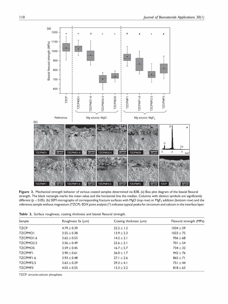
Mechanical stability and coating adhesion
The biaxial flexural strength values of different coatedsamples measured by B3B are displayed in Figure 3(a)and the mean strength values of investigated specimensare summarized in Table 2. The highest flexuralstrength of about 1000MPa was found for TZCP andTZCPMO1. At other Mg concentrations and independ-ent of the Mg source, the specimen showed a lowerstrength. It can be noticed that the strength-decreasingeffect is more pronounced with addition of MgF2 thanMgO. Within the Mg-containing coatings the tendencyof decreasing strength with increasing Mg content canbe clearly seen, except for samples with 5wt% Mg-addition.
Additional investigations of fracture surfaces(Figure 3b) corroborated these B3B results. Coatingswith a smooth and continuous morphology along theinterbonding show higher strength values in comparisonto specimens with a distinct interface between substrateand coating. The thickness of the interface layerincreased with the amount of Mg in the coating mixture,and decreased again at 5wt% Mg. EDX analysis of theinterfacial layer revealed zirconium and calcium peaks.
The coating-substrate adhesion plays a crucial rolein determining the usability and reliability of coated
Calcium magnesium phosphate Zirconia (monoclinic) Zirconia calcium oxide (cubic)
Inte
nsity
(ar
brita
ry u
nits
)
Inte
nsity
(ar
brita
ry u
nits
)
20 30 402 Theta (°)
50 20 30 402 Theta (°)
50
TZCPMO1
TZCPMO1.6
TZCPMO2.5
TZCPMO5
TZCPMF1
TZCPMF1.6
TZCPMF2.5
TZCPMF5
(a) (b)
Figure 1. GIXRD patterns of the different mixed coatings with MgO (a) and MgF2 addition (b) after heat treatment at 1500�C.
108 Journal of Biomaterials Applications 30(1)

implants for dental and orthopedic applications.30 Inthis study, the coating adhesion was investigated by ascratch test and via insertion experiments with coatedzirconia screw implants. Figure 4 shows representativetop views of the sample surface before (a) to (c) andafter (d) to (e) scratch test carried out with a sharpenedbovine femur, where the dashed black lines correspondto the scratch path. At a constant load of 7.5N thecoatings with 1wt% Mg addition maintained theirintegrity indicating a strong interbonding between coat-ing and substrate. No difference compared to the refer-ence was observed, suggesting no adverse effect of Mgon coating adhesion.
Insertion experiments with screw implants were add-itionally carried out to investigate the stability andadhesion of coatings on curved parts. The homoge-nously coated implants that show the same surfacemorphology compared to coatings obtained on planardiscs (Figure 5a) were carefully dissected after insertion
into Sawbone and bovine rip bone using a clinicalprotocol (Figure 5b). These insertions experimentswith coated zirconia implants confirmed the scratchtest results, indicating a firm adhesion of the coatingeven under load application. Images of the other inves-tigated coatings are not shown due to same achievedresults; an increase in Mg-content did not alter coatingadhesion and stability.
In vitro bioactivity assessment
The cumulative calcium and magnesium ion release inTris-HCl buffer over a period of 3 weeks is shown inFigure 6. During the first 4 days of immersion the ionicconcentration of Ca2þ and Mg2þ increased rapidly fol-lowed by gradual increase on the following days. Thisbehavior could be observed for all coatings and bothinvestigated ions. Compared to the reference sampleTZCP, Mg-addition led to a diminished Ca2þ release
MgO
1 w
t. %
1.6
wt.
%2.
5 w
t. %
5 w
t. %
TZCPMgF2
10 μm 10 μm
10 μm 10 μm
10 μm 10 μm
10 μm 10 μm
10 μm
(a) (e) (i)
(c) (g)
(d) (h)
(k)
(b) (f)(j)
0 2 4
0 2 4
keV
keV
Ca
Ca
Mg
O
O
P
Zr
*
**
Figure 2. Surface morphologies of sprayed coatings on TZ obtained by WPS after heat treatment at 1500�C. (a)–(d): coatings with
MgO addition, (e)–(h): coatings with MgF2 addition, (i): reference coating without magnesium addition. (j)–(k): EDX point analysis of
bright gray grains (*) and dark gray areas (**) of sample (a) indicate the typical major elements of calcium magnesium phosphate and
cubic zirconia, respectively.
Pardun et al. 109
1200
1100
1000
900
800
700
600
TZ
CP
TZ
CP
MO
1
TZCPMO1 TZCPMO1.6 TZCPMO2.5 TZCPMO5
TZCPMF1 TZCPMF1.6 TZCPMF2.5 TZCPMF5 TZCP
TZ
CP
MO
1.6
TZ
CP
MO
2.5
TZ
CP
MO
5
TZ
CP
MF
1
TZ
CP
MF
1.6
TZ
CP
MF
2.5
TZ
CP
MF
5
* * # + + +#
Bia
xial
flex
ural
str
engt
h (M
Pa)
♦ ♦
Reference Mg source: MgO Mg source: MgF2
20 μm 20 μm
20 μm 20 μm 20 μm 20 μm 20 μm
20 μm 20 μm
Zr
OCa
0 2 4 keV
*
(a)
(b)
Figure 3. Mechanical strength behavior of various coated samples determined via B3B. (a) Box plot diagram of the biaxial flexural
strength. The black rectangle marks the mean value and the horizontal line the median. Columns with distinct symbols are significantly
different (p< 0.05). (b) SEM micrographs of corresponding fracture surfaces with MgO (top row) or MgF2 addition (bottom row) and the
reference sample without magnesium (TZCP). EDX point analysis (*) indicates typical peaks for zirconium and calcium in the interface layer.
Table 2. Surface roughness, coating thickness and biaxial flexural strength.
Sample Roughness Sa (mm) Coating thickness (mm) Flexural strength (MPa)
TZCP 4.79� 0.39 22.2� 1.2 1034� 59
TZCPMO1 3.55� 0.38 13.9� 2.3 1023� 75
TZCPMO1.6 3.62� 0.53 14.2� 2.1 956� 68
TZCPMO2.5 3.56� 0.49 22.6� 2.1 701� 54
TZCPMO5 3.59� 0.45 16.7� 5.7 734� 32
TZCPMF1 3.90� 0.61 26.0� 1.7 942� 76
TZCPMF1.6 3.93� 0.48 27.1� 2.6 865� 71
TZCPMF2.5 3.63� 0.29 29.3� 4.1 751� 44
TZCPMF5 4.03� 0.55 13.3� 2.2 818� 63
TZCP: zirconia-calcium phosphate.
110 Journal of Biomaterials Applications 30(1)

Overview Coating morphology Cross section
TZ
CP
MF
1T
ZC
PM
O1
TZCPMF1 TZCPMF1TZCPMO1 TZCPMO1
Saw bone Rip bone
1 mm
1 mm
30 μm
30 μm
60 μm
60 μm10 μm
10 μm300 μm
300 μm
300 μm 300 μm 300 μm 300 μm
(a)
(b)
Figure 5. (a) Implant overview, coating detail and cross section of zirconia implants coated with mixed coatings via WPS after
sintering at 1500�C: MgO (top row) and MgF2 (bottom row) addition. (b) Implant coating morphology after insertion and dissection of
Sawbone (left) and bovine rip bone (right) of mixed coating with 1 wt% MgO and MgF2 addition. Arrows depict bone residues after
burn out.
TZCP
Bef
ore
scra
tch
Afte
r bo
ne s
crat
ch
TZCPMO1 TZCPMF1
(a) (b) (c)
(d) (e) (f)
100 μm 100 μm 100 μm
100 μm 100 μm 100 μm
Figure 4. SEM micrographs of the coating morphology before (top row) and after bone scratch (bottom row). The black dashed
lines represent the scratches.
Pardun et al. 111

around 75%. It can also be observed that Ca2þ releaseis influenced by the amount of Mg-addition, the higherthe amount of Mg the lower the Ca2þ release.Furthermore, MgF2 significantly reduced Ca2þ releasealready at lower concentrations in comparison to MgO.For Mg2þ release the opposite behavior was observed,higher Mg2þ concentrations were found in the super-natant with increasing content of Mg in the coating. Atthe same time there is no significant difference in Mg2þ
release between coatings with comparable amounts ofMgO or MgF2 addition.
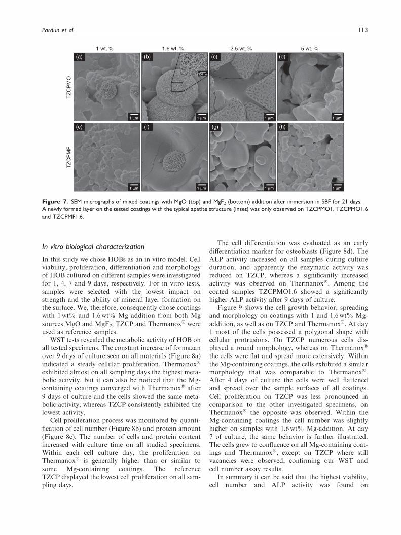
Figure 7 shows SEM images of Mg-coatings aftersoaking in SBF solution for 21 days. After immersionin SBF only samples TZCPM1, TZCPM1.6 andTZCMF1.6 exhibited a partial covering of the surfacewith a new mineral layer, which possessed the typicalglobular morphology of apatite precipitates. Withinthese samples TZCPMO1.6 showed the highest forma-tion ability. A higher magnification of this newlayer evidenced the detailed layer structure (inset inFigure 7b). On the other sample surfaces no precipita-tion was observed.
5.0
4.5
4.0
3.5
3.0
2.5
TZCPTZCPMO1
TZCPMO1.6
TZCPMO2.5
TZCPMO5
TZCPMF1
TZCPMF1.6
TZCPMF2.5
TZCPMF5
TZCPMO1
TZCPMO1.6
TZCPMO2.5
TZCPMO5
TZCPMF1
TZCPMF1.6
TZCPMF2.5
TZCPMF5
12
10
8
6
Cal
cium
ion
rele
ase
(mM
)
4
2
00 2 4 6 8 10 12
Immersion time (day)14 16 18 20 22
2.0
1.5
1.0
1.8(b)
(a)
1.6
1.4
1.2
1.0
0.8
0.6
0.4
0.2
0.0
0.5
0.0
0 2 4 6 8 10 12 14 16 18 20 22
0 2 4 6 8 10 12 14 16 18 20 22
Immersion time (day)
Immersion time (day)
Cal
cium
ion
rele
ase
(mM
)M
agne
sium
ion
rele
ase
(mM
)
Figure 6. Dissolution behavior of mixed coatings sintered at 1500�C after immersion at pH 7.4 for a period of 21 days as a function
of immersion time: calcium ion release (a) compared to the calcium ion release of a mixed coating without magnesium, and magnesium
ion release (b) of coatings with MgO (square) and MgF2 (triangle) addition.
112 Journal of Biomaterials Applications 30(1)
In vitro biological characterization
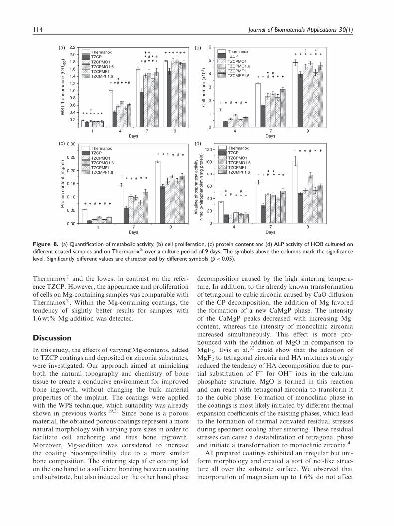
In this study we chose HOBs as an in vitro model. Cellviability, proliferation, differentiation and morphologyof HOB cultured on different samples were investigatedfor 1, 4, 7 and 9 days, respectively. For in vitro tests,samples were selected with the lowest impact onstrength and the ability of mineral layer formation onthe surface. We, therefore, consequently chose coatingswith 1wt% and 1.6wt% Mg addition from both Mgsources MgO and MgF2; TZCP and Thermanox� wereused as reference samples.
WST tests revealed the metabolic activity of HOB onall tested specimens. The constant increase of formazanover 9 days of culture seen on all materials (Figure 8a)indicated a steady cellular proliferation. Thermanox�
exhibited almost on all sampling days the highest meta-bolic activity, but it can also be noticed that the Mg-containing coatings converged with Thermanox� after9 days of culture and the cells showed the same meta-bolic activity, whereas TZCP consistently exhibited thelowest activity.
Cell proliferation process was monitored by quanti-fication of cell number (Figure 8b) and protein amount(Figure 8c). The number of cells and protein contentincreased with culture time on all studied specimens.Within each cell culture day, the proliferation onThermanox� is generally higher than or similar tosome Mg-containing coatings. The referenceTZCP displayed the lowest cell proliferation on all sam-pling days.
The cell differentiation was evaluated as an earlydifferentiation marker for osteoblasts (Figure 8d). TheALP activity increased on all samples during cultureduration, and apparently the enzymatic activity wasreduced on TZCP, whereas a significantly increasedactivity was observed on Thermanox�. Among thecoated samples TZCPMO1.6 showed a significantlyhigher ALP activity after 9 days of culture.
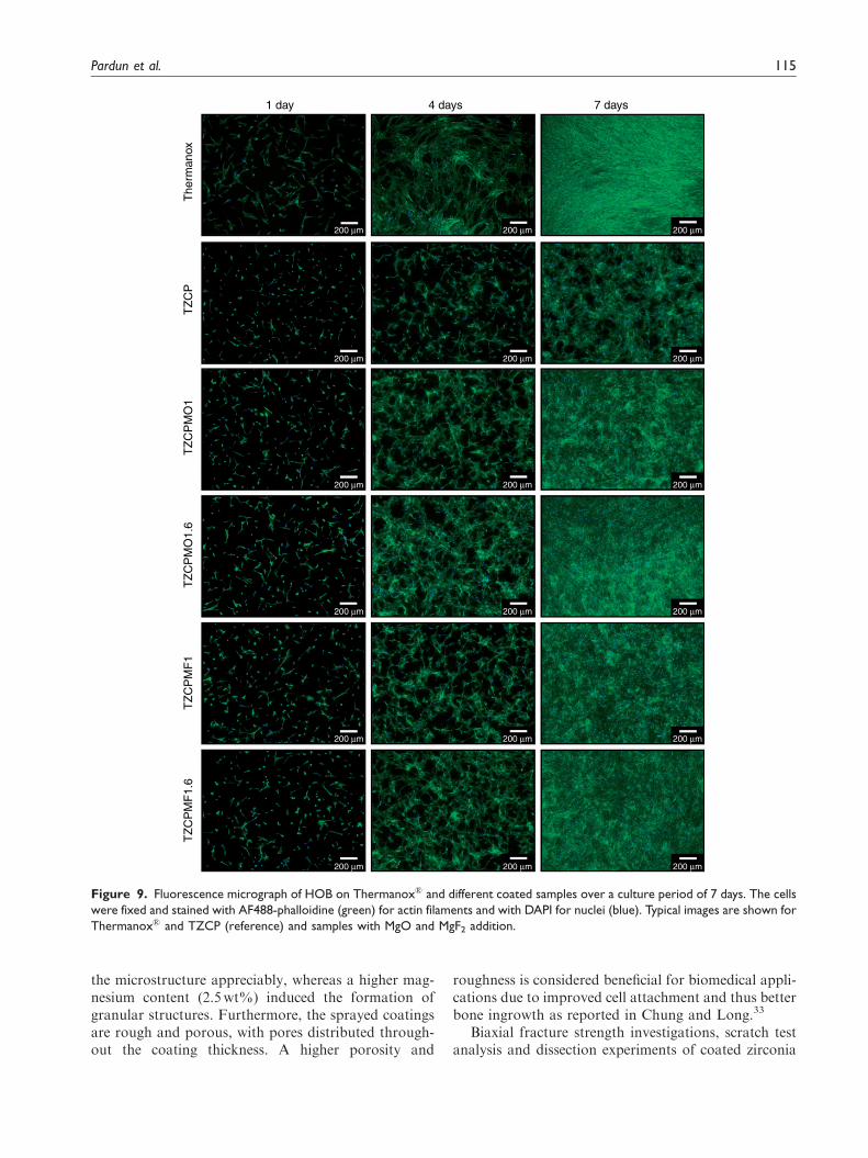
Figure 9 shows the cell growth behavior, spreadingand morphology on coatings with 1 and 1.6wt% Mg-addition, as well as on TZCP and Thermanox�. At day1 most of the cells possessed a polygonal shape withcellular protrusions. On TZCP numerous cells dis-played a round morphology, whereas on Thermanox�
the cells were flat and spread more extensively. Withinthe Mg-containing coatings, the cells exhibited a similarmorphology that was comparable to Thermanox�.After 4 days of culture the cells were well flattenedand spread over the sample surfaces of all coatings.Cell proliferation on TZCP was less pronounced incomparison to the other investigated specimens, onThermanox� the opposite was observed. Within theMg-containing coatings the cell number was slightlyhigher on samples with 1.6wt% Mg-addition. At day7 of culture, the same behavior is further illustrated.The cells grew to confluence on all Mg-containing coat-ings and Thermanox�, except on TZCP where stillvacancies were observed, confirming our WST andcell number assay results.
In summary it can be said that the highest viability,cell number and ALP activity was found on
1 wt. %
TZ
CP
MF
TZ
CP
MO
1.6 wt. % 2.5 wt. % 5 wt. %
(a)
1 μm 1 μm
300 nm
1 μm 1 μm
1 μm 1 μm 1 μm 1 μm
(b) (c) (d)
(e) (f) (g) (h)
Figure 7. SEM micrographs of mixed coatings with MgO (top) and MgF2 (bottom) addition after immersion in SBF for 21 days.
A newly formed layer on the tested coatings with the typical apatite structure (inset) was only observed on TZCPMO1, TZCPMO1.6
and TZCPMF1.6.
Pardun et al. 113
Thermanox� and the lowest in contrast on the refer-ence TZCP. However, the appearance and proliferationof cells on Mg-containing samples was comparable withThermanox�. Within the Mg-containing coatings, thetendency of slightly better results for samples with1.6wt% Mg-addition was detected.
Discussion
In this study, the effects of varying Mg-contents, addedto TZCP coatings and deposited on zirconia substrates,were investigated. Our approach aimed at mimickingboth the natural topography and chemistry of bonetissue to create a conducive environment for improvedbone ingrowth, without changing the bulk materialproperties of the implant. The coatings were appliedwith the WPS technique, which suitability was alreadyshown in previous works.19,31 Since bone is a porousmaterial, the obtained porous coatings represent a morenatural morphology with varying pore sizes in order tofacilitate cell anchoring and thus bone ingrowth.Moreover, Mg-addition was considered to increasethe coating biocompatibility due to a more similarbone composition. The sintering step after coating ledon the one hand to a sufficient bonding between coatingand substrate, but also induced on the other hand phase
decomposition caused by the high sintering tempera-ture. In addition, to the already known transformationof tetragonal to cubic zirconia caused by CaO diffusionof the CP decomposition, the addition of Mg favoredthe formation of a new CaMgP phase. The intensityof the CaMgP peaks decreased with increasing Mg-content, whereas the intensity of monoclinic zirconiaincreased simultaneously. This effect is more pro-nounced with the addition of MgO in comparison toMgF2. Evis et al.32 could show that the addition ofMgF2 to tetragonal zirconia and HA mixtures stronglyreduced the tendency of HA decomposition due to par-tial substitution of F� for OH� ions in the calciumphosphate structure. MgO is formed in this reactionand can react with tetragonal zirconia to transform itto the cubic phase. Formation of monoclinic phase inthe coatings is most likely initiated by different thermalexpansion coefficients of the existing phases, which leadto the formation of thermal activated residual stressesduring specimen cooling after sintering. These residualstresses can cause a destabilization of tetragonal phaseand initiate a transformation to monoclinic zirconia.4
All prepared coatings exhibited an irregular but uni-form morphology and created a sort of net-like struc-ture all over the substrate surface. We observed thatincorporation of magnesium up to 1.6% do not affect
2.2
2.0
1.8
1.6
1.4
1.2
1.0
0.8
0.6
0.4
0.2
0.00
0.05
0.10
0.15
0.20
0.30120
0
1
2
3
4
5
6
100
Alk
alin
e ph
osph
ates
e ac
tivity
Nm
ol p
-nitr
ophe
nol/m
in m
g pr
otei
n)
80
60
40
20
0
0.25
1 4
ThermanoxTZCPTZCPMO1TZCPMO1.6TZCPMF1TZCMPF1.6
ThermanoxTZCPTZCPMO1TZCPMO1.6TZCPMF1TZCMPF1.6
ThermanoxTZCPTZCPMO1TZCPMO1.6TZCPMF1TZCMPF1.6
ThermanoxTZCPTZCPMO1TZCPMO1.6TZCPMF1TZCMPF1.6
7 9Days
4 7 9Days
4 7 9Days
4 7 9Days
WS
T-1
abso
rban
ce (
OD
450)
Pro
tein
con
tent
(m
g/m
l)
Cel
l num
ber
(x10
5 )
+
+
+
+
+
+
+ + + + + +
+
+ * *
*
*
*
+
+
+
*
*
* * *
* * *
* * *
*
**
# #
#
###
# #
##
##
##
## #
•
• •
•
•♦
•
♦♦
♦
•♦•♦
♦
+ * # #♦
••
+
+ + + +
*
**
##
##
♦ ♦
♦ ♦
♦ ♦
♦(a) (b)
(c) (d)
Figure 8. (a) Quantification of metabolic activity, (b) cell proliferation, (c) protein content and (d) ALP activity of HOB cultured on
different coated samples and on Thermanox� over a culture period of 9 days. The symbols above the columns mark the significance
level. Significantly different values are characterized by different symbols (p< 0.05).
114 Journal of Biomaterials Applications 30(1)
the microstructure appreciably, whereas a higher mag-nesium content (2.5wt%) induced the formation ofgranular structures. Furthermore, the sprayed coatingsare rough and porous, with pores distributed through-out the coating thickness. A higher porosity and
roughness is considered beneficial for biomedical appli-cations due to improved cell attachment and thus betterbone ingrowth as reported in Chung and Long.33
Biaxial fracture strength investigations, scratch testanalysis and dissection experiments of coated zirconia
1 day
The
rman
oxT
ZC
PT
ZC
PM
O1
TZ
CP
MO
1.6
TZ
CP
MF
1T
ZC
PM
F1.
6
4 days 7 days
200 μm 200 μm 200 μm
200 μm 200 μm 200 μm
200 μm 200 μm 200 μm
200 μm 200 μm 200 μm
200 μm 200 μm 200 μm
200 μm 200 μm 200 μm
Figure 9. Fluorescence micrograph of HOB on Thermanox� and different coated samples over a culture period of 7 days. The cells
were fixed and stained with AF488-phalloidine (green) for actin filaments and with DAPI for nuclei (blue). Typical images are shown for
Thermanox� and TZCP (reference) and samples with MgO and MgF2 addition.
Pardun et al. 115
implants combined with subsequent SEM observationrevealed mechanical stability as well as coating adhe-sion of the different deposited Mg-containing coatingsand the reference coating without Mg-addition. B3Bresults show the influence of coating composition onthe mechanical strength of the bulk material. SampleTZCPMO1 exhibited the highest strength, which wassignificantly higher than all other tested coatings andcomparable with the reference TZCP. Both TZCPMO1and TZCP possess nearly the same biaxial flexuralstrength that was found for uncoated TZ and whichis approximately 1100MPa.19 Analysis of the other spe-cimens showed that the strength tends to decrease withincreasing Mg-content. Further investigations of thefracture surface revealed an interface layer betweencoating and substrate, whose thickness increases withenhanced Mg-content. This layer is most likely formedduring the sintering process. CaO, released from HAdecomposition, diffuses into the TZ substrate, as itcan be determined by EDX analysis, and probablycauses a transformation of tetragonal to cubic TZ. Inaddition, magnesium can also be integrated in TZ andlead to further stabilization of zirconia.32 However, Mgwas not detected in the interface layer by EDX analysisbecause it was not present or below detection limit ofthe method used. As can be seen from our GIXRDresults, monoclinic zirconia was detected in the coatingsbesides cubic zirconia. Both cubic and monoclinic zir-conia are known to have inferior mechanical propertiescompared to tetragonal zirconia and could affect themechanical strength.4 However, based on our B3Bresults and fracture surfaces analysis, we concludethat the interface layer is mainly responsible for thedecrease in mechanical strength due to the fact thatcubic TZ is unable to stop crack growth by phase trans-formation. This is supported by the comparison of theB3B results of specimens with Mg-containing coatingsand the reference sample TZCP. Despite the presence ofmonoclinic and cubic zirconia in the coating19 TZCPexhibits a good and with uncoated TZ comparable mech-anical strength since no interface layer between coatingand substrate was formed. Therefore, it can be hypothe-sized that the coating phase composition has only a neg-ligible influence on the strength without the existence ofan interface layer. Nevertheless, a possible increase inharmfulness of the interface layer caused by monoclinicphase cannot be excluded. Furthermore, it can be notedthat the decrease in mechanical strength clearly correlateswith the thickness of the interface layer.
Scratch tests carried out with discs and dissectionexperiments carried out with coated implants showeda firm coating adhesion. Both analysis methods yieldedthe same results suggesting no adverse effect of Mg oncoating adhesion, independent of the Mg-content.On the contrary, there is even evidence that Mg
promotes adhesion of coatings.34 In our previouswork, the low bonding strength of pure CP coating toTZ substrate was shown, which is known to be onedisadvantage.19 This improved bonding strength ofthe mixed coatings in comparison to pure HA or CPcoatings have the potential for enhanced performancein biomedical applications.
As an implant coating material, not only the coatingstability and adhesion play an important role in theperformance of implants, both chemical compositionand dissolution have also a great impact on cell behav-ior and thus on the cytotoxicity of implant materials.The results of our dissolution experiments showed thatthe release of Ca2þ was considerably reduced comparedto the reference TZCP and it further decreases withincreasing Mg-content. This observation leads to theconfirmation that the new CaMgP-phase, formedduring heat treatment, possesses a lower dissolutionrate than TCP that is present in the TZCP coatingafter sintering.19 This is also in agreement with theresults of others that showed a reduction of degrad-ation in Mg substituted CP biomaterials.35,36 Withinthe two different Mg-sources the lowest Ca2þ releasewas observed for 2.5wt% Mg-addition, this behaviorcould be attributed to the morphological change of thecoating.
The biological activity of biomaterials is associatedwith their capability of mineral layer formation when incontact with biological fluids. However, when samplesare immersed in SBF dissolution and precipitation willtake place at the same time. As a consequence, the con-centration of Ca and Mg ions and P groups increase inthe solution adjacent the specimen surface, resulting inan increase of supersaturation that is beneficial tonucleation and growth of new mineral layers. In otherwords, when precipitation is dominant a new minerallayer will form on the sample surface. Our results showthat the precipitation of a mineral layer was favored atthe surface of samples TZCPMO1 and TZCPMO1.6.In contrast, on reference sample TZCP less precipita-tion was observed after 21 days immersion (data notshown) suggesting a beneficial effect of Mg for in vitrobehavior although dissolution was lower compared toTZCP. The same observation was made by Gomeset al.36 who showed that precipitation was morefavored on samples with a less-soluble Mg substitutedb-TCP phase. All other investigated samples did notexhibit any mineral layer formation, which seemed tobe related to the Mg content in the coating and also tothe kind of additive. Coatings with MgF2 addition andwith 2.5wt% or 5wt% MgO addition showed a lowerCa2þ dissolution and thus the local concentration ofdissolved ions was too low to trigger precipitation.In case of sample TZCPMF1.6 precipitation of a newmineral layer was also detected probably due to the
116 Journal of Biomaterials Applications 30(1)
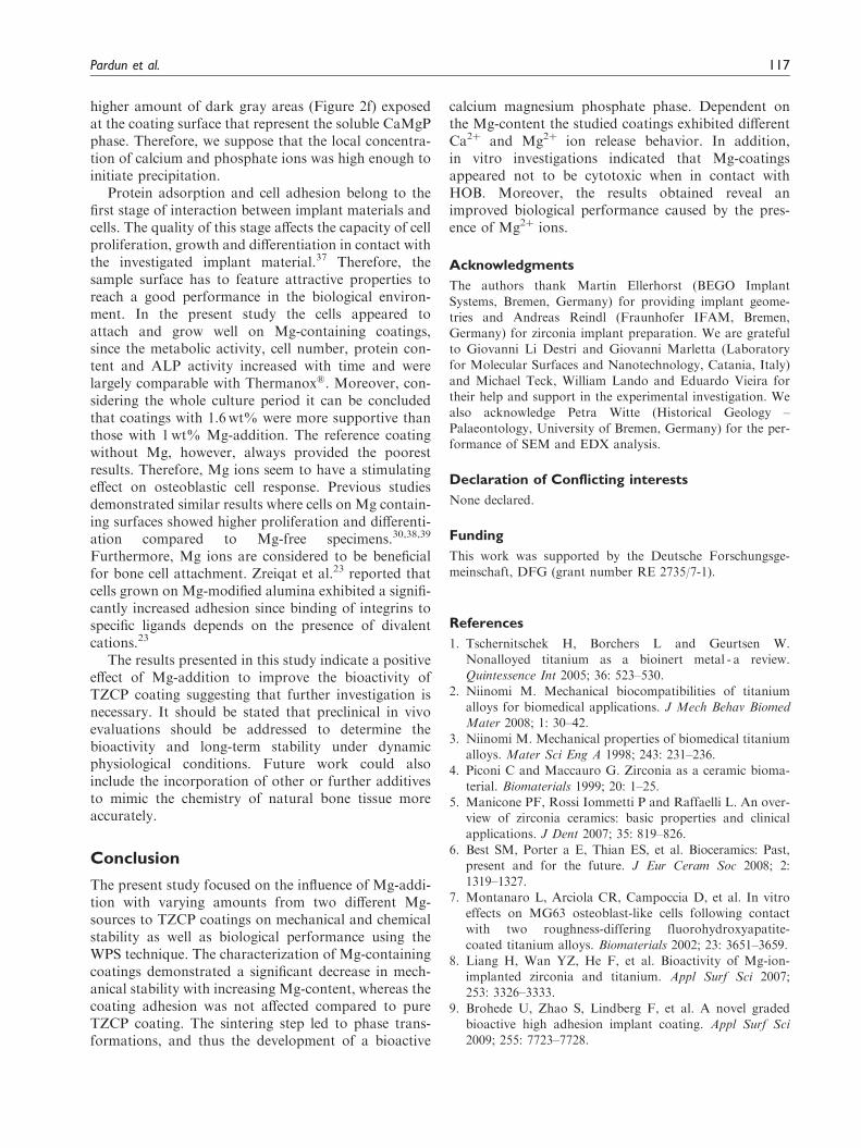
higher amount of dark gray areas (Figure 2f) exposedat the coating surface that represent the soluble CaMgPphase. Therefore, we suppose that the local concentra-tion of calcium and phosphate ions was high enough toinitiate precipitation.
Protein adsorption and cell adhesion belong to thefirst stage of interaction between implant materials andcells. The quality of this stage affects the capacity of cellproliferation, growth and differentiation in contact withthe investigated implant material.37 Therefore, thesample surface has to feature attractive properties toreach a good performance in the biological environ-ment. In the present study the cells appeared toattach and grow well on Mg-containing coatings,since the metabolic activity, cell number, protein con-tent and ALP activity increased with time and werelargely comparable with Thermanox�. Moreover, con-sidering the whole culture period it can be concludedthat coatings with 1.6wt% were more supportive thanthose with 1wt% Mg-addition. The reference coatingwithout Mg, however, always provided the poorestresults. Therefore, Mg ions seem to have a stimulatingeffect on osteoblastic cell response. Previous studiesdemonstrated similar results where cells on Mg contain-ing surfaces showed higher proliferation and differenti-ation compared to Mg-free specimens.30,38,39
Furthermore, Mg ions are considered to be beneficialfor bone cell attachment. Zreiqat et al.23 reported thatcells grown on Mg-modified alumina exhibited a signifi-cantly increased adhesion since binding of integrins tospecific ligands depends on the presence of divalentcations.23
The results presented in this study indicate a positiveeffect of Mg-addition to improve the bioactivity ofTZCP coating suggesting that further investigation isnecessary. It should be stated that preclinical in vivoevaluations should be addressed to determine thebioactivity and long-term stability under dynamicphysiological conditions. Future work could alsoinclude the incorporation of other or further additivesto mimic the chemistry of natural bone tissue moreaccurately.
Conclusion
The present study focused on the influence of Mg-addi-tion with varying amounts from two different Mg-sources to TZCP coatings on mechanical and chemicalstability as well as biological performance using theWPS technique. The characterization of Mg-containingcoatings demonstrated a significant decrease in mech-anical stability with increasing Mg-content, whereas thecoating adhesion was not affected compared to pureTZCP coating. The sintering step led to phase trans-formations, and thus the development of a bioactive
calcium magnesium phosphate phase. Dependent onthe Mg-content the studied coatings exhibited differentCa2þ and Mg2þ ion release behavior. In addition,in vitro investigations indicated that Mg-coatingsappeared not to be cytotoxic when in contact withHOB. Moreover, the results obtained reveal animproved biological performance caused by the pres-ence of Mg2þ ions.
Acknowledgments
The authors thank Martin Ellerhorst (BEGO Implant
Systems, Bremen, Germany) for providing implant geome-tries and Andreas Reindl (Fraunhofer IFAM, Bremen,Germany) for zirconia implant preparation. We are gratefulto Giovanni Li Destri and Giovanni Marletta (Laboratory
for Molecular Surfaces and Nanotechnology, Catania, Italy)and Michael Teck, William Lando and Eduardo Vieira fortheir help and support in the experimental investigation. We
also acknowledge Petra Witte (Historical Geology –Palaeontology, University of Bremen, Germany) for the per-formance of SEM and EDX analysis.
Declaration of Conflicting interests
None declared.
Funding
This work was supported by the Deutsche Forschungsge-
meinschaft, DFG (grant number RE 2735/7-1).
References
1. Tschernitschek H, Borchers L and Geurtsen W.Nonalloyed titanium as a bioinert metal - a review.Quintessence Int 2005; 36: 523–530.
2. Niinomi M. Mechanical biocompatibilities of titaniumalloys for biomedical applications. J Mech Behav BiomedMater 2008; 1: 30–42.
3. Niinomi M. Mechanical properties of biomedical titaniumalloys. Mater Sci Eng A 1998; 243: 231–236.
4. Piconi C and Maccauro G. Zirconia as a ceramic bioma-
terial. Biomaterials 1999; 20: 1–25.5. Manicone PF, Rossi Iommetti P and Raffaelli L. An over-
view of zirconia ceramics: basic properties and clinicalapplications. J Dent 2007; 35: 819–826.
6. Best SM, Porter a E, Thian ES, et al. Bioceramics: Past,present and for the future. J Eur Ceram Soc 2008; 2:1319–1327.
7. Montanaro L, Arciola CR, Campoccia D, et al. In vitroeffects on MG63 osteoblast-like cells following contactwith two roughness-differing fluorohydroxyapatite-
coated titanium alloys. Biomaterials 2002; 23: 3651–3659.8. Liang H, Wan YZ, He F, et al. Bioactivity of Mg-ion-
implanted zirconia and titanium. Appl Surf Sci 2007;253: 3326–3333.
9. Brohede U, Zhao S, Lindberg F, et al. A novel gradedbioactive high adhesion implant coating. Appl Surf Sci2009; 255: 7723–7728.
Pardun et al. 117

10. Hench LL. Bioceramics: from concept to clinic. J AmCeram Soc 1991; 74: 1487–1510.
11. Dorozhkin SV. Calcium orthophosphates. J Mater Sci
2007; 42: 1061–1095.12. Bohner M. Calcium orthophosphates in medicine: from
ceramics to calcium phosphate cements. Injury 2000;31(Suppl 4): 37–47.
13. Jarcho M. Calcium phosphate ceramics as hard tissueprosthetics. Clin Orthop Relat Res 1981; 157: 259–78.
14. Bloebaum RD, Beeks D, Dorr LD, et al.
Complications with hydroxyapatite particulate separ-ation in total hip arthroplasty. Clin Orthop Relat Res1994; 298: 19–26.
15. Overgaard S, Søballe K, Lind M, et al. Resorption ofhydroxyapatite and fluorapatite coatings in man. JBone Jt Surg 1997; 79: 654–659.
16. Ducheyne P. Bioactive ceramics. J Bone Jt Surg. 1994; 76:861–862.
17. Matsumoto TJ, An S-H, Ishimoto T, et al. Zirconia-hydroxyapatite composite material with micro porous
structure. Dent Mater 2011; 27: e205–212.18. Yugeswaran S, Yoganand CP, Kobayashi A, et al.
Mechanical properties, electrochemical corrosion and
in-vitro bioactivity of yttria stabilized zirconia reinforcedhydroxyapatite coatings prepared by gas tunneltype plasma spraying. J Mech Behav Biomed Mater
2012; 9: 22–33.19. Pardun K, Treccani L, Volkmann E, et al.
Characterization of wet powder-sprayed zirconia/calciumphosphate coating for dental implants. Clin Implant Dent
Relat Res 2013; DOI: 10.1111/cid.12071.20. Staiger MP, Pietak AM, Huadmai J, et al. Magnesium
and its alloys as orthopedic biomaterials: a review.
Biomaterials 2006; 27: 1728–1734.21. Kannan S, Lemos AF and Ferreira JMF. Synthesis and
Mechanical Performance of Biological-like
Hydroxyapatites. Chem Mater 2006; 18: 2181–2186.22. LeGeros RZ. Calcium phosphates in oral biology and
medizine. In: Myers H (ed.) Monographs in oral sciences.
Karger Basel Switzerland: 1991; pp.1–201.23. Zreiqat H, Howlett CR, Zannettino A, et al. Mechanisms
of magnesium-stimulated adhesion of osteoblastic cells tocommonly used orthopaedic implants. J Biomed Mater
Res 2002; 62: 175–184.24. Landi E, Tampieri A, Mattioli-Belmonte M, et al.
Biomimetic Mg- and Mg,CO3-substituted hydroxyapa-
tites: synthesis characterization and in vitro behaviour.J Eur Ceram Soc 2006; 26: 2593–2601.
25. Cowan JA. Structural and catalytic chemistry of magne-
sium-dependent enzymes. Biometals 2002; 15: 225–235.
26. Wiesmann HP, Tkotz T, Joos U, et al. Magnesium innewly formed dentin mineral of rat incisor. J BoneMiner Res 1997; 12: 380–383.
27. Jiang G and Shi D. Coating of hydroxyapatite on highlyporous Al2O3 substrate for bone substitutes. J BiomedMater Res 1998; 43: 77–81.
28. Borger A, Supancic P and Danzer R. The ball on three
balls test for strength testing of brittle discs: stress distri-bution in the disc. J Eur Ceram Soc 2002; 22: 1425–1436.
29. Oyane A, Kim H-M, Furuya T, et al. Preparation and
assessment of revised simulated body fluids. J BiomedMater Res A 2003; 65: 188–195.
30. Xie Y, Zhai W, Chen L, et al. Preparation and in vitro
evaluation of plasma-sprayed Mg(2)SiO(4) coating ontitanium alloy. Acta Biomater 2009; 5: 2331–2337.
31. Ruder A, Buchkremer HP, Jansen H, et al. Wet powder
spraying - a process for the production of coatings. SurfCoatings Technol 1992; 53: 71–74.
32. Evis Z, Usta M and Kutbay I. Improvement in sinter-ability and phase stability of hydroxyapatite and partially
stabilized zirconia composites. J Eur Ceram Soc 2009; 29:621–628.
33. Chung C-J and Long H-Y. Systematic strontium substi-
tution in hydroxyapatite coatings on titanium via micro-arc treatment and their osteoblast/osteoclast responses.Acta Biomater 2011; 7: 4081–4087.
34. Qi G, Zhang S, Khor KA, et al. An interfacial study ofsol–gel-derived magnesium apatite coatings on Ti6Al4Vsubstrates. Thin Solid Films 2008; 516: 5172–5175.
35. Li X, Ito A, Sogo Y, et al. Solubility of Mg-containing
beta-tricalcium phosphate at 25 degrees C. Acta Biomater2009; 5: 508–517.
36. Gomes S, Renaudin G, Jallot E, et al. Structural charac-
terization and biological fluid interaction of Sol-Gel-derived Mg-substituted biphasic calcium phosphate cer-amics. ACS Appl Mater Interfaces 2009; 1: 505–513.
37. Paital SR and Dahotre NB. Calcium phosphate coatingsfor bio-implant applications: materials, performance fac-tors, and methodologies. Mater Sci Eng R Reports 2009;
66: 1–70.38. Cai YL, Zhang JJ, Zhang S, et al. Osteoblastic cell
response on fluoridated hydroxyapatite coatings: theeffect of magnesium incorporation. Biomed Mater 2010;
5: 054114.39. Shi X, Nakagawa M, Kawachi G, et al. Surface modifi-
cation of titanium by hydrothermal treatment in Mg-con-
taining solution and early osteoblast responses. J MaterSci Mater Med 2012; 23: 1281–1290.
118 Journal of Biomaterials Applications 30(1)