magnesium orthosilicate composite jeffrey chin kong …
TRANSCRIPT

MECHANICAL PROPERTIES OF HYDROXYAPATITE-MAGNESIUM ORTHOSILICATE COMPOSITE
JEFFREY CHIN KONG LEONG
FACULTY OF ENGINEERING UNIVERSITY OF MALAYA
KUALA LUMPUR
2017
Univers
ity of
Mala
ya

MECHANICAL PROPERTIES OF HYDROXYAPATITE-MAGNESIUM ORTHOSILICATE COMPOSITE
JEFFREY CHIN KONG LEONG
THESIS SUBMITTED IN FULFILMENT OF THE REQUIREMENTS FOR THE DEGREE OF
DOCTOR OF PHILOSOPHY
FACULTY OF ENGINEERING UNIVERSITY OF MALAYA
KUALA LUMPUR
2017
Univers
ity of
Mala
ya

ii
UNIVERSITI MALAYA
ORIGINAL LITERARY WORK DECLARATION
Name of Candidate: Jeffrey Chin Kong Leong
Registration/Matric No: KHA110097
Name of Degree: Doctorate of Philosophy
Title of Project Paper/Research Report/Dissertation/Thesis (“this Work”): Mechanical Properties of Hydroxyapatite-Magnesium Orthosilicate Composite
Field of Study: Advance Materials/Nanomaterials
I do solemnly and sincerely declare that:
(1) I am the sole author/writer of this Work; (2) This Work is original; (3) Any use of any work in which copyright exists was done by way of fair dealing and for
permitted purposes and any excerpt or extract from, or reference to or reproduction of any copyright work has been disclosed expressly and sufficiently and the title of the Work and its authorship have been acknowledged in this Work;
(4) I do not have any actual knowledge nor do I ought reasonably to know that the making of this work constitutes an infringement of any copyright work;
(5) I hereby assign all and every rights in the copyright to this Work to the University of Malaya (“UM”), who henceforth shall be owner of the copyright in this Work and that any reproduction or use in any form or by any means whatsoever is prohibited without the written consent of UM having been first had and obtained;
(6) I am fully aware that if in the course of making this Work I have infringed any copyright whether intentionally or otherwise, I may be subject to legal action or any other action as may be determined by UM.
Candidate’s Signature Date
Subscribed and solemnly declared before,
Witness’s Signature Date
Name: Designation:
Univers
ity of
Mala
ya

iii
ABSTRACT
Hydroxyapatite (HA) bioceramic has attracted a great deal of attention in the past two
decades due to its similarity in terms of chemical structure to that of hard tissues.
However, a major drawback of HA is the low fracture toughness (< 1 MPam1/2)
exhibited by the ceramic. Magnesium orthosilicate ceramic, on the other hand,
possessed much higher fracture toughness and has recently been reported in the
literature as a suitable biomaterial. However, limited studies have been conducted to
investigate the combination of these two bioceramics, of which are crucial parameters
needed to substantiate its suitability as a reliable nanocomposite material. Hence for the
current research, the development of hydroxyapatite-magnesium orthosilicate composite
with improved mechanical properties was investigated. The effects and implications of
combining these two bioceramics were assessed in terms of linear shrinkage, phase
stability, bulk density, Young’s modulus, Vickers hardness, fracture toughness and
microstructural evolution. In the present research, the hydroxyapatite powder was
successfully synthesized through a novel wet chemical method and the magnesium
orthosilicate powder was produced via combination of ultrasonification and mechanical
ball milling method. Then the hydroxyapatite powder was mixed with magnesium
orthosilicate accordingly to the concentration from 10wt. % to 50wt. % using the
ultrasonification and mechanical ball milling method. Green samples were pressureless
sintered at temperatures ranging from 1000°C to 1300°C at heating rate of 10°C /
minute with a holding time of 2 hours.
A secondary phase known as whitlockite was found in all the hydroxyapatite-
magnesium orthosilicate composites after sintering. In general, the ceramic composites
exhibited low mechanical properties across all the composition investigated. However, a
high fracture toughness of 2.5 MPam1/2 was recorded for hydroxyapatite containing
Univers
ity of
Mala
ya

iv
20wt. % magnesium orthosilicate addition which indicated the potential of this
composite to be used for load bearing biomedical applications. Moreover, the SEM
graphs also demonstrated that formed whitlockite composite tends to form porous
agglomerates. Such microstructure of calcium hydroxyapatite is one of the most
frequently used bioceramics for bone and dental tissues reconstruction and for
adsorption of hazardous materials from waste water and nuclear waste disposal.
Univers
ity of
Mala
ya

v
ABSTRAK
Hydroxyapatite (HA) bioseramik mendapat banyak perhatian di sepanjang dua dekad
yang lalu disebabkan struktur kimia yang sama dengan sepertimana yang didapati di
dalam tisu keras. Walau bagaimanapun, HA mempunyai kekurangan yang utama di
mana keliatan patahnya adalah rendah (<1 MPam1/2) dipamerkan oleh seramik.
Sebaliknya, seramik magnesium orthosilicate pula memiliki keliatan patah yang lebih
tinggi dan sesuai untuk digunakan sebagai biobahan seperti yang dilaporkan di dalam
kajian kesusasteraan kebelakangan ini. Namun, kajian penyelidikan berkaitan dengan
pengabungan kedua-dua bioseramik ini, yang penting dalam menentukan kesesuaiannya
sebagai bahan komposit yang boleh dipercayai adalah terhad. Justeru itu, kajian ini
bertujuan menyelidik pembangunan hydroxyapatite-magnesium orthosilicate komposit
dengan sifat-sifat mekanikal yang lebih baik. Kesan dan implikasi gabungan kedua-dua
bioseramik ini akan dinilai dari segi pengecutan linear, kestabilan fasa, ketumpatan
pukal, Young’s modulus, nilai kekerasan, ketahanan patah dan evolusi mikrostruktur.
Dalam kajian ini, serbuk hydroxyapatite telah berjaya disintesis melalui kaedah kimia
basah novel dan serbuk magnesium orthosilicate dihasilkan melalui kombinasi
ultrasonifikasi dan kaedah bola pengisaran mekanikal. Kemudian serbuk hydroxyapatite
dicampurkan dengan forsterite mengikut kepekatan daripada 10wt. % kepada 50wt. %
menggunakan ultrasonifikasi dan kaedah bola pengisaran mekanikal. Sampel hijau
disinter tanpa tekanan pada suhu di antara 1000°C hingga 1300°C pada kadar
pemanasan 10°C / minit dengan masa yang memegang 2 jam.
Selepas pensinteran, fasa kedua yang juga dikenali sebagai whitlockite didapati di
semua komposit hydroxyapatite-magnesium orthosilicate. Secara umumnya, komposit
seramik mempunyai sifat mekanik yang rendah berbanding dengan komposisi-
komposisi yang diselidik. Walau bagaimanapun, hydroxyapatite dengan komposisi
Univers
ity of
Mala
ya

vi
20wt. % magnesium orthosilicate menunjukkan potensinya sebagai penanggung beban
di dalam aplikasi bioperubatan dengan keliatan patahnya yang tinggi sebanyak 2.5
MPam1/2. Di samping itu, graf SEM juga menunjukkan komposit whitlockite yang
terbentuk mempunyai kecenderungan untuk membentuk gumpalan berliang.
Mikrostruktur seperti kalsium hydroxyapatite adalah salah satu bioceramik yang paling
kerap digunakan untuk pembinaan semula tulang dan tisu gigi dan untuk penjerapan
bahan-bahan berbahaya daripada air kumbahan dan pembuangan sisa nuklear.
Univers
ity of
Mala
ya

vii
ACKNOWLEDGEMENTS
First and foremost, I would like to express my sincere gratitude and respect to
my project supervisor, Prof. Ir. Dr. Ramesh Singh, for his continual guidance and
encouragement given to me throughout the entire period of this research. His passion
and drive in the research field in sharing his valuable knowledge had always been
inspirational to me. He has not only shared with me his vast technical knowledge in the
area of engineering ceramics but also molded me to be a competent researcher.
Next, I would also like to thank the kind Management of University of Malaya
for the facilities support. Additionally, I would like to extend my appreciation to SIRIM
Berhad Malaysia for providing the testing instruments such as XRD machine, Vickers
hardness tester, etc.
Special thank you is also conveyed to my friends and colleagues who
voluntary willing to spend their time to guide and motivate me during the research
work: Natasha, Kelvin Chew, Ali Niakan and Christopher Chin. Their presents have
made work and life more fun.
Finally, I would like to thank my loving family and my wife Carmen Tang
who have been a great support right through the period of this research in providing
endless words of encouragement, moral support and most importantly believing in me. I
would not have made it without you people. All help, care and concern will forevermore
be appreciated and remembered.
Univers
ity of
Mala
ya

viii
In memory of my late father
Univers
ity of
Mala
ya

ix
TABLE OF CONTENTS
Page
ORIGINAL LITERARY WORK DECLARATION
ii
ABSTRACT
iii
ABSTRAK
v
ACKNOWLEDGEMENTS
vii
TABLE OF CONTENT
ix
LIST OF FIGURE
xii
LIST OF TABLES
xvi
LIST OF SYMBOLS AND ABBREVIATIONS
xvii
CHAPTER 1: INTRODUCTION & OBJECTIVES
1
1.1 Introduction
1
1.2 Problem Statement
3
1.3 Objectives of Research
5
1.4 Scope of Project
5
1.5 Thesis Structure
6
CHAPTER 2: LITERATURE REVIEW
8
2.1 Human Bone Structure
8
2.2 Biomaterials
11
2.3 Bioceramics
18
2.4 Calcium Phosphate
22
2.5 Magnesium Orthosilicate
28
Univers
ity of
Mala
ya

x
2.6 Hydroxyapatite Composites 2.6.1 Hydroxyapatite – Bioinert Composites 2.6.1.1 Hydroxyapatite – Zirconia 2.6.1.2 Hydroxyapatite – Alumina 2.6.2 Hydroxyapatite – Bioactive Composites 2.6.2.1 Hydroxyapatite – Bioactive Glass 2.6.2.2 Hydroxyapatite – AW Glass 2.6.2.3 Hydroxyapatite – Magnesium Orthosilicate 2.6.3 Hydroxyapatite – Biodegradable Composites 2.6.3.1 Hydroxyapatite – β-TCP 2.6.3.2 Hydroxyapatite – α-TCP 2.7 Summary
29
30
31
37
40
42
46
49
50
51
54
55 CHAPTER 3: METHODOLOGY
56 3.1 Powder Synthesis 3.1.1 Hydroxyapatite Powder 3.1.2 Magnesium Orthosilicate Powder 3.1.3 Hydroxyapatite – Magnesium Orthosilicate Composite Powder 3.2 X-Ray Diffraction (XRD) 3.3 Fabrication of Samples 3.4 Sintering 3.5 Grinding and Polishing 3.6 Bulk Density Measurement 3.7 Young’s Modulus Determination 3.8 Vickers Hardness Determination 3.9 Fracture Toughness Determination 3.10 Scanning Electron Microscopy and Grain Size Measurement
56
56
58
59
60
61
62
63
63
64
65
67
68
Univers
ity of
Mala
ya

xi
CHAPTER 4: RESULTS AND DISCUSSION 71 4.1 Introduction
71
4.2 Starting Powder 4.2.1 HA Powder 4.2.2 Magnesium Orthosilicate Powder 4.2.3 Hydroxyapatite-Magnesium Orthosilicate Powder 4.3 HA-MO Composite Evaluation 4.3.1 Phase Analysis 4.3.2 Shrinkage 4.3.3 Bulk Density 4.3.4 Young’s Modulus 4.3.5 Vickers Hardness 4.3.6 Fracture Toughness 4.3.7 Microstructure Evaluation 4.4 Toughening Effect of MO
71
71
73
75
77
77
81
83
85
87
90
93
100 CHAPTER 5: CONCLUSIONS AND FURTHER WORK
111 5.1 Conclusions
111
5.2 Further Work
114
REFERENCES
116 LIST OF PUBLICATIONS
130
APPENDICES APPENDIX A – CHEMICAL CALCULATIONS APPENDIX B – JCPDS FILES APPENDIX C – INSTRUMENTATIONS APPENDIX D – WATER DENSITY TABLE
131
131
133
140
144
Univers
ity of
Mala
ya

xii
LIST OF FIGURES
Figure No. Page
1.1 The applications of glasses, ceramics and composites in the human anatomy
2
2.1
The hierarchy levels of bone microstructure
10
2.2
Bone structure of a human femur
11
2.3
Interactions between living tissues and artificial materials (biomaterials)
12
2.4
Biocompatibility factors
12
2.5
Bone bonding in terms of compositional (wt. %)
21
2.6
XRD trends for HA and HA-ZrO2 specimens after sintering at 1300°C for 2 h
32
2.7
Typical micrographs of (a) HA and (b) HA-ZrO2 samples after sintering at 1300°C for 2 h. Several CaZrO3 particles in (b) are indicated by arrows
33
2.8
Size of the HA grains in the HA-CaZrO3 composites as a function of starting ZrO2 content
33
2.9
Strength and toughness of the HA-CaZrO3 composites as a function of starting ZrO2 content
34
2.10
Bending strength of HA-alumina composites
38
2.11
XRD trends of HA-alumina composites of different composition sintered at 1100°C
39
2.12
XRD trends of 30wt. % alumina sintered at different temperatures
40
2.13
Bioglass: BG, Cerabone A/W: A-W, Sintered hydroxyapatite: HA, Sintered β-tricalcium phosphate: TCP, HAPEX: HP (A- cranial repair, B – middle ear bone replacement, C – maxillofacial reconstruction, D – bioactive coating on dental root, E – alveolar ridge augmentation, F – periodontal pocket obliteration, G – spinal surgery, H – iliac crest repair, I – bone filler, and J – bioactive coating on joint stem
41
2.14
Theoretical density of HA/phosphate glass composites
43
2.15
Microstructure of HA sintered at (a)1250°C and (b)1350°C
44
2.16
Fracture toughness of HA/phosphate glass compositions
45
Univers
ity of
Mala
ya

xiii
2.17
Density of composites sintered at 1200°C and 1300°C
46
2.18 Apatite formation tested in simulated body fluid (SBF) 47
2.19
Hardness (H) and reduced elastic modulus (Er) values of HA-wollastonite composite vs. different compositions of wollastonite
49
2.20
SEM micrograph of porous β-TCP (a) 65 vol. % porosity (b) 75 vol. % porosity (c) 85 vol. % porosity
53
2.21
SEM images of HA/β-TCP grains (a) 0 wt. % (b) 10 wt. % β-TCP (c) 20 wt. % β-TCP (d) 30 wt. % β-TCP
54
3.1
HA wet chemical method process flow
58
3.2
Sintering profile
62
3.3
Block diagram of test apparatus for Young’s modulus measurement (ASTM Standards E 1876-97)
65
3.4
Schematic diagram of Vickers hardness indenter
66
3.5
Crack propagation form indentation
67
3.6
Schematic diagram showing the score given for the type of intersections
70
4.1
XRD traces of synthesized hydroxyapatite powder before sintering
72
4.2
a) SEM analysis of synthesized hydroxyapatite powder, b) EDX of synthesized hydroxyapatite powder.
73
4.3
XRD traces of synthesized magnesium orthosilicate powder before sintering.
74
4.4
a) SEM analysis of synthesized magnesium orthosilicate powder, b) EDX of synthesized magnesium orthosilicate powder.
75
4.5
XRD traces of HA-MO powders before sintering.
76
4.6
SEM micrographs of (a) Pure HA and (b) HA-50MO powder.
76
4.7
EDX of HA-MO powder.
77
4.8
XRD patterns of HA-MO samples sintered at 1000°C
78
4.9
XRD patterns of HA-MO samples sintered at 1100°C
79
Univers
ity of
Mala
ya

xiv
4.10 XRD patterns of HA-MO samples sintered at 1200°C 79
4.11 XRD patterns of HA-MO samples sintered at 1300°C
80
4.12
Shrinkage of different sintered samples as a function of sintering temperature.
82
4.13
Bulk density variation as a sintering temperature and MO content
84
4.14
Variation in bulk density with shrinkage of the HA and HA-MO composites.
84
4.15
Young’s modulus variation with sintering temperature
86
4.16
Young’s modulus variation with bulk density.
87
4.17
Vickers hardness variation with MO addition as a function of sintering temperature
89
4.18
Vickers hardness variation with MO addition as a function of bulk density
89
4.19
Fracture toughness of sintered samples as a function of sintering temperature and MO addition
92
4.20
Fracture toughness variation with MO addition as a function of bulk density
92
4.21
SEM images of samples sintered at 1000°C
96
4.22
SEM images of samples sintered at 1100°C
97
4.23
SEM images of samples sintered at 1200°C
98
4.24
SEM images of samples sintered at 1300°C
99
4.25
SEM images of samples sintered at 1300˚C. (a) HA-20MO, (b) HA
100
4.26
SEM images of (a) HA, (b) HA-10MO, (c) HA-20MO, sintered at 1300˚C.
102
4.27
(a)Propagating crack from the indent (b) Diamond shaped Vickers indentation accompanied with side cracks.
103
4.28
SEM images of fracture surface of monolithic HA.
104
4.29
XRD patterns of HA-MO samples sintered at 1300˚C.
105
Univers
ity of
Mala
ya

xv
4.30 The effect of MO on the relative density of HA when sintered at 1300˚C.
106
4.31
The effect of MO on the bulk density of HA when sintered at 1300˚C.
107
4.32
SEM images of samples sintered at 1300˚C. (a) HA-20MO, (b) HA-50MO.
107
4.33
The effect of MO on the Young’s modulus of HA when sintered at 1300˚C.
108
4.34
The effect of MO on the hardness of HA when sintered at 1300˚C.
109
4.35
The effect of MO on the fracture toughness of HA when sintered at 1300˚C.
110
Univers
ity of
Mala
ya

xvi
LIST OF TABLES
Table No. Page
2.1 Elements found in an adult’s bone
9
2.2
Biomaterials and its applications in medical devices
13
2.3
Mechanical properties of selected metallic materials compared to cortical bone
14
2.4
Mechanical properties of selected polymers
15
2.5
Mechanical properties of common bioceramics
16
2.6
Classification of composite materials
18
2.7
Types of bioactive glass-ceramics
21
2.8
The various calcium phosphates solubility product constants at 25°C and 37°C
25
2.9
Composition of physical and mechanical properties of human bone, enamel and hydroxyapatite (HA) ceramic
27
2.10
Porosity in HA-zirconia composites (%)
35
2.11
XRD phase composition of hydroxyapatite matrix-zirconia composites pressureless sintered and hot pressed
36
2.12
Density and surface profile of selected implant cylinders
38
2.13
Hardness, fracture toughness and Elastic modulus of prepared composites
50
3.1 HA-MO composition 59
3.2
Samples identifications
61
4.1
Phases present in the sintered HA and HA-MO composites
78
4.2
Fracture toughness of HA and HA-MO composites sintered at different temperature.
90
Univers
ity of
Mala
ya
xvii
LIST OF SYMBOLS AND ABBREVIATIONS
A-W Apatite wollastonite
ACP
Amorphous calcium phosphate
Al2O3
Alumina
AP
Apatite
BCP
Biphasic calcium phosphate
Ca/P Calcium phosphorus ratio
Ca10 (PO4)6 (OH)2
Hydroxyapatite
Ca3(PO4)2
Tricalcium phosphate
CaSO4
Calcium sulphate
CaZrO3
Calcium zirconate
CDHA
Calcium deficient hydroxyapatite
CHAp Carbonate apatite
CIP Cold isostatic pressing
CPC
Calcium phosphate ceramic
DCPA
Dicalcium phosphate anhydrous
DCPD
Dicalcium phosphate dehydrate
E Young’s modulus
EDX Energy dispersive X-ray
FTIR Fourier transform infrared
HA Hydroxyapatite
HA-MO Hydroxyapatite-magnesium orthosilicate
H2O
Water
Hv
Vickers hardness
i.e.
that is (id est)
JCPDS Joint Committee of Powder Diffraction Standard
Univers
ity of
Mala
ya

xviii
KIc Fracture toughness
MCPA
Monocalcium phosphate anhydrous
MCPM
Monocalcium phosphate monohydrate
Mg
Magnesium
MgO
Magnesium oxide
Mg2SiO4
Magnesium orthosilicate/forsterite
MO
Magnesium orthosilicate
OCD
Octacalcium
PMMA Polymethyl methacrylate
Q Action energy
Rpm
Revolution per minute
SC-HA Hydroxyapatite scaffold
SEM
Scanning electron microscopy
Si
Silicon
SiO2
Silicon oxide/quartz
TCP Tricalcium phosphate
TTCP Tetracalcium phosphate
UHMWPE Ultra high molecular weight polyethylene
wt. % Weight percentage
XRD X-ray diffraction
Y-TZP Yttria stabilized zirconia
ZrO2
Zirconia
α-TCP Alpha tricalcium phosphate
Β-TCP Beta tricalcium phosphate
σ Strength
Univers
ity of
Mala
ya
1
CHAPTER 1 - INTRODUCTION & OBJECTIVES
1.1 Introduction
Ceramics in general are consists of inorganic and nonmetallic materials that include
clay products, porcelain, refractory materials, pottery, abrasives, nonmetallic magnetic
materials, and glasses. In the recent 30 years, ceramics and glasses have been in the
interest as candidates for implant material since these materials exhibit highly desirable
characteristics for applications as shown in Figure 1.1. These ceramics materials which
are specially engineered for use as dental and medical implants are termed bioceramics.
The material’s characteristic of being inert in aqueous conditions and high
biocompatibility makes it as an advantage to be used in bioceramic application.
Bioceramics can be classified into three types such as materials that are implanted
inside bodies, materials that are implanted outside bodies that will be in contact with
mucous membranes and skins and materials that are used without direct contact with the
human body. The three types of materials are represented by artificial bones, crowns
and column fillers for high performance liquid chromatographies. Basically, bioceramic
have been incorporated into products used in biochemical, pharmaceutical and medical
fields.
One of the bioceramic from calcium orthophosphates family widely used by
researchers, the hydroxyapatite (HA) material, is known to have a chemical formula
(Ca10 (PO4)6 (OH)2) that correlates well with the mineral component of human such as
the hard tissues and has been widely commercialized as an artificial bone prosthetic
material that directly interface with living bones (Zhang et al., 2016). The strength
(porosity) and shapes are adjusted to supplement various bone defects and reconstruct
bone tissues. The use of dense bodies can be applied in area where strength is required,
while high porosity bodies are used in areas where involved integration with living bone
Univers
ity of
Mala
ya

2
tissues. The HA are also used to replace bone or as supplement throughout the body and
are usually processed into various sizes and shapes. Moreover, the stability in aqueous
medium with pH above 4.3 has been regarded as excellent as it was well within the
range for blood which has a pH of 7.3 (Best et al., 2008; Kalita et al., 2007).
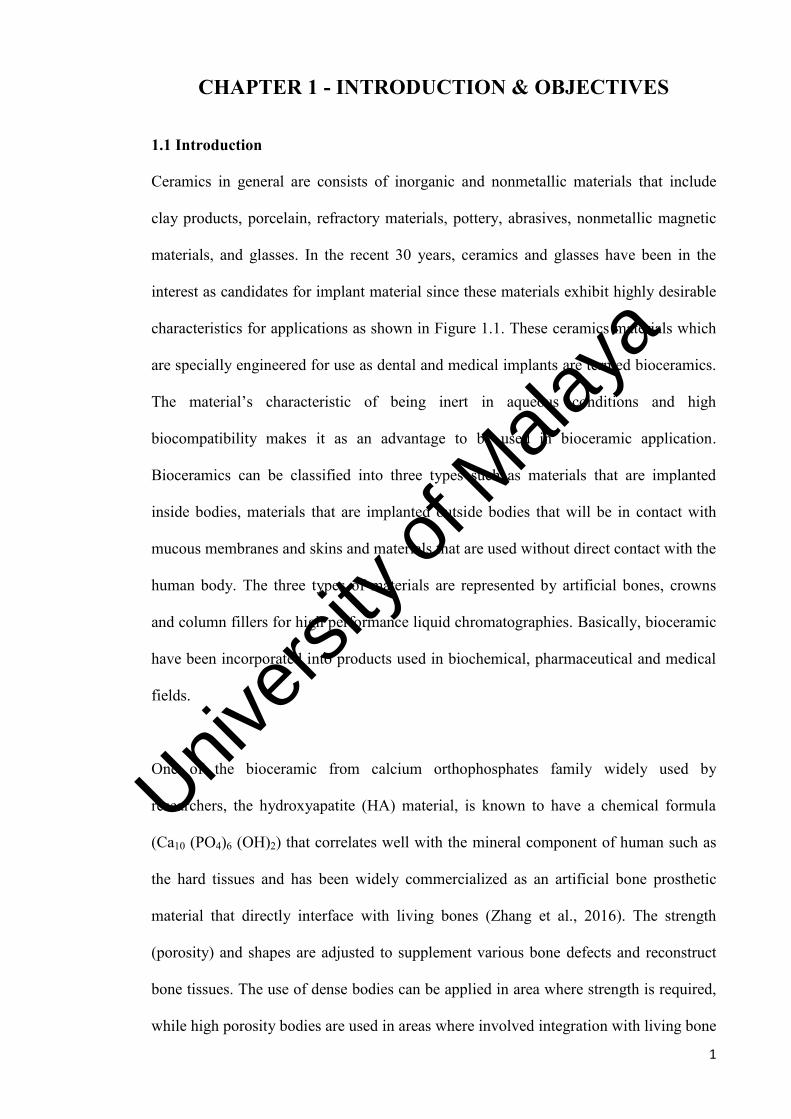
Figure 1.1: The applications of glasses, ceramics and composites in the human anatomy
(Hench and Wilson, 1993).
Univers
ity of
Mala
ya

3
Although hydroxyapatite is a promising biomaterial, its poor and unsatisfactory
mechanical properties have constraint its fullness in clinical orthopedic and dental
applications which researchers are continuously working on the improvements.
1.2 Problem Statement
Hydroxyapatite with its high biocompatibility and chemical similarity with natural bone
was introduced to be a material of interest for biomedical applications (Zakaria et al.
2013). However, due to processing difficulties and the lower mechanical properties of
HA, the applications have been limited to coatings, powders, and non-load bearing
implants only. The low mechanical properties such as poor fatigue resistance, inherent
brittleness and strength, especially its low fracture toughness (KIc) of < 1 MPam1/2 are
the major drawback for load bearing applications (Khorsand et al., 2014). Some studies
have been carried out previously by addition of dopants to enhance the low mechanical
properties of HA, however the findings showed little improvement in the fracture
toughness.
Amongst the materials which have a crucial roles in human are the magnesium and
silicon. Studies have shown that in skeletal development, silicon is necessitous and is
usually uniquely localized in the active areas of young bone (Tavangarian & Emadi,
2011). The findings from the literatures (Tavangarian & Emadi, 2011; Kharaziha &
Fathi, 2010; Siyu et al., 2007; Carlisle, 1988) showed that silicon (more than 5wt. %) is
found in the active growth areas in the bones which have a Ca/P ratio of 0.7 in the
young rats and involved in the early stage of bone calcification in physiological
conditions. Schwarz and Milne (1972) also reported that the addition of silicon in the
rats’ diet has influenced its growth. Besides silicon, magnesium is considered as the
next important element in human body as this element is closely associated with
Univers
ity of
Mala
ya

4
mineralization of calcined tissues and indirectly influences mineral metabolism which
influence the control of bone growth (Schwarz & Milne, 1972).
To improve the low mechanical properties of hydroxyapatite, there is a need to
reinforced the HA with other ceramics having better mechanical properties. Magnesium
orthosilicate (Mg2SiO4), also known as forsterite, could be a material of interest because
of the biocompatibility and higher fracture toughness. The fracture toughness of
magnesium orthosilicate ceramic has been proclaimed to be about 2.4 MPam1/2 which is
much higher than 1 MPam1/2 reported for bone implants and hydroxyapatite ceramic
(Sebdani et al., 2011; Fathi & Kharaziha, 2009).
Univers
ity of
Mala
ya

5
1.3 Objectives of Research
The main objective in conducting this research is to develop a hydroxyapatite –
magnesium orthosilicate (HA-MO) composite that exhibits better mechanical properties
while preserving its phase stability. The three-fold objectives of this research are as
follows:
1) To synthesize a HA-MO composite.
2) To establish the optimum sintering conditions of the composite that exhibits
superior mechanical properties at lower temperature suitable for biomedical
applications.
3) To establish the factors that control the properties of the composite and elucidate
the sintering mechanism of the composite.
This combination of HA-MO has not been reported widely in the literature, therefore it
is envisage that this research would generate new knowledge in the area of biomaterials.
In addition, the understanding of the various process parameters governing the
sinterability of the composites would be gained.
1.4 Scope of Project
The research will commence with an extensive literature review covering the area of
magnesium orthosilicate and hydroxyapatite in order to establish better understanding
and awareness of the current work being performed in this field. The composite will be
prepared in various compositions, by varying the magnesium orthosilicate content from
10wt. % to 50wt. % via mechanical ball milling and conventional pressureless sintering
at 1000°C to 1300°C, with ramp rate of 10°C/minute (heating and cooling) and holding
time of 2 hours for each firing. Upon sintering, the phase analysis will be carried out
Univers
ity of
Mala
ya

6
using an X-ray Diffraction (XRD) machine to evaluate the phase stability of HA. For
the grain size measurement and phase composition of HA-MO nanocomposite will be
examined through scanning electron microscope (SEM) and Energy-dispersive X-ray
(EDX) machine. Finally, the HA-MO nanocomposite will also be evaluated in terms of
mechanical properties by measuring the relative density, Vickers hardness, Young’s
modulus and fracture toughness.
1.5 Thesis Structure
In Chapter 1, a brief overview and introduction of the research area is presented. The
problem statement which lead to this research is highlighted followed by the objectives
to achieve.
Chapter 2 gives an extensive literature review on biomaterial and other researchers’
work related to HA and MO are presented. There are not many literatures available for
HA-MO composite, hence it is important to establish the fundamental of combining
these two materials.
Chapter 3 describes the framework on the synthesis technique to produce HA and MO
powder for the present work. Besides that, the experimental testing and analysis
techniques of the sintered composites will be written too. Any descriptions of the
equipment used will be included here.
The results and discussion are presented in Chapter 4. The hydroxyapatite, magnesium
orthosilicate and HA-MO powders’ characterization are discussed here, follow with
discussion on the mechanical properties and microstructural analysis. This chapter will
gives a clearer picture on the achievement of this research work.
Univers
ity of
Mala
ya

7
Finally Chapter 5 concludes the current research findings and some suggestion for
future work are given here. The appendices will contain carious experimental results,
equipment used, sample calculation including research publications.
Univers
ity of
Mala
ya

8
CHAPTER 2 - LITERATURE REVIEW
2.1 Human Bone Structure
The bulk of the human skeleton consists of bony framework which assists locomotion,
and also acts as a protective cage for internal organs. The bony framework is usually
strong and lightweight, but is also a constantly changing tissue which undergoes a
remodeling process in the entire life. Structurally, the skeleton consists of bone tissue
whereby it is formed by the inorganic and organic phases and water in the nanoscale.
Bone can be mentioned in terms of weight basis (60% inorganic, 30% organic and 10%
water) or volume basis (proportions broken down into 40%, 35%, and 25%)
respectively. (Tony, 2003; Chen et al. 2004). The bone with the inorganic phase is
referring to the ceramic that consists of mineral type of crystalline, commonly referred
to as hydroxyapatite, Ca10(PO4)6(OH)2 (Tony, 2003). The tiny apatite crystals in bone
hydroxyapatite contain impurities such as carbonate (as substitute of the phosphate
ions), potassium, magnesium, stronchloride or fluoride (as substitute of the calcium
ions), and fluoride or chloride (as subsitute of hydroxyl ions). For the bone with organic
phase, it comprises of type I collagen (90wt. %), collagen types (III and VI), and some
variety of noncollagenous proteins such as bone sialoprotein, osteonectin, osteocalcin,
and osteopontin (Boskey, 2010). Table 2.1 summarized the elements found in bones.
The hierarchical composite of the bone tissue at the micron scale and above is as shown
in Figure 2.1. At the lowest level (0.1µm) is where the mineralized collagen fibrils are
located, follow by the next level in the range of 1 to 10µm scale where two forms of the
fibrils can be arranged into known as lamellae (about 7µm thick) that contain
unidirectional fibrils in alternating angles between layers or as woven fibrils. Naturally,
the lamellar bone is commonly found in the adults (human), while the woven bone is
Univers
ity of
Mala
ya

9
found in children and large animals where rapid growth takes place, and also in the
initial healing stage of a fracture.
At the millimeter scale in different types of histological structures, lamellar bone can be
found. The primary lamellar consists of large concentric rings of lamellae similar to the
growth rings on a tree that circle the outer (2 to 3 mm) of diaphysis. In human adults,
the cortical bone is known as Haversian bone or oeteonal, where the central Haversian
canal consists of lamellae arranged in concentric cylinders, a vascular channel about 50
µm in diameter that contains nerves, variety of bone cells, and blood vessel capillaries.
Table 2.1: Elements found in an adult’s bone (Orlovskii et al., 2002; LeGeros &
Legeros, 1993).
Elements Weight (%)
Calcium 34.8
Phosphorus 15.2
Sodium 0.90
Magnesium 0.72
Potassium 0.03
Carbonates 7.40
Fluorine 0.03
Chlorine 0.13
Pyrophosphates 0.07
Other elements 0.04
Univers
ity of
Mala
ya

10
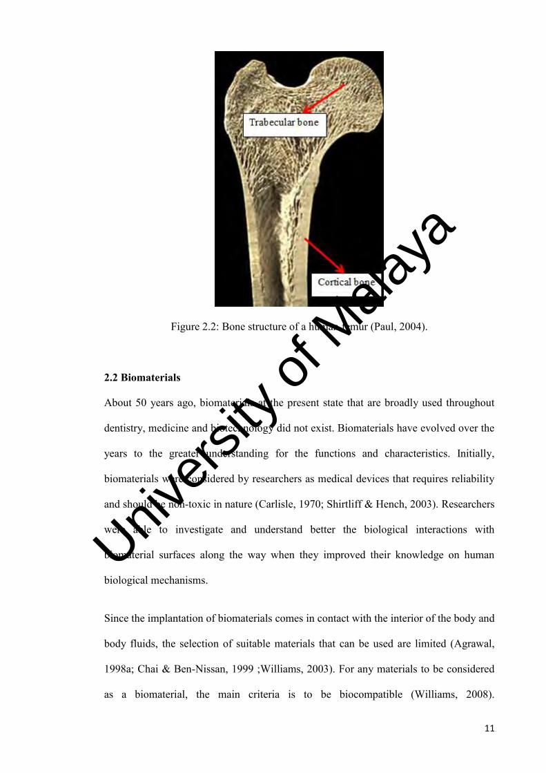
Figure 2.1: The hierarchy levels of bone microstructure (Paul, 2004).
Lastly, the tightly packed lamellar which is known as the cortical bone and highly
porous cellular solid woven bone and trabecular bone are commonly found at the
highest level of the hierarchical in the range of 1 to 5mm Basically the cortical
surrounds the trabecular bone giving that forms the bone shape or shell. For load
bearing condition, the cortical component of the bone is markedly thickened to form a
strong shaft. The internal porous framework of bones is supported by the trabecular
bone. The trabecular bone consists of stem-cell-rich bone marrow. For the growth of
new connective tissue such as muscle, cartilage, bone and tendons will require the bone
marrow. Figure 2.2 shows a typical bone structure of a human femur.
Univers
ity of
Mala
ya
11
Figure 2.2: Bone structure of a human femur (Paul, 2004).
2.2 Biomaterials
About 50 years ago, biomaterials at the present state that are broadly used throughout
dentistry, medicine and biotechnology did not exist. Biomaterials have evolved over the
years to the greater understanding for the functions and characteristics. Initially,
biomaterials were considered by researchers as medical devices that requires reliability
and should be non-toxic in nature (Carlisle, 1970; Shirtliff & Hench, 2003). Researchers
were able to investigate and understand better the biological interactions with
biomaterial surfaces along the way when they improved their knowledge on human
biological mechanisms.
Since the implantation of biomaterials comes in contact with the interior of the body and
body fluids, the selection of suitable materials that can be used are limited (Agrawal,
1998a; Chai & Ben-Nissan, 1999 ;Williams, 2003). For any materials to be considered
as a biomaterial, the main criteria is to be biocompatible (Williams, 2008).
Univers
ity of
Mala
ya

12
Biocompatibility is the interactions between biomaterial and the tissue of the human
body without causing any adverse response that affects the body.
Besides, biomaterials should exhibit characteristics of being chemical inertness, non-
thrombogenic, non-immunogenic, non-carcinogenic, non-irritant, non-toxic and stable
within the living body (Suchanek & Yoshimura, 1998; Williams, 2008), Cao et al.,
2008) that serve its functions as implant. The interactions between biomaterials and
living tissue can be summed as shown in Figure 2.3 while the factors relating to the
biocompatibility of biomaterials are indicated in Figure 2.4 (Yamanaka et al., 2006).
Figure 2.3: Interactions between living tissues and artificial materials (biomaterials)
(Yamanaka et al., 2006).
Figure 2.4: Biocompatibility factors (Yamanaka et al. 2006).
Univers
ity of
Mala
ya
13
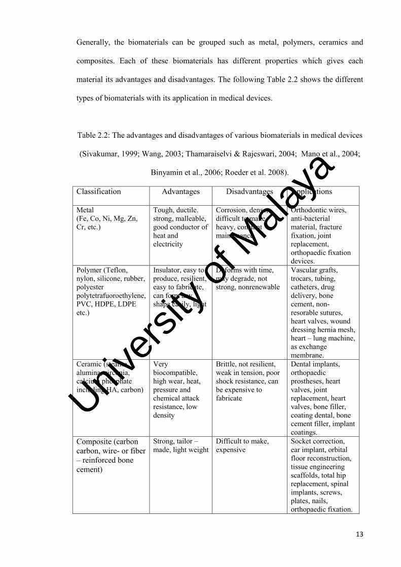
Generally, the biomaterials can be grouped such as metal, polymers, ceramics and
composites. Each of these biomaterials has different properties which gives each
material its advantages and disadvantages. The following Table 2.2 shows the different
types of biomaterials with its application in medical devices.
Table 2.2: The advantages and disadvantages of various biomaterials in medical devices
(Sivakumar, 1999; Wang, 2003; Thamaraiselvi & Rajeswari, 2004; Mano et al., 2004;
Binyamin et al., 2006; Roeder et al. 2008).
Classification Advantages Disadvantages Applications
Metal (Fe, Co, Ni, Mg, Zn, Cr, etc.)
Tough, ductile. strong, malleable, good conductor of heat and electricity
Corrosion, dense, difficult to make, heavy, constant maintenance
Orthodontic wires, anti-bacterial material, fracture fixation, joint replacement, orthopaedic fixation devices.
Polymer (Teflon, nylon, silicone, rubber, polyester polytetrafuoroethylene, PVC, HDPE, LDPE etc.)
Insulator, easy to produce, resilient, easy to fabricate, can form any shape easily, light
Deforms with time, may degrade, not strong, nonrenewable
Vascular grafts, trocars, tubing, catheters, drug delivery, bone cement, non-resorable sutures, heart valves, wound dressing hernia mesh, heart – lung machine, as exchange membrane.
Ceramic (steatite, alumina, zirconia, calcium phosphate including HA, carbon)
Very biocompatible, high wear, heat, pressure and chemical attack resistance, low density
Brittle, not resilient, weak in tension, poor shock resistance, can be expensive to fabricate
Dental implants, orthopaedic prostheses, heart valves, joint replacement, heart valves, bone filler, coating dental, bone cement filler, implant coatings.
Composite (carbon carbon, wire- or fiber – reinforced bone cement)
Strong, tailor – made, light weight
Difficult to make, expensive
Socket correction, ear implant, orbital floor reconstruction, tissue engineering scaffolds, total hip replacement, spinal implants, screws, plates, nails, orthopaedic fixation.
Univers
ity of
Mala
ya

14
One of the widely used biomaterials for implants is the metallic materials. Its
applications include simple wires and screw, to fracture-fixation plates and total joint
prostheses (artificial joints) for hips, knees, shoulders and even elbows (Dorozhkins,
2015). It is used for these applications due to attributes of stiffness, strength, toughness
and also impact resistance properties (Kulkarni et al., 2013). The metallic materials that
were initially considered as a biomaterials includes aluminum, silver, gold, stainless
steel, tantalum, vanadium steel, platinum group elements, cobalt based alloys and
titanium alloys, however due to concerns of biocompatibility and corrosion resistance,
most of the metallic materials were found to be ineffective as biomaterials (Habibovic
& Barralet, 2011). The factors mentioned have also put metallic materials less suitable
for load bearing applications thus making its usage limited. The following Table 2.3
shows some of the widely used metallic materials’ mechanical properties compared to
cortical bone.
Table 2.3: Mechanical properties of selected metallic materials compared to cortical
bone (Dee et al., 2003).
Materials Young’s
modulus, E
(GPa)
Tensile
strength,
σUTS (MPa)
Yield
strength,
σy (MPa)
Fatigue limit,
σ (MPa)
Cortical bone 15-30 70-150 30-70 -
Co-Cr alloys 210-253 655-1896 448-1606 207-950
Stainless steel 190 586-1351 221-1213 241-820
Titanium 110 760 485 300
Ti-6Al-4V 116 965-1103 896-1034 620
Univers
ity of
Mala
ya

15
On the other hand, polymers are also used for various biomedical applications as well
such as implantable devices, vascular grafts, injectable biomaterials, surgical tools and
device coatings (Dobradi et al., 2015). The use of polymers as biomaterials began due to
the need for synthetic tissue substitutes. Polymers are also used in diagnostic aids, drug
delivery and as a material for scaffolding in tissue engineering applications. However,
clinical complications will arise when the possibility of polymers releasing hazardous
chemicals occur. The hazardous chemicals can be from some unspecified additives or
chemical compounds needed for the synthesis of the polymers. It is also not suitable to
be used in biomedical applications that bear loads. Table 2.4 shows some mechanical
properties of selected polymers.
Table 2.4: Mechanical properties of selected polymers (Dee et al., 2003).
Material Tensile
strength,
σUTS (MPa)
Young’s Modulus,
E (GPa)
Elongation %
Silicone rubber 2.8 Up to 10 160
Polylactic acid 28-50 1.2-3 2-6
Polyethylene terephthalate 53 2.14 300
Nylon 6/6 76 2.8 90
Polypropylene 28-36 1.1-1.55 400-900
Polytetrafluoroethylene 17-28 0.5 120-350
Ultra high molecular weight
polyethylene (UHMWPE)
≥ 35 4-12 ≥ 300
Polymethyl methacrylate
(PMMA)
30 2.2 1.4
Univers
ity of
Mala
ya

16
The choices of implant materials changes as well as researchers established more in
depth understanding of the biomaterial mechanism and biocompatibility. Therefore,
ceramics or bioceramics became the choice as biological implants in part due to its
biocompatibility to replace and restore the function of bones (Hench, 1998; Fathi &
Hanifi, 2007; Dorozhkin, 2015). Some of the common bioceramics mechanical
properties are shown in Table 2.5. For any bioceramics to be implanted well with the
living host tissue, it needs to show a solid interface (Hench, 1998). Furthermore, the
type of materials used as implant dictates the response of tissue at the implant interface
as listed below (Hench, 1998; Dorozhkin, 2015):
a. The surrounding tissues will die if the implant material is toxic.
b. An interfacial bond forms with living tissues if the implant material is bioactive
and nontoxic.
c. A fibrous tissue of variable thickness will form if the material is biologically
inactive and nontoxic.
d. The surrounding tissue will be replaced if the implant material is biodegradable
and nontoxic.
Table 2.5: Mechanical properties of common bioceramics (Hench, 1998).
Ceramic Density
(g/cm3)
Young's
Modulus,
E (GPa)
Fracture
Toughness, KIc
(MPa.m1/2)
Compressive
Strength (MPa)
Hardness,
(Hv)
Alumina 3.98 420 3 - 5.4 4400 2300
Zirconia 5.74 - 6.08 210 6.4 - 10.9 1990 1400
Hydroxyapatite 3.05 - 3.15 80 -110 0.7 - 1.2 500 - 1000 600
Bioglass 45S5 2.6572 35 0.7 500 458
TCP 3.07 33–90 - 460 - 680 138 - 229
Univers
ity of
Mala
ya
17
Lastly, composites are materials that were developed due to the need to eliminate stress
shielding of bone and elastic modulus mismatch present in other biomaterials (Hench,
2000; Liu & Wang, 2007). It comprises of two or more combination of biomaterials
chosen from the metal, bioceramics or polymer type (Wang, 2003; Thamaraiselvi &
Rajeswari, 2004). Moreover, this material was designed with the aim of incorporating
the best characteristics from each material used (Goller et al., 2003; Binyamin et al.,
2006).
Initially, composites for biomedical applications are classified based on the type of
matrix material used (Wang, 2003). The different types of composites are classified as
polymer matrix, metal matrix or ceramic matrix composites (Wang et al., 1993; Bhaduri
& Bhaduri, 1998; Mano et al., 2004). The examples of these composites are shown in
Table 2.6 (Cao & Hench, 1996; Wang, 2003). Later, researchers used bioactivity as the
basis of classifying the different types of composites mentioned above. It can be classify
into three types such as the bioinert, bioactive and bioresorbable composites as shown in
Table 2.6 (Hench, 1991; Wang, 2003).
Originally, carbon based bioinert composite are thought to be ideal for load-bearing
orthopaedic devices as it exhibit characteristics such as lightweight, strong and have low
modulus. However, delamination under cyclic loading and chronic inflammatory
response that occurs rendered it an unsuitable biomaterial (Hench, 2000; Han et al.,
2006). Subsequently, bioactive composite which does not degrade was researched. The
research resulted in material that produces bioactive bond to bone when implanted.
Furthermore, it has mechanical properties that closely matched that of bone. The
bioresorbable composites were produced due to the need for bioactive material that
degrades and replaced by natural bones (Hench, 2000; Best et al., 2008).
Univers
ity of
Mala
ya

18
Table 2.6: Classification of composite materials (Cao & Hench, 1996; Wang, 2003).
Basis of classification
Material Matrix
Examples
Material Metal HA/TiO2, HA/Ti-6Al-4V
Ceramic HA/stainless steel, HA/glass
Polymer Carbon/PEEk, HA/HDPE
Bioactivity Bioinert carbon/carbon,
carbon/PEEK
Bioactive HA/HDPE, HA/Ti
Bioresorable TCP/PLA, TCP/PHB
2.3 Bioceramics
Bioceramics has been used for medical devices and implants for millennia. In general,
bioceramics materials can be categorized into two large groups, usually known as the
bioinert and bioactive materials (Best et al., 2008; Cao & Hench, 1996). The bioinert
materials when used as implants are considered as good biocompatibility when it
retained its mechanical and physical properties. Typical bioinert materials include
alumnia (Al2O3), zirconia (ZrO2), carbon (C), and silicon nitrades (Si3N4) (Cao &
Hench, 1996). Alumina possesses characteristics such as high hardness, high abrasion
resistance, strength and chemical inertness including good biocompatibility. This has
allowed it to be used as dental and bone implants. However, it was found that alumina
apart from being bioinert, it also induces weak tissue reaction which leads to loosening
of the implant (Best et al., 2008; Hafezi et al., 2013). On the other hand, zirconia
ceramic is known for its high toughness, high mechanical strength and good
biocompatibility. These characteristics have caught the interest to use zirconia in
orthopaedic applications and structural support (Aykul et al., 2013). Carbon, one of the
Univers
ity of
Mala
ya

19
more versatile elements, exists in many forms. It exhibit good biocompatibility with
similarity in mechanical properties as the carbon content in bones. Apart from that it
does not suffer from fatigue like other metals, polymers or even ceramics. However, as
in the case of ceramic material, it suffers from low tensile strength and brittleness
limiting its use in major load bearing application (Cao & Hench, 1996). Even though
bioinert materials are non-carcinogenic, it lacks biological response with living tissue.
The bioactive materials on the other hand can be classified further into non-resorbable
and resorbable types (Best et al., 2008; Rabiee et al., 2015). For non-resorbable
bioceramics, the bioactive materials will encourage the formation of a biological bond
between tissues and the material without degradation. Calcium phosphate ceramics,
bioactive glass-ceramics and bioactive glass are considered under this category of
material (Rabiee et al., 2015).
For calcium phosphate ceramics (CPC), the non-resorbable type is the hydroxyapatite
(HA). The HA have a chemical formula of Ca10(PO4)6(OH)2, which bear resemblance
the mineral constituent of bone and teeth (Kalita et al., 2007; He et al., 2008). Apart
from that, HA also shows excellent biocompatibility with hard tissue, skin and muscle
tissues. In addition, it can directly bond to the bone without much complication (Hsieh
et al., 2001; Murugan & Ramakrishna, 2005). Hence, it is used in various medical
applications such as periodontal treatment, alveolar ridge augmentation, dental implants
and maxillofacial surgery (Pramanik et al., 2007; Ramesh et al., 2007a). Even though
HA has been found to be beneficial for many medical application, its mechanical
properties are still low when compared to that of bone (Chu et al., 2004).
Another well-known material of bioceramics is the bioactive glass. It is a material that
derives excellent bioactivity and biocompatibility coupled with good mechanical
Univers
ity of
Mala
ya

20
properties (Thamaraiselvi & Rajeswari, 2004; Chen et al., 2006). Hench and colleague
were the first to develop a bioactive glass that uses SiO2, Na2O, CaO and P2O5 as base
component. They successfully synthesized a bioactive glass known as Bioglass® 45S5
that contains 45wt. % of SiO2, 24.5wt. % of Na2O and 24.5wt. % of CaO with addition
of 6wt. % of P2O5 to simulate the Ca/P constituents of HA (Shirtliff & Hench, 2003).
Subsequently, different compositions of bioglass were synthesized based on a SiO2-
Na2O-CaO system as shown in the compositional diagram in Figure 2.5 (Hench, 2006).
These variations of bioglass have a constant 6 wt % of P2O5 with varying SiO2-Na2O-
CaO wt. % (Hench, 1991; Cao & Hench, 1996; Hench et al., 1998; Vitale-Brovarone et
al., 2008).
Bioactive glass-ceramics are known for its high compressive strength, bending strength,
fracture toughness, interfacial bonding to bone and excellent resistance to degradation
of properties (Shirtliff & Hench, 2003). This composition of glass-ceramics phase was
modified and used by many researchers. However, the most important modification was
developed by Yamamuro and Kokubo (1992). They developed A/W
(apatite/wollastonite) bioactive glass-ceramics which have excellent mechanical
properties, biocompatible, bioactive and it is non-toxic (Kokubo et al., 2003; Best et al.,
2008). Subsequently, many variations of glass-ceramics were developed as shown in
Table 2.7 (Cao & Hench, 1996). Though glass-ceramics have been used in various
medical applications, it is still unsuitable to be used in load-bearing applications
(Kokubo et al., 2004).
Univers
ity of
Mala
ya

21
Table 2.7: Types of bioactive glass-ceramics (Cao & Hench, 1996).
Component (wt %)
Types of glass-ceramics
A/W glass-ceramics
KG Cera Ceravital®
Mina 13 Ceravital®
KGy213 Ceravital®
M8/1 Ceravital®
SiO2 34.2 46.2 46.0 38.0 50.0
Ca(PO3)2 - 25.5 16.0 13.5 7.1
CaO 44.9 20.0 33.0 31.0 -
P2O5 16.3 - - - -
Na2O - 4.8 - 4.0 5.0
MgO 4.6 2.9 5.0 - -
CaF2 0.5 - - - -
K2O - 0.4 - - -
Al2O3 - - - 7.0 1.5
Ta2O5 - - - 5.5 -
TiO2 - - - 1.0 -
B2O3 - - - - 4.0
Al(PO3)3 - - - - 2.4
SrO - - - - 20.0
La2O3 - - - - 6.0
Gd2O3 - - - - 4.0
Figure 2.5: Bone bonding in terms of compositional (wt. %) (Hench, 1991; Hench,
2006).
Univers
ity of
Mala
ya

22
Resorbable bioceramics are ceramics that progressively degrade over time when
implanted in physiological environment. As degradation occurs, it will slowly be
replaced by the host’s natural tissues (Binyamin et al., 2006). The resorbable
bioceramics includes corals, calcium sulphate and calcium phosphates ceramics (CPC)
in the form of tricalcium phosphates (TCP) (Le Huec et al., 1995; Adamopoulus &
Papadopoulus, 2007). Moreover, these ceramics also exhibits characteristics such as
bioactivity and also biocompatibility ensuring no formation of fibrous tissues layer
(Giannoudis et al., 2005; Viswananth et al., 2007).
Tricalcium phosphate (TCP) have a chemical formula of Ca3(PO4)2. Typically, TCP is
used in applications such as periodontal corrections, augmentation of bony contours and
drug delivery devices (Heymann & Passuti, 1999; Liu et al., 2008). On the other hand,
natural corals have cancellous pore that provide exceptional structure for ingrowths of
bone while allowing adsorption of nutrients and metabolism (Xu et al., 2001; Zhang et
al., 2007). Therefore, it is used for repairing of traumatised bone and replacement of
diseased bone including correction of various bone defects (Ben-Nissan et al., 2004).
Calcium sulphate (CaSO4) has been used successfully in periodontal therapy due to its
regenerative behaviour. Apart from that, it can also create barriers which isolate
connective tissues while allowing bone regeneration to occur during healing
(Adamopoulus & Papadopoulus, 2007).
2.4 Calcium Phosphate
Calcium phosphate is one of the largest and most important inorganic parts that make up
hard tissues of bone. It is similar to the crystallographic and chemical composition of
materials found in bones (Pramanik et al., 2007; Best et al., 2008). This similarity
contributes to the properties such as bioactivity and biocompatibility (Calafiori et al.,
Univers
ity of
Mala
ya

23
2007; Cengiz, et al., 2008). Therefore it is used widely in various medical applications
such as facial and oral surgery, drug carriers, dentistry and orthopaedics (Rodriguez-
Lorenzo et al., 2001; El Briak-BenAbdeslam et al., 2008).
The calcium phosphates can be grouped according to its Ca/P ratio ranging from 0.5 to
2.0 as summarized in Table 2.8. The significant of Ca/P ratio is reflected in the acidity
and the solubility of the mixture. For mixture with Ca/P < 1, the acidity and the
solubility are exceptionally high. As the Ca/P ratio increases, solubility would decreased
(with exception of TTCP and α-TCP) while acidity moves towards basicity.
Furthermore, the in vivo solubility of the material can be predicted in the order as
shown (Fernandez et al., 1999a; Aizawa et al., 1999; Bohner, 2000; Best et al., 2008):
MCPM > TTCP, α-TCP > DCPD > DCPA > OCP > β-TCP > CDHA > HA
In biomedical industry, the commercially available of calcium phosphate includes
hydroxyapatite (HA), β-tricalcium phosphate (β-TCP), biphasic calcium phosphate
(BCP), monocalcium phosphate monohydrate (MCPM) and unsintered apatite (AP)
(Kalita et al., 2007). Among those mentioned, the most widely used calcium phosphate
based ceramics are the HA and β-TCP (Slosarcyzk et al., 1996 ; Santos et al., 2004;
Thamaraiselvi & Rajeswari, 2004).
Hydroxyapatite (HA) is one of the most studied phases of calcium phosphate due to
excellent stability in aqueous media especially for pH above 4.3. Moreover, researchers
have established human blood pH to be at ~7.3 (Cao & Hench, 1996; Kalita et al., 2004;
Best et al., 2008; Ramesh et al., 2008). Apart from that, HA’s chemical formula of
Ca10(PO4)6(OH)2, correlates well with the mineral component of human hard tissues as
Univers
ity of
Mala
ya

24
shown in Table 2.9 (LeGeros & LeGeros, 1993; Suchanek et al., 1998; Suchanek et al.,
2002; Liu et al., 2004; Dorozhkin, 2007).
In addition to that, HA has a Ca/P ratio of 1.67 that is similar to stoichiometric
hydroxyapatite (Landi et al., 2000; Afshar et al., 2003; Sung et al., 2004; Kumta et al.,
2005). Moreover, it has a hexagonal crystal structure with a P63/m space group which
refers to a space group with a six-fold symmetry axis with a three-fold and a
microplane. Furthermore, the lattice constants of the hexagonal HA are a = 9.422 Å and
c = 6.883 Å, matches that of hard tissues as given in Table 2.8 (Narasaraju & Phebe,
1996; Zhang & Gonsalves, 1997; Jokanovic et al., 2006; Kalita et al., 2007; Suchanek et
al., 1997).
These similarity of HA to bone’s mineral phase has given it excellent biocompatibility
and bioactivity properties. What's more, these properties allows HA to bond with living
tissues without showing any adverse effects such as toxicity, inflammatory and
immunogenic (Murugan & Ramakrishna, 2005; Wang, et al., 2005; Mobasherpour et
al., 2007; Fathi et al., 2008).
Even though HA exhibits properties that causes no adverse effect on living tissues, there
are concern with regards to its mechanical properties. Inherently, HA is brittle, thus, this
translates into low fracture toughness (< 1 MPam1/2), low flexural strength (< 140 MPa)
and high elastic modulus (~ 120 GPa). Consequently, the usage of HA is limited to non-
load bearing applications (Ruys et al., 1995a; Muralithran & Ramesh, 2000; Donadel et
al., 2005; Ramesh et al., 2007a; Ramesh et al., 2007b; He et al., 2008).
Univers
ity of
Mala
ya

25
Table 2.8 The various calcium phosphates solubility product constants at 25oC and 37oC (Fernandez et al., 1999a; Bohner, 2000; Vallet-Regí &
González-Calbet, 2004; Bandyopadhyay et al., 2006; Kalita et al., 2007; Dorozhkin, 2007 & 2008).
Ca/P ratio Compound Chemical formula Acronym
Solubility at 25oC – log(KS)
Solubility at 37oC – log(KS)
pH stability range in aqueous solution at 25oC
2.0 Tetracalcium phosphate Ca4O(PO4)2 TTCP 38-44 37-42 a
1.67 Hydroxyapatite Ca10(PO4)6(OH)2 HA 116.8 117.2 9.5-12
1.5-1.67 Calcium-deficient hydroxyapatite e
Ca10-x(HPO4)x(PO4)6-x(OH)2-xf
(0 < x < 1) CDHA ~85.1 ~85.1 6.5-9.5
1.2-2.2 Amorphous calcium phosphate CaxHy(PO4)z · nH2O, n = 3-4.5; 15-20% H2O
ACP b b ~5-12d
1.50 β-Tricalcium phosphate β-Ca3(PO4)2 β-TCP 28.9 29.5 a
1.50 α-Tricalcium phosphate α-Ca3(PO4)2 α-TCP 25.5 25.5 a
1.33 Octacalcium Ca8H2(PO4)6 · 5H2O OCD 96.6 95.9 5.5-7.0
Univers
ity of
Mala
ya

26
Table 2.8 (continued)
Ca/P ratio Compound Chemical formula Acronym
Solubility at 25oC – log(KS)
Solubility at 37oC – log(KS)
pH stability range in aqueous solution at 25oC
1.00 Dicalcium phosphate dehydrate CaHPO4 · 2H2O DCPD 6.59 6.63 2.0-6.0
1.00 Dicalcium phosphate anhydrous CaHPO4 DCPA 6.90 7.02 c
0.50 Monocalcium phosphate monohydrate Ca(H2PO4)2·H2O MCPM 1.14 - 0.0-2.0
0.50 Monocalcium phosphate anhydrous Ca(H2PO4)2 MCPA 1.14 - c
a Hard to precipitated from aqueous solutions. b Some values found were: 25.7 ± 0.1 (pH = 7.40), 29.2 ± 0.1 (pH = 6.00), 32.7 ± 0.1 (pH = 5.28). c Stable level of temperature > 100oC. d In metastable condition e Commonly known as precipitated HA. f When x = 1, ( the boundary condition with Ca/P = 1.5).
Univers
ity of
Mala
ya

27
Table 2.9: Composition of physical and mechanical properties of human bone, enamel
and hydroxyapatite (HA) ceramic (LeGeros & LeGeros, 1993; Suchanek
et al., 1998; Dorozhkin, 2007).
Enamel Bone HA
Constituents (wt%)
Calcium, Ca2+ 36.0 24.5 39.6
Phosphorus, P 17.7 11.5 18.5
Ca/P molar ratio 1.62 1.65 1.67
Sodium, Na2+ 0.5 0.7 -
Potassium, K+ 0.08 0.03 -
Magnesium, Mg2+ 0.44 0.55 -
Carbonate CO32- 3.2 5.8 -
Flouride, F- 0.01 0.02 -
Chloride, Cl- 0.30 0.10 -
Total inorganic (mineral) 97.0 65.0 100.0
Total organic 1.0 25.0 -
Absorbed H2O 1.5 9.7 -
Traces elements: Sr2+, Ba2+,
Pb2+, Fe3+, Zn2+, Cu2+ etc.
Cyrstallographic properties
Lattice parameters (±0.003 Å)
a-axis 9.441 9.419 9.422
c-axis 6.882 6.880 6.880
Crystallinity index 70-75 33-37 100
Crystallite size, Å 1300 x 300 250 x 25-50 -
Sintering products
@ 800oC – 950oC β-TCP + HA β-TCP + HA HA + CaO
Mechanical properties
Elastic modulus (106 MPa) 0.014 0.020 0.01
Tensile strength (MPa) 70 150 100
Univers
ity of
Mala
ya
28
2.5 Magnesium Orthosilicate
Magnesium orthosilicate (Mg2SiO4), also known as forsterite, is traditionally used as an
industrial ceramic and is often sought after for its favorable refractory properties. In
view of its low heat conductivity, creep stability and good refractoriness under load,
magnesium orthosilicate is often used as thermal insulators or refractory material for
heat preservation (Xu and Wei, 2005; Li et al., 2009). Following recent developments
on magnesium orthosilicate, studies revealed that magnesium orthosilicate is chemically
stable in fuel cell environments; thus making it suitable to manufacture manifolds made
of solid oxide fuel cell (SOFC) (Kosanovic et al., 2005).
Moreover to its favorable thermal properties, Sugiyama et al. (2006) revealed that
magnesium orthosilicate possessed excellent dielectric properties, thus making it a
material suitable for millimeter-wave communication. Additionally, it was also
discovered that magnesium orthosilicate is an excellent active medium for tunable
lasers (Kosanovic et al., 2005; Tavangarian and Emadi, 2010). For instance, chromium-
doped magnesium orthosilicate (Cr4+: Mg2SiO4) lasers have a broad tunable region of
1.1 – 1.3 μm (Tavangarian & Emadi, 2011) which are also found to be ideal for optical
coherence tomography due to the lower scattering in biological tissues (Kharaziha &
Fathi, 2010; Tavangarian & Emadi, 2011; Sara et al, 2012).
Nevertheless, recent findings have established potential biomedical applications for
magnesium orthosilicate ceramics. Based on the chemical formula of magnesium
orthosilicate (Mg2SiO4), this ceramic was found to be composed from essential minerals
which are heavily involved in bone development. Studies have shown that in skeletal
development, silicon is an important element and is uniquely found in the active areas
of young bone (Xie et al., 2009; Jagdale & Bamane, 2011). The findings from the
Univers
ity of
Mala
ya

29
literatures (Schwarz & Milne, 1972; Lugo et al., 2016) showed that young rats’ bone
having a Ca/P ratio of 0.7 consists of more than 5 wt. % of silicon. The role of silicon is
mainly involved in bone calcification in the early stage of physiological conditions.
Schwarz and Milne (1972) also reported that the addition of the silicon in the rats’ diet
has an influence towards the growth. Another essential mineral which controls the bone
growth in human body and also oxidation of calcined tissues is magnesium (Legeros,
1991; Nikaido et al., 2015). Besides the significance of its minerals towards bone
development, magnesium orthosilicate was also found to exhibit bioactivity when
experimented in SBF solution (Kharaziha and Fathi, 2009).
In addition to its favorable magnesium silicate composition, magnesium orthosilicate
also demonstrated high mechanical properties; whereby recent data suggests that
magnesium orthosilicate possessed a maximum fracture toughness of 4.3 MPa·m1/2
(Kharaziha and Fathi, 2010) and this is much higher than HA. According to
documented figures, the fracture toughness of magnesium orthosilicate was established
to be within the region of 3.6 – 4.3 MPa·m1/2 (Juang & Hon, 1996; Fathi and Kharaziha,
2009; Kharaziha and Fathi, 2010). Hence, at a maximum of 4.3 MPa·m1/2, the fracture
toughness of magnesium orthosilicate is approximately 3 MPa·m1/2 more when
compared to HA (KIc = 1.18 – 1.25 MPa·m1/2 (Akao et al., 1981; Gu et al., 2002;
Kalmodia et al., 2010).
2.6 Hydroxyapatite Composites
Hydroxyapatite is a material commonly utilized in biomedical applications and
regenerative medicines for bones because of its biocompatibility and structural analogy
which are found in bone and dental tissues. However, the material has limitation where
it cannot be used in high load bearing conditions due to low mechanical properties such
Univers
ity of
Mala
ya
30
as low strength, low fracture toughness and poor resistance. To fulfil the function of
hydroxyapatite as a biological and structural material, HA composites have been
developed.
In general, no synthetic material will be completely harmonious with the living
environment. However, there are different levels of inertness associated with
bioceramic. Few factors are taken into account when it comes to the biocompatibility
when implanted such as charge, roughness, composition of material, surface wettability,
implants size and shape. For a bioceramic composite to be used as a material in
implantation, the composite must not cause any adverse effects to the blood or tissue-
material bonding, and most importantly biocompatible. This requires the bioceramic
composite to integrate naturally in the presence of blood and tissue upon implantation.
The bioceramic composites can be categorized as bioinert, bioactive and biodegradable.
2.6.1 Hydroxyapatite – Bioinert Composites
Inert bioceramics are stable materials which do not interact with tissue activity when
implanted within human body. When implanted into living organism or tissue, they
show high chemical stability or even if minor non-toxic degradation products are
released, they can be readily assimilated by the body. Some bioinert ceramics like
zirconia and alumina ceramics are used for load bearing implants as these materials
exhibited better mechanical properties than bioactive ceramics.
Knowing that mechanical properties of hydroxyapatite is unsatisfactory, the
incorporation of zirconia and alumina to form hydroxyapatite – bioinert composites can
help to improve its mechanical strength and fracture toughness.
Univers
ity of
Mala
ya

31
2.6.1.1 Hydroxyapatite – Zirconia
Hydroxyapatite is mainly used as a hard tissue implant in non-load bearing areas due to
its excellent biocompatibility in the human body. It can only be used in non-load
bearing areas because of its brittle nature. However, inert crystalline ceramics such as
zirconia can be mixed with hydroxyapatite to make composites that take advantage of
the biocompatibility of hydroxyapatite and high strength of zirconia (Evis, 2007).
Zirconia (ZrO2) is a well-known material for its mechanical properties and low toxicity,
which makes it as a viable biomaterial for dental implants and is expected to be a new
bone restorative material (Evis, 2007; An et al., 2007; Chiu et al., 2007; Drdlik et al.,
2015).
Chiu (2007) studied the ramification incorporating zirconia into the microstructural
evolution of porous hydroxyapatite. The calcium zirconate (CaZrO3) is a compound
formed from the product reaction of HA and zirconia and is bio-inert towards human
tissue (Chiu et al., 2007). The retribution of such a reaction is the consumption of the
Ca from Ha. As the amount of ZrO2 is large, HA would be consumed completely after
sintering, and α- or β- tricalcium phosphate formed (Chiu et al., 2007). In this study, 0 –
5 vol.% of 230 nm zirconia particles was added into hydroxyapatite (HA). The XRD
trends of the sintered HA and HA-ZrO2 specimens at 1300°C is shown in Figure 2.6.
No ZrO2 peaks are found in the patterns, instead calcium zirconate is found in the
sintered specimens which indicated that the reaction between HA and ZrO2 has taken
place to form CaZrO3.
Univers
ity of
Mala
ya

32
Figure 2.6: XRD trends for HA and HA-ZrO2 specimens after sintering at 1300°C for 2
h (Chiu et al., 2007).
It was reported by Chiu (2007) that the CaZrO3 can thus act as an effective grain growth
inhibitor for the HA grains. Figure 2.7a and Figure 2.7b show the typical micrographs
of HA and HA-CaZrO3 samples after sintering and Figure 2.8 shows the average size of
HA grains in the sintered composites as a function of ZrO2 content. The addition of
ZrO2 particles reduces the size of HA grains. Moreover, as the pores are smaller, the
CaZrO2 particles can act as microstructure refiner to HA.
(a)
Univers
ity of
Mala
ya

33
(b)
Figure 2.7: Typical micrographs of (a) HA and (b) HA-ZrO2 samples after sintering at
1300°C for 2 h. Several CaZrO3 particles in (b) are indicated by arrows
(Chiu et al., 2007).
Figure 2.8: Size of the HA grains in the HA-CaZrO3 composites as a function of starting
ZrO2 content (Chiu et al., 2007).
Univers
ity of
Mala
ya
34
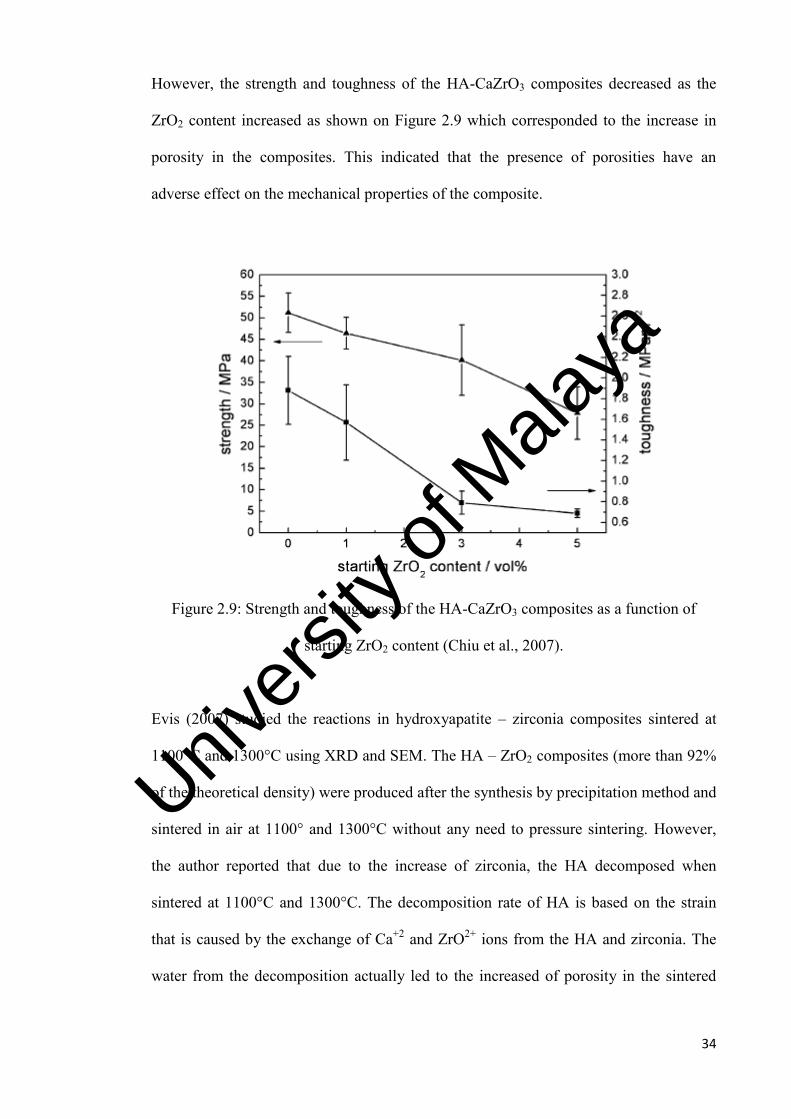
However, the strength and toughness of the HA-CaZrO3 composites decreased as the
ZrO2 content increased as shown on Figure 2.9 which corresponded to the increase in
porosity in the composites. This indicated that the presence of porosities have an
adverse effect on the mechanical properties of the composite.
Figure 2.9: Strength and toughness of the HA-CaZrO3 composites as a function of
starting ZrO2 content (Chiu et al., 2007).
Evis (2007) studied the reactions in hydroxyapatite – zirconia composites sintered at
1100°C and 1300°C using XRD and SEM. The HA – ZrO2 composites (more than 92%
of the theoretical density) were produced after the synthesis by precipitation method and
sintered in air at 1100° and 1300°C without any need to pressure sintering. However,
the author reported that due to the increase of zirconia, the HA decomposed when
sintered at 1100°C and 1300°C. The decomposition rate of HA is based on the strain
that is caused by the exchange of Ca+2 and ZrO2+ ions from the HA and zirconia. The
water from the decomposition actually led to the increased of porosity in the sintered
Univers
ity of
Mala
ya

35
samples (Evis, 2007). The porosities of the composites as determined from SEM are
presented in Table 2.9.
Table 2.10: Porosity in HA-zirconia composites ( %) (Evis, 2007).
More recently, Jadwiga et al. (2016) also reported that zirconia additive promotes
decomposition of both hydroxyapatite of natural origin as well as the synthetic one. The
reaction led to the formation of β-TCP and the CaO-ZrO2 solid solution. The research
was based on pressureless sintering performed at 1000-1300°C and hot pressing at
1050-1300°C. Table 2.10 shows the phase composition of hydroxyapatite matrix-
zirconia composites pressureless sintered and hot pressed. No free CaO was observed as
it was assume that it has dissolved in zirconia.
Univers
ity of
Mala
ya

36
Table 2.11: XRD phase composition of hydroxyapatite matrix-zirconia composites pressureless sintered and hot pressed (Jadwiga et al., 2016).
Univ
ersity
of M
alaya

37
2.6.1.2 Hydroxyapatite – Alumina
In total joint replacement, alumina (Al2O3) ceramics are being used because of excellent
biocompatibility, inertness and high wear resistance (Li et al., 1995; Carolina et al.,
2016). However, in many natural bone replacement, this material does not bond easily.
On the other hand, hydroxyapatite ceramics which have the competence to be implanted
inside bone are used as implant materials because it also helps to promote the formation
of new bone in osseointegration implant (Li et al., 1995; Mobasherpour et al., 2009;
Zhang et al., 2016). Therefore, mechanical properties of HA and HA coatings can be
improved by addition of alumina (Zhang et al., 2016).
Li et al. (1995) studied the hydroxyapatite alumina composites, with HA compositions
of 15wt. %, 25wt. %, 30wt. %, 70wt. % and pure HA. The specimens were sintered at
1275°C and implanted into 12 New Zealand White rabbits’ femoral cortical bones for
duration of 3 months. The results indicated that the bonding strength of the implants
increased as the HA content increases in the composite which indicated the significant
role of HA to the implants thru the new bone apposition. However, no linear
relationship can be drawn from HA content and bonding strength. Among the other
composites, the similar fracture interfaces and same level of bonding strength were
obtained from the pure HA and 70% HA composite through SEM. This supports the
high bonding strength transfer ability of the contact zone. For the mechanical strength
of the composites, a three-point bending test method was used to measure the strength.
The bending strength of the materials decreased with increasing HA content as shown
in Figure 2.10. The mechanical strength of HA containing ceramics increased with
increasing alumina content. The reinforcement effect of alumina can be observed in the
composite with 30% alumina in HA, where the strength was doubled compared to pure
Univers
ity of
Mala
ya

38
HA. When the volume of alumina exceeded 50%, the strength of the composites was
determined by the distribution of the HA phase in the alumina matrix.
Li et al. (1995) also reported that densities decreased with increasing HA content as
shown in Table 2.11.
Figure 2.10: Bending strength of HA-alumina composites (Li et al., 1995).
Table 2.12: Density and surface profile of selected implant cylinders (Li et al., 1995).
Materials Density (gcm-3)
Al2O3 3.97
HA 3.15
15HA/Al2O3 3.85
25HA/Al2O3 3.75
30HA/Al2O3 3.73
70HA/Al2O3 3.39
Univers
ity of
Mala
ya
39
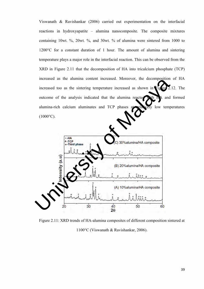
Viswanath & Ravishankar (2006) carried out experimentation on the interfacial
reactions in hydroxyapatite – alumina nanocomposite. The composite mixtures
containing 10wt. %, 20wt. %, and 30wt. % of alumina were sintered from 1000 to
1200°C for a constant duration of 1 hour. The amount of alumina and sintering
temperature plays a major role in the interfacial reaction. This can be observed from the
XRD in Figure 2.11 that the decomposition of HA into tricalcium phosphate (TCP)
increased as the alumina content increased. Moreover, the decomposition of HA
increased too as the sintering temperature increased as shown in Figure 2.12. The
outcome of the analysis indicated that the alumina reacted with HA and formed
alumina-rich calcium aluminates and TCP phases at relatively low temperatures
(1000°C).
Figure 2.11: XRD trends of HA-alumina composites of different composition sintered at
1100°C (Viswanath & Ravishankar, 2006).
Univers
ity of
Mala
ya

40
Figure 2.12: XRD trends of 30wt. % alumina sintered at different temperatures
(Viswanath & Ravishankar, 2006).
In another work, Aminzare et al. (2013) used biomimetic method to synthesize the
hydroxyapatite-alumina nanocomposite. In this work, 20wt. % alumina nanopowder
was mixed with HA before sintered at the rate of 5°C/minute to 1400°C. The results
showed that the addition of alumina was beneficial in enhancing the bending strength
by 40% and improved the hardness from 2.52 (pure HA) to 5.12 (HA-Al2O3 composite)
(Aminzare et al., 2013).
2.6.2 Hydroxyapatite – Bioactive Composites
Bioactive materials are engineered for a specific biological activity that will give strong
bonding to bone. During the implantation in the living bone, the kinetic modification of
the surface which is time dependent will takes place in the biological activity (Kim,
2001). An ion exchange reaction takes place between the bioactive implant and
surrounding body fluids which results in the formation of a biologically active calcium
phosphate layer on the implant. The layer is chemically and crystalographically
Univers
ity of
Mala
ya

41
equivalent to the mineral phase of bone (Kim, 2001). Prime examples of bioactive
materials are bioceramics such as synthetic hydroxyapatite, glass ceramic (A-W) and
bioglass. Figure 2.13 shows the clinical uses of the bioceramics mentioned earlier in
bone repairs and replacements.
Figure 2.13: Bioglass: BG, Cerabone A/W: A-W, Sintered hydroxyapatite: HA,
Sintered β-tricalcium phosphate: TCP, HAPEX: HP (A- cranial repair, B – middle ear
bone replacement, C – maxillofacial reconstruction, D – bioactive coating on dental
root, E – alveolar ridge augmentation, F – periodontal pocket obliteration, G – spinal
surgery, H – iliac crest repair, I – bone filler, and J – bioactive coating on joint stem
(Kim, 2001).
The mechanical strength of bioactive ceramics is generally lower than that of bioinert
ceramics. Therefore, over the years there are many research on uplifting the limitation
of bioactive ceramics thru combination of biomaterials to improve the mechanical
properties aspect of it.
Univers
ity of
Mala
ya

42
2.6.2.1 Hydroxyapatite – Bioactive Glass
In the SiO2 – CaO – P2O5 system, the design of bioactive glass ceramic plays an
important role as it is used for repair and replacement of diseased and damaged bone
tissue (Bogdanov et al., 2008; Rabie et al., 2015). In initial discovery, Hench et al.
(2006) explored the glass ceramic (Bioglass®), and found to bond with the living bone
without the fibrous tissue formed on the bone surfaces. The important feature of
bioactive glass is their biological activity which promotes their usage for some specific
application. Hence, the formation of hydroxyapatite on material surface can be observed
from the active materials which will be able to produce a stable bond when in contact
with bone tissue from implantation (Bianco et al., 2007; Laczka et al., 2016). It can also
be defined as their ability to induce specific cell responses leading to faster regeneration
of bone tissue. The main drawback from being used in load-bearing applications was
due to the amorphous three dimensional glass network which is believed to be the cause
of low fracture toughness and low bending strength which was reported in the range of
40-60 MPa (Dobradi et al., 2015).
Tancred et al. (1998) performed a study on the sintering of HA with glass additions.
According to the authors, the objectives for sintering HA with a glassy phase were to
enhance the densification and to improve the bioactivity of the composite
(Kangasniemi, 1993; Chern et al., 1993). In this study, synthetic hydroxyapatite was
prepared using a wet method and phosphate glass was prepared using conventional
glass-forming techniques. The HA was mixed with phosphate glass with composition of
2.5, 5, 10, 25 and 50wt. % and sintered at 4°C/minute with holding time of 3 hours at
desired temperatures of 1100°C to 1350°C. A maximum of 97.8% theoretical density
was achieved by HA at 1300°C as shown in Figure 2.14. On the other hand, composites
containing 2.5 and 5wt. % glass additions attained maximum densification above
Univers
ity of
Mala
ya

43
1250°C. For composites containing above 10wt. % achieved slightly lower
densification.
Figure 2.14: Theoretical density of HA/phosphate glass composites
(Tancred et al., 1998)
For microstructure evaluation, rapid grain growth was observed for pure HA at above
1200°C (Figure 2.15). It was reported that the average grain size increased from 1.0 ±
0.05µm at 1200°C to 2.47 ± 0.13 µm at 1350°C. Subsequently, HA with phosphate
composition up to 10wt. % resulted in high porosity when sintered at 1200°C, probably
due to reaction between the glass and HA. However, between 1200 and 1250°C, there
was a decreased in porosity for each composite as the materials reached almost
maximum density.
Univers
ity of
Mala
ya
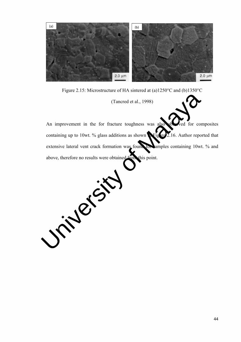
44
Figure 2.15: Microstructure of HA sintered at (a)1250°C and (b)1350°C
(Tancred et al., 1998)
An improvement in the for fracture toughness was also observed for composites
containing up to 10wt. % glass additions as shown in Figure 2.16. Author reported that
extensive lateral vent crack formation was found for samples containing 10wt. % and
above, therefore no results were obtained from this point.
(a) (b)
Univers
ity of
Mala
ya

45
Figure 2.16: Fracture toughness of HA/phosphate glass compositions (Tancred et al.,
1998)
In another work, Goller et al. (2003) sintered the naturally produced HA from human
teeth with addition of 5wt. % and 10wt. % bioglass at 1200°C and 1300°C for 4 hours.
The density of the composites varied depending on the reinforcement contents and
sintering temperatures as shown in Figure 2.17. The authors found that the bioglass
reinforcement content from 5 to 10wt. % resulted in increasing density from 2.66 to
2.72 g/cm3 when sintered at 1200°C. However, the density improvement did not show
much variation when composites were sintered at 1300°C, which was about 2.72 g/cm3
for 5 and 10wt. %.
Univers
ity of
Mala
ya

46
Figure 2.17: Density of composites sintered at 1200°C and 1300°C (Goller et al., 2003).
When composites sintered at 1200°C and 1300°C, the microhardness also displayed a
trend of decrement. The authors reported that increasing the bioglass content from 5 to
10wt. % caused the decrease in hardness from 474 to 383 HV when sintered at 1200°C
and 365 to 234 HV when sintered at 1300°C.
2.6.2.2 Hydroxyapatite – AW Glass
Since the discovery and introduction of bioactive ceramic concept (later named as
Bioglass®) by Hench and his co- workers in early 1970s for clinical applications, it has
opened new avenue to other researchers to develop further in bioactive glass which
could spontaneously bond to and integrate with living bone with improved mechanical
properties. Another researcher, Kokubo et al. (1982) reported on the discovery of a
bioactive glass ceramic, named glass-ceramic A-W (appatine-wollastonite) which has
higher mechanical strength as well as high bone bonding capability. This glass ceramic
was introduced to be the first load bearing bioactive bone substitute which it has been in
used in bulk, granular and porous forms in mainly spinal surgery (Kokubo et al., 1982).
It was later commercialized as Cerabone®. The glass ceramic A-W has two crystalline
phases; 38wt. % of oxyfluorapatite Ca10(PO4)6(O,F2) and 34wt. % wollastonite
(CaOSiO2) 50-100nm in size and a residual vitreous phase of MgO-CaO-SiO2. The
Univers
ity of
Mala
ya

47
authors also developed a simulated body fluid (SBF) test in 1990 to examine the
behavior of glass-ceramic A-W. This technique was also used to rank other materials in
terms of their bioactivity.
Kokubo (1990) observed that an apatite layer that is capable of bonding to the bone was
formed when the glass-ceramic A-W was placed in the body. The findings of the
authors showed that the bonding was so strong that when the mechanically tested
samples were retrieved, there were no signs of fracture found on the interface of the
bone implant, but rather inside of the bone structure (Kokubo, 1990). The author
believed that silanol groups were formed at the glass-ceramic surface since no
amorphous silica layer was observed between hydroxyl-carbonate apatite (HCA) layer
and the glass-ceramic A-W when tested in a simulated body fluid (SBF) as shown in
Figure 2.18. Silanol groups are believed to be the cause of the apatite layer formation as
these groups provide favourable sites needed for apatite crystal nucleation and growth.
The apatite crystals form rapidly on the surface of the glass-ceramic A-W and the
calcium and phosphate ions in the surrounding media will be consumed.
Figure 2.18: Apatite formation tested in simulated body fluid (SBF) (Kokubo, 1990)
Univers
ity of
Mala
ya

48
Other authors, such as Ryu et al. (2005) and Jun et al. (2007) experimented with
hydroxyapatite scaffolds reinforced with glass ceramic A-W coating. The authors
reported that the glass coating improved the strength as well as the bioactivity of the
HA scaffolds. It was observed that the higher alkaline phosphatase activity of the cells
successfully cultured on the scaffold coated with A-W glass than on HA scaffold alone,
which indicated that the bioactivity had been improved (Jun et al., 2007).
In another work, Encinas-Romero et al. (2010) studied the HA-wollastonite composite
powders via sol-gel processing method. The ratios of 20, 50, and 80wt. % were
synthesized and processed thru sol-gel method before evaluated through thermal
analysis, FTIR, XRD, Raman spectroscopy and SEM. The hydroxyapatite was observed
to have formed in the composites at relatively low temperature accompanied by calcium
carbonate formation between 400 to 420°C, while wollastonite remained the same. Heat
treatment and hydrochloric acid were used to produce the B-type carbonated
hydroxyapatite-wollastonite composites as final products. The compounds of the
composites experienced no decomposition upon heating between 1000°C to 1400°C.
The authors investigated and concluded on the addition of 80wt. % wollastonite had
tremendously improved the hardness by 200% when compared to pure HA, and the
reduced elastic modulus is also 45% higher as shown in Figure 2.19. In summary, the
incorporation of wollastonite has contributed to the improvement of the mechanical
properties.
Univers
ity of
Mala
ya

49
Figure 2.19: Hardness (H) and reduced elastic modulus (Er) values of HA-wollastonite
composite vs. different compositions of wollastonite (Encinas-Romero et al., 2010)
2.6.2.3 Hydroxyapatite- Magnesium Orthosilicate
In the recent developments, magnesium orthosilicate (MO) or also known as forsterite
was incorporated into HA to improve its mechanical properties (Sebdani & Fathi,
2011). Magnesium orthosilicate shows considerable fracture toughness and hardness.
Sebdani & Fathi (2011) experimented the hydroxyapatite-magnesium orthosilicate-
bioglass by sol-gel method by adding MO and bioglass to hydroxyapatite sol. The gels
were sintered at 600°C, 700°C and 800°C, followed by immersion in simulated body
fluid solutions for 1 day, 7 days, 14 days and 28 days. The authors reported that the
optimum temperature for synthesizing the composite was at 600°C to produce the
particle size of composite nanopowder in the range of 60-70 nm. Apatite layer was
formed after soaking for a day, which conclude that the composite can be good
candidate for biomedical applications (Sebdani & Fathi, 2011).
Univers
ity of
Mala
ya

50
Tavangarian & Emadi (2011) also reported on the improvement of bioactivity and
mechanical properties in hydroxyapatite-magnesium orthosilicate-bioactive glass
nanocomposite when compared to single hydroxyapatite. The composites were prepared
in varying amount of MO (10wt. %, 20wt. %, 30wt. %) by sol-gel method. The authors
summarized the findings as shown in Table 2.13. The results had determined that with
the addition of MO amount can possibly help improve the mechanical properties, and
concluded the findings as a good composite material with potential for biomedical
implantation purpose in future.
Table 2.13: Hardness, fracture toughness and Elastic modulus of prepared composites
(Tavangarian & Emadi, 2011).
Hardness (MPa) Elastic Modulus (GPa)
Fracture toughness (MPa·m1/2)
HA 2384
46 0.56
HA-10MO 2812
70 0.68
HA-20MO 3293
85 0.75
HA-30MO 3388
96 0.97
2.6.3 Hydroxyapatite – Biodegradable Composites
One of the criteria for a restorable implant is that the repair and resorption rate must be
the same or at least close to body tissues’ repair rates. Therefore, the changes of the bio-
fluid in terms of compositional and the capability of biomolecules adsorption will
influence the healing process. Various types of cell that reach the CaPO4 surface and
adsorbed layer will dictate the ways the cells respond during this process
(Bogdanoviciene et al., 2006; Witte et al., 2007; Dorozhkin, 2010; Veljovic eet al.,
2011). Tricalcium phosphate (TCP) with the chemical formula, Ca3(PO4)2 is one of the
Univers
ity of
Mala
ya

51
family under calcium phosphates known to be biodegradable. During implantation, TCP
can be dissolved with time and replaced by bone. The most common polymorphs of
TCP are the α and β forms. The formation of these phases rely on the Ca/P ratio; the α
or β tricalcium phosphate exist if Ca/P ratio is found lower than 1.67 after processing. If
the Ca/P ratio is higher than 1.67, then calcium oxide (CaO) will be found in the HA
phase (Ioku et al., 1996; Tampieri et al., 2000).
2.6.3.1 Hydroxyapatite – β-TCP
In one of the most frequently used bioceramics for dental replacement and bone is the
calcium hydroxyapatite including beta-tricalcium phosphate (β-TCP) and biphasic
calcium phosphate (BCP) which are widely used for grafts, augmentation, bone repair
or even substitution (Legeros et al., 2003; Galea et al., 2008). Calcium hydroxyapatite is
widely used for implant due to their similarity in composition to bone mineral,
bioactivity (formation of bone apatite like material on their surfaces) and their
osteoconductivity (ability to provide the appropriate scaffold for bone formation (Lala
et al., 2016). The β-TCP possesses stoichiometry similar to amorphous biologic
precursors to bone mineral, displays a Ca/P molar ratio of 1.5 (Wang et al., 2006).
In one of the interesting research work by Akira et al. (2004), the authors studied the
implantation of hydroxyapatite and β-TCP as bone substitutes after excision of bone
tumors. There were 23 patients involved for the long term study (97 months) and 30
patients for the short term study (26 months) with bone tumors treated by curettage or
resection followed by implantation of hydroxyapatite (HA) or highly purified β-TCP.
The authors found that HA was successfully incorporated into host bone in all but two
cases; moreover, no obvious evidence of HA biodegradation was observed. A single
patient exhibited late deformity following implantation of HA, while for β-TCP, at least
Univers
ity of
Mala
ya
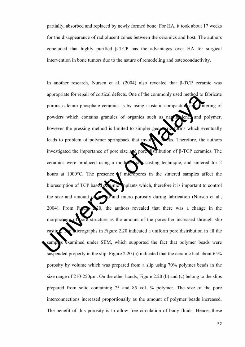
52
partially, absorbed and replaced by newly formed bone. For HA, it took about 17 weeks
for the disappearance of radiolucent zones between the ceramics and host. The authors
concluded that highly purified β-TCP has the advantages over HA for surgical
intervention in bone tumors due to the nature of remodeling and osteoconductivity.
In another research, Nursen et al. (2004) also revealed that β-TCP ceramic was
appropriate for repair of cortical defects. One of the commonly used method to fabricate
porous calcium phosphate ceramics is by using isostatic compaction and sintering of
powders which contains granules of organics such as naphthalene, and polymer,
however the pressing method is limited to simpler geometric forms which eventually
leads to problem of polymer springback that involved cracks. Therefore, the authors
investigated the importance of pore size and pore distribution of β-TCP ceramics. The
ceramics were produced using a modified slip casting technique, and sintered for 2
hours at 1000°C. The presence of micropores in the sintered samples affect the
bioresorption of TCP based ceramic implants which, therefore it is important to control
the size and amount of macro and micro porosity during fabrication (Nursen et al.,
2004). From Figure 2.20, the authors revealed that there was a change in the
morphology of pore structure as the amount of the porosifier increased through slip
casting. The micrographs in Figure 2.20 indicated a uniform pore distribution in all the
samples examined under SEM, which supported the fact that polymer beads were
suspended properly in the slip. Figure 2.20 (a) indicated that the ceramic had about 65%
porosity by volume which was prepared from a slip using 70% polymer beads in the
size range of 210-250µm. On the other hands, Figure 2.20 (b) and (c) belong to the slips
prepared from solid containing 75 and 85 vol. % polymer. The size of the pore
interconnections increased proportionally as the amount of polymer beads increased.
The benefit of this porosity is to allow free circulation of body fluids. Hence, these
Univers
ity of
Mala
ya

53
findings on porosity will be a desirable feature since it facilitates the diffusion of
calcium and phosphorus ions for mineralization (Nursen et al., 2004).
Figure 2.20: SEM micrograph of porous β-TCP (a) 65 vol. % porosity (b) 75 vol. %
porosity (c) 85 vol. % porosity (Nursen et al., 2004).
Kobayashi and Murakoshi (2013), experimented the combination of HA/β-TCP
composites. The composites were synthesized and sintered with different β-TCP content
(0, 10, 20, and 30wt. %). Based on the observation, the authors reported that a trend of
porosity increased with β-TCP content increases from 0 – 30 wt. %. The porosity
indicated the poor sinterability of HA/β-TCP powder mixture which also reflected the
low mechanical properties. For the 30 wt. % β-TCP composite, the bending strength
decreased by ~20% and Vickers hardness decreased by ~40% compared to pure HA.
However, the fracture toughness was found to have improved from 1MPa·m1/2 to 1.3
MPa·m1/2 with the 30 wt. % β-TCP composite. Moreover, Kobayashi and Murakoshi
(a) (b)
(c)
Univers
ity of
Mala
ya
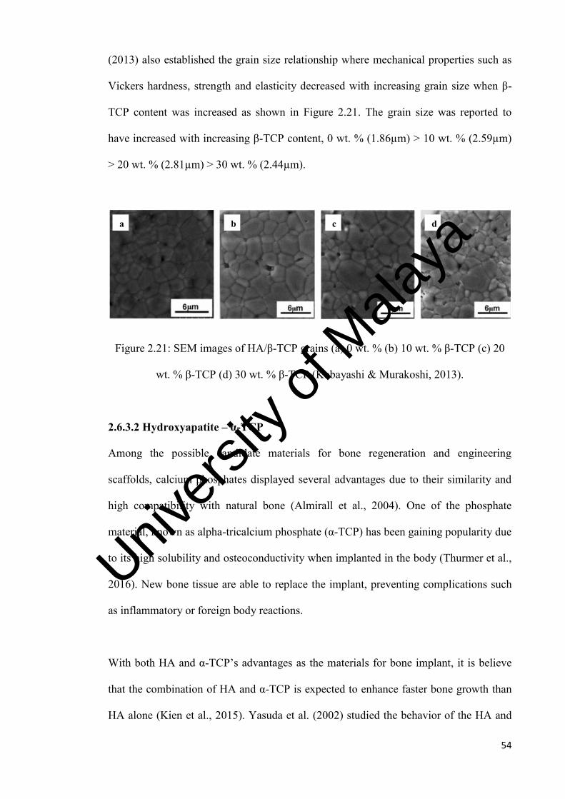
54
(2013) also established the grain size relationship where mechanical properties such as
Vickers hardness, strength and elasticity decreased with increasing grain size when β-
TCP content was increased as shown in Figure 2.21. The grain size was reported to
have increased with increasing β-TCP content, 0 wt. % (1.86µm) > 10 wt. % (2.59µm)
> 20 wt. % (2.81µm) > 30 wt. % (2.44µm).
Figure 2.21: SEM images of HA/β-TCP grains (a) 0 wt. % (b) 10 wt. % β-TCP (c) 20
wt. % β-TCP (d) 30 wt. % β-TCP (Kobayashi & Murakoshi, 2013).
2.6.3.2 Hydroxyapatite – α-TCP
Among the possible candidate materials for bone regeneration and engineering
scaffolds, calcium phosphates displayed several advantages due to their similarity and
high compatibility with natural bone (Almirall et al., 2004). One of the phosphate
material, known as alpha-tricalcium phosphate (α-TCP) has been gaining popularity due
to its high solubility and osteoconductivity when implanted in the body (Thurmer et al.,
2016). New bone tissue are able to replace the implant, preventing complications such
as inflammatory or foreign body reactions.
With both HA and α-TCP’s advantages as the materials for bone implant, it is believe
that the combination of HA and α-TCP is expected to enhance faster bone growth than
HA alone (Kien et al., 2015). Yasuda et al. (2002) studied the behavior of the HA and
c)
a)
b)
d)
Univers
ity of
Mala
ya

55
α-TCP combination in ratios of 20wt. % and 40wt. % of α-TCP. The mixed powders
were prepared through colloidal processing with 30 vol. % HA/α-TCP mixed powder
and 70 vol. % aqueous solution containing polycarboxlic from the anion dispersant.
Then, the synthesized samples were sintered at 1200°C and 1500°C for 2 hours. The
authors discovered that HA with 20 wt. % α-TCP sintered at 1500°C obtained a relative
density of about 90%, while α-TCP was difficult to be sintered at the same temperature.
On the other hand, sintered HA with 40 wt. % α-TCP exhibited a relative density at
80% only. These findings indicated that there is a potential in improving the mechanical
properties when combining HA and α-TCP as composite.
2.7 Summary
Hydroxyapatite (HA) has been successfully applied in medicine due to its excellent
biocompatibility for decades. Unfortunately the low fracture toughness (<1 MPam1/2) of
the HA ceramics was the major drawback to be used as load-bearing implants.
Therefore, the applications of HA are limited to non-load implants, fillers, coatings and
low-loaded porous implants. To overcome this problem, many researches have been
carried out to improve the mechanical properties of HA ceramics by forming a
composite body. An overview of previous researchers’ work on HA and HA composites
are deliberated in this chapter. The composites can be divided into bioinert ceramics,
bioactive ceramics and biodegrable ceramics. From the literature, magnesium
orthosilicate has the potential to be mixed with HA to improve the mechanical
properties as composite for load bearing implant purposes.
Univers
ity of
Mala
ya

56
CHAPTER 3 - METHODOLOGY
3.1 Powder Synthesis
For the current research work, the preliminary powders i.e. hydroxyapatite (HA) powder
was produced using a novel wet chemical method (Ramesh, 2004) and magnesium
orthosilicate powder was produced via combination of ultrasonification and mechanical
ball milling method. Then the HA-MO composites were finally prepared by using
similar preparation method as magnesium orthosilicate.
3.1.1 Hydroxyapatite Powder
For the preparation of hydroxyapatite powder, a novel wet chemical method was used to
synthesized pure HA (100g) with the use of precipitation from aqueous medium via
titration process by reacting calcium ion with phosphate ion based on a molar ratio Ca/P
= 1.67. During the titration process, the aqueous medium was maintained at a pH of
about 10 – 12 by the addition of small amounts of ammonium (NH3) solution (25%
concentration) (Ramesh, 2004; Tian et al., 2008). The use of commercially available
starting precursors such as calcium hydroxide, Ca(OH)2 (98% purity, BDH) and
orthophosporic acid, H3PO4 (85% purity, Merck) was in accordance with the chemical
reactions as follows:
10Ca (OH)2 + 6H3PO4 Ca10 (PO4)6 (OH)2 + 18H2O (3.1)
The synthesis process involved the preparation of orthophosphoric acid (H3PO4) and
calcium hydroxide (Ca(OH)2) solutions. The H3PO4 solution was prepared by diluting
69.19g of H3PO4 in a beaker containing 400ml of distilled water which was stirred for
10 minutes and subsequently contained in a titration funnel. This prepared acid solution
has a pH of about 1. A similar procedure was used to prepare the Ca(OH)2 solution,
Univers
ity of
Mala
ya
57
where 75.5g of Ca(OH)2 measured was mixed into a beaker containing 750ml of
distilled water and stirred for 10 minutes. The prepared Ca(OH)2 solution has a pH of
about 12. The solution was stored in a beaker which was then covered with cling film to
minimize contamination including any reaction between carbon dioxide in air with the
mixture.
The precipitation process began with H3PO4 solution dripped at a rate of 7 drops in 10
seconds into the Ca(OH)2 solution which was still in stirred condition. The titration
process usually takes about 3 hours. During the titration process, the pH of the mixture
was monitored and kept above 10.5 by the addition of NH3 solution. Once the titration
process has completed, the solution was stirred for an additional 6 hours. The precipitate
was allowed to settle overnight before filtration. The filtered precipitate was rinsed three
times at every half hour interval using distilled water and dried in the oven at 60°C
overnight. Finally the dried filtered cake was crushed and sieved (212µm) to obtain
well-defined highly crystalline HA powder. The flow of this method is shown in Figure
3.1.
Univers
ity of
Mala
ya

58
Figure 3.1: HA wet chemical method process flow.
3.1.2 Magnesium Orthosilicate Powder
Magnesium orthosilicate powder was produced via combination of ultrasonification and
mechanical ball milling method. A conventional ball milling method was employed to
mix and refine the powder particles. The starting precursors where 17.9016g of MgO
and 33.6976g of Mg3Si4O10(OH)2 weighed were mixed individually into a beaker of
100ml ethanol and undergone ultrasonification for 2 minutes before finally mixing both
of them in a beaker and ultrasonic for 30 minutes. The final solution was poured into a
high density polyethylene (HDPE) bottle and ball milled with zirconia milling balls for
3 hours prior to the drying process. After 24 hours drying in the oven at 60°C, the dried
Distilled water
Stirring
Distilled water
Stirring
Precipitation
Aging
Filtration
Drying
Crush & Sieve
HA powder
Ca(OH)2 H3PO4
Add NH3
Overnight
60°C for 24 hours
Univers
ity of
Mala
ya

59
cake of magnesium orthosilicate was sieved into powder form. Comprehensive details
on the powder ratio can be obtained in Appendix A.
3.1.3 Hydroxyapatite-Magnesium Orthosilicatae Composite Powder
The HA-magnesium orthosilicate composite powders were prepared in 30 g for each
batch of different weight percentage addition as shown in Table 3.1.
Table 3.1:HA-MO composition
Forsterite (g)
Hydroxyapatite (g)
Undoped
0 30
10wt%
3 27
20wt%
6 24
30wt%
9 21
40wt%
12 18
50wt%
15 15
The HA powders were mixed with magnesium orthosilicate accordingly to the
concentration from 10wt% to 50wt% using the ultrasonification and mechanical ball
milling method. The first step in preparing the powder was to measure the accurate
amount of powders needed for respective batches. Then both the HA and MO powders
were mixed individually into a beaker of 100ml ethanol and undergone ultrasonification
for 2 minutes before finally mixing both of them in a bigger beaker and ultrasonic for 30
minutes. The final solution was poured into a high density polyethylene (HDPE) bottle
and ball milled with zirconia milling balls for 3 hours prior to the drying process and
powder sieving.
Univers
ity of
Mala
ya

60
3.2 X-Ray Diffraction (XRD)
X-Ray diffraction (XRD) is a versatile, non-destructive analytical technique for
identification and quantitative determination of the various crystalline forms knows as
phases of compounds present in powder and solid. Besides that, XRD method is capable
of providing qualitative and quantitative information about the crystalline phase exists
in the sample (Rahaman, 1995).
In the present work, the phases present in the powders as well as the sintered samples
were determined at room temperature using X-Ray diffractometer with Cu-Kα (λ =
0.15406 nm) as the inception of the radiation using a scan speed and step scan of 6°/min
and 0.02° respectively, at 35 kV and 15 mA.
The peaks obtained were compared to standard reference JCPDS-ICDD (Joint
Committee of Powder Diffraction Standard – International Center for Diffraction Data)
files for hydroxyapatite (09-0432) and magnesium orthosilicate (34-0189). The details
of the said JCPDS-ICDD reference files are attached in Appendix B.
Besides the phase stability examination, the average crystallite size (d) can also be
determined for the hydroxyapatite, magnesium orthosilicate and the composite powders
from the peak broadening at the prominent XRD peaks at 2θ = 28°, 32° and 44° in the
XRD diffraction pattern associated with a particular planar reflection from within the
crystal unit cell using Scherrer’s formula below (Cullity & Stock, 2001).
d = 0.9λ
𝛽𝑐𝑜𝑠𝜃 (3.2)
Where,
d = Crystalline size (m)
Univers
ity of
Mala
ya
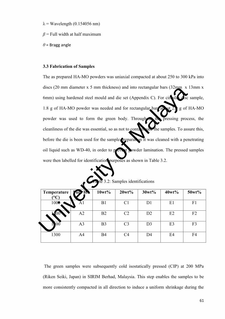
61
λ = Wavelength (0.154056 nm)
β = Full width at half maximum
θ = Bragg angle
3.3 Fabrication of Samples
The as prepared HA-MO powders was uniaxial compacted at about 250 to 300 kPa into
discs (20 mm diameter x 5 mm thickness) and into rectangular bars (32mm x 13mm x
6mm) using hardened steel mould and die set (Appendix C). For circular disc sample,
1.8 g of HA-MO powder was needed and for rectangular bar, about 2.0 g of HA-MO
powder was used to form the green body. Throughout the pressing process, the
cleanliness of the die was essential, so as not to contaminate the samples. To assure this,
before the die is been used for the sample preparation it was cleaned with a penetrating
oil liquid such as WD-40, in order to prevent powder lamination. The pressed samples
were then labelled for identification purposes as shown in Table 3.2.
Table 3.2: Samples identifications
Temperature (°C)
Pure HA 10wt% 20wt% 30wt% 40wt% 50wt%
1000
A1 B1 C1 D1 E1 F1
1100
A2 B2 C2 D2 E2 F2
1200
A3 B3 C3 D3 E3 F3
1300
A4 B4 C4 D4 E4 F4
The green samples were subsequently cold isostatically pressed (CIP) at 200 MPa
(Riken Seiki, Japan) in SIRIM Berhad, Malaysia. This step enables the samples to be
more consistently compacted in all direction to induce a uniform shrinkage during the
Univers
ity of
Mala
ya

62
sintering process and ultimately improve the densification of the sintered samples
besides preventing the samples from warping and cracking (Colling & Vasilos, 1995).
3.4 Sintering
Sintering is a process of making ceramic components from powder whereby the green
body is heated in a sintering furnace to below its melting point to allow particles
consolidation to occur. The process leads to decrease in the surface area, an increase in
compact strength and in many cases shrinkage in the compact. The densification and
grain growth can be controlled from the sintering manner. With better densification,
porosity can be reduced in a sample thereby improving the mechanical properties.
In the present research work, the conventional pressureless sintering method was used
to sinter the green compacts using standard rapid heating furnace (LT furnace, see
Appendix C) at four different temperatures of 1000°C, 1100°C, 1200°C and 1300°C. A
furnace ramp rate of 10°C/min (heating and cooling) and a holding time of 2 hours were
used at each sintering. The sintering profile used in this work is schematically shown in
Figure 3.2.
Figure 3.2: Sintering profile
Time, hrs
Temp.,
°C
2 hours
Heating 10°C/min.
Cooling 10°C/min.
Desired Temperature (T)
Univers
ity of
Mala
ya

63
3.5 Grinding and Polishing
The sintered HA-MO disc samples were firstly ground and then polished on one surface
prior to density measurement, phases analysis, Vickers hardness testing and
microstructural evaluation by Scanning Electron Microscopy (SEM). The process of
surface grinding and polishing were done manually by means of the Imtech Grinder-
Polisher (Appendix C). The samples were ground by SiC papers of 800, 1000 and 1200
grits successively, followed by polishing with 6µm and 1µm diamond paste to produce
an optically reflective surface.
3.6 Bulk Density Measurement
The bulk densities of the samples in this research were measured using the water
immersion technique based on Archimedes principle using a standard Mettler Toledo
Balance AG204 densi-meter (Appendix C). The immersion medium used in the present
research was distilled water.
The procedures to measure the bulk density can be summarized as follow:
1) Firstly, the weight of the sample in air was measured and recorded.
2) Then, the weight of the sample was measured by placing it in a dish immersed in
water after zeroing the electronic balance. The measurement was recorded.
3) Measurement was taken with care due to the sensitivity of the balance to minor
vibration. This can cause fluctuation of the reading.
4) Finally, the bulk density of the samples can be calculated using the equation
(3.3).
𝜌 =Wa
(Wa− Ww)ρw (3.3)
Univers
ity of
Mala
ya

64
Where
ρ = Bulk density of the sample
Wa = Weight of sample in air
Ww = Weight of sample in water
ρw = Density of distilled water. The temperature effect on the water density was
taken into consideration using the table provided in Appendix D.
3.7 Young’s Modulus Determination
The Young’s modulus by sonic resonance technique was determined from rectangular
samples using a commercial testing instrument (GridoSonic: MK5 “Industrial”,
Belgium). The resonant frequency of the samples was measured by monitoring and
evaluating the vibrational harmonics of the sample by a transducer where the vibrations
are physically induced in the sample by tapping. The block diagram of typical test
apparatus is shown on Figure 3.3. The modulus of elasticity is calculated from the
measured natural frequency, according to the standard test method (ASTM E1876-97).
The Young’s modulus or modulus of elasticity (E) is calculated using equation (3.4):
E = 0.9465(𝑚𝐹2
𝑏)(
𝐿
𝑡)3 T (3.4)
Where;
E = Young’s modulus (Pa)
m = Mass of bar (g)
F = Fundamental resonant frequency of the bar in flexural (Hz)
b = Width of bar (mm)
L = Length of bar (mm)
t = Thickness of bar (mm)
Univers
ity of
Mala
ya

65
T = Correction factor for fundamental flexural mode to account for finite thickness of
bar and material’s Poisson’s ratio based on equation below:
22
42
422
)536.11408.01(338.61
)173.22023.01(34.8868.0)8109.00752.01(585.61
Lt
Lt
Lt
LtT
Where;
µ = Poisson’s ratio taken as 0.22 for ceramic body (Brook, 1991)
Figure 3.3: Block diagram of test apparatus for Young’s modulus measurement
(ASTM Standards E 1876-97)
3.8 Vickers Hardness Determination
The Vickers hardness testing method was employed to ascertain the hardness of the
sintered samples. Hardness is defined as the ability of a material to resist denting as
caused by a load bearing on the material using a dimensionally standardized probe.
Indentations were made on the sintered samples using a pyramidal diamond indenter
that has a 136° tip with an applied force of 0.5 kg.
Univers
ity of
Mala
ya

66
During the test, the load is applied slowly, without impact, and held for approximately
10 seconds to create an impression. The physical quality of the indenter and the
accuracy of the applied load as defined clearly in ASTM E384-99 and ISO 14705 must
be controlled to get the correct results. The shape after indentation was then observed
under microscope to measure the diagonal length d1 and d2 respectively. The shape of
the diamond indenter and the indented shape are shown in Figure 3.4.
Figure 3.4: Schematic diagram of Vickers hardness indenter
Three indentations were made in order to obtain the average of the Vickers hardness
value. The value of Vickers hardness can be calculated using equation 3.5.
Hv = 1.854𝑃
𝑑2 (3.5)
Univers
ity of
Mala
ya

67
Where,
Hv = Vickers hardness
P = Applied load
d = average diagonal length = ([d1 + d2] /2)
3.9 Fracture Toughness Determination
The fracture toughness can be evaluated from the indentation method. The advantage of
this method is the speed, ease of testing and sample preparation, relatively low cost,
large numbers of indentation can be made quickly and the small volume of material
required. Principally, the indentation method is similar to Vickers hardness testing
procedures. Similar indentation pattern as shown in Figure 3.5 should be visible upon
indentation with existence of crack propagation.
Figure 3.5: Crack propagation from indentation (L1, L2, L3, L4 = length of cracks).
Thus, the fracture toughness (KIc) was determined from the equation 3.6 derived by
Niihara (1985);
a
Univers
ity of
Mala
ya

68
5.05.1
203.0 aHacK vIc
(3.6)
Where,
Hv = Vickers hardness (N/m2)
c = Characteristics crack length (i.e. L + a)
a = Half diagonal of the indent, µm
3.10 Scanning Electron Microscopy and Grain Size Measurement
The synthesized HA and HA-MO powders were inspected by the scanning electron
microscope (SEM). Carbon tapes were placed on metal SEM mounts and the HA and
HA-MO powders were sprinkled onto the mount with a spatula. The powders were
examined under SEM at accelerating voltage of 15 KV at magnifications up to 20,000X.
The microstructure development of the sintered samples were also examined under the
SEM. The SEM function by focusing a small spot of electrons on a thick specimen and
proceeds to scan across the specimen surface in a series of lines, using electrostatic or
electromagnetic forces. Electrons emerging from the upper specimen surface are
collected by an electron detector and used to produce image on a monitor as a series of
line.
The average grain size of sintered samples was determined from scanning electron
micrographs using the line intercept method. This technique requires measurements
taken from polished sections. In a typical analysis, a known test line is drawn on an A4
size SEM micrograph of the selected polished section and the number of intercept
between the test line and grain boundaries are counted. The test line should cover at
least 50 grains and several lines are drawn and measure before average value is taken.
Univers
ity of
Mala
ya

69
The average grain size is then calculated according to the equation proposed by
Mendelson (1969):
LD 56.1 (3.7)
where D is the average grain size and L is the measured average interception length
over a large number of grains which can be represented by:
MNCL (3.8)
Where C, M and N are the total length of the test line, the magnification of the SEM
micrograph and the number of intercepts respectively.
The technique used to count the number of intercepts was according to an international
standard test method for intercept counting (ASTME E112-96, 2004). Essentially, the
end point of a test line are not intersections and not counted unless the end appeared to
exactly touch a grain boundary, when a ‘0.5’ intersection is scored. A tangential
intersection with a grain boundary is scored as ‘1’ intersection while for intersection
coinciding with the junction of 3 grain is scored as a ‘1.5’ intersection as shown in
Figure 3.6.
Univers
ity of
Mala
ya

70
Figure 3.6: Schematic diagram showing the score given for the type of intersections
(Ramesh, 1997)
score : 0
score : 1
grain
score : 1.5
score : 0.5
test line
SEM micrograph
Univers
ity of
Mala
ya

71
CHAPTER 4 - RESULTS AND DISCUSSION
4.1 Introduction
For this research, studies were carried out to evaluate the densification behavior and
properties of hydroxyapatite-magnesium orthosilicate (HA-MO) composite. The
composite was prepared in various compositions, by varying the magnesium
orthosilicate content from 10 wt. % to 50 wt. % via mechanical ball milling and
conventional pressureless sintering at 1000°C to 1300°C, using ramp rate of 10°C /
minute for 2 hours. Before sintering, synthesized magnesium orthosilicate,
hydroxyapatite and hydroxyapatite-magnesium orthosilicate composite powders were
identified through the x-ray diffraction (XRD) and scanning electron microscope (SEM)
machine, likewise upon sintered, samples were characterized also. The mechanical
properties such as relative density, Vickers hardness, Young’s modulus and fracture
toughness were evaluated through lab works.
4.2 Starting Powder
4.2.1 HA Powder
The hydroxyapatite (HA) powder was prepared by wet chemical method. The wet
chemical method involved precipitation from an aqueous medium through the slow
titration of an orthophosphoric acid solution to calcium hydroxide based on a molar
ratio Ca/P = 1.67 where the solution was maintained at a pH of 10-12 through addition
of small amounts of ammonium solution. The XRD result of hydroxyapatite powder
before sintering as shown in Figure 4.1 is in good agreement with that of the standard
JCPDS card No. 09-432. It was observed that the peaks were generally broad, signifying
that the powder has low crystallinity. There were no other phases found in the powder.
The Ca/P ratio of the starting powder is important as the reactivity, stability and
Univers
ity of
Mala
ya

72
mechanical properties of sintered HA can be affected as reported by Liu et al. (2008)
and Raynaud et al. (2002a, b & c).
Figure 4.1: XRD traces of synthesized hydroxyapatite powder before sintering.
The SEM micrograph of HA powder shown in Figure 4.2a consists of small particles in
the range of 1-3 µm and large particles in the range of 5-10 µm. Rougher surface was
observed from the large agglomerate which consists of smaller particles. Similar
observation was also reported by Ramesh et al. (2013), Gibson et al. (2001) and Saeri et
al. (2003) for synthesized HA powder. The synthesized powder is also free of impurities
as the energy dispersive x-ray (EDX) spectrum only displayed the element of HA which
are Ca, P, and O in Figure 4.2b.
Besides, the formation of soft agglomerates which broke easily at low pressing pressure
of 250 kPa during powder compaction can be due to the process of drying the filter cake
of synthesized HA, even though the dried filter cake was grounded and sieved. Due to
the soft nature of the powders, any attempts to use higher pressures during uniaxial
Univers
ity of
Mala
ya

73
pressing of the samples were futile as powders lamination on the die surface was
observed and in some extreme cases, a layer of compacted powders separated
immediately from the green body upon ejection from the mold.
5 μm
Figure 4.2: a) SEM analysis of synthesized hydroxyapatite powder, b) EDX of
synthesized hydroxyapatite powder.
4.2.2 Magnesium Orthosilicate Powder
The magnesium orthosilicate (MO) powder was produced via combination of
ultrasonification and mechanical ball milling method. A conventional ball milling
method was employed for 3 hours to mix and refine the powder particles. The XRD
results of magnesium orthosilicate as shown in Figure 4.3 before sintering exhibited
peaks matching to the JCPDS card No. 34-0189. The SEM micrograph in Figure 4.4a
(a)
(b)
Univers
ity of
Mala
ya
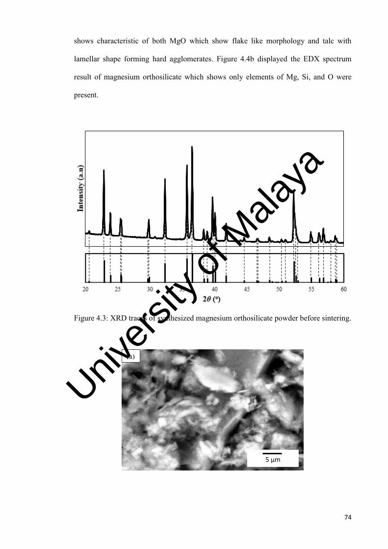
74
shows characteristic of both MgO which show flake like morphology and talc with
lamellar shape forming hard agglomerates. Figure 4.4b displayed the EDX spectrum
result of magnesium orthosilicate which shows only elements of Mg, Si, and O were
present.
Figure 4.3: XRD traces of synthesized magnesium orthosilicate powder before sintering.
5 µm
(a)
Univers
ity of
Mala
ya

75
Figure 4.4: a) SEM analysis of synthesized magnesium orthosilicate powder, b) EDX of
synthesized magnesium orthosilicate powder.
4.2.3 Hydroxyapatite-Magnesium Orthosilicate Powder
The hydroxyapatite-magnesium orthosilicate (HA-MO) powders were produced via
combination of ultrasonification and mechanical ball milling method according to
10MO, 20MO, 30MO, 40MO and 50MO composition. A conventional ball milling
method was employed for 3 hours to mix and refine the powder particles. The XRD
signatures of HA-MO powders as shown in Figure 4.5 before sintering were found to be
similar as that of the pure HA peaks matching to the JCPDS card N0. 09-0432. No
major peak shifting was observed from the XRD traces of all the HA-MO powders.
Additionally, the SEM micrographs obtained for all the HO-MO powders indicated that
the method employed to prepare the powders had negligible effect on the morphology
and agglomeration of the starting HA powder. The powder particles of both powders
(HA & HA-50MO) were in the range of 1 to 3 µm diameters as shown in Figure 4.6.
Figure 4.7 displayed the EDX spectrum result of HA-MO powder (10MO – 50MO)
(b)
Univers
ity of
Mala
ya

76
which shows only elements of Mg, Si, O, Ca, and P were present regardless of the
various MO content.
Figure 4.5: XRD traces of HA-MO powders before sintering.
Figure 4.6: SEM micrographs of (a) Pure HA and (b) HA-50MO powder.
(a) (b)
2.5 µm 2.5 µm Univers
ity of
Mala
ya

77
Figure 4.7: EDX of HA-MO powder.
4.3 HA-MO Composite Evaluation
In the present study, the HA-MO composite with various concentration from 10wt.% to
50wt.% were pressureless sintered at four different temperatures of 1000°C, 1100°C,
1200°C and 1300°C with ramp rate of 10°C/minute (heating and cooling) and a holding
time of 2 hours. Upon sintering, the HA-MO bulk samples were ground and polished on
one surface to 1µm prior to properties measurement such as phase analysis, shrinkage,
densification, Young’s modulus, hardness, fracture toughness, and microstructure
evaluation.
4.3.1 Phase Analysis
The phase stability of the sintered samples were evaluated via XRD for sintering
temperatures of 1000°C, 1100°C, 1200°C, and 1300°C and are summarized in Table
4.1.
Univers
ity of
Mala
ya

78
Table 4.1: Phases present in the sintered HA and HA-MO composites. For keys: HA =
Hydroxyapatite, MO = Magnesium orthosilicate, W = Whitlockite.
Comp. Sintering Temperature
HA
HA-10MO
HA-20MO
HA-30MO
HA-40MO
HA-50MO
1000°C HA HA+MO+W HA+MO+W HA+MO+W HA+MO+W HA+MO+W
1100°C HA HA+MO+W HA+MO+W HA+MO+W HA+MO+W HA+MO+W
1200°C
1300°C
HA
HA
HA+MO+W
HA+MO+W
HA+MO+W
HA+MO+W
HA+MO+W
HA+MO+W
HA+MO+W
HA+MO+W
HA+MO+W
HA+MO+W
Results from these four sintering temperatures indicated that complete crystalline phase
of hydroxyapatite-magnesium orthosilicate composite were obtained after cooling down
to room temperature. The comparison between XRD patterns obtained for the sintered
HA-MO composite samples are shown in Figure 4.8 to Figure 4.11.
Figure 4.8: XRD patterns of HA-MO samples sintered at 1000°C. For keys: = HA,
= whitlockite, = magnesium orthosilicate.
HA
HA-10MO
HA-20MO
HA-30MO
HA-40MO
HA-50MO
Univers
ity of
Mala
ya

79
Figure 4.9: XRD patterns of HA-MO samples sintered at 1100°C. For keys: = HA,
= whitlockite, = magnesium orthosilicate.
Figure 4.10: XRD patterns of HA-MO samples sintered at 1200°C. For keys: = HA,
= whitlockite, = magnesium orthosilicate.
HA
HA
Univers
ity of
Mala
ya

80
Figure 4.11: XRD patterns of HA-MO samples sintered at 1300°C. For keys: = HA,
= whitlockite, = magnesium orthosilicate.
The XRD phase analysis of monolithic HA in Figure 4.8 to Figure 4.11 indicated that
only pure HA was obtained when sintered at 1000°C to 1300°C. The HA peak is in
accordance to the standard JCPDS card No. 09-432. The higher the intensity of HA
peak indicates the level of crystallinity phase attained. No secondary phase was formed
in the pure HA sintered samples. At 1000°C, magnesium orthosilicate was observed to
be present in all the composites (10wt. % - 50wt. %). The intensity of MO peak
increases as amount of MO increased in the composite. Similar trend of MO peaks were
also attained in the sintering temperature between 1100°C and 1300°C, where the peak
intensity of MO appeared to increase with increasing MO content, but the difference
was minimal. The MO peaks observed are in accordance to the JCPDS card No. 34-
0189. However, secondary phase was observed to be present in all the HA-MO
composites as shown in Figure 4.8 to Figure 4.11. The addition of MO into HA
promotes the decomposition of HA to whitlockite (also known as β-TCP). This
observation was also reported by Emadi et al. (2010) where the transformation of HA to
HA
Univers
ity of
Mala
ya
81
whitlockite is associated with a chemical reaction with MO during sintering which lead
to partial thermal decomposition. It is believed that the presence of MO has caused the
dehydroxylation and decomposition to takes place. The water vapor was considered as a
product from the dehydroxylation and decomposition and the speed of the reactions
would depend on the partial pressure of H2O in the sintering atmosphere (Bernache-
Assollant et al., 2003). During the sintering of the composite samples, HA would have
loss the OH radicals which resulted to the decomposition to whitlockite.
It is also believed that the MO which contains magnesium where it inhibits apatite
crystallization in solution can act as a stabilizer for the whitlockite phase which
promotes the decomposition of HA (Ma et al., 2012; Cacciotti & Bianco, 2011; Nikaido
et al., 2015). Bigi et al. (1993) also confirmed that the presence of magnesium prevented
the transformation of calcium phosphate into HA when magnesium entered the
prenuclei structures of HA which caused distortion and prevented the growth of HA
from the structural mismatch that occurred. The decomposition of the HA to whitlockite
phase becomes obvious with the increase of the MO contents. Therefore, the composite
with higher amount of MO causes the HA to be more unstable.
4.3.2 Shrinkage
The assessment of the shrinkage was taken as the average of the diameter and the
thickness of the disc samples with respect to sintering temperatures. The resulting
shrinkage of the sintered samples is shown in Figure 4.12. Generally, the monolithic HA
samples attained higher shrinkage across the investigated range of sintering
temperatures.
The HA and HA-MO composites displayed an increasing trend manner in the shrinkage
when sintered at 1000°C, 1100°C and 1200°C. At low temperature of 1000°C, the
Univers
ity of
Mala
ya

82
sintered HA attained a shrinkage of 15.8%, while the HA-MO composites (10MO -
50MO) in the range of ~3% to ~12%. However, at 1100°C, the shrinkage increased
significantly for the sintered HA, attained at 19.1%. For the HA-MO composites, the
shrinkage increased in a linear manner till 1200°C, where a significant increase in
shrinkage were attained from ~7% to ~17% range at above 1200°C. A maximum
shrinkage of 19.7% was attained from HA sintered at 1300°C, while the composite
(HA-20MO) managed to attain about 17.7% shrinkage at the same temperature. In
addition, it was observed that shrinkage of HA-MO composites (10MO, 30MO, 50MO)
showed increasing trend and attained shrinkage of between ~15% to ~17% at 1300°C.
Figure 4.12: Shrinkage of different sintered samples as a function of sintering
temperature.
Univers
ity of
Mala
ya

83
4.3.3. Bulk Density
The effect of sintering on the bulk density of the HA and HA-MO composites is shown
in Figure 4.13. The results indicated that HA samples exhibited higher bulk density than
HA-MO composites regardless of the sintering temperature used. The bulk density of
the HA sintered at 1000°C was 2.7 g/cm3 and attained a final density of above 3 g/cm3
when sintered at 1200°C - 1300°C. Generally, sintering across the four range of
sintering temperatures (1000°C - 1300°C) resulted in very small increased in density for
the HA sintered samples.
Conversely, the HA-MO composites (10MO to 50MO) exhibited lower bulk density
across the sintering temperatures when compared to monolithic HA. For HA-10MO
only exhibited a bulk density of 2.2 g/cm3 at 1000˚C and 2.4 g/cm3 at 1300˚C which
showed a small increment. For HA-MO composites with 30MO – 50MO exhibited bulk
density in the range of ~1.7 g/cm3 to ~2.5 g/cm3 when sintered across 1000˚C to
1300˚C. This range of densities of the composites with 30MO – 50MO were generally
low when sintered below 1200˚C due to the inability of MO to consolidate and densify.
The MO requires a higher amount of activation energy to diffuse into HA. Besides,
another factor could be attributed to the hard agglomerates of magnesium orthosilicate,
which could have led to a lower packing density in the green state and also
decomposition of the HA phase in the composite matrix from the addition of MO.
Hence among the composites, HA-20MO exhibited the optimum value with a bulk
density of 1.9 g/cm3 at 1000°C and 2.7 g/cm3 at 1300°C.
Univers
ity of
Mala
ya
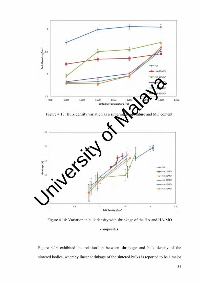
84
Figure 4.13: Bulk density variation as a sintering temperature and MO content.
Figure 4.14: Variation in bulk density with shrinkage of the HA and HA-MO
composites.
Figure 4.14 exhibited the relationship between shrinkage and bulk density of the
sintered bodies, whereby linear shrinkage of the sintered bulks is reported to be a major
Univers
ity of
Mala
ya

85
influence which affects the resulting densification values. It was observed that as the
shrinkage values increase, the bulk density values followed to increase in linear manner
except for HA-20MO composite. The onset of densification, indicated by a sharp
increase in the bulk density, for HA-20MO was between ~9% to ~17%. Generally, all
the composite shrinkage findings were lower than monolithic HA due to the complexity
of the three phases (HA, MO, whitlockite) that exist upon sintered accompanied with
porosities that make the composite to be less dense.
4.3.4 Young’s Modulus
Study carried out on the Young’s modulus (E) of HA-MO sintered samples is shown in
Figure 4.15. The HA-50MO composite exhibited a higher Young’s modulus than
monolithic HA regardless of sintering temperature. The Young’s modulus of the
monolithic HA varied almost linearly from 90 GPa at 1000°C to about 111 GPa at
1300°C. Conversely, composites containing less than 50wt. % MO exhibited a lower
Young’s modulus than the monolithic HA when sintered at 1000°C. Typical values
recorded for the composites were 29 GPa for HA-10MO, 35 GPa for HA-20MO, 64
GPa for HA-30MO and 82 GPa for the HA-40MO samples when sintered at 1000°C. At
1300˚C, HA-10MO attained 98 GPa, HA-20MO attained 105 GPA, HA-30MO attained
115 GPa and HA-40MO attained 116 GPa.
However, the elastic modulus of the composites increased rapidly with increasing
sintering temperature to attain values above 90 GPa. The maximum Young’s modulus
of 118 GPa was measured for the HA-50MO composite when sintered at 1300°C. The
increasing in Young’s modulus also reflects the reduce in porosity (Xiao et al., 2005).
These values exhibited by the composites could be beneficial when the composites are
used as implant material given their comparable value to human bones.
Univers
ity of
Mala
ya

86
According to Figure 4.16, the Young’s modulus of the HA-MO composites are not
governed by the bulk density. This finding is a bit unusual as normally the decreased in
bulk density will reflect the decline in flexural modulus of elasticity. However, thru this
result, the inclusion of MO in HA, in an increasing manner was found to be beneficial in
enhancing the stiffness of the sintered HA-MO composite as depicted by the higher
Young’s modulus attained. The pores that exist in the composite of the three phases
(HA, MO and whitlockite) could have caused more dislocation in the material which it
can be perceived that the pores had provided a mechanical interlock leading to firmer
fixation of the composite giving it better stiffness.
Figure 4.15: Young’s modulus variation with sintering temperature.
Univ
ersity
of M
alaya
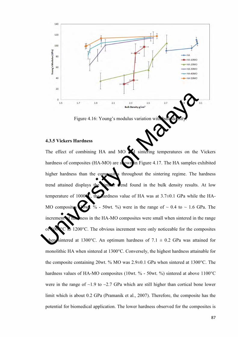
87
Figure 4.16: Young’s modulus variation with bulk density.
4.3.5 Vickers Hardness
The effect of combining HA and MO and sintering temperatures on the Vickers
hardness of composites (HA-MO) are shown in Figure 4.17. The HA samples exhibited
higher hardness than the composites throughout the sintering regime. The hardness
trend attained displays the similar trend found in the bulk density results. At low
temperature of 1000°C, the hardness value of HA was at 3.7±0.1 GPa while the HA-
MO composites (10wt. % - 50wt. %) were in the range of ~ 0.4 to ~ 1.6 GPa. The
increment of hardness in the HA-MO composites were small when sintered in the range
of 1000°C to 1200°C. The obvious increment were only noticeable for the composites
when sintered at 1300°C. An optimum hardness of 7.1 ± 0.2 GPa was attained for
monolithic HA when sintered at 1300°C. Conversely, the highest hardness attainable for
the composite containing 20wt. % MO was 2.9±0.1 GPa when sintered at 1300°C. The
hardness values of HA-MO composites (10wt. % - 50wt. %) sintered at above 1100°C
were in the range of ~1.9 to ~2.7 GPa which are still higher than cortical bone lower
limit which is about 0.2 GPa (Pramanik et al., 2007). Therefore, the composite has the
potential for biomedical application. The lower hardness observed for the composites is
Univers
ity of
Mala
ya

88
associated with the lower bulk density of the sintered body as depicted in Figure 4.18.
Tancred et al. (2001) also reported that the hardness and bulk density of HA composite
was found decreased with additive addition. Decomposition of phase and pores that
exist in the composite could be the factor of the low hardness and bulk density too.
Another attribute to the higher hardness value of the sintered samples is the grain size. It
was observed that as the grain size decreased in HA, the hardness value increased when
sintered from 1000˚C to 1300˚C. Similar observations were also found on the HA-MO
composites. Even though the composites exhibited lower hardness value than
monolithic HA, as the amount of MO increases in the composite, smaller grains were
found to be closely packed in the bimodal grain distribution resulted in higher hardness
value for the HA-MO composites.
From the results obtained and also reported by Ramesh et al. (2007b), Veljovic et al.
(2008) and Li et al. (2008b), it can be suggested that provided the grain does not exceed
a critical grain size limit, the hardness of sintered samples would be governed by bulk
density or porosity. However, once the grains exceeded the critical grain size, the
controlling parameter would be the grain size rather than bulk density.
Univers
ity of
Mala
ya

89
Figure 4.17: Vickers hardness variation with MO addition as a function of sintering
temperature.
Figure 4.18: Vickers hardness variation with MO addition as a function of bulk density.
Univers
ity of
Mala
ya
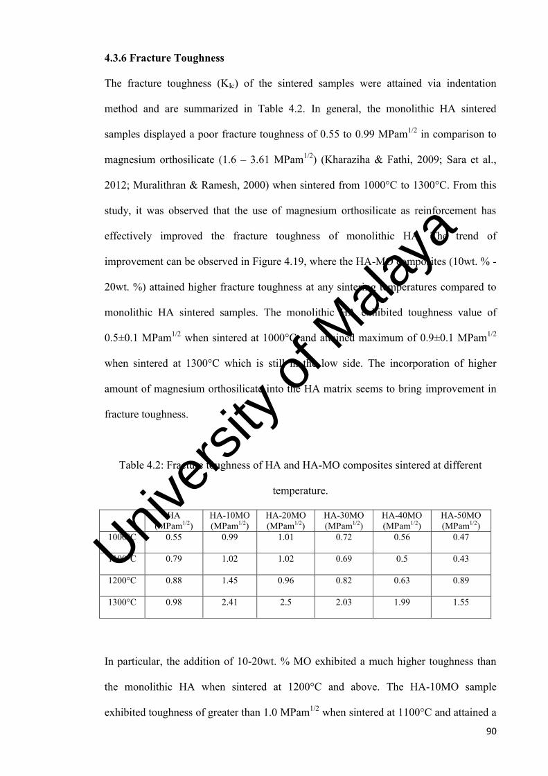
90
4.3.6 Fracture Toughness
The fracture toughness (KIc) of the sintered samples were attained via indentation
method and are summarized in Table 4.2. In general, the monolithic HA sintered
samples displayed a poor fracture toughness of 0.55 to 0.99 MPam1/2 in comparison to
magnesium orthosilicate (1.6 – 3.61 MPam1/2) (Kharaziha & Fathi, 2009; Sara et al.,
2012; Muralithran & Ramesh, 2000) when sintered from 1000°C to 1300°C. From this
study, it was observed that the use of magnesium orthosilicate as reinforcement has
effectively improved the fracture toughness of monolithic HA. The trend of
improvement can be observed in Figure 4.19, where the HA-MO composites (10wt. % -
20wt. %) attained higher fracture toughness at any sintering temperatures compared to
monolithic HA sintered samples. The monolithic HA exhibited toughness value of
0.5±0.1 MPam1/2 when sintered at 1000°C and attained maximum of 0.9±0.1 MPam1/2
when sintered at 1300°C which is still in the low side. The incorporation of higher
amount of magnesium orthosilicate into the HA matrix seems to bring improvement in
fracture toughness.
Table 4.2: Fracture toughness of HA and HA-MO composites sintered at different
temperature.
HA (MPam1/2)
HA-10MO (MPam1/2)
HA-20MO (MPam1/2)
HA-30MO (MPam1/2)
HA-40MO (MPam1/2)
HA-50MO (MPam1/2)
1000°C 0.55
0.99 1.01 0.72 0.56 0.47
1100°C 0.79 1.02 1.02 0.69 0.5 0.43
1200°C 0.88 1.45 0.96 0.82 0.63 0.89
1300°C 0.98 2.41 2.5 2.03 1.99 1.55
In particular, the addition of 10-20wt. % MO exhibited a much higher toughness than
the monolithic HA when sintered at 1200°C and above. The HA-10MO sample
exhibited toughness of greater than 1.0 MPam1/2 when sintered at 1100°C and attained a
Univers
ity of
Mala
ya

91
maximum of 2.4±0.1 MPam1/2 when sintered at 1300°C. The highest fracture toughness
of 2.5±0.1 MPam1/2 was measured at 1300°C for HA composite containing 20wt. %
MO. The HA-30MO, HA-40MO and HA-50MO only exhibited toughness in the range
of ~1.5 to ~2.0 MPam1/2 when sintered at 1300˚C as anything below exhibited low
fracture toughness value. The toughness values were in the range of ~0.5 to ~0.8
MPam1/2. This could be due to the grain boundary that was not properly formed at low
sintering temperatures.
On the other hand, the improvement in the fracture toughness of HA-10MO and HA-
20MO can be associated with the pores and presence of TCP in the composite matrix
that could have interact with the propagating crack through a stress absorption
mechanism, although the presence of such a secondary phase has been reported to be
generally detrimental to the properties of HA (Veljovic et al., 2011). Figure 4.20
indicated that the fracture toughness of the HA-MO composites was not governed by
bulk density. As discussed earlier in the attribute that help improved the stiffness of the
composites was due to grain size, and presence of the pores whereby the pores provided
the mechanical interlock which lead to the firmer fixation of the material, also
contributed to the improvement of the composites’ toughness. The fracture toughness
values for most monolithic HA reported in the literature varied at 0.6-1 MPam1/2
(Ramesh et al., 2013; Monika, 2015; Ramesh et al., 2012) and HA doped with different
additives such zirconia (Li et al., 1993); Kim et al., 2003), titania (Manjubala and
Kumar, 2000), and alumina (Champion et al., 1996) only attained a fracture toughness
in the range of 1.0 to 1.4 MPam1/2.
The indentation fracture toughness of HA-MO composites obtained in this study was
appreciably higher than those reported in many studies on dense HA ceramics. This
Univers
ity of
Mala
ya

92
finding is encouraging because it creates opportunities for these HA composites to be
used for biomedical load-bearing implants.
Figure 4.19: Fracture toughness of sintered samples as a function of sintering
temperature and MO addition.
Figure 4.20: Fracture toughness variation with MO addition as a function of bulk
density.
Univers
ity of
Mala
ya

93
4.3.7 Microstructure Evaluation
Scanning electron micrographs (SEM) of HA, and HA-MO composites (10wt. % MO,
20wt. % MO, 30wt. % MO, 40wt. % MO, 50wt. % MO) sintered at 1000°C to 1300°C
are shown in Figure 4.21 to 4.24. The ramification of magnesium orthosilicate on the
grain size and microstructure of HA-MO composites can be observed in the SEM
micrographs.
For the monolithic HA sintered samples, grain growth were observed to have taken
place when the temperature of the sintering increased. This was due to the improvement
in the homogeneity of the HA powder with increasing sintering temperature. The grain
growth of monolithic HA in Figure 4.21 (a) was not obvious when sintered at 1000°C.
As the temperature increases, the grain growth and grain boundary became more visible.
At 1200°C, the grain growth and grain boundary became visible in the sintered
monolithic HA sample as shown in Figure 4.23 (a). It was also observed that at higher
sintering temperature, the porosity reduces as compared to those sintered at 1100°C and
below, which corresponds to the formation of solid bridges between grains. The HA
grains was observed to have grown rapidly, roughly ten times from an average of
0.55µm at 1200°C to approximately 5.5µm at 1300°C as depicted in Figure 4.23 (a) and
Figure 4.24 (a). The monolithic HA exhibited a bimodal distribution of grains.
The micrographs of the HA-MO composites in Figure 4.21 (b-f) – Figure 4.24 (b-f)
(10wt. % MO, 20wt. % MO, 30wt. % MO, 40wt. % MO and 50wt. % MO) were also
observed upon sintering (1000°C - 1300°C). For HA-MO composites sintered at below
1200°C tend to display large amount of pores and well defined grains could not be
observed. The HA-MO composites sintered at 1000˚C and 1100˚C as shown in Figure
4.21 (b-f) and Figure 4.22 (b-f) indicated that the fusion state is still incomplete, hence
Univers
ity of
Mala
ya

94
no clear grain boundaries were observed. This is partly due to the inability of MO to
consolidate and densify when sintered at low temperature (Fathi & Kharaziha, 2008).
The MO requires a higher amount of activation energy and thermal energy for grain
growth to takes place in the HA-MO composite. Activation energy is the minimum
energy needed to overcome the energy barrier of the atoms present in the lattice. In
Figure 4.23 (b-f), the micrographs of the HA-MO composites sintered at 1200˚C start to
display more grain formation and visible grain boundary especially from HA-50MO.
On the other hand, the composite tends to display lesser porosity in the samples that
contain higher amount of MO. However, the addition of MO is found to promote the
decomposition of HA as indicated in the XRD results. Therefore, the decomposition
will liberate the hydroxyl in the HA matrix which increases the pore formation. As a
result, the HA-MO exhibited higher amount of porosity than monolithic HA. These
observations corresponded well to the shrinkage and bulk density of the sintered
composites discussed earlier.
Sintering at 1300°C resulted in the formation of a denser microstructure, with well-
formed grains being visible particularly for all the HA-MO composites. The average
grain sizes of the composites were much smaller than 0.55 µm. The beneficial effect of
magnesium orthosilicate in suppressing the grain growth of HA was revealed. The
higher fracture toughness exhibited by the composites could be attributed to the fine
grain size of the sintered body. However, with the existence of the molten phase which
consists of HA, MO and whitlockite upon sintering, no proper boundary was able to
form among the grains thus leaving some pores formation as observed in Figure 4.24 (b-
f). This finding could be the factor contributing to the low mechanical properties of the
HA-MO composites such as shrinkage, bulk density, and hardness due to the
Univers
ity of
Mala
ya

95
complexity of the molten phase as observed in the micrographs but the pores that exist
had helped improved the stiffness and toughness of the HA-MO composites thru the
mechanical interlock mechanism as discussed earlier. In spite of the fact that a fully
dense HA-MO composite was not achieved, an advantageous ceramic composite body
that has improved fracture toughness and has potential for load-bearing clinical implant
has been produced. Furthermore, as reported by others that bone tissue grows well into
the pores and thus increasing the strength of the implant making this HA-MO composite
as the viable bioceramics.
Univers
ity of
Mala
ya

96
Figure 4.21: SEM images of samples sintered at 1000°C. (a) HA, (b) HA-10MO,
(c) HA-20MO, (d) HA-30MO, (e) HA-40MO, (f) HA-50MO.
a b
2µm
2µm
c
2µm
d
2µm
e
2µm
f
2µm
Univers
ity of
Mala
ya

97
Figure 4.22: SEM images of samples sintered at 1100°C. (a) HA, (b) HA-10MO,
(c) HA-20MO, (d) HA-30MO, (e) HA-40MO, (f) HA-50MO.
a
2µm
2µm
b
c
2µm
d
2µm
e
2µm
2µm
f
Univers
ity of
Mala
ya

98
Figure 4.23: SEM images of samples sintered at 1200°C. (a) HA, (b) HA-10MO,
(c) HA-20MO, (d) HA-30MO, (e) HA-40MO, (f) HA-50MO.
a
2µm
b
2µm
c
2µm
d
2µm
2µm
e f
2µm
Univers
ity of
Mala
ya

99
Figure 4.24: SEM images of samples sintered at 1300°C. (a) HA, (b) HA-10MO,
(c) HA-20MO, (d) HA-30MO, (e) HA-40MO, (f) HA-50MO.
2µm
a
2µm
b
c
2µm
d
2µm
2µm
e f
2µm
Univers
ity of
Mala
ya

100
4.4 Toughening Effect of MO
The enormous improvement in fracture toughness in HA-MO composites can be viewed
in different perspectives. The decomposition of HA to whitlockite or better known as β-
TCP was being observed throughout the sintering range studied has contributed to the
high toughness of the HA-MO composites which also considered as new knowledge in
the bioceramics field. Furthermore, evidence from SEM analysis for monolithic HA
and HA-20MO sintered at 1300˚C as shown in Figure 4.25 indicated that the bimodal
grains of HA-20MO were the result from the three phases formed (HA, MO,
whitlockite) distributed quite evenly which also consist of smaller grains as observed
compared to monolithic HA when sintered at the same temperature.
Figure 4.25: SEM images of samples sintered at 1300°C. (a) HA-20MO, (b) HA
As shown in Figure 4.13, the bulk density of sintered HA-20MO was lower than HA
when sintered at 1300˚C. However, the latter exhibited higher fracture toughness as
depicted in Figure 4.19. Usually, grain size is believed to be the dominant factor
controlling the high fracture toughness of the composites. According to Hall-Petch
equation, σf = σo + kd-1/2, where σf is the fracture strength, d is the grain size, k and σo
are constants, the strength decreases with increasing grain size. From fracture
mechanics point of view, the fracture toughness of the ceramic could be correlated with
a b
2µm
2µm
Univers
ity of
Mala
ya

101
its fracture strength. Thus, the present results indicated that MO plays an important role
in suppressing grain growth and this will lead to higher fracture strength and hence
higher fracture toughness.
The incorporation of MO into HA was observed to be beneficial as the effect of MO in
suppressing the grain coarsening of HA was evident when the material was sintered at
higher temperature as typically shown in Figure 4.26. As shown in Figure 4.26 (a), the
average grain size measured for monolithic HA is about 5.5 µm when sintered at
1300˚C. The grain size decreased to less than 5.5µm with the addition of 10MO as
shown in Figure 4.26 (b) and further down to less than 0.55 µm with the addition of
20MO as shown in Figure 4.26 (c). Figure 4.6 (c) indicated the presence of bimodal
grain size from the three phases formed upon sintered (HA, MO, whitlockite).
5µm
a
Univers
ity of
Mala
ya

102
Figure 4.26: SEM images of (a) HA, (b) HA-10MO, (c) HA-20MO, sintered at 1300˚C.
A polished HA-20MO with the typical Vickers indent is shown in Figure 4.27. The
micrograph shown in Figure 4.27 (b) displayed the brittle nature of the material
evidently as the cracks propagating form the edge of the indent. When viewed under
higher magnifications, the cracks were observed to be intergranular fracture as the
cracks propagate in a “zig-zag” path rather than straight line.
b
c
5µm
5µm
Univers
ity of
Mala
ya

103
Figure 4.27: (a) Propagating crack from the indent (b) Diamond shaped Vickers
indentation accompanied with side cracks.
In contrast, the monolithic HA exhibited typical transgranular fracture, suggesting the
grain boundary was more resistant than the grain interior as shown in Figure 4.28.
Therefore, it can be suggested that with the introduction of MO into HA matrix, the
grain boundaries are weakened and there is a change in fracture mode from
transgranular to intergranular fracture, leading to an improvement in the fracture
toughness of the HA-MO composites.
1µm
a b
Univers
ity of
Mala
ya

104
Figure 4.28: SEM image of fracture surface of monolithic HA.
The observation of highest fracture toughness of 1.45 MPam1/2 for HA-10MO when
sintered at 1200˚C as compared to 0.88 MPam1/2 for monolithic HA has prompted
further work to investigate the effect by increasing the temperature to 1300˚C. As a
result, the sintering behaviour of HA with 20MO, 30MO, 40MO and 50MO sintered at
1300˚C were studied.
The phase analysis of all the MO addition samples sintered at 1300˚C is shown in
Figure 4.29. It was found that the introduction of MO content did cause the secondary
phase to occur. Nevertheless, the MO peak that corresponded to JCPDS card No. 34-
0189 was detected in these samples with the increased in intensity when more MO was
added into HA matrix. From the point of view of clinical applications as implant
material, the presence of Mg ions could be beneficial as a number of studies have shown
that there was an increased in interfacial strength for implant with HA surfaces enriched
with magnesium (Kuwahara et al., 2001; Staiger et al., 2006).
Univers
ity of
Mala
ya

105
Figure 4.29: XRD patterns of HA-MO samples sintered at 1300°C. For keys: = HA,
= whitlockite, = magnesium orthosilicate.
The shrinkage, bulk density, Young’s modulus, Vickers hardness and fracture toughness
of the composites with various MO concentrations displayed some increasing and
decreasing trend of mechanical properties as shown in Figure 4.30 to Figure 4.34
respectively. The shrinkage and bulk density of HA-MO composites was generally
lower than monolithic HA as shown in Figure 4.30 and Figure 4.31. The HA-MO
composite in Figure 4.30 was displaying an increase trend initially from 10MO to
20MO composition and peaked at 17.7% before the shrinkage began to decrease as the
MO content increased. Similarly, the bulk density as shown in Figure 4.31 of the HA-
MO composite initially displayed an increase in bulk density in the sample from 10MO
to 20MO before decreasing as the MO composition increased. These results could be
due to the lower packing density from the hard agglomerates of MO and the complexity
of the three phases (HA, MO, whitockite) that exist accompanied with porosities as
explained earlier. The bimodal grains from the three phases formed in the HA-20MO
were observed to be more evenly distributed compared to the composite with 50MO
HA-50MO
HA-40MO
HA-30MO
HA-20MO
HA-10MO
HA
Univers
ity of
Mala
ya

106
when sintered at 1300˚C, thus resulted to lesser porosities as shown in Figure 4.32. The
pores are indicated by the red circles as shown in Figure 4.32 (b). The microstructure
observed from the SEM images can be regarded as some unusual bimodal grain
distribution from the 3 phases of the HA-MO composite (HA, MO, whitlockite).
Figure 4.30: The effect of MO on the relative density of HA when sintered at 1300˚C.
Univers
ity of
Mala
ya
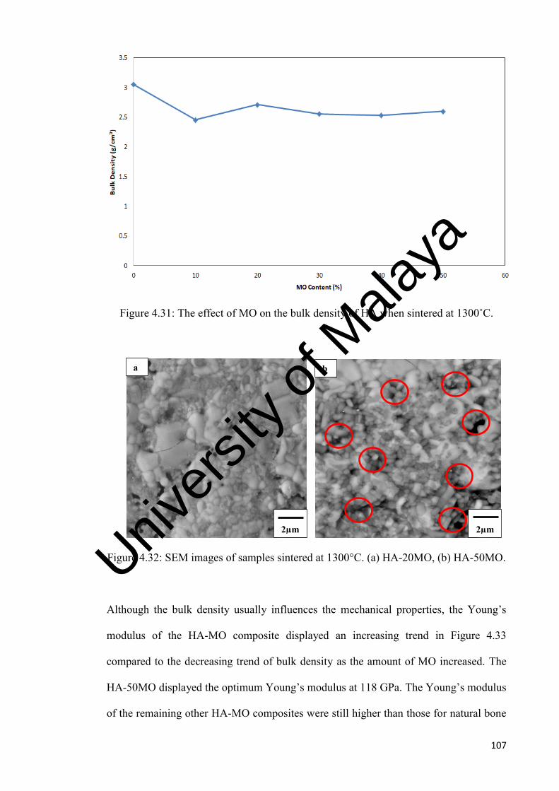
107
Figure 4.31: The effect of MO on the bulk density of HA when sintered at 1300˚C.
Figure 4.32: SEM images of samples sintered at 1300°C. (a) HA-20MO, (b) HA-50MO.
Although the bulk density usually influences the mechanical properties, the Young’s
modulus of the HA-MO composite displayed an increasing trend in Figure 4.33
compared to the decreasing trend of bulk density as the amount of MO increased. The
HA-50MO displayed the optimum Young’s modulus at 118 GPa. The Young’s modulus
of the remaining other HA-MO composites were still higher than those for natural bone
2µm
2µm
a b
Univers
ity of
Mala
ya

108
(E = 10-22 GPa) and enamel (E = 40-84 GPa) (Hench, 1991; LeGeros, 1993). It is
believed that the composite could have caused more dislocation in the bimodal grains
distribution of the material leading to firmer fixation. The mechanical interlock from the
pores which involved the arrangement of the atoms of the three phases (HA, MO,
whitlockite) could have inhibited the crack propagation by redistributing the stresses in
the regions adjacent to the crack tips thus improving the stiffness of the HA-MO
composites.
Figure 4.33: The effect of MO on the Young’s modulus of HA when sintered at 1300˚C.
However, Figure 4.34 displayed an inconsistent decreasing trend in hardness for the
HA-MO composites while monolithic HA exhibited highness hardness of 7.1 ± 0.2 GPa
when sintered at 1300˚C. The HA-MO composites were generally much lower than the
monolithic value but still above the cortical bone lower limit (0.2 GPa). This finding
was expected as the bulk density also exhibited decreasing trend when MO composition
increased. The inconsistency of the hardness value in the composite could be due to the
bimodal grain size distributed accompanied by various pores’ size in the material. The
Univers
ity of
Mala
ya

109
molten phase that exist from HA, MO and whitlockite upon sintered cannot form proper
grain boundary hence leading to random dislocation in the crystal structure leading to
the inconsistency of the hardness findings as well. Here, the addition of MO did not
bring beneficial effect to the hardness.
Figure 4.34: The effect of MO on the hardness of HA when sintered at 1300˚C.
The effect of adding MO into HA on the fracture toughness of sintered samples is
shown in Figure 4.35. It could be observed that the toughness of 0.9±0.1 MPam1/2 for
monolithic HA has been increased to 2.5±0.1 MPam1/2 when incorporated with 20MO
when sintered at 1300°C. Any further addition of MO resulted to decrease in fracture
toughness. This increased in fracture toughness could be attributed to the entire phase
formation mechanism which took place in this HA-MO composite which resulted to
smaller bimodal grain which been distributed evenly to form bridges across crack faces
acting as stress absorption mechanism which help improved the toughness of the
material.
Univers
ity of
Mala
ya

110
Figure 4.35: The effect of MO on the fracture toughness of HA when sintered at
1300˚C.
It has been concluded that the HA-MO composites show a promising direction in
improvement of fracture toughness which will definitely contribute to the new
knowledge of HA composite as few work has been done incorporating MO into HA.
Univers
ity of
Mala
ya

111
CHAPTER 5 - CONCLUSIONS & FURTHER WORK
5.1 Conclusions
In the present work, the densification behavior and properties of hydroxyapatite-
magnesium orthosilicate (HA-MO) composite were investigated. The amount of
magnesium orthosilicate in the green body was varied from 10wt. %, 20wt. %, 30wt. %,
40wt. % and 50wt. % and pressureless sintered from 1000°C to 1300°C for 2 hours. The
effects of incorporating MO with HA have been compared with the pure HA via phase
analysis, bulk density, linear shrinkage Vickers hardness, Young’s modulus, fracture
toughness and microstructural investigation. The results obtained can be concluded that:
1. The HA powder synthesized by the wet chemical precipitation method is in good
agreement with the JCPDS card. There were no other phases found in the
powder.
However, the peaks of the XRD trace were generally broad, which indicates that
the powder has low crystallinity. Large and small particles were observed from
the micrograph which formed the agglomerates.
2. The MO powder which was produced via combination of ultrasonification and
mechanical ball milling method is in good agreement with the JCPDS card. No
secondary phase was observed from the XRD result. The micrograph of MO
displayed flake like morphology and also lamellar shape forming hard
agglomerates.
3. The effect of incoporating magnesium orthosilicate (MO) to form the
composites regardless of various amount of MO (10wt. % to 50wt. %) and
sintering temperatures resulted to the decomposition of the HA phase to
Univers
ity of
Mala
ya

112
whitlockite phase (also known as β-TCP). The whitlockite phase becomes more
prominent when higher amount of MO was incorporated. The decomposition of
the composites was due to chemical reaction with MO during sintering. The MO
which contains magnesium inhibits apatite crystallization in solution which can
act as a stabilizer for the whitlockite phase which in return promotes the
decomposition of HA. Similar observation was also reported by Emadi et al.
(2010).
4. The HA-MO composites still exhibited higher amount of porosity than
monolithic HA however the HA-MO composites tend to display lesser porosity
in samples that contain higher amount of MO when sintered across the
increasing temperatures (1000°C - 1300°C). Large amount of pores and not well
defined grain size were observed in composites sintered at below 1200°C
regardless of MO contents due to the inability of MO to consolidate and densify
when sintered at low temperature. The average grain size of the composites were
found to be smaller than 0.55µm.
5. The grain growth and grain boundary for HA were only visible when sintered at
1200°C. The grains were observed to have grown rapidly, about ten times from
an average of 0.55µm at 1200°C to 5.5µm at 1300°C. A bimodal distribution of
grains was displayed by the HA grains.
6. Generally, the monolithic HA sintered samples attained higher shrinkage across
the sintering temperatures. A maximum shrinkage of 19.7% was attained from
monolithic HA while HA-20MO attained at about 17.7% shrinkage when
Univers
ity of
Mala
ya
113
sintered at 1300°C. The other HA-MO composites only attained shrinkage in the
range of ~7% to ~15%.
7. The monolithic HA sintered samples exhibited higher bulk density than HA-MO
composites regardless of the sintering temperature used. A maximum bulk
density of above 3 g/cm3 was attained by monolithic HA when sintered at
1300°C while the HA-20MO is the highest among the other composites to have
achieved bulk density of 2.7 g/cm3 at the same sintering temperature. The hard
agglomerates of magnesium orthosilicate and decomposition of the HA phase
could have led to the lower packing density in the green state.
8. For the Young’s modulus study, the HA-50MO composite exhibited a higher
Young’s modulus than monolithic HA regardless of sintering temperature. A
maximum Young’s modulus of 118 GPa was attained from the HA-50MO
composite when sintered at 1300°C while the monolithic HA attained at about
111 GPa at the same sintering temperature. The increase in Young’s modulus as
the temperatures increase reflect the reduce in porosity.
9. The hardness trend attained displays the similar trend found in the bulk density
results where the monolithic HA displays higher hardness value than the
composites. The monolithic HA hardness attained an optimum value of 7.1± 0.2
GPa when sintered at 1300°C. Conversely, the highest hardness attained by the
composite containing 20wt. % MO was 2.9± 0.1 GPa when sintered at 1300°C.
The hardness values of HA-MO composites (10wt. % - 50wt. %) sintered at
above 1100°C are still higher than cortical bone lower limit which is about 0.2
GPa.
Univers
ity of
Mala
ya

114
10. The use of magnesium orthosilicate has effectively improved the fracture
toughness of monolithic HA. In general, monolithic HA displays fracture
toughness in the range of 0.55 to 0.99 MPa·m1/2. The highest fracture toughness
of 2.5 ± 0.1 MPa·m1/2 was attained from HA-20MO composite sintered at
1300°C. This improvement in fracture toughness can be associated with the
presence of TCP in the composite matrix that could have interact with the
propagating crack through a stress absorption mechanism, although the presence
of such secondary phase is usually detrimental to the properties of HA. With the
fracture toughness improvement, the composite will create opportunities to be
used for biomedical load-bearing implants.
5.2 Further Work
Following are some suggestions for further work:
1. The use of other sintering methods such as microwave sintering and hot pressing
to prepare the HA-MO composite could be explored and compared with the
results obtained using pressureless sintering method.
2. A TEM study could be an avenue for further studies especially in elucidating the
sintering mechanism of HA-MO composite.
3. The use of two step sintering method as a possible way to suppress grain growth
of HA-MO composite in the final sintering stage. It would be interesting to
investigate if the two step sintering method could aid the densification of the
present composite.
Univers
ity of
Mala
ya

115
4. An in vitro testing through the use of simulated body fluid (SBF) can be
performed to evaluate the biocompatibility of the current HA-MO composite
samples.
5. A more insightful study of the dihydroxylation phenomenon of the composite
and the relation to XRD peak shifting of the present work could be obtained by
carrying out Raman spectroscopy studies.
6. To use various ramp rate, 5°C/minute and 15°C/minute for sintering and also
various holding time of 1 minute and 1 hour to observe the outcome on the HA-
MO composites in terms of its mechanical properties.
Univers
ity of
Mala
ya

116
REFERENCES Adamopoulus, O. & Papadopoulus, T. (2007). Nanostructured bioceramics for
maxillofacial applications. Journal of Materials Science: Materials in Medicine, 18: 1587-1597.
Afshar, A., Ghorbani, M., Ehsani, N., Saeri, M. R. & Sorrell, C. C. (2003). Some
important factors in the wet precipitation process of hydroxyapatite. Materials and Design, 24: 197-202.
Agrawal, D. K. (1998b). Microwave processing of ceramics. Current Opinion in Solid
State & Materials Science, 3: 480-485. Akira, O., Tetsuo, H., Hiroyuki, K., Naoki, K., Wenguang, G., Takeshi, K. & Naoto, E.
(2004). Comparison of hydroxyapatite and beta tricalcium phosphate as bone substitutes after exicison of bone tumors. Journal of Biomedical Materials Research, 42: 1411-1420.
Aizawa, M., Hanazawa, T., Itatani, K., Howell, F. S. & Kishioka, A. (1999).
Characterisation of hydroxyapatite powders prepared by ultrasonic spray-pyrolysis technique. Journal of Materials Science, 34: 2865-2873.
Akao, M., Aoki, H. & Kato, K. (1981). Mechanical properties of sintered hydroxyapatite for prosthetic applications. Journal of Material Science, 16: 809- 812. Almirall, A., Larrecq, G., Delgado, J. A., Martinez, S., Planell, J. A. & Ginebra, M. P.
(2004). Fabrication of low temperature macroporous hydroxyapatite scaffolds by foaming and hydrolysis of an α-TCP paste. Biomaterials, 25: 3671-3680.
Aminzare, M., Eskandari, A., Baroonian, M. H., Berenov, A., Hesabi, R., Taheri, M. &
Sadrnezhaad, S. K. (2013). Ceramics International, 39: 2197-2206. An, G. H., Wang, H. J., Kim, B. H., Jeong, Y. G., & Choa, Y. H. (2007). Fabrication and characterisation of a hydroxyapatite nanopowder by ultrasonic spray pyrolysis with salt-assisted decomposition. Materials Science and Engineering, 44: 821-824. Aykul, A., Kutbay, I., Evis, Z. & Usta, M. (2013). Effect of YF3 on the phase stability
and sinterability of hydroxyapatite – Partially stabilized zirconia composites. Ceramics International, 39: 7869-7877.
Bandyopadhyay, A., Bernard, S., Xue, W., Bose, S. (2006). Calcium phosphate-based
resorbable ceramics: Influence of MgO, ZnO, and SiO2 Dopants. Journal of the American Ceramic Society, 89: 2675-2688.
Ben-Nissan, B., Miley, A. & Vago, R. (2004). Morphology of sol-gel derived nano coated coralline hydroxyapatite. Biomaterials, 25: 4971-4975.
Univers
ity of
Mala
ya

117
Bernache-Assollant, D., Ababou, A., Champion, E., & Heughebaert, M. (2003). Sintering of calcium phosphate hydroxyapatite Ca10(PO4)6(OH)2. Calcination and particle growth. Journal of the European Ceramic Society, 23 (2): 229-241. Best, S. & Bonfield, W. (1994). Processing behavior of hydroxyapatite powders with
contrasting morphology. Journal of Materials Science: Materials in Medicine, 5: 516-521.
Best, S. M., Porter, A. E., Thian, E. S. & Huang, J. (2008). Bioceramics: Past, present and for the future. Journal of Materials Science: Materials in Medicine, 5: 516 – 521.
Bhaduri, S. & Bhaduri S. B. (1998). Recent developments in ceramic nanocomposites.
Journal of the Minerals, Metals, and Materials Society, 50: 44-51. Bianco, A., Cacciotti, I., Lombardi, M., Montanaro, L. & Gusmano, G. (2007). Thermal
stability and sintering behaviour of hydroxyapatite nanopowders. Journal of Thermal Analysis and Calorimetry, 88 (1): 237-243.
Bigi, A., Falini, G., Foresti, E., Ripamonti, A., Gazzano, M., & Roveri, N. (1993). Magnesium influence on hydroxyapatite crystallization. Journal of Inorganic Biochemistry, 49 (1): 69-78. Binyamin, G., Shafi, B. M. & Carlos, M. M. (2006). Biomaterials: A primer for surgeons. Seminar in Pediatric Surgery, 15: 276-283. Bogdanoviciene, I., Beganskiene, A., Tonsuaadu, K., Glaser, J., Meyer, H. J. & Kareiva,
A. (2006) Calcium hydroxyapatite, Ca¬10(PO4)6(OH)2 ceramics prepared by aqueous sol-gel processing. Materials Research Bulletin, 41: 1754-1762.
Bogdanov, B. I., Pashev, P. S., Hristov, J. H. & Markovska, I. G. (2008). Bioactive
fluorapatite-containing glass ceramics. Ceramics International, 35: 1651-1655. Bohner, M. (2000). Calcium orthophosphates in medicine: from ceramics to calcium
phosphate cements. International Journal of the Care of the Injured, 31: S-D37-D47.
Boskey, A. L. & Coleman, R. (2010). Critical reviews in oral biology & medicine: Aging and Bone. Journal of Dental Research, 89(12):1333-1348. Cacciotti, L. & Bianco, A. (2011). High thermally stable Mg-substituted tricalcium
phosphate via precipitation. Ceramics International, 37: 127-137. Calafiori, A. R., Di Marco, G., Martino, G. & Marotta, M. (2007). Preparation and
characterization of calcium phosphate biomaterials. Journal of Materials Science: Materials in Medicine, 18: 2331 - 2338.
Carlisle, E. M. (1970). Silicone: a possible factor in bone calcification. Science 167: 279-280. Carlisle, E. M. (1988). Silicon as a trace nutrient. Science of The Total Environment, 73:
95-106.
Univers
ity of
Mala
ya

118
Carolina, T., Chayuda, C., David, E. D. & George, V. F. (2016). Mechanical strength and damage tolerance of highly porous alumina ceramics produced from sintered particle stabilized foams. Ceramics International, 42: 8478-8487. Cao, N., Bai, Y., Ma, Q., Sui, J., Li, M. (2008). Biological behaviour of hydroxyapatite
coatings on carbon/carbon composites produced by plasma spraying. New Carbon Materials, 23 (2): 144-148.
Cao, W. & Hench, L. L. (1996). Bioactive Materials. Ceramics International, 22: 493- 507.
Cengiz, B., Gokce, Y., Yildiz, N., Aktas, Z. & Calimli, A. (2008). Synthesis and
characterization of hydroxyapatite nanoparticles. Colloids and Surfaces A: Physicochemical and Engineering Aspects, 322: 29-33.
Chai, C. S. & Ben-Nissan, B. (1999). Bioactive nanocrystalline sol-gel hydroxyapatite
coatings. Journal of Materials Science: Materials in Medicine, 10: 465-469. Chen, C. W., Oakes, C. S., Byrappa, K., Riman, R. E., Brown, K., TenHuisen, K. S. &
Janas, V. F. (2004). Synthesis, characterization, and dispersion properties of hydroxyapatite prepared by mechanochemical-hydrothermal methods. Journal of Materials Chemistry A, 14: 2425-2432.
Chen, Q. Z., Thompson, I. D. & Boccaccini, A. R. (2006). 45S5 Bioglass derived glass
ceramic scaffolds for bone tissue engineering. Biomaterials, 27: 2414-2425. Chern, L., Liu, M. L., Chen, K. S. & Ju, C. P. (1993). Plasma-sprayed hydroxyapatite-
bioactive glass coatings on Ti6A14V. Ceramic Engineering and Science Proceedings, 14: 971-981.
Chiu, C., Hsu, H. C. & Tuan, W. H. (2007). Effect of zirconia addition on the
microstructural eveloution of porous hydroxyapatite. Ceramics International, 33: 715-718.
Chu, C., Xue, X., Zhu, J. & Yin, Z. (2004). Mechanical and biological properties of
hydroxyapatite reinforced with 40 vol. % titanium particles for use as hard tissue replacement. Journal of Materials Science: Materials in Medicine, 15: 665-670.
Colling, D.C. & Vasilos, T. (1995). Industrial materials: Polymers, ceramics and
composites. Vol 2. New Jersey: Prentice Hall, Inc. Dee, K. C., Puleo, D. A. & Bizios, R. (2003). Biomaterials: An introduction to tissue
biomaterial interactions. John Wiley & Sons, Inc.: 13. Dobradi, A., Bodogh, M. E., Kovacs, K. & Balczar, I. (2015). Structure and properties of bio-glass ceramics containing natural bones. Ceramics International, 41: 4874-4881. Donadel, K., Laranjeira, M. C. M., Goncalves, V. L., Favere, V. T., de Lima, J. C. &
Prates, L. H. M. (2005). Hydroxyapatite produced by wet-chemical methods. Journal of the American Ceramic Society, 88 (8): 2230-2235.
Univers
ity of
Mala
ya

119
Dorozhkin, S. V. (2007). Calcium orthophosphates. Journal of Materials Science, 42: 1061-1095.
Dorozhkin, S. V. (2008). Calcium phosphate cements for biomedical application. Journal of Material Science, 43: 3028-3057. Dorozhkin, S. V. (2010). Bioceramics of calcium orthophosphates. Biomaterials, 31:
1465-1485. Drdlik, D., Slama, M., Hadraba, H. & Cihlar, J. (2015). Hydroxyapatite/zirconia
microfiber composites with controlled microporosity and fracture properties prepared by electrophoretic deposition. Ceramics International, 41: 11202-11212.
El Briak-BenAbdeslam, H., Ginebra, M. P., Vert, M. & Boudeville, P. (2008). Wet or
dry mechanichemical synthesis of calcium phosphates? Influence of the water content on DCPC-CaO reaction kinetics. Acta Biomaterialia, 4: 378-386.
Encinas-Romero, M. A., Aguayo-Salinas, S., Valenzuela-Garcia, J. L., Payan, S. R. &
Castilon-Barraza, F. F. (2010). Mechanical and bioactive behavior of hydroxyapatite wollastonite sintered composites. International Journal of Applied Ceramic Technology, 7: 164-177.
Evis, Z. (2007). Reactions in hydroxyapatite-zirconia composites. Ceramics
International, 33: 987-991. Fathi, M. H., Hanifi, A. & Mortazavi, V. (2008). Preparation and bioactivity evaluation
of bone-like hydroxyapatite nanopowder. Journal of Materials Processing Technology, 202: 536-542.
Fathi, M. H. & Kharaziha, M. (2009). Two step sintering of dense, nanostructural
forsterite. Material Letters, 63: 1455-1458. Fathi, M. H. & Hanifi, A. (2007). Evaluation and characterisation of nanostructure
hydroxyapatite powder prepared by simple sol-gel method. Materials Letters, 61, 3978-3983.
Fernandez, E., Gil, F. J., Ginebra, M. P., Driessens, F. C. M., Planell, J. A. & Best, S. M. (1999a). Calcium phosphate bone cements for clinical applications. Part I: Solution chemistry. Journal of Materials Science: Materials in Medicine, 10: 169-176. Galea, L. G., Bohner, M., Lemaitre, J., Kohler, T. & Muller, R. (2008). Biomaterials, 29: 3400-3407. Giannoudis, P. V., Dinopoulus, H. & Tsiridis, E. (2005). Bone substitutes: An update.
International Journal of the Care of the Injured, 365: 520-527. Gibson, I. R., Ke, S., Best, S. M. & Bonfield, W. (2001). Effect of powder characteristics on the sinterability of hydroxyapatite powders. Journal of Materials Science: Materials in Medicine, 12: 163-171.
Univers
ity of
Mala
ya

120
Goller, G., Demirkiran, H., Oktar, F. N. & Demikersen, E. (2003). Processing and characterization of bioglass reinforced hydroxyapatite composites. Ceramics International, 29: 721-724.
Gu, Y. W., Loh, N. H., Khor, K. A., Tor, S. B. & Cheang, P. (2002). Bone-like apatite
layer formation on hydroxyapatite prepared by spark plasma sintering (SPS). Biomaterials, 25: 4127-4134.
Habibovic, P. & Barralet, J. E. (2011). Bioinorganics and biomaterials: bone repair. Acta Biomaterialia. 7: 3013-3026. Hafezi, M., Talebi, A. R., Miresmaeili, S. M., Sadeghian, F. & Fesahat, F. (2013).
Histological analysis of bone repair in rat femur via nanostructured merwinite granules. Ceramics International, 39: 4575-4580.
Han, J. K., Song, H. Y., Saito, F. & Lee, B. T. (2006). Synthesis of high purity nano- sized hydroxyapatite powder by microwave-hydrothermal method. Materials Chemistry and Physics, 99: 235-239. He, L. H., Standard, O. C., Huang, T. T. Y., Latella, B. A. & Swain, M. V. (2008).
Mechanical behaviour of porous hydroxyapatite. Acta Biomaterialia, 4: 577- 586.
Hench, L. L., Splinter, R. J., Allen, W. C., & Greenlee, T. K. (1972). Bonding
mechanisms at the interface of ceramic prosthetic materials. Journal of Biomedical Materials Research, 2: 117-141.
Hench, L. L., Wilson J, eds. (1993). An introduction to bioceramics. World Scienctific,
: 1-24. Hench, L. L. (1998). Bioceramics. Journal of the American Ceramic Society, 81: 1705-
1728 Hench, L. L. (2000). The challenge of orthopaedic materials. Current Orthopaedics, 14:
7-15. Hench, L. L. (1991). Bioceramics: From concept to clinic. Journal of the American
Ceramic Society, 74: 1487-1510. Hench, L. L. (2006). The story of bioglass. Journal of Materials Science: Materials in
Medicine, 17: 967-978. Heyman, D. & Passuti, N. (1999). Bone substitutes: new concept. European Journal of
Orthopaedic Surgery & Traumatology, 9: 179-184. Hsieh, M., Perng, L., Chin, T. & Perng, H. (2001). Phase purity of sol-gel-derived
hydroxyapatite ceramic. Biomaterials, 22: 2601-2607. Ioku, K, Goto, S., Kurosawa, H., Shibuya, K., Yokozeki, H., Hayashi, T., Nakagawa, T.
(1996). Reaction of porous β-TCP with different micro-structure in vivo. Bioceramics, 9: 201-204.
Univers
ity of
Mala
ya

121
Jagdale, P.N. & Bamane, SR. (2011). Calcium hydroxyapatite bioceramics and evaluation of their in vitro biocompatibility. Research Journal of Pharmaceutical, Biological and Chemical Sciences, 2: 180-186. Jagwiga, B. M., Krzysztof, H., Maciej, S., Miroslaw, M. B., Beata, M. & Radoslaw, L.
(2016). Natural and synthethic hydroxyapatite/zirconia composites: A comparative study. Ceramics International, 42: 11126-11135.
Jokanovic, V., Izvonar, D., Dramicanin, M. D., Jokanovic, B., Zivojinovic, V., Markovic, D. & Dacic, B. (2006). Hydrothermal synthesis and nanostructure of carbonated calcium hydroxyapatite. Journal of Materials Science: Materials in Medicine, 17: 539-546. Juang, H. Y. & Hon, M. H. (1996). Effect of calcination in the sintering of
hydroxypapatite. Biomaterials, 17: 2059-2064. Jun, I. K., Song, J. H., Choi, W. Y., Koh, Y. H., Kim, H. E. & Kim, H. W. (2007). Porous hydroxyapatite scaffolds coated with bioactive apatite wollastonite glass ceramics. Journal of the American Ceramic Society, 90: 2703-2708. Kalita, S. J., Bhardwaj, A. & Bhatt, H. A. (2007). Nanocrystalline calcium phosphate
ceramics in biomedical engineering. Materials Science and Engineering: C, 27: 441-449.
Kalmodia, S., Goenka, S., Laha, T., Lahiri, D., Basu, B., & Balani, K. (2010).
Microstructure, mechanical properties and in vivo biocompatibility of spark plasma sintered hydroxyapatite-aluminum oxide carbon nanotube composite. Material Science and Engineering, 30: 1162-1169.
Kangasniemi, I. M. O. (1993). Development of Ca, P-ceramic containing bioactive glass
composites. Master Thesis, University of Leiden, The Netherlands. Kharaziha, M. & Fathi, M. (2009). Synthesis and characterization of bioactive forsterite
nanopowder. Ceramics International, 35 (6): 2449-2454. Kharaziha, M. & Fathi, M. H. (2010). Improvement of mechanical properties and
biocompatibility of forsterite bioceramic addressed to bone tissue engineering materials. Journals of the mechanical behavior of biomedical materials, 3: 530 – 537.
Khorsand, S., Fathi, M. H., & Amirkhanlou, S. (2014). Hydroxyapatite/alumina
nanocrystalline composite powders synthesized by sol-gel process for biomedical applications. International Journal of Minerals, Metallurgy, and Materials, 21 (10): 1033-1036.
Kien, P. T., Ishikawa, K. & Tsuru, K. (2015). Setting reaction of α-TCP spheres and an
acidic calcium phosphate solution for the fabrication of fully interconnected macroporous calcium phosphate. Ceramics International, 41: 13525-13531.
Kim, H. M. (2001). Bioactive ceramics: challenges and perspectives. Journal of the
Ceramic Society of Japan, 109 (4): 49-57.
Univers
ity of
Mala
ya

122
Kokubo, T., Kim, H. M. & Kawashita, M. (2003). Novel bioactive materials with different mechanical properties. Biomaterials, 24: 2161-2175. Kokubo, T. (1990). Surface chemistry of bioactive glass ceramics. Journal of Non
Crystalline Solids, 120: 138-151. Kokubo, T., Kim, H. M., Kawashita, M. & Nakamura, T. (2004). Bioactive metals:
preparation and properties. Journal of Materials Science, 15: 99-107.
Kokubo, T., Shigematsu, M., Nagashima, Y., Tashiro, M., Nakamura, T., Yamamuro, T. & Higashi, S. (1982). Apatite and wollastonite containing glass ceramics for prosthetic applications. Bulletin of the Institute for Chemical Research, 60: 260-268.
Kobayashi, S. & Murakoshi, T. (2013). Characterization of mechanical properties and
bioactivity of hydroxyapatite/β-tricalcium phosphate composites. Advanced Composite Materials, 23: 163-177.
Kosanic, C., Stubicar, N., Tomasic, N., Bermanec, V., & Stumicar, M. (2005). Synthesis of forsterite powder by combined ball milling and thermal treatment. Journal of Alloys and Compounds, 389: 306-309. Kulkarni, S. S., Yong, Y., Rys, M. J. & Lei, S. (2013). Machining assessment if nano-
crystalline hydroxyapatite bio-ceramic. Journal Manufacturing Processes, 15(4): 666-672.
Kumta, P. N., Sfeir, C., Lee, D. H., Olton, D. & Choi, D. (2005). Nanostructured calcium phosphates for biomedical applications: novel synthesis and characterization. Acta Biomaterialia, 1: 65-83. Laczka, M., Cholewa, K. & Osyczka, A. M. (2016). Bioactivity and osteoinductivity of
glasses and glassceramics and their material determinants. Ceramics International, 42: 14313-14325.
Lala, S., Satpati, B. & Pradhan, S. K. (2016). Sintering behavior and growth mechanism
of β-TCP in nanocrystalline hydroxyapatite synthesized by mechanical alloying. Ceramics International, 42: 13176-13182.
Landi, E. Tampieri, A., Celotti, G. & Sprio, S. (2000). Densification behaviour and
mechanisms of synthetic hydroxyapatites. Journal of European Ceramic Society, 20: 2377-2387.
LeGeros, R. Z. (1991). Calcium phosphates in oral biology and medicine. Monographs
in Oral Science, 15: 1-201. LeGeros, R. Z.& LeGeros, J.P. (1993). Dense hydroxyapatite: In An Introduction to
Bioceramics. Ed. By Hench, L. L & Wilson. J. (World Scientific Singapore), 9: 139-180.
LeGeros, R. Z., Lin, S., Rohanizadeh, R., Mijares, D. & LeGeros, J. P. (2003). Biphasic
calcium phosphate bioceramics: preparation, properties and applications. Journal of Materials Science: Materials in Medicine, 14: 201-209.
Univers
ity of
Mala
ya

123
Le Huec, Schaeverbeke, T., Clement, D., Faber, J. & Le Rebeller, A. (1995). Influence of porosity on the mechanical resistance of hydroxyapatite ceramics under compressive stress. Biomaterials, 16: 113-118. Li, J., Liao, H. & Hermansson, L. (1996). Sintering of partially-stabilized zirconia and
partially stabilized zirconia-hydroxyapatite composites by hot isostatic pressing and pressureless sintering. Biomaterials, 17: 1787-1790.
Li, J., Fartash, B. & Hermansson, L. (1995). Hydroxyapatite-alumina composites and
bone bonding. Biomaterials, 16: 417-422. Li, J., Wang, Q., Liu, J. H., & Li, P. (2009). Synthesis process of forsterite refractory by
iron ore tailings. Journals of environmental sciences supplement, 16: 92-295. Liu, Y., Hou, D. & Wang, G. (2004). A simple wet chemical synthesis and
characterization of hydroxyapatite nanorods. Materials Chemistry and Physics, 86: 69-73.
Liu, Y. & Wang, M. (2007). Developing a composite material for bone tissue repair.
Current Applied Physics, 7: 547-554. Liu, H., Yazici, H., Ergun, C., Webster, T. J. & Bermek, H. (2008). An in vitro evaluation of the Ca/P ratio for the cytocompatibility of nano to micron particulate calcium phosphates for bone regeneration. Acta Biomaterialia, 4: 1472-1479. Lugo, G. J., Mazon, P. & De Aza, P. N. (2016). Material processing of a new calcium
silicophosphate ceramic. Ceramics International, 42: 673-680. Ma, J., Chen, C. Z., Wang, D. G., Shao, X., Wang, C. Z. & Zhang, H. M. (2012). Effect
of MgO addition on the crystallization and in vitro bioactivity of glass ceramics in the CaO-MgO-SiO2-P2O2 system. Ceramics International, 38: 6677-6684.
Mano, J. F., Sousa, R. A., Boesel, L. F., Neves, N. M. & Reis, R. L. (2004). Bioinert,
biodegradable and injectable polymeric matrix composites for hard tissue replacement: state of the art and recent developments. Composites Science & Technology, 64: 789-817.
Mendelson, M. I. (1969). Average grain size in polycrystalline ceramics. Journal of the
American Ceramic Society, 52: 443-446. Mobasherpour, I., Heshajin, M. S., Kazemzadeh, A. & Zakeri, M. (2007). Synthesis of
nanocrystalline hydroxyaptite by using precipitation method. Journal of Alloys and Compounds, 430: 330-333.
Mobasherpour, Hashjin, M. S., Toosi, S. S. & Kamachali, R. D. (2009). Effect of the
addition ZrO2-Al2O3 on nanocrystalline hydroxyapatite bending strength and fracture toughness. Ceramics International, 35: 1569-1574.
Monika, S. (2015). Substituted hydroxyapatite for biomedical applications: A review.
Ceramics International, 41: 9203-9231.
Univers
ity of
Mala
ya

124
Muralithran. G. & Ramesh. S. (2000). The effects of sintering temperature on the properties of hydroxyapatite. Ceramics Intternational, 26: 221-230.
Murugan, R. & Ramakrishna, S. (2005). Aqueous mediated synthesis of bioresorbable
nanocrystalline hydroxyapatite. Journal of Crystal Growth, 274: 209-213. Narasaraju, T. S. B. & Phebe, D. E. (1996). Review: Some physico-chemical aspects of
hydroxylapatite. Journal of Material Science, 31: 1-21. Niihara, K. (1985). Indentation microfracture of ceramics – Its application and problems. Ceramic Society of Japan, 20: 12-18. Nikaido, T., Tsuru, K., Munar, M., Maruta, M., Matsuya, S., Nakamura, S. & Ishikawa,
K. (2015). Fabrication of β-TCP foam: Effects of magnesium oxide as phase stabilizer on its properties. Ceramics International, 32: 617-621.
Nursen, K., Muharrem, T. & Feza, K. (2004). Fabrication and characterization of porous
tricalcium phosphate ceramics. Ceramics International, 30: 205-211. Orlovskii, V.P., Komlev, V.S. & Barinov, S. M. (2002). Hydroxyapatite and
hydroxyapatite-based ceramics. Inorganic Materials, 38: 973-984. Paul, C. (2004). Bone structure and function. Retrieved from
https://deps.washington.edu/bonebio/ASBMRed/structure.html. Pramanik, S., Agarwal, A. K., Rai, K. N. & Garg, A. (2007). Development of high
strength hydroxyapatite by solid-state sintering process. Ceramics International, 33: 419-426.
Rabiee, S. M., Nazpar, N., Azizian, M., Vashaee, D. & Tayebi, L. (2015). Effect of ion
substitution on properties of bioactive glasses: A review. Ceramics International, 41: 7241-7251.
Rahaman, M.N. (1995). Ceramic processing and sintering (2nd ed). New York: Marcel
Dekker, Inc. Ramesh, S., Aw., K. L., Tolouei, T., Amiriyan, M., Tan, C. Y., Hamdi, M., Purbolaksono, J., Hassan, M. A. & Teng, W. D. (2013). Sintering properties of hydroxyapatite powders prepared using different methods. Ceramics International, 39: 111-119. Ramesh, S., Tan, C. Y., Tolouei, R., Amiriyan, M., Purbolaksono, J., Sopyan, I. & Teng, W. D. (2012). Sintering behavior of hydroxyapatite prepared from different routes. Materials and Design, 34: 148-154. Ramesh, S., Tan, C. Y., Bhaduri, S. B., Teng, W. D. & Sopyan, I. (2008). Densification
behaviour of nanocrystalline hydroxyapatite bioceramics. Journal of Materials Processing Technology, 206: 221-230.
Ramesh, S., Tan, C. Y., Sopyan, I., Hamdi, M. & Teng, W. D. (2007a). Consolidation of nanocrystalline hydroxyapatite powder. Science and Technology of Advanced Materials, 8: 124-130.
Univers
ity of
Mala
ya

125
Ramesh, S., Tan, C. Y., Bhaduri, S. B. & Teng, W. D. (2007b). Rapid densification of nanocrystalline hydroxyapatite for biomedical applications. Ceramics International, 33: 1363-1367.
Ramesh, S. (1997). The influence of transition metal oxide additions on the
manufacturing behavior & properties of yttria-tetragonal zirconia polycrystals. (Doctoral Dissertation, University of Sundreland).
Ramesh, S. (2004). A method for manufacturing hydroxyapatite bioceramic. Malaysia
Patent. No. PI. 20043325. Raynaud, S., Champion, E., Bernache-Assollant, D., & Thoma, P. (2002a). Calcium phosphate apatites with variable Ca/P atomic ratio I. Synthesis, characterization and thermal stability of powders. Biomaterials, 23: 1065-1072. Raynaud, S., Champion, E., & Bernache-Assollant, D. (2002b). Calcium phosphate apatites with variable Ca/P atomic ratio II. Calcination and sintering. Biomaterials, 23: 1073-1080. Raynaud, S., Champion, E., Bernache-Assollant, D., & Lafon, J. P. (2002c). Calcium phosphate apatites with variable Ca/P atomic ratio III. Mechanical properties and degradation in solution of hot pressed ceramics. Biomaterials, 23: 1081- 1089. Rodriguez-Lorenzo, L. M., Vallet-Regi, M. & Ferreira, J. M. F. (2001). Fabrication of
hydroxyapatite bodies by uniaxial pressing from a precipitated powder. Biomaterials, 22: 583-588.
Roeder, R. K., Converse, G. L., Kane, R. J. & Yue, W. (2008). Hydroxyapatite reinforced polymer biocomposites for synthetic bone substitutes. Biomaterials, 60: 38-45. Ruys, A. J., Wei, M., Sorrell, C. C., Dickson, M. R., Brandwood, A. & Milthorpe, B. K.
(1995a). Sintering effects on the strength of hydroxyapatite. Biomaterials, 16: 405-415.
Ryu, H. S., Lee, J. K., Kim, H. & Hong, K. S. (2005). New type of bioactive materials:
Hydroxyapatite/α-wollastonite composites. Journal of Materials Research, 20: 1154-1163.
Saeri, M. R., Afshar, A., Ghorbani, M., Ehsani, N. & Sorrell, C. C. (2003). The wet
precipitation process of hydroxyapatite. Materials Letters, 57: 4064-4069. Santos, M. H., de Oliveira, M., de Freitas Souza, L. P., Mansur, H. S. & Vasconcelos, W. L. (2004). Synthesis control and characterization of hydroxyapatite prepared by wet precipitation process. Materials Research Bulletin, 7 (4): 625-630. Sara, L. K. Y, Christopher, C. K. M., Ramesh, S., Tan, C. Y., Teng, W. D. & Sopyan, I.
(2012). Characterization of forsterite bioceramics. Advanced Materials Research, 576: 195-198.
Univers
ity of
Mala
ya

126
Schwarz, K., & Milne, D. (1972). Growth promoting effects of Si in rats. Nature, 4: 293-333. Schwarz, K. & Milne, D. (1972). Fluorine requirement for growth in the rat. Bioinorganic Chemistry, 1 (4): 331-338. Sebdani, M. M., & Fathi, M. H. (2011). Novel hydroxyapatite forsterite bioglass
nanocomposite coatings with improved mechanical properties. Journal of alloys and compound, 509: 2273-2276.
Sebdani, M. M. & Fathi, M. H. (2011). Fabrication and characterization of
hydroxyapatite-forsterite bioactive glass composite nanopowder for biomedical applications. International Journal of Applied Ceramic Technology, 8 (3): 553-559.
Sivakumar, R. (1999). On the relevance of biomaterials. Bulletin of Materials Science, 22 (3): 647-655. Shirtliff, V. J. & Hench, L., L. (2003). Bioactive materials for tissue engineering,
regeneration and repair. Journal of Materials Science, 38: 4697-4707. Siyu, N., Lee, C., & Jiang, C. (2007). Preparation and characterization of forsterite
bioceramics. Ceramics International, 33: 83-88. Slosarcyzk, A., Stobierska, E. Paszkiewicz, Z. & Gawlicki, M. (1996). Calcium
phosphate materials prepared from precipitates with various calcium-phosphorus molar ratio. Journal of the American Ceramic Society., 79 (10): 2539-2544.
Suchanek, W., Yashima, M., Kakihana, M. & Yoshimura, M. (1997). Hydroxyapatite
ceramics with selected sintering additives. Biomaterials, 18: 923-933. Suchanek, W. L., Shuk, P., Byrappa, K., Riman, R. E., TenHuisen, K. S. & Janas, V. F.
(2002). Mechanochemical hydrothermal synthesis of carbonated apatite powders at room temperature. Biomaterials, 23: 699-710.
Suchanek, W. & Yoshimura, M. (1998). Processing and properties of hydroxyapatite-
based biomaterials for use as hard tissue replacement implants. Journal of Materials. Research, 13: 94-116.
Sung, Y. M., Lee, J. C. & Yang, J. W. (2004). Crystallization and sintering characteristics of chemically precipitated hydroxyapatite nanopowder. Journal of Crystal Growth, 262: 467-472. Sugiyama, T., Tsunooka, T., Kakimoto, K., & Ohsato, H. (2006). Microwave dielectric
properties of forsterite-based solid solutions. Journals of the European Ceramic Tampieri, A., Celloti, G., Sprio, S. & Mingazzini, C. (2000). Characteristics of synthetic
hydroxyapatites and attempts to improve their thermal stability. Materials Chemistry Physics, 64: 54-61.
Univers
ity of
Mala
ya

127
Tancred, D. C., Carr, A. J. & McCormack, B. A. O. (2001). The sintering and mechanical behavior of hydroxyapatite with bioglass additions. Journal of Materials Science: Materials in Medicine, 12 (1): 81-93. Tancred, D. C., McCormack, B. A. O, & Carr, A. J. (1998). A quantitative study of the
sintering and mechanical properties of hydroxyapatite/phosphate glass composites. Biomaterials, 19: 1735-1743.
Tavangarian, F., & Emadi, R. (2010). Effects of fluorine ion and mechanical activation
on nanostructure forsterite formation mechanism. Powder Technology, 203: 180-186.
Tavangarian, F., & Emadi, R. (2011). Improving degradation rate and apatite formation
of nanostructure forsterite. Ceramics International, 38: 2275- 2280. Teamey, G. J., Boppart, S. A., Bouma, B., Brezinski, M. E., Golubovic, B., Southern, J.
F., Swanson, E. A., & Fujimoto, J. G. (1996). Conference on Lasers and Electra-Optics, OSA Technical Digest Series, 9: 57.
Thamaraiselvi, T. V. & Rajeswari, S. (2004). Biological evaluation of bioceramic
materials – A review. Trends in Biomaterials & Artificial Organs, 18: 9-17. Thurmer, M. B., Diehl, C. E. & Santos, L. A. (2016). Calcium phosphate cements based
on alpha-tricalcium phosphate obtained by wet method: Synthesis and milling effects. Ceramics International, 13: 1330-1245.
Tian, T., Jiang, D., Zhang, J. & Lin, Q. (2008). Synthesis of Si-substituted hydroxyapatite by a wet mechanochemical method. Materials Science and Engineering: C, 28, 57-63. Tony, M. K., Elise, F. M & Oscar, C. Y. (2003). Standard handbook of biomedical
engineering & design. Bone Mechanics. Mc-Graw-Hill Professional. Vallet-Regi, M. & Gonzalez-Calbet, J. M. (2004). Calcium phosphate as substitution of
bone tissues. Progress in Solid State Chemistry, 32: 1-31. Veljovic, Dy., Jancic-Haneman, R., Balac, I., Jokie, B., Putic, S., Petrovic, R., Janackonic, Dj. (2011). The effect of the shape and size of the pores on the mechanical properties of porous HAP-based bioceramics. Ceramics International, 37: 471-479. Viswanath, B. & Ravishankar, N. (2006). Interfacial reactions in hydroxyapatite/alumina nanocomposites. Scripta Materialia, 55: 863-866. Viswanath, B., Raghavan, R., Ramamurty, U. & Ravishankar, N. (2007). Mechanical
properties and anisotropy in hydroxyapatite single crystals. Scripta Materialia, 57: 361-364.
Vitale-Brovarone, C., Verne, E., Robligio, L., Martinasso, G., Canuto, R. A. & Muzio, G. (2008). Biocompatible glass-ceramics materials for bone substitution. Journal of Materials Science: Materials in Medicine, 19: 471-478.
Univers
ity of
Mala
ya

128
Wang, F., Li, M., Lu, Y. & Qi, Y. (2005). A simple sol-gel technique for preparing hydroxyaptite nanopowders. Materials Letters, 59: 916-919.
Wang, X., Fan, H., Xiao, Y., & Zhang, X. (2006). Fabrication and characterization of
porous hydroxyapatite / β-tricalcium phosphate ceramics by microwave sintering. Materials Letters, 60: 455-458.
Wang, P. E. & Chaki, T. K. (1993). Sintering behaviour and medical properties of
hydroxyapatite and dicalcium phosphate. Journal of Mateirals Science: Mateirals in Medicine, 4: 150-158.
Wang, M. (2003). Developing bioactive composite materials for tissue replacement.
Biomaterials, 24: 2133-2151. Williams, D. F. (2003). Biomaterials and tissue engineering in reconstructive surgery.
Sādhanā, 28: 563-574. Williams, D. F. (2008). On the mechanism of biocompatibility. Biomaterials, 29: 2941-
2953. Witte, F., Feyerabend, F., Maier, P., Fischer, J., Stormer, M., Blawert, C., Dietzel, W. &
Hort, N. (2007). Biodegradable magnesium-hydroxyapatite metal matrix composites. Biomaterials, 28: 2163-2174.
Xiao, F., Ye, J., Wang, Y. & Rao, P. (2005). Deagglomeration of HA during precipitation synthesis. Journal of Materials Science, 40: 5439-5442. Xie, Y., Zhai, W., Chen, L., Chang, J., Zheng, X. & Ding, C. (2009). Preparation and in
vitro evaluation of plasma sprayed Mg2SiO4 coating on titanium alloy. Acta Biomaterialia, 5: 2331-2337.
Xu, P. K. & Wei, G. Z. (2005). New process technology for refractory. Metallurgy
Industry Press, Beijing, 4: 2-5.
Xu, Y., Wang, D., Yang, L. & Tang, H. (2001). Hydrothermal conversion of coral into hydroxyapatite. Materials Characterization, 47: 83-87.
Yamanaka, A., Yokoyama, Y. & Iwamoto, H. (2006). Biomaterials in ophthalmology.
Journal of Artificial Organs, 9: 67-70. Yamamuro, T., Kokubo, T. & Ohtsuki, C. (1992). Mechanism of apatite formation on
CaO-SiO2-P2O5 glasses in a simulated body fluid. Journal of Non-Crystalline Solids, 143: 84-92.
Yasuda, H. Y., Mahara, S., Terashita, N. & Umakoshi, Y. (2002). Preparation of porous
hydroxyapatite/alpha tricalcium phosphate composites by a colloidal process. Materials Transaction, 43: 1332-1335.
Zakaria, S.M., Sharif, S.H., Othman, M. R., Yang, F., Jansen, J.A. (2013). Nano-phase
hydroxyapatite as a biomaterial in advanced hard tissue engineering. Tissue Engineering Part B Reviews, 19 (5): 431-441.
Univers
ity of
Mala
ya

129
Zhang, C., Zhang, X., Liu, C., Sun, K., & Yuan, J. (2016). Nano – alumina/hydroxyapatite composite powders prepared by in-situ chemical precipitation. Ceramics International, 42: 279-285. Zhang, S. & Gonsalves, K. E. (1997). Preparation and characterisation of thermally stable nanohydroxyapatite. Journal of Materials Science: Materials in Medicine, 8: 25-28. Zhang, Y., Wang, Y., Shi, B. & Cheng, X. (2007). A platelet derived growth factor
releasing chitosan/coral composite scaffold for periodontal tissue engineering. Biomaterials, 28: 1515-1522.
Univers
ity of
Mala
ya

130
LIST OF PUBLICATIONS
1) Densification behavior of magnesium orthosilicate-hydroxyapatite composite.
Journal of Ceramic Processing Research. 17(6): 620-625 (2016).
2) Sintering behavior and properties of magnesium orthosilicate-hydroxyapatite
ceramic. Ceramics International. 42: 15756-15761 (2016).
Univers
ity of
Mala
ya