magnetic-modified nanodiamond for bio-applications magnetic-modified nanodiamond for...
TRANSCRIPT

Magnetic-modified nanodiamond for bio-applications
Chang-You Song 1, Nikolai Perov2, Valentina Bessalova (Samsonova)2, Svetlana Norina2, Li-Chi Liu1, Zhe-Rui Lin1, Yu-Chung Lin1, Ashek-I-Ahmed1, Artashes Karmenyan 1, Olga Levinson3, Boris Zousman3,
Elena Perevedentseva1, Chia-Liang Cheng1,*
1Department of Physics, National Dong Hwa University, Taiwan 2Moscow State University, Russia
3Ray Techniques Ltd., Israel
NDNC 2016, Xi’an China, May 22-26, 2016
1
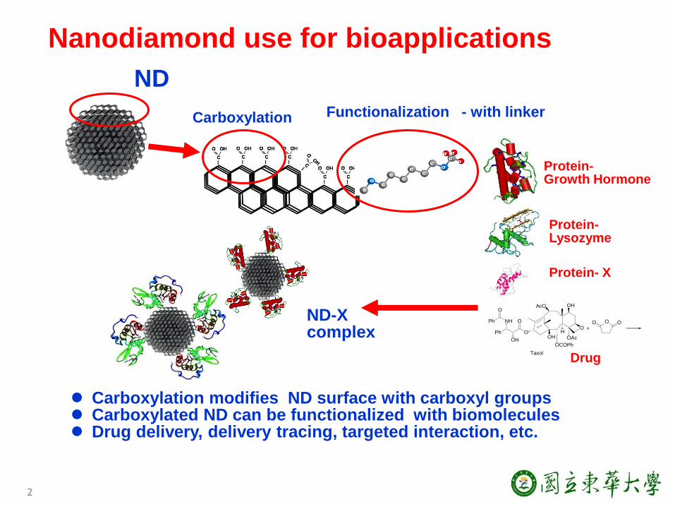
Nanodiamond use for bioapplications
Carboxylation Functionalization - with linker
2
ND
Carboxylation modifies ND surface with carboxyl groups Carboxylated ND can be functionalized with biomolecules Drug delivery, delivery tracing, targeted interaction, etc.
OO OO
OHAcO
OHO
OAc
H
OCOPh
O
Ph
NH
O
Ph
OH
+
Taxol
Protein- Growth Hormone
Protein- Lysozyme
Protein- X
Drug
ND-X complex
3
RBC
Cancer cell
Bacteria
Zebrafish embryo
Mice
Microorganisms
Nanodiamond use for bioapplications
Combine the properties of ND with magnetism. Applications of NMR, MRI, controlled drug delivery, …
Nanomedicine 2013, 8, 2041-2060
We studied ND interaction with different bio-systems and have found:
Nontoxic / bio-compatible Great property of photoluminescence High efficiency in drug delivery

The Advantages of Magnetic NPs for Bioapplications
L. H. Reddy, et. al., Chem. Rev., 2012, 112 (11), 5818–5878
4
H. Hao et al, J. Mater. Chem. B, 2014, 2, 7978-7987
(1) Drug reservoir (2) Active accumulation
(3) Magnetic Resonance Image
5
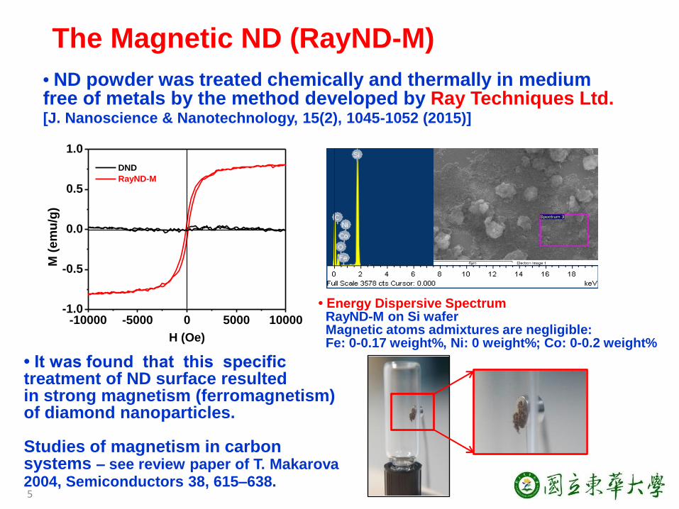
• ND powder was treated chemically and thermally in medium free of metals by the method developed by Ray Techniques Ltd. [J. Nanoscience & Nanotechnology, 15(2), 1045-1052 (2015)]
The Magnetic ND (RayND-M)
-10000 -5000 0 5000 10000-1.0
-0.5
0.0
0.5
1.0
M (
em
u/g
)
H (Oe)
DND
RayND-M
• It was found that this specific treatment of ND surface resulted in strong magnetism (ferromagnetism) of diamond nanoparticles. Studies of magnetism in carbon systems – see review paper of T. Makarova 2004, Semiconductors 38, 615–638.
• Energy Dispersive Spectrum RayND-M on Si wafer Magnetic atoms admixtures are negligible: Fe: 0-0.17 weight%, Ni: 0 weight%; Co: 0-0.2 weight%

1000 1500 2000 2500 3000 3500 4000
C-HO-H
Inte
nsit
y (
a.u
.)
wavenumber (cm-1)
Ray ND-M
cND
C=O
Characterization of Magnetic ND (RayND-M): size, surface, structure
0 100 200 300 400 5000
5
10
15
20
Nu
mb
er
(%)
Size (nm)
Average: 90 nm
• Size Distribution: Size: 90 nm.
• SEM image: High aggregation of crystallites is observed. • z-potential: -32.8 mV at pH 7.01.
6
• FTIR spectrum: The –COOH can be observed in RayND-M.
1200 1400 1600 1800
ex: 488 nm
sp2
Inte
nsi
ty (
a.u
.)
Raman shift (cm-1)
sp3
1324 cm-1
1332 cm-1
• Raman spectrum: The shift of 1332 cm-1 to 1324 cm-1 shows that size of crystallites should be small.
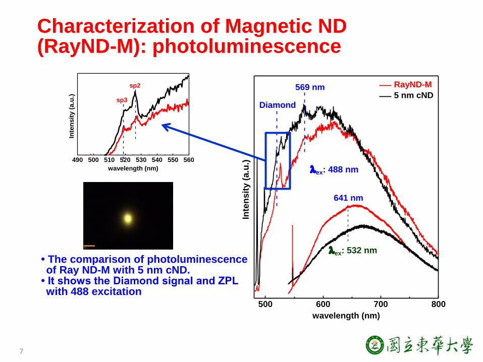
Characterization of Magnetic ND (RayND-M): photoluminescence
• The comparison of photoluminescence of Ray ND-M with 5 nm cND.
• It shows the Diamond signal and ZPL with 488 excitation
7
500 600 700 800
641 nm
RayND-M
5 nm cND
ex: 488 nm
Diamond
Inte
ns
ity
(a
.u.)
wavelength (nm)
ex: 532 nm
569 nm
490 500 510 520 530 540 550 560
sp2
Inte
nsit
y (
a.u
.)
wavelength (nm)
sp3
0 1 2 3 4 50
200
400
600
800
1000
Inte
nsit
y (
a.u
.)
Time decay (ns)
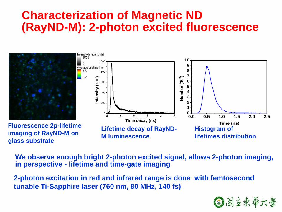
2-photon excitation in red and infrared range is done with femtosecond
tunable Ti-Sapphire laser (760 nm, 80 MHz, 140 fs)
0.0 0.5 1.0 1.5 2.0 2.50
1
2
3
4
5
6
7
8
9
10
Nu
mb
er
(10
5)
Time (ns)
Characterization of Magnetic ND (RayND-M): 2-photon excited fluorescence
Fluorescence 2p-lifetime
imaging of RayND-M on
glass substrate
Lifetime decay of RayND-
M luminescence
Histogram of
lifetimes distribution
We observe enough bright 2-photon excited signal, allows 2-photon imaging, in perspective - lifetime and time-gate imaging

Interaction with cells: Cytotoxicity in BHK
(Baby hamster kidney) cell
9
24 hr
contr
ol 1 5 10 50 100
200
0
50
100
150
* * ****
g/mlc
ell v
iab
ilit
y (
%)
24 hr
contr
ol 1 5 10 50 100
200
0
50
100
150
*
g/ml
ce
ll v
iab
ilit
y (
%)
RayND-M
RayND-M + HSA
Some concentration-dependent cytotoxicity is observed The toxicity can be significantly decreased by coating of ND particles surface with albumin (Human Serum Albumin)

Interaction with cells: Fluorescence confocal imaging
Confocal image of BHK cell after interaction with ND for 24 hrs. As Z-scan demonstrates: ND penetrates into cell (and also some ND is attached on the cell membrane). ND is well detectable in cell.
Z-scan:
Excitation Emission
RayND 514 nm 560-600 nm
Nuclei 405 nm 450-490 nm
Cytoplasm 633 nm 640-690 nm

Interaction with cells: 2-photon excitation and fluorescence lifetime imaging
Very good fluorescence signal of RayND-M at 2-photon excitation with femtosecond tunable Ti-Sapphire laser (760 nm, 80 MHz, 140 fs) Makes this ND promising for 2-photon imaging and lifetime imaging :
0 2 4 6 8 10
0
1000
2000
3000
4000
5000
6000
7000
Inte
nsit
y (
a.u
.)
Time delay (ns)
cell autofluorescence
RayND_M
0 2 4 6 8
0
1
2
3
4
5
6
nu
mb
er
(10
4)
Time (ns)
Cell with ND
M-ND
Cell
Lifetime decays allow
distinguish ND and cell signal
BHK cell with RayND-M
Histograms of lifetime distribution
Control without ND autofluorescence of BHK cell

Strong magnetism of nanodiamond is observed and the nanodiamond is characterized for it’s application in bio-medical studies.
The surface and luminescence properties give the possibilities for
biomedical applications.
In the applications magnetic and photoluminescence properties can be combined.
The nature of magnetism is unclear yet and under study.
Summary
12
13
To MOST, Ministry of Science
and Technology of Taiwan
Thank you for your attention!
Acknowledgements:

14
200 300 400 500 600 700 800
0.2
0.4
0.6
0.8
1.0
1.2
1.4
Ab
so
rpti
on
Wavelength (nm)
4 5 6 7 8 9 10-40
-35
-30
-25
-20
-15
-10
Zeta
po
ten
tial (m
V)
pH
Tools 1. LakeShore 7407 vibrating sample magnetometer (VSM) (Lake Shore Cryotronics, US), 2. Renishaw, UK, 3. - SNOM, Witec, Germany, 4. Jobin Yvon, T64000, France-Japan 5. Scanning Fluorescence Confocal Microscope (TCS-SP5, Leica, Germany) 6. System for lifetime imaging on the base of femtosecond tunable Ti-Sapphire laser (760 nm, 80 MHz, 140 fs) and PicoQuant scanner Etc…

15
Magnetic properties of carbons and nanocarbons are recently studied.
Pure diamond structure combines diamagnetic and paramagnetic compounds. But in an intermediate graphite–diamond structure spin ordering and magnetic interactions can exist [T. L. Makarova Semiconductors, 2004, 38, 6, 615–638] Ferromagnetic properties of carbon nanostructures are discussed for theoretically predicted structures and are shown experimentally. Particularly, it has been predicted that structures composed of altered C atoms with different hybridization (sp2 - and sp3 –coordinated) can have strong spontaneous magnet moment /under some conditions – strong regular ordering – difficult to realize, but on the micro-level such combination is realized in ND/ Superparamagnetic or/and ferromagnetic particles
Ferromagnetic: M
, arb
. un
it