magnetic resonance imaging environment safety in … · magnetic resonance imaging environment...
TRANSCRIPT

Magnetic Resonance
Imaging Environment
Safety in Ontario
April 2006
Healthcare Human Factors Group University Health Network

- i -
Table of Contents
List of Abbreviations 1
List of Units 1
Definitions 2
Executive Summary 3
Purpose 3
The Technology 3
The MR environment 3
Methods 3
Literature Review 3
Field Study 4
Summary of Findings 4
Conclusions 5
Issue 6
Hazards in the MR environment 6
Static magnetic field induced forces 6
RF Heating 7
Device Malfunctions 8
Existing Safety Guidelines 8
ACR Guidelines 8
ECRI Recommendations and Alerts 9
FDA Equipment Categorization 9
Health Canada Notice to Hospitals 10
MRI Safety Experts 11
FDA MAUDE Database 12
Field Study 13
Results 13
- ii -
MR Safety Officer 13
Equipment Categorization 13
Environment Architecture 13
Magnet room door 15
Floor markings 15
Equipment Location 15
Safety Labels 16
Safety Signs 16
Product Procurement 18
Implants and Contraindications 19
Thermal Issues 19
Metal and Ferromagnetic Detectors 19
Training 20
Communication 21
Interventional MR Safety 21
Summary of Results 22
Conclusions and Recommendations 24
Provincial MRI Safety Committee 24
MR Safety Officer 24
Equipment Categorization 24
Environment Architecture 25
Controlled entry in the MR environment 25
Magnet Room Door 25
Floor Markings 25
Equipment Location 25
Safety Signs 25
Product Procurement for use in the MRI Suite 25
Thermal Issues 25
Metal and Ferromagnetic Detectors 25

- iii -
MR Safe Fire Extinguishers 26
Screening Forms 26
Implants 26
Training 26
Interventional MR Safety 26
Checklist for MR Safety 26
References 27
Appendix A: Field Study Questionnaire 29
Appendix B: Sample Safety Labels 31
Appendix C: MRI Safety Sign 32
Appendix D: MRI Screening Form 33
Appendix E: Checklist for MR Safety 36

1
List of Abbreviations
ACR American College of Radiology
ASTM American Society for Testing and Materials
ECRI Emergency Care Research Institute
FDA United States Food and Drug Administration
HPFB Health Products and Food Branch of Health Canada
MoHLTC Ontario Ministry of Health and Long Term Care
MRI Magnetic Resonance Imaging
MR Magnetic Resonance
MAUDE FDA Manufacturer and User Device Experience Database
OHTAC Ontario Health Technology Advisory Committee
RF Radiofrequency
List of Units
G Gauss
kg Kilogram
T Tesla
W Watts
2
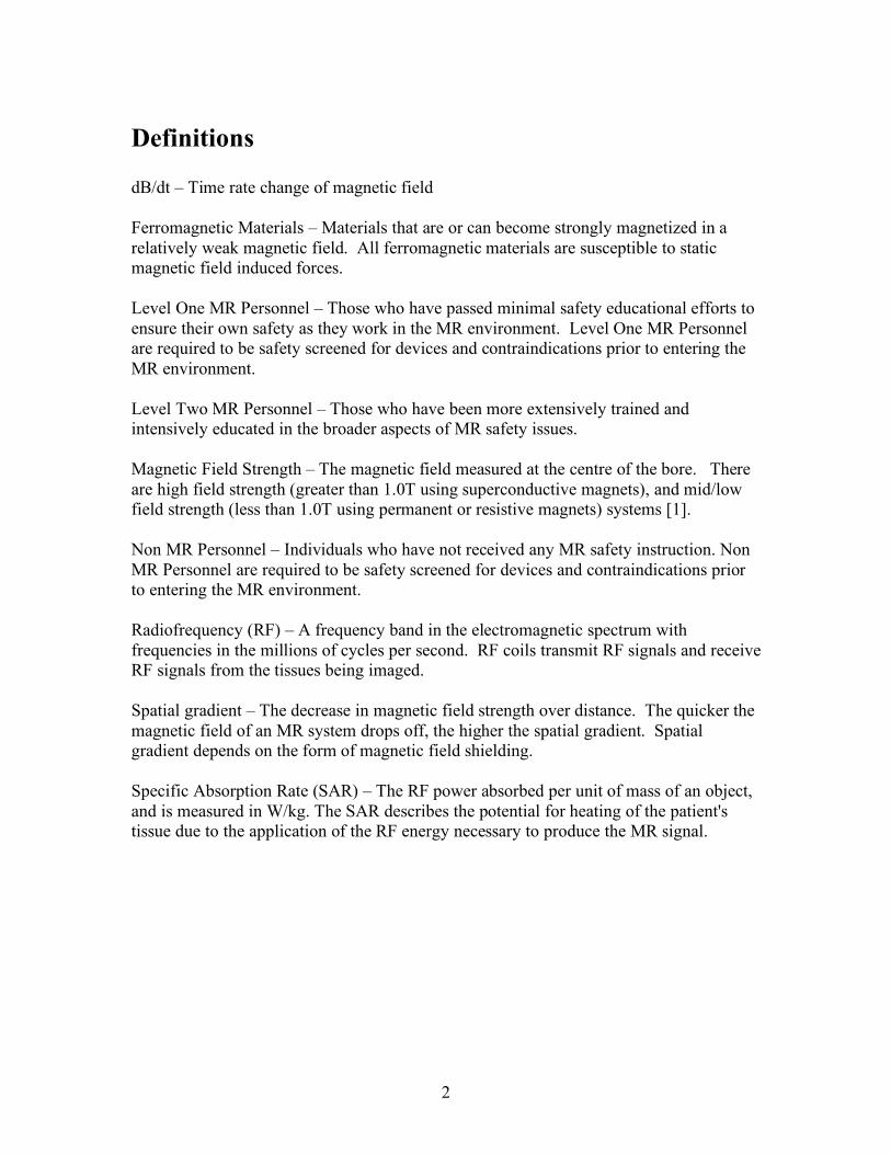
Definitions
dB/dt – Time rate change of magnetic field
Ferromagnetic Materials – Materials that are or can become strongly magnetized in a
relatively weak magnetic field. All ferromagnetic materials are susceptible to static
magnetic field induced forces.
Level One MR Personnel – Those who have passed minimal safety educational efforts to
ensure their own safety as they work in the MR environment. Level One MR Personnel
are required to be safety screened for devices and contraindications prior to entering the
MR environment.
Level Two MR Personnel – Those who have been more extensively trained and
intensively educated in the broader aspects of MR safety issues.
Magnetic Field Strength – The magnetic field measured at the centre of the bore. There
are high field strength (greater than 1.0T using superconductive magnets), and mid/low
field strength (less than 1.0T using permanent or resistive magnets) systems [1].
Non MR Personnel – Individuals who have not received any MR safety instruction. Non
MR Personnel are required to be safety screened for devices and contraindications prior
to entering the MR environment.
Radiofrequency (RF) – A frequency band in the electromagnetic spectrum with
frequencies in the millions of cycles per second. RF coils transmit RF signals and receive
RF signals from the tissues being imaged.
Spatial gradient – The decrease in magnetic field strength over distance. The quicker the
magnetic field of an MR system drops off, the higher the spatial gradient. Spatial
gradient depends on the form of magnetic field shielding.
Specific Absorption Rate (SAR) – The RF power absorbed per unit of mass of an object,
and is measured in W/kg. The SAR describes the potential for heating of the patient's
tissue due to the application of the RF energy necessary to produce the MR signal.
3
Executive Summary
Purpose In 2005, there were 58 MRI scanners in use at hospitals and private facilities in Ontario.
Of these, 53 were high field strength systems. Since then, the MoHLTC has announced
funding for nine new MRI scanners for hospitals, and three in the private not-for-profit
sector. These new machines are projected to result in more than 53,000 new MRI exams
in 2005-2006 [2].
With the increased prevalence of MRI exams in Ontario, the emerging use of MRI for
image guided surgery, and the higher field strength MRI machines used for exams, it is
important to address the potential risks to patient and staff safety in the MR environment.
The purpose of this study is to investigate the level of safety maintained at MR facilities
in Ontario, assess the MR safety literature, and provide recommendations to OHTAC on
maintaining a high level of safety at MR sites in Ontario. The scope of this study was
focused on the MR environment, including the region from the centre of the magnet bore
and extends out to the patient waiting and reception areas. The environment is divided
into four zones as defined by the American College of Radiology (ACR) [3, 4]
The Technology
The MR environment
MRI has a number of advantages over other volumetric imaging modalities. Of
significance is that MRI does not use ionizing radiation. MRI scanners are classified as
high field strength (with magnetic fields greater than 1.0 T) or mid/low field strength
(with magnetic fields less than 1.0 T). Within the MR environment, the 5G line (0.0005T)
divides the environment into safe and unsafe regions. The environment where the
magnetic field strength is less than 5G is generally considered to be safe [3]. Because of
the persistent magnetic field, and the hazards associated with magnetic fields, extra care
is required in the MR environment to ensure that injury or harm does not come to any
personnel while in the environment.
Methods
Literature Review
There is a small body of literature on safety in the MR environment. It is clear that the
MR environment is one in which caution is necessary to ensure staff and patient safety.
The literature review focused on existing MR safety guidelines, recommendations, and
standards. Additionally, the FDA MAUDE database was used to discover adverse
incidents associated with the use of MRI.

4
Field Study
A field study complemented the literature review, and assessed the level of safety
maintained at various MRI facilities throughout the province. Sites included teaching
hospitals, research facilities, community hospitals, and private-provincially insured
facilities. A standard list of questions provided insight into best practices for maintaining
a high level of MR safety.
Summary of Findings The ACR Blue Ribbon Panel for MR safety, chaired by Dr. Emanuel Kanal, published
guidelines in 2002 [3] on maintaining safety in the MR environment. In 2004 these
guidelines were updated to include new safety concerns [4]. Dr. Kanal has indicated that
an update to these guidelines will be published in 2006. The ACR guidelines are well
respected throughout the MRI community and are often used as a blueprint for MR
policies at individual facilities. The ECRI has published several recommendation
documents in their “Health Devices” publication on the topic of MRI safety [5-7]. Health
Canada publishes notices to hospitals on safety information for MRI systems [8-10].
Additionally, Dr. Frank Shellock, an MR safety expert, maintains an MR safety website
where medical devices and implants are listed according to their level of MR
compatibility [11].
Adverse incidents associated with the use of MRI are sparsely documented primarily
because only incidents resulting in death are required to be reported to the FDA.
Incidents involving injury are reported to manufacturers, a few of which are submitted to
the MAUDE database by the manufacturer. The FDA’s MAUDE database [12] reports
incidents such as projectiles, burns, and implant and device malfunctions in the MR
environment. The most notorious incident occurred in July 2001, when a 6-year-old boy
was struck in the head by a ferromagnetic oxygen cylinder that was mistakenly brought
into the MR environment while he was undergoing a postoperative scan. The oxygen
cylinder was drawn rapidly toward the MRI magnet and became a projectile. The boy
died shortly after the incident from his injuries [13].
Following a field study, it was found that not all MR facilities in Ontario follow the ACR
guidelines, and there are several inconsistencies in certain MR practices across the
province. However there are also several safety tactics successfully employed at
facilities that are practiced that do not appear in the literature. In particular the following
issues were found:
• The lack of a formal MR Safety Officer position at each hospital or hospital group
• The use of outdated MR equipment categorization
• Not utilizing the four safety zone MR environment architecture
• Not controlling access into the MR environment
• Not indicating the 5 G area through floor demarcation
• Inconsistent MR equipment labels
• The use of unclear MR warning signs
• Inadequate training for Level One MR personnel, and Non-MR Personnel

5
Conclusions These recommendations provided for MR safety in Ontario will ensure that facilities
throughout the province have access to MR safety information, and that a high level of
MR safety is maintained. The primary recommendation is to establish a provincial MRI
safety committee to maintain consistent MR safety practices in Ontario.
Recommendations for the MRI Safety Committee’s consideration include:
• Use the updated MRI categorization: MR Safe, MR Conditional, MR Unsafe
• Strictly control access to the MR environment
• Clearly indicate the 5 G perimeter on the floor surrounding the scanner.
• Assign a permanent location for equipment in the magnet room
• Use consistent MR labels on equipment used in the MR environment
• Use consistent MR signs that clearly indicated the hazards of the MR environment
• Require outpatients undergoing MRI scans to change into hospital gowns without
metal fasteners
• Provide annual training for personnel working in the MR environment

6
Issue MRI has a number of advantages over other volumetric imaging modalities. Of
significance is that MRI does not use ionizing radiation. However there are other safety
risks related to the use of the strong magnetic fields used in MRI. A MoHLTC Medical
Advisory Secretariat Health Technology Review completed in December 2003 addressed
Patient Monitoring Systems for MRI [14]. In this review, the safety risks associated with
monitoring patients during MRI exams were addressed. In a February 2004 follow-up,
the OHTAC provided safety recommendations for patient monitoring systems for MRI
[15]. In its recommendations, OHTAC suggested a safety review of Ontario MRI
facilities, and alerting MRI facilities of safety hazards to minimize risks to patients and
staff.
In addition to safety involving patient monitoring for MRI, there are several other safety
issues that must be addressed during each MRI exam. These include the proper screening
of patients for implants and contraindications, maintaining a MR environment that is free
of ferromagnetic objects, and appropriately communicating MR hazards to staff and
patients. The MR environment is highly specialized, and requires a high level of safety.
The purpose of this study is to investigate the level of safety maintained at MR facilities
in Ontario, assess the MR safety literature, and provide recommendations to OHTAC on
maintaining a high level of safety at MR sites in Ontario.
Hazards in the MR environment The use of medical devices and other equipment requires great care when in proximity to
the magnetic field of an MRI unit. There are several types of hazards in the MR
environment. Of most concern are [5]:
• Static magnetic field induced forces
• RF heating
• Device malfunctions
Static magnetic field induced forces
Two types of static magnetic field induced forces can create safety problems in the MR
environments: torque, and translational force. Torque is a rotational force that causes an
object to align parallel to the static magnetic field. Translational force is a linear force
that attracts an object into the bore of the magnet.
The closer a ferromagnetic object’s proximity to the MR system’s magnet, the higher the
spatial gradient that exists, and the greater the translational force that the object will
experience. Therefore there is a greater likelihood that the object will be drawn toward
the MR system’s magnet the closer the object is to the magnet.
Static magnetic field induced forces have a significant effect on implanted devices
(primarily torque) as well as the projectile effect (primarily translational forces) on
unrestrained ferromagnetic objects in the MR environment. Objects such as oxygen

7
cylinders, IV poles, mop buckets, and patient monitors have all been pulled into the MRI
magnetic field with potentially deadly force [5]. An example is shown in Figure 1, where
an office chair containing ferromagnetic materials came too close to the system, became a
projectile, and got lodged into the bore of the scanner magnet. The static magnetic field
is always present, and therefore poses a significant safety risk, such that safety
precautions must be taken at all times.
Figure 1: Office chair wedged into MR scanner [16]
RF Heating
The RF electromagnetic field of an MR system can induce currents in electrically
conductive materials present within the bore of the MR system. The induced current can
cause heating in the conductor, which can lead to a patient burn if the conductor is in
contact with the patient’s body. Patients have received burns from contact with
8
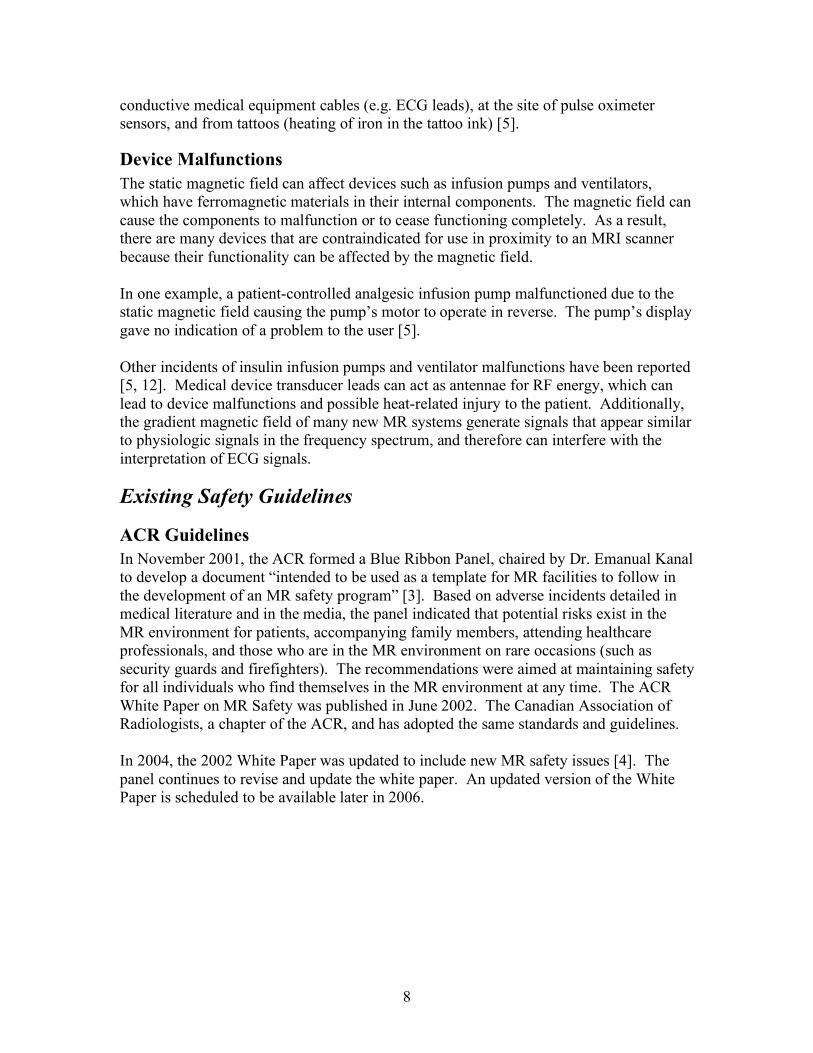
conductive medical equipment cables (e.g. ECG leads), at the site of pulse oximeter
sensors, and from tattoos (heating of iron in the tattoo ink) [5].
Device Malfunctions
The static magnetic field can affect devices such as infusion pumps and ventilators,
which have ferromagnetic materials in their internal components. The magnetic field can
cause the components to malfunction or to cease functioning completely. As a result,
there are many devices that are contraindicated for use in proximity to an MRI scanner
because their functionality can be affected by the magnetic field.
In one example, a patient-controlled analgesic infusion pump malfunctioned due to the
static magnetic field causing the pump’s motor to operate in reverse. The pump’s display
gave no indication of a problem to the user [5].
Other incidents of insulin infusion pumps and ventilator malfunctions have been reported
[5, 12]. Medical device transducer leads can act as antennae for RF energy, which can
lead to device malfunctions and possible heat-related injury to the patient. Additionally,
the gradient magnetic field of many new MR systems generate signals that appear similar
to physiologic signals in the frequency spectrum, and therefore can interfere with the
interpretation of ECG signals.
Existing Safety Guidelines
ACR Guidelines
In November 2001, the ACR formed a Blue Ribbon Panel, chaired by Dr. Emanual Kanal
to develop a document “intended to be used as a template for MR facilities to follow in
the development of an MR safety program” [3]. Based on adverse incidents detailed in
medical literature and in the media, the panel indicated that potential risks exist in the
MR environment for patients, accompanying family members, attending healthcare
professionals, and those who are in the MR environment on rare occasions (such as
security guards and firefighters). The recommendations were aimed at maintaining safety
for all individuals who find themselves in the MR environment at any time. The ACR
White Paper on MR Safety was published in June 2002. The Canadian Association of
Radiologists, a chapter of the ACR, and has adopted the same standards and guidelines.
In 2004, the 2002 White Paper was updated to include new MR safety issues [4]. The
panel continues to revise and update the white paper. An updated version of the White
Paper is scheduled to be available later in 2006.

9
The 2004 White Paper includes MR safety guidance on the following topics:
• Establishing, implementing, and maintaining current MR safety policies and
procedures
• MR Site access restriction
o MR environment zoning
o MR and non-MR personnel
o Personnel and patient screening
o Device and object screening
• MR Technologists
• Pregnancy related issues
• Pediatric MR safety concerns
• Time varying gradient magnetic field related issues
o Induced voltages
o Auditory considerations
o Thermal
• Drug delivery patches and pads
• Cryogen related issues
• Claustrophobia, anxiety, sedation, analgesia, and anesthesia
• Contrast agent safety
• Patients with intracranial aneurysm clips
• Patients with cardiac pacemakers or implantable cardioverter defibrillators
Dr. Kanal recommends that individual MR sites use the White Paper as a blueprint for
establishing MR safety policies at individual sites.
ECRI Recommendations and Alerts
The ECRI has published several recommendation documents in their “Health Devices”
publication on the topic of MR safety. In 2001, the ECRI published preliminary
guidelines on ensuring the safe use of equipment in the MR environment [5]. Since then,
the ECRI has summarized important information on MR safety, and published guidance
articles and alerts on the latest in MR safety news [6, 7].
FDA Equipment Categorization
Medical device vendors are required to communicate MR safety information regarding
devices (i.e. safety with respect to spatial gradient, SAR, etc.) in the MR environment.
1997 the United States Food and Drug Administration (FDA) defined two terms
categorizing devices that have been shown to be useful in the MR environment [5].
MR Safe – “The device, when used in the MR environment, has been
demonstrated to present no additional risk to the patient, but may affect the
quality of the diagnostic information”
MR Compatible – “The device, when used in the MR environment, is MR Safe
and has been demonstrated to neither significantly affect the quality of the
diagnostic information nor have its operations affected by the MR device”

10
As this terminology came to be used in practice, it was found that the terms were being
used incorrectly or interchangeably. There were incorrect assumptions that MR Safe
devices were also MR Compatible, which is not the case. MR Safe and MR Compatible
are not mutually exclusive. Additionally, sometimes the incorrect assumption was made
that if the device is deemed MR safe or compatible, it is safe or compatible in any
situation, in any MR environment.
In 2005, the ASTM International issued a “Standard Practice for Marking Medical
Devices and Other Items for Safety in the Magnetic resonance Environment” [17]. This
standard was developed and endorsed by the FDA as the new standard for MR device
categorization and marking. It removes the confusion from the previous categorization,
and no longer considers image quality, and primarily focuses on safety in the MR
environment.
In this standard, there are new categories for the marking and categorization of medical
devices used in the MR environment. Along with these categories, new icons for labels
are issued for ease of identifying the device and its safety rating.
MR Safe: “An item that poses no known hazards in all MR
environments.” (e.g. a plastic Petri dish)
MR Conditional: “An item that has been demonstrated to pose no
known hazards in a specified MR environment with specified
conditions of use. Field conditions that define the specified MR
environment include field strength, spatial gradient, dB/dt (time rate of
change of the magnetic field), radio frequency fields, and specific
absorption rate. Additional conditions, including specific
configurations of the item, may be required.” (e.g. a Patient Monitor)
MR Unsafe: “An item that is known to pose hazards in all MR
environments.” (e.g. Floor Buffer)
Health Canada Notice to Hospitals
The HPFB of Health Canada posts safety alerts, public health advisories, press releases
and other notices on the Health Canada web site. These are a service to health
professionals, consumers, and other interested parties. In 2005, the HPFB published two
Notices to Hospitals on safety information for MRI systems. The notices included safety
information on transdermal drug patches [8], and safety information on active
implantable medical devices and systems [9, 10].

11
MRI Safety Experts
Dr. Emanual Kanal is the director of MR services and Professor of Radiology and
Neuroradiology at the University of Pittsburgh Medical Center. He is the chair of the
American College of Radiology Blue Ribbon Panel on MR safety programs.
Dr. Frank Shellock is an Adjunct Clinical Professor of Radiology and Medicine at the
Keck School of Medicine at the University of Southern California. He is also the chair of
the Institute for Magnetic Resonance Safety, Education, and Research. His publications
(including his book and website) are used by almost all MR facilities for the verification
of devices and implants in the MR environment [1, 11].
Dr. Kanal and Dr. Shellock are well-respected MR safety experts. Their publications and
websites are widely used in the MR community for establishing safety policies and
verifying MR devices. Dr. Kanal and Dr. Shellock are active contributers to various MR
safety periodicals, websites, and email listings. They both were consulted for their
expertise for this project.

12
FDA MAUDE Database In the September 21, 2005 ECRI Audio Conference on MRI Safety and Medical Devices
[18], Mr. Jason Launders of the ECRI presented an analysis of the FDA MAUDE
database from 1995 to May 2005 [19]. He searched all reports for MRI device codes,
electrode burns, implants, aneurysm clips, pacemakers, and deaths associated with MRI.
The search found 389 unique and relevant reports. In these there were nine reported
deaths, and 302 incidents attributable to MRI technology. Of the MRI injuries, 71%
involved burns (coils, leads connected to monitoring equipment, or body loops), 10%
involved other items (e.g. implants), 10% were caused by projectiles, 4% were acoustic
injury (e.g. temporary hearing loss), 4% were fire related injuries, and 2% were caused by
internal heating (implanted leads). Metallic implant failures/burns, infusion pump
failure, pacemaker failure, aneurysm clip failure, and neurostimulator failure were
represented in the “other item” category of MRI device incidents.
While the MAUDE database cites many MRI incidents, the data are unreliable due to a
large number of unreported events. Dr. Emanual Kanal of the ACR stated, “the
percentage of accidents reported to the FDA is well, well below 10%” [18]. However the
MAUDE database can provide insight into the types of injuries and hazards in the MR
environment.

13
Field Study A field study of eleven hospitals and one private MR clinic was conducted to assess the
level of safety maintained in MR environments around the province, as well as to learn of
best practices for MR safety. This sample represents approximately 25% of the facilities
in Ontario that have MRI scanners [20]. A standard list of questions and points of
inquiry was used in this field study (Appendix A). Points of interest included equipment
categorization (i.e. MR Compatible, MR Safe), infrastructure within the MR environment
(i.e. signs, labels, floor markings), policies on patient care, and the training provided to
personnel.
Results
MR Safety Officer
Most sites do not have a formal MR Safety Officer position. Usually the charge
technologist and/or the chief MR Radiologist is responsible for the MR safety policies at
the sites. As part of the recommendations on establishing, implementing, and
maintaining current MR safety policies and procedures, the ACR recommends appointing
an MR safety officer who is responsible for ensuring that MR safe practice guidelines are
implemented and adhered to at all times.
Equipment Categorization
The use of MR safety categorization prior to August 2005, MR Safe and MR Compatible,
was assessed at each site. All sites are correctly using the MR equipment categorization.
Nearly all sites only make use of “MR Compatible” equipment to avoid confusion
between MR Safe and MR Compatible. Additionally, consistency is being maintained in
using the categorization to identify MR equipment.
Sites were unaware of the new MR Safety categories, and therefore were still using the
1997 FDA categorization. The new categorization standards for MR equipment (MR
Safe, MR Conditional, and MR Unsafe) are recommended for use by the FDA to
maintain consistency with the standards that are now being used by equipment
manufacturers [17].
Environment Architecture
The ACR recommends dividing the MR environment into four zones [4]. These zones
are shown in Figure 2 and are described as follows:
• Zone 1 is the area outside the MR environment that is accessible to the general
public.
• Zone 2 is the interface between the public Zone 1 and the controlled Zone 3 and
Zone 4. In Zone 2, patients are under the supervision of MR personnel.

14
• Zone 3 is the area where only screened personnel and objects are permitted. MR
Unsafe equipment is exchanged with MR Conditional or MR Safe equipment in
Zone 3. Access to Zone 3 is strictly restricted and should be physically controlled
from general public access.
• Zone 4 is the MR scanner room and appropriate signs should be used to
communicate this.
Figure 2: Four-zone MR environment, adapted from [4]
For sites that were visited, the patient flow from the reception area (Zone 1) to the
magnet room (Zone 4) was assessed. Controlled access beyond the reception area was
verified. Additionally, the types of controls used to prevent prohibited entry into the MR
area were noted.
There is no consistent MR suite layout primarily because of the architecture of the space
in which the MR environment is installed. The ACR recommendation of the four safety
zones is not followed across all sites. Half of the sites used only three zones.
Additionally, only a few sites control the access of non-MR personnel in the MR

15
environment. Other sites have no controlled access. As such, anyone could walk through
the hospital, into the MR environment, and directly into the magnet room. The ACR
recommends that beyond the reception desk in the MR environment, MR personnel
should escort all non-MR personnel.
Magnet room door
Zone 4 of the MR environment is typically designed with a door that swing inward to the
magnet room. If the magnet in the MR scanner is quenched, there is a positive pressure
buildup in the magnet room. This positive pressure pushes a closed door against the
stops and makes the door nearly impossible to open until the pressure on the inside of the
room is equalized with the pressure outside the room. The MAUDE database [12] lists
one asphyxiation death of a service person caused by breathing the ultracold vaporized
helium. This is also a risk for patients who are in the room when the magnet is quenched.
The positive pressure buildup may prevent technologists from attending to the patient
during an emergency situation. During the field study two technologists from different
sites mentioned that they were told by the scanner manufacturer to break the window
between Zone 3 and Zone 4 to equalize the pressure if this occurred. To prevent such
incidents and to provide the safest possible environment for patients, technologists, and
other MR personnel, the magnet room door should swing outward from the magnet room,
but should not obscure the technologists view as to who, or what, is entering the magnet
room [21]. An MR hazard sign should therefore be placed both on the inside and outside
of the magnet room door.
Floor markings
In its guidelines, the ACR recommends that where the static magnetic field strength
exceeds 5 G, the area should be clearly demarcated as being potentially hazardous [4].
Below 5 G, the static magnetic field has diminished sufficiently to pose no physical threat
to staff or patients [5].
An assessment was done as to whether sites marked the 5 G line out in the room. The
method and location of markings was noted and compared across the sites. Any other
floor markings used for safety purposes were also noted.
Two-thirds of the sites assessed included the demarcation of 5 G in the MR environment.
The method differed from site to site. These include the use of adhesive tape, and
contrasting coloured floor tiles. Some sites had an additional demarcation indicating the
proximity limit for certain equipment (e.g. an anesthesia machine that is MR Compatible
to 300 mT, or 3000 G).
Equipment Location
Occasionally MRI scans are performed on patients who require specialized monitoring
equipment [14]. This equipment can be potentially hazardous in the MR environment.
An assessment was made as to whether sites had a designated area for potentially MR
Unsafe equipment. While this is not part of the ACR guideline, it appears to be a good
strategy for ensuring potentially unsafe equipment does not come too close to the
16
scanner. This also would make MR personnel cognizant of the equipment in the MR
environment so that extra care is exercised with potentially unsafe equipment.
Very few sites were found to have dedicated space for equipment that is unsafe within the
5 G line. A few sites make use of a pole onto which the equipment can be fastened or
tethered, or a table designated for MR equipment.
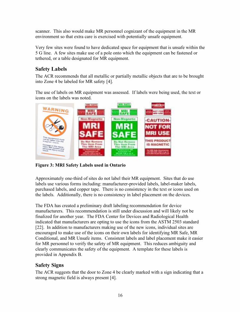
Safety Labels
The ACR recommends that all metallic or partially metallic objects that are to be brought
into Zone 4 be labeled for MR safety [4].
The use of labels on MR equipment was assessed. If labels were being used, the text or
icons on the labels was noted.
Figure 3: MRI Safety Labels used in Ontario
Approximately one-third of sites do not label their MR equipment. Sites that do use
labels use various forms including: manufacturer-provided labels, label-maker labels,
purchased labels, and copper tape. There is no consistency in the text or icons used on
the labels. Additionally, there is no consistency in label placement on the devices.
The FDA has created a preliminary draft labeling recommendation for device
manufacturers. This recommendation is still under discussion and will likely not be
finalized for another year. The FDA Center for Devices and Radiological Health
indicated that manufacturers are opting to use the icons from the ASTM 2503 standard
[22]. In addition to manufacturers making use of the new icons, individual sites are
encouraged to make use of the icons on their own labels for identifying MR Safe, MR
Conditional, and MR Unsafe items. Consistent labels and label placement make it easier
for MR personnel to verify the safety of MR equipment. This reduces ambiguity and
clearly communicates the safety of the equipment. A template for these labels is
provided in Appendix B.
Safety Signs
The ACR suggests that the door to Zone 4 be clearly marked with a sign indicating that a
strong magnetic field is always present [4].

17
An assessment was made the number and types of safety signs used in the MR
environment. The text and images on the signs were noted and compared across the sites.
There was no consistency in the signs used in MR suites across the province. While all
sites make use of safety signs, some sites make their own signs, others use purchased
signs or signs provided by the scanner manufacturer. Some signs are difficult to read,
using languages and icons that are difficult to understand. These signs are more difficult
to interpret. Examples of these signs are shown in Figure 4 and Figure 5.
Figure 4: MRI Safety Warning Signs used in Ontario

18
Figure 5: MRI Safety Warning Sign used in Ontario
Product Procurement
ASTM F2503-05 standard [17] indicates that safety testing for items that may be placed
in the MRI environment should address magnetically induced displacement force (Test
Method F 2052) [23], magnetically induced torque (Test Method F 2213) [24], and RF
heating (Test Method F 2182) [25]. Manufacturers should perform these tests and
include the results to verify the MR Safe, MR Conditional, or MR Unsafe categorization
according to the results from the testing. During device procurement, it should be
verified that the device has passed each of the three test methods, and is tagged with a
label that follows ASTM Standard F2503 and the FDA Preliminary Draft Labeling [17].
MRI Sites in Ontario follow a number of different processes when purchasing equipment
to be used in the MR environment. Equipment purchases include patient monitors and
accessories, anesthesia machines, ventilators, IV poles, and wheelchairs. Some sites
purchase MR Compatible equipment from the MR scanner manufacturer. Other sites
purchase MR Compatible equipment from the manufacturer and then verify its
compatibility with a handheld magnet.

19
Implants and Contraindications
The methods used for screening patients for implants or other contraindications were
assessed. Additionally, the method of verifying implant safety was assessed for best
practice.
All sites essentially follow the same procedure for assessing patient implants. They
obtain information on the implant from the patient, hospital records, or surgical notes.
Next, the MRISafety.com website, or “Reference Manual for Magnetic Resonance
Safety, Implants, and Devices” [1] is consulted to determine MR compatibility. If
compatibility cannot be verified, the manufacturer is contacted and information is
obtained. If the implant or contraindication cannot be assessed, the patient is not
scanned.
This procedure for verifying the compatibility of implants was found to be the preferred
practice. In addition to the MRISafety.com website and reference manual, the notices
issued by the HPFB should be used to identify implants and devices that are
contraindicated for MR use by Health Canada.
Thermal Issues
Patient burns are a serious hazard in the MR environment. Sites were asked about the
methods used to counteract patient burns while being scanned. This included policies
regarding scanning patients in their own clothing. Additionally, technologists were asked
about the incidence of patient burns at their sites.
Because of the heterogeneous materials used in patient clothing, the ACR recommends
that patients wear a “site-supplied gown with no metal fasteners” during the MR
procedure [4]. When there is the potential for a patient burn, such as with ECG
electrodes or tattoos, the ACR recommends reducing the temperature of the area through
the use of ice packs or cold compresses for the duration of the MR scan [4].
The sites visited recorded very few instances of patient burns. A few patients reported
heating in the areas where they had tattoos. To counteract heating caused by tattoos,
leads, wires, or the magnet, linens and cold compresses are used for protecting the
patient. Linens are used as a thermal insulator between limbs and the sides of the
magnet. Cold compresses are used on external wires, leads and tattoos to cool the area
during the scan. Not all sites require patients to change into hospital gowns during the
scan. One instance was reported of a patient burn as a result of conductive threading in
the patient’s clothing.
Metal and Ferromagnetic Detectors
No sites included in this study make use of metal or ferromagnetic detectors. The ACR
and ECRI do not recommend the use of metal detectors in the MR environment. Metal
detectors cannot differentiate between ferromagnetic materials and non-ferromagnetic
materials. The use of metal detectors leads to decreased vigilance because of the constant
“false positive” alarms, and could provide a false sense of security by failing to detect
small implanted devices.

20
Ferromagnetic detectors are just now becoming available. In a conversation with Mr.
Kemp Massengill, President and CEO of Mednovus Inc. [26], it was found that several
designs of passageway ferromagnetic detectors exist; however, each has its own
drawbacks. They are heavy, expensive, and not very sensitive in the centre of the
passageway. Handheld ferromagnetic detectors will soon become available for use as
well. They have a depth of sensitivity of one inch and therefore can detect small objects
on a patient. However this depth of one inch may not be adequate to detect implanted
devices (e.g. aneurism clips or shrapnel). Additionally they are not recommended for use
around the eyes because they may dislodge ferromagnetic debris in the orbits and cause
damage to a patient’s eyes. At the time of writing, further studies are being conducted on
ferromagnetic detectors. This is an evolving field, and attention should be directed
toward the results of these studies. Ferromagnetic detectors could potentially be used in
the MR environment in the future. The ACR will address ferromagnetic detectors in the
2006 version of the White Paper on MR Safety. The ECRI has not yet tested
ferromagnetic detectors, and therefore cannot approve or disapprove of their use.
Training
Two types of MR personnel are identified by the ACR [4]. Level One MR personnel are
“individuals that have passed minimal safety educational efforts to ensure their own
safety as they work within Zone 3”. Level Two MR personnel are individuals “who have
been intensively educated in the broader aspects of MR safety issues” and can work in
any of the four MR zones. Non-MR personnel are those who have not received any
formal MR safety instruction.
Each site described the types of personnel allowed into the MR environment. This
included technologists, radiologists, nurses, respiratory therapists, housekeeping staff,
and security guards. The personnel that are allowed to enter the MR environment depend
on the type of patients being scanned, and the policies of individual MR sites. When
pediatric or critical care patients are being scanned, more personnel (e.g. parents, nurses,
and respiratory therapists) are present in the MR environment. Security and
housekeeping personnel only enter the MR suite if the individual site permits their entry.
Certain sites do not permit housekeeping and security staff to enter the MR environment.
At these sites, technologists take over the housekeeping and security responsibilities.
For Level One MR personnel that were permitted entry into the MR environment, the
type of training provided was assessed. The Level One MR personnel method of
screening for devices and contraindications was noted. Finally, the techniques for
orienting newly trained MR technologists to the specific MR site were gathered and
compared across the sites.
At approximately one-third of the facilities, Level One MR personnel are permitted entry
into Zone 4 of the MR environment. Primary caregivers often accompany patients,
particularly those that are critically ill. Housekeeping and maintenance personnel visit
the MR environment for their scheduled duties.

21
Some sites provide one-time training for individuals to be designated as Level One MR
personnel. The training includes safety screening of devices, implants and other
contraindications. Technologists noted that it often difficult to schedule training time for
staff. Typically, there is no follow-up training.
There are inconsistencies in screening Level One MR personnel for devices and
contraindications. Certain sites keep the screening forms on file and simply review the
information with the individual each time they visit the MR environment. Other sites
require the individual to fill out a new screening form each time they visit the MR
environment.
Newly trained MR technologists are often paired with an experienced technologist to
orient them to the MR environment of the specific site. Most sites have an orientation
checklist that highlights the equipment, safety measures, policies and procedures for the
MR environment.
One useful practice was observed that could benefit other MR sites in Ontario. New
technologists spend the first few weeks primarily screening patients and verifying their
conclusions with an experienced technologist. This way, the technologist can practice the
essential skill of screening for contraindications and unsafe devices.
MR technologists suggested that an annual online training program would help maintain
a high level of MR safety, as it would remind individuals of the risks and hazards in the
MR environment. Following the initial training, an annual self-directed computer based
training program, followed by a self-graded interactive questionnaire is recommended as
annual training and certification for all individuals that interact with the MR environment
as part of their job.
Communication
The MR technologist community in Ontario is a fairly close-knit group. Across sites,
several technologists have worked together in the past. Technologists were asked about
the necessity and desire to communicate with other sites regarding MR safety issues.
Technologists who have worked at other sites often communicate and share ideas through
personal discussions, or an electronic bulletin board. These technologists view this as an
effective activity, and suggested that some structured means of sharing information
across the province would be very useful.
Interventional MR Safety
Most interventional MRI procedures have been in the field of neurosurgery [27-30]. MRI
is often used to monitor the progress and results of a procedure. Neurosurgeons have
documented their processes for MR safety when performing the procedures. Because
interventional MRI is not very common, there are very few guidelines for surgical
procedures that occur within the MR environment.

22
Surgeons have noted that they have made use of disposable and non-ferromagnetic
(primarily titanium) instrumentation while performing procedures [27]. If standard
surgical instrumentation is utilized, procedures are performed outside of the 5G line, and
the patient is moved back into the MR scanner for periodic imaging.
As with all MR procedures, large equipment used during interventional procedures such
as trolleys, linen hampers and anesthetic equipment should be made with non-
ferromagnetic materials wherever possible, such as non-magnetic grade of stainless steel.
The obvious risk of using ferromagnetic instrumentation and equipment is that it can
inadvertently come too close to the magnet and become a projectile. Because of this
serious risk the use of non-ferromagnetic instrumentation and specialized equipment for
interventional MRI procedures is recommended. If equipment is made of ferromagnetic
materials, it should be kept beyond the 5 G line in an area designated for potentially
unsafe equipment.
Summary of Results
The results of the field study are summarized in Table 1. It is evident from these results
that there are inconsistencies in MR safety practices throughout the province. The
inconsistency in MR safety practices could potentially lead to adverse MR incidents in
Ontario.

23
Inquiry Yes No Explanation
Do sites have an MR Safety
Officer?
0 12 No sites have appointed an MR Safety
Officer
Do sites use the 1997 FDA MR
equipment categorization?
12 0 All sites use MR Compatible
equipment
Do sites use the 2005 FDA/ASTM
MR equipment categorization?
0 12 No sites had heard of the updated
categorization
Do sites follow the 4-zone MR
environment architecture?
6 6 MR environment architecture was
dependent on the configuration of the
space allotted for the MR environment
Does the magnet room door (to
Zone 4) swing outward?
0 12 All doors to Zone 4 swing inward
Do sites mark the 5G line on the
floor of Zone 4?
8 4 Not all sites with 5G line have it
marked in a permanent manner
Do sites have a location in Zone 4
for MR Conditional equipment?
3 9 Tables, poles, and floor markings used
to indicate the location for MR
Conditional equipment
How many sites reported
accidental projectiles in the MR
suite?
2 10 Patient monitors, sand bag, ventilator
Do sites use MR safety labels on
all MR equipment?
5 7 Different types of labels are used,
depending on the site, and the
equipment
Do sites use multiple MR safety
signs?
9 3 Different signs used depending on the
MR site, and the brand of scanner
Do sites require outpatients to
change into gowns?
3 9 Patients are often scanned in their own
clothing
How many sites reported patient
burns while being scanned?
2 10 Burns from conductive material in
clothing, tattoos, body parts forming
loops
Do sites use metal detectors? 0 12 Metal detectors cause too many false
positives
Do sites use ferromagnetic
detectors?
0 12 Ferromagnetic detectors are very new,
untested technology and not widely
available
Do sites offer safety training for
staff?
12 0 All sites provide a one time in-service
Table 1: Summary of Field Study

24
Conclusions and Recommendations It is important that Ontario be proactive in promoting MR Safety at all MR sites in the
province. Reviewing the findings of the field study and the existing published MR safety
guidelines provides a number of recommendations for Ontario MR sites. The
recommendations in this section are based on the findings from the existing guidelines
and the field study of safety practice in MRI sites across Ontario. These
recommendations also include best practices found during the field study.
Provincial MRI Safety Committee
A provincial MRI safety committee should be formed to promote consistent MR safety
practices in Ontario. It should represent MRI facilities around the province, including
large research facilities, community hospitals, and independent MRI centres. The
committee should be charged with:
• Gathering and disseminating new MR safety information through the province
• Creating an annual staff training program
• Facilitating communication among MR technologists around the province
• Recording MR safety incidents, and creating a database of near-misses and
adverse MRI incidents
• Developing consistent practices to prevent MR incidents in Ontario
• Develop a consistent screening form to be used in all MR facilities in Ontario
As part of its mandate to promote MR safety in Ontario, the remaining recommendations
should be considered by the MRI Safety Committee for implementation at all MRI sites
in Ontario.
MR Safety Officer
Appoint an MR safety officer at each site or hospital group. The MR safety officer is
responsible for the regular maintenance, review, and updating of policies and procedures,
overseeing staff screening and training, as well as educating non-MR personnel on MR
safety. This responsibility should be added to a current MR technologist or radiologist
position and does not need to be a position on its own.
Equipment Categorization
Adopt the use the 2005 ASTM [17] and FDA MR equipment categorization standards
(MR Safe, MR Conditional, and MR Unsafe) to maintain consistency with the standards
that will be used by equipment manufacturers. All equipment that is used in Zone 4
should have proper MR safety labels.
All sites should make use of labels that follow the ASTM standard [17] for MR labels.
Permanent labels with the appropriate ASTM icon should be placed on all new and
existing equipment used in the MR environment.

25
Environment Architecture
The MR environment should follow the four-zone architecture outlined by the ACR [4]
for all new suites.
Controlled entry in the MR environment
Doors to Zone 2 and Zone 3 should be locked with an electronic lock. The doors to Zone
4 can be key locked, and remain unlocked while the technologist is present in Zone 3.
Technologists should act as a “gate keepers” to Zone 4 and regulate the passage of all
personnel, patients and equipment at all times.
Magnet Room Door
The magnet room door should swing outward from the magnet room, and should not
obstruct the technologist’s view of who or what is entering the magnet room. An MR
hazard safety sign should be placed on both sides of the door.
Floor Markings
The 5 G perimeter line should be marked on the floor of the magnet room in a permanent
manner, and should contrast the colour of the flooring.
Equipment Location
A clearly designated space should be dedicated to unsafe equipment in the MR
environment. The equipment should remain beyond the 5 G line when being brought into
the MR environment, and at all times while being used in the scanner room. The space
should be identified by permanent markings on the floor. This equipment should also be
physically restrained by non-ferrous means (e.g. brass chains or nylon rope).
Safety Signs
A standard MR hazard sign should be used at MR facilities across the province. The sign
developed by Dr. Frank Shellock, and available for purchase from Magmedix, Inc. [31]
was found to be the best sign in use (Appendix C). It clearly communicates the hazards
of MR by using understandable icons and language.
Product Procurement for use in the MRI Suite
During device procurement, it should be verified that the device has passed each of the
three ASTM test methods, and is tagged with a label that follows ASTM Standard F2503
and the FDA Preliminary Draft Labeling [17].
Thermal Issues
All sites should require patients to change into hospital gowns (whenever possible)
without metal fasteners, to ensure ferromagnetic or conductive items do not come into the
magnet room in error.
Metal and Ferromagnetic Detectors
The use of metal detectors in the MR environment is not recommended.

26
The use of ferromagnetic detectors is not recommended, however progress is being made
in this area and should be reconsidered in the future.
MR Safe Fire Extinguishers
Use MR safe carbon dioxide fire extinguishers in the MR environment. These are
recommended for use by the ECRI [7] for fighting small fires involving common
combustibles.
Screening Forms
The MR Safety Committee should use Dr. Frank Shellock’s screening form [11] as a
blueprint for a screening form that is used throughout Ontario. A single, comprehensive
screening form will ensure that the highest level of safety screening be observed at all
MRI facilities in the province. A copy of this form can be found in Appendix D.
Implants
Upon notification that a patient has an implanted medical device, the MR conditions of
the implant should be verified from the information provided by the implant
manufacturer, MRISafety.com, the “Reference Manual for Magnetic Resonance Safety:
Implants and Devices”, and the notices issued by HPFB of Health Canada.
Training
One-time live training should be provided to all MR personnel at each site. Following
this, MR safety training should be available for all MR personnel to complete on an
annual basis. MR Site should submit lists of individuals that have completed the annual
MR training as proof of compliance with the training process.
Sample training and testing programs are available online [32, 33] where individuals are
reminded of the hazards of their workplace and quizzed on their knowledge of these
risks. Individuals are also required to fill out a screening form at this time.
Interventional MR Safety
Use of non-ferromagnetic instrumentation and specialized equipment for interventional
MRI procedures is recommended. Electronic-based equipment such as patient monitors
and anaesthesia machines should be MR Conditional to the environment in which they
are to be used. For safety, they should be kept beyond the 5 G line, in a clearly
designated area of the MR environment.
Checklist for MR Safety
A checklist for MR safety should be developed and provided to MR facilities around the
province. The information provided in this report along with the recommendations from
the ACR, ECRI, and Health Canada should be condensed into a checklist. An outline of
the topics that should be considered in the MR Safety checklist can be found in Appendix
E.

27
References
[1] F. Shellock, Reference Manual for Magnetic Resonance Safety, Implants, and
Devices: 2005 Edition. Los Angeles: Biomechanical Research Publishing Group,
2005.
[2] "Expanding MRI Services in Ontario," in Ontario Ministry of Health and Long-
term Care, 2005.
[3] E. Kanal, J. P. Borgstede, A. J. Barkovich, C. Bell, W. G. Bradley, J. P. Felmlee,
J. W. Froelich, E. M. Kaminski, E. K. Keeler, J. W. Lester, E. A. Scoumis, L. A.
Zaremba, and M. D. Zinninger, "American College of Radiology White Paper on
MR Safety," AJR Am J Roentgenol, vol. 178, pp. 1335-47, 2002.
[4] E. Kanal, J. P. Borgstede, A. J. Barkovich, C. Bell, W. G. Bradley, S. Etheridge,
J. P. Felmlee, J. W. Froelich, J. Hayden, E. M. Kaminski, J. W. Lester, Jr., E. A.
Scoumis, L. A. Zaremba, and M. D. Zinninger, "American College of Radiology
White Paper on MR Safety: 2004 update and revisions," AJR Am J Roentgenol,
vol. 182, pp. 1111-4, 2004.
[5] ECRI, "The Safe Use of Equipment in the Magnetic Resonance Environment,"
Health Devices, vol. 30, pp. 421-444, 2001.
[6] "What's new in MR safety: the latest on the safe use of equipment in the magnetic
resonance environment," Health Devices, vol. 34, pp. 333-49, 2005.
[7] ECRI, "Magnetic Resonance Imaging Systems," Health Devices, vol. 34, pp. 117-
138, 2005.
[8] "Association of Transdermal Drug Patches with Thermal Burns During Magnetic
Resonance Imaging Procedures – Notice to Hospitals," in Health Canada Health
Products and Food Branch, 2005.
[9] "Important Safety Information on Active Implantable Medical Devices and
Systems – Notice to Hospitals," in Health Canada Health Products and Food
Branch, 2005.
[10] "Interactions Between Active Implantable Medical Devices and Systems and
Other Medical Devices – Notice to Hospitals," in Health Canada Health Products
and Food Branch, 2005.
[11] "MRISafety.com. MRI safety, bioeffects and patient management."
[12] "FDA Manufacturer and User Device Experience Database MAUDE."
[13] P. M. Colletti, "Size "H" oxygen cylinder: accidental MR projectile at 1.5 Tesla,"
J Magn Reson Imaging, vol. 19, pp. 141-3, 2004.
[14] "Patient Monitoring System for Magnetic Resonance Imaging," The Medical
Advisory Secretariat, Ontario Ministry of Health and Long-Term Care 2003.
[15] "Patient Monitoring Systems for Magnetic Resonance Imaging (MRI)," Ontario
Ministry of Health and Long-Term Care 2004.
[16] "Simply Physics. simplyphysics.com."
[17] "Standard Practice for Marking Medical Devices and Other Items for Safety in the
Magnetic Resonance Environment F2503-05," 2005.
[18] "ECRI Audio Conference on MRI Safety and Medical Devices," 2005.
[19] J. Launders, "MRI Safety, ECRI Perspectives," ECRI, 2005.

28
[20] "National Survey of Selected Medical Imaging Equipment," vol. 2005: Canadian
Institute for Health Information.
[21] "Part II: Answers to the 10 questions to ask our architect about MRI suite design,"
2005.
[22] "Conversation with Dr. Terry Woods, FDA," 2006.
[23] "Standard Test Method for Measurement of Magnetically Induced Displacement
Force on Medical Devices in the Magnetic Resonance Environment F2052-02,"
2002.
[24] "Standard Test Method for Measurement of Magnetically Induced Torque on
Medical Devices in the Magnetic Resonance Environment F2213-04," 2004.
[25] "Standard Test Method for Measurement of Radio Frequency Induced Heating
Near Passive Implants During Magnetic Resonance Imaging F2182-02a," 2002.
[26] "Conversation with Kemp Massengil, Mednovus Inc.," 2006.
[27] W. A. Hall, W. Galicich, T. Bergman, and C. L. Truwit, "3-Tesla intraoperative
MR imaging for neurosurgery," J Neurooncol, pp. 1-7, 2005.
[28] L. Gluch and D. G. Walker, "Intraoperative magnetic resonance: the future of
surgery," ANZ J Surg, vol. 72, pp. 426-36, 2002.
[29] S. G. Silverman, F. A. Jolesz, R. W. Newman, P. R. Morrison, A. R. Kanan, R.
Kikinis, R. B. Schwartz, L. Hsu, S. J. Koran, and G. P. Topulos, "Design and
implementation of an interventional MR imaging suite," AJR Am J Roentgenol,
vol. 168, pp. 1465-71, 1997.
[30] J. S. Lewin, A. Metzger, and W. R. Selman, "Intraoperative magnetic resonance
image guidance in neurosurgery," J Magn Reson Imaging, vol. 12, pp. 512-24,
2000.
[31] "Magmedix.com. Magmedix, Inc."
[32] "xrayfocus.info. Xray Safey: theory and Practice."
[33] "nann.org. National Association of Neonatal Nurses: Magnetic Resonance
Imaging Exam."

29
Appendix A: Field Study Questionnaire
MRI Safety Questions
SITE:
DATE:
TECH NAME:
TYPE OF MRI SCANNER (Manufacturer and strength):
DO YOU HAVE CRITICALLY ILL PATIENTS? Yes/No
1. Labels
- Do you make use of labels to identify if something is MRI Safe, MRI Compatible,
or Not MRI Safe?
- Any thoughts on what we should be included on the ideal safety labels? Logos,
icons, text, etc?
2. Infrastructure
- Do you deploy the 4 zones for the MRI environment (as suggested by Kannal and
the ACR)?
- Do you mark the 5G line out in the environment? If so, is it permanent (e.g.
different color tiles) or moveable (e.g. masking tape)
- What type of safety signs do you use? What is the text on them? Where were
they purchased or are they made in house?
- Do you tether any monitoring equipment in the environment to ensure it doesn’t
come too close to the magnet
- Are there any improvements that could be made to the suite you work in to make
the suite safer?
- Do you have any ideas on how to improve suite safety? Eg. Docking monitors to
the ground, using ceiling tracks, etc.
3. Training and Communication
- What are your thoughts on annual certification (like WHIMIS) for anyone
entering the MRI suites (including techs, cleaning staff, physicians, maintenance,
etc.)
- How do you currently train non-MR professionals on the dangers of MR
- Do you ever call other MR sites to ask for input into implants, contraindications,
etc? i.e. Do you communicate with other sites on MR related matters?

30
4. Implants
- Currently what is your policy on scanning patient with implants?
- How do you verify implants?
- May I have a copy of your screening form?
5. Burns
- Have you had any instances of patients being burned while in the being scanned?
And what were they caused by?
- What techniques do you use to counteract patient burns?
6. Pregnancy
- What is your policy on scanning patients that are pregnant?
7. Other policies
- What is your policy on scanning patients that may have metal in the eye?
- Do you always require the use of earplugs? What if a patient refuses to wear
them?
- Do you require patients to change into gowns?
- What MR safe equipment do you have at your facility? (e.g. fire extinguishers,
mop buckets, etc).

31
Appendix B: Sample Safety Labels
MR SAFE
3T
MR SAFE
3T
MR UNSAFE
MR Conditional
1.5 T

32
Appendix C: MRI Safety Sign

33
Appendix D: MRI Screening Form

34

35

36
Appendix E: Checklist for MR Safety
Below is an outline of the topics that should be considered in the MR Safety checklist.
A. Establish, implement, and maintain current MR safety policies and procedures
B. Static magnetic field issues: site access restrictions
a. Zoning
b. Safety infrastructure
i. Signs
ii. Labels
iii. Floor markings
c. MR and Non-MR Personnel
d. Screening
i. Patients
ii. MR personnel
iii. Devices and objects
C. MR safe practice guidelines for MR technologists
a. Certification
b. Training
D. Pregnancy related issues
E. Pediatric MR safety concerns
F. Time varying gradient magnetic field issues
a. Induced voltages
b. Auditory considerations
c. Thermal considerations
G. Drug delivery patches and pads
H. Cryogen related issues
I. Claustrophobia, anxiety, sedation-analgesia, anesthesia MR safe practice
guidelines
J. Contrast agent MR safe practices
K. MR safe practice guidelines regarding MR scanning of patients with intracranial
aneurysm clips
L. MR safe practice guidelines regarding MR scanning of patients with cardiac
pacemakers or implantable cardioverter defibrillators
M. Purchase and installation of MR Conditional equipment
a. Patient monitors
b. Ventilators
c. Anesthesia machines