magnetic resonance imaging of inner ear
DESCRIPTION
MRI in inner ear pathology Congenital Inner ear anomaliesTRANSCRIPT
MR IMAGING IN INNER EAR PATHOLOGY
Moderator-PROF & HOD . DR R.K. GOGOI
Presented by :: Sarbesh Tiwari

2
Introduction
The ear functions both as an organ of hearing and as an organ of equilibrium
3
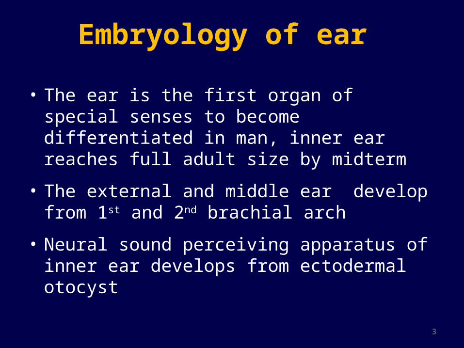
Embryology of ear
• The ear is the first organ of special senses to become differentiated in man, inner ear reaches full adult size by midterm
• The external and middle ear develop from 1st and 2nd brachial arch
• Neural sound perceiving apparatus of inner ear develops from ectodermal otocyst
4
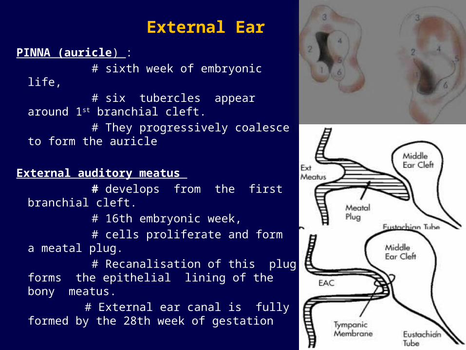
External Ear
PINNA (auricle) : # sixth week of embryonic life, # six tubercles appear around 1st
branchial cleft. # They progressively coalesce to form the
auricle
External auditory meatus # develops from the first branchial cleft. # 16th embryonic week, # cells proliferate and form a meatal plug. # Recanalisation of this plug forms the
epithelial lining of the bony meatus. # External ear canal is fully formed by the
28th week of gestation

5
MIDDLE EAR• Tympanic membrane: Develops from all
the three germinal layers. Outer epithelial layer by ectoderm, Middle fibrous layer by mesoderm & Inner mucosal layer by endoderm.
• Middle ear cavity : # Develops from endoderm of
tubotympanic recess which arises from 1st and partially from 2nd pharyngeal pouches.
# Head of Malleus and short process of incus are derived from mesoderm of 1st arch
# Rest of malleus and incus with stapes suprastructure develop from 2nd arch
# footplate and annular ligament which are derived from the otic capsule

6
Inner ear • Starts by 3rd week of fetal life and completed by 16th wks • The inner ear is derived from the ectoderm in the
region of the hindbrain. • A thickening of the ectoderm, the otic placode
becomes invaginated to form the auditory/otic vesicle.
OTIC PLACODE OTIC PIT OTIC VESICLE
7
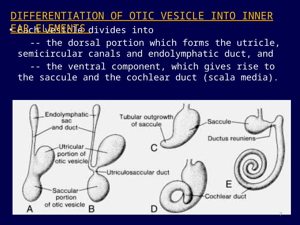
• Each vesicle divides into -- the dorsal portion which forms the utricle, semicircular
canals and endolymphatic duct, and -- the ventral component, which gives rise to the saccule and the
cochlear duct (scala media).
DIFFERENTIATION OF OTIC VESICLE INTO INNER EAR ELEMENTS

8
• Mesoderm around otocyst soon forms a cartilaginous otic capsule.
• Part of the cartilaginous shell undergoes vacuolization, and two perilymphatic spaces (scala vestibuli and scala tympani) are formed.
• Ossifies by 25 weeks

9
• Small group of cells breaks away otic capsule and along with cells of neural creast origin forms the statoacoustic ganglion.
• The ganglion subsequently splits into vestibular and cochlear nerves.

10
Anatomy of inner ear
• It lies in the petrous part of the temporal bone• Inner ear consists of osseous labyrinth that encloses
membranous labyrinth.Outer bony labyrinth 1. bony cochlea2. vestibule 3. three bony semicircular
canals4. Vestibular and cochlear aqueduct
Inner membranous labyrinth 5. Cochlear duct 6. Utricle 7. Saccule8. Three membranous
semicircular canals9. Endolymphatic system
11
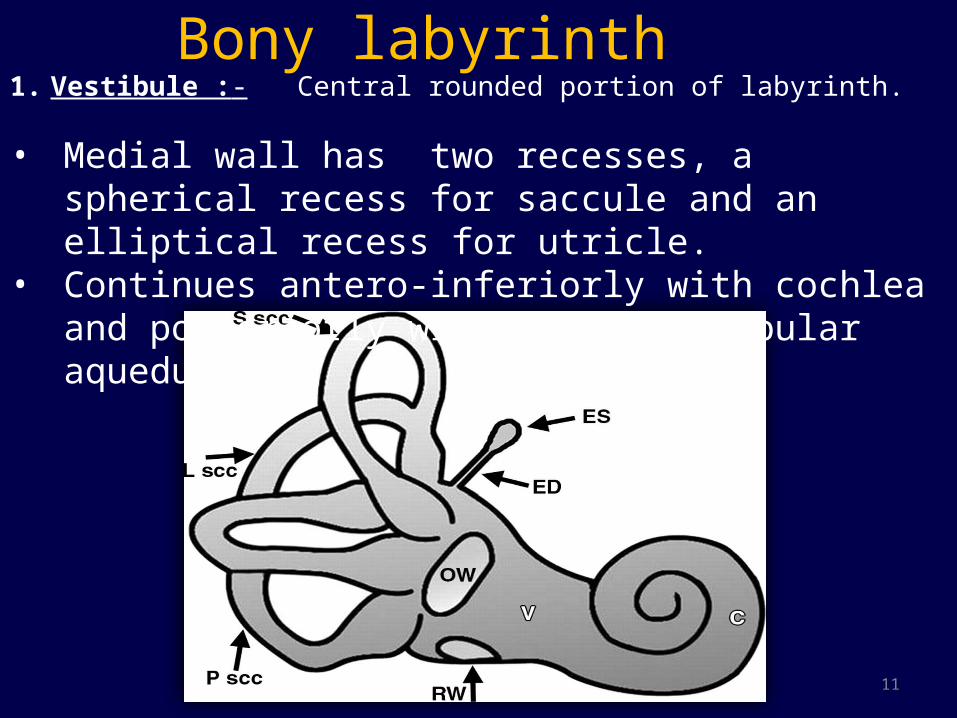
Bony labyrinth 1. Vestibule :- Central rounded portion of labyrinth.
• Medial wall has two recesses, a spherical recess for saccule and an elliptical recess for utricle.
• Continues antero-inferiorly with cochlea and posteriorly with SCC & vestibular aqueduct.
12
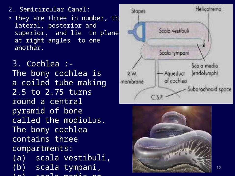
2. Semicircular Canal: • They are three in number, the
lateral, posterior and superior, and lie in planes at right angles to one another.
3. Cochlea :-The bony cochlea is a coiled tube making 2.5 to 2.75 turns round a central pyramid of bone called the modiolus. The bony cochlea contains three compartments: (a) scala vestibuli, (b) scala tympani, (c) scala media or the membranous cochlea
13
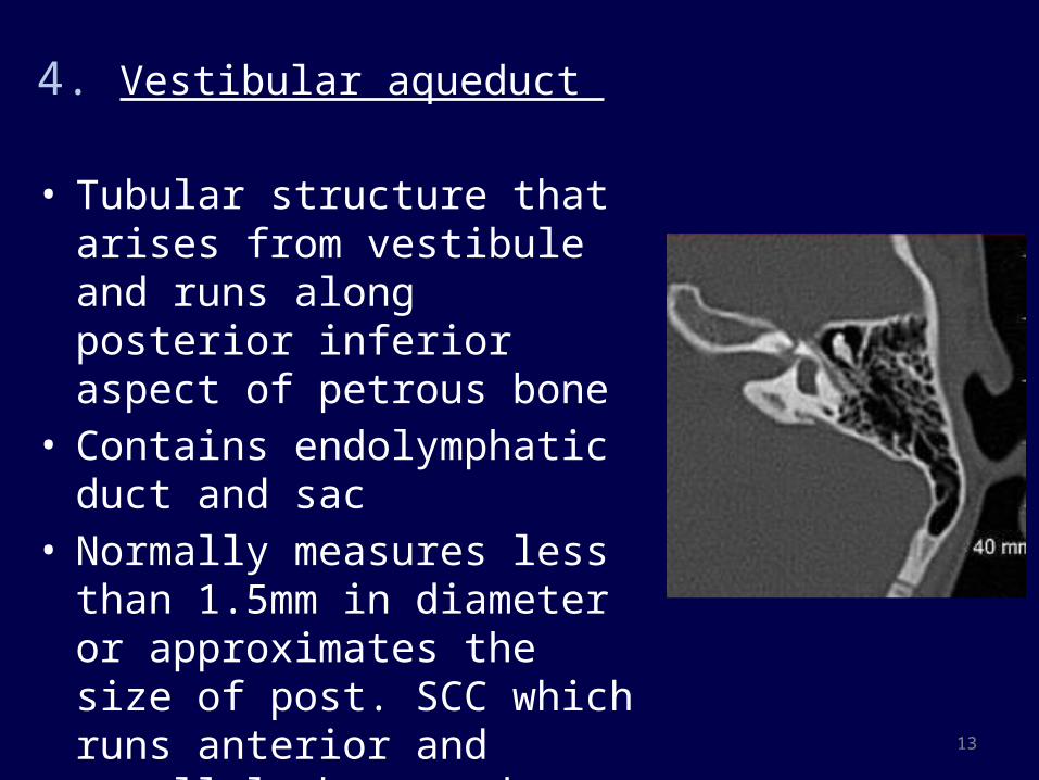
4. Vestibular aqueduct
• Tubular structure that arises from vestibule and runs along posterior inferior aspect of petrous bone
• Contains endolymphatic duct and sac
• Normally measures less than 1.5mm in diameter or approximates the size of post. SCC which runs anterior and parallel the aqueduct.
14
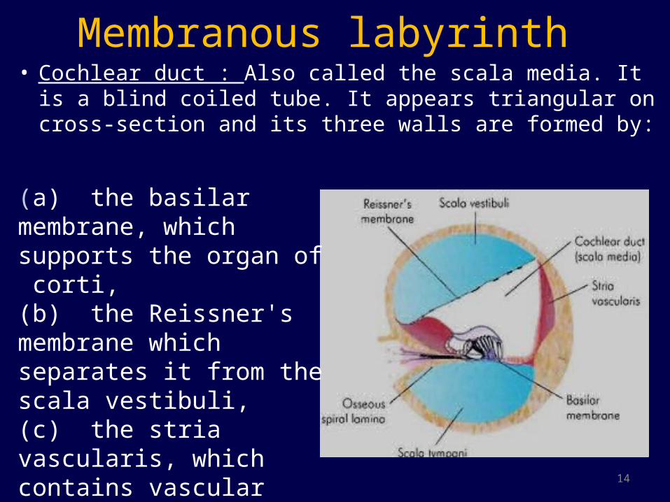
Membranous labyrinth • Cochlear duct : Also called the scala media. It is a blind
coiled tube. It appears triangular on cross-section and its three walls are formed by:
(a) the basilar membrane, which supports the organ of corti,(b) the Reissner's membrane which separates it from the scala vestibuli, (c) the stria vascularis, which contains vascular epithelium and is concerned with secretion of endolymph.
15
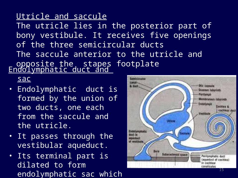
Endolymphatic duct and sac• Endolymphatic duct is formed by
the union of two ducts, one each from the saccule and the utricle.
• It passes through the vestibular aqueduct.
• Its terminal part is dilated to form endolymphatic sac which lies between the two layers of dura on the posterior surface of the petrous bone
Utricle and sacculeThe utricle lies in the posterior part of bony vestibule. It receives five openings of the three semicircular ductsThe saccule anterior to the utricle and opposite the stapes footplate

16
Internal Auditory Canal• A bony conduit that transmits VII & VIII cranial
nerves from pontomedullary junction to inner ear.
Divided by a bony lamina (falciform crest) into A. Smaller superior part• Superior vestibular N.• Facial NerveB. Larger Inferior part• Inferior vestibular N. • Cochlear nerve.

17
Blood supply of labyrinth• Arterial supply : Labyrinthine artery which is a
branch of anterior-inferior cerebellar artery
• Venous drainage : through three veins :
internal auditory vein vein of cochlear aqueduct vein of vestibular aqueduct
Transverse sinus.
Inferior petrosal sinus

18
Cross sectional anatomy of inner ear
White arrowhead : Modiolus with cochleaWhite arrow : I A C
Black arrowhead : VestibuleBlack arrow : Posterior semicircular canal
Fig.1.-----Axial HRCT of Inner Ear

19
Fig. 2– Axial heavily T2 WI Fig. 3– Axial heavily T2 WI
Cross sectional anatomy of inner ear
Axial images shows basal turn of cochlea and osseous spiral lamina
Middle and apical turns with the modiolus (arrowhead) and the spiral lamina (curved arrow) dividing the cochlea into scala vestibuli and scala tympani . The nerves are seen in CP angle.
20
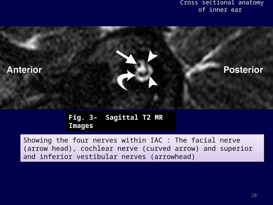
Cross sectional anatomy of inner ear
Fig. 3– Sagittal T2 MR Images
Showing the four nerves within IAC : The facial nerve (arrow head), cochlear nerve (curved arrow) and superior and inferior vestibular nerves (arrowhead)
21
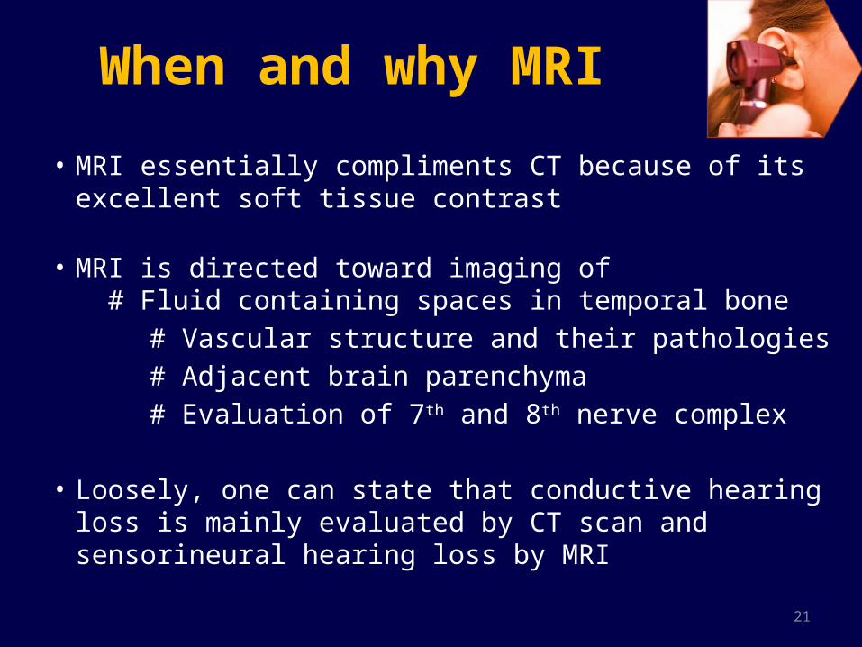
When and why MRI
• MRI essentially compliments CT because of its excellent soft tissue contrast
• MRI is directed toward imaging of # Fluid containing spaces in temporal bone
# Vascular structure and their pathologies # Adjacent brain parenchyma # Evaluation of 7th and 8th nerve complex
• Loosely, one can state that conductive hearing loss is mainly evaluated by CT scan and sensorineural hearing loss by MRI

22
IMAGING PROTOCOL -- MRI
• 1.5 or 3 Tesla MRI is preferred• Sedation used in most children• 3D volumetric CISS in axial plane
with coronal and sagittal reformation and MIP reconstruction
• Slice thickness of 0.4 – 0.7 mm• Oblique sagittal reformatted
images in plane perpendicular to 7th and 8th nerve in IAC
• Routine axial T2WI of brain to exclude CNS causes of sensorineural hearing loss
• 3D MPRAGE may be added.
• Precontrast brain with thin section through the CPA- IAC region
• MRA/ MRV as required
• Post contrast fat sat.
Inner ear Imaging Tumors and infection
23
3D CISS • Three dimensional (3D) constructive interference in
steady state (CISS) is a heavily T2 weighted fully refocused gradient echo MR sequence.
• Being heavily T2 weighted it is better suited for imaging of structures surrounded by fluid like 7th – 8th nerve complex and membranous labyrinth.
• 3D sequence , so reconstruction in any plane possible.• Other uses:-
1. Evaluation of cranial nerves 2. Diagnosis of NCC 3. Evaluation of CSF rhinorrhea 4. Evaluation of ventricular system etc.
24
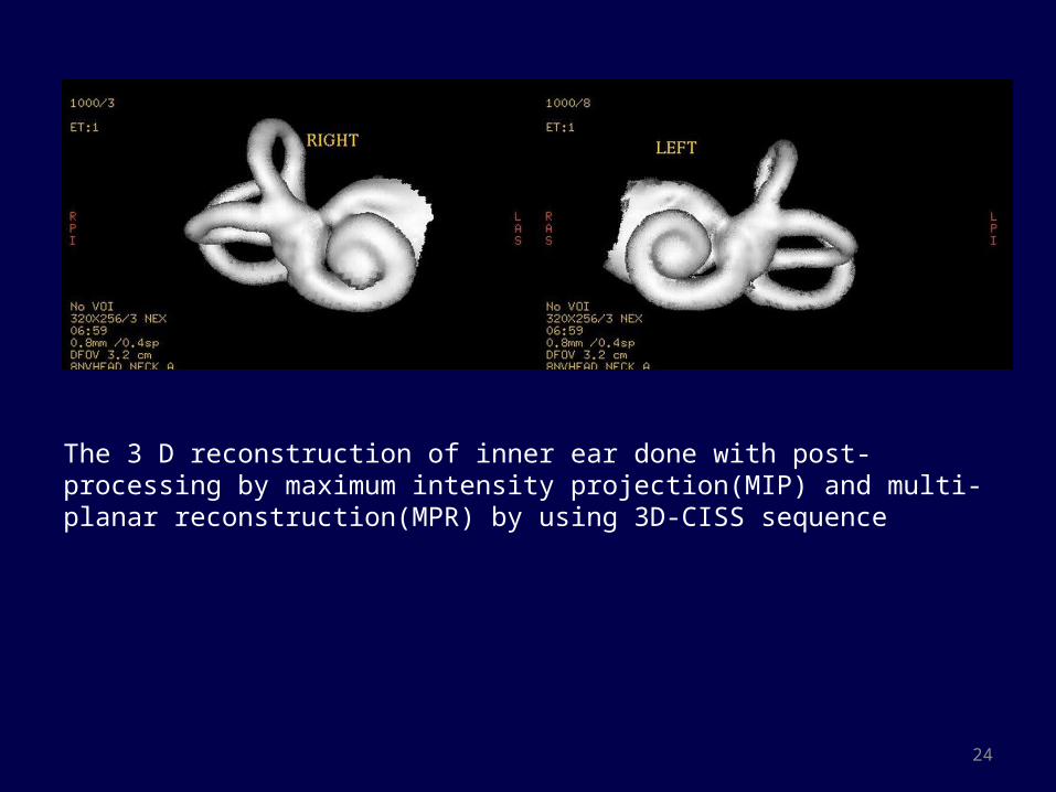
The 3 D reconstruction of inner ear done with post-processing by maximum intensity projection(MIP) and multi-planar reconstruction(MPR) by using 3D-CISS sequence

25
CONGENITAL ANOMALIES

26
Congenital malformation of inner ear Cochlear abnormalities are numerous and Jackler et al classified them on the basis
of arrested development during organogenesis
27
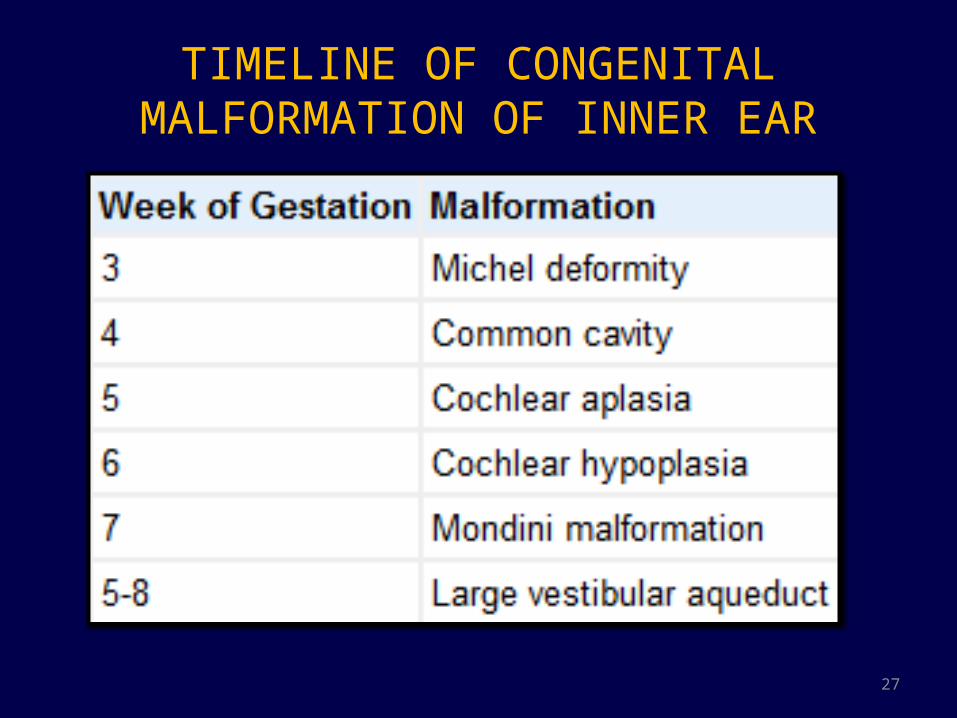
TIMELINE OF CONGENITAL MALFORMATION OF INNER EAR

28
COMPLETE LABYRINTHINE APLASIA OR MICHELE APLASIA
• Most severe inner ear deformity• Etiology : Arrested development of otic placode during 3rd
gestational week• Extremely rare – only 1% of inner ear malformation.• Unilateral/ bilateral. Unilateral cases are associated with
contralateral inner ear dysplasia.
HRCT :- # Complete absence of inner ear with hypoplasia of petrous
bone and narrow atretic IAC. # Absence of round and oval window # Flattening of medial wall of middle ear cavityMRI : 8th cranial nerve not visualized on MR images Associated with skull base, CVJ and vascular anomalies

29
Axial CT : Flat middle ear cavity and hypoplastic petrous bone
Coronal CT : Atretic internal auditory canal
Sagittal MRI : single nerve within the IAC, suggesting absent 8th nerve

30
Common cavity • Defined by absence of normal differentiation between
the cochlea and vestibule • 25% of cochlear malformation• Arrest during 4th arrest of gestation
• Associated with poor differentiation of membranous labyrinth as well resulting in severe to profound hearing loss

31
Confluence of cochlea and vestibule in a cystic cavity with no internal architecture
Common cavity
Absence of cochlear nerve
32
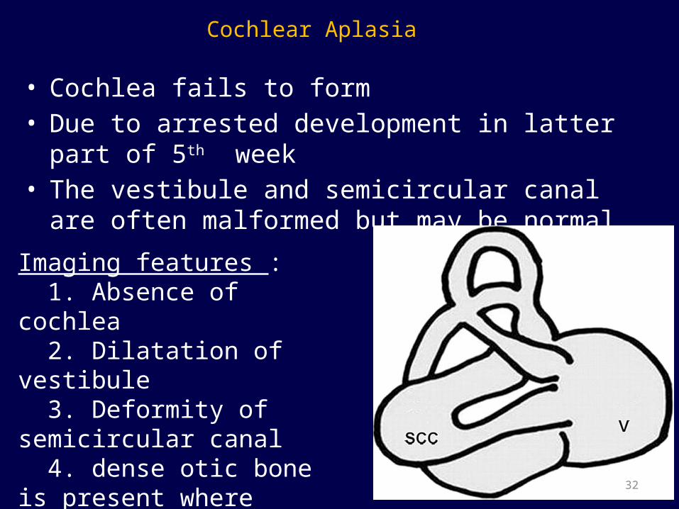
Cochlear Aplasia
• Cochlea fails to form • Due to arrested development in latter part of 5th week • The vestibule and semicircular canal are often
malformed but may be normal.
Imaging features : 1. Absence of cochlea 2. Dilatation of vestibule 3. Deformity of semicircular canal 4. dense otic bone is present where cochlea would be

33
Axial CT images shows dilated globose vestibule (arrow head) , dense sclerotic bone where cochlea should be (curved arrow) , and a stunted dilated posterior semicircular canal (straight arrow).
Coronal CT images shows malformed dilated lateral semicircular canal (straight arrow) with stunted superior semicircular canal (curved arrow)
34
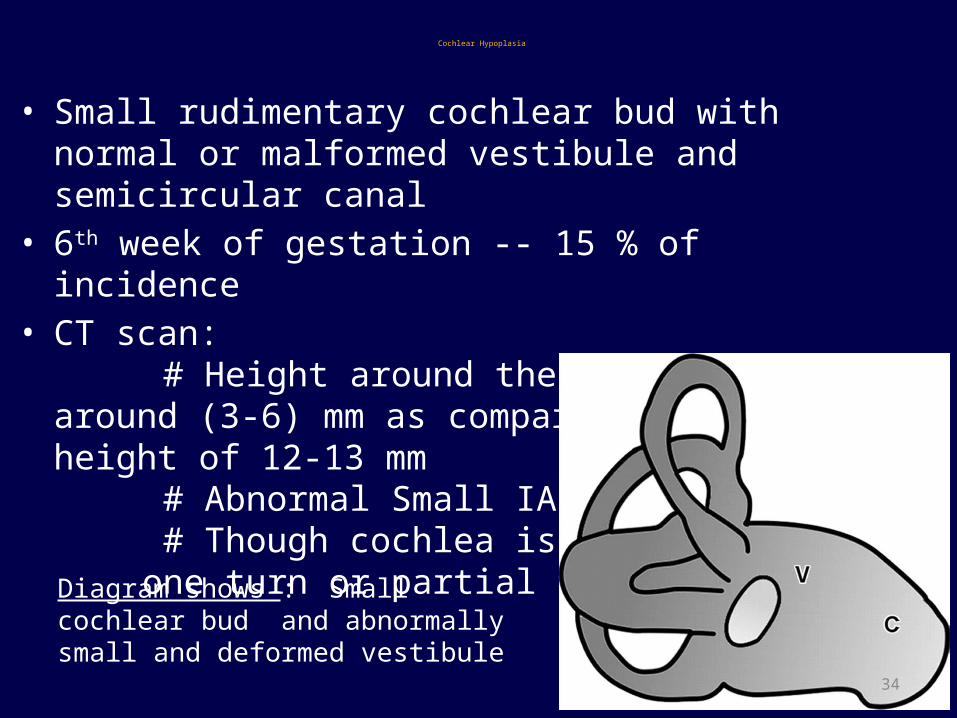
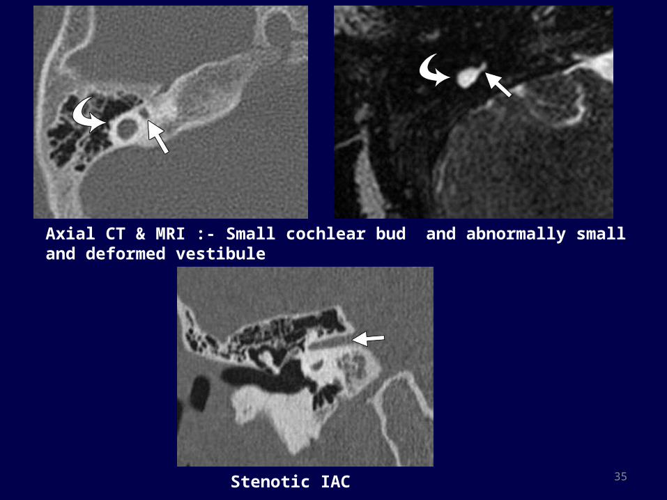
Cochlear Hypoplasia
• Small rudimentary cochlear bud with normal or malformed vestibule and semicircular canal
• 6th week of gestation -- 15 % of incidence • CT scan: # Height around the cochlea is around (3-6) mm as
compared to normal height of 12-13 mm # Abnormal Small IAC # Though cochlea is visible it has one turn or partial turn
Diagram shows : Small cochlear bud and abnormally small and deformed vestibule
35
Axial CT & MRI :- Small cochlear bud and abnormally small and deformed vestibule
Stenotic IAC
36
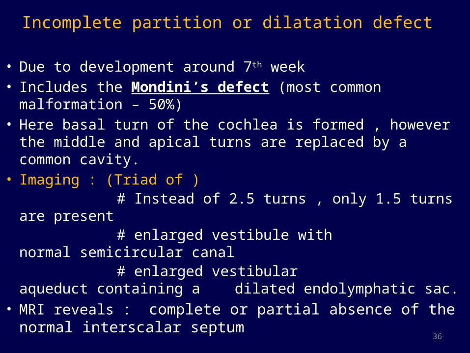
Incomplete partition or dilatation defect
• Due to development around 7th week• Includes the Mondini’s defect (most common malformation –
50%)• Here basal turn of the cochlea is formed , however the middle
and apical turns are replaced by a common cavity.• Imaging : (Triad of ) # Instead of 2.5 turns , only 1.5 turns are present # enlarged vestibule with normal semicircular canal # enlarged vestibular aqueduct containing a
dilated endolymphatic sac.• MRI reveals : complete or partial absence of the normal
interscalar septum
37
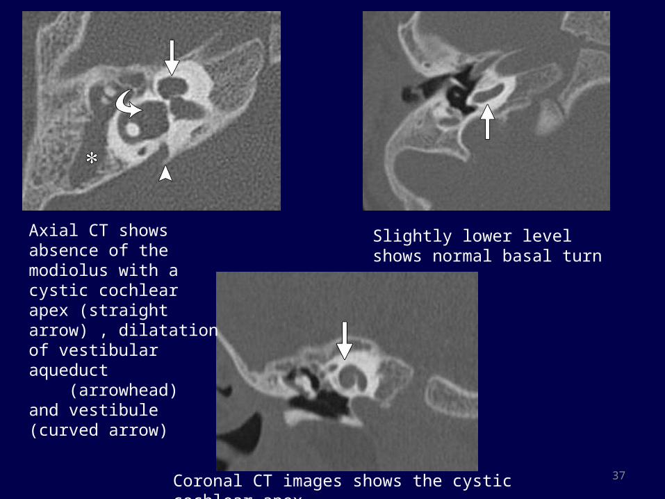
Axial CT shows absence of the modiolus with a cystic cochlear apex (straight arrow) , dilatation of vestibular aqueduct (arrowhead) and vestibule (curved arrow)
Slightly lower level shows normal basal turn
Coronal CT images shows the cystic cochlear apex
38
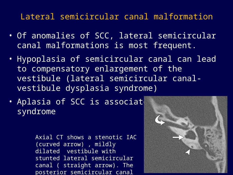
Lateral semicircular canal malformation• Of anomalies of SCC, lateral semicircular canal malformations
is most frequent.
• Hypoplasia of semicircular canal can lead to compensatory enlargement of the vestibule (lateral semicircular canal- vestibule dysplasia syndrome)
• Aplasia of SCC is associated with CHARGE syndrome
Axial CT shows a stenotic IAC (curved arrow) , mildly dilated vestibule with stunted lateral semicircular canal ( straight arrow). The posterior semicircular canal appears normal.
39
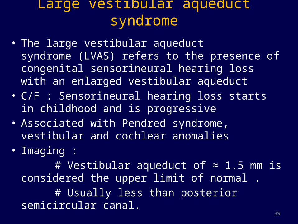
Large vestibular aqueduct syndrome
• The large vestibular aqueduct syndrome (LVAS) refers to the presence of congenital sensorineural hearing loss with an enlarged vestibular aqueduct
• C/F : Sensorineural hearing loss starts in childhood and is progressive
• Associated with Pendred syndrome, vestibular and cochlear anomalies
• Imaging : # Vestibular aqueduct of ≈ 1.5 mm is considered the
upper limit of normal . # Usually less than posterior semicircular canal.
40

41
IAC and cochlear nerve anomalies• IAC normal diameter range from 2- 8 mm, average 4mm• Diameter less than 2mm, described as Stenotic• Sagittal oblique images obtained in a plane perpendicular
to the long axis of IAC provides best depiction of the four major nerves of IAC
• Types of cochlear anomalies Type I – A Stenotic IAC with absent 8th nerve Type 2 – A common vestibulocochlear nerve with
hypoplasia or aplasia of cochlear branch 2a – associated with other inner ear anomaly 2b – No associations.

COMMON VASCULAR ANOMALIES
43
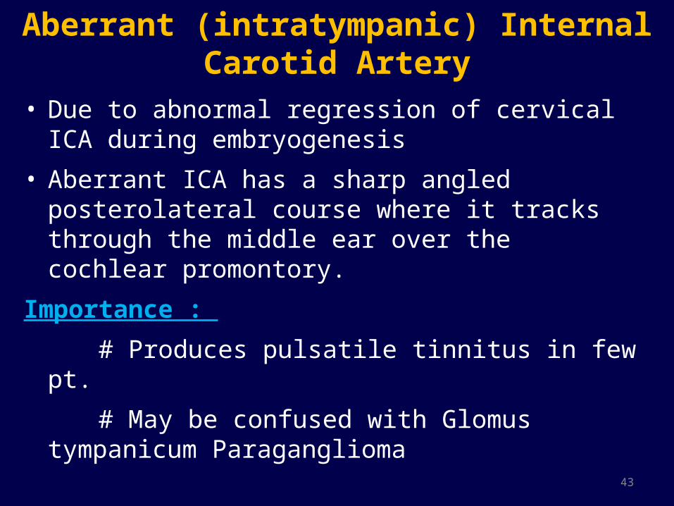
Aberrant (intratympanic) Internal Carotid Artery
• Due to abnormal regression of cervical ICA during embryogenesis
• Aberrant ICA has a sharp angled posterolateral course where it tracks through the middle ear over the cochlear promontory.
Importance :
# Produces pulsatile tinnitus in few pt.
# May be confused with Glomus tympanicum Paraganglioma
44
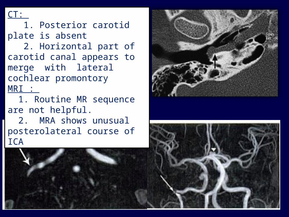
CT: 1. Posterior carotid plate is absent 2. Horizontal part of carotid canal appears to merge with lateral cochlear promontoryMRI : 1. Routine MR sequence are not helpful. 2. MRA shows unusual posterolateral course of ICA

45
• Other arterial anomalies : 1. Persistant stapedial artery 2. Persistant trigeminal Artery 3. Anomalous artery in the stria vascularis of the
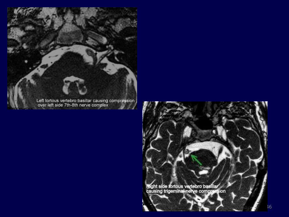
cochlea Ectatic vascular loops of AICA/PICA or tortous course
of vertebral artery may present with tinnitus due to compression of VIII nerve complex.
46

47
Venous variants
1. High riding Jugular bulb : Large jugular bulb reaching above the internal auditory canal with intact sigmoid plate
2. Dehiscent Jugular Bulb : The sigmoid plate is deficient, the bulb protrudes into the middle ear cavity. It is a common cause of a retro-tympanic vascular mass.
3. Jugular bulb diverticulum

48
Dehiscent jugular bulb
High riding jugular bulb
INFLAMMATORY CONDITIONS

50
LABYRINTHITIS
Types:1. Tympanic labyrinthitis : Infection spreads from middle ear
via oval or round window or labyrinthine fistula2. Meningogenic : Infection spreads along CSF spaces via IAC
or cochlear aqueduct. Usually bilateral.3. Hematogenic labyrinthitis : spread of infection by blood-
stream. Virus e.g, measles and mumps, syphilis etc4. Post traumatic labyrinthitis.
Inflammation of membranous labyrinth.
Viruses are the most common etiologic agents, but can be bacterial or autoimmune .
51
Imaging features:CT : # Usually normal in acute stage # Ossification of membranous labyrinth in late chronic
phaseMRI : contrast enhanced MR is the method of choice # T1- CEMR shows moderate to intense enhancement
within normal fluid filled structure of inner ear # Usually viral conditions causes subtle enhancement
and bacterial causes intense enhancement.Complication : Labyrinthitis ossificans is a Sequela of
chronic labyrinthitis, usually Pyogenic in origin.
52
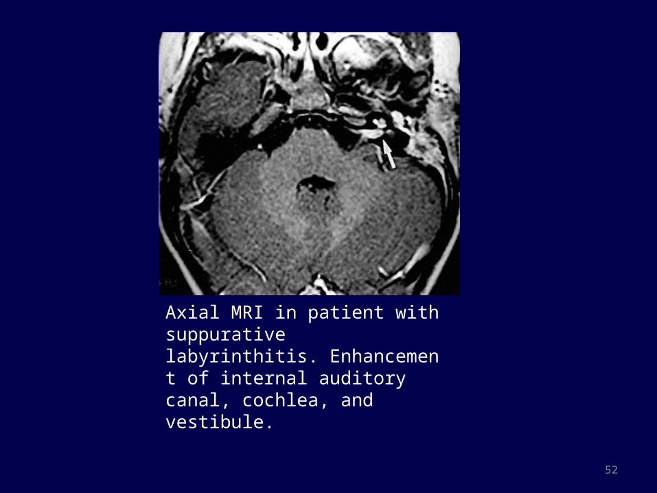
Axial MRI in patient with suppurative labyrinthitis. Enhancement of internal auditory canal, cochlea, and vestibule.

53
Labyrinthitis ossificans
• Labyrinthitis ossificans (LO) is the pathologic formation of new bone within the lumen of the otic capsule.
Etiology :- 1. Sequela of inflammation of the inner ear, e.g bacterial
meningitis or purulent labyrinthitis 2. Vascular obstruction of the labyrinthine artery 3. Autoimmune labyrinthitis etc.Imaging : CT scan :- Osseous deposition within the membranous
labyrinthMR Imaging :- Loss of the normal high signal on T2-
weighted images from displacement of the endolymphatic fluid is suggestive of this diagnosis.

54

55
CHOLESTEATOMA WITH COMPLICATION
MRI features of cholesteatoma ::--
Hypointense on T1WI & Hyperintense on T2 WINo enhancement or faint peripheral rim enhancementDelayed Contrast scan (after 45min) – continued enhancement of inflammatory or granulation tissue and not in cholesteatoma.DWI – Cholesteatoma shows restricted diffusion and are hyperintense on b= 1000/m2.

56
CHOLESTEATOMA WITH COMPLICATION
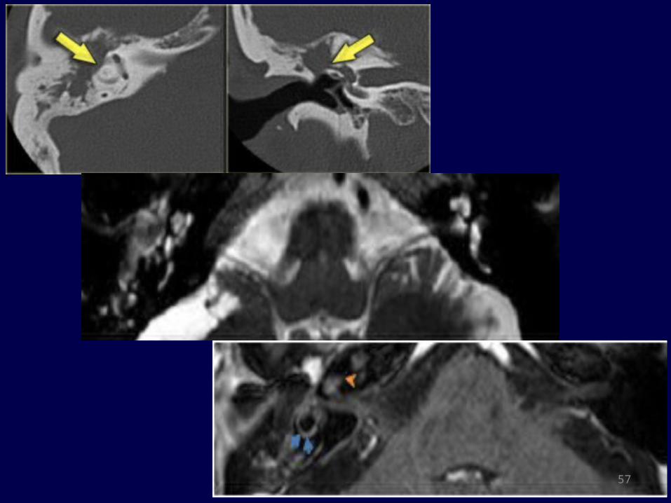
A . Labyrinthine fistula -- Most frequent complication with middle ear cholesteatoma (prevalence of 5%– 10%).
• C/F : Episodic vertigo, sensorineural hearing loss, tinnitus
• CT Findings :- 1. Dehiscent lateral semicircular canal support the
diagnosis 2. Uncommonly , dehiscence of cochlear promontory or
fistula in oval window.• MRI Findings :- a labyrinthine fistula causing labyrinthitis,
shows enhancement of the membranous labyrinth.
57
58
Other complications -
B. Perineural extension of cholesteatoma along facial nerve.
C. Erosion of the sigmoid sinus plate and consecutive thrombosis, tympanic tegmen erosion and subsequent intracranial invasion, recurrent\bacterial meningitis, and intracranial abscess are rare complications, which, nevertheless, require an urgent CT/MR imaging examination
59
Petrous apex granuloma• Cholesterol granuloma is an inflammatory granulation appearing
in response to the deposits of cholesterol crystals
• Etiology: Middle ear disorders causing mucosal edema and deposition of cholesterol crystal
• Site : Middle ear cavity followed by mastoid process and petrous apex
• C/F : Hearing loss, vertigo, headaches, tinnitus Any focal neurological deficit, especially of cranial nerves
V–VIII

60
61
Petrous apicitis• Petrous apicitis is infection with involvement of bone at
the very apex of the petrous temporal bone.
• Pathology : Osteitis developing from infected and obstructed air cells in a pneumatised petrous apex
• C/F: Presents with Gradenigo’s syndrome
1. petrous apicitis, with
2. 6th nerve palsy, and
3. Retro-orbital pain, or pain in the cutaneous distribution of the trigeminal nerve, due to extension of inflammation into Meckel's cave.

62
CT scan : 1. Erosive lysis with ill-defined
irregular edges of petrous apex2. Peripheral enhancement of
petrous apex with dural enhancement and thickening
MRI: 1. Fluid signal intensity in petrous
apex often with peripheral enhancement
2. More sensitive in detecting dural thickening and enhancement as well as leptomeningitis, cerebritis and cerebral abscess

FACIAL NERVE LESIONS

64

65

66

67
BELL’S PALSY• Bell's palsy is characterized by rapid onset lower motor neuron
facial nerve paralysis, often with resolution in 6 - 8 weeks.• Etiology : 1. Idiopathic 2. Reactivation of Herpes Simplex Virus infection in
geniculate ganglion. • Pathogenesis : Secondary to swelling and edema of the 7th
nerve within the facial nerve canal• Indication for imaging : MRI not done routinely . Indicated if :- # Decompressive surgery is being planned # Atypical: No recovery in 6 wks, recurrent palsy,
multiple cranial nerve involvement.
68
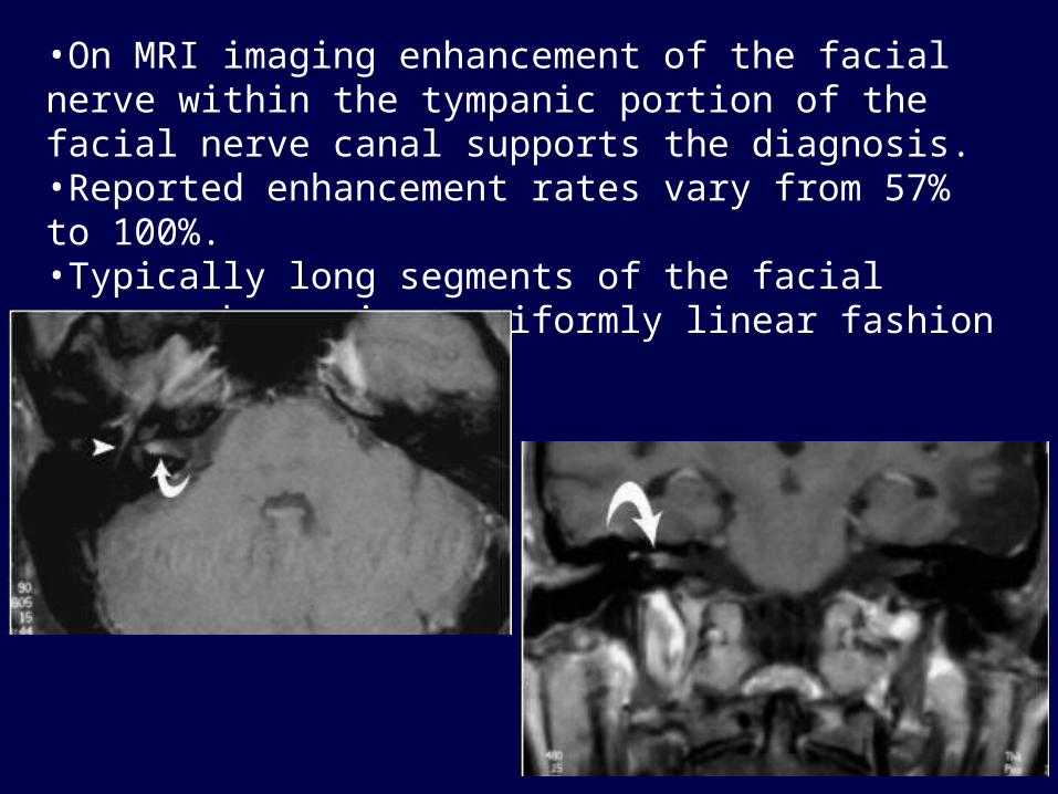
•On MRI imaging enhancement of the facial nerve within the tympanic portion of the facial nerve canal supports the diagnosis. •Reported enhancement rates vary from 57% to 100%.•Typically long segments of the facial nerve enhance in a uniformly linear fashion

69
Facial SchwannomasFacial nerve Schwannoma are uncommon tumors arising from
the Schwann cell sheathSite : Geniculate ganglion, followed by labyrinthine and
tympanic segment.Pathology : Originate from surface of the nerve, and splay the
nerve fibres over their eccentric growthC/F : 1. Persistent and gradually facial paresis. 2. Conductive hearing if tympanic segment involved
causing ossicular compression 3. In CP angle or IAC : Presents with sensorineural
deafness with facial paresis being rare in these cases. 4. Other like, tinnitus, hemifacial spasm, and otalgia
70
Imaging features HRCT :
Enhancing soft tissue density lesion along facial nerveIntracanalicular or CP angle tumor can cause bony erosion of anterosuperior portion of IAC
MRI:T1 : Iso- to hypo intense relative to gray matter T2 : Hyperintense ; large lesion may show heterogeneous signal T1 C+ (GAD) : Homogeneous enhancement with larger lesions showing cystic degeneration as focal intramural low signal intensity

71
CT SCAN : focal enlargement of the labyrinthine segment of the facial nerve
MRI: homogeneously enhancing mass filling the internal auditory canal with extension into the CP angle and labyrinthine segment
Diagnosis : Facial Nerve Schwannoma

72
The axial T1-weighted post-contrast MR image (left) shows homogeneous enhancement of the mass (between arrows). The bone algorithm CT (right)at the same level shows focal enlargement of the descending segment with extension toward the external auditory canal.Diagnosis : Schwannoma of the mastoid segment of facial nerve.
73
FACIAL NERVE HEMANGIOMA• Rare tumor of vascular origin (0.7% of all intra-temporal
tumors)• This along with other vascular malformations are termed as
Intra-temporal Benign Vascular Lesions
Age : 3rd to 6th decade without sex predilection
Site : Geniculate fossa followed by IAC
C/F: Facial nerve paralysis progressing over weeks. Sensorineural hearing loss and pulsatile tinnitus may
occur if there is erosion of otic capsule.

74
IMAGING FINDINGSMRI : # Intratemporal hemangiomas characteristically have
variable signal intensity on T1-weighted images increased signal intensity on T2-weighted imagesavid contrast enhancement.
# Low-signal-intensity foci may be seen on T1- and T2-weighted images, corresponding to the ossific matrix of the lesion
CT Scan : Enables exquisite visualization of associated bone changes
Tumor causes erosion which are irregular with indistinct margins giving a “Honeycomb” pattern of eroded bone.

75
Tumors in association with Inner ear

77
Anatomy: Cerebellopontine angle
Boundaries :- - Anterolateral surface of pons & Cerebellum - Posterior surface of petrous temporal bone
• CRANIAL NERVE - V , VII & VIII
• ARTERIES : SUP. CEREBELLAR A.AICA• VEINS :
TRIBUTARIES OF SUP PETROSAL V.

78
Imaging signs of extra-axial CPA cistern masses
1. Enlarged ipsilateral CPA cistern2. CSF/ Vascular “ Cleft” between mass and
cerebellum3. Displaced gray-white interface around mass4. Brainstem rotated5. Fourth ventricle compressed.
79
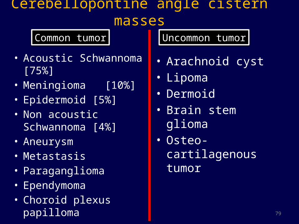
Cerebellopontine angle cistern masses
• Acoustic Schwannoma [75%]
• Meningioma [10%]• Epidermoid [5%]• Non acoustic
Schwannoma [4%]• Aneurysm• Metastasis• Paraganglioma• Ependymoma• Choroid plexus papilloma
• Arachnoid cyst• Lipoma• Dermoid• Brain stem glioma• Osteo-cartilagenous
tumor
Common tumor Uncommon tumor

80
Vestibular Schwannoma• Benign tumor arising from Schwann cells that wrap vestibulocochlear nerve• 70- 80 % of CPA lesions • Age – 5th -6th decade.• B/L acoustic Schwannoma pathognomonic for NF-2.
• Origin : Most from Inferior Vestibular Nerve, at glial-schwann cell interface
• Morphology : # Entirely intracanalicular # Intracanalicular with cisternal component ‘ Ice-
cream cone appearance’. # Rarely purely intracisternal.
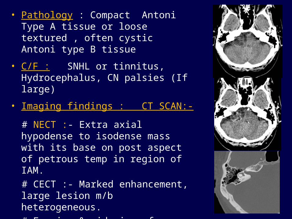
• Pathology : Compact Antoni Type A tissue or loose textured , often cystic Antoni type B tissue
• C/F : SNHL or tinnitus, Hydrocephalus, CN palsies (If large)
• Imaging findings : CT SCAN:-
# NECT :- Extra axial hypodense to isodense mass with its base on post aspect of petrous temp in region of IAM.
# CECT :- Marked enhancement, large lesion m/b heterogeneous.
# Erosion & widening of Internal Acoustic canal.
# Small lesions c/b missed d/t beam hardening artifact.
81

82
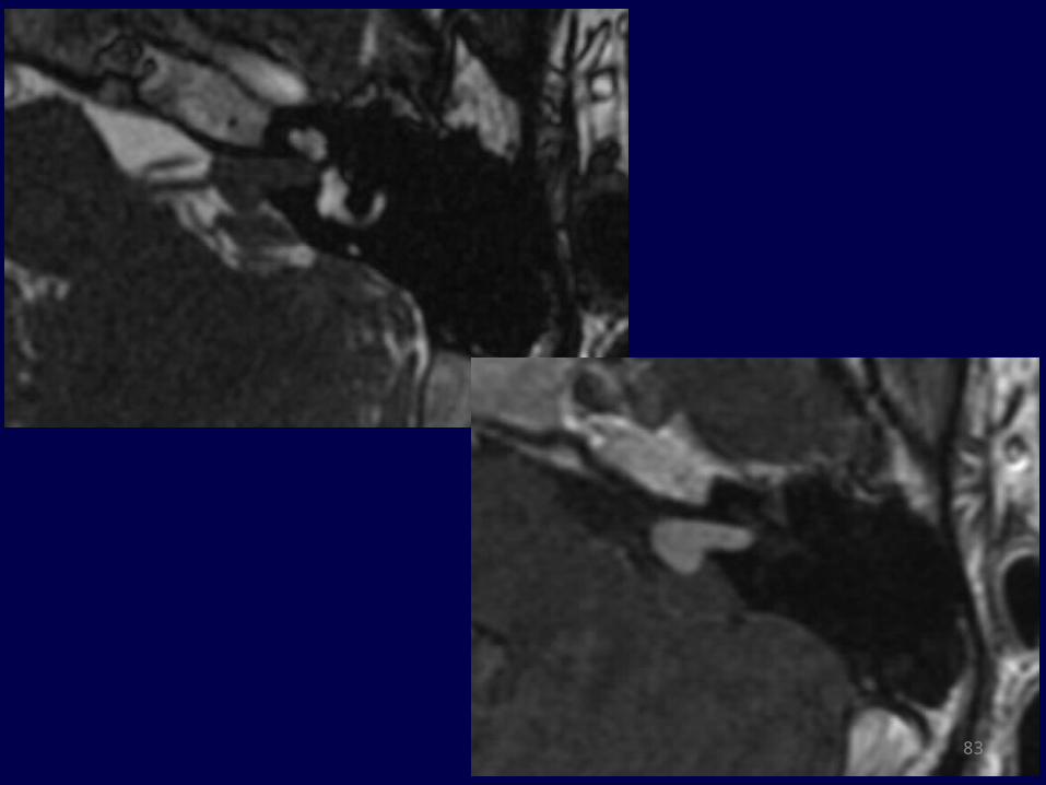
• T1WI: 2/3rd are hypointense and 1/3rd are isointense. may contain hypo intense cystic areas• T2WI : # Heterogeneously hyperintense # Small leson : "Filling defect" in high signal CSF of CPA-lAC cistern # may have associated peri-tumoural Arachnoid cysts
• T1 C+ (Gd) : # contrast enhancement is vivid # Heterogeneous in larger tumors # Occasionally, may show extension into the cochlea and dural tail of
enhancement.
MR IMAGING FEATURES
83

84
MENINGIOMA• Extra-axial neoplastic lesions arising from Arachnoid cap cells.
• 2nd most common CPA tumor (10%)
• Site : arises from the meninges covering posterior petrous bone.
• Female > Male (2-3:1) , peak age = 60yrs.
• C/F : Small Meningioma -entirely asymptomatic Large tumors – headache, paresis or neurological deficit.
• Morphology :
# "Mushroom cap" (hemispherical) with broad base towards posterior petrous wall (75%)
# Plaque-like : +/- bone invasion with hyperostosis (20%)
# Ovoid mass : mimicking Acoustic Schwannoma
85
Imaging features NECT : # Frequently hyperdense with focal areas of calcification # Bony hyperostosis of petrous boneCECT : Presence of broad dural base with dural tail and intense
enhancement is typical.MRI : # Isointense to brain parenchyma in T1 & T2WI # Blooming s/o calcification in GRE # Dural tail with other features of extra-axial lesion # May rarely extends into IAC and presents with diagnostic
dilemma.
ANGIOGRAPHY : Homogenous blush which lasts till late venous phase (Mother In Law sign)

86

87
Lipoma of internal auditory canal Rare congenital lesion often associated with
CP angle lipoma. CT Scan: Fat attenuating non enhancing lesion Presents with unilateral sensorineural hearing
loss MRI : Non enhancing lesion which is
hyperintense on T1WI & T2WI with suppression of SI on fat saturated images.
88
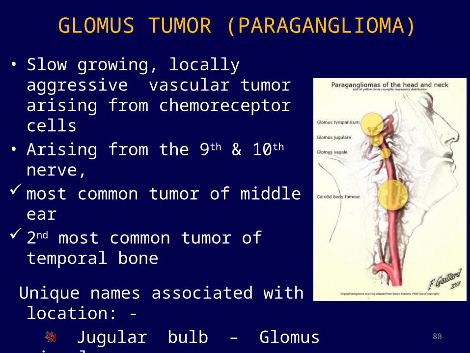
GLOMUS TUMOR (PARAGANGLIOMA)• Slow growing, locally aggressive vascular
tumor arising from chemoreceptor cells• Arising from the 9th & 10th nerve, most common tumor of middle ear 2nd most common tumor of temporal bone
Unique names associated with location: - Jugular bulb – Glomus jugulare Middle ear -- Glomus tympanicum Carotid body – Carotid body tumor Vagus nerve ganglion-- Glomus vagale
89
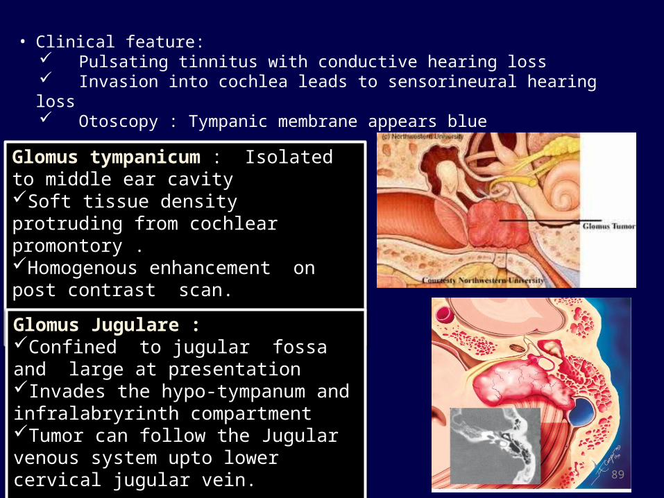
• Clinical feature: Pulsating tinnitus with conductive hearing loss Invasion into cochlea leads to sensorineural hearing loss Otoscopy : Tympanic membrane appears blue
Glomus tympanicum : Isolated to middle ear cavity Soft tissue density protruding from cochlear promontory .Homogenous enhancement on post contrast scan. Glomus Jugulare : Confined to jugular fossa and large at presentationInvades the hypo-tympanum and infralabryrinth compartment Tumor can follow the Jugular venous system upto lower cervical jugular vein.

90
MRI features : T1WI – Low signal intensityT2WI – High signal IntensityT1C+ -- Marked intense enhancementSalt – pepper appearance (T1 + T2) : Salt represents areas of
hemorrhages and pepper represents areas of flow void.

91
Perineural Spread of tumors• Common with head & neck cancer.• Nasopharyngeal CA followed by adenoid cystic carcinoma
and lymphoma.• Trigeminal and facial nerve are the commonest nerves
involvement.Features of Perineural spread :- 1. Enlargement and enhancement along the course of nerve 2. Obliteration of the fat surrounding neural foramina 3. Denervated muscles becomes atrophic with time and
replaced with fat. 4. Tumor in lateral aspect of pons should raise a suspicion.

92

LESION OF ENDOLYMPHATIC SAC & DUCT

94
Endolymphatic hydrops ( Meniere’s )• Refers to increased hydraulic pressure within inner ear
endolymphatic system.
• Etiology:
# Idiopathic (Meniere’s disease)
# Association with autoimmune disease , hormonal and metabolic condition noted (Meniere’s syndrome)
• C/F : 1. Fluctuating hearing loss
2. Episodic vertigo
3. Tinnitus
4. Aural fullness

95
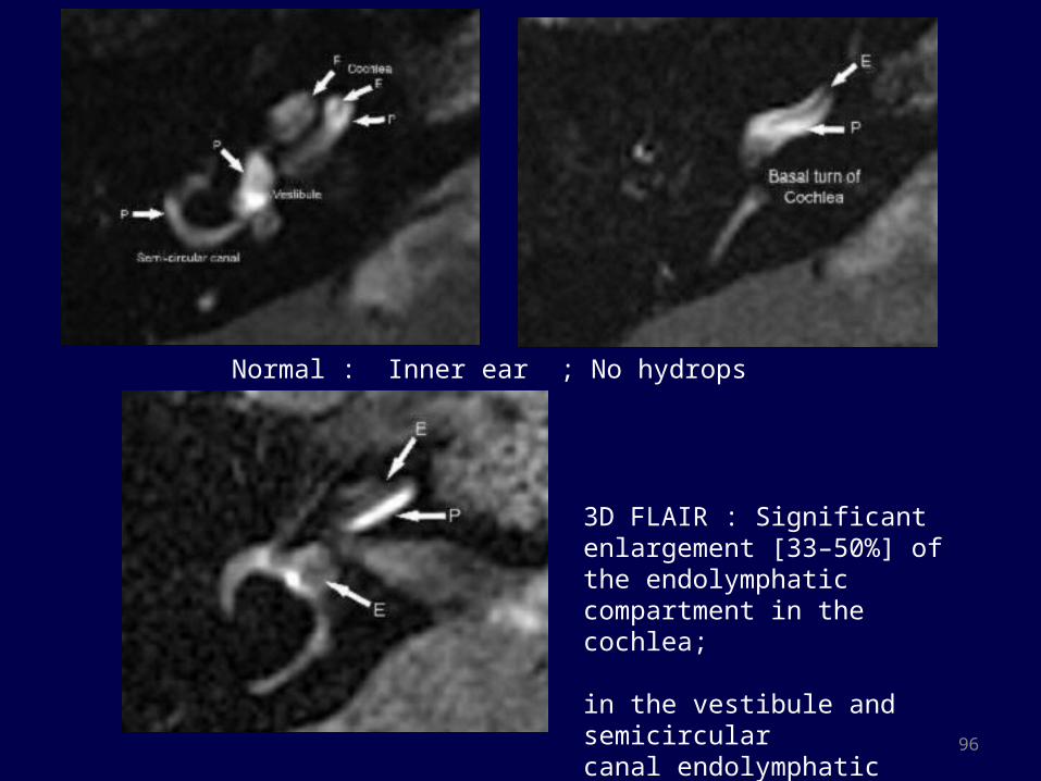
Role of MRI in Meniere’s disease• Bilateral intratympanic GBCA is being used for semi quantitative
evaluation of Meniere’s.
• 1- 1.5 ml of diluted gadolinium is injected into middle ear through a small myringotomy and evaluated after 24 hours.
• 3D FLAIR is used to evaluate inner ear
• 3Tesla is better.
• The gadolinium successfully penetrated the round window membrane, entering the perilymphatic space and delineating the contrast-enhanced perilymphatic and contrast-negative endolymphatic spaces of the inner ear
• If the non-enhancing endolymphatic area exceed the perilymphatic area, it is considered significant.
96
Normal : Inner ear ; No hydrops
3D FLAIR : Significant enlargement [33–50%] of the endolymphatic compartment in the cochlea;
in the vestibule and semicircular canal endolymphatic hydrops [>50%] has displaced almost all perilymph.
97
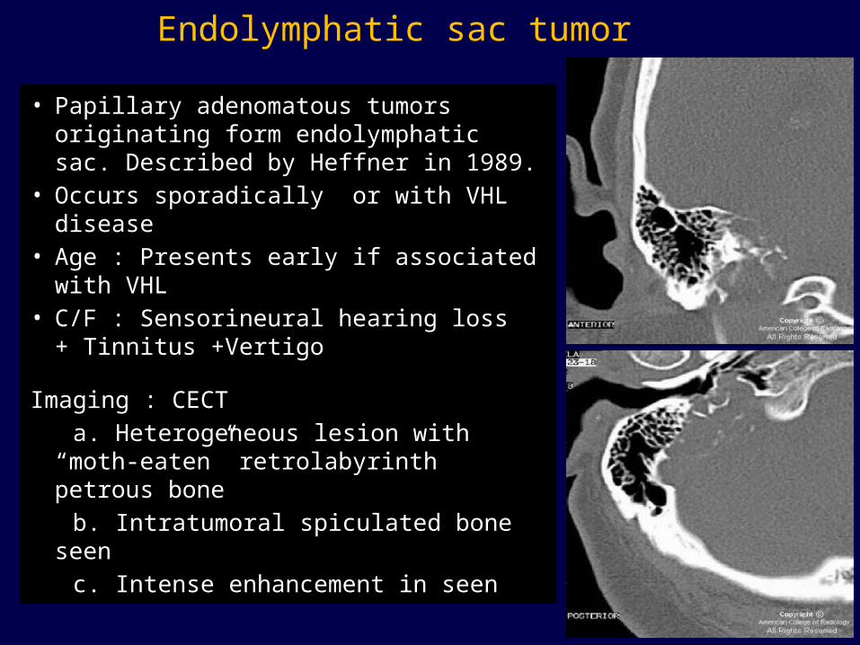
Endolymphatic sac tumor• Papillary adenomatous tumors
originating form endolymphatic sac. Described by Heffner in 1989.
• Occurs sporadically or with VHL disease• Age : Presents early if associated with
VHL• C/F : Sensorineural hearing loss +
Tinnitus +Vertigo
Imaging : CECT a. Heterogeneous lesion with “moth-
eaten” retrolabyrinth petrous bone b. Intratumoral spiculated bone seen c. Intense enhancement in seen

98
MRI Features : T1 /T2WI : Mixed signal intensity lesion where
focal high signal intensity d/t subacute hemorrhage and low signal intensity d/t calcification or hemosiderin.
Blood filled cysts and protein cyst, both appearing hyperintense on T1/T2WI suggests the diagnosis
Masses larger than 2 cm shows flow voids T1C+ : Heterogeneous enhancement
99
ISSUES WITH POST COCHLEAR IMPLANT CASES
Cochlear implants are not safe at 1.5TMR compatible CI are now availableExternal component should be removed in all casesRole of MRI is in preimplant evaluation to exclude
cochlear aplasia which is contraindication for surgery. Absent Cochlear nerve is relative contraindication.
CT scan is better for post-operative evaluation of these patients.

100
Conclusion
MR provides accurate anatomical delineation of complex soft tissue of inner ear3D reconstruction improves preimplant evaluationDetailed delineation of 7th & 8th nerve complex in temporal bone as well as membranous labyrinthDepiction of tumor size and extension into CP angle determines the approach to surgical removal.
101
References
1. Diagnostic Radiology- Neuroradiology – AIIMS – MAMC- PGI Course series . 3rd edition.
2. CT and MRI of whole body – John R Haaga 5th edition3. Joshi VM, Navlekar SK et.al -Ct and MRI imaging of the inner
ear and brain in children with sensorineural hearing loss. Radiographics. 2012 May-Jun;32(3):683-98
4. Jeremy Hornibrook, Mark Coates, Tony Goh, Philip Bird et.al MRI imaging of the inner ear for Meniere’s disease. Journal of the New Zealand Medical Association. 27 August 2010, Vol 123 No 1321
5.

102THANK YOU