magnetosome mysteries
TRANSCRIPT

mig-ma-
grad-
sta-curi-
micro-
-
-
-
-
-
--
-
Magnetosome Mysteries Despite reasonable progress elucidating magnetotactic microorganisms, many questions remain
Richard B. Frankel and Dennis A. Bazylinski
Aquatic, motile bacteria that orient and migrate along geomagnetic field Iines,known as magnetotactic bacteria, were discovered three decades ago by Richard P. Blakemore, then a graduate student at the University of Massachusetts at Amherst, Initially considered a mere curiosity, these bacteria have gained celebrity status as well as deeper respect among scientists, engineers, and the general public. Thus, magnetotactic bacteria have newfound relevance in several disciplines beyond pure microbiology, including nanobiotechnology, biomineralization, biogeochemistry, geomicrobiology, and magnetosensory behavior in animals, These bacteria are also considered relevant subject matter by those searching for signs of life on Mars,
Despite progress elucidating the how and why of magnetotaxis, many questions remain regarding the fascinating behavior of these microorganisms. Indeed, both physicists and microbiologists are trying to unlock the secrets of these microbes. Fortunately, scientists from both disciplines naturally tend to love magnetotactic bacteria, not least because they are fun and safe to study.
The Magnetotactic Bacteria
Magnetotactic bacteria are ubiquitous in aquatic habitats, including deep-ocean sediments. They generally inhabit chemically stratified water columns or sediments at or below the microaerobic redox transition zone between aerobic upper waters and lower, anaerobic regions.
Such bacteria are diverse with respect to morphology, physiology, and phylogeny. Thus magnetotactic bacteria do not represent a single,
defined, taxonomic group. Instead, the traits of magnetotaxis and magnetosome synthesis appear to be distributed among several groups of unrelated bacteria. Morphotypes include coccoid to ovoid cells; rods, vibrios, and spirilla of various dimensions; and even multicellular forms. All that have been examined are members of the domain Bacteria and possess cell walls that are characteristic of gram-negative bacteria.
Although a gliding form was recently observed, other known magnetotactic bacteria are motile by means of flagella, with some cells having a single polar flagellum, others having bipolar flagella, and still others having bundles of flagella at the ends or one side of the cell.
Magnetotactic bacteria are generally difficult to grow in pure culture, although several species can now be grown to high yields, making them amenable to biochemical analysis. Most described cultured strains are from freshwater and belong to the genus Magnetospirillum. There are also several unnamed marine species in pure culture, including vibrios known as strains MV-1 and MV-2, a marine coccus designated strain Me-1, and a marine spirillum designated strain MV-4. Yet another magnetotactic species is an anaerobic sulfate-reducing, rod-shaped bacterium, called Desulfovibrio magneticus strain RS-1. These cultured organisms, except D. magneticus, are obligate or facultative microaerophiles, and all are chemoorganoheterotrophic. The marine strains also grow chemolithoautotrophically, oxidizing reduced sulfur compounds as sources of electrons.
The Phenomenon of F I G U R E 1 Magnetotaxis and Consistency
of Bacterial Magnetosomes
The term magnetotaxis is somewhat of a misnomer because magnetotactic cells swim neither towards nor away from a magnetic field. Instead, they orient along and swim parallel or antiparallel to such fields. When observed with light microscopy in water drops, cells of each magneto-tactic species or strain display either two-way or one-way swimming behavior along the local magnetic field. They do not exhibit “run-and-tumble” motility typified by cells of Escherichia coli.
In the two-way swimming mode— for example, observed in Magneto-spirillum species in liquid media— cells are equally likely to swim parallel or antiparallel to the magnetic field direction, randomly and abruptly changing directions. In the one-way swimming mode, typified by the magnetotactic coccus strain MC-1, cells swim persistently in one direction along the magnetic field and accumulate on one side of a water drop. Bacteria of this type from Northern-hemisphere sites swim preferentially parallel to the magnetic field, corresponding to northward migration in the geomagnetic field, and are known as North-seeking (NS). Bacteria from Southern hemisphere sites swim preferentially antiparallel to the magnetic field, and are known as South-seeking (SS).
All magnetotactic bacteria contain magnetosomes, intracellular structures that consist of nanometer-scale, magnetic, iron-mineral crystals enveloped by a membrane vesicle known as the magnetosome membrane. This membrane in the genus Magnetospirillum is a lipid bilayer that anchors the crystals in the cell and serves as the locus of biological control when magneto-some crystals nucleate and grow. Empty and partially filled vesicles are observed in iron-starved cells of Magnetospirillum magnetotacticum, suggesting that the magnetosome vesicles form before mineral crystals are deposited.
Each magnetotactic species or strain exclusively produces magnetosomes containing either magnetite (Fe3O4) or greigite (Fe3S4) crystals,
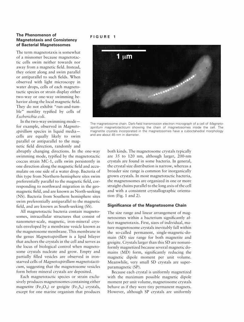
The magnetosome chain. Dark-field transmission electron micrograph of a cell of Magneto-spirillum magnetotacticum showing the chain of magnetosomes inside the cell. The magnetite crystals incorporated in the magnetosomes have a cuboctahedral morphology and are about 45 nm in diameter.
both kinds. The magnetosome crystals typically are 35 to 120 nm, although larger, 200-nm crystals are found in some bacteria. In general, the crystal size distribution is narrow, whereas a broader size range is common for inorganically grown crystals. In most magnetotactic bacteria, the magnetosomes are organized in one or more straight chains parallel to the long axis of the cell and with a consistent crystallographic orientation (Fig. 1 and 2).
Significance of the Magnetosome Chain
The size range and linear arrangement of magnetosomes within a bacterium significantly affect magnetotaxis. First, sizes of individual, mature magnetosome crystals inevitably fall within the so-called permanent, single-magnetic-domain (SD) size range for both magnetite and greigite. Crystals larger than this SD are nonuniformly magnetized because several magnetic domains (MD) form, significantly reducing the magnetic dipole moment per unit volume. Meanwhile, very small SD crystals are super-paramagnetic (SP).
Because each crystal is uniformly magnetized with the maximum possible magnetic dipole moment per unit volume, magnetosome crystals behave as if they were tiny permanent magnets.
except for one marine organism that produces However, although SP crystals are uniformly

F I G U R E 2
Magnetosome crystals have species/strain-specific shapes. Electron micrographs of magnetosome magnetite crystals in two cultured magnetotactic bacteria. (a) Cuboctahedral crystals in Magnetospirillum magnetotacticum (see Fig. 1). Small arrows indicate twinned crystals and large arrows indicate clusters of small crystals. (b) Elongated crystals in a marine magnetotactic bacterium, strain MV-1. There are two cells, each with one chain of magnetosomes. Elongated crystals imply an anisotropic crystal growth process.
magnetized, their magnetic dipole moments are not truly permanent because thermal energy can induce spontaneous magnetization reversals within each crystal, producing a time-averaged magnetic dipole moment of zero. The point is that SP crystals and MD crystals make poor permanent magnets. In practice, cells produce magnetosomes with the optimum, permanent, magnetic dipole moment by constraining the crystal size distribution to the SD size range.
The arrangement of SD magnetosome crystals in a chain maximizes the permanent magnetic dipole of the cell because individual magneto-some dipoles spontaneously orient in parallel along the chain axis. Because the magnetosome chain is fixed within the cell, an external magnetic field, such as the geomagnetic field, exerts a torque on the cellular dipole that tends to align the whole cell along the magnetic field. Thus, each cell behaves like a tiny magnetic compass needle.
But how small can a magnetic needle be and still function usefully? The answer depends on what we consider useful, but we can say that the time-averaged orientation of a compass needle in a magnetic field depends on the ratio of magnetic energy to thermal energy. The higher this ratio, the greater the orientation of the compass needle and the higher the migration speed of a swimming cell along the magnetic field. A chain of 10 to 20 magnetosomes, each 50 nm in size, is sufficient to orient a magnetotactic bacterium in the geomagnetic field (about 0.5 x10⁻⁴ T) and to enable that cell to migrate along the magnetic field at more than 90% of its forward swimming
crystals and their arrangement within the cell make it seem as though magnetotactic bacteria are experts in materials engineering, permanent magnet engineering, and statistical mechanics.
Magneto-Aerotaxis
Early models assumed that all magnetotactic bacteria are microaerophiles and indigenous in sediments. The geomagnetic field is inclined downward from horizontal in the Northern hemisphere and upward in the Southern hemisphere, with its inclination increasing from the equa
tor to the poles. NS cells in the Northern hemisphere and SS cells in the Southern hemisphere would therefore migrate downward toward the sediments along these geomagnetic field lines.
Thus magnetotaxis should help to guide cells in each hemisphere downward to less-oxygenated regions of the habitat. Once cells reach these low-oxygen microhabitats, they would presumably stop swimming and adhere to sediment particles until localized disturbances lifted them up again, after which they would eventually follow the magnetic field back down. This description corresponds to the predominant behavior of NS cells in the Northern hemisphere and SS cells in the Southern hemisphere, based on observing magnetotactic bacteria in water drops.
The discovery of large populations of magnetotactic bacteria at the microaerobic transition zone in water columns of certain chemically stratified aquatic habitats and the isolation of obligate, microaerophilic, coccoid magnetotactic bacterial strains such as MC-1 led researchers to revise that early view of magnetotaxis. The original model did not explain how bacteria in the anoxic zone of a water column benefit from magnetotaxis, nor did it explain how the magnetotactic cocci form microaerophilic bands of cells in semisolid oxygen gradients.
Indeed, when various species and strains of magnetotactic bacteria are exposed to oxygen concentration gradients within capillary tubes, these bacteria exhibit both magnetotaxis and aerotaxis, enabling them to migrate to an optimal point within the oxygen gradient. In fact, supposedly one-way magnetotactic bacteria can speed. The overall design of the magnetosome
actually back up under anoxic conditions or when exposed to lessthan-optimal oxygen concentrations. This behavior is referred to as magneto-aerotaxis.
Two different magneto-aerotactic mechanisms, termed polar and axial, are found in magnetite-producing magnetotactic bacteria. Some cocci, such as MC-1, that swim persistently in one direction along the magnetic field (NS or SS) in water drops are polar magneto-aerotactic. Others, such as the freshwater spirillum Magnetospirillum magnetotacticum that swim in either direction along the magnetic field lines with frequent, spontaneous reversals in swimming direction without turning around in water drops, are called axial magneto-aerotactic. The distinction between NS and SS does not apply to axial magneto-aerotactic bacteria.
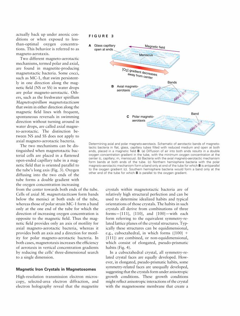
The two mechanisms can be distinguished when magnetotactic bacterial cells are placed in a flattened open-ended capillary tube in a magnetic field that is oriented parallel to the tube’s long axis (Fig. 3). Oxygen diffusing into the two ends of the tube forms a double gradient with the oxygen concentration increasing from the center towards both ends of the tube. Cells of axial M. magnetotacticum form bands below the menisci at both ends of the tube, whereas those of polar strain MC-1 form a band only at the one end of the tube for which the direction of increasing oxygen concentration is opposite to the magnetic field. Thus the magnetic field provides only an axis of motility for axial magneto-aerotactic bacteria, whereas it provides both an axis and a direction for motility for polar magneto-aerotactic bacteria. In both cases, magnetotaxis increases the efficiency of aerotaxis in vertical concentration gradients by reducing the cells’ three-dimensional search to a single dimension.
Magnetic Iron Crystals in Magnetosomes
High-resolution transmission electron microscopy, selected-area electron diffraction, and electron holography reveal that the magnetite
F I G U R E 3
Determining axial and polar magneto-aerotaxis. Schematic of aerotactic bands of magneto-tactic bacteria in flat, glass, capillary tubes filled with reduced medium and open at both ends, placed in a magnetic field B. (a) Diffusion of air into both ends results in a double oxygen concentration gradient in the tube, with the minimum oxygen concentration at the center (c, capillary; m, meniscus). (b) Bacteria with the axial magneto-aerotactic mechanism form bands at both ends of the tube. (c) Northern hemisphere bacteria with the polar magneto-aerotactic mechanism form a band only at end of the tube for which B is antiparallel to the oxygen gradient (c). Southern hemisphere bacteria would form a band only at the other end of the tube for which B is parallel to the oxygen gradient.
crystals within magnetotactic bacteria are of relatively high structural perfection and can be used to determine idealized habits and typical orientations of those crystals. The habits in such crystals all derive from combinations of three forms—{111}, {110}, and {100}—with each form referring to the equivalent symmetry-related lattice planes of the crystal structures. Typically these structures can be equidimensional, e.g., cuboctahedral, in which forms ({100} � {111}) are combined, or non-equidimensional, which consist of elongated, pseudo-prismatic habits (Fig. 4).
In a cuboctahedral crystal, all symmetry-related crystal faces are equally developed. However, in elongated, pseudo-prismatic habits, some symmetry-related faces are unequally developed, suggesting that the crystals form under anisotropic growth conditions. These growth conditions might reflect anisotropic interactions of the crystal with the magnetosome membrane that create a

F I G U R E 4
Habits of magnetosome crystals. Idealized crystal habits comprising {100}, {111}, and {110} forms corresponding to magnetosomes in various strains and species of magnetotactic bacteria. The cuboctahedron (lower left) is the only equidimensional habit. The other habits are elongated. The consistent crystal habits within a given species or strain reflect a very high degree of control over the biomineralization process.
preferred axis of elongation during the period of crystal growth.
Marine, estuarine, and salt marsh species typically produce iron sulfide magnetosomes consisting primarily of the magnetic mineral called greigite. Recognized greigite-producing magnetotactic bacteria include several types of relatively large, rod-shaped bacteria and also a magnetotactic prokaryote that is multicellular. The greigite crystals in the magnetosomes of these microorganisms are thought to form from nonmagnetic precursors such as mackinawite (tetragonal FeS). Like magnetite crystals in magnetosomes, the morphologies of the greigite crystals also appear to be species and strain specific.
Biochemistry and Gene Expression in
Magnetosome Formation
The process by which magnetotactic bacteria make and organize their magnetosomes is not completely known, although some genetic and biochemical details are starting to emerge. Partial genomic sequence data are available for two
magnetotactic bacteria, M. magnetotacticum and strain MC-1.
Some general descriptive facts provide a context for the biochemical insights that are anticipated. For instance, the cells somehow transport iron ions from outside the cell into the magnetosome membrane vesicles to form a saturated solution. Subsequent adjustment of the redox potential and pH in the vesicles causes nucleation and growth of magnetite crystals. The process is relatively rapid; iron-starved cells of Magnetospirillum gryphiswaldense form magnetosomes about 10 minutes after being exposed to 10 �M Fe(III). Because iron can amount to about 2–3% of the dry weight of magnetotactic bacteria (an astonishing amount for bacteria), the cells must have a highly efficient iron uptake system. Cells of M. gryphiswaldense take up mainly Fe(III) via a high-affinity system, and several other Magnetospirillum species produce siderophores, suggesting that they, too, can take up Fe(III).
Several important questions about magnetite synthesis need to be ad
dressed. For example, is Fe(III) or Fe(II) transported into magnetosome membrane vesicles? What Fe redox reactions are important in the biomineralization process?
A Fe(III) reductase was purified from M. magnetotacticum by Yoshihiro Fukumori and coworkers at Kanazawa University, Kanazawa, Japan. Although the Fe(III) reductase appears to be mainly cytoplasmic, it may be bound on the inner side of the cytoplasmic membrane and could reduce Fe(III) as it enters the cells. For redox conditions within magnetosome membrane vesicles such that the [Fe(III)]/[Fe(II)] ratio is about 2 (the ratio in magnetite) at an elevated pH, magnetite would be the most stable Fe-oxide phase. The vesicle could also provide sites for magnetite crystals to nucleate and grow. Interactions of the magnetosome membrane with the faces of the growing crystal could affect crystal morphology.
Researchers are looking for specific proteins, either through reverse genetics or mutant analysis, in magnetosome membranes that could explain how these minerals are produced within

such vesicles. For instance, mam (magnetosome membrane) genes appear to be conserved in a large gene cluster within both Magnetospirillum spp. and strain MC-1, and may be involved in magnetite biomineralization.
When mam genes from M. gryphiswaldense were cloned and sequenced, they fell into two gene clusters, according to Dirk Schu ler and coworkers at the Max-Planck-Institute for Marine Microbiology, Bremen, Germany. The MamA protein encoded by one of these genes shows homology to tetratricopeptide repeat proteins, while MamB is homologous to cation diffusion facilitators, and MamE to HtrA-like serine proteases. Meanwhile MamC and MamD show no homology to other known proteins. A gene cluster containing mamA and mamB is also found in M. magnetotacticum and strain MC1; here again, the clusters contain genes with no homology to known genes or proteins in established databases. No functions can be definitively assigned to any of these cluster-associated proteins.
Several magnetotactic strains, including M. gryphiswaldense and strain MV-1, relatively frequently form nonmagnetotactic mutants that lack magnetosomes. Unlike wild-
F I G U R E 5
type strains, such mutants do not accumulate iron. These mutations appear to be the result of chromosomal deletions. One mutant of M. gryphiswaldense is missing an 80-kb chromosomal segment that includes insertion sequences, mam genes, and additional genes that apparently encode magnetosome membrane proteins in the wild-type strain. Because all or most of the mam genes are located within this segment, it may represent a magnetosome island, which means it appears to contain a number of genes encoding metabolically linked functions, in this case for magnetosome synthesis. We have evidence for such a structure in strain MV-1.
The magA gene found in M. magneticum strain AMB-1 encodes a protein that appears to
Magnetite crystals from magnetotactic bacteria on Mars? Magnetite crystals released from the Martian meteorite ALH84001. Magnetite crystal morphologies of particles within the meteorite include: irregular, cuboidal, tear-drop, rounded whisker-shaped (dashed arrows), and elongated prisms (solid arrows) which have hexagonal or rectangular projections depending on orientation of the crystal. This figure was kindly provided by Kathie Thomas-Keprta.
according to Tadashi Matsunaga, Yoshiko Okamura, and coworkers at Tokyo University of Agriculture and Technology, Tokyo, Japan. When membrane vesicles from these cells contain the magA gene product, they take up iron only when ATP is supplied, indicating that this process is energy dependent. Based on analysis using a magA-luc fusion protein, the MagA protein localizes in the cell membrane and possibly the magnetosome membrane.
Another three genes encode magnetosome membrane-specific proteins. Those genes, mms6, mms16, and mpsA from Magnetospirillum strain AMB-1, are also found in the genome of M. magnetotacticum. MpsA shows homology to an acyl-CoA transferase, while Mms16 has GTPase
be a proton gradient-driving H/Fe(II) antiporter and is situated on the cytoplasmic membrane,
activity and may be involved in forming magnetosome vesicles. Mms6, the most abundant of

the three, binds to magnetite and may regulate crystal growth. Magnetite synthesized in the presence of Mms6 forms 20- to 30-nm cuboidal crystals, whereas 1- to 100-nm pleomorphic crystals form when that protein is absent.
Other Magnetosome Mysteries To Ponder
Magnetotactic bacteria construct internal, magnetic dipoles that enable such cells to orient along geomagnetic fields as they swim, yet are no longer than 1–2 �m, which is the length of the cells themselves. The construct involves a hierarchical structure, the magnetosome chain, and a mineralization process in which the mineral type, grain size, and placement in the cell are all controlled.
Primary control is presumably exerted by the magnetosome membrane through specific proteins. Yet many details of this process and others remain a mystery. While most magnetotactic bacteria have magnetosomes arranged in chains, some contain dispersed aggregates or clusters of magnetosomes, often along only one side of the cell. When it comes to orienting the cell in the geomagnetic field, this arrangement is not so efficient as when magnetosomes are aligned in chains, assuming that maximizing the ratio of magnetic dipole moment to volume of magnetite optimizes performance.
Other magnetotactic bacteria make magnetite crystals as large as 200 nm. Such crystals exceed the single-magnetic-domain size range, and so are larger than optimal. Yet the cells that make these crystals have so much magnetite that they are about 250 times more magnetic than is required for an adequate magneto-aerotactic response.
These observations suggest that we may not know all the functions of magnetosomes. Other possible functions include storing iron for future metabolic needs, sequestering potentially toxic free iron ions in the cytoplasm, and reserving electron acceptors and donors. Iron sulfide magnetosomes occasionally contain copper, a potentially toxic element, suggesting that greigite
ACKNOWLEDGMENTS
magnetosomes sometimes serve a more general detoxification purpose. Finally, magnetite crystals can disproportionate H2O2, suggesting that magnetite in magnetosomes serves as a catalase or in some related catalytic function.
Other mysteries surround magnetotaxis itself. There are many microaerobic organisms, including nonmagnetic mutants of magnetotactic bacteria, that form aerotactic bands without the aid of magnetism. Simulations of axial magnetotactic bacteria confirm that magneto-aerotaxis is more efficient than aerotaxis alone for finding optimal oxygen concentrations, meaning magnetotactic bacteria would find the optimal concentration before purely aerotactic bacteria with the same swimming speed. But this advantage holds true only at high inclinations of geomagnetic field. Many polar magnetotactic bacteria are fast swimmers, traveling about 100 body lengths per second or more, so the efficiency argument may be sound over a greater range of geomagnetic inclination for these organisms. Nevertheless, the question of whether aerotactic efficiency alone accounts for the persistence of magnetosome chains in bacteria over geologic timescales remains unanswered.
Finally, nanoscale magnetite crystals were found in the Martian meteorite ALH84001 that was recovered in Antarctica in 1984 (Fig. 5). This meteorite has a rock matrix with an age of 4.5 billion years—about when the solar system formed. Its magnetite crystals are associated with secondary carbonate minerals within the rock matrix that have an age of about 4.0 billion years, which is when Mars is thought to have had liquid water on its surface. Moreover, about 25% of the magnetite crystals have elongated, pseudo-prismatic habits and a size distribution similar to magnetosome magnetite crystals in terrestrial magnetotactic bacteria. Whether the ALH84001 crystals arose from life-forms on ancient Mars is still under discussion. But this discovery brings us to the last and the most intriguing and controversial magnetosome mystery. Were there magnetotactic bacteria on ancient Mars, and are any there now?
We thank R. Dunin-Borkowski and K. Thomas-Keprta for the generous contribution of figures. The work described here was presented at the 2003 ASM General Meeting in Washington, D.C., as part of the first symposium devoted to magnetotactic
¨bacteria. We thank other participants, including B. L. Dubbels, Y. Fukumori, Y. Okamura, and D. Schuler. We also thank our students, postdocs, and our numerous collaborators through the years. We are also very grateful to the National Science

Foundation, the Office of Naval Research, and the National Aeronautics and Space Administration for their support. Research in the D.A.B. lab is currently supported by NSF grant EAR-0311950 and NASA Johnson Space Center grant NAG 9–1115.
SUGGESTED READING
Bazylinski, D. A., and R. B. Frankel. 2004. Magnetosome formation in prokaryotes. Nature Rev. Microbiol. 2:217–230. Bauerlein, E. (ed.). 2000. Biomineralization: from biology to biotechnology and medical application. Wiley-VCH, Weinheim, Germany. Blakemore, R. P. 1982. Magnetotactic bacteria. Annu. Rev. Microbiol. 36:217–238. Dunin-Borkowski, R. E., M. R. McCartney, R. B. Frankel, D. A. Bazylinski, M. Posfai, and P. R. Buseck. 1998. Magnetic microstructure of magnetotactic bacteria by electron holography. Science 282:2868–1870. Frankel, R. B., D. A. Bazylinski, M. S. Johnson, and B. L. Taylor. 1997. Magneto-aerotaxis in marine, coccoid bacteria. Biophys. J. 73:994–1000. Matsunaga, T., and Y. Okamura. 2003. Genes and proteins involved in bacterial magnetic particle formation. Trends Microbiol. 11:536–541. McKay, C. P., E. I. Friedmann, R. B. Frankel, and D. A. Bazylinski. 2003. Magnetotactic bacteria on Earth and on Mars. Astrobiol. 3:263–270. Schu ler, D. 2004. Molecular analysis of a subcellular compartment: the magnetosome membrane in Magnetospirillum gryphiswaldense. Arch. Microbiol. 18:1–7. Spring, S., and D. A. Bazylinski. 2000. Magnetotactic bacteria. In The prokaryotes, published on the Web at http: //www.springer-ny.com/. Springer-Verlag New York, Inc., New York. Taoka, A., K. Yoshimatsu, M. Kanemori, and Y. Fukumori. 2003. Nitrite reductase from the magnetotactic bacterium Magnetospirillum magnetotacticum. Can. J. Microbiol. 49:197–206.