malaria - wisconsin state laboratory of hygiene · scraping the slide surface with a second slide...
TRANSCRIPT
12/5/2017
1

12/5/2017
2
No relevant conflicts of interest to disclose
WISCONSIN STATE LABORATORY OF HYGIENE - UNIVERSITY OF WISCONSIN
Contents
Disease background
Diagnostics Collecting blood
Preparing smears
Staining
Interpreting slides
Molecular and Antigen testing
New and emerging causes of Malaria
WISCONSIN STATE LABORATORY OF HYGIENE - UNIVERSITY OF WISCONSIN
12/5/2017
3
Disease Background
WISCONSIN STATE LABORATORY OF HYGIENE - UNIVERSITY OF WISCONSIN
Incidence
The World Health Organization estimates that in 2015 malaria caused 212 million clinical episodes, and 429,000 deaths. 90% of deaths were in sub-Saharan Africa
77% of deaths were in children under 5 years of age
Approximately 1,700 cases of malaria are
reported every year in the United States
WISCONSIN STATE LABORATORY OF HYGIENE - UNIVERSITY OF WISCONSIN
12/5/2017
4
Malaria in the USA
Malaria was declared eliminated from the United States in the early 1950’s.
Between 1957 and 2015, in the United States, 63 outbreaks of locally transmitted mosquito-borne malaria have occurred
During 1963-2015, 97 cases of transfusion-transmitted malaria were reported in the United States
WISCONSIN STATE LABORATORY OF HYGIENE - UNIVERSITY OF WISCONSIN
Plasmodium
Members of the Plasmodium genus infect a wide range of birds, mammals, reptiles, and amphibians worldwide
Transmitted by the Anopheles mosquito Can also be transmitted through transfusion or
congenitally, although this is very rare.
Over 200 species, but only 5 routinely cause human infections falciparum, vivax, ovale, malariae, and knowlesi
WISCONSIN STATE LABORATORY OF HYGIENE - UNIVERSITY OF WISCONSIN

12/5/2017
5
Disease
Infection can range from asymptomatic to life threatening.
Immune compromise, young or elderly age, pregnancy, and fetuses are at greatest risk for severe disease
Paroxysms of Fever, rigors, chills, and sweating
Headache, malaise, and myalgia
Convulsions, respiratory distress, hypoglycemia, circulatory collapse, renal failure, and coma
WISCONSIN STATE LABORATORY OF HYGIENE - UNIVERSITY OF WISCONSIN
Plasmodium Disease Features
WISCONSIN STATE LABORATORY OF HYGIENE - UNIVERSITY OF WISCONSIN
falciparum vivax ovale malariae knowlesi
Incubation period
8-11 10-17 10-17 18-40 9-12
Fever cycles 36-48 hours“Tertian”
44-48 hours“Tertian”
48 hours“Tertian”
72 hours“Quartan”
24 hours“Quotidian”
Infection duration
6-17 months 5-8 years 12-20 months 20-50 years Information lacking
Degree of parasitemia
High Low, <2% Low, <2% Low, <2% High
Averageseverity of disease
Moderate toSevere
Moderate toSevere
Mild Mild to moderate Moderate to Severe
Relapse No Yes Yes No Not likely
Area of endemicity
Large range; Tropics andsubtropics
Large range; Tropics andsubtropics except west Africa
Tropics; sub-Saharan Africa and Southeast Asia
Narrow range; tropics
Narrow Range;Southeast Asia

12/5/2017
6
Diagnostics
WISCONSIN STATE LABORATORY OF HYGIENE - UNIVERSITY OF WISCONSIN
Diagnostics
Malaria can be immediately life threatening. Testing should be ordered upon admission
Malaria testing should be available on a 24-hour STAT basis
Requests for testing should be accompanied by information related to clinical signs and symptoms, travel history, and receipt of malaria chemoprophylaxis or therapeutic antimalarial agents.
Testing methods Microscopic examination of blood films
Molecular analysis
Antigen detection
WISCONSIN STATE LABORATORY OF HYGIENE - UNIVERSITY OF WISCONSIN
12/5/2017
7
Need for Trained Specialists
Accurate interpretation relies on the availability of trained and experienced microscopists, high quality reagents, and well-maintained light microscopes.
The average laboratorian does not perform this test regularly, and may not be maintaining optimal proficiency.
WSLH offers STAT testing by experienced staff M-F 7:45am-4:30pm
STAT testing also available
WISCONSIN STATE LABORATORY OF HYGIENE - UNIVERSITY OF WISCONSIN
608-263-3280 WSLH608-258-0099 WDPH
Blood films The optimal time to test is between chills
Increases the likelihood of identifying advanced morphologies that will aid in species identification
Do not delay an initial exam upon admission
The Clinical and Laboratory Standards Institute (CLSI) recommends preparation of two thin and two thick blood films for each test
One negative set of blood films does not eliminate Plasmodium infection This should be clearly relayed back to the physician
If malaria remains a possible diagnosis, after the first set of negative smears, samples should be taken for at least 3 successive days. As often as intervals of 6 to 8 h
Positive patients should have additional testing at 24, 48, and 72 hours after initiating therapy to evaluate for drug resistance. Parasitemia usually drops within 24 hours, often by 50% or more
WISCONSIN STATE LABORATORY OF HYGIENE - UNIVERSITY OF WISCONSIN

12/5/2017
8
Blood Collection- Finger prick
Blood is ideally collected via finger prick with immediate (bedside) preparation
Prick a patient's finger with a sterile, nonreusable lancet
Let the blood flow freely blood that has to be “milked” from the finger is
diluted with tissue fluids, which decrease the number of parasites per field.
Touch a slide to the drop of blood
WISCONSIN STATE LABORATORY OF HYGIENE - UNIVERSITY OF WISCONSIN
Blood Collection- Finger prick
WISCONSIN STATE LABORATORY OF HYGIENE - UNIVERSITY OF WISCONSINCDC DPDx
12/5/2017
9
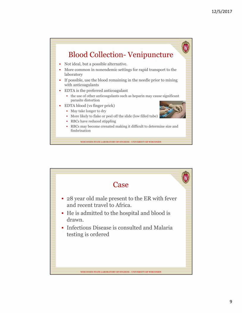
Blood Collection- Venipuncture Not ideal, but a possible alternative.
More common in nonendemic settings for rapid transport to the laboratory
If possible, use the blood remaining in the needle prior to mixing with anticoagulants
EDTA is the preferred anticoagulant the use of other anticoagulants such as heparin may cause significant
parasite distortion
EDTA blood (vs finger prick) May take longer to dry
More likely to flake or peel off the slide (low filled tube)
RBCs have reduced stippling
RBCs may become crenated making it difficult to determine size and fimbrination
WISCONSIN STATE LABORATORY OF HYGIENE - UNIVERSITY OF WISCONSIN
Case
28 year old male present to the ER with fever and recent travel to Africa.
He is admitted to the hospital and blood is drawn.
Infectious Disease is consulted and Malaria testing is ordered
WISCONSIN STATE LABORATORY OF HYGIENE - UNIVERSITY OF WISCONSIN
12/5/2017
10
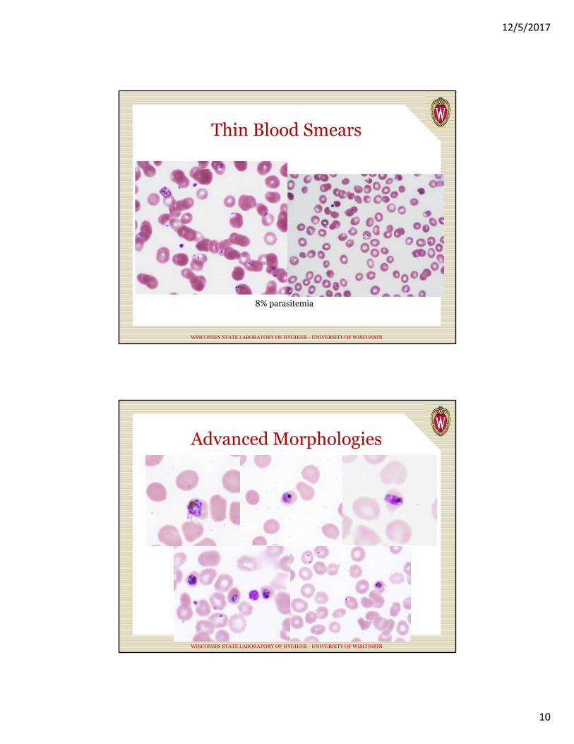
Thin Blood Smears
WISCONSIN STATE LABORATORY OF HYGIENE - UNIVERSITY OF WISCONSIN
8% parasitemia
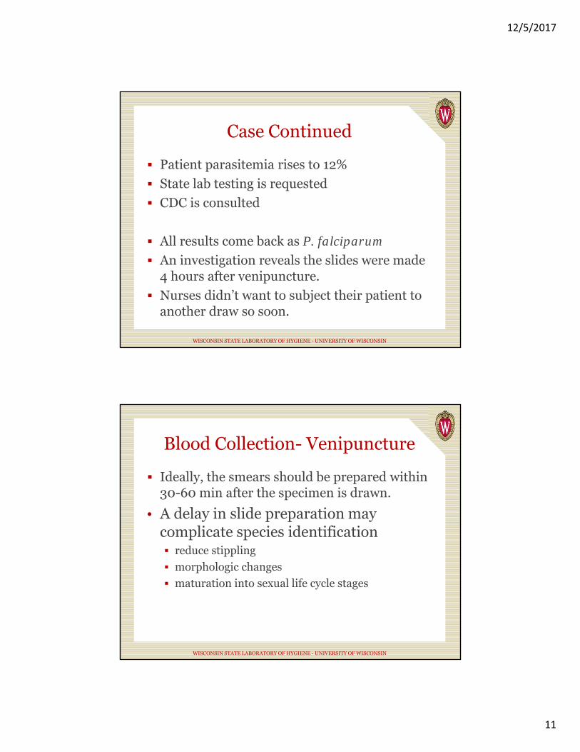
Advanced Morphologies
WISCONSIN STATE LABORATORY OF HYGIENE - UNIVERSITY OF WISCONSIN
12/5/2017
11
Case Continued
Patient parasitemia rises to 12%
State lab testing is requested
CDC is consulted
All results come back as P. falciparum
An investigation reveals the slides were made 4 hours after venipuncture.
Nurses didn’t want to subject their patient to another draw so soon.
WISCONSIN STATE LABORATORY OF HYGIENE - UNIVERSITY OF WISCONSIN
Blood Collection- Venipuncture
Ideally, the smears should be prepared within 30-60 min after the specimen is drawn.
• A delay in slide preparation may complicate species identification reduce stippling
morphologic changes
maturation into sexual life cycle stages
WISCONSIN STATE LABORATORY OF HYGIENE - UNIVERSITY OF WISCONSIN

12/5/2017
12
Slide types Thin Films: A drop of blood is spread over a
large surface area Species identification is better because parasite and
RBC morphologies are more clear
Parasitemia measurements are more accurate
Sensitivity lower than thick films
Thick films: A larger amount of blood is examined in a smaller area 10 to 20 times more sensitive
Can be examined faster
Species identification is more difficult
WISCONSIN STATE LABORATORY OF HYGIENE - UNIVERSITY OF WISCONSIN
Slide Preparation Thin film
One drop of blood (~50uL) near the end
Spread the drop using a second slide held at 45 degrees
Thick film 1-2 drops of blood should be spread to the size of a dime or nickel (1.5-
2.0 cm)
Should be just barely able to read newsprint through the wet slide
Most common error is to use too much blood
Stir the slide for 30 second to remove fibrin
Not necessary if using EDTA blood
WISCONSIN STATE LABORATORY OF HYGIENE - UNIVERSITY OF WISCONSIN MCM v11, CLSI M15-A

12/5/2017
13
Slide Preparation
WISCONSIN STATE LABORATORY OF HYGIENE - UNIVERSITY OF WISCONSINCDC DPDx
Slide Preparation Allow the film to air dry (room temperature) in a dust-free area.
Thin- at an angle
Thick- horizontal (8-12 hours)
Slides may be placed under a light fan to hasten drying (1-4 hours)
Never apply heat to a film Heat fixes the blood, causing the erythrocytes to remain intact during
staining
This leads to stain retention and an inability to identify the parasites
If thick films are to be stained at a later time, they should be lysed (laked) before storage 10 min in Giemsa buffer
Or, quickly dipped in H2O (up to 10 sec)
WISCONSIN STATE LABORATORY OF HYGIENE - UNIVERSITY OF WISCONSIN

12/5/2017
14
Troubleshooting Avoid fingerprints by only holding the sides
Store in a dust proof container
Use absolutely clean, grease-free slides Best results are obtained when slides are cleaned
Immerse in 70% Ethanol, drain on a paper towel, polish with a lint free cloth
WISCONSIN STATE LABORATORY OF HYGIENE - UNIVERSITY OF WISCONSIN
Troubleshooting- Flaking
WISCONSIN STATE LABORATORY OF HYGIENE - UNIVERSITY OF WISCONSIN

12/5/2017
15
Troubleshooting- Flaking If too much blood is used or any grease remains on the slide, the
blood may flake off during staining.
At the correct thickness you should be just able to read text through the blood
Lyse the smear immediately after drying to reduce flaking
Scraping the slide surface with a second slide during preparation improves adherence
Fresh blood is less likely to flake
Norgan, et. al. A method for reducing the sloughing of thick blood films for malaria diagnosis. Malaria Journal 2013 12:231
Other Points to Remember
Blood films should be stained on the same day or within a few days of collection Prolonged storage may result in stain retention
It is not recommended to combine thick and thin on the same slide It can delay reporting while waiting for drying
Detection of blood parasites by automated instruments is not recommended Failure to detect parasitemias have been reported
WISCONSIN STATE LABORATORY OF HYGIENE - UNIVERSITY OF WISCONSIN

12/5/2017
16
Staining
Giemsa is the stain of choice Color descriptions in texts and at DPDx are based
on Giemsa and may not be the same with other stains
Wright or Wright-Giemsa can be used but species determination may be more difficult Schuffner’s dots and Maurer’s clefts may not
visible or as clear
It is better to do one stain well than several stains poorly.
WISCONSIN STATE LABORATORY OF HYGIENE - UNIVERSITY OF WISCONSIN
Giemsa Staining Protocol1. Prepare fresh Giemsa
2. Pour 40 ml of Giemsa buffer into a second jar. Add 20 μl (2 drops) of Triton X-100.
3. Thin smears only: Fix slides in absolute methanol, 1-5 seconds and air dry
4. Place slides into the Giemsa stain for 45-60 minutes.
5. Remove thin smear slides and rinse by dipping 3-4 times in the Giemsa buffer. Thick smears should be left in buffer for 5 minutes.
6. Air dry slides upright in a rack. Fan may be used to shorten dry time.
WISCONSIN STATE LABORATORY OF HYGIENE - UNIVERSITY OF WISCONSIN CLSI M15-A

12/5/2017
17
Alternate Giemsa Protocols
WISCONSIN STATE LABORATORY OF HYGIENE - UNIVERSITY OF WISCONSIN CLSI M15-A
Examination of Slides
Examination of blood films for parasites should always be considered a STAT procedure.
All results should be relayed by telephone to the physician as soon as possible.
All positive results should be reported to public health agencies.
WISCONSIN STATE LABORATORY OF HYGIENE - UNIVERSITY OF WISCONSIN

12/5/2017
18
Examination of Slides Blood smears should first be examined at low power (100x) to
evaluate for microfilariae.
Thin films: Smears should be examined in an area where there is a single layer of
distinctive red blood cells.
Depending on the experience of the microscopist, examination usually takes 15-20 min.
Due to variations in speed a minimum of 300 oil immersion fields at 1000X should be examined.
More fields should be examined if something suspicious is found.
Thick films: Examination of a thick film usually requires 5-10 min (~100 oil
immersion fields)
WISCONSIN STATE LABORATORY OF HYGIENE - UNIVERSITY OF WISCONSIN
Determination of Parasitemia The number of parasite per 100 RBCs or per uL of blood
Thin smears: Preferred The percent of infected RBCs is determined by enumerating the number of
infected RBCs in relation to the number of uninfected RBCs. A minimum of 500 RBCs or 10 fields total should be counted.
(# infected RBCs ÷ Total # RBCs counted) × 100
= Percent Infected RBCs
Notes:
Multiply-infected RBCs are counted as one.
Gametocytes are not figured in calculations.
Thick smears: Not ideal The number of parasites/μl of blood is determined by enumerating the number of
parasites in relation to the standard number of WBCs/μl (8000).
# Parasites × (8000 ÷ # WBCs counted)
= # parasites per μL of blood
WISCONSIN STATE LABORATORY OF HYGIENE - UNIVERSITY OF WISCONSIN
12/5/2017
19
WISCONSIN STATE LABORATORY OF HYGIENE - UNIVERSITY OF WISCONSIN
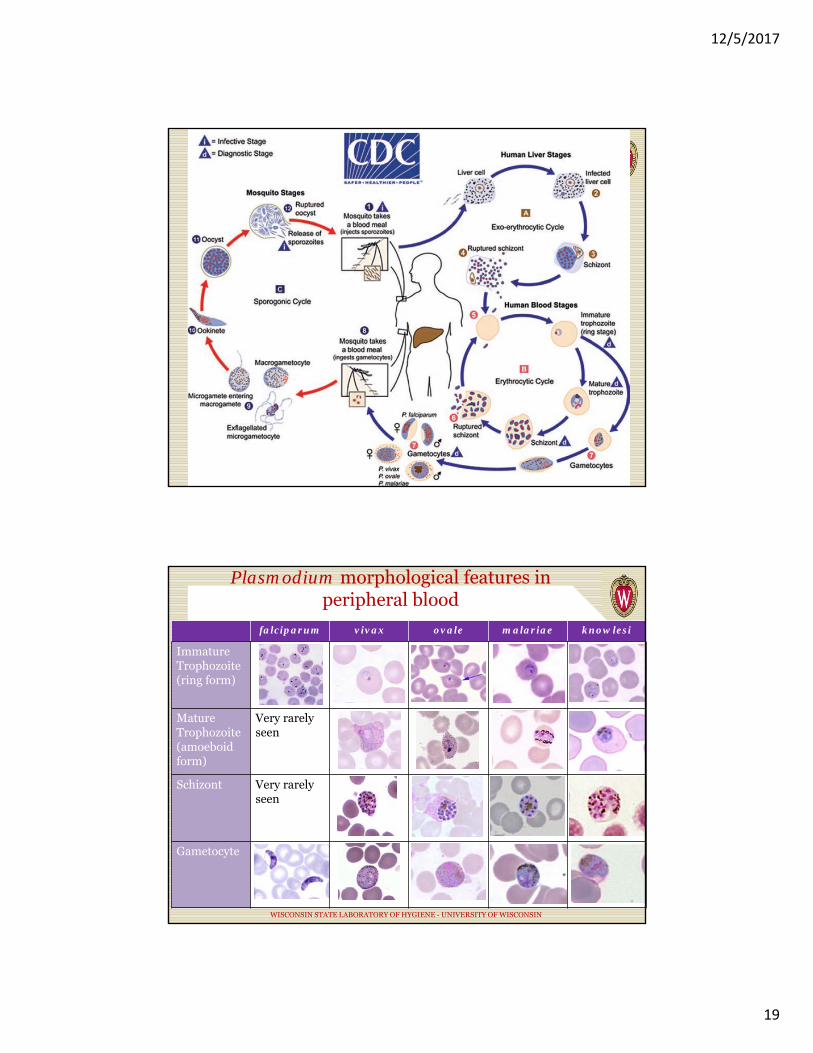
Plasmodium morphological features in peripheral blood
WISCONSIN STATE LABORATORY OF HYGIENE - UNIVERSITY OF WISCONSIN
falciparum vivax ovale malariae knowlesi
Immature Trophozoite (ring form)
Mature Trophozoite (amoeboid form)
Very rarely seen
Schizont Very rarely seen
Gametocyte

12/5/2017
20
Plasmodium morphological features in peripheral blood
WISCONSIN STATE LABORATORY OF HYGIENE - UNIVERSITY OF WISCONSIN
falciparum vivax ovale malariae knowlesi
Immature Trophozoite (ring form)
Small, delicate, 1-2 chromatin dots;occasional appliqué forms; normal RBC; occasional Maurer’s clefts
Large cytoplasm, large chromatin dot; occasional Schuffner’s dots; normal to large RBC
Average ring; normal to large RBC; occasional Schuffner’s dots
Average ring; normal RBC; rarely, Ziemann's stippling
Delicate cytoplasm; 1 to 2 chromatin dots; occasional appliqué forms; normal RBC; rarely, Sinton and Mulligan's stippling
Mature Trophozoite (amoeboid form)
Very rarely seen; compact cytoplasm; dark pigment; normal RBC
Large amoeboid; yellowish-brown pigment; Schuffner’s dots; fine, large RBC
Compact; dark-brown pigment; Schuffner’s dots; normal to large RBC
Occasional band forms; dark-brown pigment; normal RBC; rarely, Ziemann's stippling
Occasional band forms; coarse, dark-brown pigment; rarely, normal RBC; Sinton and Mulligan's stippling
Schizont Very rarely seen; 8 to 24 small merozoites; dark pigment, clumped in one mass; normal RBC
12 to 24 merozoites; yellowish-brown pigment; Schuffner’s dots; large RBC
4 to 14 merozoites with large nuclei, clustered around dark-brown pigment; Schuffner’s dots; normal to large RBC
6 to 12 merozoites with large nuclei, clustered around coarse, dark-brown pigment; rarely, normal RBC; Ziemann's stippling
Up to 16 merozoites with large nuclei, clustered around coarse, dark-brown pigment; normal RBC; rarely, Sinton and Mulligan's stippling
Gametocyte Rare; bananashaped; dark pigment
Large and round; brown pigment; Schuffner’s dots; large RBC
Round to oval; brown pigment; Schuffner’s dots; normal to large RBC
Round to oval; rarely, Ziemann's stippling
Round to oval; rarely, Sinton and Mulligan's stippling
Babesia vs Malaria
WISCONSIN STATE LABORATORY OF HYGIENE - UNIVERSITY OF WISCONSIN
Babesia Malaria
Tear drop shaped ring forms
Round signet ring forms
Size variabilityWhite central vacuole
Fewer parasites per red cell
Higher parasitemias Schuffner’s dots
Extracellular forms Advanced forms
Maltese cross Hemazoin pigment

12/5/2017
21
When You Find a Positive All positive Malaria patients should be
reported to WSLH and WDHS.
Please send a thick and 2 thin smears along with EDTA blood to WSLH
Confirmatory testing is fee exempt and provides percent parasitemia, species identification, and for new patients, molecular confirmation of ID.
WSLH is also able to send blood on to the CDC if drug susceptibility testing is needed.
WISCONSIN STATE LABORATORY OF HYGIENE - UNIVERSITY OF WISCONSIN
Molecular Detection of Malaria WSLH Protocol EDTA blood
Extract genomic DNA
Real-Time PCR
Targets the 18S rRNA gene
Melt-curve analysis
Able to detect mixed infections
New probe based PCR currently in development- greater specificity
Any sample that is smear positive but PCR indeterminate will be sent to the CDC
Mangold et al . Real-Time PCR for Detection and Identification of Plasmodium spp. Journal of clinical Microbiology, Vol. 43, No. 5, May 2005, p. 2435-2440,
12/5/2017
22
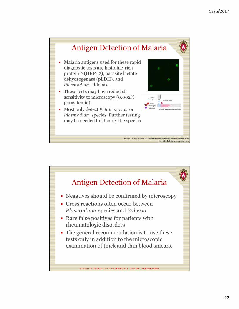
Antigen Detection of Malaria
Malaria antigens used for these rapid diagnostic tests are histidine-rich protein 2 (HRP- 2), parasite lactate dehydrogenase (pLDH), and Plasmodium aldolase
These tests may have reduced sensitivity to microscopy (0.002% parasitemia)
Most only detect P. falciparum or Plasmodium species. Further testing may be needed to identify the species
Sulzer AJ, and Wilson M. The fluorescent antibody test for malaria. CritRev Clin Lab Sci 1971;2:601-609.
Antigen Detection of Malaria
Negatives should be confirmed by microscopy
Cross reactions often occur between Plasmodium species and Babesia
Rare false positives for patients with rheumatologic disorders
The general recommendation is to use these tests only in addition to the microscopic examination of thick and thin blood smears.
WISCONSIN STATE LABORATORY OF HYGIENE - UNIVERSITY OF WISCONSIN
12/5/2017
23
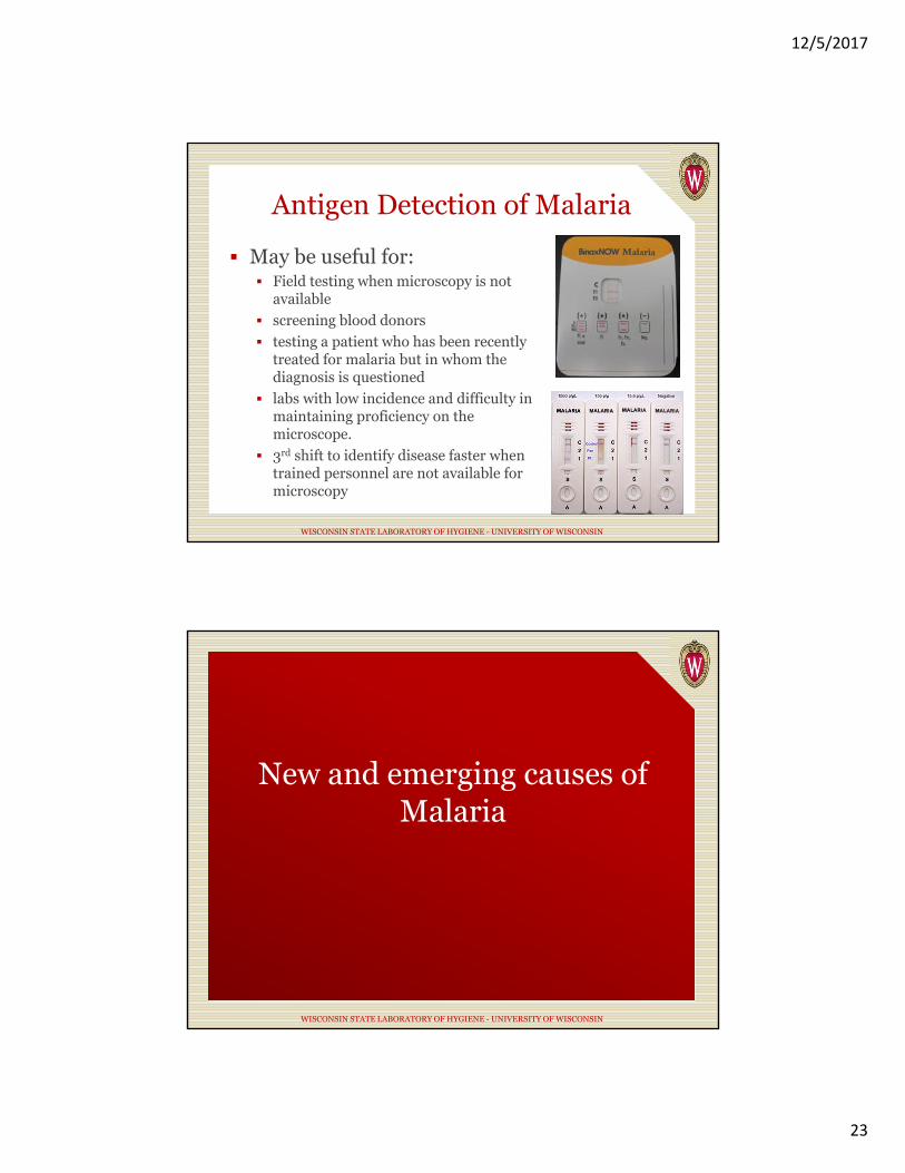
Antigen Detection of Malaria
May be useful for: Field testing when microscopy is not
available
screening blood donors
testing a patient who has been recently treated for malaria but in whom the diagnosis is questioned
labs with low incidence and difficulty in maintaining proficiency on the microscope.
3rd shift to identify disease faster when trained personnel are not available for microscopy
WISCONSIN STATE LABORATORY OF HYGIENE - UNIVERSITY OF WISCONSIN
New and emerging causes of Malaria
WISCONSIN STATE LABORATORY OF HYGIENE - UNIVERSITY OF WISCONSIN

12/5/2017
24
P. knowlesi
Geographically confined to Southeast Asia
Predominantly infects macaque monkeys
First naturally acquired human case 1965
Reporting has greatly increased since 2004
Cause of up to 70% of malaria cases in some areas
WISCONSIN STATE LABORATORY OF HYGIENE - UNIVERSITY OF WISCONSIN
Other Simian Plasmodium species and their look alikes
MMWR. March 13, 2009 / 58(09);229-232
12/5/2017
25
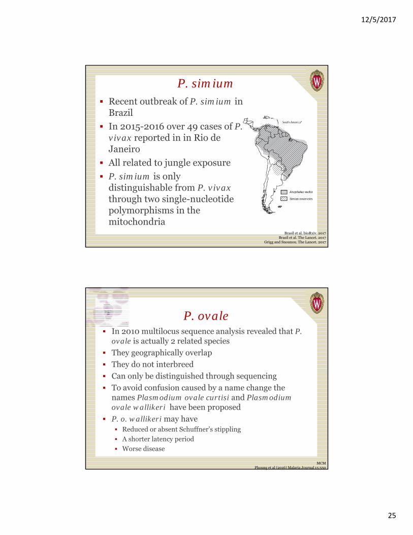
P. simium Recent outbreak of P. simium in
Brazil
In 2015-2016 over 49 cases of P. vivax reported in in Rio de Janeiro
All related to jungle exposure
P. simium is only distinguishable from P. vivax through two single-nucleotide polymorphisms in the mitochondria
Brasil et al. bioRxiv. 2017Brasil et al. The Lancet. 2017
Grigg and Snounou. The Lancet. 2017
P. ovale In 2010 multilocus sequence analysis revealed that P.
ovale is actually 2 related species
They geographically overlap
They do not interbreed
Can only be distinguished through sequencing
To avoid confusion caused by a name change the names Plasmodium ovale curtisi and Plasmodium ovale wallikeri have been proposed
P. o. wallikeri may have Reduced or absent Schuffner’s stippling
A shorter latency period
Worse disease
MCM Phoung et al (2016) Malaria Journal 15:550

12/5/2017
26
Conclusions
Malaria is still a prevalent and dangerous pathogen.
Lab diagnosis can be challenging and requires well trained staff
New technologies are developing to reduce subjectivity and enhance sensitivity.
Simian Plasmodium species are able to cause disease in humans and may emerge as major causes of Malaria
WISCONSIN STATE LABORATORY OF HYGIENE - UNIVERSITY OF WISCONSIN
References
CLSI M15-A
Manual of Clinical Microbiology v11
CDC DPDx
Phoung et al (2016) Malaria Journal 15:550
MMWR 58(09);229-232 CDC
Brasil et al. bioRxiv. 2017
Brasil et al. The Lancet. 2017
Grigg and Snounou. The lancet. 2017
Andrew P Norgan, Heather E Arguello, Lynne M Sloan, Emily C Fernholz and Bobbi S Pritt. A method for reducing the sloughing of thick blood films for malaria diagnosis. Malaria Journal 2013 12:231
Mangold et al . Real-Time PCR for Detection and Identification of Plasmodium spp. Journal of clinical Microbiology, Vol. 43, No. 5, May 2005, p. 2435-2440
Sulzer AJ, and Wilson M. The fluorescent antibody test for malaria. Crit Rev ClinLab Sci 1971;2:601-609.
WISCONSIN STATE LABORATORY OF HYGIENE - UNIVERSITY OF WISCONSIN