malignant hepatic tumors in pediatric patients: … · university hospital “policlinico-vittorio...
TRANSCRIPT
INTRODUCTION
Liver tumors account for roughly 1% of all pedi-atric neoplasms and, in two-thirds of cases, they aremalignant1,2. Among primary hepatic neoplasms inchildren, epithelial (hepatocyte-derived) tumors aremore common than the mesenchymal ones3. Clinical presentation is relatively uniform with
abdominal enlargement; unfortunately, specific
symptoms develop late. A differential diagnosis ofliver tumors in children can be obtained consider-ing some factors, such as the age of the child, clin-ical information, laboratory tests (especially AFP)and imaging findings.Imaging plays a fundamental role in characteriz-
ing lesions, staging and evaluating the adequatetreatment and the outcome. Although the radiologi-cal findings of some tumors can overlap, knowledge
1
MALIGNANT HEPATIC TUMORS IN PEDIATRIC PATIENTS: CLINICAL AND RADIOLOGICAL FEATURES
M.L. GIUNTA1, F. ROCCASALVA1, G. ATTINÀ1, G. CAPPELLO1, V. DI BENEDETTO2, M.G. SCUDERI2, G. BELFIORE1, A.G. MUSUMECI1, P.V. FOTI1, A. DI CATALDO3, G. PETRILLO1, L. SALVATORELLI4, S. PALMUCCI1
1Radiodiagnostic and Radiotherapy Unit, University Hospital “Policlinico-Vittorio Emanuele”, Catania, Italy.2Department of Pediatric Surgery, University Hospital “Policlinico-Vittorio Emanuele”, Catania, Italy.3Pediatric Hematology and Oncology Unit, University Hospital “Policlinico-Vittorio Emanuele”, Catania, Italy.4Department of Medical and Surgical Sciences and Advanced Technologies, G.F. Ingrassia, University Hospital “Policlinico-Vittorio Emanuele”, Anatomic Pathology Section, University of Catania, Catania, Italy.
Abstract: Hepatic tumors represent only 1% of all pediatric neoplasms; unfortunately, in two-thirds of cases, they are malignant. Clinical presentation is often non-specific, causing late diagnosis.
Imaging plays a fundamental role in characterizing lesions, staging and evaluating the adequatetreatment and the outcome. For staging disease, The Pretreatment Extent of Disease (PRETEXT) sys-tem could be assessed using Computed Tomography (CT) or Magnetic Resonance (MR).
Main malignant liver tumors include: hepatoblastoma, hepatocellular carcinoma, fibrolamellarcarcinoma, undifferentiated embryonal sarcoma, rabdomiosarcoma, epitheliod hemangioendothe-lioma, hepatic lymphoma, angiosarcoma, liver metastases.
Clinical and radiological features of these neoplasms are briefly reported in this pictorial review,emphasizing morphological signs observed with ultrasonography, computed tomography andmagnetic resonance. However, the diagnosis could not be easy, because the majority of tumors isvery often heterogeneous in appearance. A multidisciplinary approach is recommended, in order tomake a correct diagnosis and help clinicians in management of patients.
Keywords: Hepatic neoplasms, Imaging, Clinical and radiological features.
Corresponding Author: Stefano Palmucci, MD; e-mail: [email protected]
WCRJ 2015; 2 (1): e481

2
dated in June 2005, is used for staging and riskstratification of liver tumors5,9-11. The system was first developed for hepatoblas-
toma, and then applied to all pediatric malignantliver tumors. Using Couinaud’s system of seg-mentation of the liver, PRETEXT number is cal-culated subtracting the highest number ofcontiguous liver segments that are not involved bytumor from four5. PRETEXT system is summa-rized in tables 1 and 2.
HEPATOBLASTOMA
Clinical features
Hepatoblastoma is the most common tumor amongprimary hepatic malignancy in childhood12. Riskfactors include preterm birth, low-birth-weight,Beckwith-Wiedemann syndrome and Gardner syn-drome3,12,13.Analysing the epidemiology of the tumor in
different regions, hepatoblastoma has been report-ed as the most common primary liver tumor inWestern countries, whereas HCC is the most com-mon malignant hepatic lesion encountered inEastern regions14.In 90% of cases it is seen in patients younger
than 5 years of age, and a predominance in maleof 1,7:1 is described3,14. This tumor originatesfrom embryonic and fetal hepatocytes mixed withmesenchymal elements15.The most frequent clinical finding is an ab-
dominal enlargement associated with increasedserum alpha-fetoprotein level in 90-95% of pa-tients12-14. In some cases, the level of human chori-onic gonadotropin can be also elevated withprecocious puberty15,16. Hepatoblastoma cancause thrombosis of portal and hepatic venousstructures and metastasis to regional lymph-nodes, lungs, bone and brain12. According toAgarwala, hepatoblastoma is a solitary mass in80% of cases, but in 20% of patients it is multifo-cal with lung metastasis14.
Imaging features
Imaging findings usually are variable in relationwith the histologic type: epithelial (60% of cases),mixed (epithelial and mesenchymal, about 30%),and anaplastic type (10% of cases). The mostcommon type is the epithelial one. Mixed andanaplastic types are characterized by presence ofosteoid, cartilaginous or fibrous elements, andhaemorrhagic and necrotic components, giving aheterogeneous appearing15,17. Recently, hepato-
of the specific US, CT and MR imaging features ofeach malign liver tumor is helpful to assess the cor-rect diagnosis. In addition, in pediatric radiology therisk of radiation exposure and sedation must becarefully evaluated, and an integrated approach us-ing different modalities is often mandatory.Ultrasonography (US) represents the first-lev-
el imaging modality in a child with suspected ab-dominal mass because of its lack of ionizingradiation and no need for sedation. Of course thefinding of a hepatic tumor is an indication for fur-ther imaging evaluation3,4.Multidetector Computed Tomography (MD-
CT) is an important technique for the preoperativeplanning of treatment for patients with malignantneoplasms of the liver. The standard CT approachtends to use relatively low-dose techniques and toavoid unenhanced and multiphase images5,6.MR provides multiplanar imaging and very high
contrast resolution on images, without any ionizingradiation exposure. On the other hand, it requireslong image acquisition time and patient collaboration(breath-hold, etc); breath-hold acquisition may not bepossible in the case of sedated patients, and dedicatedsoftware (navigator, respiratory-triggered acquisi-tion) is needed in order to maintain high quality ofimages7. According to Kolbe et al8, the use of hepato-cyte-specific contrast agents can be useful to charac-terize liver lesions in pediatric patients.Aim of this pictorial review is to describe the
main clinical and radiological features of a malig-nant hepatic tumor. For each lesion, after a briefsummary of clinical features, we provide radio-logical descriptions in order to increase knowl-edge of imaging features of these malignanthepatic diseases.
PRETEXT SYSTEM
The Pretreatment Extent of Disease (PRETEXT)system, designed by the International ChildhoodLiver Tumor Strategy Group (SIOPEL) and up-
PRETEXT number Definition
I One section is involved and three adjoining sections are free
II One or two sections are involved, but two adjoining sections are free
III Two or three sections are involved, and no two adjoining sections are free
IV All four sections are involved
Table 1. The Pretreatment Extent of Disease (PRETEXT)system5, 9-11, for staging pediatric liver malignancies.

blastoma is classified into epithelial type - whichincludes fetal (Figure 1), embryonal, macrotra-becular and anaplastic subtypes - and mixed ep-ithelial/mesenchymal subtypes.US: echogenicity and echotexture are very
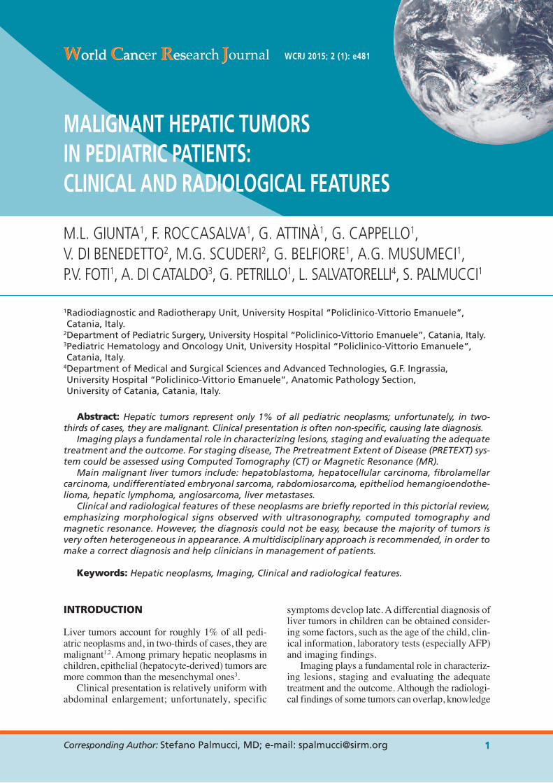
variable (Figures 2 to 4). Epithelial type tumor ap-pears as a well delineated, multilobulated, and sep-
tated hypoechoic mass15. Mixed hepatoblastoma isheterogeneous with possible hypoechoic fibroticsepta, echogenic shadowing calcifications andanechoic foci of haemorrhage or necrosis withinthe tumor3. In a previous work by de Campo et al18,heterogeneity of tumor has been demonstrated in astudy population including 8 hepatoblastomas18.
MALIGNANT HEPATIC TUMORS IN PEDIATRIC PATIENTS
3
Caudate Lobe C1 Tumor involving the caudate lobe All C1 patients are at least PRETEXT IIInvolvement (C) C0 All other patientsExtrahepatic abdominal E0 No evidence of tumor spread in abdomen Add suffix “a” if ascites is presentdisease (E) E1 Direct extension into adjacent organs
or diaphragmE2 Peritoneal nodules
Tumor focality (F) F0 Solitary tumorF1 Two or more tumors
Tumor rupture or H0 No intraperitoneal haemorrhageintraperitoneal H1 Imaging and clinical findingshaemorrhage (H) of intraperitoneal haemorrhageDistant metastases (M) M0 No metastases Add suffix or suffixes to indicate
M1 Any metastases locationLymph node N0 No nodal metastasesmetastases (N) N1 Abdominal nodal metastases
N2 Extra-abdominal nodal metastasis (withor without abdominal nodal metastasis)
Portal vein P0 No involvement Add suffix “a” if intravascular tumor involvement (P) P1 Involvement of either the left or right is present
branch of the portal veinP2 Involvement of the main portal vein
Involvement ofthe IVC and/or hepatic veins (V) V0 No involvement Add suffix “a” if intravascular tumor
V1 Involvement of the hepatic veins is presentV2 Involvement of two hepatic veinsV3 Involvement of all three hepatic veins
and/or the IVC
Table 2. The Pretreatment Extent of Disease (PRETEXT) system5, 9-11. Additional elements for staging malignancies, including: caudate lobe involvement, extrahepatic abdominal disease, tumor focality, tumor rupture of intraperitoneal heamorrhage, distant metastases, lymph node metastases, portal vein involvement, involvement of IVC (Inferior Vena Cava) and/or hepatic veins.
Figure 1. Epatoblastoma, fe-tal-type. A, Nests of neoplas-tic epatocytes are set in afibrous stroma (haematoxylinand eosin, original magnifi-cation, x80); B, At highermagnification, cytologicaldetails of neoplastic cells canbe better appreciated (haema-toxylin and eosin, originalmagnification, x200).
strates lower enhancement than normal liverparenchyma22. Fibrotic septa appear hypointenseon both T1-weighted and T2-weighted acquisitionsand enhanced after administration of gadolini-um3,15. In the epatobiliary phase – acquired 20 min-utes after the injection of hepatospecific agents(Gd-EOB-DTPA) – the lesion appears hypointenseand sharply demarcated from the liver parenchy-ma23. According to a review published by Meyers,this sharp demarcation is very useful to evaluate therelationship between the tumor and the adjacentstructures such as vessels and biliary tree22.
HEPATOCELLULAR CARCINOMA
Clinical features
HCC is the second most common tumor amongprimary hepatic malignancy in childhood6.
4
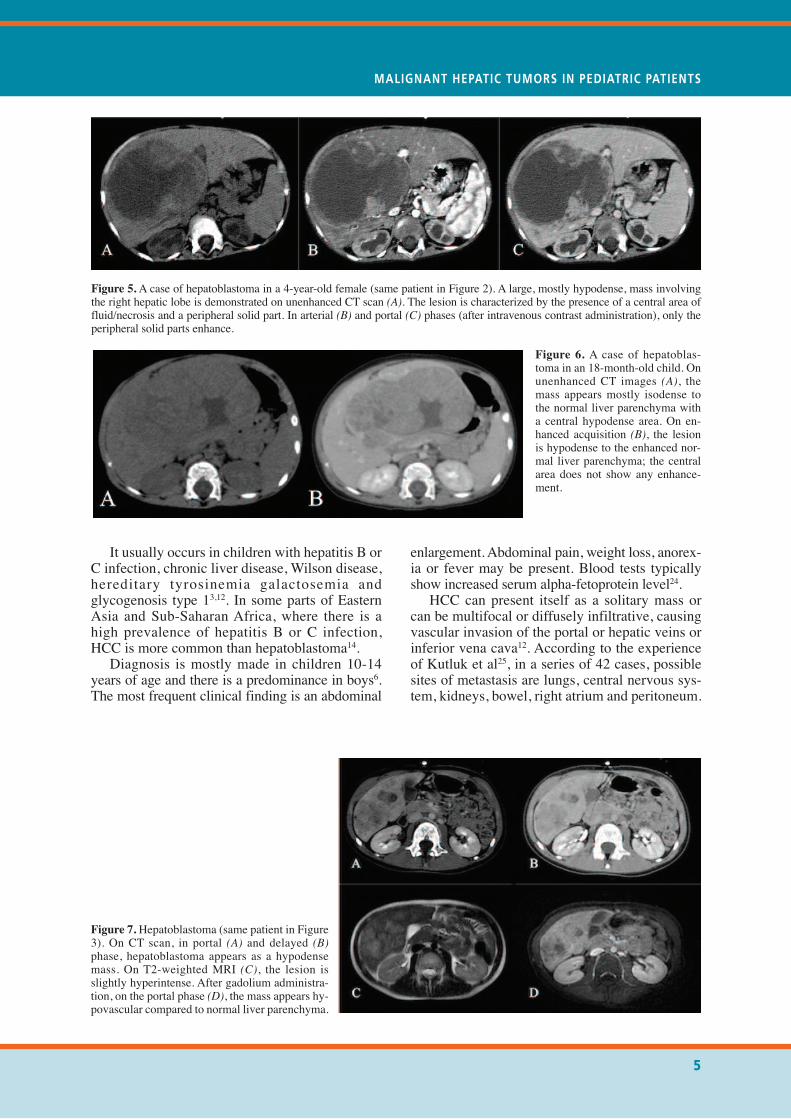
Cystic appearance of hepatoblastoma has been de-scribed by Miller19. In this case, a differential diag-nosis is needed from simple hepatic cysts:generally, the presence of internal septations ishelpful for assessing the correct diagnosis19.CT: hepatoblastoma is mostly hypodense on
unenhanced and contrast-enhanced images, al-though it is possible to find areas of enhancement,especially along the periphery and septations(Figures 5 and 6). CT scan is also the best imagingmodality to detect chunky calcifications or os-seous foci within the tumor12. In a series of 50 cas-es of hepatoblastoma retrospectively analysed, theCT pattern of calcifications and lobulations withseptations has been considered a diagnostic mark-er to make a correct differential diagnosis, namelybetween hepatoblastoma and other liver neoplas-tic masses20. MRI: hepatoblastoma is predominantly hy-
pointense on T1-weighted images and hyperintenseon T2-weighted sequences (Figure 7)21. MRI signalis homogeneous in the epithelial type and heteroge-neous in the mixed and anaplastic pattern. After ad-ministration of contrast medium, in arterious,portal and delayed phases, hepatoblastoma demon-
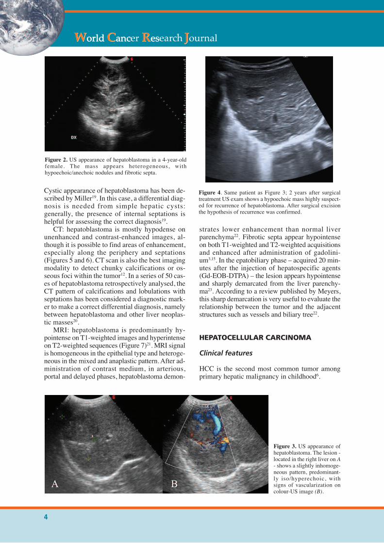
Figure 4. Same patient as Figure 3; 2 years after surgicaltreatment US exam shows a hypoechoic mass highly suspect-ed for recurrence of hepatoblastoma. After surgical excisionthe hypothesis of recurrence was confirmed.
Figure 2. US appearance of hepatoblastoma in a 4-year-oldfemale. The mass appears heterogeneous, withhypoechoic/anechoic nodules and fibrotic septa.
Figure 3. US appearance ofhepatoblastoma. The lesion -located in the right liver on A- shows a slightly inhomoge-neous pattern, predominant-ly iso/hyperechoic, withsigns of vascularization oncolour-US image (B).
It usually occurs in children with hepatitis B orC infection, chronic liver disease, Wilson disease,hereditary tyrosinemia galactosemia andglycogenosis type 13,12. In some parts of EasternAsia and Sub-Saharan Africa, where there is ahigh prevalence of hepatitis B or C infection,HCC is more common than hepatoblastoma14.Diagnosis is mostly made in children 10-14
years of age and there is a predominance in boys6.The most frequent clinical finding is an abdominal
enlargement. Abdominal pain, weight loss, anorex-ia or fever may be present. Blood tests typicallyshow increased serum alpha-fetoprotein level24.HCC can present itself as a solitary mass or
can be multifocal or diffusely infiltrative, causingvascular invasion of the portal or hepatic veins orinferior vena cava12. According to the experienceof Kutluk et al25, in a series of 42 cases, possiblesites of metastasis are lungs, central nervous sys-tem, kidneys, bowel, right atrium and peritoneum.
MALIGNANT HEPATIC TUMORS IN PEDIATRIC PATIENTS
5
Figure 5.A case of hepatoblastoma in a 4-year-old female (same patient in Figure 2). A large, mostly hypodense, mass involvingthe right hepatic lobe is demonstrated on unenhanced CT scan (A). The lesion is characterized by the presence of a central area offluid/necrosis and a peripheral solid part. In arterial (B) and portal (C) phases (after intravenous contrast administration), only theperipheral solid parts enhance.
Figure 6. A case of hepatoblas-toma in an 18-month-old child. Onunenhanced CT images (A), themass appears mostly isodense tothe normal liver parenchyma witha central hypodense area. On en-hanced acquisition (B), the lesionis hypodense to the enhanced nor-mal liver parenchyma; the centralarea does not show any enhance-ment.
Figure 7. Hepatoblastoma (same patient in Figure3). On CT scan, in portal (A) and delayed (B)phase, hepatoblastoma appears as a hypodensemass. On T2-weighted MRI (C), the lesion isslightly hyperintense. After gadolium administra-tion, on the portal phase (D), the mass appears hy-povascular compared to normal liver parenchyma.

Imaging features
Imaging features of HCC found in young patientsare similar to those seen in adults12.US: US appearance of HCC is variable. While
small HCCs are mostly hypoechoic (even thoughthey may be iso- or hyperechoic), larger tumorsappear more heterogenous because of the pres-ence within the lesion of fat, necrosis or haemor-rhage. Sometimes it is possible to see the tumorcapsule as a thin hypoechoic halo15. Doppler eval-uation shows high-velocity arterial flow6.CT: HCC usually appears as an isodense or
slightly hypodense mass on unenhanced CT. Afteradministration of contrast medium, because of itspredominant supply by the hepatic artery, the tu-mor shows arterial phase hyperenhancement, fol-lowed by rapid wash out on portal phase3,12,26.If present, the tumor capsule appears as a hy-
podense rim on unenhanced images with enhance-ment in the delayed phase3,27,28.MRI: HCC’s appearance is generally heteroge-
neous on T1-weighted images because of the pres-ence within the tumor of areas of haemorrhage,necrosis, fat and calcification. It mostly appearshyperintense on T2-weighted images15.After administration of gadolinium, the tumor
shows arterial phase hyperenhancement with rap-id portal phase wash out.If present, the tumor capsule appears hy-
pointense on both T1-weighted and T2-weightedimages with delayed enhancement after gadolini-um administration3,27.Using hepatospecific contrast agents in de-
layed post-contrast hepatobiliary phase MRI,HCC is usually hypointense8,12.
FIBROLAMELLAR CARCINOMA (FLC)
Clinical features
Fibrolamellar carcinoma (FLC) is a particularvariant of HCC observed in adolescents andyoung adult patients. It arises in patients withoutchronic liver disease and there is no associationwith serum alpha-fetoprotein level12,29,30. General-ly patients complain of abdominal pain or abdom-inal distension.Usually fibrolamellar carcinoma presents itself
as a solitary mass although in 10-15% of casessatellite lesions can be present12. Typically it contains a central scar of myxoid
tissue; in 68% of cases, there are calcificationswithin the lesion, usually located in the centralscar29.
Metastasis in the loco-regional lymph nodesand intra-peritoneal spread are possible14. Othersecondary sites of disease involvement are lungsand adrenal glands30.
Imaging features
US: FLC appears as a circumscribed mass pre-dominantly isoechoic or hyperechoic (Figure 8A),with a hyperechoic central scar possibly contain-ing shadowing echogenic calcifications3.CT: On unenhanced scan, FLC may appear
isodense or hypodense to the adjacent liver. Aftercontrast medium administration, tumor shows hy-perenhancement during the arterial phase withvariable attenuation during the portal venousphase (Figures 8B and 8C). The central scar is hy-podense to the rest of the tumor, showing en-hancement on delayed phase acquisitions30; in ourexperience, these imaging features resemble thoseof adult patients.MRI: FLC is slightly hypointense on T1-
weighted images and slightly hyperintense on T2-weighted images21. The central scar is typicallyhypointense on T2-weighted acquisitions and thatis the most important feature for the differentialdiagnosis with FNH12.After gadolinium administration, the tumor en-
hances more than adjacent liver. The central scarshows lack of enhancement during arterious andvenous phases but a partial enhancement is seenon delayed phases21.
UNDIFFERENTIATED EMBRYONAL SARCOMA (UES)
Clinical features
Undifferentiated sarcoma of the liver is the thirdcommonest malignant liver tumor in children andit represents less than 5% of all pediatric malig-nant liver tumors31,32. It was first described in1978 by Stocker and Ishak33. It is an aggressive tu-mor of mesenchymal origin mostly diagnosed inchildren 6-10 years of age with a slight male pre-dominance3; about 88% has been reported in chil-dren less than 15 years in age34. At the beginning,the prognosis was considered poor, with a mediansurvival of less than one year. However, since thelate 1980s long-term survivors were reported, dueto chemotherapy performed before surgical resec-tion34-36. The most common presenting symptomare abdominal pain, weight loss, fever and weak-ness37. Laboratory liver function tests and alpha-fetoprotein are usually normal37,38.
6

UES usually presents itself as a solitary welldemarcated large mass with a fibrous pseudocap-sule (Figure 9). Metastases can involve the lung,pleura and peritoneum. Rare complications arespontaneous liver rupture and abdominal haemor-rhage39.
Imaging features
Imaging shows a heterogeneous mass with septa,necrosis, and haemorrhagic areas37.US: UES appears as a solid isoechoic or hy-
perechoic mass (Figure 10) with small anechoicspaces corresponding to foci of necrosis, oldhaemorrhage or cystic degeneration3.CT: Abdominal CT usually reveals the pres-
ence of a large well-circumscribed mass, with hy-podense appearance (Figure 11)40,41. Unlike itssolid ultrasound appearance, on CT scan UESshows a predominantly fluid aspect with internalsepta and peripheral rim of enhancement on de-layed images after administration of contrast
medium, corresponding to the fibrous pseudocap-sule3,39,41. However, also homogeneous enhance-ment has been reported in literature42.
MALIGNANT HEPATIC TUMORS IN PEDIATRIC PATIENTS
7
Figure 8.A case of fibrolamellar HCC in a 12-year-old child. US exam (A) demonstrates a predominantly isoechoic mass, con-taining small hyperechoic central foci. The lesion shows heterogeneous enhancement on arterial phase (B); a slight wash-out isobserved on delayed phase (C) image.
Figure 9. Undifferentiated sarcoma: A, Neo-plastic proliferation is separated from the nor-mal liver by a thick fibrous pseudocapsule(haematoxylin and eosin, original magnifica-tion, x60). B, Higher magnification showinglarge atypical, spindle to stellate-shaped cellswith hyperchromatic nuclei, set in a myxoidstroma. Some cells contain in their cytoplasmnumerous eosinophilic globules of varioussizes. This latter finding is frequently encoun-tered in this tumor (haematoxylin and eosin,original magnification x 150).
Figure 10. Imaging of same patient in Figure 9. On US im-ages, the undifferentiated embryonal sarcoma appears as alarge heterogenous mass, with small anechoic and hypere-choic areas.

Discrepancy of internal architecture on US and CTcould be considered a very useful diagnostic markerto differentiate UES from other hepatic masses41.MRI: UES appears hypointense on T1-weight-
ed and hyperintense on T2-weighted images (Fig-ure 11)40; focal areas of haemorrhage appearhyperintense on T1-weighted images and hy-pointense on T2-weighted images. After gadolini-um administration, intralesional nodules couldenhance, showing hyperintense signal. The fibrot-ic pseudocapsule is hypointense on both T1-weighted and T2-weighted images3.Fluid-content, depicted both on CT and MR
images, requires a careful differentiation from hy-datic cyst and liver abscess43,44.
RHABDOMYOSARCOMA
Clinical features
Liver rhabdomyosarcoma arises mostly in males,representing 1% of all liver tumors in childhood3,14.Hystologically, it has been differentiated into
four types: pleomorphic, alveolar, embryonal andbotryoidal45. Plemorphic rabdmiosarcoma is mostfrequent in adults, whereas embryonal type hasbeen reported more commonly in infants and chil-dren45. Botriod rabdomiosarcoma is generally in-dicated also as “rabdomiosarcoma of the biliarytract”.This tumor usually shows itself as right upper
abdominal mass with symptoms related to visceralcompression, jaundice, fever, anorexia or vomiting,lethargy or malaise; however, its onset could be in-sidious, often represented only by aspecific symp-toms such as anorexia and generalized weakness46.Less frequently, more or less in 17% of patients, itpresents with spontaneous rupture of the tumor.
Alpha-fetoprotein is usually within normalrange, LDH levels are very elevated and an eleva-tion of predominantly conjugated bilirubin and al-kaline phosphatase is also possible3,14. Metastaticdisease at diagnosis is visible in 30% of patients3.
Imaging features
Radiological findings of rhabdomyosarcoma arevarious; usually, rhabdomyosarcoma of the biliarytract appears as a large hilar mass with biliary ductdilatation47-48.US: Tumor appears as a single heterogeneous
hypoechoic mass or multiple hypoechoic lesionsseparated by septa; biliary dilatation could also beappreciated.CT: The CT pattern is variable and the mass
can be hypo- or hyperdense on the unenhanced-images, sometimes with biliary dilatation48. Aftercontrast injection, the lesion can demonstrate aheterogeneous globular enhancement, but some-times there is no enhancement. MR: On MRI, the tumor usually appears as a
partially cystic lesion, hypointense on T1-weight-ed images and hyper-intense on T2-weighted ac-quisitions, simulating a choledochal cyst3.
EPITHELIOD HEMANGIOENDOTHELIOMA (EHE)
Clinical features
Epithelioid hemangioendothelioma (EHE) is arare neoplasm of vascular origin with intermedi-ate malignant potential49. It most commonly af-fects females and it has an incidence of <0.1 per100,000 population12,50. EHE grows slowly with asurvival of 10 years in about 65% of patients51.
8
Figure 11. A case of undifferentiated embryonalsarcoma in a 3-year-old female (same patient inFigures 9-10). Unenhanced CT (A) shows a largewell-circumscribed mass in the left hepatic lobe,with a predominantly fluid aspect. After contrastinjection, on arterial phase (B) and delayed phase(C) images, the internal septa and the solid fi-brotic parts demonstrate progressive enhance-ment, with a heterogeneous pattern. OnT2-weighted-image (D), the lesion appears hy-perintense with hypointense fibrotic pseudocap-sule and internal septa.
Frequently this pathological entity is inciden-tally discovered because of its lack of symptoms3.It can present with one or more liver lesions
(nodular subtype), which are at the beginning lo-cated peripherally and can be associated with cap-sular retraction due to fibrotic reaction.Progressively lesions enlarge and coalesce toform confluent masses (diffuse subtype)12.Metastases can involve lung, bone, lymph
nodes, spleen and peritoneum3.
Imaging features
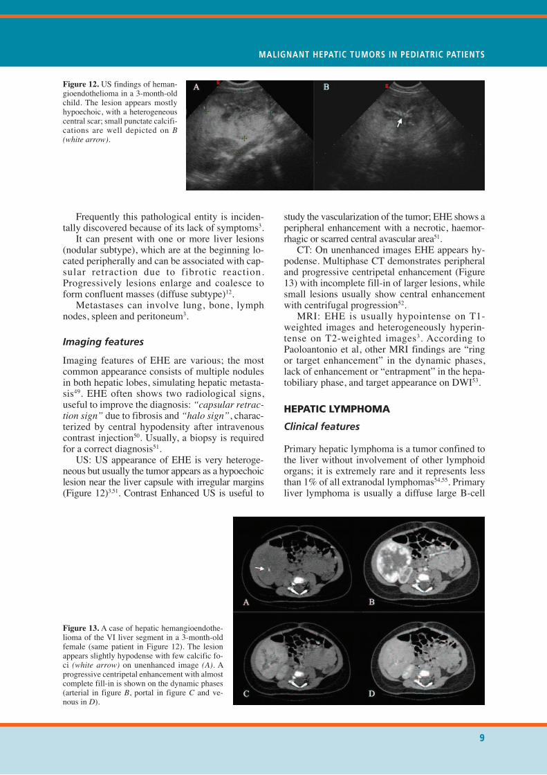
Imaging features of EHE are various; the mostcommon appearance consists of multiple nodulesin both hepatic lobes, simulating hepatic metasta-sis49. EHE often shows two radiological signs,useful to improve the diagnosis: “capsular retrac-tion sign” due to fibrosis and “halo sign”, charac-terized by central hypodensity after intravenouscontrast injection50. Usually, a biopsy is requiredfor a correct diagnosis51.US: US appearance of EHE is very heteroge-
neous but usually the tumor appears as a hypoechoiclesion near the liver capsule with irregular margins(Figure 12)3,51. Contrast Enhanced US is useful to
study the vascularization of the tumor; EHE shows aperipheral enhancement with a necrotic, haemor-rhagic or scarred central avascular area51.CT: On unenhanced images EHE appears hy-
podense. Multiphase CT demonstrates peripheraland progressive centripetal enhancement (Figure13) with incomplete fill-in of larger lesions, whilesmall lesions usually show central enhancementwith centrifugal progression52.MRI: EHE is usually hypointense on T1-
weighted images and heterogeneously hyperin-tense on T2-weighted images3. According toPaoloantonio et al, other MRI findings are “ringor target enhancement” in the dynamic phases,lack of enhancement or “entrapment” in the hepa-tobiliary phase, and target appearance on DWI53.
HEPATIC LYMPHOMA
Clinical features
Primary hepatic lymphoma is a tumor confined tothe liver without involvement of other lymphoidorgans; it is extremely rare and it represents lessthan 1% of all extranodal lymphomas54,55. Primaryliver lymphoma is usually a diffuse large B-cell
MALIGNANT HEPATIC TUMORS IN PEDIATRIC PATIENTS
9
Figure 12. US findings of heman-gioendothelioma in a 3-month-oldchild. The lesion appears mostlyhypoechoic, with a heterogeneouscentral scar; small punctate calcifi-cations are well depicted on B(white arrow).
Figure 13. A case of hepatic hemangioendothe-lioma of the VI liver segment in a 3-month-oldfemale (same patient in Figure 12). The lesionappears slightly hypodense with few calcific fo-ci (white arrow) on unenhanced image (A). Aprogressive centripetal enhancement with almostcomplete fill-in is shown on the dynamic phases(arterial in figure B, portal in figure C and ve-nous in D).
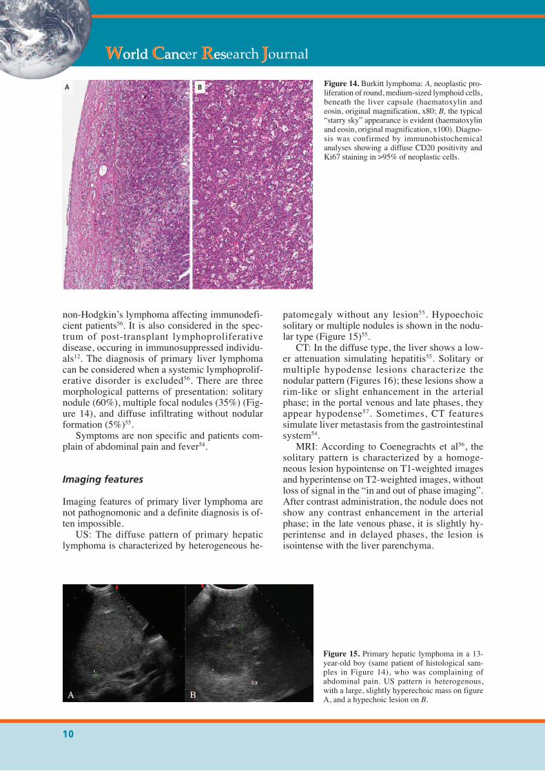
non-Hodgkin’s lymphoma affecting immunodefi-cient patients56. It is also considered in the spec-trum of post-transplant lymphoproliferativedisease, occuring in immunosuppressed individu-als12. The diagnosis of primary liver lymphomacan be considered when a systemic lymphoprolif-erative disorder is excluded56. There are threemorphological patterns of presentation: solitarynodule (60%), multiple focal nodules (35%) (Fig-ure 14), and diffuse infiltrating without nodularformation (5%)55.Symptoms are non specific and patients com-
plain of abdominal pain and fever54.
Imaging features
Imaging features of primary liver lymphoma arenot pathognomonic and a definite diagnosis is of-ten impossible.US: The diffuse pattern of primary hepatic
lymphoma is characterized by heterogeneous he-
patomegaly without any lesion55. Hypoechoicsolitary or multiple nodules is shown in the nodu-lar type (Figure 15)55.CT: In the diffuse type, the liver shows a low-
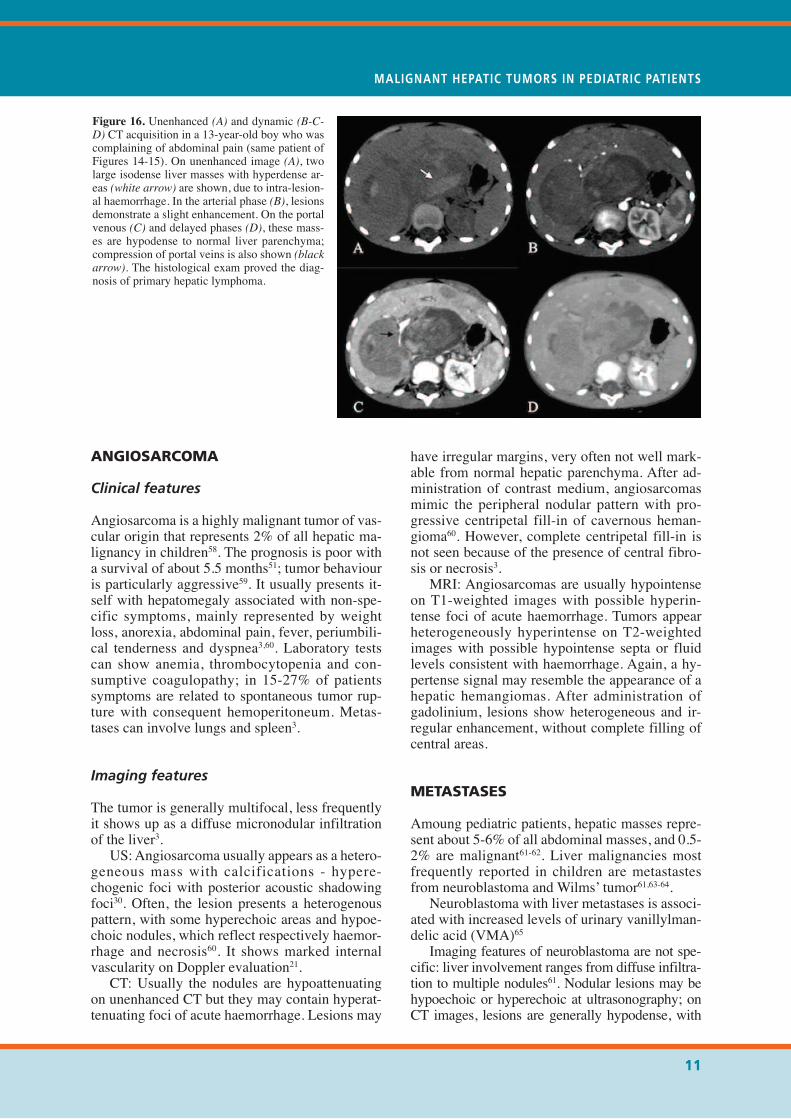
er attenuation simulating hepatitis55. Solitary ormultiple hypodense lesions characterize thenodular pattern (Figures 16); these lesions show arim-like or slight enhancement in the arterialphase; in the portal venous and late phases, theyappear hypodense57. Sometimes, CT featuressimulate liver metastasis from the gastrointestinalsystem54.MRI: According to Coenegrachts et al56, the
solitary pattern is characterized by a homoge-neous lesion hypointense on T1-weighted imagesand hyperintense on T2-weighted images, withoutloss of signal in the “in and out of phase imaging”.After contrast administration, the nodule does notshow any contrast enhancement in the arterialphase; in the late venous phase, it is slightly hy-perintense and in delayed phases, the lesion isisointense with the liver parenchyma.
10
Figure 14. Burkitt lymphoma: A, neoplastic pro-liferation of round, medium-sized lymphoid cells,beneath the liver capsule (haematoxylin andeosin, original magnification, x80; B, the typical“starry sky” appearance is evident (haematoxylinand eosin, original magnification, x100). Diagno-sis was confirmed by immunohistochemicalanalyses showing a diffuse CD20 positivity andKi67 staining in >95% of neoplastic cells.
Figure 15. Primary hepatic lymphoma in a 13-year-old boy (same patient of histological sam-ples in Figure 14), who was complaining ofabdominal pain. US pattern is heterogenous,with a large, slightly hyperechoic mass on figureA, and a hypechoic lesion on B.
ANGIOSARCOMA
Clinical features
Angiosarcoma is a highly malignant tumor of vas-cular origin that represents 2% of all hepatic ma-lignancy in children58. The prognosis is poor witha survival of about 5.5 months51; tumor behaviouris particularly aggressive59. It usually presents it-self with hepatomegaly associated with non-spe-cific symptoms, mainly represented by weightloss, anorexia, abdominal pain, fever, periumbili-cal tenderness and dyspnea3,60. Laboratory testscan show anemia, thrombocytopenia and con-sumptive coagulopathy; in 15-27% of patientssymptoms are related to spontaneous tumor rup-ture with consequent hemoperitoneum. Metas-tases can involve lungs and spleen3.
Imaging features
The tumor is generally multifocal, less frequentlyit shows up as a diffuse micronodular infiltrationof the liver3.US: Angiosarcoma usually appears as a hetero-
geneous mass with calcifications - hypere-chogenic foci with posterior acoustic shadowingfoci30. Often, the lesion presents a heterogenouspattern, with some hyperechoic areas and hypoe-choic nodules, which reflect respectively haemor-rhage and necrosis60. It shows marked internalvascularity on Doppler evaluation21.CT: Usually the nodules are hypoattenuating
on unenhanced CT but they may contain hyperat-tenuating foci of acute haemorrhage. Lesions may
have irregular margins, very often not well mark-able from normal hepatic parenchyma. After ad-ministration of contrast medium, angiosarcomasmimic the peripheral nodular pattern with pro-gressive centripetal fill-in of cavernous heman-gioma60. However, complete centripetal fill-in isnot seen because of the presence of central fibro-sis or necrosis3.MRI: Angiosarcomas are usually hypointense
on T1-weighted images with possible hyperin-tense foci of acute haemorrhage. Tumors appearheterogeneously hyperintense on T2-weightedimages with possible hypointense septa or fluidlevels consistent with haemorrhage. Again, a hy-pertense signal may resemble the appearance of ahepatic hemangiomas. After administration ofgadolinium, lesions show heterogeneous and ir-regular enhancement, without complete filling ofcentral areas.
METASTASES
Amoung pediatric patients, hepatic masses repre-sent about 5-6% of all abdominal masses, and 0.5-2% are malignant61-62. Liver malignancies mostfrequently reported in children are metastastesfrom neuroblastoma and Wilms’ tumor61,63-64.Neuroblastoma with liver metastases is associ-
ated with increased levels of urinary vanillylman-delic acid (VMA)65Imaging features of neuroblastoma are not spe-
cific: liver involvement ranges from diffuse infiltra-tion to multiple nodules61. Nodular lesions may behypoechoic or hyperechoic at ultrasonography; onCT images, lesions are generally hypodense, with
MALIGNANT HEPATIC TUMORS IN PEDIATRIC PATIENTS
11
Figure 16. Unenhanced (A) and dynamic (B-C-D) CT acquisition in a 13-year-old boy who wascomplaining of abdominal pain (same patient ofFigures 14-15). On unenhanced image (A), twolarge isodense liver masses with hyperdense ar-eas (white arrow) are shown, due to intra-lesion-al haemorrhage. In the arterial phase (B), lesionsdemonstrate a slight enhancement. On the portalvenous (C) and delayed phases (D), these mass-es are hypodense to normal liver parenchyma;compression of portal veins is also shown (blackarrow). The histological exam proved the diag-nosis of primary hepatic lymphoma.
lower enhancement than normal hepatic parenchy-ma61. MR imaging has been considered superior toCT in detecting hepatic metastases from neuroblas-toma66 at stage 4S, when liver parenchyma is char-acterized by diffuse metastatic infiltration.Liver secondary lesions from Wilms’ tumor
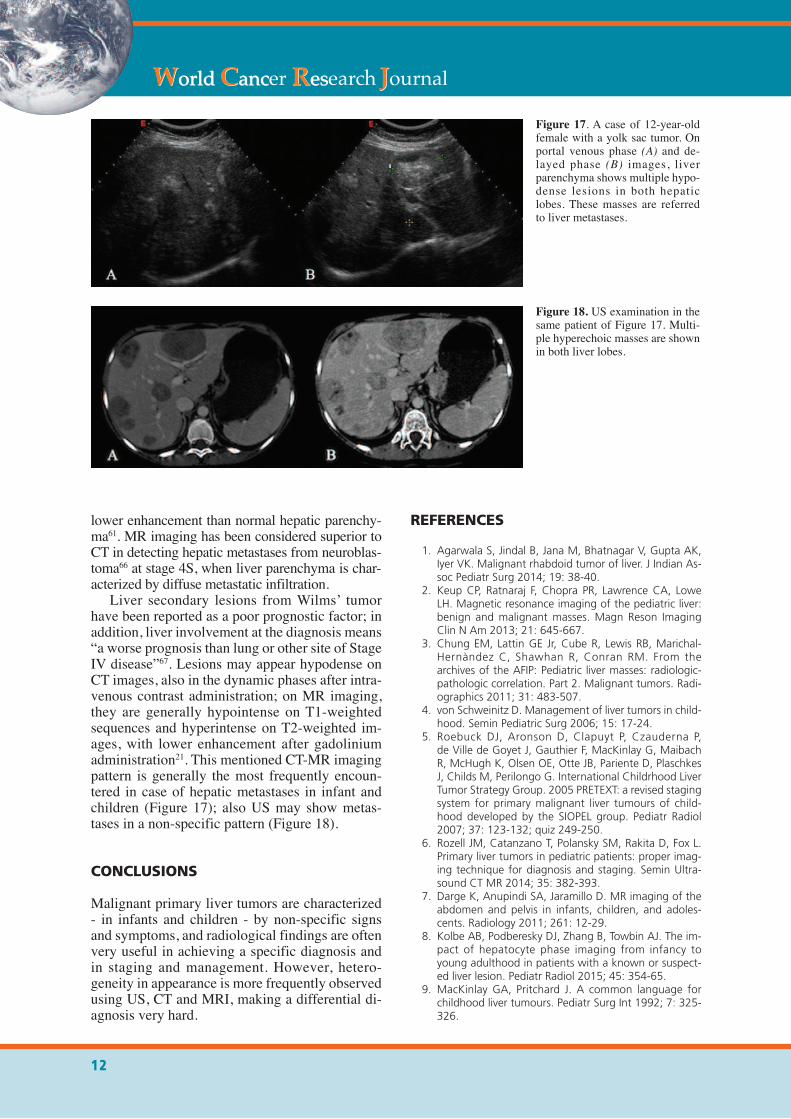
have been reported as a poor prognostic factor; inaddition, liver involvement at the diagnosis means“a worse prognosis than lung or other site of StageIV disease”67. Lesions may appear hypodense onCT images, also in the dynamic phases after intra-venous contrast administration; on MR imaging,they are generally hypointense on T1-weightedsequences and hyperintense on T2-weighted im-ages, with lower enhancement after gadoliniumadministration21. This mentioned CT-MR imagingpattern is generally the most frequently encoun-tered in case of hepatic metastases in infant andchildren (Figure 17); also US may show metas-tases in a non-specific pattern (Figure 18).
CONCLUSIONS
Malignant primary liver tumors are characterized- in infants and children - by non-specific signsand symptoms, and radiological findings are oftenvery useful in achieving a specific diagnosis andin staging and management. However, hetero-geneity in appearance is more frequently observedusing US, CT and MRI, making a differential di-agnosis very hard.
REFERENCES
1. Agarwala S, Jindal B, Jana M, Bhatnagar V, Gupta AK,Iyer VK. Malignant rhabdoid tumor of liver. J Indian As-soc Pediatr Surg 2014; 19: 38-40.
2. Keup CP, Ratnaraj F, Chopra PR, Lawrence CA, LoweLH. Magnetic resonance imaging of the pediatric liver:benign and malignant masses. Magn Reson ImagingClin N Am 2013; 21: 645-667.
3. Chung EM, Lattin GE Jr, Cube R, Lewis RB, Marichal-Hernàndez C, Shawhan R, Conran RM. From thearchives of the AFIP: Pediatric liver masses: radiologic-pathologic correlation. Part 2. Malignant tumors. Radi-ographics 2011; 31: 483-507.
4. von Schweinitz D. Management of liver tumors in child-hood. Semin Pediatric Surg 2006; 15: 17-24.
5. Roebuck DJ, Aronson D, Clapuyt P, Czauderna P,de Ville de Goyet J, Gauthier F, MacKinlay G, MaibachR, McHugh K, Olsen OE, Otte JB, Pariente D, PlaschkesJ, Childs M, Perilongo G. International Childrhood LiverTumor Strategy Group. 2005 PRETEXT: a revised stagingsystem for primary malignant liver tumours of child-hood developed by the SIOPEL group. Pediatr Radiol2007; 37: 123-132; quiz 249-250.
6. Rozell JM, Catanzano T, Polansky SM, Rakita D, Fox L.Primary liver tumors in pediatric patients: proper imag-ing technique for diagnosis and staging. Semin Ultra-sound CT MR 2014; 35: 382-393.
7. Darge K, Anupindi SA, Jaramillo D. MR imaging of theabdomen and pelvis in infants, children, and adoles-cents. Radiology 2011; 261: 12-29.
8. Kolbe AB, Podberesky DJ, Zhang B, Towbin AJ. The im-pact of hepatocyte phase imaging from infancy toyoung adulthood in patients with a known or suspect-ed liver lesion. Pediatr Radiol 2015; 45: 354-65.
9. MacKinlay GA, Pritchard J. A common language forchildhood liver tumours. Pediatr Surg Int 1992; 7: 325-326.
12
Figure 17. A case of 12-year-oldfemale with a yolk sac tumor. Onportal venous phase (A) and de-layed phase (B) images, liverparenchyma shows multiple hypo-dense lesions in both hepaticlobes. These masses are referredto liver metastases.
Figure 18. US examination in thesame patient of Figure 17. Multi-ple hyperechoic masses are shownin both liver lobes.
10. Pritchard J, Brown J, Shafford E, Perilongo G, Brock P,Dicks-Mireaux C, Keeling J, Phillips A, Vos A, PlaschkesJ. Cisplatin, doxorubicin, and delayed surgery for child-hood hepatoblastoma: a successful approach – resultsof the first prospective study of the International Socie-ty of Pediatric Oncology. J Clin Oncol 2000; 18: 3819-3828.
11. Aronson DC, Schnater JM, Staalman CR, Weverling GJ,Plaschkes J, Perilongo G, Brown J, Phillips A, Otte JB,Czauderna P, MacKinlay G, Vos A. Predictive value ofthe pretreatment extent of disease system in hepato-blastoma: results from the International Society of Pedi-atric Oncology Liver Tumor Study Group SIOPEL-1study. J Clin Oncol 2005; 23: 1245-1252.
12. Hegde SV, Dillman JR, Lopez MJ, Strouse PJ. Imaging ofmultifocal liver lesions in children and adolescents. Can-cer Imaging 2013; 12: 516-529.
13. Roebuck DJ. Assessment of malignant liver tumors inchildren. Cancer Imaging 2009; 9 Spec No A: S98-S103.
14. Agarwala S. Primary Malignant Liver Tumors in Chil-dren. Indian J Pediatr 2012; 79: 793-800.
15. Helmberger TK, Ros PR, Mergo PJ, Tomczak R, ReiserMF. Pediatric liver neoplasms: a radiologic-pathologiccorrelation. Eur Radiol 1999; 9: 1339-1347.
16. Marino S, Caruso M, Magro G, D’Amico S, La Spina M,Moscheo C, Russo G, Di Cataldo A. Hepatoblastomapresenting as precocius puberty: a case report. J PediatrEndocrinol Metab 2015; 28: 429-432.
17. Stocker JT. Hepatic tumors in children. Clin Liver Dis2001; 5: 259-281, viii-ix.
18. de Campo M, de Campo JF. Ultrasound of primary he-patic tumours in childhood. Pediatric Radiology 1988;19: 19-24.
19. Miller JH. The ultrasonographic appearance of cystichepatoblastoma. Radiology 1981; 138: 141-143.
20. Dachman AH, Pakter RL, Ros PR, Fishman EK, GoodmanZD, Lichtenstein JE. Hepatoblastoma: radiologic-patho-logic correlation in 50 cases. Radiology 1987; 164: 15-19.
21. Adeyiga AO, Lee EY, Eisenberg RL. Focal hepatic mass-es in pediatric patients. AJR Am J Roentgenol 2012;199: W422-440.
22. Meyers AB, Towbin AJ, Geller JI, Podberesky DJ. Hepa-toblastoma imaging with gadoxetate disodium-en-hanced MRI—typical, atypical, pre- and post-treatmentevaluation. Pediatr Radiol 2012; 42: 859-866.
23. Meyers AB, Towbin AJ, Serai S, Geller JI, Podberesky DJ.Characterization of pediatric liver lesions with gadoxe-tate disodium. Pediatr Radiol 2011; 41: 1183-1197.
24. Ni YH, Chang MH, Hsu HY, Hsu HC, Chen CC, ChenWJ, Lee CY. Hepatocellular carcinoma in childhood.Clinical manifestations and prognosis. Cancer 1991;68: 1737-1741.
25. Kutluk T, Yalcin B, Ekinci S, Kale G, Akyuz C, Aydin B,Varan A, Demir HA, Buyukpamukcu M. Primary liver tu-mor in children: Hacettepe experience. Turk J Pediatr2014; 56: 1-10.
26. Vu TL, Qureshi W, Turan N, Yonkers S, Stallings C,Semelka RC. Applied Radiology 2010; 39: 8-19.
27. Yu SC, Yeung DT, So NM. Imaging features of hepato-cellular carcinoma. Clin Radiol 2004; 59: 145-156.
28. Palmucci S. Focal liver lesions detection and characteri-zation: The advantages of gadoxetic acid-enhanced liv-er MRI. World J Hepatol 2014; 6: 477-485.
29. Ichikawa T, Federle MP, Grazioli L, Madariaga J,Nalesnik M, Marsh W. Fibrolamellar hepatocellular car-cinoma: imaging and pathologic findings in 31 recentcases. Radiology 1999; 213: 352-361.
30. Pedrassa BC, da Rocha EL, Kierszenbaum ML, BormannRL, Torres LR, D’Ippolito G. Uncommon hepatic tumors:iconographic essay - Part 1. Radiol Bras 2014; 47: 310-316.
31. Merli L, Mussini C, Gabor F, Branchereau S, Martelli H,Pariente D, Guérin F. Pitfalls in the Surgical Manage-ment of Undifferentiated Sarcoma of the Liver and Ben-efits of Preoperative Chemotherapy. Eur J Pediatr Surg2015; 25: 132-137.
32. Ismail H, Dembowska-Baginska B, Broniszczak D,Kalincinski P, Maruszewski P, Kluge P, Swieszkowska E,Kosciesza A, Lembas A, Perek D. Treatment of undiffer-entiated embryonal sarcoma of the liver in children—single center experience. Journal of Pediatric Surgery2013; 48: 2202-2206.
33. Stocker JT, Ishak KG. Undifferentiated (embryonal) sar-coma of the liver: report of 31 cases. Cancer 1978; 42:336-348.
34. Baron PW, Majlessipour F, Bedros AA, Zuppan CW, Ben-Youssef R, Yanni G, Ojogho ON, Concepcion W. Undif-ferentiated embryonal sarcoma of the liver successfullytreated with chemotherapy and liver resection. J Gas-trointest Surg 2007; 11: 73-75.
35. Babin-Boilletot A, Flamant F, Terrier-Lacombe MJ, Mars-den HB, van Unnik A, Deméocq F, Zucker JM, Voûte PA,Otten J, Behar C, Valayer J. Primitive malignant nonep-ithelial hepatic tumors in children. Med Pediatr Oncol1993; 21: 634-639.
36. Kadomatsu K, Nakagawara A, Zaizen Y, Nagoshi M,Tsuneyoshi M, Fukushige T, Haigou A, Suita S. Undiffer-entiated (embryonal) sarcoma of the liver: report ofthree cases. Surg Today 1992; 22: 451-455.
37. Chocarro G, Amesty MV, Hernàndez F, Chenu BG, OrtìzR, Hernàndes S, Sànches A, Gàmes M, Santamaria ML,Tovar JA. Embryonal sarcoma of the liver. Pediatr SurgInt 2013; 29: 1261-1266.
38. Iqbal K, Xian ZM, Yuan C. Undifferentiated liver sarco-ma – rare entity: a case report and review of the litera-ture. J Med Case Rep 2008; 2: 20.
39. Küpeli S, Yalçın B, Çil BE, Akçören Z, Büyükpamukçu M.Undifferentiated embryonal sarcoma of the liver in achild complicated by haemorrhage. Pediatr Radiol2008; 38: 1259-1261.
40. Crider MH, Hoggard E, Manivel JC. Undifferentiated(embryonal) sarcoma of the liver. Radiographics 2009;29: 1665-1668.
41. Moon WK, Kim WS, Kim IO, Yeon KM, Yu IK, Choi BI,Han MC. Undifferentiated embryonal sarcoma of theliver: US and CT findings. Pediatr Radiol 1994; 24: 500-503.
42. Gondu GR, Eppa VR, Sowdepalli A, Narasimhan M,Ardhanari R. A rare case of childhood undifferentiatedembryonal sarcoma of the liver managed successfully.ACG Case Rep J 2014; 1: 74-75.
43. Halefoglu AM, Oz A. Primary undifferentiated embry-onal sarcoma of the liver misdiagnosed as hydatid cystin a child: a case report and review of the literature.JBR-BTR. 2014; 97: 248-250.
44. Hanafiah M, Yahya A, Zuhdi Z, Yaacob Y. A case of anundifferentiated embryonal sarcoma of the liver mim-icking a liver abscess. Sultan Qaboos Univ Med J 2014;14: e578-581.
45. Rajamahendran R, Amudhan A, Prabhakaran R, Kan-nan D, Chandramohan SM. Embryonal rhabdomyosar-coma of liver in a 16-year-old boy: A rare case report.Int J Hepatobiliary Pancreat Dis 2014; 4: 52-56.
46. Haider N, Nadim MS, Piracha MN. Primary embryonalrhabdomyosarcoma of the liver in a young male. J CollPhysicians Surg Pak. 2013; 23: 750-751.
MALIGNANT HEPATIC TUMORS IN PEDIATRIC PATIENTS
13
47. Friedburg H, Kauffmann GW, Böhm N, Fiedler L, JobkeA. Sonographic and computed tomographic features ofembryonal rhabdomyosarcoma of the biliary tract. Pedi-atr Radiol 1984; 14: 436-438.
48. Donnelly LF, Bisset GS 3rd, Frush DP. Diagnosis please.Case 2: Embryonal rhabdomyosarcoma of the biliarytree. Radiology 1998; 208: 621-623.
49. Choi KH, Moon WS. Epithelioid hemangioendothe-lioma of the liver. Cli Mol Hepatic 2013; 19: 315-319.
50. Neofytou K, Chrysochos A, Charalambous N, Dietis M,Petridis C, Andreou C, Petrou A. Hepatic epithelioid he-mangioendothelioma and the danger of misdiagnosis:report of a case. Case Rep Oncol Med 2013; 2013:243939.
51. Schweitzer N, Soda B, Gebel M, Manns MP, Boozari B.Gray scale and contrast-enhanced ultrasound imagingof malignant liver tumors of vascular origin. United Eu-ropean Gastroenterol J 2015; 3: 63-71.
52. Chen Y, Yu RS, Qiu LL, Jiang DY, Tan YB, Fu YB. Con-trast-enhanced multiple-phase imaging features in he-patic epithelioid hemangioendothelioma. World JGastroenterol 2011; 17: 3544-3553.
53. Paolantonio P, Laghi A, Vanzulli A, Grazioli L, MoranaG, Ragozzino A, Colagrande S. MRI of hepatic epithe-lioid hemangioendothelioma (HEH). J Magn Reson Im-aging 2014; 40: 552-558.
54. Pedrassa BC, da Rocha EL, Kierzenbaum ML, BormannRL, Francisc VV, D’Ippolito G. Uncommon hepatic tu-mors: iconographic essay - Part 2. Radiol Bras 2014; 47:374-379.
55. Lu Q, Zhang H, Wang WP, Jin YJ, Ji ZB. Primary non-Hodgkin’s lymphoma of the liver: sonographic and CTfindings. Hepatobiliary Pancreat Dis Int 2015; 14: 75-81.
56. Coenegrachts K, Vanbeckevoort D, Deraedt K, VanSteenbergen W. Mri findings in primary non-Hodgkin’slymphoma of the liver. JBR-BTR 2005; 88: 17-19.
57. Maher MM, McDermott SR, Fenlon HM, Conroy D,O’Keane JC, Carney DN, Stack JP. Imaging of primarynon-Hodgkin’s lymphoma of the liver. Clin Radiol 2001;56: 295-301.
58. Fernandez-Pineda I, Cabello-Laureano R. Differential di-agnosis and management of livers in infants. World JHepatol 2014; 6: 486-495.
59. Litten JB, Tomlinson GE. Liver tumors in children. On-cologist 2008; 13: 812-820.
60. Emre S, McKenna GJ. Liver tumors in children. PediatrTransplantation 2004; 8: 632-638.
61. Rivard DC, Lowe LH. Radiological reasoning: multiplehepatic masses in an infant. AJR Am J Roentgenol2008; 190(6 Suppl): S46-52.
62. Davey MS, Cohen MD. Imaging of gastrointestinal ma-lignancy in childhood. Radiol Clin North Am 1996; 34:717-742.
63. Cohen MD, Siddiqui A. Liver metastases in Wilms’ tu-mour. Clin Radiol. 1982; 33: 539-40.
64. David R, Lamki N, Fan S, Singleton EB, Eftekhari F, Shirk-hoda A, Kumar R, Madewell JE. The many faces of neu-roblastoma. Radiographics 1989; 9: 859-82.
65. Bond JV. Neuroblastoma metastatic to the liver in in-fants. Arch Dis Child 1976; 51: 879-882.
66. Nour-Eldin NE, Abdelmonem O, Tawfik AM, NaguibNN, Klingebiel T, Rolle U, Schwabe D, Harth M, El-toukhy MM, Vogl TJ. Pediatric primary and metastaticneuroblastoma: MRI findings: pictorial review. MagnReson Imaging 2012; 30: 893-906.
67. Ehrlich PF, Ferrer FA, Ritchey ML, Anderson JR, GreenDM, Grundy PE, Dome JS, Kalapurakal JA, Perlman EJ,Shamberger RC. Hepatic metastasis at diagnosis in pa-tients with Wilms tumor is not an independent adverseprognostic factor for stage IV Wilms tumor: a reportfrom the Children’s Oncology Group/National WilmsTumor Study Group. Ann Surg 2009; 250: 642-648.
14