mammary gland growth factors: roles in normal development and in
TRANSCRIPT
Mammary Gland Growth Factors: Roles inNormal Development and in Cancer
Nancy E. Hynes1 and Christine J. Watson2
1Friedrich Miescher Institute for Biomedical Research, Maulbeerestrasse 66, CH-4058 Basel, Switzerland2Department of Pathology, University of Cambridge, Tennis Court Road, Cambridge CB2 1QP, United Kingdom
Correspondence: [email protected]
Normal development of the mammary gland proceeds via interactions between theepithelium and the mesenchyme that start during embryogenesis and continue during puber-tal outgrowth and differentiation. The function of specific peptide growth factors that bindmembers of the receptor tyrosine kinase family and the cytokine receptor family are requiredat each stage. In many cases the peptides are produced in one compartment and act on recep-tors in the other compartment. One of the striking differences between normal developmentand cancer is the loss of this cross-talk. Mammary tumor cells often produce a peptide andexpress the receptor on the same cell leading to autocrine activation of signaling pathways,a mechanism that is characteristic for cancer cells. We will discuss different peptides inthe context of normal development and cancer in this review.
Development of the mouse mammary glandbegins at embryonic day 10.5 with forma-
tion of the milk line, a thickening of the ecto-derm that extends from the anterior to theposterior limb buds. By E11.5 five placode pairsinvaginate into the surrounding stroma andundergo limited branching morphogenesis. Atbirth a rudimentary ductal tree is present in astromal fat pad. At the onset of puberty ovarianhormones induce ductal elongation until thefat pad is filled with a system of ducts and sidebranches. During pregnancy the glands fillwith secretory alveolar units that on parturitionproduce copius amounts of milk for the off-spring. As the pups are weaned, these alveoliare removed by programmed cell death cou-pled with extensive tissue remodeling, an event
termed involution. All these processes are underthe control of a wide variety of hormonal andpeptide factors. In this review, which is concen-trated on peptide growth factors, we will discussthose factors that have important roles in nor-mal development and have been implicated inbreast cancer.
RECEPTOR TYROSINE KINASES ANDPEPTIDE LIGANDS IN NORMALDEVELOPMENT AND CANCER
The superfamily of receptor tyrosine kinases(RTK) includes approximately 60 members di-vided into 19 subfamilies. Each receptor hasan extracellular ligand binding region, a singlemembrane-spanning region, and a cytoplasmic
Editors: Mina J. Bissell, Kornelia Polyak, and Jeffrey Rosen
Additional Perspectives on The Mammary Gland as an Experimental Model available at www.cshperspectives.org
Copyright # 2010 Cold Spring Harbor Laboratory Press; all rights reserved; doi: 10.1101/cshperspect.a003186
Cite this article as Cold Spring Harb Perspect Biol 2010;2:a003186
1
on February 1, 2018 - Published by Cold Spring Harbor Laboratory Press http://cshperspectives.cshlp.org/Downloaded from

tyrosine kinase-containing domain. In this re-view we will discuss the epidermal growth factorreceptor (EGFR) or ErbB family and the EGF-related ligands, the fibroblast growth factor re-ceptor (FGFR), and FGFs as well as insulin-likegrowth factors (IGFs) and their receptors, men-tioning their involvement in normal develop-ment of the mammary gland and in breast cancer.
EGF FAMILY OF GROWTH FACTORS ANDTHEIR RECEPTORS
There are four members of the ErbB RTK family:EGFR, ErbB2, ErbB3, and ErbB4. Peptides in theEGF family bind ErbB receptors and based ontheir receptor specificity are divided into threegroups: The first includes EGF, transforminggrowth factor (TGF)-a, amphiregulin (AR), andepigen (EPG), which bind EGFR; the secondincludes betacellulin (BTC), heparin-bindingEGF (HB-EGF), and epiregulin (EPR), whichbind both EGFR and ErbB4; the third group,the neuregulins (NRGs), forms two subgroupsbased on their capacity to bind ErbB3 and
ErbB4 (NRG-1 and NRG-2) or ErbB4 (NRG-3and NRG-4) (Hynes and Lane 2005) (Fig. 1).
Ligand binding initiates signaling by caus-ing specific homo- or heterodimeric receptorformation and activation of the cytoplasmickinase domain that phosphorylates tyrosinesin the tail region of each receptor. Phosphoryla-tion triggers the association of specific signalingmolecules whose binding initiates downstreamsignaling events. Of note, ErbB3 has impairedkinase activity and only acquires signaling po-tential, i.e., tyrosine phosphorylation, whendimerized with another receptor. Furthermore,ErbB2 does bind any of the EGF-family ligands,however, it is activated via heterdimerizationwith the other ligand-bound receptors (Hynesand Lane 2005) and is their preferred hetero-dimerization partner (Graus-Porta et al. 1997).
FGF FAMILY OF PEPTIDE GROWTHFACTORS AND RECEPTORS
Members of the FGF ligand family and theirreceptors form a complex network with four
EGFTGFα
AREPG
BTCHB-EGF
EPR
NRG-1NRG-2
NRG-3NRG-4
431
2
Figure 1. EGF-family peptides and ErbB Receptors. The four ErbB receptors are shown: (1) EGFR, (2) ErbB2,(3) ErbB3, and (4) ErbB4. The EGF-family peptides bind specific ErbB receptors: EGF, TGF-a,AR, and EPGbind EGFR; BTC, HB-EGF, EPR have dual specificity, binding both EGFR and ErbB4; NRG-1 and NRG-2bind ErbB3 and ErbB4; NRG-3 and NRG-4 bind ErbB4 (Hynes and Lane 2005). Various roles for specificligands (boxed in red) and all the ErbB receptors have been described in normal mammary glanddevelopment. In breast cancer, overexpressed ErbB2 together with ErbB3 function as a unit to driveproliferation of specific subsets of tumors (Holbro et al. 2003). EGFR is overexpressed in 30%–60% ofbasal-like breast cancers. See text for more information.
N.E. Hynes and C.J. Watson
2 Cite this article as Cold Spring Harb Perspect Biol 2010;2:a003186
on February 1, 2018 - Published by Cold Spring Harbor Laboratory Press http://cshperspectives.cshlp.org/Downloaded from

FGFR genes that undergo alternative splicingto yield minimally seven FGFRs. Alternativesplicing of FGFR 1–3 in the D3 region (III)of the ectodomain yields, e.g., FGFR1-IIIb andFGFR1-IIIc, that bind different FGFs and areexpressed in epithelial or mesenchmal compart-ments, respectively. The 18 FGFs are dividedinto six subfamilies; the FGF 11-14 subfamilyhas been renamed fibroblast homologous fac-tors because their core structure is similar toother FGFs, however, several key residues forreceptor binding are missing and they do notbind FGFRs (reviewed in Itoh and Ornitz2004; Beenken and Mohammadi 2009).
Each FGF ligand binds a specific FGFR(Fig. 2), which enforces receptor dimerization,kinase activation and autophosphorylation ofspecific tyrosine residues in the cytoplasmic
domain (Mohammadi et al. 1996; Lew et al.2009). In addition to the receptor, FRS2, amajor adaptor linking FGFRs to the ERK andPI3K pathways becomes phosphorylated onspecific tyrosine residues (Kouhara et al. 1997;Ong et al. 2001).
INSULIN, IGFs, AND THEIR RECEPTORS
The peptides insulin and IGF1 bind to variousmembers of the insulin receptor (IR)-IGF1receptor (IGF1R) family. The receptors are tet-rameric structures composed of half receptorseach of which has an extracellular ligand-binding a chain and a transmembrane span-ning b chain with cytoplasmic tyrosine kinaseactivity. IGF1 binds IGF1R as well as a hybridcomposed of an IR and an IGF1R. Moreover,
FGFR activationFGF subfamilies
FGF1
FGF1FGF2
FGF4
FGF4FGF5FGF6
FGF7
FGF3FGF7FGF10FGF22
FGF8
FGF8FGF17FGF18
FGF9
FGF9FGF16FGF20
FGF19 (endocrine)
FGF19FGF21FGF23
R1IIIb R1IIIc R2IIIb R2IIIc R3IIIb R3IIIc R4
++ ++++++ + + + +
+ + + ++ ++ + +
(+)
+ ++
+ ++ +
+ + + ++ + + +
+ + +
+++++++++(+)++++++
+++
+ +++
++ +
Figure 2. Members of the FGF family are divided into six subgroups of closely related peptides. Each ligandbinds to a specific subset of the FGFRs. () refers to the fact that there is still uncertainty regarding thebinding. The table summarizes results from many laboratories and some relevant publications are as follows:(Ornitz et al. 1996; Zhang et al. 2006; Kurosu et al. 2006, 2007; Suzuki et al. 2008; Schwertfeger 2009).
Peptide Factors and Cytokines in the Mammary Gland
Cite this article as Cold Spring Harb Perspect Biol 2010;2:a003186 3
on February 1, 2018 - Published by Cold Spring Harbor Laboratory Press http://cshperspectives.cshlp.org/Downloaded from

the bioavailability of IGF1 is controlled byIGF binding proteins (IGFBP) (reviewed in DeMeyts 2004). As for other RTKs, ligand bindingstimulates multiple intracellular pathways. Onespecific characteristic of the IR-IGF1R familyis their dependence on insulin responsive sub-strate (IRS) family members, in particular IRS-1/2, for intracellular signaling.
ROLES FOR EGF-FAMILY PEPTIDES, FGFs,AND IGF1 AND THEIR RECEPTORS INMAMMARY GLAND DEVELOPMENT
Embryonic Development ofthe Mammary Gland
Development of the mouse mammary glandbegins in at embryonic day E10.5 and by E11.5,five placode pairs invaginate into the surround-ing stroma and undergo branching morpho-genesis. FGF10 and its receptor FGFR2-IIIbhave important roles during embryonic mam-mary gland development. Starting at E10.5,somatic-expressed FGF10 acts on FGFR2-IIIbin the ectoderm to initiate induction and posi-tioning of the mammary line and placode for-mation. The ligand and the receptor are bothessential for induction of four of the fiveplacode pairs (Mailleux et al. 2002; Veltmaatet al. 2006). Considering upstream regulatorsof the pathway, it has been shown that FGF10levels are reduced in somites lacking the tran-scription factor GLI3, and the mammary lineand placode three fail to form in its absence(Veltmaat et al. 2006).
NRG3 was discovered as a hypomorphic allelecausing hypoplastic mammary buds in skamutant mice (Howard et al. 2005). NRG3 isexpressed in the mesenchyme adjacent to siteswhere placodes form and acts to promotemammary morphogenesis. ErbB4, the NRG3receptor, as well as the other ErbB receptors,are expressed at sites of epithelial–mesenchy-mal interactions during morphogenesis of theembryonic mammary gland (Wansbury et al.2008). Intriguingly, embryonic mammary pla-code formation appears to be normal in ErbB4null mice (Tidcombe et al. 2003). Indeed,embryos deleted for each of the four ErbB
receptors appear to form normal mammaryplacodes (Wansbury et al. 2008). This suggeststhat while NRG3 is limiting in the processof placode formation, the ability of an indi-vidual ErbB receptor to form a heterodimerwith the other family members provides suf-ficient signaling to allow embryonic placodeformation.
The IGF/IGF1R axis has an essential rolein embryonic bud development. Intriguingly, itwas observed that embryos deficient in P190-B,a member of the RhoGTPase activating (Rho-GAP) family that interacts with integrins, hadsmaller mammary buds. The defect was in bothcompartments, because the epithelial bud pro-liferation was lower and the underlying mesen-chyme was aberrant. A similar phenotype wasobserved in embryonic mammary buds lackingIRS-1/2 (Heckman et al. 2007). Taken together,the results suggest that IGF1/IGFR1 signalingthrough a RhoGAP and the IRS effector pro-teins has an essential role in the epithelialmesenchymal interactions needed for propermammary bud morphogenesis.
Ductal Outgrowth and Differentiation
The rudimentary epithelial tree in the mam-mary fat pad remains quiescent until pubertyonset when ovarian steroid hormone produc-tion commences and induces ductal outgrowth.Bulbous structures on the tips of the ducts,the terminal end buds (TEBs) are highly pro-liferative and penetrate into the fat pad as theducts elongate. TEBs bifurcate and secondarybranches form during this process, until theentire fat pad is filled with a network of bran-ched ducts. During repeated estrous cycles theductal network increases in complexity andside-branches grow under progesterone control.Under control of prolactin, alveolar structuresbud off the ductal system during pregnancy,and these differentiate into milk-producingstructures. Steroid hormones, growth hormone(GH), and prolactin are the master regulatorsof mammary growth and pregnancy-induceddifferentiation, whereas peptide factors of theEGF and FGF family, and IGF-1 have specificroles during these processes.
N.E. Hynes and C.J. Watson
4 Cite this article as Cold Spring Harb Perspect Biol 2010;2:a003186
on February 1, 2018 - Published by Cold Spring Harbor Laboratory Press http://cshperspectives.cshlp.org/Downloaded from
During ductal outgrowth multiple FGFs:FGF1, FGF2, FGF4, FGF7, and FGF10, as wellas FGFR1 and FGFR2 are expressed (reviewedin Schwertfeger 2009). TEBs have high levelsof FGFR2, which is essential at this stage be-cause FGFR2 null glands penetrate the fat padmore slowly and show fewer branch pointscompared with WT controls (Lu et al. 2008).Indeed, it has been shown that the epithelialspecific FGFR2-IIIb isoform is responsible forductal outgrowth (Parsa et al. 2008). Duringpregnancy and lactation some FGFs are ex-pressed (Coleman-Krnacik and Rosen 1994),however, to our knowledge a specific role forthe FGF/FGFR signaling network at this devel-opmental stage has not been described.
The pituitary GH is essential for pubertalmammary development (reviewed in Hoveyet al. 2002); IGF1, which is produced in themammary gland, acts as the local effector ofGH (reviewed in Wood et al. 2000; Kleinbergand Ruan 2008; Kleinberg et al. 2009). Duringpostnatal development of the gland, IGF1 isproduced in both stromal and epithelial cells(Loladze et al. 2006) and, in its absence, TEBsfail to develop correctly and the glands showlimited outgrowth potential (Ruan and Klein-berg 1999). These results, together with datashowing that embryonic IGF1R null mammarybuds transplanted into WT hosts also possesslimited outgrowth potential and show defectsin TEB proliferation (Bonnette and Hadsell2001), illustrate the importance of the IGF1axis for mammary development. The RhoGAP,p190-B mentioned previously, also modulatesthe IGF1/IGF1R axis during pubertal out-growth. In p190-B heterozygous females theductal outgrowth rate is slower and decreasedlevels of IRS-1/2 were observed in TEBs (Chak-ravarty et al. 2003). Interestingly, pregnancyhormones partially overcome the proliferationdefect of the IGF1R null epithelium, potentiallydue to increased insulin sensitivity (discussedin Bonnette and Hadsell 2001). IGF1 is alsorequired for lobuloalveolar development ofthe mammary gland (Loladze et al. 2006).
Multiple EGF-family ligands and all of theErbB receptors are expressed in the mammarygland during ductal outgrowth, pregnancy-
induced alveolar differentiation, and in lac-tation (Schroeder and Lee 1998). EGF-familyligands are produced as transmembrane precur-sors that on cleavage bind and activate ErbBreceptors. Members of the ADAM (a disintegrinand metalloproteinase) family of proteases, aswell as matrix metalloproteinases (MMP) cleaveand release EGF-related ligands (Sandersonet al. 2006; Bergers and Coussens 2000). Dur-ing pubertal outgrowth EGFR is strongly ex-pressed in TEBs and adjacent stroma (Colemanet al. 1988) and waved-2 mutants with a kinase-impaired EGFR show less outgrowth (Sebastianet al. 1998). Reciprocal transplant experimentsbetween WT epithelium and EGFR mutantstroma and the reverse, revealed that stromalEGFR is required during this phase (Wiesenet al. 1999).
The evidence pointing to epithelial-pro-duced AR as the EGFR activating ligand at thisdevelopmental stage is very strong. Expressionof AR, but none of the other EGFR ligands, isvery high during puberty (Sternlicht et al. 2005).Moreover, AR appears to be a direct target ofestradiol since upon injection of the steroidinto 3-wk-old ovariectomized mice, AR expres-sion is strongly up-regulated (Ciarloni et al.2007). Finally, AR null epithelial grafts do notgrow in WT mammary fat pads (Ciarloni et al.2007). Interestingly, ADAM17 null mice have aphenotype very similar to that of AR null miceand it was shown that in the absence of this pro-tease there is impaired cleavage and release ofAR (Sternlicht et al. 2005). Thus, AR is requiredspecifically downstream from estradiol in theepithelial compartment, where it is released byADAM17 and acts on stromal EGFR to promoteductal elongation during puberty. This suggestsa model whereby stromal cells respond to ARand produce soluble factors that act back onthe epithelium. Some interesting candidatesare expressed at this developmental stage in-cluding FGFs, IGF1, and HGF. Because absenceof the epithelial FGFR2-IIIb also impairs puber-tal outgrowth (Parsa et al. 2008), it is tempt-ing to speculate that stromal-produced FGFs,e.g., FGF7 or FGF10, are the ligands acting onthe epithelium. Indeed, it has been shownthat FGF2 acts downstream from EGFR and
Peptide Factors and Cytokines in the Mammary Gland
Cite this article as Cold Spring Harb Perspect Biol 2010;2:a003186 5
on February 1, 2018 - Published by Cold Spring Harbor Laboratory Press http://cshperspectives.cshlp.org/Downloaded from

supports growth of EGFR null mammary orga-noids in culture (Sternlicht et al. 2005). Despitethe fact that stromal ErbB2 is also active at pub-erty (Sebastian et al. 1998), analyses carried outon ErbB2 null mammary epithelium showedthat the receptor has a limiting role in the epi-thelium, not in the stroma. In the absence ofepithelial ErbB2, impaired ductal elongationand branching (Jackson-Fisher et al. 2004;Andrechek et al. 2005), as well as alterations inTEB structures were observed (Jackson-Fisheret al. 2004) during pubertal outgrowth. ErbB2requires a heterodimerization partner for li-gand-induced activation and likely candidatesfor this role are NRG1 and ErbB3. On the onehand, implantation of NRG1 slow-release pel-lets promoted ductal outgrowth (Jones et al.1996). On the other hand, transplantation ofmammary buds from ErbB3 null embryosinto recipient WT fat pads showed that in theabsence of ErbB3 pubertal outgrowth wasseverely impaired and TEBs showed increasedapoptosis (Jackson-Fisher et al. 2008).
EGFR, ErbB2, and ErbB3 each have pro-liferative and survival roles during mammaryoutgrowth. In contrast, ErbB4 has an essentialrole late in pregnancy and in lactation (reviewedin Stern 2008). Initial studies showed that dur-ing lactation ErbB4 was strongly phosphory-lated on tyrosine (Schroeder and Lee 1998),indicative of a role at this developmental stage.A direct examination of ErbB4 null mammaryepithelium showed that pubertal outgrowthwas normal, however, alveolar differentiationand lactation were impaired (Tidcombe et al.2003). Moreover, the defect was shown to bedue to impaired phosphorylation of the tran-scription factor Stat5, which has an essentialrole in mammary gland differentiation andis activated during lactation by ErbB4 (Joneset al. 1999). ErbB4 is the only member of thefamily that undergoes g-secretase cleavage,which releases the intracellular cytoplasmicdomain of the receptor, allowing its nuclearentry (Ni et al. 2001; Sardi et al. 2006). Thisis interesting in light of the finding that in lactat-ing mammary glands this domain of ErbB4 ispresent in the nucleus (Long et al. 2003). Theother three ErbB family members have also
been detected in the nucleus of cells in variousnormal tissues, although this has not beendescribed in the mammary gland (reviewed inWang and Hung 2009).
There are two candidates for the ligand acti-vating ErbB4 during late stages of pregnancyand in lactation. One is NRGa1, which whendeleted has a transient effect on alveolar devel-opment late in pregnancy and early in lactation(Li et al. 2002). In addition, there is evidencesuggesting that HB-EGF might mediate ErbB4activation during lactation. The heparin-sulfateproteoglycan CD44 has been shown to bindMMP-7 and pro-HB-EGF on the surface ofmammary epithelial cells and this binding isrequired for HB-EGF processing. A lactationdefect has been observed in CD44 null females.Moreover, neither HB-EGF processing, norErbB4 activation were observed in mammaryglands lacking the proteoglycan (Yu et al. 2002).
RTKs AND MAMMARY CANCER
A prevailing theme during the proliferativephase of mammary gland development is para-crine receptor activation by ligands produced inother cells. One of the most striking differencesbetween normal development and cancer is theability of the estrogen receptor (ER) positivecancer cells to proliferate; in the normal glandthe ER positive cells do not divide but respondby producing paracrine factors that act onother cells, AR being a good example. This char-acteristic appears to be lost in cancer, some-thing that distinguishes the normal mammarygland from breast cancer (Anderson and Clarke2004). In this section we will briefly review can-cer roles for the peptides discussed in normaldevelopment.
FGFs AND FGFRs IN CANCER
The association between FGF and mammarycancer was first established in MMTV-inducedtumors, where proviral insertional mutagenesisled to FGF3 transcriptional activation (Peterset al. 1989). A recent large-scale screen revealedthat the FGF pathway was activated in .65% ofMMTV-induced tumors. Integration sites were
N.E. Hynes and C.J. Watson
6 Cite this article as Cold Spring Harb Perspect Biol 2010;2:a003186
on February 1, 2018 - Published by Cold Spring Harbor Laboratory Press http://cshperspectives.cshlp.org/Downloaded from

adjacent to FGFR1 and FGFR2 in additionto FGF3, 4, 6, 8, and 10 (Theodorou et al.2007). Interestingly, FGF8 and FGF10, ligandsfor FGFR2 (Fig. 2), are also overexpressed inhuman breast cancer (Marsh et al. 1999; Theo-dorou et al. 2004).
FGFR1, FGFR2, and FGFR4 have each beenimplicated in breast cancer development. The8p11-12 amplicon harboring FGFR1 has beendetected in 8%–10% of breast cancers (Adnaneet al. 1991; Ray et al. 2004; Reis-Filho et al. 2006)and correlates with poor outcome in patientswith ER positive tumors (Elsheikh et al. 2007),suggestive of a role in endocrine resistance.FGFR1 is overexpressed in some, but not alltumors with the amplicon, likely reflecting thecomplexity of this amplicon. Irrespective ofthis, elevated levels of the receptor have beendetected in lobular breast carcinomas (Reis-Filho et al. 2006; Xian et al. 2009). Genome-wide screens aimed at uncovering breast cancerassociated genes identified single-nucleotidepolymorphisms (SNP) in introns of FGFR2(Easton et al. 2007; Hunter et al. 2007). Oneof the SNPs alters the binding of Oct-1/Runx2to the DNA and, using an in vitro tran-scriptional assay it was shown that this SNPincreased FGFR2 transcription (Meyer et al.2008). Primary tumors with this SNP showelevated FGFR2 levels (Nordgard et al.2007). FGFR2 amplification (10q26) has alsobeen detected in a subgroup of triple negative(negative for ER, PR, and high ErbB2) breastcancers (Turner et al. 2010). High levels ofthe FGFR4Arg388 allele have been correlatedwith poor response to chemotherapy (Thussbaset al. 2006) and endocrine therapy (Meijeret al. 2008). Introduction of this allele intoa transgenic mammary tumor model led tomore rapid tumor development and tumorprogression (Seitzer et al. 2010). Other clinicalobservations on the ligands and receptors arepresented in Figure 3.
EGF-FAMILY PEPTIDES AND ErbBRECEPTORS IN CANCER
In contrast to the FGFs that are often activatedby MMTV insertional mutagenesis (Theodorou
et al. 2007), we are not aware of any reportsshowing that EGF-family peptides or their re-ceptors are activated by the provirus. However,mammary transgenic models, most driven bythe MMTV promoter, have been made withmultiple peptides and with the EGFR andErbB2. MMTV-NRG transgenics show persis-tent TEBs and mammary cancers arise with along latency (Krane and Leder 1996). MMTV-EGFR transgenics develop hyperplasias andcancer (Brandt et al. 2000). Mammary tumorsarising from transgenic expression of TGFaand ErbB2/Neu have been extensively analyzed(reviewed in Cardiff and Wellings 1999; Mikae-lian et al. 2004). Moreover, MMTV-ErbB2/Neumice are often used to test the importance ofparticular proteins in tumor outgrowth andmetastasis formation (White et al. 2004; Lahlouet al. 2007).
A vast body of evidence suggests that theEGF-family peptides and their receptorshave an important role in human breast cancer.ERBB2 gene amplification was the first consis-tent genetic alteration found in approximately25% of primary tumors (Slamon et al. 1987;van de Vijver et al. 1987; Berger et al. 1988). Be-cause this was discovered more than 20 yr ago,numerous studies on the role of this receptorin breast cancer and on its potential as a thera-peutic target have been published (reviewedin Hynes and Lane 2005). The basal-like sub-type of breast cancer, also referred to as triplenegative due to lack of ER, PR (progesteronereceptor), and ErbB2, has a very aggressive clin-ical behavior (Nielsen et al. 2004). EGFR isexpressed at moderate to high levels in 30%–60% of basal-like breast cancer (Reis-Filhoand Tutt 2008; Thike et al. 2009). Clinical trialstesting the potential of EGFR tyrosine kinaseinhibitors are ongoing in breast cancer (seeClinicalTrials.gov).
IGF-1 AND IGF1R IN CANCER
There are numerous studies suggesting thatIGF-1 and its receptor have important rolesin breast cancer (Sachdev and Yee 2001; Pollaket al. 2004). The IGF/IGF1R axis impactson many aspects of the tumor via its ability
Peptide Factors and Cytokines in the Mammary Gland
Cite this article as Cold Spring Harb Perspect Biol 2010;2:a003186 7
on February 1, 2018 - Published by Cold Spring Harbor Laboratory Press http://cshperspectives.cshlp.org/Downloaded from
to act on the Ras/MAPK/ERK proliferativepathway and the PI3K survival pathway. In-deed, even the ratio of IGF-1 to its bindingprotein IGFBP-3 has been shown to modifybreast cancer risk in BRCA mutation carriers(Mitchell et al. 2006). It is also interesting tonote that down-regulation of IGFBP-3 leadingto up-regulation of IGF1-R signaling andPI3K pathway activation was described as amechanism underlying resistance to an ErbB
inhibitor (Guix et al. 2008). This shows notonly the importance of IGF-1/IGF1R signalingin activating the PI3K pathway, but also suggeststhat in the clinic up-regulation of this pathwayshould be monitored when ErbB blockinginhibitors are used. Recently, a mouse modelof basal-like breast cancer that is dependenton IGF1R has been described and in the futureshould be useful for testing targeted therapies(Klinakis et al. 2009).
FGF8
FGF8
FGF7
FGF3
FGF7
FGF10
MMTVintegration
siteDeregulation in human breast cancers
FGFR1
FGFR2
FGFR4
+(2)
+(1)
+(4)
+(3)
+(1)
located in the 11q13 amplicon found amplified in ~18% of breast cancers(5, 6, 7)
located in the 10q26 ampliconamplified in 4–12% of breast cancers(12)
SNPs are associated with breast cancer risk(15, 16)
FGF4
FGF6
FGF4 +(1) located in the 11q13 amplicon no data about expression or amplification
Expressed in breast cancers(8, 9)
expressed in normal breast and in breast cancers (50% of samples)higher expression than in normal breast(10, 11)
elevated expression in ~10% of breast cancers(3)
located in the 8p11-12 ampliconamplified in ~10% of breast cancers(12, 13)
potential target in lobular breast carcinoma(14)
high FGFR4 expression predicts poor response to endocrine therapies(17)
SNP is associated with decreased survival and resistance toadjuvant chemotherapy(18, 19)
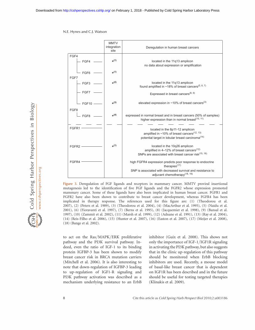
Figure 3. Deregulation of FGF ligands and receptors in mammary cancer. MMTV proviral insertionalmutagenesis led to the identification of five FGF ligands and the FGFR2 whose expression promotedmammary cancer. Some of these ligands have also been implicated in human breast cancer. FGFR1 andFGFR2 have also been shown to contribute to breast cancer development, whereas FGFR4 has beenimplicated in therapy response. The references used for this figure are: (1) (Theodorou et al.2007), (2) (Peters et al. 1989), (3) (Theodorou et al. 2004), (4) (MacArthur et al. 1995), (5) (Naidu et al.2001), (6) (Fioravanti et al. 1997), (7) (Berns et al. 1995), (8) (Jacquemier et al. 1998), (9) (Bansal et al.1997), (10) (Zammit et al. 2002), (11) (Marsh et al. 1999), (12) (Adnane et al. 1991), (13) (Ray et al. 2004),(14) (Reis-Filho et al. 2006), (15) (Hunter et al. 2007), (16) (Easton et al. 2007), (17) (Meijer et al. 2008),(18) (Bange et al. 2002).
N.E. Hynes and C.J. Watson
8 Cite this article as Cold Spring Harb Perspect Biol 2010;2:a003186
on February 1, 2018 - Published by Cold Spring Harbor Laboratory Press http://cshperspectives.cshlp.org/Downloaded from

CYTOKINES AND THEIR RECEPTORS INNORMAL DEVELOPMENT AND CANCER
Cytokines comprise a large family of solubleproteins that mediate cell communication andinduce potent biological responses often byinducing the expression of receptors, co-receptors, or adaptors. Their receptors can begrouped into families that share similar struc-tures and signal through the Jak/Stat pathway.Notably, unlike the EGF, FGF, and IGF fam-ilies of receptors, cytokine receptors do nothave intrinsic kinase activity. They rely insteadon receptor-associated Jak kinases (Haan et al.2006). In mammals there are four Jaks that areubiquitously expressed with the exception of
Jak3. On ligand binding, the Jaks transphos-phorylate each other and the receptor chainsto create docking sites for Stats that are thenalso tyrosine phosphorylated by the Jaks to cre-ate active Stat dimers. These subsequently trans-locate to the nucleus, bind to palindromicmotifs (TTCCNGGAA), and activate transcrip-tion of sets of target genes that are specific foreach Stat (Fig. 4).
CYTOKINE RECEPTOR FAMILIES
There are five main receptor families that havedifferent receptor chains (usually two or three)and are activated by different ligands: the
Early pregnancyIL-4, IL-13
Mid-pregnancy/lactationPRL
InvolutitonLIP, OSM
IL-4Rα IL-13Rα1 PRLR PRLA LIFR/OSMR p130g
P P
P
P
P
Shp-1
P
SOCS5
JAK1
Y
YYY
Y
Tyk2
?
P PJAK1 JAK1JAK2 JAK2
CIS1
PIAS1/3
Stat5Stat3
Stat3
Stat3
Stat5
Target gene
Pregnant
Stat6
Stat1 Stat3
Stat1
Stat5
Ges
tatio
n
Lactation
Shp-1/2
YY
YP
P P
P
P
P
PP
P
P
PP
P
P
P
P
Y
Y
Y
Y
Y
Y
Y
Y
Y
Y Y
Y YStat6P
SOCS1 SOCS1
SOCS1
Involution
AdultYoung virgin
Target genes
Stat6 Stat3
Lineage commitment InvoluutionGATA3 OSMR
C-Maf IGFBP5NFAT1/NFATc2 CEBPβ/δ
Stat3
Pik3r1
SlpiSOCS3SAA1
SAA2
Lactation
Whey acidic proteinCaesin alpha, betaSocs2
Stat5
Puberty
Lactating
Figure 4. Cytokine signaling through the Jak/Stat pathway in mammary gland development. Sequentialactivation of Stat6 and Stat5 control alveologenesis during pregnancy. These Stats are activated in response tospecific ligands, IL-4 and IL-13 (Stat6) and Prl (Stat5) that bind to receptors of the cytokine family. Duringinvolution, cell death is mediated by Stat3, which is activated by LIF and then by OSM, which share thecommon gp130 receptor. Stat signaling is negatively regulated by the family of SOCS proteins that are directtranscriptional targets of each Stat factor. Once activated, Stats bind to similar consensus sequences butactivate transcription of distinct sets of target genes to control mammary gland development.
Peptide Factors and Cytokines in the Mammary Gland
Cite this article as Cold Spring Harb Perspect Biol 2010;2:a003186 9
on February 1, 2018 - Published by Cold Spring Harbor Laboratory Press http://cshperspectives.cshlp.org/Downloaded from

interferon (IFN), gp130, gC, IL-3, and singlechain families (Schindler and Plumlee 2008).The corresponding ligands are diverse andinclude the interleukins (IL), leptin, leukemiainhibitory factor (LIF), and prolactin (Prl). Dif-ferent combinations of JAKs associate with eachreceptor, for example, Jak1 and Jak2 bind to theIFNg receptor. The exception is the single chainfamily where the receptor is a homodimer andJak2 is invariably the associated kinase. Themost relevant member of this family is the Prlreceptor (PRLR).
THE STAT FAMILY OF TRANSCRIPTIONFACTORS
There are seven mammalian Stats (Stats1–6, 5a,and 5b) that are approximately 91 kDa in sizeand are distributed in pairs on three differ-ent chromosomes. They appear to have arisenfrom a single primordial gene with the Stat3and Stat5a/b pair being most closely related tohomologs found in Dictyostelium, Caenorhab-ditis elegans, and Drosophila (Kisseleva et al.2002).
Avariety of biochemical and genetic studieshave revealed which Stats are downstream effec-tors of particular cytokines. Although Stat2 isspecific for IFN-I, Stat4 for IL-12, IL-23, andIL-27, and Stat6 for IL-4 and IL-13, a muchwider range of cytokines signal through Stat1,Stat3, and Stats5a/b. The IL-6 family (IL-6,IL-11, IL-31, LIF, CNTF, CLC/CLF, NP, CT1,and OSM), the IL-10 family (IL-10, IL-19,IL-20, IL-22, IL-24, and IL-26), and G-CSF,Leptin, and IL-21 all activate Stat3 and theIL-3 family (IL-3, IL-5, and GM-CSF), thesingle chain family (GH, Prl, Tpo, and Epo),and the gC family (IL-2, IL-7, IL-9, IL-15,and IL-21) activate Stat5a and Stat5b that are96% identical at the amino acid level. Apartfrom the response to Prl (Stat5a) and GH(Stat5b) these Stats are not entirely functionallyredundant and, interestingly, appear to bindDNA differently as Stat5a preferentially formsteteramers whereas Stat5b favors dimers (Ver-dier et al. 1998). Stat1 is the only Stat down-stream from IFNg but is also activated bysome cytokines of the gp130 family and EGF.
THE ROLES OF CYTOKINES AND STATS INMAMMARY GLAND DEVELOPMENT
Development of the embryonic mammarygland proceeds unperturbed in mice that aredeficient for Stats 1, 2, 4, 5, and 6. Thus, theseStats are unlikely to be important in mammarystem cells or early tissue development. Becausedeletion of Stat3 results in early embryoniclethality, it has not been possible to determinewhether Stat3 is important at this stage of devel-opment. However, given that Stat3 is impli-cated in self-renewal of embryonic stem cellsin culture, it would be interesting to determineif Stat3 has a role in establishing or maintainingembryonic mammary stem cells.
The adult mammary gland is quite differentand, individual members of the Stat family haveimportant roles. Stats 1 and 3 are constitutivelyexpressed but Stat1 is tyrosine phosphorylatedonly in virgin gland and in late involution, fol-lowing remodeling of the gland, while Stat3activity is highest on the day of birth and duringthe first 6 days of involution (Watson 2006).Stat6 is expressed also throughout adult mam-mary gland development, and is activatedduring pregnancy before Stat5. Thus, there arediscrete windows of Stat activity in the adultmammary gland. The expression of cytokinesis less well known although a role for LIF inductal elongation during puberty has been de-scribed (Kritikou et al. 2003).
Genetic studies in mice have revealed rolesfor Stats 3, 5a and b, and 6 in the adult glandduring a pregnancy cycle whereas ablation ofStat1 has not been reported to have a majoreffect. Stat6 is activated by the type-2 cytokines,IL-4 and IL-13, in T cells and is associatedwith changes in chromatin structure and tran-scriptional up-regulation of GATA-3, c-Maf,and NFAT1/NFATc2. Surprisingly, in mam-mary gland, Stat6 is also activated by IL-4 andIL-13 and this correlates with the expres-sion of IL4Ra and GATA-3 in the epithelium(Khaled et al. 2007). Importantly, in the absenceof either Stat6 or IL-4 and IL-13, lobuloalveolardevelopment is substantially delayed with num-bers of alveoli reduced by about 70% althoughthis defect is resolved by late pregnancy when
N.E. Hynes and C.J. Watson
10 Cite this article as Cold Spring Harb Perspect Biol 2010;2:a003186
on February 1, 2018 - Published by Cold Spring Harbor Laboratory Press http://cshperspectives.cshlp.org/Downloaded from
it is likely that levels of GATA-3 are sufficient tospecify and maintain the lineage in the absenceof Stat6. GATA-3 is critically important as dem-onstrated by two similar studies in which Gata3was conditionally deleted at different stages ofmammary development (Kouros-Mehr et al.2006; Asselin-Labat et al. 2007). Deletion ofGata3 in alveolar cells during pregnancy re-sulted in a block in alveolar differentiation andfailed lactogenesis (Asselin-Labat et al. 2007).Doxycycline-inducible Cre-mediated deletionof Gata3 in late pregnancy resulted also in thedisruption of ductal architecture (Kouros-Mehret al. 2006). Using FACS to isolate subpopula-tions of epithelial cells revealed that the size ofthe luminal progenitor pool increases signif-icantly in Gata3-null mice. Because overex-pression of GATA3 in null cells induced theexpression of the milk proteins b-casein andWAP, in the absence of lactogenic hormonestimulation, it seems reasonable to concludethat GATA-3 promotes the differentiation oflineage-restricted progenitor cells (Asselin-Labat et al. 2007).
Levels of GATA-3 are high also in the mam-mary epithelium of virgin mice and it is oneof the earliest transcription factors to be ex-pressed in mammary gland, being detectablein the embryonic placode. The importance ofGATA-3 is illustrated further by the failure toform TEBs and a severe reduction in ductal out-growth in the absence of GATA-3. A transcrip-tional link between GATA-3 and FOXA1 wasuncovered using microarray and promoteranalysis coupled with chromatin immuno-precipitation to identify a functional GATA-3binding site in the FOXA1 promoter. Interest-ingly, FOXA1, is required for ERa transcrip-tional activity. Thus, GATA-3 is required atvarious stages of mammary gland development,placing this transcription factor earlier in theluminal lineage than Stat5 and Stat6.
It is interesting that phosphorylated Stat5levels are reduced in Stat6-deficient mammaryglands. Whether this is a consequence of re-duced levels of IL-5, which is known to activateStat5, rather than Prl is not known. A hierarchyof signaling from IL-4/IL-13 through Stat6and GATA3 is thus an important constituent
of commitment to the alveolar luminal lineage.Notably, GATA3 is highly expressed in breastcancers of the luminal A subtype, which alsoexpress ERa (Sorlie et al. 2003).
Stats 5a and 5b have long been known to beessential mediators of lobuloalveolar develop-ment (Miyoshi et al. 2001; Cui et al. 2004).These two Stat5 gene have different roles reflect-ing their upstream activators Prl (Stat5a) andGH (Stat5b) although they can compensatefor each other, as shown by the rescue of im-paired alveologenesis and failed lactation inthe Stat5a knockout mouse by Stat5b (Liuet al. 1998). It can be concluded that Stats5aand b are essential for normal differentiationand survival of alveolar mammary epithelialcells but that because ductal morphogenesis isunaffected in their absence, this requirement isspecific to the alveolar lineage. This distin-guishes Stat5 from GATA-3, which is requiredin both ductal and alveolar lineages. Recently,transplantation studies of FACS-sorted cells sug-gested that alveolar progenitor cells could not begenerated from a presumptive earlier progeni-tor in the absence of Stats5a and b. Notably,whereas GATA-3 expression is unperturbed inStat5a/b null cells, the expression of Elf5 isseverely diminished (Yamaji et al. 2009).
Once again, there are parallels with thehematopoietic system because Stat5a/b havebeen shown to control stem and progenitorcell fate and loss of a single allele of Stat5a/bresulted in increased HSC cycling and depletionof the long-term HSC pool (Wang et al. 2009).Furthermore, results obtained with mice com-pletely null for Stat5a/b reveal that these factorsare required for the normal development of alllymphoid cells (Yao et al. 2006).
Taken together, the data suggest that thereare at least two alveolar lineages: a Prl-Stat5 con-trolled lineage where Elf5 is an essential reg-ulator and a Stat6-GATA-3 lineage that may beERa positive. It is interesting that these lineagesare not independent of each other because abla-tion of either Stat5 or GATA-3 results in a loss ofdifferentiated epithelium perhaps indicatingessential interactions between cells of these lin-eages. Because GATA-3 is expressed in virgingland whereas Stat6 is activated in only a few
Peptide Factors and Cytokines in the Mammary Gland
Cite this article as Cold Spring Harb Perspect Biol 2010;2:a003186 11
on February 1, 2018 - Published by Cold Spring Harbor Laboratory Press http://cshperspectives.cshlp.org/Downloaded from

ductal cells in the virgin, it may be that GATA-3is switched off in a subset of alveolar cells duringpregnancy to be replaced by Elf5.
Stat3 is essential for the initiation of apop-tosis and remodeling following forced wean-ing (Chapman et al. 1999). In the absence ofStat3, involution is dramatically delayed andthe reversible phase can be extended for up to6 d (Humphreys et al. 2002). Stat3 is activatedby a number of cytokines. In involuting mam-mary gland, the upstream ligand is LIF, shownusing LIF deficient mice (Kritikou et al. 2003)or by implanting LIF pellets into lactatingmammary glands (Schere-Levy et al. 2003).Another upstream regulator of Stat3 activityin involution is TGFb3 (Nguyen and Pollard2000). Interestingly, IL-6 does not regulateStat3 activity in involution (Zhao et al. 2002)although it does in breast cancer cells, suggest-ing perhaps that different signals downstreamfrom their cognate receptors specifies eithera proliferative or death response to specificStat3-activating cytokines. Recently, it hasbeen shown that the receptor for oncostatin M(OSMR) is required for the activation of Stat3during the second phase of involution (Tiffenet al. 2008) where it may substitute for LIFwhich peaks at 12 hours involution. BecauseOSMR is transcriptionally regulated by Stat3,this provides positive feedback on the pathwayenhancing the Stat3 signal in the second phase(Tiffen et al. 2008).
Thus, cytokines and their Stat targets haveimportant roles in lineage commitment, sur-vival, and death of mammary gland epithelium.
CYTOKINE SIGNALING AND BREASTCANCER
As mentioned earlier, GATA-3 expression isstrongly correlated with the Luminal A subtypeof breast tumors, which have the best prognosticoutcome (Sorlie et al. 2003). Low GATA-3 ex-pression is an indicator of poor clinical out-come. In mouse models of breast cancer,GATA-3 suppresses tumor dissemination andmaintains differentiation.
Several Stats, and in particular Stat3, havebeen shown to be constitutively activated in a
wide range of blood and solid tumors andare frequently activated in breast carcinoma(Watson and Miller 1995) (Fig. 5). The ap-parent paradox between the death-inducingfunction of Stat3 in involution and its onco-genic and survival function in breast tumors,has prompted extensive investigation. Apartfrom the different cell types involved, namelyfully differentiated alveolar cells in involutioncompared with ductal cells in most breast carci-nomas, Stat3 activation results in the secretionof cytokines. Thus, chronic Stat3 activity intumor cells results in paracrine signaling to cellsin the tumor microenvironment including infil-trating immune cells. It is well established thatinflammation has tumor-promoting poten-tial and active Stat3 is often found at the inva-sive edge of tumors, adjacent to inflammatorycells (Bromberg and Wang 2009). Stat3 is alsoa key mediator of anti-inflammatory responsesdownstream from the IL-10 receptor and hasrecently been shown to enable tumors to evadethe immune system by inhibiting the matura-tion of dendritic cells and macrophages (Wanget al. 2004). Accordingly, inhibition of Stat3 inmacrophages induced an antitumor immuneresponse in a rat model of breast cancer (Sunet al. 2006). Recently, using an activated ErbB2mouse model of breast cancer, it was shownthat although Stat3 does not affect the initiationof tumors, it dramatically increases lung meta-stasis (Ranger et al. 2009). Ablation of Stat3 inthis model was associated with a reduction ininflammatory and angiogenic responses. Thus,breast cancers with active Stat3 condition othercells in the tumor microenvironment by secret-ing cytokines that promote both metastasis andimmune evasion.
Recently, a role for IL-6 in self-renewal ofbreast cancer stem cells was proposed (Sansoneet al. 2007). Using the so-called mammosphereassay, where stem cells and their derivativesare cultured in floating spheroids, it was shownthat breast tumor cells express higher levels ofIL-6 mRNA than nontumor cells from thesame patient although secretion of IL-6 by thesecells was not measured. Furthermore, IL-6 treat-ment promoted spheroid growth in a Notch-3-dependent manner. IL-6 has been shown also to
N.E. Hynes and C.J. Watson
12 Cite this article as Cold Spring Harb Perspect Biol 2010;2:a003186
on February 1, 2018 - Published by Cold Spring Harbor Laboratory Press http://cshperspectives.cshlp.org/Downloaded from

be a potent growth factor in ERa positive breasttumors (Sasser et al. 2007). Overexpression ofIL-6 in MCF-7 breast cancer cells repressed E-cadherin expression and induced an EMT phe-notype, and xenografts of these cells showedincreased proliferation and advanced tumorgrade (Sullivan et al. 2009). Interestingly, ithas been shown recently that EMT inducesstem cell-like properties (Mani et al. 2008).Cooperation between Stat3 and EGFR hasbeen shown to be involved in EMT by mediatingthe expression of the TWIST gene (Lo et al.2007). So, Stat3 could influence tumor spreadby directly inducing EMT via up-regulation ofTWIST expression and down-regulation of E-cadherin levels and enhancing self-renewalproperties.
Stat3 and Stat5 have quite different rolesin normal mammary gland development and
have been suggested to play opposing roles incell fate with Stat3 being pro-apoptotic andStat5 prosurvival (Clarkson et al. 2006). A sub-set of breast tumors have both Stat3 and Stat5constitutive activity and a recent study showedthat these tumors are more differentiated thantumors with only Stat3 activity and further-more, have more favorable prognostic charac-teristics (Walker et al. 2009). These data inbreast tumors contrast with those in transgenicmice that develop tumors as a consequence ofoverexpression of either normal or activatedStat5 although these also have a differentiatedphenotype (Iavnilovitch et al. 2004). Recentwork has revealed that expression of constitu-tively active Stat5a suppresses the motility andinvasiveness of MCF7 cells while in contrast,constitutively active Stat5b has no effect sug-gesting that expression of Stat5a, but not Stat5b,
IL-1βTh17
IL-17
IL-12
Mature DC
TAMIL-1β
pStat3
pStat3
pStat3
pStat3
Tumour cells
IL-6IL-23
Immature DC
IL-6IL-10
Non-tumor cells
IL-1β IL-6
IL-10
Figure 5. Constitutive Stat3 activity in tumors affects the surrounding microenvironment. Chronic Stat3activation in tumor cells results in secretion of IL-1b, IL-6, and IL-10; these in turn act on nontumor cells,T helper 17 cells (Th17) and tumor associated macrophages (TAM) resulting in further secretion ofcytokines, which act in a feedback loop to promote growth and differentiation of the tumor cells.Stat3-directed secretion of IL-10 by the tumor cells also results in inhibition of antitumor immunity, forexample by inhibiting the maturation of dendritic cells (DCs). Thus, Stat3 and the cytokines that it induces,promote tumor invasion and metastasis. Diagram adapted from Pensa et al. (2009).
Peptide Factors and Cytokines in the Mammary Gland
Cite this article as Cold Spring Harb Perspect Biol 2010;2:a003186 13
on February 1, 2018 - Published by Cold Spring Harbor Laboratory Press http://cshperspectives.cshlp.org/Downloaded from
could be an indicator of better prognosis inbreast cancer (Tang et al. 2010).
Although there is little evidence that Stat6 isoncogenic, Stat6 deficient mice are significantlymore resistant to tumors arising from 4T1 mam-mary adenocarcinoma cells compared withwild-type BALB/c mice (Jensen et al. 2003).Furthermore, ablation of Stat6 results in en-hanced survival of mice in which 4T1 primarytumors have been surgically excised (Ostrand-Rosenberg et al. 2002). This has been proposedto result from the polarization of the immuneresponse toward a type 1 cytokine profile be-cause Stat6 KO mice are unable to generate atype 2 response.
Thus, the Stats that are important for nor-mal alveologenesis in mammary gland are alsoimportant in tumorigenesis either by promot-ing tumor growth (Stat5), generating an inflam-matory tumor microenvironment (Stat3), or byfacilitating immune evasion (Stat6 and Stat3).
ACKNOWLEDGMENTS
We would like to thank Drs. G. MacDonald andJ. Dey (Basel) and Dr. J. Wickenden (Cam-bridge) for useful discussions and help withthe figures. Work in the Hynes laboratory issupported by the Novartis Research Founda-tion. Work in the Watson laboratory is fundedby the BBSRC and the Breast Cancer Campaign.
REFERENCES
Adnane J, Gaudray P, Dionne CA, Crumley G, Jaye M,Schlessinger J, Jeanteur P, Birnbaum D, Theillet C.1991. BEK and FLG, two receptors to members of theFGF family, are amplified in subsets of human breast can-cers. Oncogene 6: 659–663.
Anderson E, Clarke RB. 2004. Steroid receptors and cellcycle in normal mammary epithelium. J MammaryGland Biol Neoplasia 9: 3–13.
Andrechek ER, White D, Muller WJ. 2005. Targeted disrup-tion of ErbB2/Neu in the mammary epithelium results inimpaired ductal outgrowth. Oncogene 24: 932–937.
Asselin-Labat ML, Sutherland KD, Barker H, Thomas R,Shackleton M, Forrest NC, Hartley L, Robb L, GrosveldFG, van der Wees J, et al. 2007. Gata-3 is an essential reg-ulator of mammary-gland morphogenesis and luminal-cell differentiation. Nat Cell Biol 9: 201–209.
Bange J, Prechtl D, Cheburkin Y, Specht K, Harbeck N,Schmitt M, Knyazeva T, Muller S, Gartner S, Sures I,
et al. 2002. Cancer progression and tumor cell motilityare associated with the FGFR4 Arg(388) allele. CancerRes 62: 840–847.
Bansal GS, Cox HC, Marsh S, Gomm JJ, Yiangou C, Luq-mani Y, Coombes RC, Johnston CL. 1997. Expressionof keratinocyte growth factor and its receptor in humanbreast cancer. Br J Cancer 75: 1567–1574.
Beenken A, Mohammadi M. 2009. The FGF family: Biology,pathophysiology and therapy. Nat Rev Drug Discov 8:235–253.
Berger MS, Locher GW, Saurer S, Gullick WJ, WaterfieldMD, Groner B, Hynes NE. 1988. Correlation ofc-erbB-2 gene amplification and protein expression inhuman breast carcinoma with nodal status and nucleargrading. Cancer Res 48: 1238–1243.
Bergers G, Coussens LM. 2000. Extrinsic regulators of epi-thelial tumor progression: Metalloproteinases. CurrOpin Genet Dev 10: 120–127.
Berns EM, Foekens JA, van Staveren IL, van Putten WL, deKoning HY, Portengen H, Klijn JG. 1995. Oncogeneamplification and prognosis in breast cancer: Relation-ship with systemic treatment. Gene 159: 11–18.
Bonnette SG, Hadsell DL. 2001. Targeted disruption of theIGF-I receptor gene decreases cellular proliferationin mammary terminal end buds. Endocrinology 142:4937–4945.
Brandt R, Eisenbrandt R, Leenders F, Zschiesche W, Binas B,Juergensen C, Theuring F. 2000. Mammary gland specifichEGF receptor transgene expression induces neoplasiaand inhibits differentiation. Oncogene 19: 2129–2137.
Bromberg J, Wang TC. 2009. Inflammation and cancer: IL-6and STAT3 complete the link. Cancer Cell 15: 79–80.
Cardiff RD, Wellings SR. 1999. The comparative pathologyof human and mouse mammary glands. J MammaryGland Biol Neoplasia 4: 105–122.
Chakravarty G, Hadsell D, Buitrago W, Settleman J, RosenJM. 2003. p190-B RhoGAP regulates mammary ductalmorphogenesis. Mol Endo 17: 1054–1065.
Chapman RS, Lourenco PC, Tonner E, Flint DJ, Selbert S,Takeda K, Akira S, Clarke AR, Watson CJ. 1999. Suppres-sion of epithelial apoptosis and delayed mammary glandinvolution in mice with a conditional knockout of Stat3.Genes Dev 13: 2604–2616.
Ciarloni L, Mallepell S, Brisken C. 2007. Amphiregulin is anessential mediator of estrogen receptor alpha function inmammary gland development. Proc Natl Acad Sci 104:5455–5460.
Clarkson RW, Boland MP, Kritikou EA, Lee JM, FreemanTC, Tiffen PG, Watson CJ. 2006. The genes induced bysignal transducer and activators of transcription(STAT)3 and STAT5 in mammary epithelial cells definethe roles of these STATs in mammary development. MolEndocrinol 20: 675–685.
Coleman-Krnacik S, Rosen JM. 1994. Differential temporaland spatial gene expression of fibroblast growth factorfamily members during mouse mammary gland develop-ment. Mol Endocrinol 8: 218–229.
Coleman S, Silberstein GB, Daniel CW. 1988. Ductal mor-phogenesis in the mouse mammary gland: Evidence sup-porting a role for epidermal growth factor. Dev Biol 127:304–315.
N.E. Hynes and C.J. Watson
14 Cite this article as Cold Spring Harb Perspect Biol 2010;2:a003186
on February 1, 2018 - Published by Cold Spring Harbor Laboratory Press http://cshperspectives.cshlp.org/Downloaded from
Cui Y, Riedlinger G, Miyoshi K, Tang W, Li C, Deng CX,Robinson GW, Hennighausen L. 2004. Inactivation ofStat5 in mouse mammary epithelium during pregnancyreveals distinct functions in cell proliferation, survival,and differentiation. Mol Cell Biol 24: 8037–8047.
De Meyts P. 2004. Insulin and its receptor: Structure, func-tion and evolution. Bioessays 26: 1351–1362.
Easton DF, Pooley KA, Dunning AM, Pharoah PD, Thomp-son D, Ballinger DG, Struewing JP, Morrison J, Field H,Luben R, et al. 2007. Genome-wide association studyidentifies novel breast cancer susceptibility loci. Nature447: 1087–1093.
Elsheikh SE, Green AR, Lambros MBK, Turner NC, GraingeMJ, Powe D, Ellis IO, Reis-Filho JS. 2007. FGFR1 ampli-fication in breast carcinomas: A chromogenic in situhybridisation analysis. Breast Cancer Res 9: R23.
Fioravanti L, Cappelletti V, Coradini D, Miodini P, BorsaniG, Daidone MG, Di Fronzo G. 1997. int-2 Oncogeneamplification and prognosis in node-negative breast car-cinoma. Int J Cancer 74: 620–624.
Graus-Porta D, Beerli RR, Daly JM, Hynes NE. 1997.ErbB-2, the preferred heterodimerization partner of allErbB receptors, is a mediator of lateral signaling.EMBO J 16: 1647–1655.
Guix M, Faber AC, Wang SE, Olivares MG, Song Y, Qu S,Rinehart C, Seidel B, Yee D, Arteaga CL, et al. 2008.Acquired resistance to EGFR tyrosine kinase inhibitorsin cancer cells is mediated by loss of IGF-binding pro-teins. J Clin Invest 118: 2609–2619.
Haan C, Kreis S, Margue C, Behrmann I. 2006. Jaks andcytokine receptors–an intimate relationship. BiochemPharmacol 72: 1538–1546.
Heckman BM, Chakravarty G, Vargo-Gogola T, Gonzales-Rimbau M, Hadsell DL, Lee AV, Settleman J, Rosen JM.2007. Crosstalk between the p190-B RhoGAP and IGFsignaling pathways is required for embryonic mammarybud development. Dev Biol 309: 137–149.
Holbro T, Beerli RR, Maurer F, Koziczak M, Barbas CF 3rd,Hynes NE. 2003. The ErbB2/ErbB3 heterodimer func-tions as an oncogenic unit: ErbB2 requires ErbB3 to drivebreast tumor cell proliferation. Proc Natl Acad Sci 100:8933–8938.
Hovey RC, Trott JF, Vonderhaar BK. 2002. Establishing aframework for the functional mammary gland: Fromendocrinology to morphology. J Mammary Gland BiolNeoplasia 7: 17–38.
Howard B, Panchal H, McCarthy A, Ashworth A. 2005.Identification of the scaramanga gene implicates Neure-gulin3 in mammary gland specification. Genes Dev 19:2078–2090.
Humphreys RC, Bierie B, Zhao L, Raz R, Levy D, Hennigh-ausen L. 2002. Deletion of Stat3 blocks mammary glandinvolution and extends functional competence of thesecretory epithelium in the absence of lactogenic stimuli.Endocrinology 143: 3641–3650.
Hunter DJ, Kraft P, Jacobs KB, Cox DG, Yeager M, Hankin-son SE, Wacholder S, Wang Z, Welch R, Hutchinson A,et al. 2007. A genome-wide association study identifiesalleles in FGFR2 associated with risk of sporadic post-menopausal breast cancer. Nat Genet 39: 870–874.
Hynes NE, Lane HA. 2005. ERBB receptors and cancer: Thecomplexity of targeted inhibitors. Nat Rev Cancer 5:341–354.
Iavnilovitch E, Cardiff RD, Groner B, Barash I. 2004. Dereg-ulation of Stat5 expression and activation causes mam-mary tumors in transgenic mice. Int J Cancer 112:607–619.
Itoh N, Ornitz DM. 2004. Evolution of the Fgf and Fgfr genefamilies. Trends Genet 20: 563–569.
Jackson-Fisher AJ, Bellinger G, Breindel JL, Tavassoli FA,Booth CJ, Duong JK, Stern DF. 2008. ErbB3 is requiredfor ductal morphogenesis in the mouse mammary gland.Breast Cancer Res 10: R96.
Jackson-Fisher AJ, Bellinger G, Ramabhadran R, Morris JK,Lee KF, Stern DF. 2004. ErbB2 is required for ductal mor-phogenesis of the mammary gland. Proc Natl Acad Sci101: 17138–17143.
Jacquemier J, Sun ZZ, Penault-Llorca F, Geneix J, Devilard E,Adelaide J, Birnbaum D. 1998. FGF7 protein expressionin human breast carcinomas. J Pathol 186: 269–274.
Jensen SM, Meijer SL, Kurt RA, Urba WJ, Hu HM, Fox BA.2003. Regression of a mammary adenocarcinoma inSTAT6-/- mice is dependent on the presence ofSTAT6-reactive T cells. J Immunol 170: 2014–2021.
Jones FE, Jerry DJ, Guarino BC, Andrews GC, Stern DF.1996. Heregulin induces in vivo proliferation and differ-entiation of mammary epithelium into secretory lobu-loalveoli. Cell Growth Differ 7: 1031–1038.
Jones FE, Welte T, Fu XY, Stern DF. 1999. ErbB4 signaling inthe mammary gland is required for lobuloalveolar devel-opment and Stat5 activation during lactation. J Cell Biol147: 77–88.
Khaled WT, Read EK, Nicholson SE, Baxter FO, Brennan AJ,Came PJ, Sprigg N, McKenzie AN, Watson CJ. 2007. TheIL-4/IL-13/Stat6 signalling pathway promotes luminalmammary epithelial cell development. Development134: 2739–2750.
Kisseleva T, Bhattacharya S, Braunstein J, Schindler CW.2002. Signaling through the JAK/STAT pathway, recentadvances and future challenges. Gene 285: 1–24.
Kleinberg DL, Ruan W. 2008. IGF-I, GH, and sex steroideffects in normal mammary gland development. J Mam-mary Gland Biol Neoplasia 13: 353–360.
Kleinberg DL, Wood TL, Furth PA, Lee AV. 2009. Growthhormone and insulin-like growth factor-I in the transi-tion from normal mammary development to preneoplas-tic mammary lesions. Endocr Rev 30: 51–74.
Klinakis A, Szabolcs M, Chen G, Xuan S, Hibshoosh H,Efstratiadis A. 2009. Igfr1 as a therapeutic target in amouse model of basal-like breast cancer. Proc Natl AcadSci 106: 2359–2364.
Kouhara H, Hadari YR, Spivak-Kroizman T, Schilling J, Bar-Sagi D, Lax I, Schlessinger J. 1997. A lipid-anchoredGrb2-binding protein that links FGF-receptor activationto the Ras/MAPK signaling pathway. Cell 89: 693–702.
Kouros-Mehr H, Slorach EM, Sternlicht MD, Werb Z. 2006.GATA-3 maintains the differentiation of the luminal cellfate in the mammary gland. Cell 127: 1041–1055.
Krane IM, Leder P. 1996. NDF/heregulin induces persis-tence of terminal end buds and adenocarcinomas in the
Peptide Factors and Cytokines in the Mammary Gland
Cite this article as Cold Spring Harb Perspect Biol 2010;2:a003186 15
on February 1, 2018 - Published by Cold Spring Harbor Laboratory Press http://cshperspectives.cshlp.org/Downloaded from
mammary glands of transgenic mice. Oncogene 12:1781–1788.
Kritikou EA, Sharkey A, Abell K, Came PJ, Anderson E,Clarkson RW, Watson CJ. 2003. A dual, non-redundant,role for LIF as a regulator of development and STAT3-mediated cell death in mammary gland. Development130: 3459–3468.
Kurosu H, Choi M, Ogawa Y, Dickson AS, Goetz R, Eliseen-kova AV, Mohammadi M, Rosenblatt KP, Kliewer SA,Kuro-o M. 2007. Tissue-specific expression of bKlothoand fibroblast growth factor (FGF) receptor isoformsdetermines metabolic activity of FGF19 and FGF21. JBiol Chem 282: 26687–26695.
Kurosu H, Ogawa Y, Miyoshi M, Yamamoto M, Nandi A,Rosenblatt KP, Baum MG, Schiavi S, Hu MC, Moe OW,et al. 2006. Regulation of fibroblast growth factor-23 sig-naling by klotho. J Biol Chem 281: 6120–6123.
Lahlou H, Sanguin-Gendreau V, Zuo D, Cardiff RD,McLean GW, Frame MC, Muller WJ. 2007. Mammaryepithelial-specific disruption of the focal adhesion kinaseblocks mammary tumor progression. Proc Natl Acad Sci104: 20302–20307.
Lew ED, Furdui CM, Anderson KS, Schlessinger J. 2009. Theprecise sequence of FGF receptor autophosphorylation iskinetically driven and is disrupted by oncogenic muta-tions. Sci Signal 2: ra6.
Li L, Cleary S, Mandarano MA, Long W, Birchmeier C, JonesFE. 2002. The breast proto-oncogene, HRGa regu-lates epithelial proliferation and lobuloalveolar develop-ment in the mouse mammary gland. Oncogene 21:4900–4907.
Liu X, Gallego MI, Smith GH, Robinson GW, Hennighau-sen L. 1998. Functional rescue of Stat5a-null mammarytissue through the activation of compensating signalsincluding Stat5b. Cell Growth Differ 9: 795–803.
Lo HW, Hsu SC, Xia W, Cao X, Shih JY, Wei Y, Abbruzzese JL,Hortobagyi GN, Hung MC. 2007. Epidermal growth fac-tor receptor cooperates with signal transducer and activa-tor of transcription 3 to induce epithelial-mesenchymaltransition in cancer cells via up-regulation of TWISTgene expression. Cancer Res 67: 9066–9076.
Loladze AV, Stull MA, Rowzee AM, Demarco J, Lantry JH3rd, Rosen CJ, Leroith D, Wagner KU, Hennighausen L,Wood TL. 2006. Epithelial-specific and stage-specificfunctions of insulin-like growth factor-I during postnatalmammary development. Endocrinology 147: 5412–5423.
Long W, Wagner KU, Lloyd KC, Binart N, Shillingford JM,Hennighausen L, Jones FE. 2003. Impaired differentia-tion and lactational failure of Erbb4-deficient mammaryglands identify ERBB4 as an obligate mediator of STAT5.Development 130: 5257–5268.
Lu P, Ewald AJ, Martin GR, Werb Z. 2008. Genetic mosaicanalysis reveals FGF receptor 2 function in terminal endbuds during mammary gland branching morphogenesis.Dev Biol 321: 77–87.
MacArthur CA, Shankar DB, Shackleford GM. 1995. Fgf-8,activated by proviral insertion, cooperates with theWnt-1 transgene in murine mammary tumorigenesis.J Virol 69: 2501–2507.
Mailleux AA, Spencer-Dene B, Dillon C, Ndiaye D,Savona-Baron C, Itoh N, Kato S, Dickson C, Thiery JP,Bellusci S. 2002. Role of FGF10/FGFR2b signaling during
mammary gland development in the mouse embryo.Development 129: 53–60.
Mani SA, Guo W, Liao MJ, Eaton EN, Ayyanan A, Zhou AY,Brooks M, Reinhard F, Zhang CC, Shipitsin M, et al.2008. The epithelial-mesenchymal transition generatescells with properties of stem cells. Cell 133: 704–715.
Marsh SK, Bansal GS, Zammit C, Barnard R, Coope R,Roberts-Clarke D, Gomm JJ, Coombes RC, JohnstonCL. 1999. Increased expression of fibroblast growth factor8 in human breast cancer. Oncogene 18: 1053–1060.
Meijer D, Sieuwerts AM, Look MP, van Agthoven T, FoekensJA, Dorssers LC. 2008. Fibroblast growth factor receptor 4predicts failure on tamoxifen therapy in patients withrecurrent breast cancer. Endocr Relat Cancer 15: 101–111.
Meyer KB, Maia AT, O’Reilly M, Teschendorff AE, Chin SF,Caldas C, Ponder BA. 2008. Allele-specific up-regulationof FGFR2 increases susceptibility to breast cancer. PLoSBiol 6: e108.
Mikaelian I, Blades N, Churchill GA, Fancher K, KnowlesBB, Eppig JT, Sundberg JP. 2004. Proteotypic classifica-tion of spontaneous and transgenic mammary neo-plasms. Breast Cancer Res 6: R668–679.
Mitchell G, Antoniou AC, Warren R, Peock S, Brown J,Davies R, Mattison J, Cook M, Warsi I, Evans DG, et al.2006. Mammographic density and breast cancer risk inBRCA1 and BRCA2 mutation carriers. Cancer Res 66:1866–1872.
Miyoshi K, Shillingford JM, Smith GH, Grimm SL, WagnerKU, Oka T, Rosen JM, Robinson GW, Hennighausen L.2001. Signal transducer and activator of transcription(Stat) 5 controls the proliferation and differentiation ofmammary alveolar epithelium. J Cell Biol 155: 531–542.
Mohammadi M, Dikic I, Sorokin A, Burgess WH, Jaye M,Schlessinger J. 1996. Identification of six novel autophos-phorylation sites on fibroblast growth factor receptor 1and elucidation of their importance in receptor activa-tion and signal transduction. Mol Cell Biol 16: 977–989.
Naidu R, Wahab NA, Yadav M, Kutty MK, Nair S. 2001.Detection of amplified int-2/FGF-3 gene in primarybreast carcinomas using differential polymerase chainreaction. Int J Mol Med 8: 193–198.
Nguyen AV, Pollard JW. 2000. Transforming growth factorbeta3 induces cell death during the first stage of mam-mary gland involution. Development 127: 3107–3118.
Ni CY, Murphy MP, Golde TE, Carpenter G. 2001. g-Secre-tase cleavage and nuclear localization of ErbB-4 receptortyrosine kinase. Science 294: 2179–2181.
Nielsen TO, Hsu FD, Jensen K, Cheang M, Karaca G, Hu Z,Hernandez-Boussard T, Livasy C, Cowan D, Dressler L,et al. 2004. Immunohistochemical and clinical character-ization of the basal-like subtype of invasive breast carci-noma. Clin Cancer Res 10: 5367–5374.
Nordgard SH, Johansen FE, Alnaes GI, Naume B, Borresen-Dale AL, Kristensen VN. 2007. Genes harbouring sus-ceptibility SNPs are differentially expressed in the breastcancer subtypes. Breast Cancer Res 9: 113.
Ong SH, Hadari YR, Gotoh N, Guy GR, Schlessinger J, Lax I.2001. Stimulation of phosphatidylinositol 3-kinase byfibroblast growth factor receptors is mediated by coordi-nated recruitment of multiple docking proteins. ProcNatl Acad Sci 98: 6074–6079.
N.E. Hynes and C.J. Watson
16 Cite this article as Cold Spring Harb Perspect Biol 2010;2:a003186
on February 1, 2018 - Published by Cold Spring Harbor Laboratory Press http://cshperspectives.cshlp.org/Downloaded from

Ornitz DM, Xu J, Colvin JS, McEwen DG, MacArthur CA,Coulier F, Gao G, Goldfarb M. 1996. Receptor specificityof the fibroblast growth factor family. J Biol Chem 271:15292–15297.
Ostrand-Rosenberg S, Clements VK, Terabe M, Park JM,Berzofsky JA, Dissanayake SK. 2002. Resistance tometastatic disease in STAT6-deficient mice requireshemopoietic and nonhemopoietic cells and is IFN-gdependent. J Immunol 169: 5796–5804.
Parsa S, Ramasamy SK, De Langhe S, Gupte VV, Haigh JJ,Medina D, Bellusci S. 2008. Terminal end bud mainte-nance in mammary gland is dependent upon FGFR2bsignaling. Dev Biol 317: 121–131.
Pensa S, Watson CJ, Poli V. 2009. Stat3 and theinflammation/acute phase response in involution andbreast cancer. J Mammary Gland Biol Neoplasia 14:121–129.
Peters G, Brookes S, Smith R, Placzek M, Dickson C. 1989.The mouse homolog of the hst/k-FGF gene is adjacent toint-2 and is activated by proviral insertion in some virallyinduced mammary tumors. Proc Natl Acad Sci 86:5678–5682.
Pollak MN, Schernhammer ES, Hankinson SE. 2004.Insulin-like growth factors and neoplasia. Nat Rev Cancer4: 505–518.
Ranger JJ, Levy DE, Shahalizadeh S, Hallett M, Muller WJ.2009. Identification of a Stat3-dependent transcriptionregulatory network involved in metastatic progression.Cancer Res 69: 6823–6830.
Ray ME, Yang ZQ, Albertson D, Kleer CG, Washburn JG,Macoska JA, Ethier SP. 2004. Genomic and expressionanalysis of the 8p11-12 amplicon in human breast cancercell lines. Cancer Res 64: 40–47.
Reis-Filho JS, Simpson PT, Turner NC, Lambros MB, JonesC, Mackay A, Grigoriadis A, Sarrio D, Savage K, Dexter T,et al. 2006. FGFR1 emerges as a potential therapeutic tar-get for lobular breast carcinomas. Clin Cancer Res 12:6652–6662.
Reis-Filho JS, Tutt AN. 2008. Triple negative tumours:A critical review. Histopathology 52: 108–118.
Ruan W, Kleinberg DL. 1999. Insulin-like growth factor I isessential for terminal end bud formation and ductal mor-phogenesis during mammary development. Endocrinol-ogy 140: 5075–5081.
Sachdev D, Yee D. 2001. The IGF system and breast cancer.Endocr Relat Cancer 8: 197–209.
Sanderson MP, Dempsey PJ, Dunbar AJ. 2006. Control ofErbB signaling through metalloprotease mediated ecto-domain shedding of EGF-like factors. Growth Factors24: 121–136.
Sansone P, Storci G, Tavolari S, Guarnieri T, Giovannini C,Taffurelli M, Ceccarelli C, Santini D, Paterini P, MarcuKB, et al. 2007. IL-6 triggers malignant features in mam-mospheres from human ductal breast carcinoma andnormal mammary gland. J Clin Invest 117: 3988–4002.
Sardi SP, Murtie J, Koirala S, Patten BA, Corfas G. 2006.Presenilin-dependent ErbB4 nuclear signaling regulatesthe timing of astrogenesis in the developing brain. Cell127: 185–197.
Sasser AK, Sullivan NJ, Studebaker AW, Hendey LF, Axel AE,Hall BM. 2007. Interleukin-6 is a potent growth factor for
ER-a-positive human breast cancer. FASEB J 21:3763–3770.
Schere-Levy C, Buggiano V, Quaglino A, Gattelli A, CirioMC, Piazzon I, Vanzulli S, Kordon EC. 2003. Leukemiainhibitory factor induces apoptosis of the mammary epi-thelial cells and participates in mouse mammary glandinvolution. Exp Cell Res 282: 35–47.
Schindler C, Plumlee C. 2008. Inteferons pen the JAK-STATpathway. Semin Cell Dev Biol 19: 311–318.
Schindler C, Levy DE, Decker T. 2007. JAK-STAT signaling:From interferons to cytokines. J Biol Chem 282:20059–20063.
Schroeder JA, Lee DC. 1998. Dynamic expression and acti-vation of ERBB receptors in the developing mouse mam-mary gland. Cell Growth Differ 9: 451–464.
Schwertfeger KL. 2009. Fibroblast growth factors in develop-ment and cancer: Insights from the mammary and pros-tate glands. Curr Drug Targets 10: 632–644.
Sebastian J, Richards RG, Walker MP, Wiesen JF, Werb Z,Derynck R, Hom YK, Cunha GR, DiAugustine RP.1998. Activation and function of the epidermal growthfactor receptor and erbB-2 during mammary gland mor-phogenesis. Cell Growth Differ 9: 777–785.
Seitzer N, Mayr T, Streit S, Ullrich A. 2010. A single nucleo-tide change in the mouse genome accelerates breast can-cer progression. Cancer Res 70: 802–812.
Slamon DJ, Clark GM, Wong SG, Levin WJ, Ullrich A,McGuire WL. 1987. Human breast cancer: Correlationof relapse and survival with amplification of theHER-2/neu oncogene. Science 235: 177–182.
Sorlie T, Tibshirani R, Parker J, Hastie T, Marron JS, Nobel A,Deng S, Johnsen H, Pesich R, Geisler S, et al. 2003.Repeated observation of breast tumor subtypes in inde-pendent gene expression data sets. Proc Natl Acad Sci100: 8418–8423.
Stern DF. 2008. ERBB3/HER3 and ERBB2/HER2 duet inmammary development and breast cancer. J MammaryGland Biol Neoplasia 13: 215–223.
Sternlicht MD, Sunnarborg SW, Kouros-Mehr H, Yu Y, LeeDC, Werb Z. 2005. Mammary ductal morphogenesisrequires paracrine activation of stromal EGFR viaADAM17-dependent shedding of epithelial amphiregu-lin. Development 132: 3923–3933.
Sullivan NJ, Sasser AK, Axel AE, Vesuna F, Raman V, Ram-irez N, Oberyszyn TM, Hall BM. 2009. Interleukin-6induces an epithelial-mesenchymal transition phenotypein human breast cancer cells. Oncogene 28: 2940–2947.
Sun Z, Yao Z, Liu S, Tang H, Yan X. 2006. An oligonucleotidedecoy for Stat3 activates the immune response of macro-phages to breast cancer. Immunobiology 211: 199–209.
Suzuki M, Uehara Y, Motomura-Matsuzaka K, Oki J,Koyama Y, Kimura M, Asada M, Komi-Kuramochi A,Oka S, Imamura T. 2008. bKlotho is required for fibro-blast growth factor (FGF) 21 signaling through FGFreceptor (FGFR) 1c and FGFR3c. Mol Endocrinol 22:1006–1014.
Tang JZ, Zuo ZH, Kong XJ, Steiner M, Yin Z, Perry JK, ZhuT, Liu DX, Lobie PE. 2010. Signal transducer and activa-tor of transcription (STAT)-5A and STAT5B differentiallyregulate human mammary carcinoma cell behavior.Endocrinology 151: 43–55.
Peptide Factors and Cytokines in the Mammary Gland
Cite this article as Cold Spring Harb Perspect Biol 2010;2:a003186 17
on February 1, 2018 - Published by Cold Spring Harbor Laboratory Press http://cshperspectives.cshlp.org/Downloaded from
Theodorou V, Boer M, Weigelt B, Jonkers J, van der Valk M,Hilkens J. 2004. Fgf10 is an oncogene activated by MMTVinsertional mutagenesis in mouse mammary tumors andoverexpressed in a subset of human breast carcinomas.Oncogene 23: 6047–6055.
Theodorou V, Kimm MA, Boer M, Wessels L, Theelen W,Jonkers J, Hilkens J. 2007. MMTV insertional mutagene-sis identifies genes, gene families and pathways involvedin mammary cancer. Nat Genet 39: 759–769.
Thike AA, Cheok PY, Jara-Lazaro AR, Tan B, Tan P, Tan PH.2009. Triple-negative breast cancer: Clinicopathologicalcharacteristics and relationship with basal-like breastcancer. Mod Pathol.
Thussbas C, Nahrig J, Streit S, Bange J, Kriner M, Kates R,Ulm K, Kiechle M, Hoefler H, Ullrich A, et al. 2006.FGFR4 Arg388 allele is associated with resistance to adju-vant therapy in primary breast cancer. J Clin Oncol 24:3747–3755.
Tidcombe H, Jackson-Fisher A, Mathers K, Stern DF, Gass-mann M, Golding JP. 2003. Neural and mammary glanddefects in ErbB4 knockout mice genetically rescued fromembryonic lethality. Proc Natl Acad Sci 100: 8281–8286.
Tiffen PG, Omidvar N, Marquez-Almuina N, Croston D,Watson CJ, Clarkson RW. 2008. A dual role for oncostatinM signaling in the differentiation and death of mammaryepithelial cells in vivo. Mol Endocrinol 22: 2677–2688.
Turner N, Lambros MB, Horlings HM, Pearson A, sharpe R,Natrajan R, Geyer FC, van Kouwenhove M, Kreike B,Mackay A, et al. 2010. Integrative molecular profiling oftriple negatvie breast cancers identifies amplicon driversand potential therapeutic targets. Oncogene doi:10.1038/onc.2009.489.
Van de Vijver M, van de Bersselaar R, Devilee P, CornelisseC, Peterse J, Nusse R. 1987. Amplification of the neu(c-erbB-2) oncogene in human mammmary tumors isrelatively frequent and is often accompanied by amplifi-cation of the linked c-erbA oncogene. Mol Cell Biol 7:2019–2023.
Veltmaat JM, Relaix F, Le LT, Kratochwil K, Sala FG, van Vee-len W, Rice R, Spencer-Dene B, Mailleux AA, Rice DP,et al. 2006. Gli3-mediated somitic Fgf10 expression gra-dients are required for the induction and patterning ofmammary epithelium along the embryonic axes. Devel-opment 133: 2325–2335.
Verdier F, Rabionet R, Gouilleux F, Beisenherz-Huss C,Varlet P, Muller O, Mayeux P, Lacombe C, GisselbrechtS, Chretien S. 1998. A sequence of the CIS gene promoterinteracts preferentially with two associated STAT5Adimers: A distinct biochemical difference betweenSTAT5A and STAT5B. Mol Cell Biol 20: 389–401.
Walker SR, Nelson EA, Zou L, Chaudhury M, Signoretti S,Richardson A, Frank DA. 2009. Reciprocal effects ofSTAT5 and STAT3 in breast cancer. Mol Cancer Res 7:966–976.
Wang S-C, Hung M-C. 2009. Nuclear translocation of theepidermal growth factor receptor family membrane tyro-sine kinase receptors. Clin Cancer Res 15: 6484–6489.
Wang Z, Li G, Tse W, Bunting KD. 2009. Conditional dele-tion of STAT5 in adult mouse hematopoietic stem cellscauses loss of quiescence and permits efficient nonabla-tive stem cell replacement. Blood 113: 4856–4865.
Wang T, Niu G, Kortylewski M, Burdelya L, Shain K, ZhangS, Bhattacharya R, Gabrilovich D, Heller R, Coppola D,et al. 2004. Regulation of the innate and adaptive immuneresponses by Stat-3 signaling in tumor cells. Nat Med 10:48–54.
Wansbury O, Panchal H, James M, Parry S, Ashworth A,Howard B. 2008. Dynamic expression of Erbb pathwaymembers during early mammary gland morphogenesis.J Invest Dermatol 128: 1009–1021.
Watson CJ. 2006. Involution: Apoptosis and tissue remodel-ling that convert the mammary gland from milk factoryto a quiescent organ. Breast Cancer Res 8: 203.
Watson CJ, Miller WR. 1995. Elevated levels of members ofthe STAT family of transcription factors in breast carci-noma nuclear extracts. Br J Cancer 71: 840–844.
White DE, Kurpios NA, Zuo D, Hassell JA, Blaess S,Mueller U, Muller WJ. 2004. Targeted disruption ofb1-integrin in a transgenic mouse model of humanbreast cancer reveals an essential role in mammarytumor induction. Cancer Cell 6: 159–170.
Wiesen JF, Young P, Werb Z, Cunha GR. 1999. Signalingthrough the stromal epidermal growth factor receptor isnecessary for mammary ductal development. Develop-ment 126: 335–344.
Wood TL, Richert MM, Stull MA, Allar MA. 2000. Theinsulin-like growth factors (IGFs) and IGF binding pro-teins in postnatal development of murine mammaryglands. J Mammary Gland Biol Neoplasia 5: 31–42.
Xian W, Pappas L, Pandya D, Selfors LM, Derksen PW,drBruin M, Gray NS, Jonkers J, Rosen JM, Brugge JS.2009. Fibroblast growth factor receptor 1-transformedmammary epithelial cells are dependent on RSK activityfor growth and survival. Cancer Res 69: 2244–2251.
Yamaji D, Na R, Feuermann Y, Pechhold S, Chen W, Robin-son GW, Hennighausen L. 2009. Development of mam-mary luminal progenitor cells is controlled by thetranscription factor STAT5A. Genes Dev 23: 2382–2387.
Yao Z, Cui Y, Watford WT, Bream JH, Yamaoka K, HissongBD, Li D, Durum SK, Jiang Q, Bhandoola A, et al. 2006.Stat5a/b are essential for normal lymphoid developmentand differentiation. Proc Natl Acad Sci 103: 1000–1005.
Yu WH, Woessner JF Jr, McNeish JD, Stamenkovic I. 2002.CD44 anchors the assembly of matrilysin/MMP-7 withheparin-binding epidermal growth factor precursor andErbB4 and regulates female reproductive organ remodel-ing. Genes Dev 16: 307–323.
Zammit C, Coope R, Gomm JJ, Shousha S, Johnston CL,Coombes RC. 2002. Fibroblast growth factor 8 isexpressed at higher levels in lactating human breast andin breast cancer. Br J Cancer 86: 1097–1103.
Zhang X, Ibrahimi OA, Olsen SK, Umemori H, Moham-madi M, Ornitz DM. 2006. Receptor specificity of thefibroblast growth factor family. The complete mamma-lian FGF family. J Biol Chem 281: 15694–15700.
Zhao L, Melenhorst JJ, Hennighausen L. 2002. Loss of inter-leukin 6 results in delayed mammary gland involution: Apossible role for mitogen-activated protein kinase andnot signal transducer and activator of transcription 3.Mol Endocrinol 16: 2902–2912.
N.E. Hynes and C.J. Watson
18 Cite this article as Cold Spring Harb Perspect Biol 2010;2:a003186
on February 1, 2018 - Published by Cold Spring Harbor Laboratory Press http://cshperspectives.cshlp.org/Downloaded from

16, 20102010; doi: 10.1101/cshperspect.a003186 originally published online JuneCold Spring Harb Perspect Biol
Nancy E. Hynes and Christine J. Watson CancerMammary Gland Growth Factors: Roles in Normal Development and in
Subject Collection The Mammary Gland as an Experimental Model
Gland Development and CancerOn the Role of the Microenvironment in Mammary
Derek RadiskyMetalloproteinases Couple Form with FunctionOn How Mammary Gland Reprogramming
Bonnie F. Sloane
Breast Cancer BiologyOn Using Functional Genetics to Understand
Kornelia PolyakMammary Gland DevelopmentOn Molecular Mechanisms Guiding Embryonic
Gertraud W. Robinson
the Mammary GlandOn Oncogenes and Tumor Suppressor Genes in
Rushika M. Perera and Nabeel Bardeesy
On Stem Cells in the Human BreastMark A. LaBarge
CancerOn Leukocytes in Mammary Development and
Cyrus M. GhajarDiscovery, Function, and Current StatusOn Murine Mammary Epithelial Stem Cells:
Jeffrey M. Rosen
Differentiation and Breast TumorigenesisOn Chromatin Remodeling in Mammary Gland
Kornelia Polyak
On In Vivo Imaging in CancerDavid Piwnica-Worms
On Hormone Action in the Mammary GlandJ.M. Rosen
Models of Breast CancerThe Utility and Limitations of Mouse−−in Context
Choosing a Mouse Model: Experimental Biology
Alexander D. Borowsky
Breast Cancer Biology in Mammary Development andβTGF-
Harold Moses and Mary Helen Barcellos-Hoff Tumor ProgressionMechanosignaling in Normal Development and Mammary Gland ECM Remodeling, Stiffness, and
Pepper Schedin and Patricia J. Keely
Engineered MiceHouse Mouse to the Development of GeneticallyBiologist: From the Initial Observations in the A Compendium of the Mouse Mammary Tumor
Robert D. Cardiff and Nicholas Kenney
Mammary Gland DevelopmentMolecular Mechanisms Guiding Embryonic
Pamela Cowin and John Wysolmerski
http://cshperspectives.cshlp.org/cgi/collection/ For additional articles in this collection, see
Copyright © 2010 Cold Spring Harbor Laboratory Press; all rights reserved
on February 1, 2018 - Published by Cold Spring Harbor Laboratory Press http://cshperspectives.cshlp.org/Downloaded from