management of back pain - heart of england nhs ... • clinical features • differential diagnosis...
TRANSCRIPT

MANAGEMENT OF BACK PAIN
MR. NAVIN FURTADO BSc MBBS MSc(Eng) FRCS(Neuro.Surg)
Consultant Neurosurgeon & Spinal SurgeonQueen Elizabeth Hospital Birmingham


Objectives
• Clinical Features• Differential Diagnosis
• Management Pathways• NHS England Pathfinder Project
– National Pathway of Care for Low Back Pain and Radicular Pain (2014)
• Surgical causes and cases• Physiotherapy/Triage & Treat Practitioner

Birmingham City• 1,085,400 population
• Younger population • 45.7% < 30yrs• 12.9% > 65yrs
• Growing city - 1% per year from 2001
Birmingham Metropolitan Area ~ 4 million
QEHB7 x Spine Consultants
ROH10 x Spine Consultants
Birmingham Spine Services

Epidemiology
• Low back pain without radicular pain – one of the most common musculo-skeletal conditions presenting to GPs– 80 % of all people will get back pain at some point in their life – access rates have increased from 231 to 295 per 1,000 (2005 to 2010)– Hospital Episode Statistics (HES) data for 2010/2011:
• over 70,000 procedures for low back pain in England • around 67,000 facet joint injections
• Low back pain with radicular pain – access rates have risen from 74 to121 per 100,000 population– Hospital Episode Statistics (HES) data for 2010/2011:
• 76,304 interventional procedures were undertaken for lumbar radicular pain
• 11,674 decompressions were carried out for spinal stenosis

Spinal Surgery in the Press

Definitions
• Radicular pain:– pain radiating down the leg following a dermatomal distribution – the two most common causes of radicular pain are prolapsed
intervertebral disc and spinal stenosis
• Non-specific back pain:– back pain but without nerve root involvement
• Specific back pain:– pain that may be amenable to treatment
• Symptoms defined as:– Acute: duration <6 weeks– Persistent: 6 weeks – 3 months– Chronic: duration >3 months

Differential Diagnosis
Spine Pathology
Oncology
Other: CSF
VascularCongenital
Degenerative PainDeformity
Spinal Reconstruction:
TumourTraumaInfection

Clinical Examination of Lumbar Spine
LOOK
Patient standing in neutral position:• Inspect from front, sides
and back• Note:
• Deformity • Soft tissue abnormalities
e.g. hairy patch (spinabifida)
• Surgical scars
FEEL
• Midline spinousprocesses
• Paraspinal soft tissues• Note: alignment and
focal tenderness
MOVE
• Forward flexion• Touch toes – how far
reached• Hip flexion plays a part
• Extension• 10 – 20o
• Lateral flexion• Lateral rotation
Lower Limb Neuro
+/- DRE

Treatment Objectives
Patient remains independent with return to previous activities and employment in
the shortest time possible.
Treatment
Investigations
Symptoms


Pathfinder Project-National Pathway of Care
for Low Back Pain and Radicular Pain 2014

Red Flags
• Cauda Equina Syndrome and/or cord compression – History:
• urinary retention/incontinence • Faecal incontinence • Altered perianal sensation • Limb weakness
– Examination • Limb weakness • Generalised neurological deficit/gait disturbance • Hyper-reflexia, clonus, extensor plantar response • Saddle anaesthesia
• Reduced anal tone/squeeze (if circumstances permit) • Urinary retention

Red Flags
• Suspicion of tumour or infection:– Past history of cancer (all patients should have been given an alert card
about MSCC following their initial presentation ) – Unexplained weight loss – Other symptoms suggestive of malignancy – Unwell/Fever – Raised inflammatory markers ESR>50, PCV < 30 – Age <10 years or Age >60 years – Pain - Thoracic or persisting non-mechanical – Possible immunosuppression – IVDU, HIV, Chemotherapy, Steroids.

Yellow Flags• Attitudes - towards the current problem. Does the patient feel that with appropriate help
and self-management they will return to normal activities?
• Beliefs - the patient feels they have something serious causing their problem - usually cancer. 'Faulty' beliefs can lead to catastrophisation.
• Compensation - Is the patient awaiting payment for an accident/injury at work/RTA?
• Diagnosis - Inappropriate communication can lead to patients misunderstanding what is meant, the most common examples being 'your disc has popped out' or 'your spine is crumbling'.
• Emotions - Patients with other emotional difficulties such as on-going depression and/or anxiety states are at a high risk of developing chronic pain.
• Family - There tends to be two problems with families, either over bearing or under supportive.
• Work - The worse the relationship, the more likely they are to develop chronic LBP.

Waddell’s Signs
Superficial tenderness, not in keeping with
anatomical boundaries.
Regional symptoms not conforming to anatomical
principles.
Pain on movements not affecting the lower back
Lack of pain after distraction• e.g. patients being unable to
straight-leg raise, but able to sit on the examination couch with legs straight and hips flexed to 90°.
Overreaction to stimuli.

Acute Pain Management
• Patients with acute low back pain:– self-manage with simple analgesia– minimal bed rest (up to a maximum of 48 hours) depending on the
severity of pain followed by progressive resumption of their normal activity
• Majority of patients will improve naturally assisted by good primary care management including physiotherapy/ hands on manipulation
• For those that do not respond:– an early risk assessment should be conducted– should be actively managed by the appropriate therapists

Keele STarT Back Screening Tool

Pathfinder Project-National Pathway of Care
for Low Back Pain and Radicular Pain 2014

Surgical Causes of Back Pain
Spine Pathology
Oncology
Other: CSF
VascularCongenital
Degenerative PainDeformity
Spinal Reconstruction:
TumourTraumaInfection

Symptoms need to correlate with imaging!
• Boden et al (1990)– The most cited paper in lumbar spine surgery– MRI findings in 67 individuals without back pain, sciatica, and
neurogenic claudication. – Study showed that common indications for surgery (e.g ., herniated
discs, spinal stenosis) can be incidentally found when scanning patients without neurological symptoms.
– The study also showed that as patients aged, these incidental findings increased in frequency.
– The study implied correctly that to recommend surgery for a patient, there should be a clear correlation of symptoms and radiographicalfindings and certainly should not be based on radiographical findings alone.
• Indications should be clear and unambiguous prior to surgery

Spinal Infection
OsteomyelitisDiscitisEpidural Abscess

• Spinal Tumour
• Extradural• Intradural
• Extra-medullary
• Intra-medullary


Spinal Tumour

• Spinal Tumour
• Intradural• Extra-
medullary

Degenerative Pain• Disc Disease• Lumbar Spondylosis• Spinal Stenosis

Degenerative Pain• Disc Disease• Lumbar Spondylosis• Spinal Stenosis
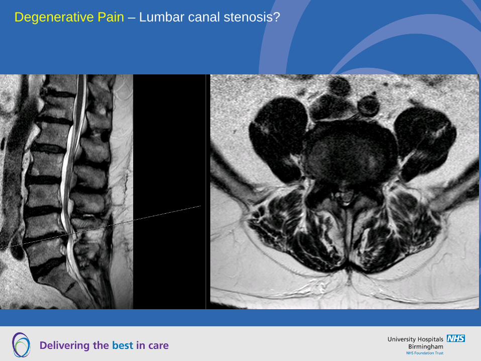
Degenerative Pain – Lumbar canal stenosis?

Degenerative Pain – Severe cervical myelopathy!
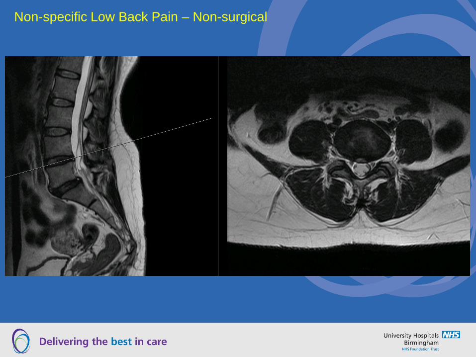
Non-specific Low Back Pain – Non-surgical

Non-specific Low Back Pain - Non-surgical

Specific Low Back Pain

Specific Low Back Pain

Specific Low Back Pain - Spondylolisthesis

Specific Low Back Pain - Spondylolisthesis

Adult Degenerative Deformity

QUESTIONS?