management of biochemical recurrence after radical prostatectomy & radiation therapy
TRANSCRIPT

GAURAV NAHAR
DNB UROLOGY(STD.)
MMHRC, MADURAI
Evaluation and Management of Prostate-specific Antigen
Recurrence After Radical Prostatectomy for Localized
Prostate Cancer

INTRODUCTION
Radical prostatectomy(RP)- the primary curative procedure for the treatment of localized prostate cancer.
Approximately one third of all patients still demonstrate disease recurrence after surgery.
For the majority, first sign of recurrent disease is a rising PSA level without either clinical or radiographic evidence of disease—the so-called ‘PSA recurrence’ or ‘biochemical failure’.

Rising PSA levels after RP may be due to 1. a local recurrence in the prostatic bed, 2. occult distant metastases or 3. a combination of both.
Quite difficult to identify recurrent lesions accurately at an early stage of PSA recurrence.
Local recurrence may be cured using salvage external-beam radiotherapy, whereas distant metastases require systemic hormonal therapy.

Majority of patients with PSA recurrence after RP- relatively young and healthy; hence treatment for PSA recurrence should aim not only to improve survival but also to preserve the quality of life.

PSA is a glycoprotein produced primarily by epithelial cells lining the acini and ducts of prostate gland.
Serum PSA levels are normally very low.
Elevated serum PSA level-d/t disruption of normal prostatic architecture- an important marker of many prostate diseases- BPH, prostatitis, and prostate cancer.


Treatment optionsT1/T2 disease
• The standard approaches for men with organ-confined T1/T2 prostate cancer are– radical prostatectomy (RP)– external beam radiation therapy (EBRT),– brachytherapy, and – active surveillance
Choice of therapy is largely a matter of patient preference.
No evidence that cure rate is different with RP, EBRT, or brachytherapy when patients are stratified based upon prognostic characteristics.

Intermediate- or high-risk T1/T2 prostate cancer
For these patients definitive treatment rather than active surveillance
Intermediate-risk disease- EBRT, brachytherapy, or RP
High-risk disease- ADT plus EBRT or RP plus adjuvant EBRT

Advantages of main treatment for early prostate cancer: EBRT
Effective long term control with high dose RxLow risk of urinary incontinenceWide range of agesWhen combined with hormonal therapy, offers a
chance of cure in high-risk of diseaseTreatments can eradicate extension of tumor beyond
the margins of prostate

Advantages of main treatment for early prostate cancer: Brachytherapy
Cancer control rate equal to surgery and EBRT for organ-confined tumor
Quicker than EBRT (one treatment)
Available for cure in a wide range of ages and in those with comorbidities

Advantages of main treatment for early prostate cancer: Radical Prostatectomy
Effective long-term cancer control Prediction of prognosis can be more precise based on
pathologic features in specimenPelvic lymph node dissection is possible through the
same incisionPSA failure easy to predict

Advantages of main treatment for early prostate cancer: Active Observation
Reduces overtreatment
Avoids or postpones treatment-associated complications
Has no effect on work or social activities

Contraindications to main treatment options for early prostate cancer
RP: High operative risk, ‘medical age’ of 70 or more, neurogenic bladder, morbid fear of surgery
Active observation: High grade tumors, pt preference, expected survival of 10 or more years.

DEFINITION OF PSA RECURRENCE AFTER RP
PSA usually reaches an undetectable level within 21–30 days after radical prostatectomy.
Persistently detectable or subsequent rising serum PSA levels (typical limit of detection is 0.05 ng/ml) after RP indicate either residual prostate cancer or recurrence.

AUA Guideline Update Panel recommended using a cut point ≥ 0.2 ng/mL, with a second confirmatory level ≥ 0.2 ng/mL, to define surgical failure.
Memorial Sloan-Kettering Cancer Center (MSKCC) demonstrated best cut point to predict the probability of metastatic progression was > 0.4 ng/mL, followed by another rise.

EAU guidelines on prostate cancer: serum PSA level of >0.2 ng/ml- residual or recurrent disease & major risk of progression when the PSA level reaches 0.4 ng/ml.
Prostate-Specific Antigen Working Group recommendation: PSA value ≥ 0.4 ng/mL, 8 weeks or more after RP and rises on a subsequent measurement.
Eight weeks is ample time to allow PSA levels to clear, given a half-life of 2 to 3 days.

EAU guidelines for follow-up of prostate cancer after treatment with curative intent, PSA measurement + DRE at 3, 6 and 12 months after treatment, then every 6 months until 3 years, and thereafter annually.
45% developed recurrence in first 2 years after RP, 76% within first 5 years, and the remaining 23% >5 years after surgery. Hence a prolonged PSA follow-up is necessary after RP.

No definite consensus regarding PSA cut-off point for defining PSA recurrence after RP, a PSA level of 0.2 ng/ml on conventional assays is the most acceptable cut-off point for PSA recurrence based on a clinical point of view.

PSA RECURRENCE AFTER RADIATION THERAPY
Biochemical failure after radiation therapy (ASTRO) as three consecutive PSA rises, optimally separated by 3 months between measurements, beginning at least 2 years after the start of radiation therapy.
Time of failure is midpoint between the nadir and the first confirmed rise, or any rise significant enough to trigger therapy.

ASTRO Phoenix Criteria recommend that biochemical failure be defined as a PSA rise of 2 ng/mL above the post-treatment nadir, whether or not the patient received hormonal therapy in conjunction with radiation therapy.
The date at which that level was reached would be the date of relapse.
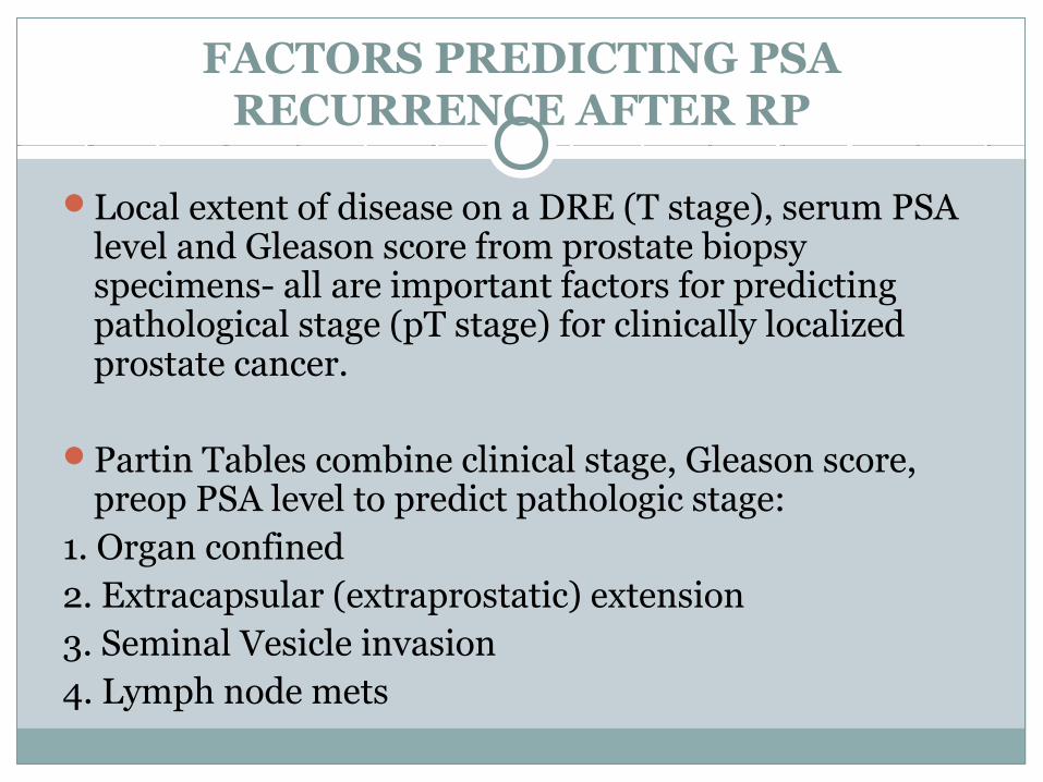
FACTORS PREDICTING PSA RECURRENCE AFTER RP
Local extent of disease on a DRE (T stage), serum PSA level and Gleason score from prostate biopsy specimens- all are important factors for predicting pathological stage (pT stage) for clinically localized prostate cancer.
Partin Tables combine clinical stage, Gleason score, preop PSA level to predict pathologic stage:
1. Organ confined2. Extracapsular (extraprostatic) extension3. Seminal Vesicle invasion4. Lymph node mets

Pretreatment risk stratification for prostate cancer

Using Partin tables, information regarding the probability of various pathological stages, such as organ-confined disease, extraprostatic extension, and seminal vesicle or lymph node involvement, is provided pre-operatively.
Such pathological stages can serve as an excellent surrogate for outcome after RP.

For majority of patients, biochemical relapse occurs far earlier than the development of radiographically evident findings or findings on physical examination or by biopsy.
Low pretreatment PSA levels, lower-grade tumors, low clinical or pathologic staging, late time from definitive local therapy to PSA relapse, and long PSADTs generally indicate a low likelihood of developing distant radiographically apparent metastases.

Serum PSA level between 10 and 20 ng/ml-intermediate risk for PSA recurrence, while serum PSA level >20 ng/ml represent a high-risk for developing PSA recurrence after RP.
Gleason grade ≥4, or a Gleason score >7 on RP specimens is predictive of a high-risk for PSA recurrence.

Histopathological determinants and molecular markers have been evaluated to predict PSA recurrence and survival.
p53 tumor suppressor gene expression, bcl-2 protooncogene expression, expression of Ki-67 & p27, apoptotic index, DNA ploidy and tumor angiogenesis (microvessel density):- all are possible predictive factors of PSA recurrence after RP.

TESTS TO DETERMINE SITE OF RECURRENCE
Current methods of detecting recurrence whether in prostatectomy bed, an irradiated gland, or metastatic sites such as bone or lymph nodes, are of very limited value.
Bone Scintigraphy:Bone scintigraphy will only detect metastatic disease
that interferes with normal osteoblast/osteoclast interactions to produce abnormal bone deposition. Areas of marrow involvement that do not impact bone metabolism will remain undetected.

No single PSA value predicts scan positivity, although PSAs will be well above 20 to 30 ng/mL before bone scintigraphy reflects metastatic disease.
Tracer uptake in areas of trauma, infection, or inflammation can easily be mistaken for metastatic disease.

CT SCAN:CT scans are not sufficiently sensitive for detecting
local recurrence until increasing rate of PSA becomes >20 ng/ml per year.
CT is suboptimal for detection of metastasis as it has a lower limit of detection of 0.5 cm & the scans are nonspecific, making it difficult to distinguish scar tissue or fibrosis from tumor.

Sensitivity & specificity of MRI and MR spectroscopy are improving; most useful for detecting nodal and bony metastases. But not sufficiently useful early in the course of PSA recurrence.
Positron emission tomography (PET-CT using FDG, 18F choline, 11C choline, 11C acetate), a biochemical imaging modality, still investigational, cannot accurately distinguish post-operative scars from local recurrence.

PROSTASCINT (Antibody based imaging/ Immunoscintigraphy):
Approved by the U.S. FDA to detect occult metastatic disease in early prostate cancer, also indicated for a rising PSA and a negative or equivocal standard metastatic evaluation when there is a high clinical suspicion of metastatic disease.
Based on a murine antibody, 7E11, combined with indium-111 to target the internal domain of PSMA, a transmembrane type II glycoprotein found on normal prostate tissue and prostate cancers.

PREDICTING LOCAL Vs SYSTEMIC RECURRENCE
Combination of Gleason score, pathological stage and serum PSA velocity 1 year after surgery best distinguished local recurrence from distant metastases.
PSADT and Gleason score are highly prognostic for clinical outcome.


TREATMENT OF PSA RECURRENCE
Depends on the site of recurrence: namely local, systemic or a combination of both.
Treatment options for presumed local recurrence include external beam radiotherapy and, for presumed distant metastasis, hormonal therapy.
Observation only is also one of the treatment options regardless of recurrence site.

Routine tests cannot identify site of recurrence untilnPSA reaches 20–50 ng/ml, at which level effectiveness of radiotherapy can no longer be expected.
Therefore, treatment is mainly selected according to the pathological findings of RP specimen and post-operative serum PSA parameters.

OBSERVATION:
Natural course from PSA recurrence to development of metastatic disease or prostate cancer-specific death is quite long.
Hence observation with delayed hormonal therapy for symptomatic or metastatic disease can be a valid treatment option.

RADIATION THERAPY:Salvage radiotherapy is the recommended
terminology for curative-intended radiation for post-operative PSA recurrence as opposed to adjuvant radiotherapy administered shortly after RP based on adverse pathological findings.
Candidates must have a life expectancy of >10 years, since salvage radiation therapy is sometimes associated with high morbidity.

Preoperative PSA level, pre-radiotherapy PSA level and seminal vesicle involvement are significant risk factors for actuarial biochemical disease-free survival following post-operative radiotherapy.
ASTRO Consensus Panel demonstrated a serum PSA level of 1.5 ng/ml as the threshold level for optimal success rates.
European Consensus Group recommended a PSA level of 1.0–1.5 ng/ml as appropriate cut-off point to initiate salvage radiotherapy for presumed local recurrence.

Dose of radiation:ASTRO Consensus Panel- 64.8Gy radiation to the
prostatic bed.European Consensus group- 64 Gy, with 1.8-2.0 Gy
per fraction.
Predictors of disease progression following salvage radiotherapy: negative/close margins, an absence of extracapsular extension, presence of seminal vesicle invasion, a Gleason score of 8–10, a pre-radiotherapy PSA level >2.0 ng/ml, a PSA doubling time of ≤10 months.

Hormonal therapy may increase sensitivity to irradiation, may be effective for possible distant metastases in such patients.
But the European Consensus Group mentioned that hormonal therapy is not standard in patients receiving salvage radiotherapy.

HORMONAL THERAPY:Androgen deprivation therapy by surgical(B/L
scotal orchiectomy) or medical castration using a LH-RH agonist or antiandrogens may improve survival.
PSA level at which hormonal therapy should be initiated remains unclear, though time to metastatic disease was delayed on starting at PSA level ≤5 ng/ml than at PSA level ≤10mg/dl.

INTERMITTENT HORMONAL THERAPY- this concept introduced to avoid the side effects of hormonal therapy.
Long-term efficacy remains unclear.
Finasteride may have an ability to delay disease progression patients in PSA recurrence after RP; long-term studies are required.

CONCLUSION
Clinical state of “Rising PSA/Biochemical recurrence” after RP- second in size only to localized disease.
Unique in that patients are characterized by an absence of symptoms, radiographic findings or pathologic findings—standard measures of treatment effects.

PSADT is one of the most common elements for stratifying patients & allocating Rx.
Patients can be divided into three groups based on prognosis:
low-risk patients are unlikely to develop metastases or symptoms or die of their disease and should be managed expectantly;
intermediate-risk patients receive androgen deprivation or can be considered for investigational approaches designed to slow the disease to the point where the patient dies of other causes; and
high-risk patients (those with PSADTs of ≤9 months) can be considered for androgen deprivation or ideally, enrolled in a clinical trial(TAX3503- Docetaxel 10 cycles; Mitoxantrone & Prednisone).

Thank You