management of retinoblastoma - human health … · chance of inheritance. • chromosomal anomaly...
TRANSCRIPT

MANAGEMENT OF RETINOBLASTOMA

INTRODUCTION
Most common intraocular malignancy of childhood arising from embryonic neural retinal cell.
Unifocal/ Multifocal.
Unilateral (70%)/ Bilateral (30%).
Sporadic (94%)/ Familial (6%).
Non hereditary (50-60%)/ Hereditary (40-50%).
EPIDEMIOLOGY
Median age
• Unilateral: 2 years, 80% below 3 - 4 years.
• Bilateral: < 12 months.
Incidence
• 1 in 15,000-20,000 live births in the US, higher in developing countries
including India
• No racial or gender predeliction.
Congenital Anomalies
• Associated in 0.05% cases of retinoblastoma.
• Cleft palate, dentinogenesis imperfecta, incontinentia pigmenti etc. with no
mental impairment.

FORMS OF PRESENTATION
SPORADIC (Non- hereditary)
• Unilateral, unifocal.
• 60% of all cases.
• Present later.
• Children of the affected are
normal.
• Chromosomal anomaly is a
somatic mutation.
• Relatives have a low risk of RB
development.
FAMILIAL (Hereditary)
• 85% bilateral, multifocal.
• 40% of all cases.
• Present earlier.
• Children of the affected have 45%
chance of inheritance.
• Chromosomal anomaly is a germline
mutation.
• Relatives have a high risk of RB
development.
• Autosomal dominant with high
penetrance.

GENETICS
• RB represents a prototypical model demonstrating genetic etiology of
cancer.
• It is caused by mutation of the RB gene, a TSG on long arm of
chromosome 13 (13q14.1-q14.2).
• Normal individual inherits two copies of this gene one from each parent.
ALFRED KNUDSON’S TWO HIT HYPOTHESIS (1971)
Two separate loss of function mutations are required to inactivate
both the homologous loci of the RB gene for malignant phenotype
to be expressed

Two mutations are required for the development of retinoblastoma.
Sporadic retinoblastoma
» Child starts with two wild type alleles (RB+/RB+).
» Both alleles must mutate to produce the disease (RB/RB).
» Probability of both mutations occurring in the same cell is low; only one
tumor forms (e.g., one eye).
» First hit occurs after conception in utero or in early childhood in retinal
cells.
» All cells in body are not affected as germ cells are not involved.
» Second somatic mutation results in loss of other normal allele.
GENETICS-SPORADIC RB

Hereditary Retinoblastoma
» Child starts with heterozygous alleles (RB/RB+).
» Only one mutation is required to produce disease (RB/RB).
» Mutations resulting in loss of heterozygosity (LOH) are more probable in rapidly dividing cells, and multiple tumors occur (e.g., both eyes).
» First hit occurs in utero in germ cells before conception or is inherited from a parent.
» All cells of body affected.
» Second hit occurs in any retinal cell.
» Increased risk for second malignancies
GENETICS-HEREDITARY RB


PATTERNS OF GROWTH
TUMOR
ENDOPHYTIC MIXED DIFFUSE INFILTRATING EXOPHYTIC
Arises from inner
layers of retina.
Fills the vitreous
cavity
Anteriorly reaches
aqueous venous
channels
May permeate
through lymphatic
channels.
Visual disturbance
& white eye reflex.
Arises from outer
layers of retina.
Fills the subretinal
space.
Posteriorly causes
serous RD.
Choroidal invasion
through Bruch’s
membrane.
Proptosis & RD.
Most common
growth pattern
No mass, only
signs of
endophthalmitis.
Older age 6 yrs.
Pseudohypopyon
resembling
inflammatory
reaction.
Diagnosis
delayed
UL & sporadic

NATURAL HISTORY
• Rapidly progressive tumor
• Untreated fills the eye & completely involves the globe.
• Metastasis (BM, bone, LNs and liver) is rare at presentation.
ROUTES OF SPREAD
Direct local
Tumor infiltration
Anterior spread to
Conjunctiva,
Eyelids &
Extra ocular tissue
Hematogenous
dissemination
From orbital, bone or
lymphatic invasion
Subarachnoid
Space Of
optic nerve
Choroid invasion
Scleral invasion
Orbital soft
tissue, bone &
brain invasion
CSF dissemination
To brain & spine
Lymphatic
dissemination

TRILATERAL RB
• Primary Retinoblastoma of pineal & parasellar sites.
• Single tumor.
• Well differentiated: more rosettes, fleurettes & photoreceptor differentiation.
• Majority – familial retinoblastoma.
• Usually fatal due to meningeal spread, median survival of 9 months.
• Appear years (median time 40 months) after successful treatment of primary.
• Main cause of death in RB patients between 3 to 8 years.
• D/D: Metastatic or recurrent retinoblastoma.

HISTOPATHOLOGY
Also –
• Calcification +++
• Necrosis ++
• Multifocality.
• Composed of uniform small round or polygonal
mitotically active cells.
• Viable tumor cells surround blood vessels & form
pseudorosettes.
• Cells are arranged in three characteristic types:
• Flexner-Wintersteiner rosette: characteristic of
RB but also seen in pineoblastoma &
medulloepithelioma. Cells resembles
retinoblasts of embryo.
• Homer-Wright rosette.
• Fleurette

CLINICAL PRESENTATION
Developed countries: present with signs rather than symptoms, IO tumor without local extension.
Developing countries: diagnosed only after an enlarged eye or gross orbital extension.
Leucokoria (60%): lack of red reflex of the eye in large tumors, RD, retrolental mass
or vitreous opacification due tumor cells which is often noticed by the mother.
Strabismus (20%): disruption of fusional reflex due to loss of central vision from a
tumor in the macula.
Rubeosis iridis (17%): in advanced cases due to extensive tumor necrosis releasing
angiogenic factors.
Heterochromia.
Spontaneous hyphaema
Glaucoma: neovascular or closed angle.
Pseudohypopyon: seeding of ant. chamber in endophytic or diffuse infiltrating
tumors.
Pain: glaucoma or inflammation.
Proptosis.

DIAGNOSTIC EVALUATION
HISTORY: Administration of oxygen at birth, eating
of dirt, association with dogs & Family history of RB.
SYMPTOMS & SIGNS: Ocular as well as systemic.
OPHTHALMOSCOPIC EUA
• Indirect ophthalmoscopy with pupillary dilation & general anesthesia.
• Number, size, location (anterior or posterior), laterality, disc diameter, subretinal fluid or seeds noted and degree of exophthalmos measured.
• Detailed mapping done with appropriate diagrams & description (relation with ora serrata, optic disc & macula).
• Creamy pink or snow white mass projecting into the vitreous.
• Poorly developed stroma gives way to tumor bits forming vitreal seeds
• RD, vitreal opacification & h’ge make diagnosis difficult.

STAGING
• Though most cases are diagnosed clinically, imaging is done:
– Confirm diagnosis.
– Estimate tumor size.
– Document intralesional calcium.
– Assess for spread of tumor into optic nerve, choroid, sclera & orbit.
– Detect ectopic disease in pineal or suprasellar region.
• Differentiating RB from other ocular lesions in child presenting with atypical
features (only RD or opaque vitreous, atypical mass).

OCULAR ULTRASOUND
• Demonstrates a mass more echogenic than the vitreous on B mode & highly
reflective intrinsic echoes of fine calcifications on A mode.
• RD may also be seen in exophytic tumors.
• Accuracy: 80% (limited by vitreal opacities & RD).
• Limited evaluation of medial & lateral extension, extraocular disease.
• Color doppler displays normal & tumor vasculature & differentiates subretinal or
choroidal h’ge from neoplasms

CT SAN / MRI SCAN
• 90% show calcification
• Dense homogenous
• Extension to choroid, vitreous &
sclera not reliable.
• Detects intracranial disease
• 3D multiplanar capability.
• Hyperintense to vitreous on T1
& markedly hypointense on T2
• Delineation of ON, IO & EO spread
• Differentites between tumor, RD
& subretinal fluid.

STAGING SYSTEMS-CLINICAL

STAGING SYSTEMS-CLINICAL

PROGNOSTIC FACTORS
• Optic nerve invasion.
• Massive choroidal invasion, CB: increased possibility of hematogenous
spread (60%) & extension to extrascleral tissues (6 years DFS 90% in IO
disease versus 10% for EO disease).
• Gross extraorbital extension has >90% risk of metastasis.
• Poorly differentiated tumor.
• Anterior chamber invasion: mortality 20 to 80%.
• Large tumor with vitreous seeding.
• Rubeosis iridis.
• Glaucoma.
• Bilateral tumors behave poorly as mortality result from second cancers &
trilateral RB.
• Trilateral RB has almost 100% fatality.

EXTENT OF
INVASION OF
OPTIC NERVE
MORTALITY
RATE
SURVIVAL
Superficial 10% Similar to
uninvolved
ON 90% Upto Lamina
cribrosa
29%
Posterior to
Lamina cribrosa
42% 60%
Positive
transected margin
80% 20%
Stump >5mm better better

MANAGEMENT OF RB
• Multidisciplinary approach: Ocular oncologist, pediatric oncologist, radiation oncologist, radiologist and child psychologist.
• Treatment is tailored to each individual.
• Goals of treatment:
– Save life.
– Preserve vision or salvage eye (i.e. avoid enucleation).
– Minimize any complications or side effects of therapy.
• Choice of therapy:
– Risk of metastatic disease.
– Systemic status.
– Laterality of disease/ size/ location of tumor.
– Visual prognosis.
– Risk of second cancers.
TREATMENT TECHNIQUES
• ENUCLEATION
• EXENTERATION
• EBRT
• FOCAL THERAPIES
– Plaque Radiotherapy
– Laser Photocoagulation
– Cryotherapy
– Thermotherapy
– Chemothermotherapy
• CHEMOREDUCTION
– Intravenous
– Subconjunctival
– Transpupillary
• SYSTEMIC CHEMOTHERAPY
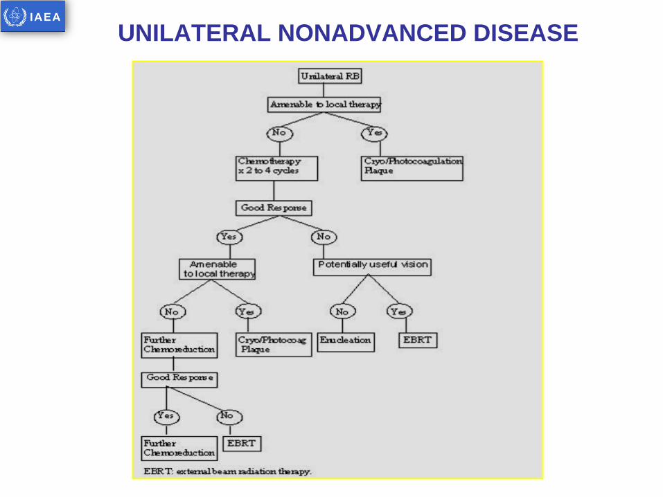
UNILATERAL NONADVANCED DISEASE

UNILATERAL ADVANCED DISEASE

FLOW CHART: BILATERAL DISEASE

ENUCLEATION
INDICATIONS
• Unilateral or bilateral RB completely filling the globe with no hope of visual salvage due to damage to entire retina.
• Tumor invasion in optic nerve, choroid, AC, pars plana or orbit.
• Painful glaucoma with loss of vision.
• Tumor unresponsive to other forms of conservative treatment.
• Inability to examine retina secondary to vitreous h’ge or cataract following conservative therapy.
PROCEDURE
• Involves removal of the eye leaving behind lids and extraocular muscles but removing the longest possible segment (10 to 15mm) of optic nerve in continuity with the globe.
• Care should be taken to avoid perforation of the globe to prevent seeding.
– Scleral perforation at the site of muscle insertions.
– Traction sutures in the muscles.
• Optic nerve snares or clamps should be avoided to prevent crush artefact which may be misinterpreted as invasion by tumor.
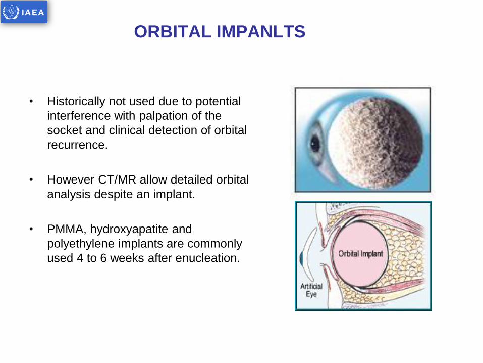
ORBITAL IMPANLTS
• Historically not used due to potential
interference with palpation of the
socket and clinical detection of orbital
recurrence.
• However CT/MR allow detailed orbital
analysis despite an implant.
• PMMA, hydroxyapatite and
polyethylene implants are commonly
used 4 to 6 weeks after enucleation.

ADJUVANT THERAPY AFTER ENUCLEATION
Orphanet Journal of Rare Diseases 2006, 1:31

CHEMOTHERAPY
GOALS OF CHEMOTHERAPY
• Reduction of tumor size → RD dealt with focal therapy is the standard
of care in early stage disease.
• Reduce the use of EBRT which reduces second malignancies and
orbitofacial growth anomalies in early stage.
• Reduce the need of enucleation in early stage.
• Reduce the risk of local and systemic relapse in advanced stage.
• Improve survival in metastatic disease.
• Neoadjuvant
– IORB - BL disease, UL disease not amenable to local therapy
(6 to 12 cycles).
– EORB – Orbit/bone involvement, metastatic spread
• Adjuvant: High risk histopathological features.
• Salvage: Recurrent disease in an only eye.

EYE PRESERVATION AS PER STAGE
EYES PRESERVED
RE I to III RE IV to V
EBRT ALONE 53% 45%
EBRT+SALVAGE 96% 66%
CT ALONE 29% 11%
CT +SALVAGE 94% 66%
CTRT - 75%
Visual results Limited by
• Macular involvement
• Tractional Retinal Detachment
• Hemorrhage

CRYOTHERAPY
• Rapid freezing forms intracellular crystals which ruptures tumor cells and causes vascular occlusion.
• Under GA, pencil like probe is placed precisely on the sclera directly behind the intraocular focus of RB.
• Fails if overlying vitreous seeding present.
• 1 or 2 sessions at 1 month interval are required.
• Indication: Small primary or recurrent tumor in anterior retina i.e. equatorial and peripheral region or post EBRT residual tumor < 2mm thick and < 3.5 mm diameter.
• Complications: vitreous hemorrhage, choroidal effusion, retinal detachment, localized periretinal fibrosis and retinal tear.

PHOTOCOAGULATION
• Argon/ Diode laser/ Xenon arc.
• Light is focused through dilated pupil under GA and the feeding vessels are
coagulated which results in involution of tumor.
• Indications:
– Small primary or recurrent tumor in posterior part of retina < 2.5 mm
thick and < 4.5 mm diameter.
– Retinal neovascularization due to radiation retinopathy.
• Most tumors require 2 to 3 sessions to be cured.
• Contraindications:
– Tumor located at or near macula or pupillary area.
– Mushroom shaped tumors
– Tumors arising from a vitreous base.

THERMOTHERAPY
• Ultrasound/microwave/infrared radiation used to deliver hyperthermia to
tumor.
• 42 to 600C (which is below coagulation threshold) of heat produces a grey
white scar but does not photocoagulate retinal vessels.
• Synergistic effect with CTRT
• Indications:
Thermotherapy alone: small tumors outside retinal arcade < 3mm diameter
and 2 to 3mm thick without vitreous or sub-retinal seeds produces control
rates of 86%
• Thermochemotherapy (TCT): Useful for larger tumors after tumor shrinkage
following 2 to 3 cycles when they satisfy above size criteria (thickness>4mm
associated with higher recurrences).

THERMOTHERAPY
• Mechanism of action:
– Membrane damage.
– Protein denaturation.
– Chromosomal damage.
– Disruption of biochemical pathways.
– Ischemic necrosis.
• Schedule:
– Thermotherapy alone: 300MW power for >/= 10 mins upto45 to 600C at 1 monthly interval for 3 sessions produces grey white scar.
– TCT: 42 to 450C for 5 to 20 mins depending on size (upto 15 mm diameter) produces a light grey scar.
• Complications: focal iris atrophy and focal para axial lenticular opacity.
• Advantage: suitable for small tumors adjacent to fovea and optic nerve in which plaque therapy or laser photocoagulation would possibly induce more profound visual loss.
• Disadvantage: Time consuming, tedious process that requires careful observations, recordings, subjective and treatment adjustments required in response to subtle tumor changes.
THERMOTHERAPY

LOCAL ADMINISTERED CARBOPLATIN
• Being evaluated at present for
advanced intraocular RB since
achieves high concentration in
vitreous humor.
• Subconjunctival: levels peak at 1 hr
and diminishes thereafter slowly.
• Iontophoretic: levels slowly peak at 6
hrs.
• Could be combined with focal
therapies and avoid systemic
administration.
Abramson DH. Opthalmology 1999; 106:1947-50
Hayden BH. Arch Opthalmol 2000;118:1549-54
Simpson AE. Arch Opthalmol 2002;120:1069-74

SCLERAL PLAQUE THERAPY
• 1929: Foster, Moore and Scott used Ra seeds
• 1948: Henry Stallard pioneered and refined the technique, initial Ra
applicator was replaced by cobalt 60 plaque.
– Curved applicator to fit the eye with suture holes for fixing.
– Left in place for 3 to 7 days to deliver 40 Gy to tumor apex and 100 to
200 Gy to tumor base.
– Disadvantage: No external shielding resulting in high radiation dose to
orbital bones and the surgeon.
• 1970-80’s: other radio-isotopes used e.g. I125, Ir192, Ru106
Plaque Energy Half life Penetration
Co60 1.33-1.7MV 5.2 years
I125 27-25Kev 60 days upto 10mm
Ir192 295-612Kev 74 days
Ru106 3.5Mev (β) 368 days upto 6mm

I125 CLAWS FOR WHOLE EYE-STANNARD

EXTERNAL BEAM RADIOTHERAPY
• Indications:
– Lesions close to macula or optic nerve.
– Larger tumors with vitreous seeding.
– Recurrent disease.
– Adjuvant postoperative radiotherapy after enucleation in high risk pathologic features.
– Palliative radiotherapy
– Progression after chemoreduction
• Target volume: Entire retina upto ora serrata and atleast 1 cm of ON accepting the potential for cataract formation.
– All retinal cells have neoplastic potential resulting in recurrences in retina as well as vitreous.
– RB is a multifocal disease.
– Tumor may even spread subretinally.

TARGET VOLUME
EBRT does not prevent the appearance of new tumors in clinically uninvolved retina.
Therefore, the traditional belief that external beam radiation can treat the retina “prophylactically” should be seriously questioned.
Focal treatment modalities (plaque brachytherapy, photocoagulation and/or cryotherapy), when clinically feasible, should be considered the treatment of choice for intraocular retinoblastoma.
EBRT should be considered only when focal treatment modalities are not clinically indicated.
Hernandez, IJROBP, 1996
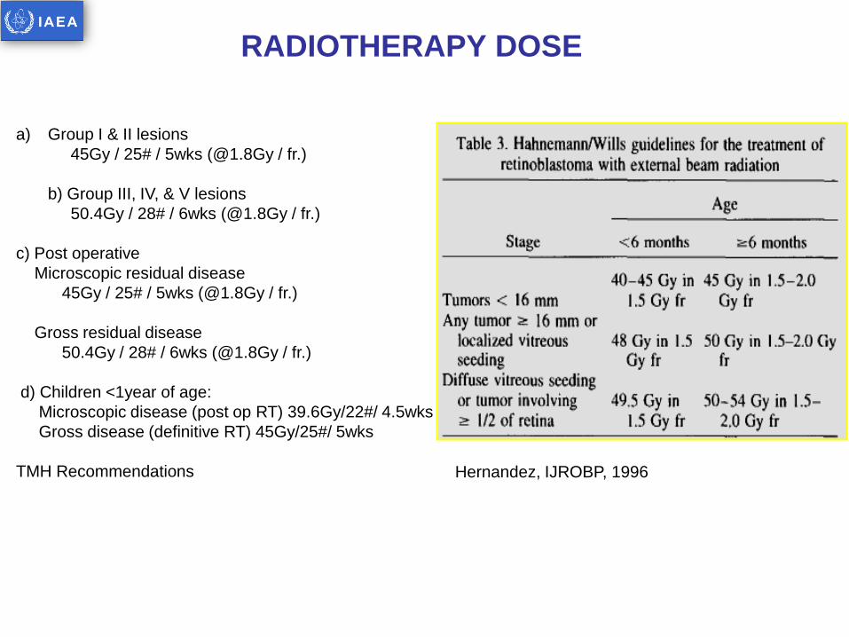
RADIOTHERAPY DOSE
Hernandez, IJROBP, 1996
a) Group I & II lesions
45Gy / 25# / 5wks (@1.8Gy / fr.)
b) Group III, IV, & V lesions
50.4Gy / 28# / 6wks (@1.8Gy / fr.)
c) Post operative
Microscopic residual disease
45Gy / 25# / 5wks (@1.8Gy / fr.)
Gross residual disease
50.4Gy / 28# / 6wks (@1.8Gy / fr.)
d) Children <1year of age:
Microscopic disease (post op RT) 39.6Gy/22#/ 4.5wks
Gross disease (definitive RT) 45Gy/25#/ 5wks
TMH Recommendations

EBRT TECHNIQUES
Described by Reese in 1930
Orthovoltage beams attempting lens sparing
High dose to bone: Bony deformities

Single En-Faced Megavoltage Beam
Ant. Border at Bony Orbital Margin
High Dose to Bony Orbit

Anterior border at the limbus for lateral field.
Half beam block anteriorly.
4 to 6 MV photon field.
Eyes closed to spare minor salivary glands
and eyelids.
Anterior field with electrons to prevent
underdosing of AC and prevent exit dose
1. Classic single temporal portal 3×4 cm,
anterior border at lateral bony canthus with
posterior 150 tilt (D-shaped field).
2. Anterolateral differentially weighted beams
with anterior lens shield.
Underdose of anterior structures of eye.
Higher recurrences.
Cataract formation still higher.
Hernandez, IJROBP, 1996

3D-CONFORMAL RT
• Four non co-planar fields
• All anterior oblique field: sup,inf, med, lat.
• Less orbital hypoplasia.
• Minimize dose to opposite eye, optic
chiasma, post. Pituitary, upper cervical
spine.
• Tumor = 95% & orbit = 50%.
• More homogenous dose distribution.
• Less vitreal recurrence.

INTENSITY MODULATED RT (IMRT)
RT DOSE: 45-50Gy

Significant Reduction in Volume of Bony Wall of Orbit in
High Dose Region

Recurrence occurs usually within 3 yr.
Follow up done for indefinite period for diagnosis of second malignancy and
tumor control
OPTHALMOSCOPIC EXAMINATION :
• First year: every 2-3 months .
• Second year: every 3-4 months.
• 3-5 years: every 6 months.
• > 5 years : every one year..
FOLLOW UP

SECOND CANCERS
• Subsequent cancer risk in 963 hereditary patients (SIR, 19; 95% CI, 16 to 21) exceeded the risk in 638 nonhereditary Rb patients (SIR, 1.2; 95% CI, 0.7 to 2.0).
• Radiation further increased the risk of another cancer in hereditary patients by 3.1-fold (95% CI, 2.0 to 5.3).
• Hereditary patients continued to be at significantly increased risk for sarcomas, melanoma, and cancers of the brain and nasal cavities.
• The cumulative incidence for developing a new cancer at 50 years after diagnosis of Rb was 36% (95% CI, 31% to 41%) for hereditary and 5.7% (95% CI, 2.4% to 11%) for nonhereditary patients.
Klienerman, JCO 2005

SUMMARY
Retinoblastomas should be treated by a group of specialists skilled in management
of childhood malignancies.
Team should include Ophthalmologist, Medical Oncologists, Radiation Oncologists,
Ocular surgeon, Genetic counselors
Multimodality treatment comprising Chemotherapy, Focal Therapies, Radiotherapy,
& Surgery results in optimal outcomes
Customization of treatment is necessary based on disease status, risk factors, &
Response to therapy
Radiation therapy in the form of plaque bracytherapy & EBRT are useful modalities
for achieving local control
EBRT is extremely effective in palliation of locally advanced & metastatic disease