management of tuberculous pleural effusion
TRANSCRIPT
Management of Tuberculous Pleural Effusion
Beenish Ajmal, Kanza Ijaz ,Khawaja Tahir Mahmood Department of pharmacy, Lahore College for Women University, Lahore
Abstract Tuberculous (TB) pleural infusion is a buildup of fluid in the space between the lining of the lung and the lung tissue (pleural space) after a severe, usually long-term infection with tuberculosis. A study was carried out among 20patients of TB pleural effusion, of which 3 could be followed for a period of 3months. The aims of the study were to assess the patient compliance with the management methods for tuberculous pleural effusion, the end results of treatment of pleural effusion with anti-TB drugs and aspiration methods, the relapse rate over a 3 months period, patient satisfaction with DOTS medicines as well as aspiration therapy, to prevent the subsequent development of active tuberculosis & to relieve symptoms. The methods employed to accomplish these aims were development of a medication history/patient interview form. Patients with tuberculous pleural effusion were interviewed & national treatment guidelines were compared with international treatment guidelines. The results revealed that 90% were satisfied with the DOTS policy, with respect to management of tuberculous pleural effusion, at the end of treatment whereas 10% expressed dissatisfaction with this policy.
Key words: Aspiration, DOTS, Management, Tuberculous Pleural Effusion INTRODUCTION Tuberculous (TB) pleural infusion is a buildup of fluid in the space between the lining of the lung and the lung tissue (pleural space) after a severe, usually long-term infection with tuberculosis [1] Tuberculous pleural effusion is one of the most common forms of extra pulmonary tuberculosis (TB). The immediate cause of the effusion is a delayed hypersensitivity response to mycobacterium antigens in the pleural space. For this reason microbiological analyses are often negative and limited by the lengthy delay in obtaining results [2] The clinical importance of pleural effusions ranges from incidental manifestations of cardiopulmonary diseases to symptomatic inflammatory or malignant diseases, as shown in the image below, requiring urgent evaluation and treatment [3] Treatment of tuberculous pleural effusion will always involve a combination of many drugs (usually four drugs). The medicines are continued until lab tests show which medicines work best. The outlook is excellent if tuberculous pleural effusion is diagnosed early and treatment is begun quickly. Tuberculous pleural effusion
can cause permanent lung damage if not treated early. The highest diagnostic yield was obtained by histology (85%), followed by culture of pleural biopsy (37%) and pleural fluid culture (36%) [4] Closed pleural biopsy remains the most effective diagnostic method, and ADA level is a cheap diagnostic method in countries with a high prevalence of TB [5] There is no doubt that pleuroscopy-guided biopsy is of great value for TPE diagnosis; however, sensitivity and specificity of noninvasive tests, especially ADA, can help to distinguish between TB and malignancy [6] The investigation of pleural effusion has been greatly assisted by advancements in pleural fluid analysis. In the case of tuberculous pleural effusion, diagnosis traditionally requires the demonstration of acid fast bacilli in the pleural space using microbiological or histological techniques. [7] Pleural effusion accounts for 22.1% of cases of pediatric pulmonary tuberculosis. Parenchymal consolidation is the most common associated radiographic finding. Bacteriologic confirmation was achieved in 56.4% of cases. A short course of chemotherapy is effective [8]
Beenish Ajmal et al / J Biomed Sci and Res., Vol 3 (1), 2011,302-307
302
If the patherapy sunderlyingpatient hashould bePneumoniapulmonaryexudative are usuallymedical transudateeffusions ceven if thspecific trto provide effusions dthe effusioThe manastatus of thCurrently, chemothermonth regand 2-drugchest tubmanagemepleural Efthe ever ingroup of p METHODStudy wasHospital, Lrandomly followed fhistory taradiologicabiochemicaspiration Each sampmycobactehistologicafindings, biochemicanalyzed. effusion wwere takesignificantas age, sexclinical d
atient has should beg heart faias an exude made ta, cancey emboliseffusions [
y managed disorder.
s or exudatecausing sevhe cause is eatment is relief. The
depends on on [10] agement dephe patient,
the rapy for tubgimen, with g continuatibe drainageent of both mffusion. Thincreasing coatients [12]
DS s carried oLahore. 20
selected, for a perioaking and al findings, al profiles and biops
ple of pleurerium and al exam
cytologicaal analysisPatients w
were interven on a prt and relevax, duration diagnosis a
a transudae directed ilure or cirdative effuto define er, tubercm accoun
[9] Transudby treating
Howeves, refractor
vere respiratunderstood
available, cmanagemethe underly
pend on thseverity of standard
berculosis co a 4-drug iion phase [1e is effecmalignant ais approachost of hospit.
out at Gulapatients ofof which
od of 3monphysical
hematologwere rec
sy were alral tissue w
the rest mination. al, microbs of pleurwith tuberciewed and redesigned ant points wof hospitaliand treatm
ative effustoward
rrhosis. If usion, attem
the etioloculosis, nt for m
dative effusithe underly
ver, whery large pletory symptod and disecan be drai
ent of exudaying etiolog
he performathe sympto
short-couomprises a intensive ph11] Out-patctive for and suppurah would redtal care for
ab Devi Chf any age w
3 could nths. Thoro
examinatiogical and serorded. Plelso perform
was culturedwas sent
Macroscobiological ral fluid wculous plecase histoPerforma
were noted sization, cau
ment. Natio
sion, the the
mpts ogy. and
most ions ying ether eural oms, ase-ined ative y of
ance oms. urse six-
hase tient
the ative duce this
hest were
be ough ons, rum
eural med. d for
for opic and
were eural ories and
such uses, onal
treatment internationInclusion c 20patie
females PatientsExclusion Renal
insuffichigh vaOf pleu
MultiplPatientspleural
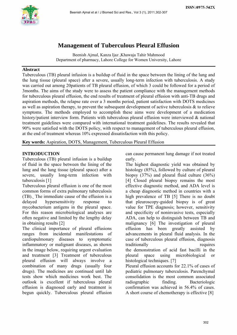
Patient Neonat RESULTSData of 20effusion wsetting. Tanalyzed du
Fig 1: AWPLEURAL
Fig 2: PRPLEURAL
01020304050607080
Know
25
0
10
20
30
40
50
60
70
ma
guidelines al treatmentcriteria ents of eithes randomly s of all agescriteria
insufficiciency: Patialues ural fluid Adle pathologs with moeffusion.
’s refusal tes were exc
S 0 patients
was studied The followuring the stu
WARENESS EFFUSION
REVELANCEFFUSION
ws doesn't know
5
75
ales females
65
35
were cot guidelines
er sex i.e. bselected
s were inclu
iency aients may
denosine degy of plere than on
cluded from
with tuberin a specia
wing paraudy:
ABOUT TU
CE OF TU
s
ompared ws.
both male a
uded
and/or lipresent fau
eaminase levural effusi
ne etiology
m the study
culous pleualized hospameters w
UBERCULO
UBERCULO
with
and
iver ulty
vel ion: of
ural pital were
OUS
OUS
Beenish Ajmal et al / J Biomed Sci and Res., Vol 3 (1), 2011,302-307
303
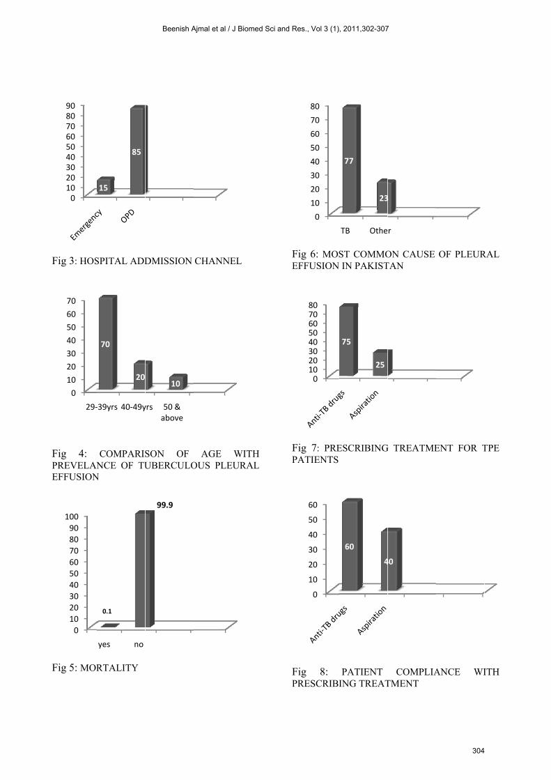
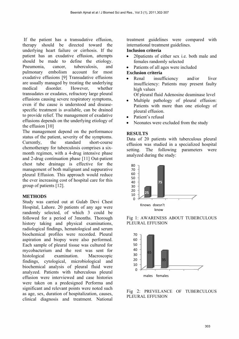
Fig 3: HOS
Fig 4: PREVELANEFFUSION
Fig 5: MOR
0102030405060708090
0
10
20
30
40
50
60
70
29‐
0
10
20
30
40
50
60
70
80
90
100
SPITAL ADD
COMPARISNCE OF TU
N
RTALITY
15
85
‐39yrs 40‐49y
70
20
yes no
0.1
DMISSION
SON OF UBERCULO
yrs 50 & above
10
99.9
CHANNEL
AGE WOUS PLEUR
WITH RAL
Fig 6: MOSEFFUSION
Fig 7: PREPATIENTS
Fig 8: PRESCRIBI
0
10
20
30
40
50
60
70
80
01020304050607080
0
10
20
30
40
50
60
ST COMMO IN PAKIST
ESCRIBING
PATIENT ING TREAT
TB Othe
77
23
75
25
60
4
ON CAUSE TAN
G TREATME
COMPLIATMENT
r
3
40
OF PLEUR
ENT FOR T
ANCE WI
RAL
TPE
ITH
Beenish Ajmal et al / J Biomed Sci and Res., Vol 3 (1), 2011,302-307
304
Fig 9: PRESCRIBACCORDASTANDAR
Fig 10: ACCORDA
Fig 11: SAWITH DOT
0
10
20
30
40
50
60
0
10
20
30
40
50
60
70
Lo
0102030405060708090
RATIONABING ANCE WITRD TREATM
PREVELANCE WITH
ATISFACTITS MEDICIN
40
60
ow Middle
70
20
90
10
AL vs. TREATME
H CRITERMENT GUID
LANCE OH ECONOM
ION FROMNES
High
5
IRRATIONENT
RIA GIVENDELINES
OF TPE IC STATUS
M TREATME
NAL IN
N IN
IN
S
ENT
Fig 12: RE
Fig 13: SPATIENTS
Fig 14: PEVARIOUS
0102030405060708090100
Ye
0
10
20
30
40
50
60
Righsid
ELAPSE
SIDE OF WITH TB P
RCENTAGS SYMPTOM
es No
3
97
ht e
left side
58
40
CHEST INPLEURAL E
GE OF PATMS
both sides
2
NVOLVED EFFUSION
TIENTS WI
IN
ITH
Beenish Ajmal et al / J Biomed Sci and Res., Vol 3 (1), 2011,302-307
305
DISCUSSION Common presentation of tuberculous pleural effusion were fever, breathlessness, cough, night sweat, loss of appetite, weight loss and chest pain. Cough was mainly unproductive. Haemoptysis was rare. Generally clinical symptoms and physical signs do not positively help for definitive diagnosis of TPE [13] Diagnostic pleural aspiration and pleural biopsy could be performed by a single session of procedure. Results of cytological and microbiological examination as well as pleural biopsy could be obtained within 3 to 5 days. The procedure needed to be repeated in patients with non informative pleural fluid and pleural biopsy examinations. The natural history of an untreated tuberculous pleural effusion is characterized by spontaneous resolution in 4 to 16 weeks with subsequent development of active pulmonary TB in 43 to 65% of cases. These data emphasize the importance of proper diagnosis and treatment of tuberculous pleural effusions. According to the directly observed treatment short-course guidelines, severely ill patients with extensive or bilateral pleural effusions and sputum positivity are given treatment under category I (treated during intensive phase with four drugs: isoniazid, rifampin, pyrazinamide, and ethambutol for 2 months followed by continuation phase of 4 months with isoniazid and rifampin).[14] Those with a solitary TB pleural effusion should be treated with isoniazid, rifampin, and pyrazinamide for 2 months followed by 4 months of two drugs, isoniazid and rifampin. Especially in loculated TB pleural effusions, there can be delayed resorption of pleural fluid even after completion of 6 months of treatment [15] Under the Revised National Tuberculosis Control Programme, patients who are sputum negative after 3 smear examinations are subjected to radiological examination after they fail to respond to a course of antibiotics for a period of two weeks. Those showing radiological evidence of pleural effusion are examined physically and investigated further by tuberculin testing and diagnostic aspiration. Those confirmed to be suffering from
tuberculous pleural effusion by naked eye examination; biochemical tests and cytology of pleural fluid are given a fixed schedule of drugs. The treatment is stopped after six or eight months as per category, and outcome is reported as “Treatment Completed” [16]. CONCLUSION Involvement of pleural surfaces in patients with tuberculosis is still one of the most common extra-pulmonary manifestations of thoracic tuberculosis. Six months intermittent regimens are now considered standard and effective therapy in most parts of the world. Failure to improve with antituberculous therapy may be due to concurrent mixed infection with anaerobic bacteria. Steroids accelerate the resolution of symptoms and reabsorption of fluid. Isoniazid, Ethambutol, Rifampicin, Pyrizamide and Streptomycin for six months were reported to be the course of management. However in pregnancy Streptomycin was strongly contraindicated. A relapse rate of 3% after 3 months of follow up was noted. The patients of pleural effusion who are treated under DOTS often fail to get convinced about the successful results of treatment and question the treating physician about the basis of his assessment for stopping treatment. ACKNOWLEDGEMENTS It is a great pleasure for us to express our hearty thanks and gratitude to our vice chancellor Dr. Bushra Mateen, Registrar Ms Shaista Vine and head of department Dr. Hafeez Ikram for their special guidance and kindness to our department.It is our utmost pleasure to avail this opportunity to express gratitude and deep sense of obligation to our teachers Ms Fatima Amin and Ms Mariyam for their valuable and dexterous guidance.Very special thanks to Dr. Muhammad Akram Choudhary for giving us the chance to work in Gulab Devi Hospital under his guidance. A very special thanks to Dr. Ijaz Nasir, Dr. Farida Ijaz and other working staff to provide us with every aspect of information which we needed and for all their kind and highly respected attitude
Beenish Ajmal et al / J Biomed Sci and Res., Vol 3 (1), 2011,302-307
306
REFERENCES [1]. Philadelphia, Pa: Saunders Elsevier, Cecil
Medicine; 2007, 23rd Ed: chap 345. [2]. José m. porcel, tuberculous pleural effusion,
2010, 187, 263-270 [3]. Jeffrey rubins, md, Pleural effusion, 2010,
medscape [4]. Richter C, Perenboom R, Swai AB, Kitinya
J, Mtoni I, Chande H, Kazema RR, Chuwa LM, Mwakyusa DH, Maselle SY , Diagnosis of tuberculosis in patients with pleural effusion in an area of HIV infection and limited diagnostic facilities, 1994, 46(5), 293-7.
[5]. Salazar-Lezama M, Quiroz-Rosales H, Bañales-Méndez JL, Sánchez-Guzmán M, Villarreal-Velarde H, Báez-Saldaña R, Azcona-Martínez E, Selman-Lama M, Diagnostic methods of primary tuberculous pleural effusion in a region with high prevalence of tuberculosis, a study in Mexican population, 1997, 49(6), 453-6.
[6]. Zarić B, Kuruc V, Milovancev A, Markovic M, Sarcev T, Canak V, Pavlović S, Differential diagnosis of tuberculous and malignant pleural effusions: what is the role of adenosine deaminase, 2008, 186, 233-240
[7]. McGrath EE, Anderson PB, Diagnostic tests for tuberculous pleural effusion, 2010, 29, 1187-1193
[8]. José m. Merino, md, Isabel carpintero, md, Teresa alvarez, md, Jesus Rodrigo, md, Jesus Sanchez, md and José m. Coello, md, tuberculous pleural effusion in children, 1999, 115, 126-30
[9]. Porcel jm, Light rw, diagnostic approach to pleural effusion in adults, 2006, 73(7), 1211-20.
[10]. Jeffrey rubins, md, pleural effusion: treatment & medication, 2010, medscape
[11]. W.w. Yew, c. Lange and c.c. Leung, treatment of tuberculosis updates 2010, 2010, 10, 1183
[12]. Adeoye po, salami AK, koledoye a, early experience with outpatient tube drainage for management of pleural collections, 2009, 28(6), 364-7.
[13]. Zay Soe, Wunna Hla Shwe, Soe Moe, A Study on Tuberculous Pleural Effusion, 2010, 2, 32-48
[14]. Paul garner, jimmy volmink, directly observed treatment for tuberculosis, 2003; 327, 823
[15]. Arun Gopi, Sethu M. Madhavan, Surendra K. Sharma, Steven A. Sahn, Diagnosis and Treatment of Tuberculous Pleural Effusion in 2006, 2007, 131, 880-889
[16]. Arora vk, sarin r., revised national tuberculosis control programme: Indian perspective.2000, 42(1):21-6.
Beenish Ajmal et al / J Biomed Sci and Res., Vol 3 (1), 2011,302-307
307