manual for health professionals - international agency for...
TRANSCRIPT

1
Manual forHealth Professionals
Directorate General of Health ServicesMinistry of Health and Family Welfare
Government of India
November 2005
Manuals for Training in Cancer Control

2
CONTENTS
Foreword 03
Preface 05
1. Magnitude of Cancers 07
2. Principles of Cancer Control 15
A. Primary preventionB. Early detectionC. ScreeningD. Diagnosis and treatment
3. Common Cancers 33A. Cancer of the oral cavityB. Cancer of the uterine cervixC. Cancer of the breast
4. National Cancer Control 58Programme (NCCP)
3
FOREWORD
India is one of the few countries in the world to have a National Cancer Control Programme.The programme was conceived with the objectives of providing preventive and curativeservices through public education and enhancement of treatment facilities.
We have been able to develop 23 Regional Cancer Centres and several Oncology Wingsin India, which provide comprehensive cancer care services. One of the major limitationsof the programme is the late stage at presentation of common cancers thus reducing thechances of survival. There is a need to increase awareness among the community regardingprevention and early detection of cancers. The programme is developing IEC materialsfor the same. Once the population is armed with the necessary information, it is expectedthat the health system should be geared to tackle the increased demand for care. Therehave to be trained health care professionals to support the needs of the community. Thiscan be addressed by proper training and sensitisation of general practitioners and healthcare providers.
These manuals are developed for training health professionals and specific modules havebeen prepared for Cytology, Palliative care and Tobacco cessation. The facilitator’s manualwill assist the trainers to conduct the programmes. The manuals are self-explanatory andthe health professionals will be able to use them on their own.
(S. P. AGARWAL)
Dr. S. P. AGARWALM. S. (Surg.) M. Ch. (Neuro)
DIRECTOR GENERAL
GOVERNMENT OF INDIADIRECTORATE GENERAL OF HEALTH SERVICES
NIRMAN BHAVAN, NEW DELHI - 110011TEL. NO. 23018438, 23019063
FAX NO. 91-11-23017924Dated: 13th September, 2005
Hkkjr ljdkj
LokLF; lsok egkfuns’kky;
fuekZ.k Hkou] uà fnYyh & 110011

4

5
PREFACE
Demographic and epidemiological transitions and changes in lifestyle are leading to theemergence of cancer and other chronic diseases as public health problems in India. Cancerpattern in India reveals the predominance of tobacco related cancers, which are amenableto primary prevention. Cancer Registries in different parts of the country reveal that majorityof cancer cases present in an advanced stage and makes treatment options prolonged andexpensive. Therefore, the National Cancer Control Programme has placed its emphasis onprevention, early detection, enhancement of therapy facilities and provision of pain andpalliative care. Comprehensive legislation on tobacco by the Government of India will helpto control the tobacco related cancers. The programme has been able to augment thetreatment capacity and to address the geographical gaps in cancer care services. Awarenessand early detection programmes are undertaken through District Cancer ControlProgrammes.
Health care personnel have a major role in providing awareness, promoting early detection,prompt referral to a cancer treatment facility and in providing pain relief and palliative care.The knowledge and skills in the above areas have to be enhanced and these manuals havebeen developed in response to this need. This set of manuals, which consists of a facilitators’manual and separate manuals for health professionals, cytology, tobacco cessation andpalliative care, is an attempt at providing the minimum required capacity. The manuals areself explanatory and will help the trainers, who will be from Regional Cancer Centres andother cancer treatment centres.
The manuals and the compact disc will be widely disseminated and same will be availableon the website of the Ministry of Health and Welfare. The National Cancer Control Programmewill urge that these may be used in cancer control training programmes in various settings.
K. RAAMAMOORTHYJoint SecretaryTele: 23061706Fax: 23061398E-mail: [email protected]
GOVERNMENT OF INDIAMINISTRY OF HEALTH & FAMILY WELFARE
NIRMAN BHAVAN, NEW DELHI - 110011

6

7
Cancer is a group of diseases characterized by uncontrolled cell multiplication which canoccur in any living tissue in any site in the human body. Cancer develops in several phasesdepending on the type of tissue affected. Figure 1 shows the phases in cancer development.Control of communicable diseases and demographic changes have led to the emergenceof cancer and other non-communicable diseases as major public health problems in India.
Figure 1: Typical phases of cancer development
(Source- National Cancer Control Programmes – Policies and Managerial Guidelines. World Health Organization.
2nd Edition, 2002)
Incidence of cancer is the most reliable indicator of occurrence of cancer and is generatedfrom population based cancer registries (PBCRs). PBCRs also provide data on cancersurvival and mortality. Prevalence (number of persons living with the disease at any giventime) of cancer can be estimated using the information on cancer incidence and survival.
Global scenario
Cancer is emerging as a major problem globally; both in more developed and in lessdeveloped countries. Annually there are over 10 million new cases of cancer and more than6 million deaths due to cancer (12% of all deaths) worldwide. The contribution of thedeveloping world to this figure is more than half. By 2020, the number of new cancer casesis expected to reach at least 15 million a year and cancer deaths 10 million a year. Figure 2shows the share of the more developed and less developed countries, in cancer incidencein 2000 and projected incidence in 2020. The common sites of cancer in the world arepresented in Table 1.
Magnitude of Cancers
1
Magnitude of Cancers

8
(Source- National Cancer Control Programmes – Policies and Managerial Guidelines. World Health Organization.
2nd Edition, 2002)
Figure 2: Share of the more developed and less developed countries, in cancer incidence in 2000and projected incidence in 2020.
Table 1: Incidence of common cancers in 2000 – World, More and Less DevelopedCountries
(Source- World Cancer Report, WHO, IARC, 2003)
Rank World More developed Less developedcountries countries
1 Lung Lung Lung
2 Stomach Prostate Stomach
3 Prostate Colon/rectum Liver
4 Colon/rectum Stomach Oesphagus
5 Liver Bladder Colon/rectum
1 Breast Breast Breast
2 Uterine cervix Colon/rectum Uterine cervix
3 Colon/rectum Lung Stomach
4 Lung Stomach Lung
5 Stomach Corpus uteri Colon/rectum
Males
Females

9
Magnitude of Cancers
Over time, certain trends have been visible for some types of cancer. Since 1950, theincidence of stomach cancer has declined by more than 50% in most countries. Lung cancer,by contrast, rose dramatically in the 20th century. Lung cancer incidence in men has showna decline in more developed countries in the 1980s, although incidence in less developedcountries continues to rise. Incidence among women also has shown a rise.
National scenario
A network of cancer registries was started across the country under the National CancerRegistry Programme (NCRP) by the Indian Council of Medical Research (ICMR) in 1982.There are at present six population based cancer registries (five urban and one rural) andfive hospital based registries (HBCRs) generating data on cancer in the country underNCRP. The PBCR collects information regarding all new cancer cases occurring in a definedpopulation. PBCRs provide data on cancer incidence, survival and mortality. The HBCRrecords information on cancer patients attending a specific hospital with focus on clinicalcare and hospital services. HBCRs provide information on length and quality of survival inrelation to site, stage and treatment and help to assess quality of hospital care and cancerservices. Figure 3 shows the leading cancers in various PBCRs in the country, includingsome outside NCRP.

10
Figure 3: Leading cancers among women (Blue) and men (Red) in various cancer registriesacross India
● 0.8 million new cases/yr
● 2.4 million prevalent cases
● Tobacco Related Cancers (TRC) are amenable for primary prevention.
● 48% cancers in men and 20% in women are due to tobacco.
● Oral cancer - can be diagnosed early and treated successfully
● 13% of cancers in women (uterine cervix) can be potentially screened and prevented
● 9% of cancers in women (breast) can be detected early and treated effectively.

11
Five of the PBCRs are urban – Bangalore, Bhopal, Chennai, Delhi and Mumbai. Barshi inMaharashtra is the sole representative for rural areas. Crude incidence rates in the PBCRsvary from 37.3 per 100,000 (Barshi) to 86.7 per 100,000 (Chennai) among males, and 44.1per 100,000 (Barshi) to 101.2 per 100,000 (Chennai) among females. Cancer incidence inIndia is estimated to be around 70-90 per 100,000 population with 700,000 – 900,000 newcases of cancer every year. If survival is taken as three years on an average, at any giventime there will be about 2,500,000 cancer patients in the country. In 2000, 5,50,000 deathsin the country were due to cancer.
Cancer pattern as seen in the Population-based Cancer Registries (NCRP-PBCR) in Indiaare shown in Tables 2 and 3.
Table 2: Five most common cancers in Men as seen in PBCR in India
Rank Bangalore Bhopal Chennai Delhi Mumbai Barshi (Rural)
1 Stomach Lungs Stomach Lungs Lungs Hypopharynx
2 Oesophagus Oral cavity# Lungs Larynx Oesophagus Oesophagus
3 Lungs Tongue Oesophagus Prostate Larynx Liver
4 Hypopharynx Oesophagus Tongue Brain Oral cavity# Rectum
5 Prostate Hypopharynx Oral cavity# NonHodgkins Prostate Oral cavity#
lymphoma
# Oral cavity includes gum, floor of mouth and other mouth cancers, and excludes lip, and tongue
Table 3: Five most common cancers in Women as seen in PBCR in India
Rank Bangalore Bhopal Chennai Delhi Mumbai Barshi (Rural)
1 Breast Breast Uterine Breast Breast Uterinecervix cervix
2 Uterine Uterine Breast Uterine Uterine Breastcervix cervix cervix cervix
3 Oral cavity# Oral cavity# Stomach Ovary Ovary Oesophagus
4 Oesophagus Ovary Ovary Gall bladder Oesophagus Ovary
5 Ovary Oesophagus Oesophagus Body Oral cavity# Oral cavity#
ueterus
# Oral cavity includes gum, floor of mouth and other mouth cancers, and excludes lip, and tongue
(Source- NCRP – Two-year report of the Population based Cancer Registries 1997-98. ICMR)

12
Manual for Health Professionals
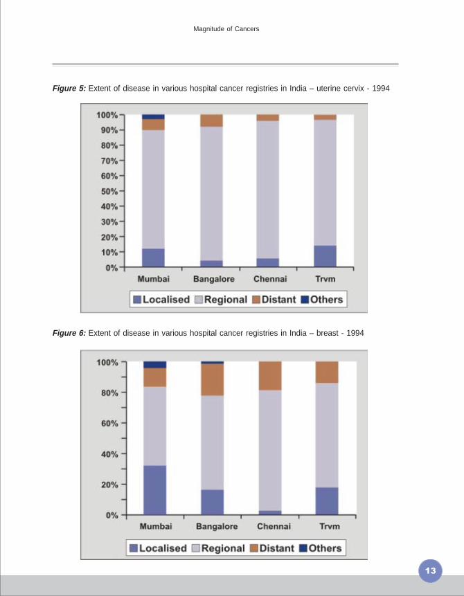
Stage at presentation (local, regional, distant spread) is the most important determinant ofsurvival in cancer. HBCRs provide information on stage at presentation and hospital basedsurvival. The stage at presentation of the three common cancers, in different hospital registriesis shown in figures 4, 5 and 6. The majority of patients present with disease beyond theorgan of origin, when the likelihood of cure is considerably reduced. This leads to the highnumbers of deaths among cancer patients. If these cancers could be detected early, andtreatment instituted, many deaths could be averted.
Figure 4: Extent of disease in various hospital cancer registries in India -Buccal mucosa-1994
13
Magnitude of Cancers
Figure 5: Extent of disease in various hospital cancer registries in India – uterine cervix - 1994
Figure 6: Extent of disease in various hospital cancer registries in India – breast - 1994

14
Key messages
� Cancer is a group of diseases with common characteristics
� Uncontrolled multiplication of cells
� Tendency for local and distant spread
� Cancer is the cause of 12% deaths worldwide
� PBCRs provide data on cancer incidence and survival
� Incidence of cancer in India is 70-90 per 100,000.
� The most common cancers in India are:
� Cancers of the lungs, stomach, and oral cavity among men
� Cancers of the uterine cervix and breast among women
� HBCRs provide data on length and quality of survival in relation
to site and stage at presentation and management
� Stage at presentation is the most important
determinant of survival in cancer
Manual for Health Professionals

15
Principles of Cancer Control
Table 4: Causes of cancer deaths in the United States of America (USA)
The cellular changes that characterize cancers are initiated by various degreesof interaction between host factors and exogenous agents. Life-style relatedfactors are the most important and preventable among them. Studies conducted in the USAhave identified the major risk factors of cancers, and these have been listed in Table 4.
Cause of Cancer Best estimate (%) Range of acceptableestimates (%)
Tobacco 30 25-40
Alcohol 3 2-4
Diet 35 10-70
Reproductive andsexual behaviour 7 1-13
Occupation 4 2-8
Pollution 2 1-5
Industrial by-products 1 1-2
Medicines andmedical procedures 1 0.5-3
Geophysical factors 3 2-4
Infection 10 1-?
(Source- National Cancer Control Programmes – Policies and Managerial Guidelines. World Health Organization.
2nd Edition, 2002)
The relative importance of these factors in cancer causation in India may differ to someextent from the figures quoted in Table 4. Tobacco, for example, is responsible for aroundhalf of all cancers in men and 20% cancers in women in India. Knowledge of these factorscan serve as the basis of cancer control.
2

16
Manual for Health Professionals
Cancer control consists of prevention, early detection, treatment, and palliative care. Ajudicious mix of these approaches, when used for specific cancers, can substantially reducecancer burden. Table 5 illustrates the approaches for some common groups of cancers.
Table 5: Approaches in cancer control
Approach Cancers Strategy
Prevention Tobacco-related Tobacco control/ cessationcancers
Early detection Oral/Breast/Cervix Propagation of awarenessand self-examinationOpportunistic examinationDiagnostic support
Diagnosis and treatment Common cancers TrainingTreatment guidelinesInfrastructureReferral practices
Palliative care All advanced cancers Oral morphine availability,Human resourcedevelopmentCommunity participation

17
Principles of Cancer Control
2A. Primary Prevention
Primary Prevention aims to reduce the incidence of disease by risk factor modification. Arisk factor for a disease is an attribute or exposure that increases the probability of gettingthe disease. As exogenous risk factors including personal habits play a major role in theaetiology of cancer, modifying risk factor exposure may prevent many cancers. Among theactivities for prevention, emphasis should be placed on:
● Tobacco control
● Health education relating to sexual and reproductive factors associated with cancer
● Avoiding alcohol use
● Healthy diet
● Physical activity and avoidance of obesity
Tobacco
Tobacco is the single most important modifiable risk factor for cancer. Of all cancers inIndia, 34% are due to tobacco (48% of cancers in men and 20% of cancers in women).Tobacco smoke contains approximately 4000 chemicals of which at least 438 can causecancer. Tobacco smoking causes cancer of the lung, larynx and oesophagus. Smoking isalso associated with cancers of the pancreas, bladder, pelvis of the kidneys, ureter andsquamous cell carcinoma of the uterine cervix. Tobacco chewing is the most important riskfactor for cancer of the oral cavity. Inhalation of secondary smoke, known as “passivesmoking” is a unique feature of smoking. It results in increased risk of cancers among non-smokers exposed to tobacco smoke.
Male tobacco use prevalence in 1998-1999 was 46.5% (National Family Health Survey – 21998-1999). The prevalence of tobacco use in women was 13.8% in this survey. The NationalHousehold Survey of Drug and Alcohol abuse conducted in 25 states in 2002 reported aprevalence of 55.8% in males aged 12 – 60 years of age. Data from these surveys indicatethat tobacco use prevalence is higher among males than females and among older agegroups than younger age groups.
Tobacco control involves health promotion and education, advocacy, support for cessation,community mobilization, taxation and other fiscal measures, livelihood alternatives,regulation, legislation and enforcement. Policy-level interventions would include levy oftaxes (to raise prices of tobacco products and act as a disincentive for purchase), regulationof tobacco products (for constituents, emissions, health warnings, and misleading health
18
Manual for Health Professionals
claims) and measures to reduce supply (ban on sale to youth, curbs on smuggling, andprogrammes to aid tobacco farmers and workers to switch over to alternative livelihoods).
Interventions at community level would involve programmes for empowering people,especially vulnerable sections, with the knowledge, motivation and skills required to abstainfrom or abandon the use of the tobacco habit. This includes creation of suitable environmentsto stimulate, support and sustain healthy lifestyle choices such as tobacco free norms atschools, worksites and homes.
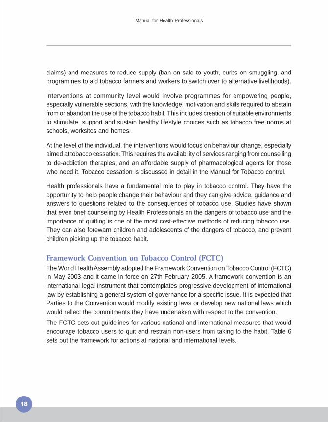
At the level of the individual, the interventions would focus on behaviour change, especiallyaimed at tobacco cessation. This requires the availability of services ranging from counsellingto de-addiction therapies, and an affordable supply of pharmacological agents for thosewho need it. Tobacco cessation is discussed in detail in the Manual for Tobacco control.
Health professionals have a fundamental role to play in tobacco control. They have theopportunity to help people change their behaviour and they can give advice, guidance andanswers to questions related to the consequences of tobacco use. Studies have shownthat even brief counseling by Health Professionals on the dangers of tobacco use and theimportance of quitting is one of the most cost-effective methods of reducing tobacco use.They can also forewarn children and adolescents of the dangers of tobacco, and preventchildren picking up the tobacco habit.
Framework Convention on Tobacco Control (FCTC)
The World Health Assembly adopted the Framework Convention on Tobacco Control (FCTC)in May 2003 and it came in force on 27th February 2005. A framework convention is aninternational legal instrument that contemplates progressive development of internationallaw by establishing a general system of governance for a specific issue. It is expected thatParties to the Convention would modify existing laws or develop new national laws whichwould reflect the commitments they have undertaken with respect to the convention.
The FCTC sets out guidelines for various national and international measures that wouldencourage tobacco users to quit and restrain non-users from taking to the habit. Table 6sets out the framework for actions at national and international levels.

19
Principles of Cancer Control
Framework for national action
● Comprehensive ban on advertising Requires partnerships withincountries
● Protection against second-hand smoke
● Prohibition of youth access
● Prominent health warnings
● Testing and regulation of contents
● Increase in tobacco taxes
● Cessation programmes
● Alternative crops
● Surveillance
Framework for international cooperation
● Ban on cross-border advertising Requires partnerships amongcountries
● Prevention of illicit trade
● Scientific and legal cooperation
● Technical assistance
● Financial support for FCTCimplementation (bilateral andmultilateral channels) Monitoring
(Source – Report on Tobacco control in India, Government of India, 2004)
Table 6: Framework for national action and international cooperation in FCTC
Cigarettes and Other Tobacco Products Act, 2003
A tobacco control legislation entitled ‘The Cigarettes and Other Tobacco Products (Prohibitionof Advertisement and Regulation of Trade and Commerce, Production, Supply andDistribution) Act, 2003’ became an Act of Parliament on 18 th May 2003. This comprehensivepiece of legislation, intended to protect and improve public health, encompasses a widearray of evidence-based strategies to reduce tobacco consumption. This legislation bringsthe entire range of tobacco products under the jurisdiction of the Central Government forthe purpose of this Act and is enforceable across all states. Some of the key provisions andpenalties under the Act are listed in Table 7.

20
Manual for Health Professionals
Table 7: Key provisions and penalties of the Cigarettes and Other Products Act, 2003
Provisions Penalties
Prohibition on direct and indirectadvertisements of tobacco products,with the exception of advertising atpoints of sale and on tobacco packs.Ban on gifts, prizes, scholarships orsponsorship of sports or other culturalevents using the trademark or brandnames of tobacco products
Prohibition of smoking in public places
Prohibition on sale of tobacco productsto persons below 18 years of ageProhibition on sale of tobacco within aradius of 100 yards of educationalinstitutions
Legible and conspicuous display ofhealth warnings on not less than one ofthe largest panels of the tobaccopackage with text of warning in the samelanguage as that used on the packIndication of tar and nicotine contentson the package with maximumpermissible limits as prescribed
Advertisement is to be forfeited anddisposed of. First conviction punishablewith imprisonment of up to two years orfine up to Rs 1000, or both. Subsequentconvictions punishable withimprisonment of up to five years and fineof up to Rs 5000
Offences would be made compoundablewith a fine of up to Rs 200
Offences would be compoundable withsummary trials and a fine of up to Rs 200
Producer/manufacturer – imprisonmentup to two years or fine up to Rs 5000, orboth for first offence, and imprisonmentup to five years and fine up to Rs 10,000for subsequent convictionsSeller/distributor – imprisonment up to oneyear or fine up to Rs 1000, or both forfirst offence, and imprisonment up to twoyears and fine up to Rs 3000 forsubsequent convictions
(Source –Report on Tobacco control in India, Government of India, 2004)

21
Principles of Cancer Control
Alcohol
Increasing alcohol consumption is associated with cancers of the mouth, pharynx (excludingnasopharynx), larynx, oesophagus and liver. The risk relationship between cancer and alcoholis nearly a linear relationship with the risk increasing with increasing amount of alcoholconsumed. Co-existence of tobacco habits can have a multiplicative effect on developmentof cancer.
Control of alcohol requires actions similar to those for tobacco control. The actions shouldbe targeted towards individual and community and include taxation, general public education,encouraging highly vulnerable groups like young people to avoid starting consumption etc.
Sexual and Reproductive FactorsSexual and reproductive factors are associated with cancer of the uterine cervix and breast.Sexual behaviour factors, like young age at first sexual activity, multiple sexual partnersand poor sexual hygiene, are associated with cancer of the uterine cervix. Human PapillomaVirus (HPV) has now been identified as the etiological agent responsible for cervical cancer.HPV prevalence increases with high risk sexual behaviour and poor sexual hygiene.
Late age at marriage, nulliparity, and late menopause have been linked to breast cancer,but the underlying mechanism is probably uninterrupted exposure to oestrogen for prolongedperiods in all these cases.
Education regarding sexual hygiene and safe sexual behaviour may be provided for preventionof cancer cervix. Safe sexual behaviour protects women from the risk of cervical cancer bypreventing infection with HPV. Breast cancer is not preventable to any large extent. Earlydetection of breast cancer is the main strategy for improving survival in breast cancer.
DietVarious studies in the past two decades suggest the role of diet in human cancers. Dietaryfactors are responsible for many cancers in the Western countries. Changing dietary patternswill lead to increased contribution of diet in cancer causation in India also. It is generallyagreed that diets rich in animal fats, especially red meats, increase the risk for cancer. It isalso widely accepted that diets high in fresh vegetables and fruits, and fibre reduce risk forcancer.
Certain basic measures may help in reducing risk of cancer:● Avoid being underweight or overweight● Engage in regular physical activity● Consumption of alcohol is not recommended● Limit consumption of salted foods● Choose predominantly plant based diets rich in fruits and vegetables● Restrict the intake of red-meat (beef, pork, lamb) and preserved meat

22
OccupationOccupational cancers constitute 5-10% of all cancers. Increased risk of lung cancer hasbeen seen in workers engaged in manufacture of rubber tyres in developing countries,textile workers, ship and dockyard workers and wood workers. Higher risk of bladder cancerwas seen in workers of chemical and pharmaceutical plants.
Limiting exposure to potentially carcinogenic substances through protective gear, frequentrotation of workers, mechanized handling of such chemicals and similar mechanisms mayhelp reduce cancers from occupational exposures.
InfectionInfections with various agents are implicated in the aetiology of certain cancers as shown inTable 8. Control of cancers caused by or associated with infections depends upon successin combating the infection concerned. Measures include eliminating reservoirs and sourcesof infection, preventing transmission, increasing host immunity through vaccination, andeffective treatment of those infected.
Table 8: Infective agents associated with cancers and measures for prevention
Infective agent Cancer PreventionHuman Papilloma virus
Hepatitis B and HepatitisC virus
Epstein- Barr virus
Schistosomahaematobium
Clonorchis sinensis
Helicobacter Pylori
Cancer of the UterineCervix, Oesophagealcarcinoma, Anal cancer,Penile cancer, Oral cancer
Hepatocellular carcinomacan occur from chronicactive infection
Burkitt Lymphoma,Nasopharyngealcarcinoma
Bladder cancer
Cholangiocarcinoma
Stomach cancer
Safe sexual practices,avoiding multiple sexualpartners
Universal precautions,Safe sexual practices,vaccine for Hepatits B
No specific interventions
Preventing water pollutionwith human waste, treatingpatients, controllingintermediate hosts (snails)
Preventing water pollutionwith human waste, treatingpatients, controllingintermediate hosts (snail,fish), avoid eating raw fish
Treating patients withsymptomatic infection
Manual for Health Professionals

23
Principles of Cancer Control
2B. Early Detection
Early detection of cancer is the detection of disease at a stage in its natural history wherethe chance of cure is high. Early detection is only part of a wider strategy that includesdiagnosis, treatment and follow-up.
Many cancers that are potentially curable at early stages are detected only in advancedstages. Diagnosis of such cancers at a stage where treatment is effective could have amajor impact on the disease outcome. Certain symptoms and signs may be early indicatorsof some cancers. These include:
● Unexplained change in bowel or bladder habit
● A white patch or ulcer in the mouth that does not heal
● Obvious change in a mole or wart, like rapid increase in size, bleeding or ulceration
● Bleeding from body’s orifices eg – haematuria
● Persistent indigestion/ difficulty in swallowing/ difficulty in breathing
● Persistent fever unresponsive to treatment
● Unexplained loss of weight
● Chronic cough or hoarseness of voice especially in a smoker
All people should be aware of these warning signs. The presence of any of these featuresdoes not mean a definitive diagnosis of cancer. Such changes may occur in other benignconditions also. However, any such sign not responding to appropriate treatment warrantsimmediate medical attention and prompt management.
It is also important to train people to detect cancers in the early stage with self-examinationof the oral cavity and breast. Health professionals should be trained for early detection andprompt referral of suspected cases.

24
2C. ScreeningScreening is the presumptive identification of unrecognised disease or defects by means oftests, examination or other procedures that can be applied rapidly. Screening is based onthe concept that there is a detectable pre-clinical phase of the disease being screened, anddetection at this stage markedly alters disease prognosis . The success of screening dependson having sufficient numbers of trained personnel to perform the screening tests withadequate coverage of target populations, and on the availability of facilities that can undertakesubsequent diagnosis, treatment and follow-up. The target disease should be a commonform of cancer with high associated morbidity and mortality, and test procedures should beacceptable, safe and relatively inexpensive. Screening is recommended for cancers ofuterine cervix and breast, only if resources permit.
Screening for Cervical cancer
Cervical smear cytology is the standard screening test for cervical cancer. It is an easy andeffective method revealing the presence of pre-cancerous lesions as well as in situ or veryearly invasive cancer. Screening should preferably begin at 35 years of age, as at youngerages dysplasia detected through screening rarely progresses to cancer, but adds toprogramme cost in treatment (Figure 7). The important requirement for cervical cytology isthe availability of good laboratory services so that accurate diagnosis is possible. Screeningprogrammes may be initiated in a defined population if adequate trained manpower andfacilities are available. The most important aspects of a screening programme are itsorganization and management. All women in the target population should be invited forscreening, unique identification numbers provided for follow up, and reliable laboratoryfacilities and personnel made available. The screened population has to be providedappropriate interventions and follow up. At least 80% of the target population has to becovered if reduction in incidence is to be achieved.
Manual for Health Professionals
Note: CIN = cervicalintraepithelial neoplasia
Figure 7 : Screening for cervical cancer

25
Principles of Cancer Control
Alternative strategies such as visual inspection are being tested for use in low-resourcesettings where laboratory facilities for cervical cytology are inadequate. Test performanceof Visual Inspection with Acetic acid (VIA) suggests that it has similar sensitivity to that ofcervical cytology in detecting cervical intraepithelial neoplasia, but has lower specificity.Further studies are underway to judge how appropriate and feasible it will be to introduceVIA-based cervical cancer screening programmes on a population-wide basis. There isincreasing interest in the use of HPV DNA testing for screening. The test, however, requiresfinancial and sophisticated technical resources. Details of some of the methods are providedin the section on cervical cancer. However, (more than the tests) it is the health system,with the required resources and services for the follow up management of those withabnormal test results that determines the outcome of any such programme.
Screening for cancer of breastMammography is an effective screening test for breast cancer, and can reduce mortalitydue to breast cancer if used with appropriate follow-up. Unfortunately, it is an expensivetest that requires great care and expertise both to perform and in the interpretation of results.It is therefore currently not a viable option for many countries. Breast self-examination hasnot been proven to reduce breast cancer mortality. Early diagnosis of breast cancer, bypromoting breast awareness among all women and clinical breast examinations for womenpreferably in the age group 40-69 years, should be encouraged.
Appropriate diagnostic facilities and referral practices have to be established to ensure thatearly detection and screening programmes result in the desired results.

26
2D. Diagnosis and Treatment
Diagnostic Methods
The diagnostic procedures in oncology are for diagnosis, determining the extent of thedisease, deciding the treatment options available and evaluating the patient during follow-up. Clinical evaluation is the first and the most important step in the diagnosis of malignancy.It requires the health profesional to be alert to the early warning signals. A thorough historyand clinical examination of any suspicious symptom or sign is mandatory. Clinical suspicionof malignancy can be confirmed by various diagnostic methods described below.
Radiological Evaluation
Various imaging methods are:
● X ray
● Fluoroscopy
● Mammography
● Ultrasound
● C.T.Scan
● Magnetic Resonance Imaging
● Positron Emission Tomography
Nuclear Medicine
● Radio nuclide scan and Radioactivity uptake studies e.g. Thyroid, Bone
Biochemical Evaluation
This is generally done to know the organ functions, like liver function tests, and renal function tests.
Endoscopy
In oncology endoscopy is useful to:
● Detect the site of primary cancer
● Evaluate the extent of lesion
● Perform biopsy
● Perform certain therapies like endoprosthesis for oesophageal stenosis, lasertherapy, etc.
Manual for Health Professionals

27
Principles of Cancer Control
Pathological Evaluation
Pathological evaluation is an important method for confirmation of clinical diagnosis andincludes:
● Haematological Examination: Examination of peripheral blood smear and bonemarrow.
● Cytological Examination:
� Exfoliative cytology: examination of exfoliated cells; e.g.female genital tract,oral cavity, urinary tract (urine examination), gastrointestinal lesions (gastriclavage) etc.
� Fine Needle Aspiration Cytology (FNAC): to obtain material from organs thatdo not shed cells spontaneously. Example: Breast, Thyroid, etc.
� Aspiration of body fluids: to rule out or confirm malignant effusions. Example:pleural fluid, peritoneal fluid.
● Biopsy: A small chunk of tissue is removed from the suspicious site and subjectedto histopathological examination. It may be:
� Excisional biopsy in small tumours
� Incisional/ Punch biopsy in skin and mucosal lesions
� Cone biopsy in uterine cervix
� Needle biopsy in bone marrow, solid tumours of abdomen and pelvic organs.
Immunological Evaluation
Some cancers release biologic or biochemical substances, in the form of hormones,enzymes, and antigens, into the circulation. The measurement of these substances in bloodcan be useful in the detection and diagnosis of some types of cancers. Such chemicals arecalled tumour markers. Some common tumour markers and the conditions in which theymay be raised are listed in Table 9.

28
Tumour marker
Alpha feto protein (AFP)
Beta human chorionicgonadotrophin (B-hCG)
Carcino embryonicantigen (CEA)
CA-125
Prostate specific antigen
Malignancies
Hepatoblastoma,Nonseminomatous germcell tumour testis, Non-dysgerminomatous germcell tumour ovary,Hepatocellular carcinoma,
Choriocarcinoma, Testiculargerm cell tumours
Colorectal cancers, Breastcancer, Cholangiocarcinoma,Stomach cancer
Epithelial ovarian cancer
Prostate cancer
Non-malignantconditions
Cirrhosis, Hepatitis
Hypogonadism,Hydatidiform mole
Smoking, Fatty liver,Hepatitis
Pregnancy, Menstruation,Endometriosis, Ascites,Pleural effusion
Prostatitis, Benignprostatic hyperplasia,Prostatic manipulation
(source – Role of Tumor markers and Recent advances in cancer diagnosis, Manisha Bhutani, Amish Vora and VinodKochupillai, Fifty Years of Cancer Control in India. National Cancer Control Programme. Government of India. 2002)
Staging of cancer
Staging is used to assess the extent of the spread of the disease in the body. It is anindication of prognosis, and is used as a guide to determine the type and extent of treatmentrequired.
TNM classification- The TNM classification for tumours has been adopted by theInternational Union against Cancer, and has been extended for many sites of cancer. Thisis a detailed clinical staging which is arrived at by the clinician by ascertaining the extent ofthe primary tumour (T), lymph node involvement (N), and presence of metastases (M). Theinformation so obtained is scored. The details of scoring are specific to each type of cancer.
Other systems of staging include the FIGO (International Federation of Gynaecology andObstetrics) staging for cancers of the uterine cervix and body of the uterus, and the Duke’ssystem of staging for cancer of the rectum.
Manual for Health Professionals
Table 9: Some common tumour markers and the conditions in which they may be raised

29
Principles of Cancer Control
Principles of TreatmentThe primary goals of cancer treatment are cure ideally, prolongation of useful life if possible,and improvement in quality of life always. The principal methods of treatment are surgery,radiotherapy, and chemotherapy (including hormonal manipulation). Each of these modalitieshas a well-established role, and can be used for cure or for palliation. Appropriate combinationand sequencing of these modalities can be adopted for specific cancers.
Surgery
Surgery plays an important role in the diagnosis, staging and treatment of localised cancers.Where other modalities form the mainstay of treatment, surgery can contribute throughremoval of tumour masses, palliation and treatment of some complications.Surgery requires the support of other specialties including anaesthesiology, bloodtransfusion services, pathology (specially oncopathology) and critical care nursing. In earlystage solid tumours, surgery that encompasses a sufficient margin of normal tissue iscurative. These include early stage cancers of the breast, oral cavity, uterine cervix, colon,prostate and the skin. Surgery is also used post chemotherapy or radiotherapy to providelocal cancer control and better chances for adjuvant therapy. In certain solid tumours,surgery is critical for reducing bulk (cytoreduction). Surgery is valuable in oncologyemergencies, to relieve bowel obstruction, promote cessation of bleeding, closeperforations, relieve compression, and drain ascites or pleural effusions. Apart fromtreatment, surgery for reconstruction and rehabilitation can improve function and cosmeticappearance and enhance quality of life for patients.
Radiotherapy
Radiotherapy is one of the most important methods of curing local cancer. Radiotherapy isthe method of treating diseases with “ionising radiation”. The ionising radiation causesdamage to certain vital structures within the cells. The cells are either damaged or arerendered incapable of further multiplication. These damaging effects on normal cells areless and reversible whereas the damage in the abnormal cell is irreversible. This differentialis the principle of radiotherapeutic treatment.
Radiotherapy is a capital-intensive specialty, requiring high technology equipment andskilled technicians, found only in tertiary centres. Radiotherapy may be teletherapy(administered from a distance) or brachytherapy (treatment with radioactive substanceswithin body cavities or tissues). Teletherapy may be administered by cobalt machines orby accelerators. Clinical outcomes are identical with both machines. Brachytherapy maybe delivered by low dose rate (LDR) devices using caesium and high dose rate (HDR)devices using iridium or cobalt. HDR can be used for treatment of a wider variety of cancersthan LDR and reduces the need for hospital bed occupancy, but demands more expertiseand has higher costs.

30
Manual for Health Professionals
Radiotherapy is one of the most important methods of curing local cancer. It is also oftenadministered before or after surgery. Such treatment either facilitates surgery orconsolidates surgical gains, and reduces local recurrence of disease. Palliative radiotherapyis of value in cases of pain secondary to bone metastasis and tumours causing bleedingor compressive syndromes.Radiotherapy can cause various side effects. Patients may notice loss of appetite, nausea,and occasionally vomiting persisting for a week. The symptoms are mild in nature and seenin about 10% of patients, and are easily controlled by medicines. Other side effects dependon the site irradiated and can include mucositis and bone marrow depression. Long-termside effects are also observed.
Chemotherapy
Chemotherapy is the use of cytotoxic drugs against cancer. Cancer cells are damaged tothe extent that they cannot survive. Normal cells are also damaged but to a lesser degree.
Chemotherapy is curative in certain cancers e.g. Hodgkin disease, high-grade non-Hodgkinlymphomas; palliative in many cancers, and used as adjuvant therapy for some cancersincluding breast cancer, ovarian cancer and colorectal cancer. The goal of adjuvant therapy(treatment given in addition to primary definitive therapy in the absence of macroscopicresidual disease) is to avoid metastases, prolong life and improve quality of life.Chemotherapy is ineffective in hepatobiliary cancers, pancreatic cancer, thyroid cancer,and central nervous system cancers among others.
Acute side effects of chemotherapy are usually self-limited and reversible. Fall in bloodcount, hair loss, nausea, vomiting, constipation, diarrhea, anaemia, and depression of theimmune system are some of the side-effects. There may be drug specific side effects likecardiotoxicity, nephrotoxicity, neurotoxicity.
Palliative care
Palliative care is an approach that improves the quality of life of patients and their familiesfacing a life-threatening illness. This is done through prevention and relief of suffering bymeans of early identification, accurate assessment and treatment of pain and physical,psychosocial and spiritual problems. Palliative care involves a multidisciplinary teamapproach. Further details on palliative care are provided in the Manual on Palliative Care.
31
Principles of Cancer Control
Issues that need to be kept in mind for all cancers:
● Prompt referral of patients with any suspicion of cancer for appropriatemanagement
● Compliance of the patient with medical advice
● Provision of psychosocial services for the patient, and the family
● Rehabilitation: Physical, psychological and social rehabilitation so that the affectedindividual is able to take care of self, be emotionally stable, and be able to workand socialize, to the extent possible.
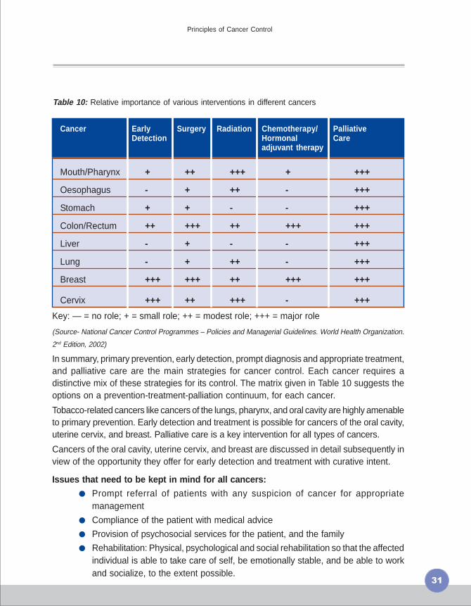
Table 10: Relative importance of various interventions in different cancers
Cancer Early Surgery Radiation Chemotherapy/ PalliativeDetection Hormonal Care
adjuvant therapy
Mouth/Pharynx + ++ +++ + +++
Oesophagus - + ++ - +++
Stomach + + - - +++
Colon/Rectum ++ +++ ++ +++ +++
Liver - + - - +++
Lung - + ++ - +++
Breast +++ +++ ++ +++ +++
Cervix +++ ++ +++ - +++
Key: — = no role; + = small role; ++ = modest role; +++ = major role
(Source- National Cancer Control Programmes – Policies and Managerial Guidelines. World Health Organization.
2nd Edition, 2002)
In summary, primary prevention, early detection, prompt diagnosis and appropriate treatment,and palliative care are the main strategies for cancer control. Each cancer requires adistinctive mix of these strategies for its control. The matrix given in Table 10 suggests theoptions on a prevention-treatment-palliation continuum, for each cancer.
Tobacco-related cancers like cancers of the lungs, pharynx, and oral cavity are highly amenableto primary prevention. Early detection and treatment is possible for cancers of the oral cavity,uterine cervix, and breast. Palliative care is a key intervention for all types of cancers.
Cancers of the oral cavity, uterine cervix, and breast are discussed in detail subsequently inview of the opportunity they offer for early detection and treatment with curative intent.

32
� Primary prevention� Avoid use of tobacco in any form
� Avoid alcohol
� Promote physical activity
� Eat plenty of fruits and vegetables
� Practise Safe sexual behaviour
� Early detection of cancers
� Breast awareness
� Awareness in community regarding early warning signs of commoncancers (Oral/Breast/Cervix)
� Opportunistic check up for oral, breast and cervical cancer
� Prompt referral and appropriate management
Prompt referral of any suspicious case is the mostimportant step towards cure.
� Diagnostic methods:� Clinical history & examination – first and most important
� Radiological examination
� Pathological examination
� Diagnostic procedures help us to know:
� The type of cancer
� The extent (staging) of cancer
� Treatment options and prognosis
� Follow-up evaluation
� Treatment Modalities: � Surgery � Radiotherapy � Chemotherapy
� Treatment modalities can be used with intention of cure or palliation, andalone or in combination depending on type and extent of disease.
� Goal of treatment is ideally cure if possible and improvement in quality oflife always.
Key messages
Manual for Health Professionals

33
Common Cancers
3A. Cancer of the Oral CavityOral cancer is one of the ten most common cancers in the world. In India, oral cancer,including cancers of the lip, tongue, gum and floor of mouth, is one the most commoncancers, and may be the commonest in many regions. Oral cancer is both preventable andcurable. There is usually a long natural history and most cases of oral cancer arise frompre-cancerous lesions. Therefore there is ample opportunity for intervention before actualmalignancy develops. Also oral cancer responds well to surgery and radiation if detectedearly.
Risk factorsTobacco chewing is the single most important risk factor for oral cancer. Other risk factorsinclude alcohol use, betel nut chewing, and chronic trauma to oral mucosa by sharp toothor ill-fitting dentures. Chronic exposure to these risk factors causes changes in the oralmucosa and these changes are visible as pre-cancerous lesions. Over time, malignancymay develop in these lesions.
Pre-cancerous lesionsPre-cancerous lesions or conditions are local or generalized disturbances that predisposeto malignancy in a particular site. Leucoplakia, erythroplakia, palatal changes associatedwith reverse smoking or beedi smoking and submucous fibrosis are local pre-cancerouslesions. Plummer Vinson syndrome, syphilis, and erosive lichen planus are generalisedpre-cancerous conditions.
All these conditions are amenable to early diagnosis, and treatment is possible in manycases.
LeucoplakiaThis is defined as a white patch that cannot becharacterized as any other disease clinically orpathologically (Figure 8). They can be of 4 types:
� Homogeneous leucoplakia: Low risk of cancer� Ulcerated or erosive leucoplakia: High risk of
cancer� Speckled or nodular leucoplakia: High risk of
cancer� Verrucous leucoplakia: Very high risk of
cancerTwo or more types of leucoplakia may bepresent in the oral cavity at the same time.Confirmatory diagnosis is by biopsy.
3
Figure 8: Homogeneous leukoplakiainvolving dorsum and right lateral borderof tongue.

34
Treatment of leucoplakia:
Treatment is planned on the basis of individual cases. In all cases, patients must be advisedto quit the tobacco habit. Routine follow-up observation allows early detection of anycancerous change in the lesions.
Erythroplakia:
This is a bright, velvety area sometimes surrounded byfaint plaques which cannot be characterized as any otherlesion clinically or pathologically (Figure 9). About 90%of these lesions show cellular dysplasia or malignancy.The risk of malignancy in erythroplakia is higher than inleucoplakia. Hence all cases of erythroplakia need tobe biopsied. Treatment of erythroplakia is similar totreatment of leucoplakia.
Palatal changes due to beedies and reverse smoking:There is palatal keratosis, excrescences around the openings of minor salivary glands,white patches, red areas, ulceration and melanin pigmentation (Figure 9). All such lesionsshould be subjected to biopsy and treatment is to be instituted accordingly. Cessation ofthe tobacco habit is an essential part of treatment.
Traumatic Ulcers / Keratosis:Sharp cusps, remaining root stumps, mal-aligned teeth and unscientifically fabricateddentures can cause traumatic ulcers or keratosis on the lateral margins of the tongue orbuccal mucosa. Any such ulcer in the mouth that does not heal within one month of antibiotic/antiseptic treatment should be viewed with suspicion. The irritant like sharp tooth, ill-fittingdentures etc. should be removed immediately and the lesion reassessed after two weeks.Any lesion that persists should be biopsied and managed accordingly.
Oral Submucous Fibrosis (SMF):This is characterized by blanching of the oral mucosa,difficulty in tolerating spicy foods and slowlyprogressive inability to open the mouth and protrudethe tongue Figure 10. In some cases there may beinvolvement of soft palate resulting in nasal voice.
Manual for Health Professionals
Figure 9: Erythroplakia in the rightlateral border of tongue
Figure 10: An elderly lady with OralSubmucous fibrosis. Restricted mouthopening and depappillated tongue canbe appreciated in this lady who is achronic chewer of tobacco.
SMF may be localized or generalized. Diagnosis is byvisualisation or palpation of fibrous bands, loss ofelasticity of buccal mucosa and atrophy of tongue. Ingeneralized SMF, the entire oral mucosa is atrophic.Once detected, the patient should be advised to stopthe tobacco habit, and should be regularly followed up.

35
Common Cancers
Oral Cancer:
The most common cancer seen inthe oral cavity is squamous cellcarcinoma. It presents as apainless ulcer, mass or fissure(Figure 11). As the diseaseadvances, patient may haveexcessive salivation, trismus, anddifficulty in chewing, swallowing orspeaking, depending on theinvolvement. There may also becervical lymphadenopathy. Distantmetastases are uncommon in oralcancers.
Figure 11: Oral cancer
Early detection
I. Self Examination of oral cavity:This is important for dectecting oral lesions atan early stage.
Figure 12: Self Examination of oral cavity
36
When to do self-examination:
All habitual tobacco users should do it once a month
How to do it:
� Rinse the mouth with water and stand before a mirror in adequate light
� Look in the mirror for any abnormal white or red patch, ulcer or roughened area,granular area or swelling in the mouth (Figure 12).
� If any such area is seen, the suspicious area should be felt with the fingers.Normal oral mucosal is soft and pink.
� Consult a doctor if any abnormal area is found.
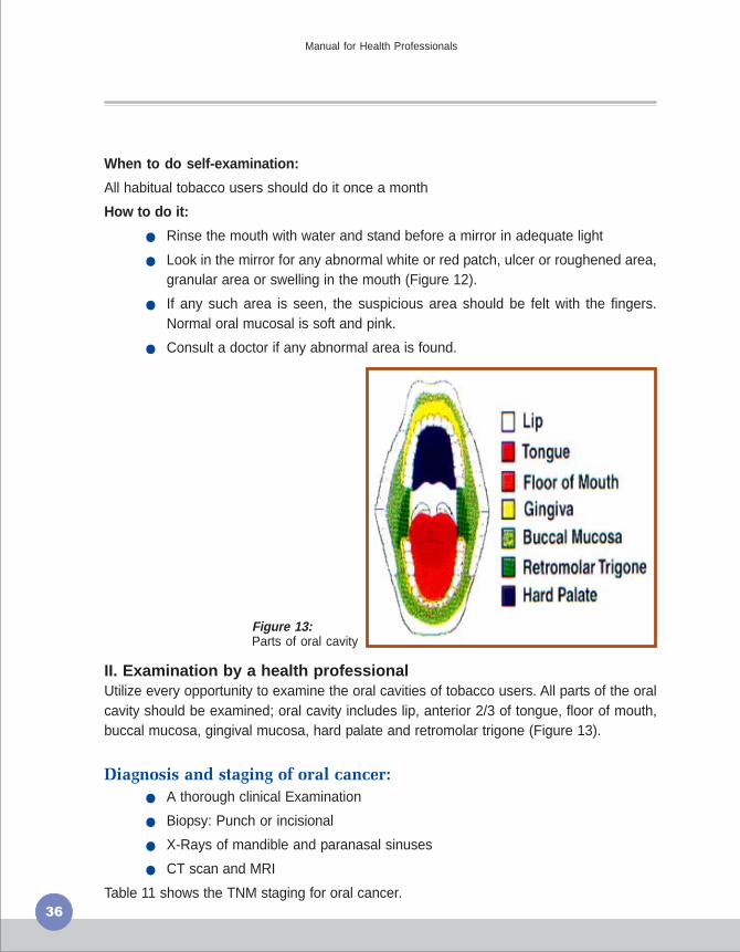
II. Examination by a health professionalUtilize every opportunity to examine the oral cavities of tobacco users. All parts of the oralcavity should be examined; oral cavity includes lip, anterior 2/3 of tongue, floor of mouth,buccal mucosa, gingival mucosa, hard palate and retromolar trigone (Figure 13).
Diagnosis and staging of oral cancer:
� A thorough clinical Examination
� Biopsy: Punch or incisional
� X-Rays of mandible and paranasal sinuses
� CT scan and MRI
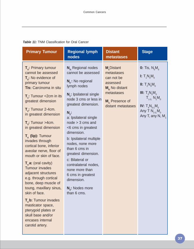
Table 11 shows the TNM staging for oral cancer.
Figure 13:Parts of oral cavity
Manual for Health Professionals
37
Common Cancers
Primary Tumour Regional lymph Distant Stagenodes metastases
Table 11: TNM Classification for Oral Cancer
0: Tis, N0M
0
I: T1N
0M
0
II: T2N
0M
0
III: T3N0M0
T1-3,
N1M
0
IV: T4N
0-1M
0
Any T N2-3,
M0
Any T, any N, M1
TX: Primary tumourcannot be assessedT0: No evidence ofprimary tumourTis: Carcinoma in situ
T1: Tumour <2cm in itsgreatest dimension
T2: Tumour 2-4cm.in greatest dimension
T3: Tumour >4cm.in greatest dimension
T4 (lip): Tumourinvades throughcortical bone, inferioraveolar nerve, floor ofmouth or skin of face.
T4 a: (oral cavity)Tumour invadesadjacent structurese.g. through corticalbone, deep muscle oftoung, maxillary sinus,skin of face.
T4 b: Tumour invadesmasticator space,pterygoid plates orskull base and/orencases internalcarotid artery.
NX: Regional nodescannot be assessed
N0 : No regionallymph nodes
N1: lpsilateral singlenode 3 cms or less ingreatest dimension.
N2:a: lpsilateral singlenode > 3 cms and<6 cms in greatestdimension.
b: Ipsilateral multiplenodes, none morethan 6 cms ingreatest dimension.
c: Bilateral orcontralateral nodes,none more than6 cms in greatestdimension.
N3: Nodes morethan 6 cms.
MxDistantmetastasescan not beassessedM0: No distantmetastases
M1: Presence ofdistant metastases

38
Manual for Health Professionals
Management of Oral Cancer
Management may be through surgery, radiotherapy, chemotherapy, or a combination ofmodalities. Figure 14 presents a flow chart of management of any person with a suspiciousoral lesion.
Figure 14: flowchart for management of patient with an oral lesion

39
Common Cancers
� Oral cancer is preventable and curable if detected early
� Tobacco chewing is the most important risk factor
� Health professionals can
� Examine oral cavity of all patients with history of tobacco use
� Advocate cessation of tobacco and alcohol use
� Teach Oral self-examination
� Ask clients to report to the health centre if they spot any lesion that lookssuspicious
� Ensure prompt referral of patients with suspicious lesions
� Provide pain relief and palliative care
Key messages
40
3B. Cancer of the Uterine Cervix
Cervical cancer is the third most common cancer among women in the world and the leadingcause of death from cancer among women in developing countries. In India more than100,000 new cases of cervical cancer occur each year and nearly 75,000 women die annuallyfrom this disease.
Human Papilloma virus infection, which is a sexually transmitted infection, is the primarycause of this cancer. HPV prevalence increases with multiple sexual partners for eitherspouse, and poor genital hygiene of both partners.
Symptoms of cancer of the uterine cervix:
In the early stages, there will be no symptoms. By the time symptoms appear, disease mayhave already spread. Common symptoms are:
� Post-menopausal bleeding
� Post-coital bleeding
� Intermenstrual bleeding
� Blood stained discharge per vaginum
� Excessive seropurulent discharge
� Backache
� Lower abdominal pain
Cervical cancer develops slowly over 10 to 15 years. Initially, abnormal cells develop in theepithelial layer of the cervix [Cervical Intra-epithelial Neoplasia (CIN)]. If left undetectedand untreated, such pre-cancerous lesions can progress to invasive cancer. Treatment ofpre-cancerous lesions is simpler and chance of cure is higher. In contrast, treatment ofinvasive cancer is more difficult, and chances of cure decrease with advancing stage ofcancer. Cancer cervix, due to its slow progression from pre-cancerous lesion to malignancy,and easy accessibility to examination, gives us ample opportunity for early detection andthus considerably improved prognosis. Early detection may be through opportunisticexamination of women attending outpatient clinics or through a systematic programme ofscreening.
Screening for cervical cancer can be considered in women aged 35 to 50 years, as thechances of detecting pre-cancerous lesions are maximum in this age group. The successof screening is determined by the coverage of women in the target age group rather thanthe frequency of screening. Once in a lifetime screening for all women between 35 and 50years will achieve good results at relatively low cost (Table 12). Pap smear is the establishedmethod for screening of cervical cancer. It has been effective in reducing the incidence andmortality due to cervical cancer in large programmatic settings. However, being a laboratory-
Manual for Health Professionals
41
Common Cancers
based test, pap smear requires appropriate infrastructure and skilled manpower. Also, itseffectiveness as a screening test decreases if quality is not maintained. Visual inspectiontests are being researched as low-cost alternatives to Pap smear.
Table 12: Reduction in the cumulative rate of invasive cervical cancer for women aged 35-64years, with different frequencies of screening (80% compliance and moderately sensitive test)
(Source - National Cancer Control Programmes – Policies and Managerial Guidelines.World Health Organization. 2nd Edition, 2002)
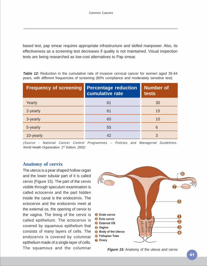
Anatomy of cervix
The uterus is a pear shaped hollow organand the lower tubular part of it is calledcervix (Figure 15). The part of the cervixvisible through speculum examination iscalled ectocervix and the part hiddeninside the canal is the endocervix. Theectocervix and the endocervix meet atthe external os, the opening of cervix tothe vagina. The lining of the cervix iscalled epithelium. The ectocervix iscovered by squamous epithelium thatconsists of many layers of cells. Theendocervix is covered by columnarepithelium made of a single layer of cells.The squamous and the columnar
Frequency of screening Percentage reduction Number ofcumulative rate tests
Yearly 61 30
2-yearly 61 15
3-yearly 60 10
5-yearly 55 6
10-yearly 42 3
Figure 15: Anatomy of the uterus and cervix

42
epithelia meet near the external os and form the squamo-columnar junction (SCJ). Thesquamo-columnar junction is a distinct line between the red velvet-like columnar epitheliumand the smooth pinkish white squamous epithelium and is usually situated just outside theexternal os. The location of the SCJ is not constant throughout the life of the woman. At ayounger age the SCJ may move down on the ectocervix and as the woman grows old itmay move up and get hidden inside the endocervix. The area just outside and adjacent tothe SCJ is known as the Transformation Zone (TZ). Most of the cervical precancers (CIN)usually arise from this area.
Pap smear:The ectocervix and the endocervix are scraped to collect cells that are spread on aglass slide, stained in the laboratory and examined under microscope. Depending onthe features of the cells seen under microscope the cytopathologist (or a trainedtechnologist) can report the smear as ‘negative’ (normal) or ‘positive’ (abnormalitiessuspicious of low grade or high grade CIN). The details of fixation and staining aregiven in the Manual for Cytology.
Manual for Health Professionals
Figure 16: Cusco's Speculum and Ayre's Spatula
43
Common Cancers
Requirements
Examination gloves
Speculum (Cusco’s self-retaining type preferred)
Ayre’s spatula (Figure16) and endocervical brush (if available)
Glass slide
Diamond tipped pencil to write on glass slide (if the glass slide has frosted edge anordinary pencil can be used to label it)
Focusing light
Coplin’s jar
95% ethyl alcohol
Cytology form
Procedure:
Procedure should be explained to the woman.
Clean a glass slide with dry cotton and label it.
The woman should lie down on her back with legs folded (lithotomy position not required).
Insert the speculum gently and expose the cervix.
Note any abnormal discharge, bleeding or growth in the cervix.
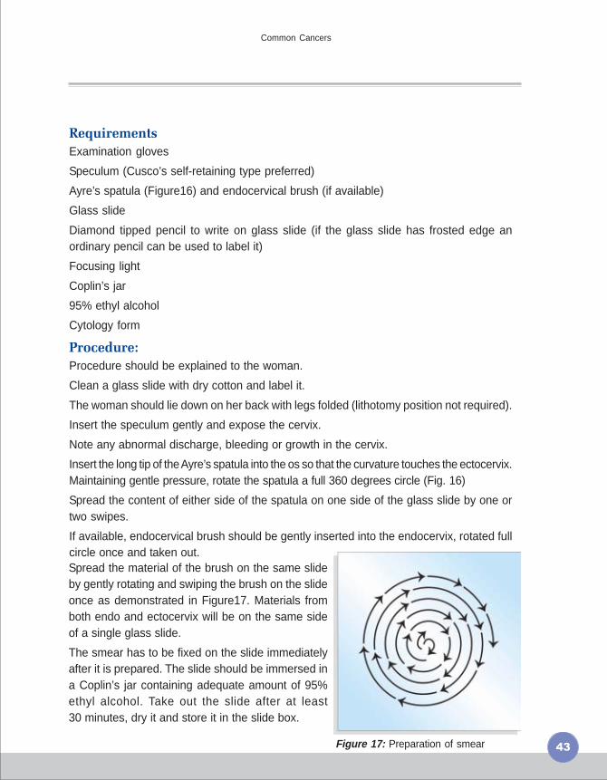
Insert the long tip of the Ayre’s spatula into the os so that the curvature touches the ectocervix.Maintaining gentle pressure, rotate the spatula a full 360 degrees circle (Fig. 16)
Spread the content of either side of the spatula on one side of the glass slide by one ortwo swipes.
If available, endocervical brush should be gently inserted into the endocervix, rotated fullcircle once and taken out.Spread the material of the brush on the same slideby gently rotating and swiping the brush on the slideonce as demonstrated in Figure17. Materials fromboth endo and ectocervix will be on the same sideof a single glass slide.
The smear has to be fixed on the slide immediatelyafter it is prepared. The slide should be immersed ina Coplin’s jar containing adequate amount of 95%ethyl alcohol. Take out the slide after at least30 minutes, dry it and store it in the slide box.
Figure 17: Preparation of smear

44
Figure 19: Visual inspection with aceticacid showing acetowhitening
On inspection:
� Normal cervix will have reddishendocervix and paler ectocervix.(Figure18)
� Ulcer: peeled away epitheliumresulting in denuded area
� Erosion: endocervix may be everted(ectropion) and there is extensivereddish area
Bleeds on touch: to be mentionedspecifically in the report. Slight bleedingmay be insignificant.
Growth: Friable growth with irregularsurface and which bleeds on touch isusually malignant. Smooth growths withregular surface are generally benign.
Figure 18: Normal Cervix
Manual for Health Professionals
Alternative strategies
Unaided Visual Inspection:
Requirements:Examination gloves
Speculum (Cusco’s self-retainingtype preferred)
Focusing light (with halogen bulb preferred)
Procedure:Procedure should be explained to the woman.
Introduce a steel cusco’s speculum lubricatedwith water or saline into the vagina gently in theclosed position
Open the speculum gently so that there is no bleeding.Bleeding will obscure the picture and inspection becomes difficult.
Look for any discharge:
� Whitish curdy discharge: candidal infection
� Yellowish, frothy purulent discharge: infection with trichomonas vaginalis
� Mop any discharge with a dry cotton swab for better visualization of cervix

45
Common Cancers
Hard to touch: If the cervix feels hard and there is resistance, it should be recorded.
Any other findings should be noted down, eg. Cysts, hypertrophy, etc.
Unaided visual inspection was used historically as a method of detecting cervical cancer atan early stage, but is no longer employed for this purpose.
Visual Inspection using 4% Acetic acid (VIA):
Acetic acid causes dehydration of the cells and some surface coagulation of proteins therebyreducing the transparency of the epithelium. These changes are more prominent in abnormalepithelium and can be easily distinguished on naked eye inspection. (Figure19)
Requirements:
Examination gloves
Speculum (Cusco’s self-retaining type preferred)
Cotton tipped swabs
Freshly prepared 5% acetic acid (to be produced at least once a week by diluting 5 ml ofglacial acetic acid with 95 ml of distilled water)
Focusing light (with halogen bulb preferred)
VIA forms
Procedure:
Procedure should be explained to the woman.
The woman should lie down on her back with legs folded (lithotomy position not required).
Insert the speculum gently and expose the cervix.
Note any abnormal discharge, bleeding or growth in the cervix.
Apply adequate amount of acetic acid to the cervix using the cotton swabs. Wait for 1minute to note the changes.
Identify the squamo-columnar junction as the line joining the pink smooth squamousepithelium with the red velvet like columnar epithelium. Look for white patches. If there areno white patches in the ectocervix the test is negative. All the aceto-white patches are notconsidered positive.
If there is a white patch, its density, margin and the relationship to the SCJ should be noted.Table 13 gives the detailed criteria for categorizing VIA test results as negative or positiveor invasive cancer.

46
Table 13: Criteria for categorizing VIA test results
VIA category Description
Negative ● No aceto-white lesions ● Transparent lesions or faint patchylesions without definite margins ● Nabothian cysts becomingaceto-white ● Faint line like aceto-whitening at the junctionof columnar and squamous epithelium ● Aceto-white lesionsfar away from the transformation zone
Positive ● Distinct, opaque aceto-white area ● Margin should be welldefined, may or may not be raised ● Abnormality close to thesquamocolumnar junction in the transformation zone and notfar away from the os.
Invasive cancer Obvious growth or ulcer in the cervix. Acetowhite area maynot be visible because of bleeding
Manual for Health Professionals
Visual inspection with Lugol’sIodine:Requirements and procedure are similarto that for VIA, except that Lugol’s iodinesolution is used instead of acetic acid.Normal Cells containing glycogen takeup iodine and turn brown. Cells that areabnormal appear yellowish white as theydo not contain glycogen and therefore donot turn brown. The columnar epitheliumalso does not stain brown. However theneoplastic cells appear paler and thickerthan the columnar epithelium. (Figure 20)
Biopsy is taken from any such suspiciousareas, particularly the squamocolumnarjunction.
(Lugol’s iodine: Dissolve 6 gms of potassium iodide in 100ml distilled water. Add 4 gms ofiodine to this and dissolve properly.)
Figure 20: Visual inspection with Lugol’s iodine

47
Common Cancers
Documentation and delivery of test results
Screening will be beneficial only if those testing positive in the screening are investigatedfurther and appropriately treated. This is true also for those being tested opportunistically. Itis essential that proper records be maintained of all the women who are subjected to thetest (Pap smear or Visual Inspection) and their test results. It should be possible to identifyand contact women who have tested positive even if they do not report back to the healthcentre.
Management of women with abnormal tests
All cases of suspicious smears or visual inspections should be subjected to colposcopy forbetter visualization. Biopsy, either by endocervical curettage or cervical cone biopsy shouldbe done in all suspicious cases on colposcopy. For such investigations, women should bepromptly referred to the nearest centre performing these investigations. Figures 21 and 22depict the sequential management of women with abnormal test results on Pap and VIA.

48
Manual for Health Professionals
Furt
her
eval
uatio
n an
d m
anag
emen
t af
ter
Pap
sm
ear
cyto
logy
Figure 21
CIN
: C
ervi
cal I
ntra
epith
elia
l neo
plas
ia.
LEE
P:
Loop
Ele
ctro
surg
ical
Exc
isio
nal P
roce
dure
.
AS
CU
S: A
typi
cal s
quam
ous
cells
of
unsp
ecifi
ed s
igni
fican
ce.
AG
US
: Aty
pica
l gla
ndul
ar c
ells
of
unsp
ecifi
ed s
igni
fican
ce.

49
Common Cancers
Furt
her
eval
uatio
n an
d m
anag
emen
t af
ter
scre
enin
g by
VIA
Figure 22

50
Manual for Health Professionals
Stages of cervical cancer:Table 14: Cervical cancer staging (FIGO)
Stage Lesion
Stage 1 Lesion confined to cervix
Stage 1a (i) Microinvasion < 3mm from basement membrane with nolymph or vascular spread
Stage 1a (ii) Invasion >3mm, but <5mm deep and 7mm across
Stage 1b Invasion of the rest of the cervix
Stage 2 Invasion into vagina, but not pelvic wall
Stage 2a Invasion of upper 2/3 of vagina, but not parametrium
Stage 2b Invasion of parametrium
Stage 3 Invasion of lower vagina or pelvic wall or causingureteric obstruction
Stage 4 Invasion of bladder or rectal mucosal or beyondthe true pelvis.
Management of cervical cancers:
Cervical Intraepithelial Neoplasia (CIN) is asymptomatic and is diagnosed only on routinescreening. This is a histological diagnosis.
Treatment of CIN includes Cryotherapy (ablation) and Loop Electrosurgical Excisionalprocedure (LEEP). Both are outpatient procedures. Follow-up of patients 6 monthly for 2years is important.
Once the tumour has broken through the basement membrane, it penetrates the cervicalstroma directly or through vascular channels and spreads to adjacent structures. This is thestage of cervical cancer.
Cervical cancer can be treated by surgery, radiotherapy, or chemotherapy, or a combinationof the three.

51
Common Cancers
� Cancer of the uterine cervix is curable if detected early and treatedpromptly
� HPV infection is the etiological agent for cancer cervix
� Early detection is possible through opportunistic examination of womenattending out-patient clinics
� Screening of asymptomatic women through an organized approach canreduce the incidence of and mortality in cervical cancer
� Health professionals can
� Stress the importance of genital hygiene
� Ensure prompt treatment of genital infections
� Conduct opportunistic check up of women attending out-patient clinics
� Ensure prompt referral and appropriate treatment
� Provide pain relief and palliative care
Key messages

52
3C. Cancer of the Breast
Breast cancer is the commonest cancer among women all over the world. In India, it is thesecond most common cancer among women after cancer of the uterine cervix and isemerging as the commonest cancer in urban centres. Data from Hospital Based CancerRegistry (HBCR) show that only about 15% of patients present in localized stage. RegionalLymph nodes are involved in around 75% at the time of presentation and about 10% havedistant metastases at the time of presentation.
Risk factors
Some of the risk factors for breast cancer are� Reproductive and hormonal factors – the older a women is when she has her first child,
the greater her chance of having breast cancer. Women who begin menstruation early(before age 12), have menopause late ( after age 55)or never had children are also atgreater risk. Women who take menopausal hormone therapy (oestrogen and progesterone)for five years or more after menopause also appear to have an increased risk.
� Family History: Risk of cancer increases in women with a first-degree relative withbilateral breast disease. Familial occurrence of breast cancer has been linked tomutations in Brca 1 and Brca 2 genes. Such mutations are associated with an increasedrisk of breast cancer, and also an earlier age at onset.
� Other factors:� Being obese after menopause: women who are obese after menopause are at
higher risk of breast cancer. Fatty tissue produces oestrogen, and thereforeobese women are likely to have higher levels of oestrogen compared to thinwomen. This may predispose them to breast cancer.
� Physical inactivity: women who are physically inactive throughout life have agreater risk of having breast cancer. Physical activity may reduce risk bypreventing weight gain.
� Alcohol intake: some studies suggest that the risk of breast cancer increaseswith increased intake of alcoholic beverages.
Prompt diagnosis of breast cancer in the early stage is very important. This is possible byincreasing the level of awareness among women and health care professionals. The followingmethods may be used for early detection -
� Breast awareness and breast self examination (BSE): The first person to detectany lump in the breast is the woman herself. For this, it is essential that everywoman be aware of the size, shape and consistency of her breasts, and knowwhen there is an abnormal change in any of these.
Manual for Health Professionals

53
Common Cancers
� Clinical Breast Examination (CBE): This is tobe performed by a physician, trained nurse ora health worker. It is recommended that womenmay be examined for any lump in the breastwhen they have come for other reasons.
� Mammography: This is a soft-tissueradiography where a small radiation dose of0.1 rads is delivered. With this method it ispossible to detect lesions as small as 1 mm.
Breast awareness: Being breast aware is beingfamiliar with one’s breasts. Every woman should knowhow her breasts look and feel so that she is able tonotice any unusual change.To achieve this, everywoman must examine her breasts from time to time. There need not be any set manner fordoing this. It is preferable to examine the breast once a month, ten days after the menstrualperiod with the flat of the hand (Figure 23).
Every woman should be aware of the following signs –
� A change in size
� A nipple that is pulled in or changed in position or shape
� A rash on or around the nipple
� Discharge from one or both nipples
� Puckering or dimpling of skin
� Lump or thickening in the breast
� Constant pain in the breast or armpit
In case a woman notices any such change, she should promptly visit the health centre ora health professional.
Breast examination by a health professional -� The breasts are inspected with the patient lying down to look for any asymmetry.
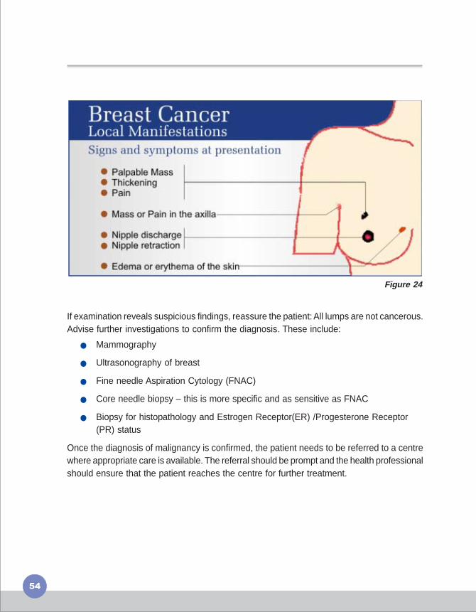
� Then with the flat of the hand, both the breasts are palpated in a circular mannerstarting from the nipple and areolae in a clockwise manner towards the peripheryand the axillary tail of the breast in sitting and lying down position. Then the axilla,supraclavicular region and liver are also examined. Figure 24 shows thesymptoms and signs of Breast Cancer.
Figure 23
54
Figure 24
If examination reveals suspicious findings, reassure the patient: All lumps are not cancerous.Advise further investigations to confirm the diagnosis. These include:
� Mammography
� Ultrasonography of breast
� Fine needle Aspiration Cytology (FNAC)
� Core needle biopsy – this is more specific and as sensitive as FNAC
� Biopsy for histopathology and Estrogen Receptor(ER) /Progesterone Receptor(PR) status
Once the diagnosis of malignancy is confirmed, the patient needs to be referred to a centrewhere appropriate care is available. The referral should be prompt and the health professionalshould ensure that the patient reaches the centre for further treatment.
55
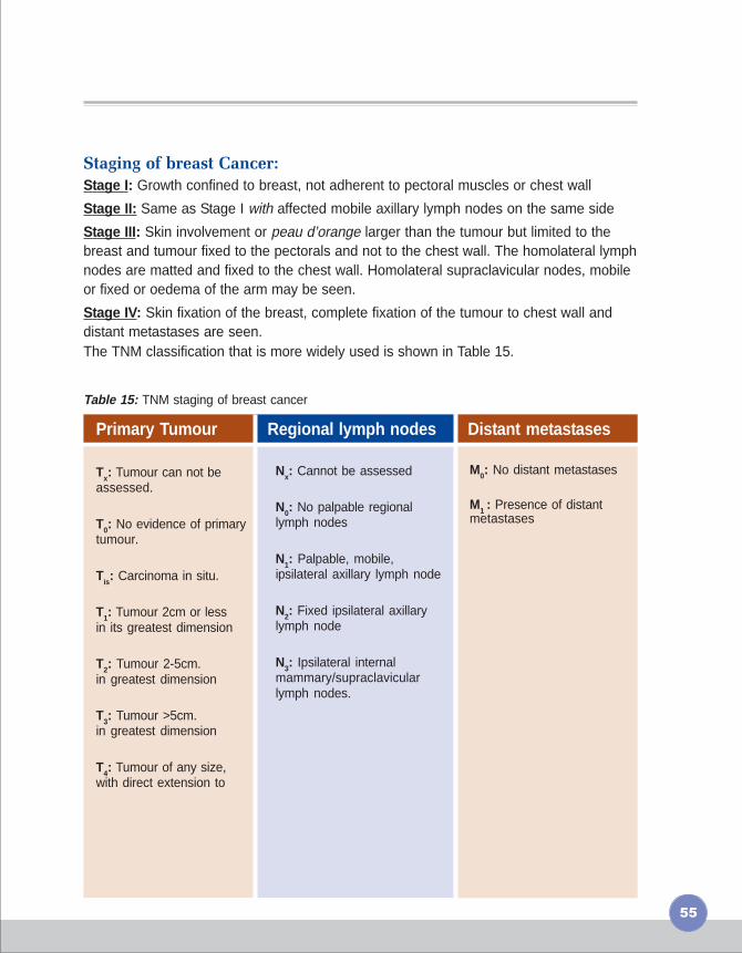
Staging of breast Cancer:
Stage I: Growth confined to breast, not adherent to pectoral muscles or chest wall
Stage II: Same as Stage I with affected mobile axillary lymph nodes on the same side
Stage III: Skin involvement or peau d’orange larger than the tumour but limited to thebreast and tumour fixed to the pectorals and not to the chest wall. The homolateral lymphnodes are matted and fixed to the chest wall. Homolateral supraclavicular nodes, mobileor fixed or oedema of the arm may be seen.
Stage IV: Skin fixation of the breast, complete fixation of the tumour to chest wall anddistant metastases are seen.The TNM classification that is more widely used is shown in Table 15.
Table 15: TNM staging of breast cancer
Primary Tumour Regional lymph nodes Distant metastases
Tx: Tumour can not beassessed.
T0: No evidence of primarytumour.
Tis: Carcinoma in situ.
T1: Tumour 2cm or lessin its greatest dimension
T2: Tumour 2-5cm.in greatest dimension
T3: Tumour >5cm.in greatest dimension
T4: Tumour of any size,with direct extension to
Nx: Cannot be assessed
N0: No palpable regionallymph nodes
N1: Palpable, mobile,ipsilateral axillary lymph node
N2: Fixed ipsilateral axillarylymph node
N3: Ipsilateral internalmammary/supraclavicularlymph nodes.
M0: No distant metastases
M1 : Presence of distantmetastases

56
Manual for Health Professionals
Management of breast cancer
Breast cancer is managed by surgery, radiotherapy, chemotherapy (including hormonetherapy), or a combination of the three. Figure 25 shows the management of a person witha suspicious breast lump in a flowchart.
Figure 25
57
Common Cancers
� Breast cancer is curable if detected early
� Health professionals can –
� Create ‘breast awareness’ among clients and ask them to report if a lumpis felt
� Offer clinical breast examinations to women aged 40-69 years
� Reassure – all lumps are not cancer
� Ensure prompt referral and appropriate management
� Provide pain relief and palliative care
Key messages
58
National Cancer Control Programme
The National Cancer Control Programme was started in 1975-76 with the objectives of:
● Primary prevention of tobacco related cancers
● Secondary prevention of cancers amenable to early diagnosis, such as cancer of theuterine cervix
● Extension and strengthening of therapeutic services including pain relief on a nationalscale through regional cancer centres, medical and dental colleges
The components of the programme are
� Regional Cancer Centres (RCCs): The RCCs are envisaged as specialized centresproviding comprehensive cancer care to the patients from their respective regions,undertaking awareness generation and early detection activities and offering specializedtraining to health professionals and conducting cancer research. There are 22 RCCs inthe country at present (Figure 26). RCCs are provided financial assistance through thescheme for ‘Assistance for RCCs.
� Strengthening of other cancer treatment facilities – Medical institutions are providedgrants to enhance their cancer treatment facilities through purchase of radiotherapyand other equipment under the Oncology Wing development scheme.
� District Cancer Control Programme (DCCP) - The DCCP aims to make the district thehub of cancer control activities. The focus is on prevention of cancers, early detection,minimal treatment of common cancers, appropriate referral and provision of supportivecare in the district.
� Activities through Non-governmental Organizations (NGOs) - This scheme is meant toextend financial assistance to NGOs working in cancer control.
Guidelines for the various schemes under NCCP are available with the Ministry of Healthand Family Welfare, New Delhi.
Manual for Health Professionals
4

59
National Cancer Control Programme
Regional Cancer Centres in India
Figure 26

60
Manual for Health Professionals
Role of Health Professionals in NCCP
Health professionals have the following roles to play under the National CancerControl Programme.
Prevention of cancers –� Create awareness about the ills of tobacco and advocate avoidance
� Encourage and assist habitual tobacco users to quit the habit
� Promote healthy dietary practices and physical activity
Early detection of cancers –� Create awareness about the early warning signs of cancer
� Encourage breast awareness
� Encourage oral self-examination
� Create awareness about symptoms of cervical cancer
� Examine, as a routine, the oral cavity of patients with history of tobacco use
� Offer clinical breast examination to any woman over 35 years presenting to the healthcentre
� If facilities exist, perform a pap smear test for every woman at least once in her lifetime,between 35 and 40 years of age
� Promptly refer any person with a suspicious lesion for accurate diagnosis and appropriatetreatment
Treatment of cancers -� Ensure that every patient complies with therapy advised
� If follow up care is required at the health centre level, make sure that detailed instructionsare provided by the treating institution
Palliative care -� Ensure that the patient is free from pain as far as possible. Learn and practice the WHO
step-ladder approach of pain management; refer to the appropriate centre for oralmorphine
� Achieve control of unwanted symptoms to the extent possible.
� Provide psychological support to the patient to accept the diagnosis and treatment
� Involve the family in diagnosis, treatment and care as far as possible
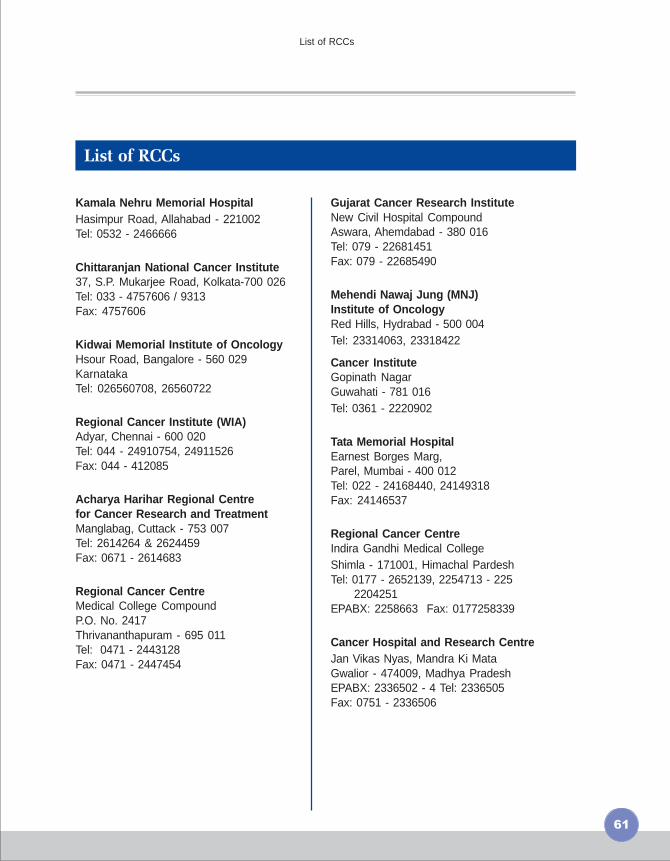
61
List of RCCs
List of RCCs
Kamala Nehru Memorial HospitalHasimpur Road, Allahabad - 221002Tel: 0532 - 2466666
Chittaranjan National Cancer Institute37, S.P. Mukarjee Road, Kolkata-700 026Tel: 033 - 4757606 / 9313Fax: 4757606
Kidwai Memorial Institute of OncologyHsour Road, Bangalore - 560 029KarnatakaTel: 026560708, 26560722
Regional Cancer Institute (WIA)Adyar, Chennai - 600 020Tel: 044 - 24910754, 24911526Fax: 044 - 412085
Acharya Harihar Regional Centrefor Cancer Research and TreatmentManglabag, Cuttack - 753 007Tel: 2614264 & 2624459Fax: 0671 - 2614683
Regional Cancer CentreMedical College CompoundP.O. No. 2417Thrivananthapuram - 695 011Tel: 0471 - 2443128Fax: 0471 - 2447454
Gujarat Cancer Research InstituteNew Civil Hospital CompoundAswara, Ahemdabad - 380 016Tel: 079 - 22681451Fax: 079 - 22685490
Mehendi Nawaj Jung (MNJ)Institute of OncologyRed Hills, Hydrabad - 500 004Tel: 23314063, 23318422
Cancer InstituteGopinath NagarGuwahati - 781 016Tel: 0361 - 2220902
Tata Memorial HospitalEarnest Borges Marg,Parel, Mumbai - 400 012Tel: 022 - 24168440, 24149318Fax: 24146537
Regional Cancer CentreIndira Gandhi Medical CollegeShimla - 171001, Himachal PardeshTel: 0177 - 2652139, 2254713 - 225 2204251EPABX: 2258663 Fax: 0177258339
Cancer Hospital and Research CentreJan Vikas Nyas, Mandra Ki MataGwalior - 474009, Madhya PradeshEPABX: 2336502 - 4 Tel: 2336505Fax: 0751 - 2336506

62
Indian Rotary Cancer Institute (AIIMS)Ansari NagarNew Delhi - 110029Fax: 2658921
Rashtra Sant Tukdoji (RST)Regional Cancer HospitalTukdoji Chowk, Manewada RoadNagpur - 440 027, MaharastraTel: 0712 - 2748995, 2744441Fax: 91 - 0712 - 2754546
Jawahar Lal Nehru Institute ofPostgraduate Medical Education &ResearchPondicherry Cancer Care SocietyPondicherry - 605006Tel: 0431 - 2272901, 227238, 2272389Fax: 2272235, 2272067
Indira Gandhi Instituteof Medical SciencesSheikhpura, Patna - 800 014, BiharTel: 0612 - 2287225, 2282329Fax: 0162 - 2287225
Acharya Tulsi Regional Cancer Trustand Research Institute (RCC)S.P.Medical CollegeBikaner, Rajasthan - 334003Tel: 0151 - 2226300 - 304
Manual for Health Professionals
Regional Cancer CentrePt. B.D. Sharma Post GraduateInstitute of Medical SciencesRohtak, HaryanaTel: 01262 - 2211307, 2213893Fax: 01262 - 2211308
Ambedhkar HospitalPt. J.N.M. Medical Collage and RCCRaipur, ChhatisgarhTelefax: 071 - 2221275
Civil HospitalAizawal, MizoramTelefax:- 0389231715Tel: 0389 - 2323236Fax: 0389 - 2315211 2322403
Post Graduate Institute of MedicalEducation & ResearchChandigarh - 160 012Tel: 0172 - 274506Fax: 0172 - 2744401, 2745078
Sanjay Gandhi Post Graduate Instituteof Medical Sciences (SGPGI)Raibareli Road, Lucknow - 2260134Tel: 0522 - 2668004 - 008, 2668700Fax: 0522 - 2668017, 2668129

63
Acknowledgements
ACKNOWLEDGEMENTS
Compiled and edited by
Dr. Cherian VargheseNational Professional Officer(Non-communicable diseases and Mental Health)Office of the WHO Representative to IndiaNew Delhi
Dr. Kavita VenkataramanNational Consultant (NMH)Office of the WHO Representative to IndiaNew Delhi
Dr. Sadhana BhagwatNational Consultant (Cancer)Ministry of Health and Family WelfareNew Delhi
ContributorsPartha Basu, CNCI, KolkataHead, Gynecologic OncologyChittaranjan National Cancer Institute37, S. P. Mukherjee RoadCalcutta, India, 700026
Reviewed byKidwai Memorial Insitute of Oncology, BangaloreTata Memorial Hospital, MumbaiGujarat Cancer Research Institute
The material in this publication does not imply the expression of any opinion whatsoever on the part ofthe World Health Organization concerning the legal status of any country, territory, city or area or of itsauthorities, or concerning the delimitation of its frontiers or boundaries. The mention of specific companiesor of certain manufacturers’ products does not imply that they are endorsed or recommended by theWorld Health Organization in preference to others of a similar nature that are not mentioned. Theresponsibility for the interpretation and use of the material lies with the reader.

64
Manual for Health Professionals
References / Suggested Reading
1. Agarwal S. P., Rao Y. N., Gupta Sudhir. Eds. Fifty Years of Cancer Control in India.National Cancer Control Programme. Directorate General Health Services. Ministryof Health And Family Welfare. Government of India. 2002.
2. Mathew B., Wesley R. S. Eds. Handbook on Cancer Control. World HealthOraganization, Regional Cancer Centre, Trivandrum.
3. National Cancer Control Programmes – Policies and Managerial Guidelines. WorldHealth Organization. 2nd Edition.
4. National Cancer Registry Programme – Five Year Consolidated Report of the HospitalBased Cancer Registries 1994-1998. Indian Council Of Medical Research, New Delhi.
5. National Cancer Registry Programme – Two-Year Report of the Population BasedCancer Registries 1997-1998. Indian Council Of Medical Research, New Delhi.
6. Nayak C.R., Nayak S. Eds. Handbook on Oncology. Acharya Harihar Regional CancerCentre, Cuttack.
7. Prevention and Early Detection of Cancer.Regional Cancer Centre, JIPMER,Pondicherry.
8. Reddy K. Srinath, Gupta Prakash C. Eds. Report on Tobacco Control in India. Ministryof Health and Family Welfare, Government of India, 2004.
9. Sankaranarayanan R., Wesley R. S. A Practical Manual on Visual Screening forCervical Neoplasia. World Health Organization – International Agency for Researchon Cancer. IARC Technical Publication No.41.
10. Stewart, Bernard W., Kleihues, Paul. Eds. World Cancer Report. WHO, IARC 2003.
11. Training Programme for Health Workers, Nurses and Medical Officers – Manual.Institute Rotary Cancer Hospital, All India Institute of Medical Sciences.